The role of neoadjuvant (HER)2-targeted therapies in (HER)2-overexpressing breast cancers
Upload
independentCategory
view
0download
0
Restoration of Podocyte Structure and Improvement ofChronic Renal Disease in Transgenic MiceOverexpressing ReninAnne-Cecile Huby1, Maria-Pia Rastaldi2, Kathleen Caron3, Oliver Smithies4, Jean-Claude Dussaule1,5,6,
Christos Chatziantoniou1,5*
1 INSERM UMR 702, Paris, France, 2 Fondazione IRCCS Ospedale Maggiore Policlinico & Fondazione D’Amico per la Ricerca sulle Malattie Renali, Milano, Italy,
3 Department of Molecular and Cell Physiology, University of North Carolina, Chapel Hill, North Carolina, United States of America, 4 Department of Pathology and
Laboratory Medicine, University of North Carolina, Chapel Hill, North Carolina, United States of America, 5 Universite Pierre et Marie Curie-Paris VI, UMR S 702, Paris, France,
6 AP-HP, Hopital St-Antoine, Department of Physiology, Paris, France
Abstract
Background: Proteinuria is a major marker of the decline of renal function and an important risk factor of coronary heartdisease. Elevated proteinuria is associated to the disruption of slit-diaphragm and loss of podocyte foot processes, structuralalterations that are considered irreversible. The objective of the present study was to investigate whether proteinuria can bereversed and to identify the structural modifications and the gene/protein regulation associated to this reversal.
Methodology/Principal Findings: We used a novel transgenic strain of mouse (RenTg) that overexpresses renin at a constanthigh level. At the age of 12-month, RenTg mice showed established lesions typical of chronic renal disease such as peri-vascular and periglomerular inflammation, glomerular ischemia, glomerulosclerosis, mesangial expansion and tubular dilation.Ultrastructural analysis indicated abnormal heterogeneity of basement membrane thickness and disappearance of podocytefoot processes. These structural alterations were accompanied by decreased expressions of proteins specific of podocyte(nephrin, podocin), or tubular epithelial cell (E-cadherin and megalin) integrity. In addition, since TGFb is considered the majorpro-fibrotic agent in renal disease and since exogenous administration of BMP7 is reported to antagonize the TGFb-inducedphenotype changes in kidney, we have screened the expressions of several genes belonging in the TGFb/BMP superfamily. Wefound that the endogenous inhibitors of BMPs such as noggin and Usag-1 were several-fold activated inhibiting the action ofBMPs and thus reinforcing the deleterious action of TGFb.Treatment with an AT1 receptor antagonist, at dose that did notdecrease arterial pressure, gradually reduced albuminuria. This decrease was accompanied by re-expression of podocin,nephrin, E-cadherin and megalin, and reappearance of podocyte foot processes. In addition, expressions of noggin and Usag-1were markedly decreased, permitting thus activation of the beneficial action of BMPs.
Conclusions/Significance: These findings show that proteinuria and alterations in the expression of proteins involved inthe integrity and function of glomerular and renal epithelial phenotype are reversible events when the local action ofangiotensin II is blocked, and provide hope that chronic renal disease can be efficiently treated.
Citation: Huby A-C, Rastaldi M-P, Caron K, Smithies O, Dussaule J-C, et al. (2009) Restoration of Podocyte Structure and Improvement of Chronic Renal Disease inTransgenic Mice Overexpressing Renin. PLoS ONE 4(8): e6721. doi:10.1371/journal.pone.0006721
Editor: Carmine Zoccali, L’ Istituto di Biomedicina ed Immunologia Molecolare, Consiglio Nazionale delle Ricerche, Italy
Received May 13, 2009; Accepted July 12, 2009; Published August 21, 2009
Copyright: � 2009 Huby et al. This is an open-access article distributed under the terms of the Creative Commons Attribution License, which permitsunrestricted use, distribution, and reproduction in any medium, provided the original author and source are credited.
Funding: Supported by grants from the Institut National de la Sante et de la Recherche (C.C.), University Pierre et Marie Curie (C.C. & JC.D.), Sanofi France (C.C. &JC.D.) and National Institutes of Health, HL049277 (O.S.). AC.H. was a doctoral fellow of the French Ministry of National Education (Ecole Doctorale de Physiologie& Physiopathologie, ED 394). The funders had no role in study design, data collection and analysis, decision to publish, or preparation of the manuscript.
Competing Interests: The authors have declared that no competing interests exist.
* E-mail: [email protected]
Introduction
Numerous clinical studies defined proteinuria as a major marker
of the decline of renal function. In addition, several studies
demonstrated that proteinuria is an important risk factor of
coronary heart disease and suggested to incorporate proteinuria
into the assessment of an individual’s cardiovascular risk [1].
Proteinuria occurs when the structure of podocytes, peculiar
ramified glomerular cells, is destroyed by disruption of the slit-
diaphragm and loss of foot processes. It is generally believed that
this structural alteration is the crucial step characterizing the
irreversibility of chronic kidney disease.
Our group has investigated over the last years the mechanisms
involved in the development of renal fibrosis in order to identify
targets for therapy [2–8] and was among the first groups to report
that regression of renal disease was feasible following therapy with
angiotensin II receptor antagonists, at least in experimental models
of hypertensive nephropathy [9–10]. These results were indepen-
dently confirmed and extended to additional experimental models
of nephropathy by other investigators [11–14]. However, a major
criticism about reversibility of chronic kidney disease in rodents
was that the disease was induced in young animals not suffering for
a long period from a chronic disease like hypertension or diabetes
(as it usually occurs in humans) and that therapy was induced
PLoS ONE | www.plosone.org 1 August 2009 | Volume 4 | Issue 8 | e6721

before reaching huge proteinuria or an important destruction of
podocyte structure.
To address these issues in the present study, we used a novel
model [15–16] of hypertension-induced renal disease mimicking
closer the kinetics and the physiopathological characteristics of
human nephroangiosclerosis. We found that these mice are
hypertensive and display albuminuria as early as 2–3 month old,
and that these pathological features are accentuated with age and
are accompanied by functional and structural alterations typical of
chronic renal disease including loss of podocyte foot processes. We
decided to start treatment with an AT1 receptor antagonist when
the animals reached the age of 12-months, thus, to treat aged
animals that have been proteinuric for a long period of their life-
span. We found that treatment with an AT1 receptor antagonist
induced reappearance of foot processes and of proteins charac-
terizing normal slit diaphragms, and re-established the normal
phenotype of tubular epithelial cells. These changes were
accompanied by a shift in the equilibrium between pro and anti-
fibrotic members of the TGFb/BMP superfamily.
This study, by showing that long lasting proteinuria, disorga-
nisation of podocyte structure and phenotype change of tubular
epithelium can be reversed supports the notion that chronic renal
disease can be efficiently treated.
Results
RenTg mice as a model to study hypertension-induceddisease
RenTg mice were generated and described previously [15]. A
major advantage of this transgenic strain is that renin is produced
ectopically (in the liver, and thus its release is independent of renal
perfusion pressure and electrolyte concentration at the macula
densa) at a genetically controlled rate allowing a ‘‘standardized’’
increase of endogenous synthesis of angiotensin II [15]. RenTg
mice display elevated systolic blood pressure as early as 2 month
old (14668 compared to 11064 mm Hg for age matched wild
type, p,0.01); at the age of 3 month they show slightly increased
albuminuria (18.363.5 vs 1.560.1 g/mol creat, p,0.01). At this
early age, renal morphology as revealed by Masson’s trichrome
appears to be normal (data not shown).
RenTg mice develop chronic kidney diseaseArterial blood pressure and urinary excretion of albumin
progressively increased with age to reach at 12 months values
highly elevated compared to age matched wild type controls
(p,0.001, Fig. 1). Histological examination of kidneys of 12-
month old RenTg revealed the presence of well-established lesions
in all renal compartments such as peri-vascular and periglomer-
ular inflammation, fibrinoid-like deposits within renal vessels,
glomerular ischemia, glomerulosclerosis, mesangial expansion and
tubular dilation (Fig. 2B). Fibrillar collagen assessed by red Sirius
staining, examined with polarized light and measured by
morphometric analysis, increased two-fold in RenTg mice
(p,0.01, Fig. 3B). Accordingly, the expression of agents
promoting fibrogenesis, such as collagen type I and FSP-1, or
indicating renal inflammation, such as MCP-1, were several-fold
increased compared to age-matched normotensive controls
(p,0.05, p,0.05 and p,0.01, respectively, Fig. 4).
Ultrastructural modifications in kidneys of RenTg miceTo assess that proteinuria is associated with important
modifications of podocyte structure, ultrastructural analysis by
electron microscopy was performed in the renal cortex of 12
month-old RenTg mice and their wild type controls. Figure 5B
shows a representative example of the lesions found in all samples
of RenTg mice. In particular, podocyte foot processes lost their
normal shape, and displayed numerous areas of effacement. The
glomerular basement membrane displayed abnormal thickness.
Additionally, mesangial expansion was evident.
To provide a cellular assessment of the above-described
modifications, we investigated the expression of two proteins
essential for the normal structure of podocytes: nephrin and
podocin. As shown in Figure 6, nephrin and podocin mRNA
expressions were 10- and 5-fold decreased in RenTg, compared to
age-matched wild type controls. In addition, two tubular proteins,
E-cadherin (an index of normal epithelial phenotype) and megalin
(involved in protein reabsorption) were also significantly decreased
(Fig. 7B and 7F).
Figure 1. Effect of AT1 receptor antagonism on blood pressureand albuminuria. Systolic blood pressure remained unchangedwhereas albuminuria decreased to almost normal levels in 12 monthold RenTg mice during administration of the AT1 receptor antagonistirbesartan; in triangles are shown the values for age-matched wild typecontrols. Values are mean6SEM; n = 5, 9 and 13 for wild type and RenTgmice before and after irbesartan, respectively; * P,0,05 or ** P,0,01 vsWT; ## P,0,001 vs RenTg.doi:10.1371/journal.pone.0006721.g001
Reversal of Proteinuria
PLoS ONE | www.plosone.org 2 August 2009 | Volume 4 | Issue 8 | e6721

Restoration of podocyte structure, tubular epithelialphenotype and decrease of proteinuria following AT1receptor antagonism
To test whether long lasting proteinuria and the related
structural lesions can be reversed, 12 month-old RenTg mice
were treated with irbesartan for a period of 6 weeks. In parallel,
irbesartan was also administered in a group of aged matched wild
type controls (n = 5). At the dose used, irbesartan did not affect
blood pressure (17063 mm Hg, Fig. 1A). Despite the lack of anti-
hypertensive effect, albuminuria progressively decreased in
irbesartan-treated mice (p,0.01 vs RenTg, Fig. 1B). Irbesartan
administration did not affect blood pressure nor albuminuria in
wild type mice (12763 mm Hg and 0.960.2 g/mol creat,
respectively)
This irbesartan-induced decrease in albuminuria in RenTg
mice was accompanied by improvement in renal cortical
morphology (Fig. 2C), regression of fibrillar interstitial collagen
content (Fig. 3C) and normalization of the expression of collagen
type I, FSP-1 and MCP-1 (Fig. 4). Irbesartan administration had
no effect on renal morphology, fibrogenesis or inflammation in
wild type mice (data not shown).
Most important, though mesangial expansion and GBM
thickness were not influenced by treatment, podocyte structure
recovered almost completely. (Fig. 5C).
The re-establishment of podocyte structure following AT1
receptor antagonism in RenTg mice was associated to the
induction of nephrin and podocin expressions at normal levels
(Fig. 6) and to re-appearance of E-cadherin and megalin in the
proximal tubule (Fig. 7C and 7G). Irbesartan administration did
not change expression of the above-mentioned proteins in wild
type mice.
Changes in the TGFb/BMP equilibrium duringprogression and reversal of Chronic Kidney Disease inRenTg mice
To get insights on the mechanisms accompanying the above-
described structural and functional changes, measurement of gene
expressions of pro- and anti-fibrotic genes of the TGFb/BMP
Figure 2. Improvement of renal histology following AT1receptor antagonism. Representative example of renal corticalhistology revealed by Mason’s trichrome in 12 month old wild type(A) and RenTg mice before (B) and after (C) 6 weeks of irbesartanadministration. Note the substantial improvement of the renal histologyfollowing therapy with the AT1 receptor antagonist. Bar = 200 mm.doi:10.1371/journal.pone.0006721.g002
Figure 3. Decrease of renal fibrosis during AT1 receptorantagonism. Representative example of fibrillar collagen content inthe renal interstitium revealed by Sirius red staining and polarized lightin wild type (A) and RenTg mice before (B) and after (C) irbesartantreatment. Quantification by morphometric analysis is shown on thelower panel (D). Fibrillar collagen content was decreased to normallevels in RenTg mice after irbesartan administration. Values are aremean6SEM; n = 5, 9 and 13 for wild type and RenTg mice before andafter irbesartan, respectively; ** P,0,01 vs WT; ## P,0,01 vs RenTg.Bar = 200 mm.doi:10.1371/journal.pone.0006721.g003
Reversal of Proteinuria
PLoS ONE | www.plosone.org 3 August 2009 | Volume 4 | Issue 8 | e6721

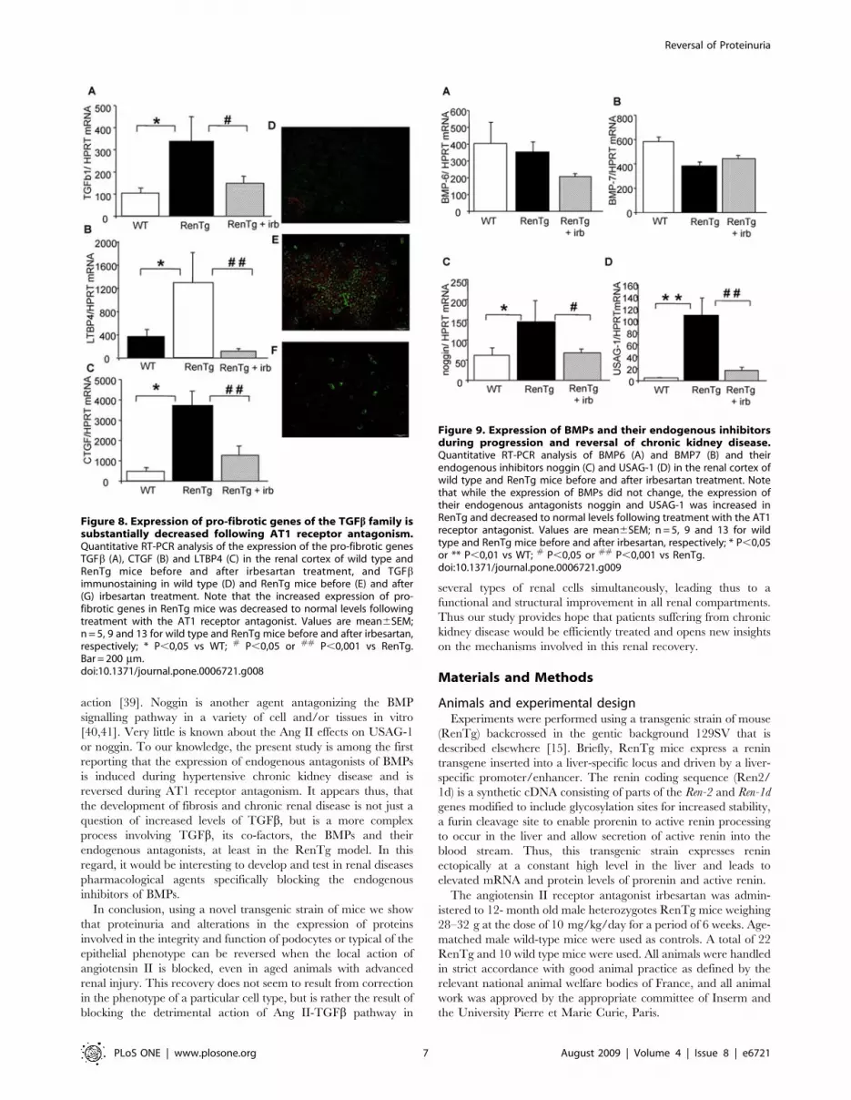
superfamily were performed (Figs. 8–9). Thus, expressions of the
TGFb itself, and of proteins acting either as co-factors (such as
CTGF) and/or helping its action (such as Latent Transforming
Growth Factor beta Binding Protein 4 or LTBP4) were several fold
increased in 12 mo old RenTg mice (Fig. 8A–C). These differences
in mRNA expressions were accompanied by the abnormal
appearance of TGFb within glomeruli and tubular epithelium in
RenTg mice (Fig. 8E). AT1 receptor antagonism strongly inhibited
this activation and reduced TGFb, CTGF and LTBP4 expressions
to normal levels (Fig. 8A–C & F). Irbesartan administration did
not change expression of these genes in wild type mice.
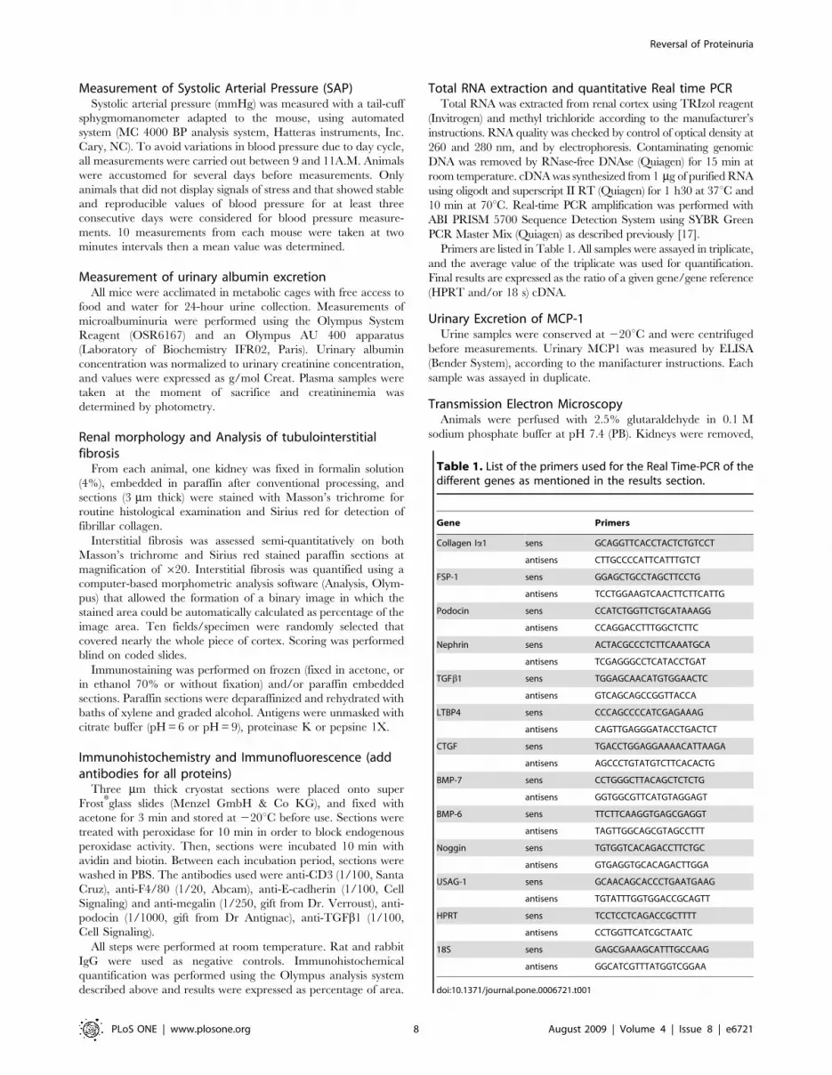
Interestingly, the expression of proteins antagonizing the action
of TGFb such as BMP6 and BMP7 did not change (Fig. 9A&B) in
12 mo-old RenTg mice. However, the expression of agents acting
as endogenous inhibitors/antagonists of BMPs such as noggin and
uterine sensitization-associated gene-1 (USAG-1) were several fold
increased in the renal cortex of 12 mo-old RenTg mice
(Fig. 9C&D). Administration of the AT1 receptor antagonist
decreased noggin and USAG-1 expressions back to normal levels
(Fig. 9). Irbesartan administration had no effect on the expression
of these genes in wild type mice.
Discussion
In this study we investigated whether proteinuria and the
associated structural and phenotype alterations can be reversed
when therapy starts at advanced stages of renal disease, and we
examined the mechanisms participating in this reversibility. An
important novel finding is that podocyte lesions such as loss of foot
processes and basal membrane disorganization are reversible
phenomena when the local action of Ang II is blocked. An
additional finding is that Ang II antagonism restores the normal
phenotype of tubular cells and reverses renal vascular inflammation.
Furthermore, we provide some clues about the mechanisms involved
in the reversal of renal fibrosis: it appears that this recovery was
due to a resetting of the TGFb/BMPs equilibrium towards a more
efficient action of the anti-fibrotic members of this family.
Regression of proteinuria and nephroangio-, glomerulo- or
tubulointerstitial sclerosis by blocking the action of the renin-
angiotensin system has been documented in several experimental
models of progressive renal disease, such as chronic nitric oxide
synthase inhibition, renal mass ablation, puromycin aminonucleo-
side [18–20]. However, a major scepticism about the reversibility of
chronic kidney disease in rodents compared to humans, is that the
disease was induced in most cases in young animals not suffering for
a long period from a chronic disease like hypertension or diabetes (as
is usually the case in humans), and that therapy was induced before
reaching high levels proteinuria or an important destruction of
podocyte structure for a prolonged period. To address these issues in
the present study, we used a novel model [15,16] of Ang II- induced
renal disease mimicking closer the kinetics and the physiopatho-
logical characteristics of hypertension-associated human renal
disease. These mice are hypertensive and display proteinuria
(albuminuria) as early as 2–3 mo old, and these pathologies are
accentuated with age and are accompanied by functional and
structural alterations typical of chronic renal disease including loss
of foot processes of podocytes and phenotype changes of renal
epithelial cells towards the mesenchymal phenotype.
We decided to start treatment with an AT1 receptor antagonist
when the animals reached the age of 12-mo, and thus, to treat
relatively aged animals that had been proteinuric for a long period
of their life-span. We found that treatment with an AT1 receptor
antagonist induced reappearance of foot processes and de novo
expression of nephrin and podocin (proteins indicating normal
function of slit diaphragm) reducing thus, urinary protein
(albumin) excretion. In agreement to our findings, downregulation
of nephrin gene that was totally prevented by angiotensin-
converting enzyme inhibitor and AT1 receptor blocker, was
observed in a model of progressive renal disease (passive Heymann
nephritis) [21]. In addition, Ang II induced redistribution of F-
actin and zonula ocludents-1 (ZO-1) in a murine cell line of
podocytes in vitro; these structural changes were mediated by the
AT1 receptor and were associated to an increased permeability of
albumin across podocyte monolayers. [22]. The repair of the
glomerular function and the reversal of proteinuria can be due
either to restructuring of existing and/or to de novo generation of
podocytes. The first hypothesis, has been proposed for the reversal
of glomerulosclerosis during ACE inhibition in the renal ablation
model [23]. Alternatively, podocyte repopulation through remod-
elling of Bowman’s capsule epithelial cells contributed to
Figure 4. AT1 receptor antagonism led to decreased expres-sion of pro-fibrotic and pro-inflammatory factors in thekidney. Quantitative RT-PCR analysis of the expression of fibrogenicfactors such as collagen type I chain a1 (A) and FSP-1 (B) in the renalcortex and urinary excretion of the pro-inflammatory factor MCP-1 (C) inwild type and RenTg mice before and after irbesartan treatment. Notethe substantial decrease of these factors following AT1 receptorantagonism. Values are mean6SEM; n = 5, 9 and 13 for wild type andRenTg mice before and after irbesartan, respectively; * P,0,05 or **P,0,01 vs WT; ## P,0,01 vs RenTg.doi:10.1371/journal.pone.0006721.g004
Reversal of Proteinuria
PLoS ONE | www.plosone.org 4 August 2009 | Volume 4 | Issue 8 | e6721

regression of renal disease in the Munich Wistar Fromter rat
model of glomerular injury [24]. It is possible that the underlying
mechanism of repair depends on the model and/or conditions of
renal injury (abrupt hemodynamic changes in the renal ablation,
much slower progressive decline in the MWF model). Our study is
among the first using ultrastructural analysis and reporting re-
appearance of foot processes and improvement of slit diaphragm
barrier following therapy with AT1 receptor blockade.
In addition to glomerular, RenTg mice displayed also tubular
epithelial lesions typical of chronic kidney disease. Thus, the
expression of megalin, a tubular protein contributing to protein
reabsorption and endocytosis was decreased in 12 mo-old RenTg
mice, whereas it returned to normal levels following AT1 receptor
antagonism. In agreement with an Ang II-induced effect on
megalin expression, treatment with Ang II decreased mRNA and
protein expression of megalin in a renal tubular cell line in vitro;
this decreased expression resulted to a suppression of cellular
uptake and degradation of albumin by these cells [25]. Decrease or
loss of E-cadherin expression is a typical alarm signal indicating
disappearance of the normal epithelial phenotype, whereas
induction of FSP-1 expression is associated to the appearance of
myofibroblasts and interstitial fibrosis. Thus, in addition to
megalin decrease, RenTg mice showed decreased expression of
E-cadherin concomitant to the appearance of tubulointerstitial
fibrosis and increased expression of FSP-1; AT1 receptor
antagonism reversed to normal these phenomena. Since numerous
in vitro studies showed that E-cadherin loss and increased FSP-1
expression are induced by the action of TGFb [26–28] and since
TGFb is considered as a major mediator of the pro-fibrotic action
of Ang II [29–31], it is logical to propose that the changes in E-
cadherin and FSP-1 expressions in the RenTg mice are due to the
Ang II-induced activation of TGFb pathway.
Figure 5. Restoration of podocyte structure following AT1 receptor antagonism. Representative examples of ultrastructural analysisperformed by electron microscopy in wild type (A) and RenTg mice before (B) and after (C) irbesartan treatment. The structural alterations ofpodocytes in RenTg mice such as loss of podocyte foot processes and abnormal thickness of the basal membrane (B) were substantially improvedafter antagonism of the AT1 receptor (C). Bar = 2 mm.doi:10.1371/journal.pone.0006721.g005
Reversal of Proteinuria
PLoS ONE | www.plosone.org 5 August 2009 | Volume 4 | Issue 8 | e6721

To address the above issue, we measured the expression levels of
several members of the TGFb superfamily. Comforting our
hypothesis, expression levels of TGFb, or of agents assisting or
acting as co-factors such as CTGF and LTBP4 were upregulated
in 12 mo old RenTg mice. Interestingly, the abnormal upregula-
tion of TGFb protein expression was evident in glomerular and
tubular interstitial compartments of the kidney (Fig. 8E). This
observation suggests that TGFb was the common mediator of
podocyte and tubular phenotype changes in RenTg mice. We
have reported previously that Ang II induced collagen I synthesis
and renal fibrosis through activation of TGFb [9,32]. In addition,
systemic infusion of Ang II into normal rats increased renal CTGF
mRNA and protein levels and induced renal injury; AT1 receptor
antagonism prevented CTGF increase and the development of
renal disease [33]. Other studies observed increased levels of
CTGF associated to the development of proteinuria and interstitial
fibrosis in murine models of diabetic nephropathy; in these studies,
administration of CTGF antisense reduced proteinuria and serum
creatinine and mesangial expansion [34]. Recent data strongly
suggest LTBP-4 as an important regulator of TGFb activation and
the TGFb-induced fibroblast differentiation associated to the
development of fibrosis in tissues such as lung [35]. Our data
suggest that a similar interaction between TGFb and LTBP-4 also
occurs during renal disease. The TGFb superfamily includes the
BMPs, which in addition to their bone morphogenic action are
also antagonists of the pro-fibrotic action of TGFb. In this regard,
it has been shown that exogenous administration of pharmaco-
logical doses of BMP7 reversed renal disease in several models of
experimental renal disease in rodents [36,37]. In our model, the
gene expression of major BMPs, such as BMP4 and BMP7
remained unchanged in the renal cortex of 12 mo-old RenTg mice
before or after AT1 receptor antagonism. Of note, the expression
of agents described as endogenous antagonists of BMPs such as
noggin and USAG-1 strongly increased with the progression of
renal disease in RenTg, and decreased to normal levels during
AT1 receptor treatment. Recent studies showed that the USAG-1
is abundantly expressed in the kidney, and in kidney injury
models, the ratio of USAG-1 to BMP-7 expression decreased with
kidney damage but increased after subsequent kidney regeneration
[38]. In addition, mice lacking USAG-1 are resistant to renal
injury. Thus, USAG1-/- mice exhibited prolonged survival and
preserved renal function in acute (cisplatin-induced nephrotoxici-
ty) or chronic (unilateral ureteral obstruction) renal injury models.
The preservation of the renal function in USAG-1-/- mice was
attributed to the enhancement of endogenous BMP signaling and
Figure 6. Reestablishment of normal podocyte phenotype afterAT1 receptor antagonism. Quantitative RT-PCR analysis of theexpression of genes involved in the podocyte structure such as podocin(A) and nephrin (B) in the renal cortex of wild type and RenTg micebefore and after irbesartan treatment, and representative examples ofpodocin expression in glomeruli of wild type (C) and RenTg mice before(D) and after (E) irbesartan treatment. The expressions of podocin andnephrin in the RenTg mice returned to control values followingirbesartan administration. Bar = 50 mm. Values are mean6SEM; n = 5, 9and 13 for wild type and RenTg mice before and after irbesartan,respectively; ** P,0,01 vs WT; ## P,0,001 vs RenTg.doi:10.1371/journal.pone.0006721.g006
Figure 7. Therapy with AT1 receptor antagonist led toreappearance of proteins characterizing normal function ofproximal tubules. Representative examples of the expression ofproteins typical of the structure and function of proximal tubules suchas E-cadherin and megalin. E-cadherin immunostaining in wild type (A)and RenTg mice before (B) and after (C) irbesartan treatment.Quantification by morphometric analysis is presented in D. Megalinexpression in wild type (E) and RenTg mice before (F) and after (G)irbesartan treatment. Quantification by morphometric analysis ispresented in H. Note that the blunted expression of both proteins inRenTg mice was increased to normal levels following treatment withthe AT1 receptor antagonist. Bar = 200 mm. Values are mean6SEM;n = 5, 9 and 13 for wild type and RenTg mice before and after irbesartan,respectively; ** P,0,01 vs WT; # P,0,05 or ## P,0,001 vs RenTg.doi:10.1371/journal.pone.0006721.g007
Reversal of Proteinuria
PLoS ONE | www.plosone.org 6 August 2009 | Volume 4 | Issue 8 | e6721
action [39]. Noggin is another agent antagonizing the BMP
signalling pathway in a variety of cell and/or tissues in vitro
[40,41]. Very little is known about the Ang II effects on USAG-1
or noggin. To our knowledge, the present study is among the first
reporting that the expression of endogenous antagonists of BMPs
is induced during hypertensive chronic kidney disease and is
reversed during AT1 receptor antagonism. It appears thus, that
the development of fibrosis and chronic renal disease is not just a
question of increased levels of TGFb, but is a more complex
process involving TGFb, its co-factors, the BMPs and their
endogenous antagonists, at least in the RenTg model. In this
regard, it would be interesting to develop and test in renal diseases
pharmacological agents specifically blocking the endogenous
inhibitors of BMPs.
In conclusion, using a novel transgenic strain of mice we show
that proteinuria and alterations in the expression of proteins
involved in the integrity and function of podocytes or typical of the
epithelial phenotype can be reversed when the local action of
angiotensin II is blocked, even in aged animals with advanced
renal injury. This recovery does not seem to result from correction
in the phenotype of a particular cell type, but is rather the result of
blocking the detrimental action of Ang II-TGFb pathway in
several types of renal cells simultaneously, leading thus to a
functional and structural improvement in all renal compartments.
Thus our study provides hope that patients suffering from chronic
kidney disease would be efficiently treated and opens new insights
on the mechanisms involved in this renal recovery.
Materials and Methods
Animals and experimental designExperiments were performed using a transgenic strain of mouse
(RenTg) backcrossed in the gentic background 129SV that is
described elsewhere [15]. Briefly, RenTg mice express a renin
transgene inserted into a liver-specific locus and driven by a liver-
specific promoter/enhancer. The renin coding sequence (Ren2/
1d) is a synthetic cDNA consisting of parts of the Ren-2 and Ren-1d
genes modified to include glycosylation sites for increased stability,
a furin cleavage site to enable prorenin to active renin processing
to occur in the liver and allow secretion of active renin into the
blood stream. Thus, this transgenic strain expresses renin
ectopically at a constant high level in the liver and leads to
elevated mRNA and protein levels of prorenin and active renin.
The angiotensin II receptor antagonist irbesartan was admin-
istered to 12- month old male heterozygotes RenTg mice weighing
28–32 g at the dose of 10 mg/kg/day for a period of 6 weeks. Age-
matched male wild-type mice were used as controls. A total of 22
RenTg and 10 wild type mice were used. All animals were handled
in strict accordance with good animal practice as defined by the
relevant national animal welfare bodies of France, and all animal
work was approved by the appropriate committee of Inserm and
the University Pierre et Marie Curie, Paris.
Figure 8. Expression of pro-fibrotic genes of the TGFb family issubstantially decreased following AT1 receptor antagonism.Quantitative RT-PCR analysis of the expression of the pro-fibrotic genesTGFb (A), CTGF (B) and LTBP4 (C) in the renal cortex of wild type andRenTg mice before and after irbesartan treatment, and TGFbimmunostaining in wild type (D) and RenTg mice before (E) and after(G) irbesartan treatment. Note that the increased expression of pro-fibrotic genes in RenTg mice was decreased to normal levels followingtreatment with the AT1 receptor antagonist. Values are mean6SEM;n = 5, 9 and 13 for wild type and RenTg mice before and after irbesartan,respectively; * P,0,05 vs WT; # P,0,05 or ## P,0,001 vs RenTg.Bar = 200 mm.doi:10.1371/journal.pone.0006721.g008
Figure 9. Expression of BMPs and their endogenous inhibitorsduring progression and reversal of chronic kidney disease.Quantitative RT-PCR analysis of BMP6 (A) and BMP7 (B) and theirendogenous inhibitors noggin (C) and USAG-1 (D) in the renal cortex ofwild type and RenTg mice before and after irbesartan treatment. Notethat while the expression of BMPs did not change, the expression oftheir endogenous antagonists noggin and USAG-1 was increased inRenTg and decreased to normal levels following treatment with the AT1receptor antagonist. Values are mean6SEM; n = 5, 9 and 13 for wildtype and RenTg mice before and after irbesartan, respectively; * P,0,05or ** P,0,01 vs WT; # P,0,05 or ## P,0,001 vs RenTg.doi:10.1371/journal.pone.0006721.g009
Reversal of Proteinuria
PLoS ONE | www.plosone.org 7 August 2009 | Volume 4 | Issue 8 | e6721
Measurement of Systolic Arterial Pressure (SAP)Systolic arterial pressure (mmHg) was measured with a tail-cuff
sphygmomanometer adapted to the mouse, using automated
system (MC 4000 BP analysis system, Hatteras instruments, Inc.
Cary, NC). To avoid variations in blood pressure due to day cycle,
all measurements were carried out between 9 and 11A.M. Animals
were accustomed for several days before measurements. Only
animals that did not display signals of stress and that showed stable
and reproducible values of blood pressure for at least three
consecutive days were considered for blood pressure measure-
ments. 10 measurements from each mouse were taken at two
minutes intervals then a mean value was determined.
Measurement of urinary albumin excretionAll mice were acclimated in metabolic cages with free access to
food and water for 24-hour urine collection. Measurements of
microalbuminuria were performed using the Olympus System
Reagent (OSR6167) and an Olympus AU 400 apparatus
(Laboratory of Biochemistry IFR02, Paris). Urinary albumin
concentration was normalized to urinary creatinine concentration,
and values were expressed as g/mol Creat. Plasma samples were
taken at the moment of sacrifice and creatininemia was
determined by photometry.
Renal morphology and Analysis of tubulointerstitialfibrosis
From each animal, one kidney was fixed in formalin solution
(4%), embedded in paraffin after conventional processing, and
sections (3 mm thick) were stained with Masson’s trichrome for
routine histological examination and Sirius red for detection of
fibrillar collagen.
Interstitial fibrosis was assessed semi-quantitatively on both
Masson’s trichrome and Sirius red stained paraffin sections at
magnification of 620. Interstitial fibrosis was quantified using a
computer-based morphometric analysis software (Analysis, Olym-
pus) that allowed the formation of a binary image in which the
stained area could be automatically calculated as percentage of the
image area. Ten fields/specimen were randomly selected that
covered nearly the whole piece of cortex. Scoring was performed
blind on coded slides.
Immunostaining was performed on frozen (fixed in acetone, or
in ethanol 70% or without fixation) and/or paraffin embedded
sections. Paraffin sections were deparaffinized and rehydrated with
baths of xylene and graded alcohol. Antigens were unmasked with
citrate buffer (pH = 6 or pH = 9), proteinase K or pepsine 1X.
Immunohistochemistry and Immunofluorescence (addantibodies for all proteins)
Three mm thick cryostat sections were placed onto super
FrostHglass slides (Menzel GmbH & Co KG), and fixed with
acetone for 3 min and stored at 220uC before use. Sections were
treated with peroxidase for 10 min in order to block endogenous
peroxidase activity. Then, sections were incubated 10 min with
avidin and biotin. Between each incubation period, sections were
washed in PBS. The antibodies used were anti-CD3 (1/100, Santa
Cruz), anti-F4/80 (1/20, Abcam), anti-E-cadherin (1/100, Cell
Signaling) and anti-megalin (1/250, gift from Dr. Verroust), anti-
podocin (1/1000, gift from Dr Antignac), anti-TGFb1 (1/100,
Cell Signaling).
All steps were performed at room temperature. Rat and rabbit
IgG were used as negative controls. Immunohistochemical
quantification was performed using the Olympus analysis system
described above and results were expressed as percentage of area.
Total RNA extraction and quantitative Real time PCRTotal RNA was extracted from renal cortex using TRIzol reagent
(Invitrogen) and methyl trichloride according to the manufacturer’s
instructions. RNA quality was checked by control of optical density at
260 and 280 nm, and by electrophoresis. Contaminating genomic
DNA was removed by RNase-free DNAse (Quiagen) for 15 min at
room temperature. cDNA was synthesized from 1 mg of purified RNA
using oligodt and superscript II RT (Quiagen) for 1 h30 at 37uC and
10 min at 70uC. Real-time PCR amplification was performed with
ABI PRISM 5700 Sequence Detection System using SYBR Green
PCR Master Mix (Quiagen) as described previously [17].
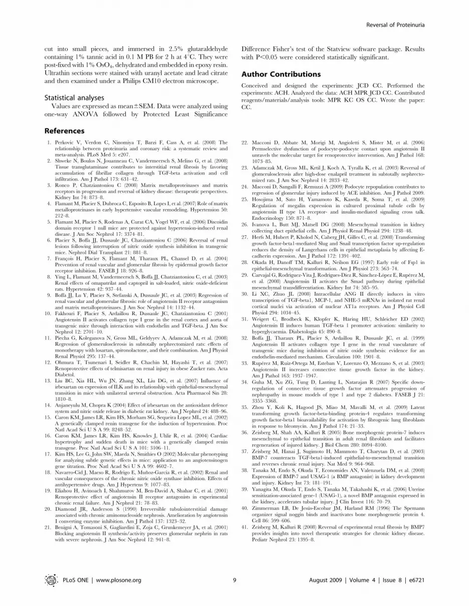
Primers are listed in Table 1. All samples were assayed in triplicate,
and the average value of the triplicate was used for quantification.
Final results are expressed as the ratio of a given gene/gene reference
(HPRT and/or 18 s) cDNA.
Urinary Excretion of MCP-1Urine samples were conserved at 220uC and were centrifuged
before measurements. Urinary MCP1 was measured by ELISA
(Bender System), according to the manifacturer instructions. Each
sample was assayed in duplicate.
Transmission Electron MicroscopyAnimals were perfused with 2.5% glutaraldehyde in 0.1 M
sodium phosphate buffer at pH 7.4 (PB). Kidneys were removed,
Table 1. List of the primers used for the Real Time-PCR of thedifferent genes as mentioned in the results section.
Gene Primers
Collagen Ia1 sens GCAGGTTCACCTACTCTGTCCT
antisens CTTGCCCCATTCATTTGTCT
FSP-1 sens GGAGCTGCCTAGCTTCCTG
antisens TCCTGGAAGTCAACTTCTTCATTG
Podocin sens CCATCTGGTTCTGCATAAAGG
antisens CCAGGACCTTTGGCTCTTC
Nephrin sens ACTACGCCCTCTTCAAATGCA
antisens TCGAGGGCCTCATACCTGAT
TGFb1 sens TGGAGCAACATGTGGAACTC
antisens GTCAGCAGCCGGTTACCA
LTBP4 sens CCCAGCCCCATCGAGAAAG
antisens CAGTTGAGGGATACCTGACTCT
CTGF sens TGACCTGGAGGAAAACATTAAGA
antisens AGCCCTGTATGTCTTCACACTG
BMP-7 sens CCTGGGCTTACAGCTCTCTG
antisens GGTGGCGTTCATGTAGGAGT
BMP-6 sens TTCTTCAAGGTGAGCGAGGT
antisens TAGTTGGCAGCGTAGCCTTT
Noggin sens TGTGGTCACAGACCTTCTGC
antisens GTGAGGTGCACAGACTTGGA
USAG-1 sens GCAACAGCACCCTGAATGAAG
antisens TGTATTTGGTGGACCGCAGTT
HPRT sens TCCTCCTCAGACCGCTTTT
antisens CCTGGTTCATCGCTAATC
18S sens GAGCGAAAGCATTTGCCAAG
antisens GGCATCGTTTATGGTCGGAA
doi:10.1371/journal.pone.0006721.t001
Reversal of Proteinuria
PLoS ONE | www.plosone.org 8 August 2009 | Volume 4 | Issue 8 | e6721
cut into small pieces, and immersed in 2.5% glutaraldehyde
containing 1% tannic acid in 0.1 M PB for 2 h at 4uC. They were
post-fixed with 1% OsO4, dehydrated and embedded in epoxy resin.
Ultrathin sections were stained with uranyl acetate and lead citrate
and then examined under a Philips CM10 electron microscope.
Statistical analysesValues are expressed as mean6SEM. Data were analyzed using
one-way ANOVA followed by Protected Least Significance
Difference Fisher’s test of the Statview software package. Results
with P,0.05 were considered statistically significant.
Author Contributions
Conceived and designed the experiments: JCD CC. Performed the
experiments: ACH. Analyzed the data: ACH MPR JCD CC. Contributed
reagents/materials/analysis tools: MPR KC OS CC. Wrote the paper:
CC.
References
1. Perkovic V, Verdon C, Ninomiya T, Barzi F, Cass A, et al. (2008) The
relationship between proteinuria and coronary risk: a systematic review andmeta-analysis. PLoS Med 5: e207.
2. Shweke N, Boulos N, Jouanneau C, Vandermeersch S, Melino G, et al. (2008)Tissue transglutaminase contributes to interstitial renal fibrosis by favoring
accumulation of fibrillar collagen through TGF-beta activation and cell
infiltration. Am J Pathol 173: 631–42.3. Ronco P, Chatziantoniou C (2008) Matrix metalloproteinases and matrix
receptors in progression and reversal of kidney disease: therapeutic perspectives.Kidney Int 74: 873–8.
4. Flamant M, Placier S, Dubroca C, Esposito B, Lopes I, et al. (2007) Role of matrix
metalloproteinases in early hypertensive vascular remodeling. Hypertension 50:212–8.
5. Flamant M, Placier S, Rodenas A, Curat CA, Vogel WF, et al. (2006) Discoidindomain receptor 1 null mice are protected against hypertension-induced renal
disease. J Am Soc Nephrol 17: 3374–81.6. Placier S, Boffa JJ, Dussaule JC, Chatziantoniou C (2006) Reversal of renal
lesions following interruption of nitric oxide synthesis inhibition in transgenic
mice. Nephrol Dial Transplant 21: 881–8.7. Francois H, Placier S, Flamant M, Tharaux PL, Chansel D, et al. (2004)
Prevention of renal vascular and glomerular fibrosis by epidermal growth factorreceptor inhibition. FASEB J 18: 926–8.
8. Ying L, Flamant M, Vandermeersch S, Boffa JJ, Chatziantoniou C, et al. (2003)
Renal effects of omapatrilat and captopril in salt-loaded, nitric oxide-deficientrats. Hypertension 42: 937–44.
9. Boffa JJ, Lu Y, Placier S, Stefanski A, Dussaule JC, et al. (2003) Regression ofrenal vascular and glomerular fibrosis: role of angiotensin II receptor antagonism
and matrix metalloproteinases. J Am Soc Nephrol 14: 1132–44.10. Fakhouri F, Placier S, Ardaillou R, Dussaule JC, Chatziantoniou C (2001)
Angiotensin II activates collagen type I gene in the renal cortex and aorta of
transgenic mice through interaction with endothelin and TGF-beta. J Am SocNephrol 12: 2701–10.
11. Piecha G, Koleganova N, Gross ML, Geldyyev A, Adamczak M, et al. (2008)Regression of glomerulosclerosis in subtotally nephrectomized rats: effects of
monotherapy with losartan, spironolactone, and their combination. Am J Physiol
Renal Physiol 295: 137–44.12. Ohmura T, Tsunenari I, Seidler R, Chachin M, Hayashi T, et al. (2007)
Renoprotective effects of telmisartan on renal injury in obese Zucker rats. ActaDiabetol.
13. Liu BC, Xia HL, Wu JN, Zhang XL, Liu DG, et al. (2007) Influence ofirbesartan on expression of ILK and its relationship with epithelial-mesenchymal
transition in mice with unilateral ureteral obstruction. Acta Pharmacol Sin 28:
1810–8.14. Anjaneyulu M, Chopra K (2004) Effect of irbesartan on the antioxidant defence
system and nitric oxide release in diabetic rat kidney. Am J Nephrol 24: 488–96.15. Caron KM, James LR, Kim HS, Morham SG, Sequeira Lopez ML, et al. (2002)
A genetically clamped renin transgene for the induction of hypertension. Proc
Natl Acad Sci U S A 99: 8248–52.16. Caron KM, James LR, Kim HS, Knowles J, Uhlir R, et al. (2004) Cardiac
hypertrophy and sudden death in mice with a genetically clamped renintransgene. Proc Natl Acad Sci U S A 101: 3106–11.
17. Kim HS, Lee G, John SW, Maeda N, Smithies O (2002) Molecular phenotypingfor analyzing subtle genetic effects in mice: application to an angiotensinogen
gene titration. Proc Natl Acad Sci U S A 99: 4602–7.
18. Navarro-Cid J, Maeso R, Rodrigo E, Munoz-Garcıa R, et al. (2002) Renal andvascular consequences of the chronic nitric oxide synthase inhibition. Effects of
antihypertensive drugs. Am J Hypertens 9: 1077–83.19. Eliahou H, Avinoach I, Shahmurov M, Ben-David A, Shahar C, et al. (2001)
Renoprotective effect of angiotensin II receptor antagonists in experimental
chronic renal failure. Am J Nephrol 21: 78–83.20. Diamond JR, Anderson S (1990) Irreversible tubulointerstitial damage
associated with chronic aminonucleoside nephrosis. Amelioration by angiotensinI converting enzyme inhibition. Am J Pathol 137: 1323–32.
21. Benigni A, Tomasoni S, Gagliardini E, Zoja C, Grunkemeyer JA, et al. (2001)
Blocking angiotensin II synthesis/activity preserves glomerular nephrin in ratswith severe nephrosis. J Am Soc Nephrol 12: 941–8.
22. Macconi D, Abbate M, Morigi M, Angioletti S, Mister M, et al. (2006)
Permselective dysfunction of podocyte-podocyte contact upon angiotensin II
unravels the molecular target for renoprotective intervention. Am J Pathol 168:
1073–85.
23. Adamczak M, Gross ML, Krtil J, Koch A, Tyralla K, et al. (2003) Reversal of
glomerulosclerosis after high-dose enalapril treatment in subtotally nephrecto-
mized rats. J Am Soc Nephrol 14: 2833–42.
24. Macconi D, Sangalli F, Remuzzi A (2009) Podocyte repopulation contributes to
regression of glomerular injury induced by ACE inhibition. Am J Pathol 2009.
25. Hosojima M, Sato H, Yamamoto K, Kaseda R, Soma T, et al. (2009)
Regulation of megalin expression in cultured proximal tubule cells by
angiotensin II type 1A receptor- and insulin-mediated signaling cross talk.
Endocrinology 150: 871–8.
26. Ivanova L, Butt MJ, Matsell DG (2008) Mesenchymal transition in kidney
collecting duct epithelial cells. Am J Physiol Renal Physiol 294: 1238–48.
27. Herfs M, Hubert P, Kholod N, Caberg JH, Gilles C, et al. (2008) Transforming
growth factor-beta1-mediated Slug and Snail transcription factor up-regulation
reduces the density of Langerhans cells in epithelial metaplasia by affecting E-
cadherin expression. Am J Pathol 172: 1391–402.
28. Okada H, Danoff TM, Kalluri R, Neilson EG (1997) Early role of Fsp1 in
epithelial-mesenchymal transformation. Am J Physiol 273: 563–74.
29. Carvajal G, Rodrıguez-Vita J, Rodrigues-Dıez R, Sanchez-Lopez E, Ruperez M,
et al. (2008) Angiotensin II activates the Smad pathway during epithelial
mesenchymal transdifferentiation. Kidney Int 74: 585–95.
30. Li XC, Zhuo JL (2008) Intracellular ANG II directly induces in vitro
transcription of TGF-beta1, MCP-1, and NHE-3 mRNAs in isolated rat renal
cortical nuclei via activation of nuclear AT1a receptors. Am J Physiol Cell
Physiol 294: 1034–45.
31. Weigert C, Brodbeck K, Klopfer K, Haring HU, Schleicher ED (2002)
Angiotensin II induces human TGF-beta 1 promoter activation: similarity to
hyperglycaemia. Diabetologia 45: 890–8.
32. Boffa JJ, Tharaux PL, Placier S, Ardaillou R, Dussaule JC, et al. (1999)
Angiotensin II activates collagen type I gene in the renal vasculature of
transgenic mice during inhibition of nitric oxide synthesis: evidence for an
endothelin-mediated mechanism. Circulation 100: 1901–8.
33. Ruperez M, Ruiz-Ortega M, Esteban V, Lorenzo O, Mezzano S, et al. (2003)
Angiotensin II increases connective tissue growth factor in the kidney.
Am J Pathol 163: 1937–1947.
34. Guha M, Xu ZG, Tung D, Lanting L, Natarajan R (2007) Specific down-
regulation of connective tissue growth factor attenuates progression of
nephropathy in mouse models of type 1 and type 2 diabetes. FASEB J 21:
3355–3368.
35. Zhou Y, Koli K, Hagood JS, Miao M, Mavalli M, et al. (2009) Latent
transforming growth factor-beta-binding protein-4 regulates transforming
growth factor-beta1 bioavailability for activation by fibrogenic lung fibroblasts
in response to bleomycin. Am J Pathol 174: 21–33.
36. Zeisberg M, Shah AA, Kalluri R (2005) Bone morphogenic protein-7 induces
mesenchymal to epithelial transition in adult renal fibroblasts and facilitates
regeneration of injured kidney. J Biol Chem 280: 8094–8100.
37. Zeisberg M, Hanai J, Sugimoto H, Mammoto T, Charytan D, et al. (2003)
BMP-7 counteracts TGF-beta1-induced epithelial-to-mesenchymal transition
and reverses chronic renal injury. Nat Med 9: 964–968.
38. Tanaka M, Endo S, Okuda T, Economides AN, Valenzuela DM, et al. (2008)
Expression of BMP-7 and USAG-1 (a BMP antagonist) in kidney development
and injury. Kidney Int 73: 181–191.
39. Yanagita M, Okuda T, Endo S, Tanaka M, Takahashi K, et al. (2006) Uterine
sensitization-associated gene-1 (USAG-1), a novel BMP antagonist expressed in
the kidney, accelerates tubular injury. J Clin Invest 116: 70–79.
40. Zimmerman LB, De Jesus-Escobar JM, Harland RM (1996) The Spemann
organizer signal noggin binds and inactivates bone morphogenetic protein 4.
Cell 86: 599–606.
41. Zeisberg M, Kalluri R (2008) Reversal of experimental renal fibrosis by BMP7
provides insights into novel therapeutic strategies for chronic kidney disease.
Pediatr Nephrol 23: 1395–8.
Reversal of Proteinuria
PLoS ONE | www.plosone.org 9 August 2009 | Volume 4 | Issue 8 | e6721
Copyright © 2022 FDOKUMEN