Angiotensin II Contributes to Podocyte Injury by Increasing TRPC6 Expression via an NFAT-Mediated...
14
Cardiovascular, Pulmonary, and Renal Pathology Angiotensin II Contributes to Podocyte Injury by Increasing TRPC6 Expression via an NFAT-Mediated Positive Feedback Signaling Pathway Tom Nijenhuis,* † Alexis J. Sloan, ‡ Joost G.J. Hoenderop, † Jan Flesche, ‡ Harry van Goor, § Andreas D. Kistler, ‡ Marinka Bakker,* Rene J.M. Bindels, † Rudolf A. de Boer, ¶ Clemens C. Möller, ‡ Inge Hamming, § Gerjan Navis, Jack F.M. Wetzels,* Jo H.M. Berden,* Jochen Reiser, ‡ Christian Faul, ‡ and Johan van der Vlag* From the Departments of Nephrology * and Physiology, † Radboud University Nijmegen Medical Centre, Nijmegen, the Netherlands; the Division of Nephrology and Hypertension, ‡ Department of Medicine, University of Miami Miller School of Medicine, Miami, Florida; and the Departments of Pathology, § Cardiology, ¶ and Nephrology, University Medical Centre Groningen, Groningen, the Netherlands The transient receptor potential channel C6 (TRPC6) is a slit diaphragm–associated protein in podocytes involved in regulating glomerular filter function. Gain-of-function mutations in TRPC6 cause heredi- tary focal segmental glomerulosclerosis (FSGS), and several human acquired proteinuric diseases show increased glomerular TRPC6 expression. Angiotensin II (AngII) is a key contributor to glomerular disease and may regulate TRPC6 expression in nonrenal cells. We demonstrate that AngII regulates TRPC6 mRNA and protein levels in cultured podocytes and that An- gII infusion enhances glomerular TRPC6 expression in vivo. In animal models for human FSGS (doxoru- bicin nephropathy) and increased renin-angiotensin system activity (Ren2 transgenic rats), glomerular TRPC6 expression was increased in an AngII-depen- dent manner. TRPC6 expression correlated with glo- merular damage markers and glomerulosclerosis. We show that the regulation of TRPC6 expression by An- gII and doxorubicin requires TRPC6-mediated Ca 2 influx and the activation of the Ca 2 -dependent pro- tein phosphatase calcineurin and its substrate nuclear factor of activated T cells (NFAT). Accordingly, cal- cineurin inhibition by cyclosporine decreased TRPC6 expression and reduced proteinuria in doxorubicin nephropathy, whereas podocyte-specific inducible expression of a constitutively active NFAT mutant in- creased TRPC6 expression and induced severe pro- teinuria. Our findings demonstrate that the deleteri- ous effects of AngII on podocytes and its pathogenic role in glomerular disease involve enhanced TRPC6 expression via a calcineurin/NFAT positive feedback signaling pathway. (Am J Pathol 2011, 179:1719 –1732; DOI: 10.1016/j.ajpath.2011.06.033) The glomerular capillary filtration barrier consists of en- dothelial cells, glomerular basement membrane (GBM), and visceral epithelial cells or podocytes linked by the slit diaphragm. The slit diaphragm is a complex of intercon- nected proteins that connect podocyte foot processes, which provides both physical linkage and a signaling unit that regulates podocyte behavior. 1 Damage to the glo- merular capillary filter, in particular at the level of the podocyte and the slit diaphragm, is of crucial importance in the pathophysiology of proteinuria. 1 Previously, the transient receptor potential channel C6 (TRPC6) has been identified as a novel slit diaphragm–associated pro- Supported by a Kolff Career Stimulation Grant from the Dutch Kidney Foundation (KJPB 07.0001), a grant from the Genzyme Renal Innovations Program and a Ruby Diabetes Research Grant (2009.80.118) (T.N.), a EURYI award (J.H.), a grant from the Swiss National Science Foundation (PBZHP3-128278) and an Amgen-FROMO fellowship (A.D.K.), grants from the US National Institutes of Health (grants DK073495 and DK089394) (J.R.), and a Young Investigator Career Development Grant from the NephCure Foundation and a National Scientist Development Grant from the American Heart Association (C.F.). Accepted for publication June 10, 2011. T.N., A.J.S., J.G.J.H., and J.F. contributed equally to this work. Address reprint requests to Johan van der Vlag, Ph.D., Nephrology Research Laboratory (279), Nijmegen Centre for Molecular Life Sciences, Department of Nephrology, Radboud University Nijmegen Medical Cen- tre, P.O. Box 9101, 6500 HB Nijmegen, The Netherlands; or Christian Faul, Ph.D., Division of Nephrology and Hypertension, Leonard Miller School of Medicine, University of Miami, 1580 NW 10 th Ave. (R-762), Miami, FL 33136. E-mail: [email protected] or [email protected]. The American Journal of Pathology, Vol. 179, No. 4, October 2011 Copyright © 2011 American Society for Investigative Pathology. Published by Elsevier Inc. All rights reserved. DOI: 10.1016/j.ajpath.2011.06.033 1719
Transcript of Angiotensin II Contributes to Podocyte Injury by Increasing TRPC6 Expression via an NFAT-Mediated...
The American Journal of Pathology, Vol. 179, No. 4, October 2011
Copyright © 2011 American Society for Investigative Pathology.
Published by Elsevier Inc. All rights reserved.
DOI: 10.1016/j.ajpath.2011.06.033
Cardiovascular, Pulmonary, and Renal Pathology
Angiotensin II Contributes to Podocyte Injury byIncreasing TRPC6 Expression via an NFAT-Mediated
Positive Feedback Signaling PathwayTom Nijenhuis,*† Alexis J. Sloan,‡
Joost G.J. Hoenderop,† Jan Flesche,‡
Harry van Goor,§ Andreas D. Kistler,‡
Marinka Bakker,* Rene J.M. Bindels,†
Rudolf A. de Boer,¶ Clemens C. Möller,‡
Inge Hamming,§ Gerjan Navis,�
Jack F.M. Wetzels,* Jo H.M. Berden,*Jochen Reiser,‡ Christian Faul,‡ andJohan van der Vlag*From the Departments of Nephrology * and Physiology,† Radboud
University Nijmegen Medical Centre, Nijmegen, the Netherlands;
the Division of Nephrology and Hypertension,‡ Department of
Medicine, University of Miami Miller School of Medicine, Miami,
Florida; and the Departments of Pathology,§ Cardiology,¶ and
Nephrology,� University Medical Centre Groningen, Groningen,
the Netherlands
The transient receptor potential channel C6 (TRPC6)is a slit diaphragm–associated protein in podocytesinvolved in regulating glomerular filter function.Gain-of-function mutations in TRPC6 cause heredi-tary focal segmental glomerulosclerosis (FSGS), andseveral human acquired proteinuric diseases showincreased glomerular TRPC6 expression. AngiotensinII (AngII) is a key contributor to glomerular diseaseand may regulate TRPC6 expression in nonrenal cells.We demonstrate that AngII regulates TRPC6 mRNAand protein levels in cultured podocytes and that An-gII infusion enhances glomerular TRPC6 expressionin vivo. In animal models for human FSGS (doxoru-bicin nephropathy) and increased renin-angiotensinsystem activity (Ren2 transgenic rats), glomerularTRPC6 expression was increased in an AngII-depen-dent manner. TRPC6 expression correlated with glo-merular damage markers and glomerulosclerosis. Weshow that the regulation of TRPC6 expression by An-gII and doxorubicin requires TRPC6-mediated Ca2�
influx and the activation of the Ca2�-dependent pro-tein phosphatase calcineurin and its substrate nuclear
factor of activated T cells (NFAT). Accordingly, cal-cineurin inhibition by cyclosporine decreased TRPC6expression and reduced proteinuria in doxorubicinnephropathy, whereas podocyte-specific inducibleexpression of a constitutively active NFAT mutant in-creased TRPC6 expression and induced severe pro-teinuria. Our findings demonstrate that the deleteri-ous effects of AngII on podocytes and its pathogenicrole in glomerular disease involve enhanced TRPC6expression via a calcineurin/NFAT positive feedbacksignaling pathway. (Am J Pathol 2011, 179:1719–1732;DOI: 10.1016/j.ajpath.2011.06.033)
The glomerular capillary filtration barrier consists of en-dothelial cells, glomerular basement membrane (GBM),and visceral epithelial cells or podocytes linked by the slitdiaphragm. The slit diaphragm is a complex of intercon-nected proteins that connect podocyte foot processes,which provides both physical linkage and a signaling unitthat regulates podocyte behavior.1 Damage to the glo-merular capillary filter, in particular at the level of thepodocyte and the slit diaphragm, is of crucial importancein the pathophysiology of proteinuria.1 Previously, thetransient receptor potential channel C6 (TRPC6) hasbeen identified as a novel slit diaphragm–associated pro-
Supported by a Kolff Career Stimulation Grant from the Dutch KidneyFoundation (KJPB 07.0001), a grant from the Genzyme Renal InnovationsProgram and a Ruby Diabetes Research Grant (2009.80.118) (T.N.), aEURYI award (J.H.), a grant from the Swiss National Science Foundation(PBZHP3-128278) and an Amgen-FROMO fellowship (A.D.K.), grantsfrom the US National Institutes of Health (grants DK073495 andDK089394) (J.R.), and a Young Investigator Career Development Grantfrom the NephCure Foundation and a National Scientist DevelopmentGrant from the American Heart Association (C.F.).
Accepted for publication June 10, 2011.
T.N., A.J.S., J.G.J.H., and J.F. contributed equally to this work.
Address reprint requests to Johan van der Vlag, Ph.D., NephrologyResearch Laboratory (279), Nijmegen Centre for Molecular Life Sciences,Department of Nephrology, Radboud University Nijmegen Medical Cen-tre, P.O. Box 9101, 6500 HB Nijmegen, The Netherlands; or Christian Faul,Ph.D., Division of Nephrology and Hypertension, Leonard Miller School ofMedicine, University of Miami, 1580 NW 10th Ave. (R-762), Miami, FL 33136.
E-mail: [email protected] or [email protected].1719
1720 Nijenhuis et alAJP October 2011, Vol. 179, No. 4
tein in podocytes.2 Gain-of-function mutations in TRPC6have been shown to cause autosomal dominant focalsegmental glomerulosclerosis (FSGS), and enhancedpodocyte expression of wild-type and mutant TRPC6leads to glomerular damage.2–5
TRP channels are involved in several renal processesand diseases, ranging from tubular Ca2� and Mg2� re-absorption, through osmoregulation, to polycystic kidneydisease.6–9 Podocytes express TRPC6, and co-immuno-precipitation studies demonstrate that TRPC6 is associ-ated with the slit diaphragm proteins nephrin and podo-cin, suggesting that TRPC6 is involved in signalingevents at the slit diaphragm.2,10 The slit diaphragm com-plex is mechanically and functionally linked to the actincytoskeleton. Cytoskeletal rearrangement has been sug-gested to underlie foot process effacement, which is a cru-cial early event in the pathophysiology of proteinuria.4 Sev-eral gain-of-function TRPC6 mutations have been identifiedin the TRPC6 encoding gene.2–4,11,12 In addition, glomeru-lar TRPC6 expression is increased in acquired human pro-teinuric diseases, including nonfamilial FSGS and membra-nous glomerulopathy.4 Taken together, it is likely thatenhanced Ca2� influx due to an increased number of func-tional TRPC6 channels at the cell surface and/or enhancedchannel activity compromises the structural integrity of thepodocyte, leading to proteinuria.
TRPC6 is a receptor-operated cation channel, whichcan be activated by angiotensin II (AngII) through stim-ulation of the angiotensin type 1 receptor (AT1R) andsecondary generation of diacylglycerol.3,13,14 AngII is akey contributor to the pathogenesis of glomerular dis-ease, and the antiproteinuric effects of angiotensin-con-verting enzyme (ACE) inhibition and AT1R blockade areundisputed.15,16 In nonrenal cells, AngII activates TRPC6currents and enhances TRPC6 transcription.14,17,18 Incardiomyocytes, AngII induces a TRPC6 and Ca2�-de-pendent calcineurin/nuclear factor of activated T cells(NFAT) positive feedback loop, leading to increasedTRPC6 transcription, driving cardiac hypertrophy.14,18
Podocytes also express both AT1R and AT2R, and AngIIhas detrimental effects in podocytes.15,16,19,20 AngII in-creases intracellular Ca2� levels and induces changes inthe actin cytoskeleton.21–23 When the AT1R is overex-pressed in podocytes, transgenic rats develop podocytedamage and glomerulosclerosis.24 Furthermore, the over-expression of renin in mice induces podocyte damage andproteinuria, pathological effects that can be ameliorated bytreating these transgenic animals with angiotensin receptorblockers (ARBs).25 In analogy to cardiomyocytes, AngII-induced Ca2�-calcineurin-NFAT–mediated transcription ofTRPC6 could also occur in podocytes; therefore, AngIIcould cause an up-regulation of TRPC6 expression, whichresults in elevated intracellular Ca2� levels in podocytes inacquired proteinuric disease.
The aims of this study were to determine whether AngIIregulates TRPC6 expression in podocytes, to gain insightinto the downstream effectors of AngII/TRPC6-mediatedsignaling, and to evaluate its in vivo significance in exper-
imental proteinuric glomerular disease.Materials and Methods
Animal Studies
Unilateral doxorubicin nephropathy was induced in ratsby temporary clipping of the left renal artery and vein,followed by injection of 1.5 mg/kg of doxorubicin (Sigma-Aldrich, Zwijndrecht, the Netherlands) via the tail vein.After 12 minutes, when doxorubicin was cleared from thecirculation, the clamp was removed. Bilateral doxorubicinnephropathy was induced by injection of 5 mg/kg ofdoxorubicin. Animals were treated with the ARB L158,809(150 mg per liter of drinking water) from week 6 to 12 afterinduction of doxorubicin nephropathy. Additional animalsreceived the ACE inhibitor (ACEi) lisinopril (75 mg perliter of drinking water) from week 6 to 18 after induction ofdoxorubicin nephropathy. Cyclosporine (20 mg/kg; dis-solved in 0.5 mL of olive oil) or vehicle (0.5 mL of olive oil)was administered by daily oral gavage from week 4 to 6after doxorubicin injection. For the AngII infusion studies,Wistar rats received a continuous AngII infusion (435 ng/kg/min) by subcutaneous osmotic minipumps during 3 weeks.Before termination, animals were housed in metaboliccages for 24 hours. Male homozygous TGR(mRen2)27(Ren2 transgenic) rats and age-matched Sprague-Dawleyrats were purchased from the Max Delbrück Center forMolecular Medicine (Berlin-Buch, Berlin, Germany). Wild-type and Ren2 transgenic rats were treated with a non-hypotensive dose of the ARB candesartan (0.05 mg/kg/d)with osmotic minipumps (Alzet model 2004) for 4 weeks.The animal ethics committees of the Radboud UniversityNijmegen and the University Medical Centre Groningenapproved all animal studies.
Generation of Inducible Transgenic MiceOverexpressing Constitutive Active NFATc1 inPodocytes
The transgenic TetO-HA-NFATc1nuc mouse line was gen-erated in the laboratory of Dr. Gerald Crabtree and pro-vided by Dr. Seung K. Kim (both from Stanford University,Stanford, California).26 In NFATc1nuc, the serine residuesthat are dephosphorylated by calcineurin are substitutedwith alanine residues, rendering it constitutively nuclear,constitutively active, and insensitive to nuclear kinases.27
These single transgenic mice were mated with podocin–reverse tetracycline-controlled transactivator (rtTA) miceto generate double transgenic doxycycline-induciblepodocin-rtTA/TetO-HA-NFATc1nuc mice.28 Transgenicmice were genotyped using specific primer sets. Podo-cin-rtTA/TetO-HA-NFATc1nuc F1 littermates were matedto obtain F2 double transgenic mice for experimentalprocedures. Transgene expression was induced in podo-cytes by adding doxycycline (Sigma-Aldrich; 2 mg/mL in7% sucrose, pH � 5) to the drinking water of 6- to8-week-old double transgenic mice for 4 days. Simulta-neously, the mice were fed a special diet chow contain-ing doxycycline (2000 ppm). Control animals were eithersingle transgenic mice that also received doxycycline
or double transgenic mice that received no doxycy-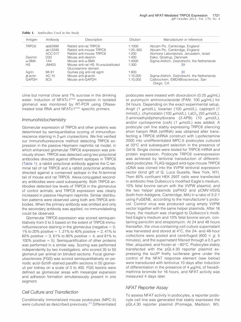
AngII and NFAT-Mediated TRPC6 Expression 1721AJP October 2011, Vol. 179, No. 4
cline but normal chow and 7% sucrose in the drinkingwater. Induction of NFATc1nuc expression in isolatedglomeruli was monitored by RT-PCR using DNase-treated total RNA and NFATc1nuc specific primers.
Immunohistochemistry
Glomerular expression of TRPC6 and other proteins wasdetermined by semiquantitative scoring of immunofluo-rescence staining in 2-�m cryosections. We first verifiedour immunofluorescence method detecting TRPC6 ex-pression in the passive Heymann nephritis rat model, inwhich enhanced glomerular TRPC6 expression was pre-viously shown. TRPC6 was detected using two polyclonalantibodies directed against different epitopes in TRPC6(Table 1): a rabbit polyclonal antibody against the C-ter-minal tail of rat TRPC6 and a rabbit polyclonal antibodydirected against a conserved epitope in the N-terminaltail of mouse and rat TRPC6. Alexa-conjugated second-ary antibodies were used subsequently. Both TRPC6 an-tibodies detected low levels of TRPC6 in the glomerulusof control animals, and TRPC6 expression was clearlyincreased in passive Heymann nephritis. Similar distribu-tion patterns were observed using both anti-TRPC6 anti-bodies. When the primary antibody was omitted and onlythe secondary antibody was applied, no immunolabelingcould be observed.
Glomerular TRPC6 expression was scored semiquan-titatively from 0 to 5 based on the extent of TRPC6 immu-nofluorescence staining in the glomerulus (negative � 0,1% to 20% positive � 1, 21% to 40% positive � 2, 41% to60% positive � 3, 61% to 80% positive � 4, and 81% to100% positive � 5). Semiquantification of other proteinswas performed in a similar way. Scoring was performedindependently by two investigators, who scored 35 to 50glomeruli per animal on blinded sections. Focal glomer-ulosclerosis (FGS) was scored semiquantitatively on pe-riodic acid–Schiff–stained paraffin sections in 50 glomer-uli per kidney on a scale of 0 to 400. FGS lesions weredefined as glomerular areas with mesangial expansionand adhesion formation simultaneously present in onesegment.
Cell Culture and Transfection
Conditionally immortalized mouse podocytes (MPC-5)
Table 1. Antibodies Used in the Study
Antigen Antibody Description
TRPC6 ab62999 Rabbit anti-rat TRPC6ab12249 Rabbit anti-mouse TRPC6ACC-017 Rabbit anti-mouse TRPC6
Desmin D33 Mouse anti-desmin�-SMA 1A4 Mouse anti-�-SMAHS JM403 Mouse anti-rat HS: N-unsubstitut
Glucosamine domainAgrin MI-91 Guinea pig anti-rat agrin�-actin AC-15 Mouse anti-�-actinGAPDH 6C5 Mouse anti-GAPDH
were cultured as described previously.31 Differentiated
podocytes were treated with doxorubicin (0.25 �g/mL)or puromycin aminonucleoside (PAN; 100 �g/mL) for24 hours. Depending on the exact experimental setup,AngII (1 �mol/L), losartan (100 �mol/L), captopril (1mmol/L), chymostatin (100 �mol/L), LaCl3 (50 �mol/L),2-aminoethyldiphenylborane (2-APB) (10 �mol/L),and/or cyclosporine (csA) (1 �mol/L) was added. Apodocyte cell line stably expressing TRPC6 silencingshort hairpin RNA (shRNA) was obtained after trans-fecting a TRPC6 shRNA construct with Lipofectamine2000 into undifferentiated MPC-5 podocytes culturedat 33°C and subsequent selection in the presence ofG418. Single clones were tested for TRPC6 mRNA andprotein expression. Podocyte TRPC6 overexpressionwas achieved by lentiviral transduction of differenti-ated podocytes. FLAG-tagged wild-type mouse TRPC6cDNA was cloned into the VVPW lentiviral expressionvector (kind gift of G. Luca Gusella, New York, NY).Then 80% confluent HEK 293T cells were transfectedin antibiotic-free Dulbecco’s modified Eagle’s medium,10% fetal bovine serum with the VVPW plasmid, andthe two helper plasmids psPAX2 and pCMV-VSVG(both from Addgene, Cambridge, MA) in a ratio of 3:2:1using FuGENE, according to the manufacturer’s proto-col. Control virus was produced using empty VVPWvector together with the same helper plasmids. After 16hours, the medium was changed to Dulbecco’s modi-fied Eagle’s medium and 10% fetal bovine serum, con-taining penicillin and streptomycin. At 24 and 48 hoursthereafter, the virus-containing cell culture supernatantwas harvested and stored at 4°C, the 24- and 48-hourcollections were pooled and centrifuged (600 � g; 5minutes), and the supernatant filtered through a 0.5 �mfilter, aliquoted, and frozen at �80°C. Podocytes stablytransfected with the pGL4.30 reporter plasmid ex-pressing the luc2P firefly luciferase gene under thecontrol of the NFAT response element (see below)were transduced with lentivirus 10 days after inductionof differentiation in the presence of 4 �g/mL of hexadi-methrine bromide for 16 hours, and NFAT activity wasmeasured 4 days later.
NFAT Reporter Assay
To assess NFAT activity in podocytes, a reporter podo-cyte cell line was generated that stably expresses the
Dilution Manufacturer or reference
1:1000 Abcam Plc, Cambridge, England1:25–300 Abcam Plc, Cambridge, England1:200 Alomone Laboratories, Jerusalem, Israel1:400 Dako, Glostrup, Denmark1:4000 Sigma-Aldrich, Zwijndrecht, the Netherlands1:300 29
1:800 301:10,000 Sigma-Aldrich, Zwijndrecht, the Netherlands1:10,000 Calbiochem, EMD4Biosciences, San
Diego, CA
ed
pGL4.30 reporter plasmid (Promega, Madison, WI).

1722 Nijenhuis et alAJP October 2011, Vol. 179, No. 4
pGL4.30 includes the luc2P firefly luciferase gene un-der the control of the NFAT response element. Stablytransfected clones were selected in the presence ofhygromycin (300 �g/mL). Cells were differentiated for10 days before lentiviral transduction and for 14 daysbefore drug treatment. Stable cell lines were probedwith Bright-Glo Luciferase Assay System (Promega),and luminescence was measured on a SpectraMax Lluminescence microplate reader (Molecular Devices,Sunnyvale, CA).
To assess NFAT activity in TRPC6 knockdown cells,undifferentiated wild-type and TRPC6 stable knockdownpodocytes were transiently transfected with the pGL4.30reporter plasmid-expressing the luc2P firefly luciferasegene under the control of the NFAT response elementand the pGL4.74 plasmid-expressing hRluc Renilla lucif-erase as an internal control to correct for transfectionefficiency (Promega). Transiently transfected cells wereassayed with Dual-Glo Luciferase Assay System (Pro-mega).
Real-Time Quantitative RT-PCR Analysis
Total RNA was isolated from cultured podocytes andRNA was reverse transcribed (Transcriptor Kit; RocheDiagnostics, Mannheim, Germany). Real-time quantita-tive PCR was performed using SYBR Green Supermix(Roche Diagnostics) on a MyiQ Real-Time PCR Detec-tion System (Bio-Rad Laboratories, Hercules, CA).TRPC6 expression was quantified by the delta-delta CT
method using glyceraldehyde 3-phosphate dehydro-genase (GAPDH) as the housekeeping gene. Samplesizes of 5 to 8 separate mouse podocyte cultures wereused per experimental condition per experiment. Re-sults were confirmed in at least two distinct experi-ments.
Immunocytochemistry
Podocytes grown on collagen A–coated plastic SlideFlasks(NUNC, Roskilde, Denmark) were fixed and incubated witha rabbit polyclonal anti-mouse TRPC6 antibody (Table 1).Alexa-conjugated secondary antibodies were applied,cells were embedded, and images were collected asdescribed above.
Immunoblotting
Podocytes or isolated glomeruli were lysed in a 20mmol/L Tris pH 8 buffer containing 500 mmol/L NaCl,0.5% (wt/vol) 3-[(3-cholamidopropyl)dimethylammonio]-1-propanesulfonate (CHAPS), 1% (vol/vol) Triton X-100, 2pg of pepstatin, and the Complete Mini cocktail of pro-tease inhibitors (Roche Diagnostics). Protein concentra-tion was determined and samples containing equalamounts of protein were resolved on a 10% (wt/vol) SDS-PAGE gel and blotted to polyvinylidene difluoride mem-branes (Bio-Rad Laboratories). Blots were incubated withrabbit polyclonal anti-mouse TRPC6 antibody, mouse an-ti-�-actin antibody, or mouse anti-GAPDH antibody (Ta-
ble 1) and subsequently with peroxidase-labeled sec-ondary antibodies. Proteins were visualized usingchemiluminescence, and signal intensity was determineddigitally.
Ca2� Imaging Studies
Imaging of intracellular Ca2� concentration ([Ca2�]i)with Fura-2 was performed in selected populations ofcells with an inverted fluorescence microscope setup.In brief, conditionally immortalized mouse podocytesexpressing either a control shRNA or TRPC6 shRNAconstruct were incubated with 5 �mol/L Fura-2-AM(Sigma-Aldrich) for 45 minutes at room temperature.Cells were treated with 100 �mol/L 1-oleoyl-2-acetyl-sn-glycerolin (OAG) in the presence or absence of2-APB, and [Ca2�]i was measured. After stimulation,the extracellular buffer was exchanged with 2 mmol/LCa2� to distinguish membrane-associated channel-de-pendent changes in [Ca2�]i. Fluorescence data arepresented as a 340/380-nm ratio. Data from selectedcell populations were averaged, and statistical analy-sis was performed on multiple experiments.
Statistical Analysis
Data are expressed as mean � SEM. Statistical compar-isons were analyzed by one-way analysis of variance andFisher’s multiple comparison. P � 0.05 was consideredsignificant.
Results
Glomerular TRPC6 Expression Correlates withPodocyte Injury in Doxorubicin Nephropathy
Unilateral doxorubicin nephropathy was induced and re-sulted in a significant proteinuria (161 � 52 mg/24 h,246 � 14 mg/24 h, and 388 � 28 mg/24 h at 6, 18, and30 weeks, respectively, after doxorubicin injection). Glo-merular TRPC6 protein expression was increased indoxorubicin-exposed kidneys when compared with con-trol kidneys (Figure 1, A and B). TRPC6 expression andproteinuria increased over time (Figure 1B), and protein-uria tended to increase with higher TRPC6 protein levels(r2 � 0.2029; P � 0.16) (Figure 1C). FGS as a directmeasure of glomerular injury was significantly increasedin doxorubicin-exposed kidneys (8 � 3 versus 1 � 1 at 6weeks, 47 � 11 versus 12 � 4 at 18 weeks, and 89 � 24versus 33 � 7 at 30 weeks). The FGS score correlatedwith TRPC6 protein levels (r2 � 0.8028; P � 0.0001)(Figure 1D). TRPC6 expression was also enhanced inbilateral doxorubicin nephropathy (Figure 1E), as werecortical TRPC6 mRNA levels (Figure 1F), suggestingthat doxorubicin nephropathy alters transcription ofTRPC6. In glomeruli from doxorubicin nephropathyrats, TRPC6 co-localized with desmin, which is amarker for injured podocytes, whereas in control kid-neys desmin and TRPC6 expression levels were low(Figure 1G). No co-localization was found for TRPC6
and glomerular �-smooth muscle actin (�-SMA), which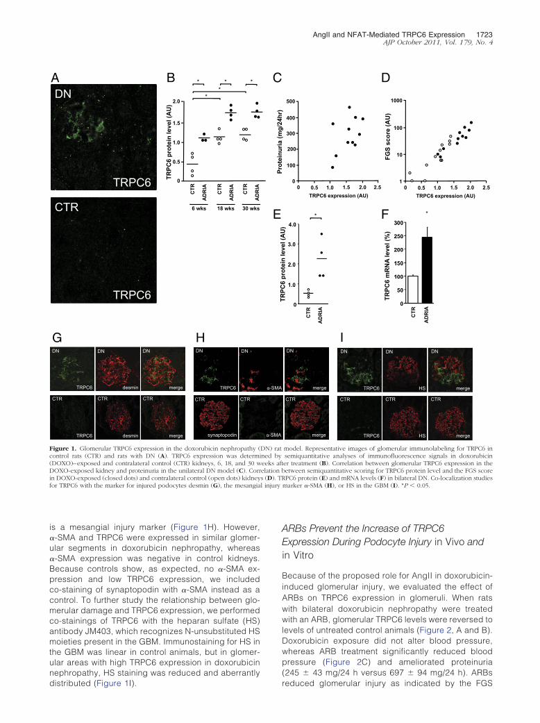
AngII and NFAT-Mediated TRPC6 Expression 1723AJP October 2011, Vol. 179, No. 4
is a mesangial injury marker (Figure 1H). However,�-SMA and TRPC6 were expressed in similar glomer-ular segments in doxorubicin nephropathy, whereas�-SMA expression was negative in control kidneys.Because controls show, as expected, no �-SMA ex-pression and low TRPC6 expression, we includedco-staining of synaptopodin with �-SMA instead as acontrol. To further study the relationship between glo-merular damage and TRPC6 expression, we performedco-stainings of TRPC6 with the heparan sulfate (HS)antibody JM403, which recognizes N-unsubstituted HSmoieties present in the GBM. Immunostaining for HS inthe GBM was linear in control animals, but in glomer-ular areas with high TRPC6 expression in doxorubicinnephropathy, HS staining was reduced and aberrantly
A
CTR
TRPC6
B
TRPC6 desmin merge
TRPC6 desmin merge
CTR CTR CTR
DN DN DN
G H
TRPC6
DN DN
CTR CTR
synaptopodin
DN
TRPC6 TRPC
6 pr
otei
n le
vel (
AU
) 2.0
1.5
1.0
0.5
0
CTR
AD
RIA
CTR
AD
RIA
CTR
AD
RIA
6 wks 18 wks 30 wks
* * * *
*
Figure 1. Glomerular TRPC6 expression in the doxorubicin nephropathy (control rats (CTR) and rats with DN (A). TRPC6 expression was determ(DOXO)–exposed and contralateral control (CTR) kidneys, 6, 18, and 30 wDOXO-exposed kidney and proteinuria in the unilateral DN model (C). Corrin DOXO-exposed (closed dots) and contralateral control (open dots) kidneyfor TRPC6 with the marker for injured podocytes desmin (G), the mesangia
distributed (Figure 1I).
ARBs Prevent the Increase of TRPC6Expression During Podocyte Injury in Vivo andin Vitro
Because of the proposed role for AngII in doxorubicin-induced glomerular injury, we evaluated the effect ofARBs on TRPC6 expression in glomeruli. When ratswith bilateral doxorubicin nephropathy were treatedwith an ARB, glomerular TRPC6 levels were reversed tolevels of untreated control animals (Figure 2, A and B).Doxorubicin exposure did not alter blood pressure,whereas ARB treatment significantly reduced bloodpressure (Figure 2C) and ameliorated proteinuria(245 � 43 mg/24 h versus 697 � 94 mg/24 h). ARBs
D
F
TRPC6 HS merge
DN DN DN
TRPC6 HS merge
CTR CTR CTR
I
merge
DN
CTR
merge
FGS
scor
e (A
U)
1000
100
10
1 0.5 1.0 1.5 2.0 2.5
TRPC6 expression (AU) 0
500
400
300
200
100
0
Prot
einu
ria (m
g/24
hr)
0.5 1.0 1.5 2.0 TRPC6 expression (AU)
0 2.5
TRPC
6 pr
otei
n le
vel (
AU
)
*
()
CTR
AD
RIA
TRPC
6 m
RN
A le
vel (
%)
CTR
AD
RIA
*
model. Representative images of glomerular immunolabeling for TRPC6 insemiquantitative analyses of immunofluorescence signals in doxorubiciner treatment (B). Correlation between glomerular TRPC6 expression in theetween semiquantitative scoring for TRPC6 protein level and the FGS scorePC6 protein (E) and mRNA levels (F) in bilateral DN. Co-localization studies
marker �-SMA (H), or HS in the GBM (I). *P � 0.05.
C
E
-SMA
-SMA
DN) ratined byeeks aftelation bs (D). TRl injury
reduced glomerular injury as indicated by the FGS
1724 Nijenhuis et alAJP October 2011, Vol. 179, No. 4
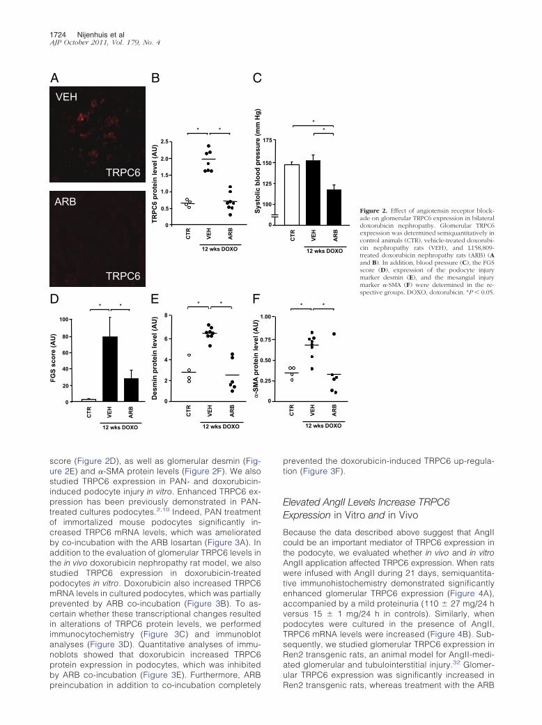
score (Figure 2D), as well as glomerular desmin (Fig-ure 2E) and �-SMA protein levels (Figure 2F). We alsostudied TRPC6 expression in PAN- and doxorubicin-induced podocyte injury in vitro. Enhanced TRPC6 ex-pression has been previously demonstrated in PAN-treated cultures podocytes.2,10 Indeed, PAN treatmentof immortalized mouse podocytes significantly in-creased TRPC6 mRNA levels, which was amelioratedby co-incubation with the ARB losartan (Figure 3A). Inaddition to the evaluation of glomerular TRPC6 levels inthe in vivo doxorubicin nephropathy rat model, we alsostudied TRPC6 expression in doxorubicin-treatedpodocytes in vitro. Doxorubicin also increased TRPC6mRNA levels in cultured podocytes, which was partiallyprevented by ARB co-incubation (Figure 3B). To as-certain whether these transcriptional changes resultedin alterations of TRPC6 protein levels, we performedimmunocytochemistry (Figure 3C) and immunoblotanalyses (Figure 3D). Quantitative analyses of immu-noblots showed that doxorubicin increased TRPC6protein expression in podocytes, which was inhibitedby ARB co-incubation (Figure 3E). Furthermore, ARB
A
Syst
olic
blo
od p
ress
ure
(mm
Hg)
C
100
80
60
40
20
0
FGS
scor
e (A
U)
CTR
VEH
AR
B
12 wks DOXO
* * D * *
CTR
VEH
AR
B
12 wks DOXO
Des
min
pro
tein
leve
l (A
U)
E 8
6
4
2
0
-SM
A pr
otei
n le
vel (
AU
)
F 1
0
0
0
2.5
2.0
1.5
1.0
0.5
0
* *
CTR
VEH
AR
B
12 wks DOXO
TRPC
6 pr
otei
n le
vel (
AU
)
B VEH
ARB
TRPC6
TRPC6
preincubation in addition to co-incubation completely
prevented the doxorubicin-induced TRPC6 up-regula-tion (Figure 3F).
Elevated AngII Levels Increase TRPC6Expression in Vitro and in Vivo
Because the data described above suggest that AngIIcould be an important mediator of TRPC6 expression inthe podocyte, we evaluated whether in vivo and in vitroAngII application affected TRPC6 expression. When ratswere infused with AngII during 21 days, semiquantita-tive immunohistochemistry demonstrated significantlyenhanced glomerular TRPC6 expression (Figure 4A),accompanied by a mild proteinuria (110 � 27 mg/24 hversus 15 � 1 mg/24 h in controls). Similarly, whenpodocytes were cultured in the presence of AngII,TRPC6 mRNA levels were increased (Figure 4B). Sub-sequently, we studied glomerular TRPC6 expression inRen2 transgenic rats, an animal model for AngII-medi-ated glomerular and tubulointerstitial injury.32 Glomer-ular TRPC6 expression was significantly increased in
* *
CTR
VEH
AR
B
12 wks DOXO
* *
CTR
VEH
AR
B
12 wks DOXO
Figure 2. Effect of angiotensin receptor block-ade on glomerular TRPC6 expression in bilateraldoxorubicin nephropathy. Glomerular TRPC6expression was determined semiquantitatively incontrol animals (CTR), vehicle-treated doxorubi-cin nephropathy rats (VEH), and L158,809-treated doxorubicin nephropathy rats (ARB) (Aand B). In addition, blood pressure (C), the FGSscore (D), expression of the podocyte injurymarker desmin (E), and the mesangial injurymarker �-SMA (F) were determined in the re-spective groups. DOXO, doxorubicin. *P � 0.05.
2.0
150
125
100
0
175
.00
.75
.50
.25
0
Ren2 transgenic rats, whereas treatment with the ARB
AngII and NFAT-Mediated TRPC6 Expression 1725AJP October 2011, Vol. 179, No. 4
candesartan in a nonhypotensive dose significantly re-duced this effect (Figure 4C). This finding suggeststhat increased TRPC6 expression in Ren2 transgenicrats is AngII dependent. Alternatively, a direct effect ofrenin on TRPC6 expression via the (pro)renin receptorthat is expressed by podocytes could cause this find-ing.33 However, renin application to differentiated cul-tured podocytes did not induce TRPC6 expression(Figure 4D). Because our results suggest that AngIIregulates TRPC6 expression in podocyte injury, we
A TR
PC6
mR
NA
leve
l (%
)
B * *
TRPC
6 m
RN
A le
vel (
%)
* *
DOXO DOXO + ARB RTC
TRPC6
C
D
F E
0
100
200
300
400
500
1 2 3
* *
TRPC
6 / β
-act
in
prot
ein
leve
l (%
)
CTR
DO
XO
DO
XO
+ A
RB
0
50
100
150
200
250
300
350
1 2 3
CTR
DO
XO
AR
B +
D
OXO
TRPC
6 m
RN
A le
vel (
%)
0
50
100
150
200
250
300
1 2 3 0
100
200
300
400
500
600
1 2 3
* *
CTR
PAN
PAN
+
AR
B
CTR
DO
XO
DO
XO
+ A
RB
CTR DOXO DOXO + ARB
β-actin
100
150
75 50
37
Figure 3. Effect of angiotensin receptor blockade on TRPC6 mRNA andprotein levels in in vitro podocyte injury. TRPC6 mRNA levels were deter-mined in untreated cultured podocytes (CTR) and podocytes incubated for24 hours with PAN or co-incubated with losartan (PAN � ARB) (n � 5–6separate podocyte cultures per experimental condition) (A). The effect ofdoxorubicin (DOXO) and co-incubation with DOXO and losartan (DOXO �ARB) on TRPC6 mRNA levels (B). TRPC6 protein expression visualized byimmunocytochemistry of cultured podocytes (C). Immunoblot analysis ofcell lysates derived from DOXO- and/or losartan-treated podocytes forTRPC6 and �-actin. Molecular weights indicated in kiloDaltons.(D). Intensityof TRPC6 immunoblot signals was quantified by densitometry and TRPC6protein levels were normalized to �-actin (E). Effect of preincubation withARB 1 hour before DOXO application (ARB � DOXO) on TRPC6 mRNAlevels (F). *P � 0.05.
evaluated the effect of lowering AngII levels in bilateral
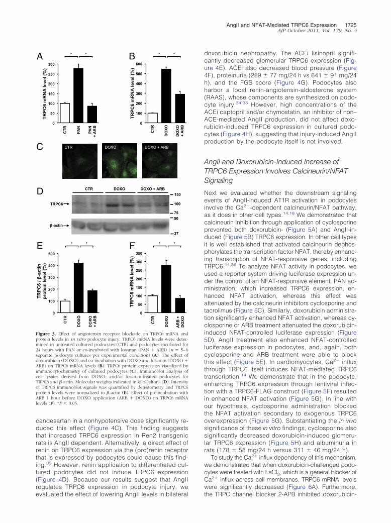
doxorubicin nephropathy. The ACEi lisinopril signifi-cantly decreased glomerular TRPC6 expression (Fig-ure 4E). ACEi also decreased blood pressure (Figure4F), proteinuria (289 � 77 mg/24 h vs 641 � 91 mg/24h), and the FGS score (Figure 4G). Podocytes alsoharbor a local renin-angiotensin-aldosterone system(RAAS), whose components are synthesized on podo-cyte injury.34,35 However, high concentrations of theACEi captopril and/or chymostatin, an inhibitor of non–ACE-mediated AngII production, did not affect doxo-rubicin-induced TRPC6 expression in cultured podo-cytes (Figure 4H), suggesting that injury-induced AngIIproduction by the podocyte itself is not involved.
AngII and Doxorubicin-Induced Increase ofTRPC6 Expression Involves Calcineurin/NFATSignaling
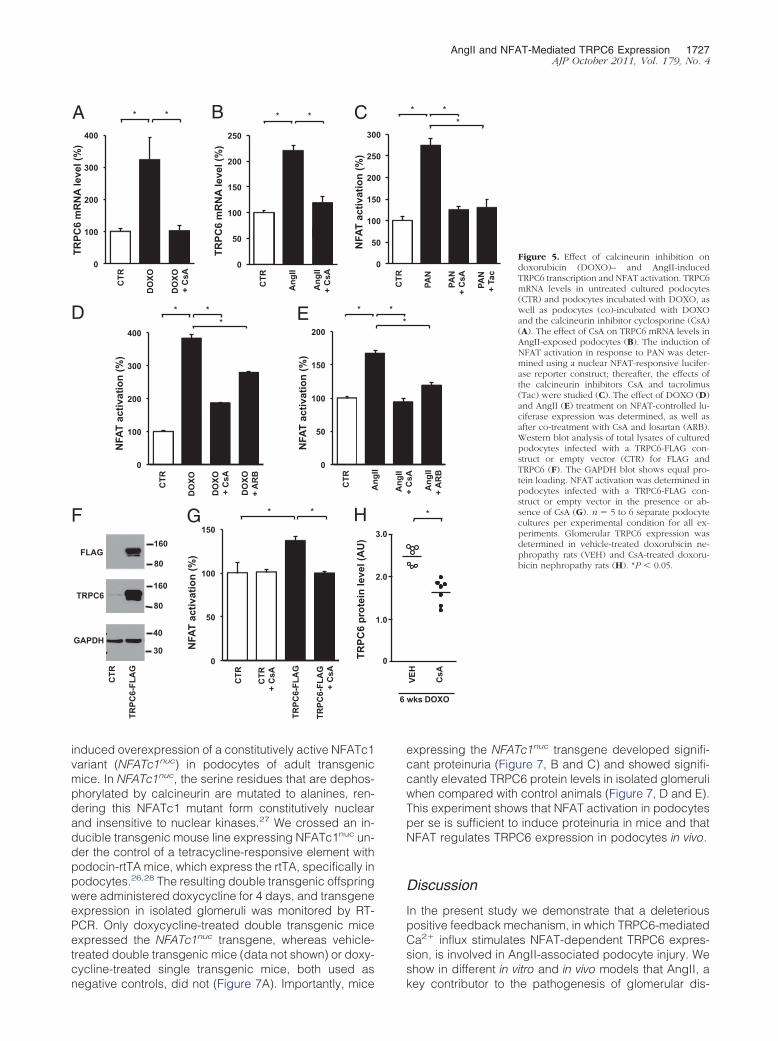
Next we evaluated whether the downstream signalingevents of AngII-induced AT1R activation in podocytesinvolve the Ca2�-dependent calcineurin/NFAT pathway,as it does in other cell types.14,18 We demonstrated thatcalcineurin inhibition through application of cyclosporineprevented both doxorubicin- (Figure 5A) and AngII-in-duced (Figure 5B) TRPC6 expression. In other cell typesit is well established that activated calcineurin dephos-phorylates the transcription factor NFAT, thereby enhanc-ing transcription of NFAT-responsive genes, includingTRPC6.14,36 To analyze NFAT activity in podocytes, weused a reporter system driving luciferase expression un-der the control of an NFAT-responsive element. PAN ad-ministration, which increased TRPC6 expression, en-hanced NFAT activation, whereas this effect wasattenuated by the calcineurin inhibitors cyclosporine andtacrolimus (Figure 5C). Similarly, doxorubicin administra-tion significantly enhanced NFAT activation, whereas cy-closporine or ARB treatment attenuated the doxorubicin-induced NFAT-controlled luciferase expression (Figure5D). AngII treatment also enhanced NFAT-controlledluciferase expression in podocytes, and, again, bothcyclosporine and ARB treatment were able to blockthis effect (Figure 5E). In cardiomyocytes, Ca2� influxthrough TRPC6 itself induces NFAT-mediated TRPC6transcription.14 We demonstrate that in the podocyte,enhancing TRPC6 expression through lentiviral infec-tion with a TRPC6-FLAG construct (Figure 5F) resultedin enhanced NFAT activation (Figure 5G). In line withour hypothesis, cyclosporine administration blockedthe NFAT activation secondary to exogenous TRPC6overexpression (Figure 5G). Substantiating the in vivosignificance of these in vitro findings, cyclosporine alsosignificantly decreased doxorubicin-induced glomeru-lar TRPC6 expression (Figure 5H) and albuminuria inrats (178 � 58 mg/24 h versus 311 � 46 mg/24 h).
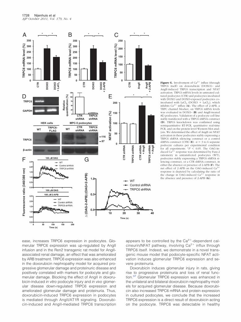
To study the Ca2� influx dependency of this mechanism,we demonstrated that when doxorubicin-challenged podo-cytes were treated with LaCl3, which is a general blocker ofCa2� influx across cell membranes, TRPC6 mRNA levelswere significantly decreased (Figure 6A). Furthermore,
the TRPC channel blocker 2-APB inhibited doxorubicin-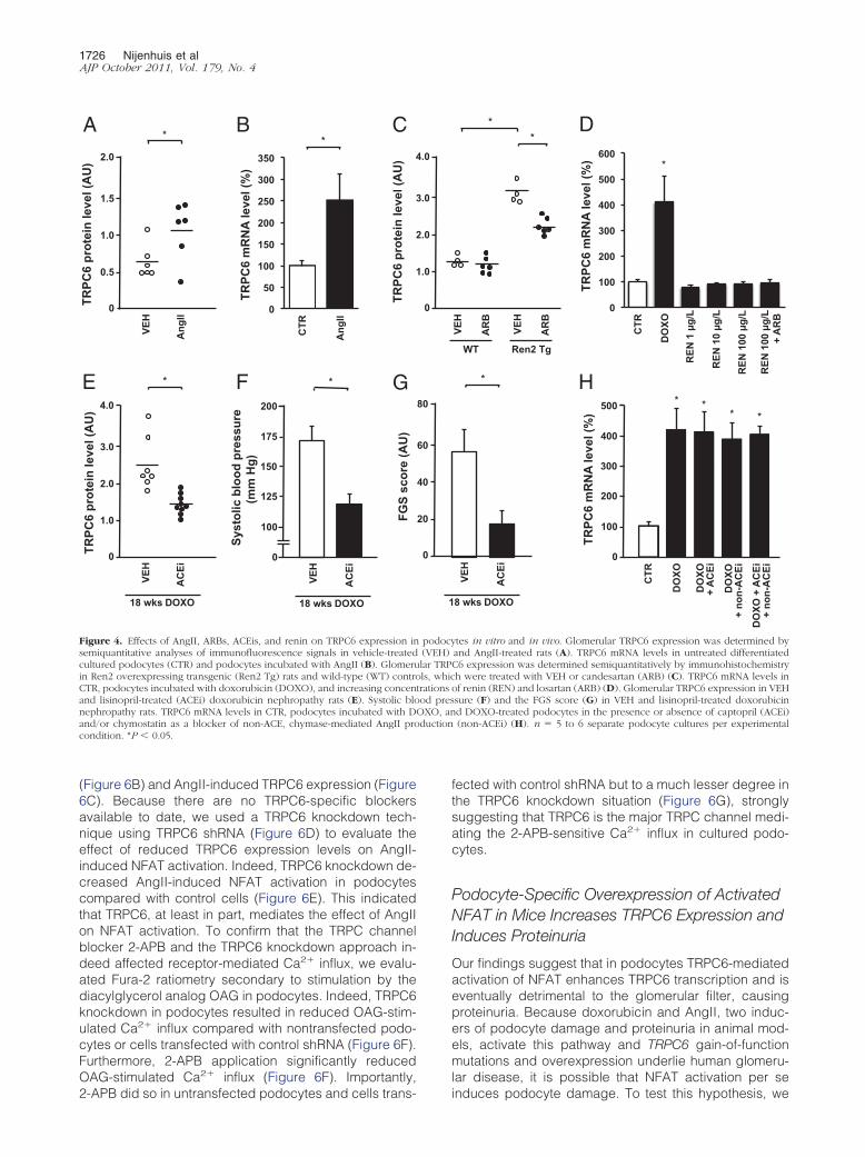
duction
1726 Nijenhuis et alAJP October 2011, Vol. 179, No. 4
(Figure 6B) and AngII-induced TRPC6 expression (Figure6C). Because there are no TRPC6-specific blockersavailable to date, we used a TRPC6 knockdown tech-nique using TRPC6 shRNA (Figure 6D) to evaluate theeffect of reduced TRPC6 expression levels on AngII-induced NFAT activation. Indeed, TRPC6 knockdown de-creased AngII-induced NFAT activation in podocytescompared with control cells (Figure 6E). This indicatedthat TRPC6, at least in part, mediates the effect of AngIIon NFAT activation. To confirm that the TRPC channelblocker 2-APB and the TRPC6 knockdown approach in-deed affected receptor-mediated Ca2� influx, we evalu-ated Fura-2 ratiometry secondary to stimulation by thediacylglycerol analog OAG in podocytes. Indeed, TRPC6knockdown in podocytes resulted in reduced OAG-stim-ulated Ca2� influx compared with nontransfected podo-cytes or cells transfected with control shRNA (Figure 6F).Furthermore, 2-APB application significantly reducedOAG-stimulated Ca2� influx (Figure 6F). Importantly,
A
TRPC
6 m
RN
A le
vel (
%)
0
50
100
150
200
250
300
350
1 2 CTR
Ang
II
*
E
TRPC
6 pr
otei
n le
vel (
AU
) 2.0
3.0
2.0
1.0
0
4.0
VEH
*
AC
Ei
18 wks DOXO
75
100
125
150
175
200
F 2.0
150
125
100
175
Syst
olic
blo
od p
ress
ure
(mm
Hg)
0
200
VEH
AC
Ei
*
18 wks DOXO
G 1
FGS
scor
e (A
U)
C
TRPC
6 pr
otei
n le
vel (
AU
) 2
TRPC
6 pr
otei
n le
vel (
AU
) 2.0
1.5
1.0
0.5
0
2.0 VE
H
*
Ang
II
B
Figure 4. Effects of AngII, ARBs, ACEis, and renin on TRPC6 expression insemiquantitative analyses of immunofluorescence signals in vehicle-treatedcultured podocytes (CTR) and podocytes incubated with AngII (B). Glomeruin Ren2 overexpressing transgenic (Ren2 Tg) rats and wild-type (WT) controCTR, podocytes incubated with doxorubicin (DOXO), and increasing concenand lisinopril-treated (ACEi) doxorubicin nephropathy rats (E). Systolic blonephropathy rats. TRPC6 mRNA levels in CTR, podocytes incubated with Dand/or chymostatin as a blocker of non-ACE, chymase-mediated AngII procondition. *P � 0.05.
2-APB did so in untransfected podocytes and cells trans-
fected with control shRNA but to a much lesser degree inthe TRPC6 knockdown situation (Figure 6G), stronglysuggesting that TRPC6 is the major TRPC channel medi-ating the 2-APB-sensitive Ca2� influx in cultured podo-cytes.
Podocyte-Specific Overexpression of ActivatedNFAT in Mice Increases TRPC6 Expression andInduces Proteinuria
Our findings suggest that in podocytes TRPC6-mediatedactivation of NFAT enhances TRPC6 transcription and iseventually detrimental to the glomerular filter, causingproteinuria. Because doxorubicin and AngII, two induc-ers of podocyte damage and proteinuria in animal mod-els, activate this pathway and TRPC6 gain-of-functionmutations and overexpression underlie human glomeru-lar disease, it is possible that NFAT activation per se
VEH
AC
Ei
*
8 wks DOXO
0
100
200
300
400
500
1 2 3 4 5
2.0
300
200
100
400
TRPC
6 m
RN
A le
vel (
%)
0
500 * * * *
CTR
DO
XO
DO
XO
+ A
CEi
DO
XO
+ no
n-A
CEi
DO
XO +
AC
Ei
+ no
n-A
CEi
VEH
AR
B
VEH
AR
B
* *
WT Ren2 Tg
0
100
200
300
400
500
600 *
D
TRPC
6 m
RN
A le
vel (
%)
CTR
DO
XO
REN
1 µ
g/L
REN
10
µg/L
REN
100
µg/
L
REN
100
µg/
L +
AR
B
H
tes in vitro and in vivo. Glomerular TRPC6 expression was determined byand AngII-treated rats (A). TRPC6 mRNA levels in untreated differentiatedC6 expression was determined semiquantitatively by immunohistochemistryh were treated with VEH or candesartan (ARB) (C). TRPC6 mRNA levels in
of renin (REN) and losartan (ARB) (D). Glomerular TRPC6 expression in VEHsure (F) and the FGS score (G) in VEH and lisinopril-treated doxorubicind DOXO-treated podocytes in the presence or absence of captopril (ACEi)(non-ACEi) (H). n � 5 to 6 separate podocyte cultures per experimental
0
25
50
75
00
80
60
40
20
0
1
.0
3.0
2.0
1.0
0
4.0
podocy(VEH)lar TRPls, whic
trationsod pres
OXO, an
induces podocyte damage. To test this hypothesis, we
AngII and NFAT-Mediated TRPC6 Expression 1727AJP October 2011, Vol. 179, No. 4
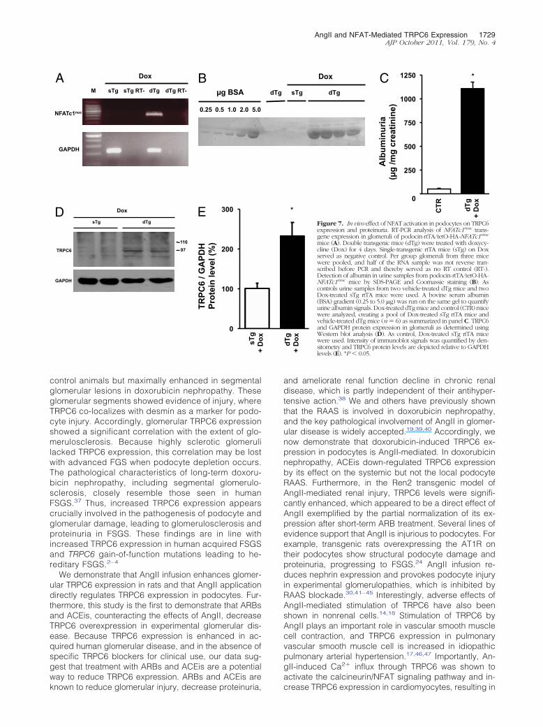
induced overexpression of a constitutively active NFATc1variant (NFATc1nuc) in podocytes of adult transgenicmice. In NFATc1nuc, the serine residues that are dephos-phorylated by calcineurin are mutated to alanines, ren-dering this NFATc1 mutant form constitutively nuclearand insensitive to nuclear kinases.27 We crossed an in-ducible transgenic mouse line expressing NFATc1nuc un-der the control of a tetracycline-responsive element withpodocin-rtTA mice, which express the rtTA, specifically inpodocytes.26,28 The resulting double transgenic offspringwere administered doxycycline for 4 days, and transgeneexpression in isolated glomeruli was monitored by RT-PCR. Only doxycycline-treated double transgenic miceexpressed the NFATc1nuc transgene, whereas vehicle-treated double transgenic mice (data not shown) or doxy-cycline-treated single transgenic mice, both used as
A
0
100
200
300
400
1 2 3
TRPC
6 m
RN
A le
vel (
%)
* * C
TR
DO
XO
DO
XO
+ C
sA
B
0
50
100
150
200
250
1 2 3
TRPC
6 m
RN
A le
vel (
%)
* *
CTR
Ang
II
Ang
II +
CsA
5
10
15
20
25
30
NFA
T ac
tivat
ion
(%)
C
0
50
100
150
1 2 3 4
NFA
T ac
tivat
ion
(%)
CTR
CTR
+
CsA
TRPC
6-FL
AG
TRPC
6-FL
AG
+
CsA
* *
160
TRPC6
80
160
80
40
30
FLAG
GAPDH
CTR
TRPC
6-FL
AG
F
0
100
200
300
400
1 2 3 4
D
NFA
T ac
tivat
ion
(%)
CTR
DO
XO
DO
XO
+ C
sA
DO
XO
+ A
RB
* * *
0
50
100
150
200
1 2
E N
FAT
activ
atio
n (%
)
CTR
*
H
TRPC
6 pr
otei
n le
vel (
AU
)
G
negative controls, did not (Figure 7A). Importantly, mice
expressing the NFATc1nuc transgene developed signifi-cant proteinuria (Figure 7, B and C) and showed signifi-cantly elevated TRPC6 protein levels in isolated glomeruliwhen compared with control animals (Figure 7, D and E).This experiment shows that NFAT activation in podocytesper se is sufficient to induce proteinuria in mice and thatNFAT regulates TRPC6 expression in podocytes in vivo.
Discussion
In the present study we demonstrate that a deleteriouspositive feedback mechanism, in which TRPC6-mediatedCa2� influx stimulates NFAT-dependent TRPC6 expres-sion, is involved in AngII-associated podocyte injury. Weshow in different in vitro and in vivo models that AngII, a
PAN PAN+CsA PAN+FK506
* *
PAN
PAN
+
CsA
PAN
+
Tac
*
4
+ C
sA
Ang
II +
AR
B
VEH
*
CsA
wks DOXO
Figure 5. Effect of calcineurin inhibition ondoxorubicin (DOXO)– and AngII-inducedTRPC6 transcription and NFAT activation. TRPC6mRNA levels in untreated cultured podocytes(CTR) and podocytes incubated with DOXO, aswell as podocytes (co)-incubated with DOXOand the calcineurin inhibitor cyclosporine (CsA)(A). The effect of CsA on TRPC6 mRNA levels inAngII-exposed podocytes (B). The induction ofNFAT activation in response to PAN was deter-mined using a nuclear NFAT-responsive lucifer-ase reporter construct; thereafter, the effects ofthe calcineurin inhibitors CsA and tacrolimus(Tac) were studied (C). The effect of DOXO (D)and AngII (E) treatment on NFAT-controlled lu-ciferase expression was determined, as well asafter co-treatment with CsA and losartan (ARB).Western blot analysis of total lysates of culturedpodocytes infected with a TRPC6-FLAG con-struct or empty vector (CTR) for FLAG andTRPC6 (F). The GAPDH blot shows equal pro-tein loading. NFAT activation was determined inpodocytes infected with a TRPC6-FLAG con-struct or empty vector in the presence or ab-sence of CsA (G). n � 5 to 6 separate podocytecultures per experimental condition for all ex-periments. Glomerular TRPC6 expression wasdetermined in vehicle-treated doxorubicin ne-phropathy rats (VEH) and CsA-treated doxoru-bicin nephropathy rats (H). *P � 0.05.
0
0
0
0
0
0
0
CTR
CTR
3
Ang
II
Ang
II
* *
2.0
2.0
1.0
0
6
3.0
key contributor to the pathogenesis of glomerular dis-
1728 Nijenhuis et alAJP October 2011, Vol. 179, No. 4
ease, increases TRPC6 expression in podocytes. Glo-merular TRPC6 expression was up-regulated by AngIIinfusion and in the Ren2 transgenic rat model for AngII-associated renal damage, an effect that was amelioratedby ARB treatment. TRPC6 expression was also enhancedin the doxorubicin nephropathy model for acquired pro-gressive glomerular damage and proteinuric disease andpositively correlated with markers for podocyte and glo-merular damage. Blocking the effect of AngII in doxoru-bicin-induced in vitro podocyte injury and in vivo glomer-ular disease down-regulated TRPC6 expression andameliorated glomerular damage and proteinuria. Thus,doxorubicin-induced TRPC6 expression in podocytesis mediated through AngII/AT1R signaling. Doxorubi-
MPC-5 podocytes HEK cells
TRPC6
0
50
100
150
200
250
300
350
1 2 3 0
50
100
150
200
250
300
350
1 2 3
A
TRPC
6 m
RN
A le
vel (
%)
B * *
TRPC
6 m
RN
A le
vel (
%)
* * C
TR
DO
XO
DO
XO
+ La
Cl 3
CTR
DO
XO
+ 2-
APB
C
5
10
15
20
NFA
T ac
tivat
ion
(%)
E TRPC6
GAPDH
CTR shRNA
TRPC6 shRNA
Rel
ativ
e
quan
tific
atio
n (%
) D
β-actin
TRPC6 shRNA
CTR shRNA WT TRPC6-
FLAG WT
97 116
F
Fluo
resc
ence
ratio
(340
/380
nm
)
∆ (O
AG
-2-A
PB):(
OA
G+2
-APB
)
G
DO
XO
Time (s)
cin-induced and AngII-mediated TRPC6 transcription
appears to be controlled by the Ca2�-dependent cal-cineurin/NFAT pathway, involving Ca2� influx throughTRPC6 itself. Indeed, we demonstrate in a novel trans-genic mouse model that podocyte-specific NFAT acti-vation induces glomerular TRPC6 expression and se-vere proteinuria.
Doxorubicin induces glomerular injury in rats, givingrise to progressive proteinuria and loss of renal func-tion.37 Glomerular TRPC6 expression was enhanced inthe unilateral and bilateral doxorubicin nephropathy mod-els for acquired glomerular disease. Because doxorubi-cin also increased TRPC6 mRNA and protein expressionin cultured podocytes, we conclude that the increasedTRPC6 expression is a direct result of doxorubicin acting
1 2 3
CTR
Ang
II
Ang
II +
2-A
PB
* *
T R T R P C T R T R P
TRPC
6 sh
RN
A
CTR
shR
NA
+ A
ngII
TRPC
6 sh
RN
A +
Ang
II
* *
(s)
Figure 6. Involvement of Ca2� influx (throughTRPC6 itself) on doxorubicin (DOXO)– andAngII-induced TRPC6 transcription and NFATactivation. TRPC6 mRNA levels in untreated cul-tured podocytes (CTR) and podocytes incubatedwith DOXO and DOXO-exposed podocytes co-incubated with LaCl3 (DOXO � LaCl3), whichinhibits Ca2� influx (A). The effect of 2-APB, aTRPC channel blocker, on TRPC6 mRNA levelswas evaluated in DOXO- (B) and AngII-treated(C) podocytes. Validation of a podocyte cell linestably transfected with a TRPC6 shRNA construct(D). TRPC6 knockdown was confirmed usingsemiquantitative RT-PCR, quantitative real-timePCR, and on the protein level Western blot anal-ysis. We determined the effect of AngII on NFATactivation in these podocytes stably expressing aTRPC6 shRNA silencing construct or a controlshRNA construct (CTR) (E). n � 3 to 6 separatepodocyte cultures per experimental conditionfor all experiments. *P � 0.05. The OAG-in-duced Ca2� response was determined by Fura-2ratiometry in untransfected podocytes (WT),podocytes stably expressing a TRPC6 shRNA si-lencing construct, or a CTR shRNA construct, ineither the absence or presence of 2-APB (F). Thenet effect of 2-APB on the OAG-induced Ca2�
response is depicted by calculating the ratio ofthe change in OAG-induced Ca2� response inthe absence and presence of 2-APB (G).
0
50
100
150
200
250
TRPC
6 m
RN
A le
vel (
%)
0
0
0
0
0
CC
TR s
hRN
A
Time
on the podocyte. TRPC6 was detectable in healthy
+
AngII and NFAT-Mediated TRPC6 Expression 1729AJP October 2011, Vol. 179, No. 4
control animals but maximally enhanced in segmentalglomerular lesions in doxorubicin nephropathy. Theseglomerular segments showed evidence of injury, whereTRPC6 co-localizes with desmin as a marker for podo-cyte injury. Accordingly, glomerular TRPC6 expressionshowed a significant correlation with the extent of glo-merulosclerosis. Because highly sclerotic glomerulilacked TRPC6 expression, this correlation may be lostwith advanced FGS when podocyte depletion occurs.The pathological characteristics of long-term doxoru-bicin nephropathy, including segmental glomerulo-sclerosis, closely resemble those seen in humanFSGS.37 Thus, increased TRPC6 expression appearscrucially involved in the pathogenesis of podocyte andglomerular damage, leading to glomerulosclerosis andproteinuria in FSGS. These findings are in line withincreased TRPC6 expression in human acquired FSGSand TRPC6 gain-of-function mutations leading to he-reditary FSGS.2– 4
We demonstrate that AngII infusion enhances glomer-ular TRPC6 expression in rats and that AngII applicationdirectly regulates TRPC6 expression in podocytes. Fur-thermore, this study is the first to demonstrate that ARBsand ACEis, counteracting the effects of AngII, decreaseTRPC6 overexpression in experimental glomerular dis-ease. Because TRPC6 expression is enhanced in ac-quired human glomerular disease, and in the absence ofspecific TRPC6 blockers for clinical use, our data sug-gest that treatment with ARBs and ACEis are a potentialway to reduce TRPC6 expression. ARBs and ACEis are
NFATc1nuc
GAPDH
Dox
sTg M sTg RT- dTg dTg RT-
0.25 0.5 1.0 2.0 5.0
µg BSA B A
dTg sTg
Dox
97 116
GAPDH
TRPC6
D E TR
PC6
/ GA
PDH
Pr
otei
n le
vel (
%)
0
100
200
300
sTg
known to reduce glomerular injury, decrease proteinuria,
and ameliorate renal function decline in chronic renaldisease, which is partly independent of their antihyper-tensive action.38 We and others have previously shownthat the RAAS is involved in doxorubicin nephropathy,and the key pathological involvement of AngII in glomer-ular disease is widely accepted.19,39,40 Accordingly, wenow demonstrate that doxorubicin-induced TRPC6 ex-pression in podocytes is AngII-mediated. In doxorubicinnephropathy, ACEis down-regulated TRPC6 expressionby its effect on the systemic but not the local podocyteRAAS. Furthermore, in the Ren2 transgenic model ofAngII-mediated renal injury, TRPC6 levels were signifi-cantly enhanced, which appeared to be a direct effect ofAngII exemplified by the partial normalization of its ex-pression after short-term ARB treatment. Several lines ofevidence support that AngII is injurious to podocytes. Forexample, transgenic rats overexpressing the AT1R ontheir podocytes show structural podocyte damage andproteinuria, progressing to FSGS.24 AngII infusion re-duces nephrin expression and provokes podocyte injuryin experimental glomerulopathies, which is inhibited byRAAS blockade.30,41–45 Interestingly, adverse effects ofAngII-mediated stimulation of TRPC6 have also beenshown in nonrenal cells.14,18 Stimulation of TRPC6 byAngII plays an important role in vascular smooth musclecell contraction, and TRPC6 expression in pulmonaryvascular smooth muscle cell is increased in idiopathicpulmonary arterial hypertension.17,46,47 Importantly, An-gII-induced Ca2� influx through TRPC6 was shown toactivate the calcineurin/NFAT signaling pathway and in-
sTg dTg
Dox
0
250
500
750
1000
1250
1 2
Alb
umin
uria
(µ
g /m
g cr
eatin
ine)
*
dTg
+
Dox
CTR
*
dTg
+
Dox
C
Figure 7. In vivo effect of NFAT activation in podocytes on TRPC6expression and proteinuria. RT-PCR analysis of NFATc1nuc trans-gene expression in glomeruli of podocin-rtTA/tetO-HA-NFATc1nuc
mice (A). Double transgenic mice (dTg) were treated with doxycy-cline (Dox) for 4 days. Single-transgenic rtTA mice (sTg) on Doxserved as negative control. Per group glomeruli from three micewere pooled, and half of the RNA sample was not reverse tran-scribed before PCR and thereby served as no RT control (RT-).Detection of albumin in urine samples from podocin-rtTA/tetO-HA-NFATc1nuc mice by SDS-PAGE and Coomassie staining (B). Ascontrols urine samples from two vehicle-treated dTg mice and twoDox-treated sTg rtTA mice were used. A bovine serum albumin(BSA) gradient (0.25 to 5.0 �g) was run on the same gel to quantifyurine albumin signals. Dox-treated dTg mice and control (CTR) micewere analyzed, creating a pool of Dox-treated sTg rtTA mice andvehicle-treated dTg mice (n � 6) as summarized in panel C. TRPC6and GAPDH protein expression in glomeruli as determined usingWestern blot analysis (D). As control, Dox-treated sTg rtTA micewere used. Intensity of immunoblot signals was quantified by den-sitometry and TRPC6 protein levels are depicted relative to GAPDHlevels (E). *P � 0.05.
dTg
Dox
crease TRPC6 expression in cardiomyocytes, resulting in
1730 Nijenhuis et alAJP October 2011, Vol. 179, No. 4
cardiomyocyte hypertrophy.14,18 A recent study by Eckelet al48 showed that AngII-induced albuminuria was notprevented but was significantly ameliorated in TRPC6knockout compared with wild-type mice. Altogether, thisdemonstrates that AngII-induced and calcineurin/NFAT-mediated TRPC6 expression in the podocyte is an impor-tant mediator in the pathogenesis of podocyte injury andglomerular disease.
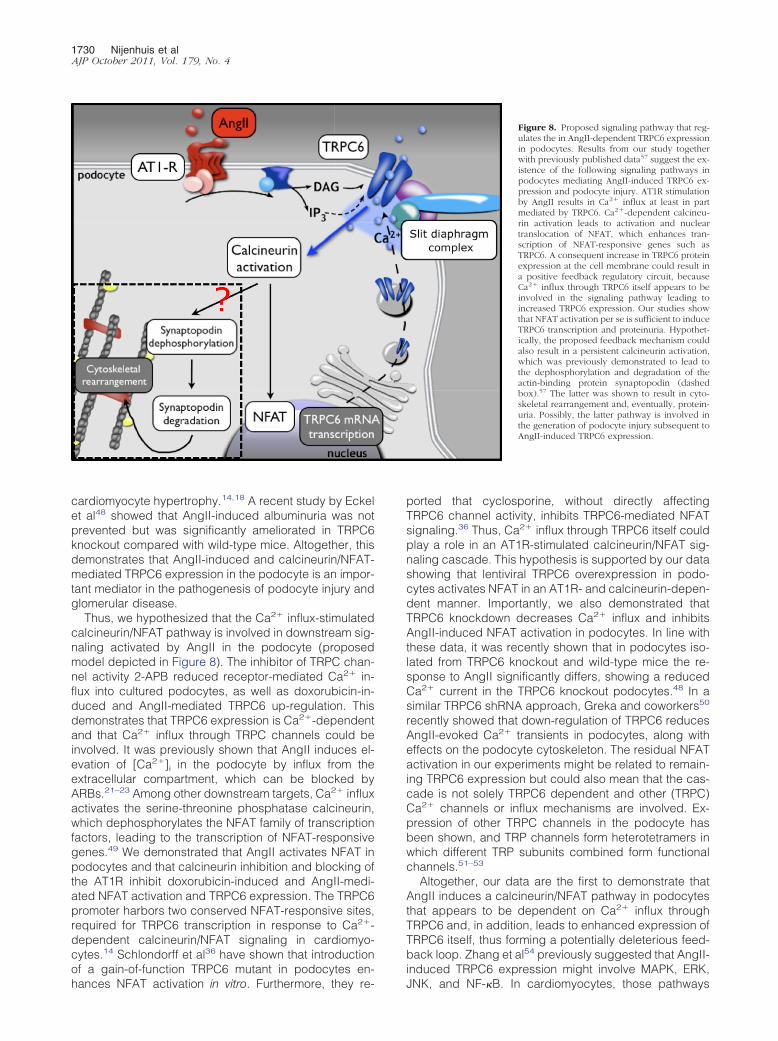
Thus, we hypothesized that the Ca2� influx-stimulatedcalcineurin/NFAT pathway is involved in downstream sig-naling activated by AngII in the podocyte (proposedmodel depicted in Figure 8). The inhibitor of TRPC chan-nel activity 2-APB reduced receptor-mediated Ca2� in-flux into cultured podocytes, as well as doxorubicin-in-duced and AngII-mediated TRPC6 up-regulation. Thisdemonstrates that TRPC6 expression is Ca2�-dependentand that Ca2� influx through TRPC channels could beinvolved. It was previously shown that AngII induces el-evation of [Ca2�]i in the podocyte by influx from theextracellular compartment, which can be blocked byARBs.21–23 Among other downstream targets, Ca2� influxactivates the serine-threonine phosphatase calcineurin,which dephosphorylates the NFAT family of transcriptionfactors, leading to the transcription of NFAT-responsivegenes.49 We demonstrated that AngII activates NFAT inpodocytes and that calcineurin inhibition and blocking ofthe AT1R inhibit doxorubicin-induced and AngII-medi-ated NFAT activation and TRPC6 expression. The TRPC6promoter harbors two conserved NFAT-responsive sites,required for TRPC6 transcription in response to Ca2�-dependent calcineurin/NFAT signaling in cardiomyo-cytes.14 Schlondorff et al36 have shown that introductionof a gain-of-function TRPC6 mutant in podocytes en-
hances NFAT activation in vitro. Furthermore, they re-ported that cyclosporine, without directly affectingTRPC6 channel activity, inhibits TRPC6-mediated NFATsignaling.36 Thus, Ca2� influx through TRPC6 itself couldplay a role in an AT1R-stimulated calcineurin/NFAT sig-naling cascade. This hypothesis is supported by our datashowing that lentiviral TRPC6 overexpression in podo-cytes activates NFAT in an AT1R- and calcineurin-depen-dent manner. Importantly, we also demonstrated thatTRPC6 knockdown decreases Ca2� influx and inhibitsAngII-induced NFAT activation in podocytes. In line withthese data, it was recently shown that in podocytes iso-lated from TRPC6 knockout and wild-type mice the re-sponse to AngII significantly differs, showing a reducedCa2� current in the TRPC6 knockout podocytes.48 In asimilar TRPC6 shRNA approach, Greka and coworkers50
recently showed that down-regulation of TRPC6 reducesAngII-evoked Ca2� transients in podocytes, along witheffects on the podocyte cytoskeleton. The residual NFATactivation in our experiments might be related to remain-ing TRPC6 expression but could also mean that the cas-cade is not solely TRPC6 dependent and other (TRPC)Ca2� channels or influx mechanisms are involved. Ex-pression of other TRPC channels in the podocyte hasbeen shown, and TRP channels form heterotetramers inwhich different TRP subunits combined form functionalchannels.51–53
Altogether, our data are the first to demonstrate thatAngII induces a calcineurin/NFAT pathway in podocytesthat appears to be dependent on Ca2� influx throughTRPC6 and, in addition, leads to enhanced expression ofTRPC6 itself, thus forming a potentially deleterious feed-back loop. Zhang et al54 previously suggested that AngII-induced TRPC6 expression might involve MAPK, ERK,
Figure 8. Proposed signaling pathway that reg-ulates the in AngII-dependent TRPC6 expressionin podocytes. Results from our study togetherwith previously published data57 suggest the ex-istence of the following signaling pathways inpodocytes mediating AngII-induced TRPC6 ex-pression and podocyte injury. AT1R stimulationby AngII results in Ca2� influx at least in partmediated by TRPC6. Ca2�-dependent calcineu-rin activation leads to activation and nucleartranslocation of NFAT, which enhances tran-scription of NFAT-responsive genes such asTRPC6. A consequent increase in TRPC6 proteinexpression at the cell membrane could result ina positive feedback regulatory circuit, becauseCa2� influx through TRPC6 itself appears to beinvolved in the signaling pathway leading toincreased TRPC6 expression. Our studies showthat NFAT activation per se is sufficient to induceTRPC6 transcription and proteinuria. Hypothet-ically, the proposed feedback mechanism couldalso result in a persistent calcineurin activation,which was previously demonstrated to lead tothe dephosphorylation and degradation of theactin-binding protein synaptopodin (dashedbox).57 The latter was shown to result in cyto-skeletal rearrangement and, eventually, protein-uria. Possibly, the latter pathway is involved inthe generation of podocyte injury subsequent toAngII-induced TRPC6 expression.
JNK, and NF-�B. In cardiomyocytes, those pathways
AngII and NFAT-Mediated TRPC6 Expression 1731AJP October 2011, Vol. 179, No. 4
were also described, and receptor-activated TRPC6-me-diated NFAT activation was shown to inhibit JNK andERK.55 To demonstrate that NFAT activation in podocytesis independently capable of enhancing TRPC6 expres-sion in vivo and inducing proteinuria, we generated atransgenic mouse model in which a constitutively activeNFAT mutant could be induced in a podocyte-specificmanner. As originally hypothesized, we demonstratedthat induction of NFAT results in increased glomerularTRPC6 expression and the development of severe pro-teinuria. On submission of this work another articleappeared56 that also describes the effect of NFAToverexpression and the subsequent development ofproteinuria; however, the involvement of TRPC6 wasnot specifically studied in that article. Our data confirmthat the activation of NFAT in podocytes is sufficient toinduce proteinuria and substantiate that a positivefeedback loop involving NFAT-induced TRPC6 expres-sion contributes to the induction and/or maintenance ofproteinuric disease.
The current findings add an additional downstreampathway secondary to calcineurin signaling in podo-cytes, in addition to the dephosphorylation of the actin-binding protein synaptopodin by calcineurin as we pre-viously reported.57 Synaptopodin is vitally important inmaintaining the podocyte actin cytoskeleton, and itsdephosphorylation results in subsequent cathepsinL–mediated degradation, cytoskeletal disorganization,and proteinuria.57,58 Thus, our data contribute to ahypothetical mechanism by which AngII-induced andNFAT-mediated TRPC6 overexpression, as addressedin our experiments, fuel persistent calcineurin activa-tion, leading to synaptopodin degradation as de-scribed previously, eventually perpetuating podocyteinjury (Figure 8). For this hypothesis to be proven, itremains to be established whether Ca2� influx throughTRPC6 directly induces calcineurin-mediated dephos-phorylation of synaptopodin.
In conclusion, we have demonstrated an AngII-in-duced, TRPC6-dependent, NFAT-mediated feedbackmechanism driving TRPC6 transcription in podocytes.This finding underlines the crucial role of TRPC6 in thepathogenesis of podocyte injury and proteinuria.
References
1. Faul C, Asanuma K, Yanagida-Asanuma E, Kim K, Mundel P: Actinup: regulation of podocyte structure and function by components ofthe actin cytoskeleton. Trends Cell Biol 2007, 17:428–437
2. Reiser J, Polu KR, Moller CC, Kenlan P, Altintas MM, Wei C, Faul C,Herbert S, Villegas I, Avila-Casado C, McGee M, Sugimoto H, BrownD, Kalluri R, Mundel P, Smith PL, Clapham DE, Pollak MR: TRPC6 isa glomerular slit diaphragm-associated channel required for normalrenal function. Nat Genet 2005, 37:739–744
3. Winn MP, Conlon PJ, Lynn KL, Farrington MK, Creazzo T, HawkinsAF, Daskalakis N, Kwan SY, Ebersviller S, Burchette JL, Pericak-Vance MA, Howell DN, Vance JM, Rosenberg PB: A mutation in theTRPC6 cation channel causes familial focal segmental glomeruloscle-rosis. Science 2005, 308:1801–1804
4. Moller CC, Wei C, Altintas MM, Li J, Greka A, Ohse T, Pippin JW,Rastaldi MP, Wawersik S, Schiavi S, Henger A, Kretzler M, Shankland
SJ, Reiser J: Induction of TRPC6 channel in acquired forms of pro-teinuric kidney disease. J Am Soc Nephrol 2007, 18:29–365. Krall P, Canales CP, Kairath P, Carmona-Mora P, Molina J, Carpio JD,Ruiz P, Mezzano SA, Li J, Wei C, Reiser J, Young JI, Walz K: Podo-cyte-specific overexpression of wild type or mutant trpc6 in mice issufficient to cause glomerular disease. PLoS One 2010, 5:e12859
6. Clapham DE: TRP channels as cellular sensors. Nature 2003, 426:517–524
7. Hsu YJ, Hoenderop JG, Bindels RJ: TRP channels in kidney disease.Biochim Biophys Acta 2007, 1772:928–936
8. Nijenhuis T, Vallon V, van der Kemp AW, Loffing J, Hoenderop JG,Bindels RJ: Enhanced passive Ca2� reabsorption and reducedMg2� channel abundance explains thiazide-induced hypocalciuriaand hypomagnesemia. J Clin Invest 2005, 115:1651–1658
9. Woudenberg-Vrenken TE, Bindels RJ, Hoenderop JG: The role oftransient receptor potential channels in kidney disease. Nat RevNephrol 2009, 5:441–449
10. Moller CC, Flesche J, Reiser J: Sensitizing the slit diaphragm withTRPC6 ion channels. J Am Soc Nephrol 2009, 20:950–953
11. Zhu B, Chen N, Wang ZH, Pan XX, Ren H, Zhang W, Wang WM:Identification and functional analysis of a novel TRPC6 mutation as-sociated with late onset familial focal segmental glomerulosclerosis inChinese patients. Mutat Res 2009, 664:84–90
12. Santin S, Ars E, Rossetti S, Salido E, Silva I, Garcia-Maset R, GimenezI, Ruiz P, Mendizabal S, Nieto JL, Pena A, Camacho JA, Fraga G,Cobo MA, Bernis C, Ortiz A, de Pablos AL, Sanchez-Moreno A, PintosG, Mirapeix E, Fernandez-Llama P, Ballarin J, Torra R: TRPC6 muta-tional analysis in a large cohort of patients with focal segmentalglomerulosclerosis. Nephrol Dial Transplant 2009, 24: 3089–3096
13. Hofmann T, Obukhov AG, Schaefer M, Harteneck C, Gudermann T,Schultz G: Direct activation of human TRPC6 and TRPC3 channels bydiacylglycerol. Nature 1999, 397:259–263
14. Kuwahara K, Wang Y, McAnally J, Richardson JA, Bassel-Duby R, HillJA, Olson EN: TRPC6 fulfills a calcineurin signaling circuit duringpathologic cardiac remodeling. J Clin Invest 2006, 116:3114–3126
15. Ruggenenti P, Perna A, Gherardi G, Garini G, Zoccali C, Salvadori M,Scolari F, Schena FP, Remuzzi G: Renoprotective properties of ACE-inhibition in non-diabetic nephropathies with non-nephrotic protein-uria. Lancet 1999, 354:359–364
16. Ruggenenti P, Perna A, Loriga G, Ganeva M, Ene-Iordache B, Tur-turro M, Lesti M, Perticucci E, Chakarski IN, Leonardis D, Garini G,Sessa A, Basile C, Alpa M, Scanziani R, Sorba G, Zoccali C, RemuzziG: Blood-pressure control for renoprotection in patients with non-diabetic chronic renal disease (REIN-2): multicentre, randomisedcontrolled trial. Lancet 2005, 365:939–946
17. Saleh SN, Albert AP, Peppiatt CM, Large WA: Angiotensin II activatestwo cation conductances with distinct TRPC1 and TRPC6 channelproperties in rabbit mesenteric artery myocytes. J Physiol 2006,577:479–495
18. Onohara N, Nishida M, Inoue R, Kobayashi H, Sumimoto H, Sato Y,Mori Y, Nagao T, Kurose H: TRPC3 and TRPC6 are essential forangiotensin II-induced cardiac hypertrophy. EMBO J 2006, 25:5305–5316
19. van den Hoven MJ, Waanders F, Rops AL, Kramer AB, van Goor H,Berden JH, Navis G, van der Vlag J: Regulation of glomerular hepa-ranase expression by aldosterone, angiotensin II and reactive oxygenspecies. Nephrol Dial Transplant 2009, 24:2637–2645
20. Harrison-Bernard LM, Navar LG, Ho MM, Vinson GP, el-Dahr SS:Immunohistochemical localization of ANG II AT1 receptor in adultrat kidney using a monoclonal antibody. Am J Physiol 1997, 273:F170 –177
21. Henger A, Huber T, Fischer KG, Nitschke R, Mundel P, SchollmeyerP, Greger R, Pavenstadt H: Angiotensin II increases the cytosoliccalcium activity in rat podocytes in culture. Kidney Int 1997, 52:687–693
22. Nitschke R, Henger A, Ricken S, Gloy J, Muller V, Greger R, Paven-stadt H: Angiotensin II increases the intracellular calcium activity inpodocytes of the intact glomerulus. Kidney Int 2000, 57:41–49
23. Gloy J, Henger A, Fischer KG, Nitschke R, Mundel P, Bleich M,Schollmeyer P, Greger R, Pavenstadt H: Angiotensin II depolarizespodocytes in the intact glomerulus of the rat. J Clin Invest 1997,99:2772–2781
24. Hoffmann S, Podlich D, Hahnel B, Kriz W, Gretz N: Angiotensin II type1 receptor overexpression in podocytes induces glomerulosclerosis
in transgenic rats. J Am Soc Nephrol 2004, 15:1475–1487
1732 Nijenhuis et alAJP October 2011, Vol. 179, No. 4
25. Huby AC, Rastaldi MP, Caron K, Smithies O, Dussaule JC, Chatzian-toniou C: Restoration of podocyte structure and improvement ofchronic renal disease in transgenic mice overexpressing renin. PLoSOne 2009, 4:e6721
26. Winslow MM, Pan M, Starbuck M, Gallo EM, Deng L, Karsenty G,Crabtree GR: Calcineurin/NFAT signaling in osteoblasts regulatesbone mass. Dev Cell 2006, 10:771–782
27. Beals CR, Clipstone NA, Ho SN, Crabtree GR: Nuclear localization ofNF-ATc by a calcineurin-dependent, cyclosporin-sensitive intramo-lecular interaction. Genes Dev 1997, 11:824–834
28. Shigehara T, Zaragoza C, Kitiyakara C, Takahashi H, Lu H, Moeller M,Holzman LB, Kopp JB: Inducible podocyte-specific gene expressionin transgenic mice. J Am Soc Nephrol 2003, 14:1998–2003
29. van den Born J, van den Heuvel LP, Bakker MA, Veerkamp JH,Assmann KJ, Berden JH: A monoclonal antibody against GBM HSinduces an acute selective proteinuria in rats. Kidney Int 1992, 41:115–123
30. Raats CJ, Bakker MA, Hoch W, Tamboer WP, Groffen AJ, van denHeuvel LP, Berden JH, van den Born J: Differential expression ofagrin in renal basement membranes as revealed by domain-specificantibodies. J Biol Chem 1998, 273:17832–17838
31. Shankland SJ, Pippin JW, Reiser J, Mundel P: Podocytes in culture:past, present, and future. Kidney Int 2007, 72:26–36
32. Lee MA, Bohm M, Paul M, Bader M, Ganten U, Ganten D: Physiologicalcharacterization of the hypertensive transgenic rat TGR(mREN2)27.Am J Physiol 1996, 270:E919–929
33. Ichihara A, Kaneshiro Y, Takemitsu T, Sakoda M, Itoh H: The (pro)reninreceptor and the kidney. Semin Nephrol 2007, 27:524–528
34. Durvasula RV, Petermann AT, Hiromura K, Blonski M, Pippin J, Mun-del P, Pichler R, Griffin S, Couser WG, Shankland SJ: Activation of alocal tissue angiotensin system in podocytes by mechanical strain.Kidney Int 2004, 65:30–39
35. Durvasula RV, Shankland SJ: Activation of a local renin angiotensinsystem in podocytes by glucose. Am J Physiol Renal Physiol 2008,294:F830–839
36. Schlondorff J, Del Camino D, Carrasquillo R, Lacey V, Pollak MR:TRPC6 mutations associated with focal segmental glomerulosclerosiscause constitutive activation of NFAT-dependent transcription. Am JPhysiol Cell Physiol 2009, 296:C558–569
37. Pippin JW, Brinkkoetter PT, Cormack-Aboud FC, Durvasula RV,Hauser PV, Kowalewska J, Krofft RD, Logar CM, Marshall CB, OhseT, Shankland SJ: Inducible rodent models of acquired podocytediseases. Am J Physiol Renal Physiol 2009, 296:F213–229
38. Reiser J, Mundel P: Dual effects of RAS blockade on blood pressureand podocyte function. Curr Hypertens Rep 2007, 9:403–408
39. Kramer A, van den Hoven M, Rops A, Wijnhoven T, van den HeuvelL, Lensen J, van Kuppevelt T, van Goor H, van der Vlag J, Navis G,Berden JH: Induction of glomerular heparanase expression in rats withadriamycin nephropathy is regulated by reactive oxygen species andthe renin-angiotensin system. J Am Soc Nephrol 2006, 17:2513–2520
40. Kramer AB, van der Meulen EF, Hamming I, van Goor H, Navis G:Effect of combining ACE inhibition with aldosterone blockade onproteinuria and renal damage in experimental nephrosis. Kidney Int2007, 71:417–424
41. Hiramatsu N, Hiromura K, Shigehara T, Kuroiwa T, Ideura H, SakuraiN, Takeuchi S, Tomioka M, Ikeuchi H, Kaneko Y, Ueki K, Kopp JB,Nojima Y: Angiotensin II type 1 receptor blockade inhibits the devel-opment and progression of HIV-associated nephropathy in a mousemodel. J Am Soc Nephrol 2007, 18:515–527
42. Ideura H, Hiromura K, Hiramatsu N, Shigehara T, Takeuchi S, To-mioka M, Sakairi T, Yamashita S, Maeshima A, Kaneko Y, Kuroiwa T,Kopp JB, Nojima Y: Angiotensin II provokes podocyte injury in murine
model of HIV-associated nephropathy. Am J Physiol Renal Physiol2007, 293:F1214–122143. Benigni A, Tomasoni S, Gagliardini E, Zoja C, Grunkemeyer JA,Kalluri R, Remuzzi G: Blocking angiotensin II synthesis/activity pre-serves glomerular nephrin in rats with severe nephrosis. J Am SocNephrol 2001, 12:941–948
44. Remuzzi A, Gagliardini E, Sangalli F, Bonomelli M, Piccinelli M, Be-nigni A, Remuzzi G: ACE inhibition reduces glomerulosclerosis andregenerates glomerular tissue in a model of progressive renal dis-ease. Kidney Int 2006, 69:1124–1130
45. Kawachi H, Koike H, Shimizu F: Molecular structure and function ofthe slit diaphragm: expression of nephrin in proteinuric states and indeveloping glomeruli. Nephrol Dial Transplant 2002, 17 Suppl9:20–22
46. Weissmann N, Dietrich A, Fuchs B, Kalwa H, Ay M, Dumitrascu R,Olschewski A, Storch U, Mederos y Schnitzler M, Ghofrani HA, Scher-muly RT, Pinkenburg O, Seeger W, Grimminger F, Gudermann T:Classical transient receptor potential channel 6 (TRPC6) is essentialfor hypoxic pulmonary vasoconstriction and alveolar gas exchange.Proc Natl Acad Sci U S A 2006, 103:19093–19098
47. Yu Y, Fantozzi I, Remillard CV, Landsberg JW, Kunichika N, PlatoshynO, Tigno DD, Thistlethwaite PA, Rubin LJ, Yuan JX: Enhanced ex-pression of transient receptor potential channels in idiopathic pulmo-nary arterial hypertension. Proc Natl Acad Sci U S A 2004, 101:13861–13866
48. Eckel J, Lavin PJ, Finch EA, Mukerji N, Burch J, Gbadegesin R, Wu G,Bowling B, Byrd A, Hall G, Sparks M, Zhang ZS, Homstad A, Barisoni L,Birbaumer L, Rosenberg P, Winn MP: TRPC6 enhances angiotensinII-induced albuminuria. J Am Soc Nephrol 2011, 22:526–535
49. Gwack Y, Feske S, Srikanth S, Hogan PG, Rao A: Signalling totranscription: store-operated Ca2� entry and NFAT activation in lym-phocytes. Cell Calcium 2007, 42:145–156
50. Tian D, Jacobo SM, Billing D, Rozkalne A, Gage SD, Anagnostou T,Pavenstadt H, Hsu HH, Schlondorff J, Ramos A, Greka A: Antagonis-tic regulation of actin dynamics and cell motility by TRPC5 andTRPC6 channels. Sci Signal 2010, 3:ra77
51. Goel M, Sinkins WG, Zuo CD, Estacion M, Schilling WP: Identificationand localization of TRPC channels in the rat kidney. Am J PhysiolRenal Physiol 2006, 290:F1241–1252
52. Hoenderop JG, Voets T, Hoefs S, Weidema F, Prenen J, Nilius B,Bindels RJ: Homo- and heterotetrameric architecture of the epithelialCa2� channels TRPV5 and TRPV6. EMBO J 2003, 22:776–785
53. Hofmann T, Schaefer M, Schultz G, Gudermann T: Subunit compo-sition of mammalian transient receptor potential channels in livingcells. Proc Natl Acad Sci U S A 2002, 99:7461–7466
54. Zhang H, Ding J, Fan Q, Liu S: TRPC6 up-regulation in Ang II-inducedpodocyte apoptosis might result from ERK activation and NF-{kappa}B translocation. Exp Biol Med (Maywood) 2009, 234:1029–1036
55. Nishida M, Onohara N, Sato Y, Suda R, Ogushi M, Tanabe S, Inoue R,Mori Y, Kurose H: Galpha12/13-mediated up-regulation of TRPC6negatively regulates endothelin-1-induced cardiac myofibroblast for-mation and collagen synthesis through nuclear factor of activated Tcells activation. J Biol Chem 2007, 282:23117–23128
56. Wang Y, Jarad G, Tripathi P, Pan M, Cunningham J, Martin DR, LiapisH, Miner JH, Chen F: Activation of NFAT signaling in podocytescauses glomerulosclerosis. J Am Soc Nephrol 2010, 21:1657–1666
57. Faul C, Donnelly M, Merscher-Gomez S, Chang YH, Franz S, Delf-gaauw J, Chang JM, Choi HY, Campbell KN, Kim K, Reiser J, MundelP: The actin cytoskeleton of kidney podocytes is a direct target of theantiproteinuric effect of cyclosporine A. Nat Med 2008, 14:931–938
58. Asanuma K, Kim K, Oh J, Giardino L, Chabanis S, Faul C, Reiser J,Mundel P: Synaptopodin regulates the actin-bundling activity of al-
pha-actinin in an isoform-specific manner. J Clin Invest 2005, 115:1188–1198