Cholesterol and hematopoietic stem cells: inflammatory mediators of atherosclerosis
Upload
independentCategory
view
1download
0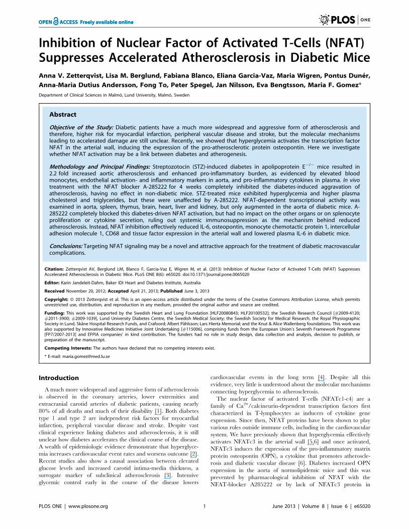
Inhibition of Nuclear Factor of Activated T-Cells (NFAT)Suppresses Accelerated Atherosclerosis in Diabetic MiceAnna V. Zetterqvist, Lisa M. Berglund, Fabiana Blanco, Eliana Garcia-Vaz, Maria Wigren, Pontus Duner,
Anna-Maria Dutius Andersson, Fong To, Peter Spegel, Jan Nilsson, Eva Bengtsson, Maria F. Gomez*
Department of Clinical Sciences in Malmo, Lund University, Malmo, Sweden
Abstract
Objective of the Study: Diabetic patients have a much more widespread and aggressive form of atherosclerosis andtherefore, higher risk for myocardial infarction, peripheral vascular disease and stroke, but the molecular mechanismsleading to accelerated damage are still unclear. Recently, we showed that hyperglycemia activates the transcription factorNFAT in the arterial wall, inducing the expression of the pro-atherosclerotic protein osteopontin. Here we investigatewhether NFAT activation may be a link between diabetes and atherogenesis.
Methodology and Principal Findings: Streptozotocin (STZ)-induced diabetes in apolipoprotein E2/2 mice resulted in2.2 fold increased aortic atherosclerosis and enhanced pro-inflammatory burden, as evidenced by elevated bloodmonocytes, endothelial activation- and inflammatory markers in aorta, and pro-inflammatory cytokines in plasma. In vivotreatment with the NFAT blocker A-285222 for 4 weeks completely inhibited the diabetes-induced aggravation ofatherosclerosis, having no effect in non-diabetic mice. STZ-treated mice exhibited hyperglycemia and higher plasmacholesterol and triglycerides, but these were unaffected by A-285222. NFAT-dependent transcriptional activity wasexamined in aorta, spleen, thymus, brain, heart, liver and kidney, but only augmented in the aorta of diabetic mice. A-285222 completely blocked this diabetes-driven NFAT activation, but had no impact on the other organs or on splenocyteproliferation or cytokine secretion, ruling out systemic immunosuppression as the mechanism behind reducedatherosclerosis. Instead, NFAT inhibition effectively reduced IL-6, osteopontin, monocyte chemotactic protein 1, intercellularadhesion molecule 1, CD68 and tissue factor expression in the arterial wall and lowered plasma IL-6 in diabetic mice.
Conclusions: Targeting NFAT signaling may be a novel and attractive approach for the treatment of diabetic macrovascularcomplications.
Citation: Zetterqvist AV, Berglund LM, Blanco F, Garcia-Vaz E, Wigren M, et al. (2013) Inhibition of Nuclear Factor of Activated T-Cells (NFAT) SuppressesAccelerated Atherosclerosis in Diabetic Mice. PLoS ONE 8(6): e65020. doi:10.1371/journal.pone.0065020
Editor: Karin Jandeleit-Dahm, Baker IDI Heart and Diabetes Institute, Australia
Received November 20, 2012; Accepted April 21, 2013; Published June 3, 2013
Copyright: � 2013 Zetterqvist et al. This is an open-access article distributed under the terms of the Creative Commons Attribution License, which permitsunrestricted use, distribution, and reproduction in any medium, provided the original author and source are credited.
Funding: This work was supported by the Swedish Heart and Lung Foundation [HLF20080843; HLF20100532]; the Swedish Research Council [#2009-4120;#2011-3900; #2009-1039], Lund University Diabetes Centre, the Swedish Medical Society; the Swedish Society for Medical Research, the Royal PhysiographicSociety in Lund, Skane Hospital Research Funds, and Crafoord; Albert Pahlsson; Lars Hierta Memorial; and the Knut & Alice Wallenberg foundations. This work wasalso supported by Innovative Medicines Initiative Joint Undertaking [#115006], comprising funds from the European Union’s Seventh Framework Programme[FP7/2007-2013] and EFPIA companies’ in kind contribution. The funders had no role in study design, data collection and analysis, decision to publish, orpreparation of the manuscript.
Competing Interests: The authors have declared that no competing interests exist.
* E-mail: [email protected]
Introduction
A much more widespread and aggressive form of atherosclerosis
is observed in the coronary arteries, lower extremities and
extracranial carotid arteries of diabetic patients, causing nearly
80% of all deaths and much of their disability [1]. Both diabetes
type 1 and type 2 are independent risk factors for myocardial
infarction, peripheral vascular disease and stroke. Despite vast
clinical experience linking diabetes and atherosclerosis, it is still
unclear how diabetes accelerates the clinical course of the disease.
A wealth of epidemiologic evidence demonstrate that hyperglyce-
mia increases cardiovascular event rates and worsens outcome [2].
Recent studies also show a causal association between elevated
glucose levels and increased carotid intima-media thickness, a
surrogate marker of subclinical atherosclerosis [3]. Intensive
glycemic control early in the course of the disease lowers
cardiovascular events in the long term [4]. Despite all this
evidence, very little is understood about the molecular mechanisms
connecting hyperglycemia to atherosclerosis.
The nuclear factor of activated T-cells (NFATc1-c4) are a
family of Ca2+/calcineurin-dependent transcription factors first
characterized in T-lymphocytes as inducers of cytokine gene
expression. Since then, NFAT proteins have been shown to play
various roles outside immune cells, including in the cardiovascular
system. We have previously shown that hyperglycemia effectively
activates NFATc3 in the arterial wall [5,6] and once activated,
NFATc3 induces the expression of the pro-inflammatory matrix
protein osteopontin (OPN), a cytokine that promotes atheroscle-
rosis and diabetic vascular disease [6]. Diabetes increased OPN
expression in the aorta of normolipidemic mice and this was
prevented by pharmacological inhibition of NFAT with the
NFAT-blocker A285222 or by lack of NFATc3 protein in
PLOS ONE | www.plosone.org 1 June 2013 | Volume 8 | Issue 6 | e65020

NFATc3 deficient mice [6]. Additional experimental evidence
supports a role for NFAT as a regulator of genes able to promote
vascular dysfunction and potentially, a pro-atherogenic vascular
phenotype [7,8,9]. NFAT promotes vascular smooth muscle cell
(VSMC) proliferation and migration [7,10], and plays a role in
neointima formation and in the regulation of cyclooxygenase 2
(Cox2) expression after vascular injury [11,12,13]. NFAT
contributes to the development of angiotensin II-induced hyper-
tension, via down-regulation of potassium channel expression
[14,15]. Moreover, NFAT controls the alternative splicing of
allograft inflammatory factor-1 (AIF-1), resulting in products
differentially associated to parameters defining human plaque
phenotype and stability [16].
Together, these observations led us to hypothesize that NFAT
may act as a glucose-sensor in the vessel wall, translating changes
in Ca2+ signals into changes in gene expression that lead to
macrovascular disease in diabetes. To more directly test this
hypothesis and in the context of an atherosclerosis-prone
experimental model, we investigate the effects of NFAT-signaling
inhibition on atherosclerotic plaque formation and inflammatory
burden in diabetic and non-diabetic apolipoprotein (Apo)E
deficient mice.
Materials and Methods
AnimalsThis study was carried out in strict accordance with the
recommendations in the Guide for the Care and Use of
Laboratory Animals of the National Institutes of Health. All
protocols were approved by the local ethics review board at Lund
University and the Malmo/Lund Animal Care and Use Commit-
tee (Permit Number: M29-12). Animals were anaesthetized with
ketamine hydrochloride and xylazine (i.p.; 2.5 mg and 7.5 mg/
100 g body weight, respectively) and euthanized by exsanguina-
tion through cardiac puncture for blood collection. Depth of
anesthesia was assessed by the toe-pinch reflex procedure and
absence of muscular tone. All efforts were made to minimize
suffering. Adult ApoE2/2 (B6.129P2-Apoetm1Unc/J; Charles River,
Sulzfeld, Germany; n = 196), C57BL/6J (stock number 000664;
Charles River; n = 25) and FVBN 9x-NFAT-luciferase reporter
mice (NFAT-luc; n = 72) [17] were used.
Study DesignTreatment protocols are summarized in Figure 1. Protocol I:
22 weeks old ApoE2/2 mice received intraperitoneal (i.p.)
injections of streptozocin (STZ; Sigma-Aldrich, Stockholm,
Sweden; 60 mg/kg body weight, pH 4.5) or vehicle (citrate buffer)
once a day for 5 days at the start of the experiments, as previously
described [6]. One group of mice (n = 24) was euthanized 4 weeks
after the first STZ/vehicle injection, while additional 2 groups
received daily i.p. injections of the NFAT blocker A-285222
(0.29 mg/kg body weight) or vehicle (saline) for 1 (n = 81) or 4
weeks (n = 43) until termination. Protocols II and III: NFAT-luc
mice were used. In protocol II (n = 29), diabetes was induced as in
protocol I and mice received daily i.p. injections of A-285222
(0.15 mg/kg body weight) or saline until termination at day 16. In
protocol III (n = 36), mice were fed a normal chow (R3;
Lantmannen, Kimstad, Sweden) or a high fat diet (HFD;
R638:0.15% cholesterol, 21% fat; Lantmannen) during 4 or 8
weeks. Protocol IV: C57BL/6J mice (n = 25) received daily i.p.
injections of A-285222 (0.29 mg/kg body weight) or vehicle for 4
weeks until termination.
A-285222 inhibits all NFAT family members and was provided
by Abbott Laboratories (Abbott Park, IL). Body weight and blood
glucose, measured in whole venous blood (One-Touch, LifeScan
Inc., Milipitas, CA) were monitored once a week. Animals had free
access to tap water, fed normal chow diet (except in protocol III).
For en face and cross sectional measurements of plaque (4- and 8-
weeks groups), the aorta and heart were dissected out after whole
body perfusion with phosphate-buffered saline (PBS) and stored in
Histochoice (Amresco Inc, Solon, OH) at 4uC for fixation until
further processing. For mRNA expression measurements in the
aortic arch and experiments involving splenocytes and monocytes
(5-weeks group), whole body perfusion with PBS was performed,
after which aortas were dissected free of connective tissue and snap
frozen, and whole spleens were weighed and stored in ice cold PBS
until further processing. The 8-week version of protocol I was
repeated in an additional group of diabetic mice (n = 25) for
mRNA and protein expression measurements in the aortic arch
and for liver histology. For the pharmacokinetics of A-285222,
additional ApoE2/2 (n = 23) and FVBN (n = 7) mice were used.
Histological Evaluation of Atherosclerosis, Spleen andLiver
En face preparations of the aorta were performed as described
before [18]. Briefly, aortas were fixed in Histochoice, dipped in
78% methanol and stained for 40 min in 0.16% Oil Red O
(ORO) dissolved in 78% methanol/0.2 mol/L NaOH, after which
they were washed in 78% methanol and distilled water. Cover slips
were mounted with water-soluble mounting media L-550A
(Histolab, Goteborg, Sweden). Lipids are stained red, which
makes the plaques bordeaux-colored. Lipid (ORO), macrophages
(Moma-2; monocyte/macrophage 2), a-smooth muscle actin (a-
SMA) and collagen contents were evaluated in cross-sections
(10 mm) of the aortic root as described before [18]. Rat anti-
Moma-2 primary antibody (1 mg/mL; BMA Biomedicals, Augst,
Switzerland), mouse anti-alpha-SMA (0.42 mg/mL, Sigma-Al-
drich) and biotinylated secondary IgG antibodies (Vector Labo-
ratories, Burlingame, CA) were used. Sections were counter-
stained with Harris hematoxylin for determination of subvalvular
lesion area, expressed both in mm2 and as percentage of total cross
sectional area to correct for potential structural differences in the
arterial wall between groups [19]. Media and lumen areas were
also determined based on the Harris hematoxylin staining.
Specificity of immune staining was confirmed by the absence of
staining when primary or secondary antibodies were omitted from
the protocol. Sections (5–6 per mouse) were analyzed under blind
conditions by computer-aided morphometry (Image-Pro Plus,
Media Cybernetics, Bethesda, MD and BioPix iQ 2.0 software,
Biopix AB, Gothenburg, Sweden, for en face and cross sections
respectively). Expression of tissue factor (TF) and osteopontin
(OPN) was also evaluated in the aortic root using confocal
immunofluorescence microscopy as described before [6]. Sections
were stained with primary rabbit antibodies, anti-OPN (0.5 mg/
mL, IBL, Hamburg, Germany) or anti-TF (10 mg/mL, American
Diagnostica, Stamford, CT) and secondary antibody, DyLight
649 anti-rabbit IgG (1:400 and 1:500, for OPN and TF,
respectively; Jackson ImmunoResearch, West Grove, PA); and
counterstained with the nucleic acid dye SYTOX Green (1:3000,
Molecular Probes, Invitrogen, Paisley, UK). Sections (3–6 per
mouse) were examined under blind conditions at 20X in a Zeiss
LSM 5 Pascal laser scanning confocal microscope and mean
fluorescence intensities of OPN and TF in the plaque were
quantified using the Zeiss LSM 5 analysis software and ImageJ
(version 1.47 m), respectively.
Spleen and liver cryosections (10 mm) were fixed with
Histochoice and stained with hematoxylin and eosin (H&E). Liver
sections were also stained with ORO and hematoxylin. For
NFAT in Diabetes-Induced Atherosclerosis
PLOS ONE | www.plosone.org 2 June 2013 | Volume 8 | Issue 6 | e65020

quantification of liver fat content (ORO), three sections per mouse
were analyzed under blind conditions by computer-aided
morphometry (BioPix iQ 2.0 software, Biopix AB, Gothenburg,
Sweden).
Luciferase Reporter AssayLuciferase activity was measured in tissue homogenates from
the aortic arch, spleen, thymus, brain, heart, liver and kidney.
Assays were performed as previously described [5,7]. Optical
density was measured using a Tecan Infinite M200 instrument
(Tecan Nordic AB, Molndal, Sweden) and data expressed as
relative luciferase units (RLU) per mg protein. Protein concentra-
tion was determined with the EZQ protein quantification kit
(Molecular Probes, Invitrogen, Paisley, UK) or the DC Protein
Assay (Bio-Rad Laboratories Sundbyberg, Sweden).
Plasma Cholesterol, Triglycerides and CytokinesPlasma cholesterol and triglycerides were measured by color-
imetric assays (InfinityTM-Cholesterol and InfinityTM-Triglyceride;
Thermo Scientific, Middletown, VA) as described before [20].
Plasma cytokines were measured using a pro-inflammatory 7-plex
kit (Meso Scale Discovery, Rockville, MD). The lower detection
limit for each cytokine was within the range described by the
manufacturer. Plasma OPN and soluble (s) vascular cell adhesion
molecule 1 (VCAM-1) levels were assayed using Quantikine mouse
OPN and sVCAM-1 ELISA kits (R&D Systems, Abingdon UK).
Absorbance was measured at 450 nm and the lower limits of
detection were 5.7 pg/mL and 0.31 ng/mL, respectively. All
assays were performed according to the manufacturers’ instruc-
tions.
Quantitative RT-PCR (qRT-PCR)RNA was extracted from the aortic arch using TRI Reagent BD
(Sigma-Aldrich) and a protocol for simultaneous isolation of RNA,
DNA and protein, according to the manufacturer’s instructions.
cDNA synthesis and real-time PCR were performed as previously
described [21], using TaqMan Gene Expression assays for IL-6
(Mm00446190_m1), OPN (Mm00436767_m1), monocyte chemo-
tactic protein 1 (MCP-1; Mm00441242_m1), intercellular adhe-
sion molecule 1 (ICAM-1; Mm00516023_m1), VCAM-1
(Mm01320970_m1), IL-1b (Mm01336189_m1), Cox-2
(Mm00478374_m1), IL-10 (Mm00439614_m1), TF
Figure 1. Study design. Protocol I: 22 weeks old ApoE2/2 mice received intraperitoneal (i.p.) injections of STZ or vehicle as previously described[6]. One group of mice was euthanized 4 weeks after the first STZ/vehicle injection, while additional 2 groups received daily i.p. injections of the NFATblocker A-285222 (0.29 mg/kg body weight) or vehicle (saline) for 1 or 4 weeks until termination. Protocols II and III: NFAT-luc mice were used. Inprotocol II, diabetes was induced as in protocol I and mice received daily i.p. injections of A-285222 (0.15 mg/kg body weight) or saline untiltermination at day 16. In protocol III, mice were fed a normal chow or a high fat diet (HFD; 0.15% cholesterol, 21% fat) during 4 or 8 weeks. ProtocolIV: C57BL/6J mice received daily i.p. injections of A-285222 (0.29 mg/kg body weight) or vehicle for 4 weeks until termination. Arrows indicate timeof termination; diabetes (black bars), control (white bars), A-285222-treated (hatched bars) and high fat diet (grey bars).doi:10.1371/journal.pone.0065020.g001
NFAT in Diabetes-Induced Atherosclerosis
PLOS ONE | www.plosone.org 3 June 2013 | Volume 8 | Issue 6 | e65020

(Mm00436948_m1), CD68 (Mm03047340_m1) with HPRT
(Mm00446968_m1) and b-actin (Mm00607939_s1) as endogenous
controls.
Western BlottingFollowing RNA extraction from the aortic arch, protein was
precipitated from the phenol-ethanol supernatant obtained after
sedimentation of the DNA pellet. After a series of washes, the
protein-containing pellet was dried and dissolved in SDS sample
buffer (62.5 mmol/L Tris-HCl pH 6.8, 2% SDS, 10% glycerol,
5% 2-mercaptoethanol and 0.001% bromophenol blue). Alterna-
tively, arteries were homogenized directly in SDS sample buffer as
previously described [6]. Protein concentration was determined
with the EZQ protein quantification kit (Molecular Probes). An
equal amount of protein was loaded onto 12.5% Tris-HCl gels
(Bio-Rad Laboratories) and separated by gel electrophoresis.
Proteins were transferred to polyvinylidene difluoride membranes
(Bio-Rad Laboratories), blocked in 3% BSA/5% non-fat dry milk
and incubated with primary anti-TF (2 mg/mL in 3% BSA,
American Diagnostica, Stamford, CT) or to nitrocellulose
membranes (Bio-Rad Laboratories), blocked in 5% BSA and
incubated with anti-CD68 (0.82 mg/mL in 5% milk, DakoCyto-
mation, Glostrup, Denmark). HRP-conjugated secondary anti-
body (Cell Signaling, Danvers, MA) was used and bands detected
with chemiluminescence (Supersignal West Dura, Pierce Biotech-
nology, Rockford, IL). b-actin (1:3000) or a-tubulin (1:5000; both
Figure 2. NFAT inhibition suppresses accelerated atherosclerosis in diabetes. (A) En face lesion area in the aortic arch of control anddiabetic female ApoE2/2 mice treated for 4 weeks with A-285222 or saline (protocol I). Mice were 30 weeks old at the time of analysis, 8 weeks afterthe first STZ or vehicle injection. Data is expressed as percentage of total aortic arch area (n = 9–12 mice/group). Two-way ANOVA for the effect ofdiabetes and the drug revealed significant interaction between factors (**P,0.005). Bonferroni post-test yielded **P,0.01 vs. non-diabetic controlmice and ##P,0.01 vs. diabetic saline-treated mice. (B) Representative en face preparations of aortas from ApoE2/2 mice treated as in A and stainedwith ORO (bordeaux-colored). Scale = 2 mm. (C) Blood glucose (mmol/L) and (D) body weight (g) values for mice in panel A. (E) Merged data from themeasurements in panel A and en face data obtained 4 weeks after the first STZ or vehicle injection (n = 9–13 mice/group). Control non-diabetic (blue);diabetic (red); A-285222-treated (dotted lines); saline treated (unbroken lines).doi:10.1371/journal.pone.0065020.g002
NFAT in Diabetes-Induced Atherosclerosis
PLOS ONE | www.plosone.org 4 June 2013 | Volume 8 | Issue 6 | e65020

from GenScript Corporation, Piscataway, NJ) were used as
loading controls.
Splenocyte Proliferation and Cytokine ProductionSplenocytes were isolated as previously described [22]. Briefly,
single cell suspensions were prepared by pressing spleens through a
70-mm cell strainer (BD Falcon, Franklin Lakes, NJ). Erythrocytes
were removed using red blood cell lysing buffer (Sigma-Aldrich).
Cells were cultured in culture medium containing 10% heat-
inactivated FCS, 1 mmol/L sodium pyruvate, 10 mmol/L Hepes,
50 U of penicillin, 50 mg/mL streptomycin, 0.05 mmol/L b-
mercaptoethanol, and 2 mmol/L L-glutamine (RPMI 1640,
GIBCO, Paisley, UK) in 96-well round bottom plates (Sarstedt,
Numbrecht, Germany). For proliferation assay, 26105 cells/well
were cultured with or without DynabeadsH coupled to anti-CD3
and anti-CD28 antibodies for T-cell activation and expansion
(bead to cell ratio 1:1, Invitrogen, Life Technologies, Carlsbad,
CA); alternatively, with or without 2.5 mg/mL concanavalin A
(ConA; Sigma-Aldrich) for 88 hours. To measure DNA synthesis,
the cells were pulsed with 1 mCi [methyl-3H]thymidine (Amer-
sham Biosciences, Uppsala, Sweden); during the last 16 hours,
macromolecular material was harvested on glass fiber filters using
a Filter Mate Harvester (Perkin Elmer, Buckinghamshire, UK) and
analyzed using a liquid scintillation counter (Wallac 1450
MicroBeta, Ramsey, MN). For cytokine production, 16106
cells/well were cultured in 48-well plates (Sarstedt, Numbrecht,
Germany) with or without anti-CD3/CD28 dynabeads (bead to
cell ratio 1:1, Invitrogen) for 48 hours, after which cytokine
concentrations in the culture media were measured using a Th1/
Th2 9-plex ultra-sensitive kit (Meso Scale Discovery). The lower
detection limit for each cytokine was within the range described by
the manufacturer.
Table 1. Blood glucose, body weight, plasma cholesterol and triglyceride values in mice undergoing the different experimentalprotocols.
Genotype and treatmentBlood glucose(mmol/L) Body weight (g)
Total cholesterol(mmol/L)
Triglycerides(mmol/L)
Protocol I (ApoE2/2)
4 weeks:
Control (n = 13) 8.860.8 22.961.4 10.861.5 0.4860.13
Diabetes (n = 11) 19.664.7*** 21.661.9 16.963.5*** 0.5560.22
5 weeks:
Control (n = 9) 9.161.1 30.262.0 10.661.4 1.4460.36
Diabetes (n = 11) 23.465.5*** 29.461.7 17.962.4** 2.7161.02
Control+A-285222 (n = 8) 9.660.5 30.662.1 13.663.2 2.1461.03
Diabetes+A-285222 (n = 11) 23.265.8*** 28.362.6* 17.265.6 2.5761.56
8 weeks:
Control (n = 12) 9.061.6 24.361.7 10.261.3 0.6560.22
Diabetes (n = 10) 17.765.5*** 21.362.2*** 15.664.4*** 1.0460.38**
Control+A-285222 (n = 12) 9.060.4 23.461.3 11.761.4 0.6860.19
Diabetes+A-285222 (n = 9) 19.863.5*** 20.861.0** 16.064.7** 0.9360.25
Protocol II (NFAT-luc)
Control (n = 11) 8.961.0 22.563.0 2.3060.33 1.7460.39
Diabetes (n = 8) 15.467.2* 21.961.4 2.4960.33 2.2060.30
Diabetes+A-285222 (n = 10) 14.765.4* 22.762.3 2.4560.36 1.9160.75
Protocol III (NFAT-luc)
4 weeks:
Control (n = 12) 10.361.2 30.864.3 3.1960.34 1.3160.56
High fat diet (n = 8) 11.461.6 39.067.7** 5.5961.25*** 0.8060.38
8 weeks:
Control (n = 9) 9.760.7 35.965.6 3.3960.67 1.2460.53
High fat diet (n = 7) 12.763.0** 43.765.0* 6.0061.01*** 1.0460.40
Protocol IV (C57Bl6/J)
Control (n = 13) 12.862.7 25.263.4 2.7560.73 0.5060.13
Control+A-285222 (n = 12) 11.761.9 26.064.5 2.4260.46 0.4960.15
Values represent mean 6 SD. Blood glucose values are averaged during the experiments (from week 2 until termination). Body weight and lipids values were measuredat termination. For protocol 1 (4 weeks), student’s t-test yielded ***p,0.001 vs. non-diabetic mice. For protocol 1 (5 and 8 weeks), two-way ANOVA (for the effects ofdiabetes and A-285222) revealed no interactions between factors. Bonferroni post-tests yielded *,0.05, **p,0.01, ***p,0.001 for comparisons between control anddiabetic mice receiving the same treatment. For protocol 2, one-way ANOVA and Bonferroni post-tests yielded *,0.05, vs non-diabetic control. For protocol 3, two-wayANOVA (for the effects of high fat diet and diet duration) revealed no interactions. Bonferroni post-tests yielded *,0.05, **p,0.01, ***p,0.001 for comparisonsbetween mice fed high fat diet and controls.doi:10.1371/journal.pone.0065020.t001
NFAT in Diabetes-Induced Atherosclerosis
PLOS ONE | www.plosone.org 5 June 2013 | Volume 8 | Issue 6 | e65020

Flow Cytometry AnalysisCells from blood and splenocytes were analyzed as previously
described [23,24,25]. Blood cells were stained with the following
fluorochrome-conjugated antibodies after blocking of FC receptors
for 5 minutes: PerCP/Cy5.5-anti-CD62L (L-selectin), PE/Cy7-
anti-Ly-6c, (BioLegend, San Diego, CA) and APC-anti-CD115
(eBioscience, San Diego, CA). Splenocytes were stained for PE/
Cy7-anti-CD3, Pacific Blue-anti-CD4, APC-anti-CD25 after
blocking of FC receptors for 5 minutes. Cells where then
permeabilized and thereafter stained with PE-anti-Foxp3 (Biole-
gend). For interferon (IFN)-c measurements, splenocytes (56105
cells/cell culture well) were incubated with phorbol 12-myristate
13-acetate (PMA; 10 ng), ionomycin (0.2 mg), and brefeldin A
(1 mg, all from Sigma) for 4 hours at 37uC. Stimulated cells were
then stained for CD3 and CD4 (as above). Cells were thereafter
permeabilized and stained with PE-anti-IFN-c (Biolegend). Mea-
surements were performed using a CyAn ADP flow cytometer
(Beckman Coulter, Brea, CA) and analyzed with FlowJo7.6
software (Tree Star, Ashland, OR). Mononuclear leukocytes were
gated from the forward scatter (FSC)/side scatter (SSC). Single
stained samples were used to correct for fluorescence spillover in
multicolor analyses, and gate boundaries were set by fluorescence-
minus-one (FMO) controls.
Gas Chromatography Mass Spectroscopy (GC/MS)For the pharmacokinetics of A-285222, blood was collected
from adult ApoE2/2 mice (n = 23) by cardiac puncture at different
time points (30 min, 1, 2, 4, 6, 12 and 24 hours) after i.p. injection
of A-285222 (0.29 mg/kg body weight in saline solution). Plasma
was isolated and a known concentration (2.5 mmol/L) of the
analogous inactive compound A-216491 (Abbott Park, IL) was
added as an internal standard to all samples. Samples were
randomized and run in duplicates. Samples (300 mL) were
extracted twice with ethyl acetate (400 mL), followed by evapora-
tion. The dried residues were finally re-dissolved in chloroform
(30 mL) and analyzed by GC/MS on an Agilent 6890N gas
chromatograph (Agilent, Santa Clara, CA) coupled to a Leco
Pegasus III TOFMS electron impact TOF (time-of-flight) mass
spectrometer (Leco Corp., St. Joseph, MI). Identification was
based on mass spectra and retention indexes, calculated from the
injection of a homologous series of n-alkanes. The concentration of
A-285222 in plasma was determined using a calibration curve
calculated from analyses of plasma from untreated mice, spiked
with known concentrations of A-285222 and A-216491. Plasma A-
285222 levels were also determined in mice from protocols I (5
and 8 weeks), II and IV, from blood collected at the time of
euthanasia (i.e.,24 h after the last i.p. injection of A-285222).
These measurements were performed in duplicate using pooled
plasma from 6–12 mice for each experimental condition. Plasma
from the groups treated with saline served as negative controls. In
Figure 3. Inhibition of NFAT reduces the lipid contents in the plaque of diabetic mice. (A) ORO stained cross-sections of the aortic rootfrom control and diabetic female ApoE2/2 mice treated with or without A-285222 for 4 weeks (Protocol I). Sections were counter-stained with Harrishematoxylin. Scale = 500 mm. (B) Summarized morphometric data from sections stained as in A showing ORO positive area in the plaques. Two-wayANOVA for the effect of diabetes and the drug revealed significant interaction between factors (P,0.05). Post-test yielded #P,0.05 vs. diabeticsaline-treated mice (n = 9–12 mice/group). (C–E) Summarized morphometric data from the same animals as in B showing monocytes/macrophages-2(Moma-2, C), collagen (D) and a-smooth muscle actin (a-SMA, E) positive areas in the plaques. Two-way ANOVA revealed significant effect of diabetes(P,0.001 for Moma-2 and collagen; P,0.05 for a-SMA), Bonferroni post-test yielded *P,0.05 and ***P,0.001 vs. non-diabetic saline-treated mice(n = 9–12 mice/group).doi:10.1371/journal.pone.0065020.g003
NFAT in Diabetes-Induced Atherosclerosis
PLOS ONE | www.plosone.org 6 June 2013 | Volume 8 | Issue 6 | e65020

a separate experiment to evaluate efficacy of i.p. administration,
plasma levels of A-285222 after intracardiac (i.c.) injection of the
drug were compared to levels after i.p. administration (n = 7 mice).
StatisticsResults are expressed as means 6 SEM if not otherwise
specified. Statistical analysis was performed using GraphPad
software (Prism 5.0). For parametric data, significance was
determined using Student’s t-test, one- or two-way ANOVA as
specified in the text, followed by Bonferroni post hoc tests. Non-
parametric data was analyzed using Mann-Whitney or Kruskal-
Wallis test followed by Dunn’s post-test.
Results
In vivo Inhibition of NFAT Prevents the Diabetes-inducedAggravation of Atherosclerosis in the Aortic Arch
Atherosclerosis prone 22 week old ApoE2/2 mice were treated
as outlined in PROTOCOL I (Figure 1). A 2.2 fold increase in
atherosclerotic plaque area (25.4% vs. 11.6%; p,0.01) was
observed in the aortic arch of diabetic mice when compared to
aged-matched non-diabetic controls, as assessed by en face
measurements of ORO stained area eight weeks after the first
STZ-injection (Figure 2A–B). In vivo treatment with the NFAT
blocker A-285222 for the last 4 weeks of the experiment
completely abrogated (p,0.01) the effect of diabetes on lesion
area (Figure 2A–B). There was no effect of A-285222 on
atherosclerosis in non-diabetic ApoE2/2 mice. As expected,
STZ-treated mice had higher blood glucose and lower weight
gain than control mice, but A-285222 had no impact on these
parameters (Figure 2C–D and Table 1). A similar effect of diabetes
was observed in the descending aorta (2.1 fold increase in plaque
area), but the overall plaque area was lower than in the arch and
the effect of A-285222 less pronounced (Figure S1).
Lesion area was also evaluated in a separate group of control
and diabetic mice 4 weeks after the first STZ or vehicle injection,
when mice were 26 weeks of age. At this earlier time point diabetes
had no evident effect on lesion size in the aortic arch (Figure 2E).
Figure 2E also shows the accelerated development of plaque in the
Figure 4. In vivo treatment with A-285222 effectively blocks diabetes-induced NFAT-dependent transcriptional activity. (A) NFAT-luciferase activity in the aortic arch, spleen, thymus, brain, heart, liver and kidney from control and diabetic female NFAT-luc mice treated with A-285222 or saline (Protocol II). Values are expressed as relative luciferase units (RLU) per mg protein (n = 8–11 mice/group for aortic arch, spleen andthymus; n = 4–8 mice/group for brain, heart, liver and kidney). **P,0.01 vs. non-diabetic control mice and ##P,0.01 vs. diabetic saline-treated group.(B) Basal (non-diabetic) NFAT-dependent transcriptional activity (RLU mg21) in the different tissues examined (n = 4–11 mice/group).doi:10.1371/journal.pone.0065020.g004
NFAT in Diabetes-Induced Atherosclerosis
PLOS ONE | www.plosone.org 7 June 2013 | Volume 8 | Issue 6 | e65020

diabetic mice, which is completely prevented by NFAT inhibition,
and the well-recognized effect of age on atherosclerosis [26] in the
non-diabetic groups.
Atherosclerosis was also examined in cross-sections of the aortic
root from the same animals used for the en face measurements
shown in Figure 2A–B. Total cross sectional area calculated from
the hematoxylin staining was larger in diabetic than non-diabetic
mice (1.686106 um2 vs. 1.236106 um2; P,0.001). This increased
area in the diabetic mice is the combined result of increased
plaque (6.596105 um2 vs. 4.036105 um2; P,0.001) and lumen
(9.046105 um2 vs. 6.656105 um2; P,0.001) area, while a
concomitant thinning of the media was observed (1.156105 um2
vs. 1.836105 um2; P,0.001), especially in regions beneath the
atherosclerotic plaques. Erosion and focal dilatation of the medial
layer have been described in the aortic root of ApoE2/2 mice
when fed an atherogenic diet with cholate or when bred on specific
backgrounds (C3H or 128SvJ) [27,28]. Our results show that
diabetes (without the atherogenic diet or specific background) is a
sufficient drive for medial wall erosion; in line with work showing
structural modifications and disruption of the arterial media as a
result of diabetes in STZ-treated C56B/J6 mice [19]. When the
subvalvular plaque area was expressed as percentage of total cross
sectional area, it was 16.5% larger in diabetic than in control
untreated mice (p,0.05); whereas the effect of diabetes was
attenuated in A-285222-treated mice (9.6% increase, n.s.).
Interestingly, inhibition of NFAT resulted in reduced lipid
contents in the plaque of diabetic mice (Figure 3A–B). Even
though plaque macrophage, collagen and a-SMA contents were
increased in the aortic root of diabetic mice when compared to
non-diabetic controls, values were not affected by A-285222
treatment (Figure 3C–E and Figure S2A). Higher expression of
OPN, but not of TF was observed in aortic root sections from
diabetic mice when compared to non-diabetic controls; but values
were not affected by A-285222 treatment either (Figure S2A–B).
In agreement with previous studies [20], total plasma choles-
terol was significantly elevated in diabetic ApoE2/2 mice when
compared to non-diabetic controls at 4, 5 and 8 weeks (Table 1).
Plasma triglycerides were also elevated after 8 weeks. However,
the reduced atherosclerosis is less likely due to a lipid lowering
effect, since A-285222 had no effect on plasma lipids, regardless if
the mice were diabetic or not (Table 1). In agreement to what
others have reported [29], ApoE2/2 mice on chow diet had very
little fat accumulation in the liver (,2%). No signs of liver steatosis
were observed after treatment of ApoE2/2 mice with A-285222
(Figure S3).
In vivo Treatment with A-285222 Effectively BlocksDiabetes-induced NFAT-dependent TranscriptionalActivity
Previous studies from our group have shown that A-285222 is a
potent blocker of glucose-induced NFAT-dependent transcrip-
tional activity and NFAT-target genes [6,7] in VSMCs and intact
vessels in vitro [5,6]. Here we demonstrate that in vivo treatment
with A-285222 completely blocks the diabetes-induced NFAT-
dependent transcriptional activity in the aortic arch of NFAT-luc
mice (Figure 4A, PROTOCOL II). Of all organs examined,
NFAT was selectively activated in the aorta of diabetic mice, with
a tendency to increased activation in kidneys, but no effect in
spleen, thymus, brain, heart or liver. A-285222 treatment was
effective only if NFAT had been previously activated as in the
aortic arch, and possibly in the kidneys. Diabetic mice had
significant hyperglycemia at this time-point, but unchanged body
weight and plasma lipids (Table 1). A-285222 had no effect on any
of these parameters (Table 1). As shown in Figure 4B, basal levels
of NFAT activity varied depending on the organ. Consistent with
what others have reported, the highest levels were observed in
brain, kidney and heart; and the lowest in spleen and liver [17].
Figure 5. In vivo NFAT inhibition reduces diabetes-induced elevation of plasma IL-6 levels. The effect of diabetes and NFAT inhibition onplasma IL-6, IFN-c, IL-12p70, IL-1b, IL-10, KC, TNF-a, OPN and sVCAM-1 was studied 8 weeks after the first STZ/vehicle injection in female ApoE2/2
mice treated with A-285222 or saline for the last 4 weeks of the experiment (Protocol I, n = 9–12 mice/group). Only for IL-6, two-way ANOVA for theeffect of diabetes and the drug revealed a significant interaction between factors (*P,0.05). Bonferroni post-test yielded ***P,0.001 vs. non-diabeticcontrol and #P,0.05 vs. diabetic saline-treated group. All other cytokines except KC were increased by diabetes. For parametrically distributed data(IL-1b and OPN), two-way ANOVA followed by Bonferroni post-test yielded **P,0.01 and ***P,0.01 vs. corresponding non-diabetic groups. For therest of the cytokines, which were non-parametrically distributed, Kruskal-Wallis followed by Dunn’s post-test yielded *P,0.05, **P,0.01 and***P,0.01 vs. corresponding non-diabetic groups.doi:10.1371/journal.pone.0065020.g005
NFAT in Diabetes-Induced Atherosclerosis
PLOS ONE | www.plosone.org 8 June 2013 | Volume 8 | Issue 6 | e65020

A separate set of NFAT-luc mice were fed HFD or chow diet for
4 or 8 weeks and NFAT-luciferase activity measured in the aorta
(PROTOCOL III). Even though plasma cholesterol levels were
significantly higher in mice fed HFD when compared to controls
(Table 1), no differences in NFAT-luciferase activity were
observed (Figure S4). Body weight increased with HFD, and a
small (3 mmol/L) but significant increase in blood glucose was
observed after 8 weeks of HFD, while triglycerides were not
significantly affected (Table 1).
Effect of Diabetes and NFAT-signaling Inhibition onSystemic and Plaque Inflammation
Plasma cytokines, levels and phenotype of circulating monocytes
and expression of inflammatory genes in the aortic arch were
examined in ApoE2/2 mice undergoing PROTOCOL I. Plasma
IL-6, IFN-c, IL-12p70, IL-1b, IL-10, tumor necrosis factor (TNF)a,
OPN and sVCAM-1 were significantly increased in diabetic mice 8
weeks after the first STZ injection, whereas keratinocyte-derived
chemokine (KC) was not affected (Figure 5). Treatment with A-
285222 for 4 weeks blunted the effect of diabetes on IL-6 levels, but
had no significant effects on the other plasma cytokines (Figure 5).
Already early after the onset of diabetes, circulating monocytes were
elevated in diabetic ApoE2/2 mice when compared to non-diabetic
controls, as evidenced by higher percentages of CD115 (M-CSF
receptor) expressing blood mononuclear cells (Figure 6B). Treat-
ment with A-285222 had no effect on blood monocyte levels.
Neither diabetes nor NFAT inhibition had any impact on the
fraction of CD115 positive cells expressing the adhesion molecule
CD62L (L-selectin) or on the fractions of inflammatory (CD115+Ly6Chigh) or patrolling (CD115+ Ly6Clow) blood monocytes
(Figure 6C–E).
Also early after the onset of diabetes, several markers of
inflammation and endothelial activation including MCP-1,
VCAM-1, IL-1b, Cox2 and TF were significantly increased at
the mRNA level in the aortic wall of diabetic mice when compared
to controls; and trends towards increased IL-6 and ICAM-1 were
observed (Figure S5). Treatment with A-285222 for 4 weeks
significantly reduced IL-6, OPN, MCP-1 and ICAM-1 mRNA in
the aortic arch of diabetic mice, while levels of VCAM-1, IL-1b,
Cox2 and IL-10 were not affected (Figure 7A). Expression of the
macrophage marker CD68 and cellular TF were significantly
lowered after A-285222 treatment in diabetic mice, both at the
mRNA and protein level (Figure 7B–C).
To determine whether NFAT signaling inhibition affected the
capacity of immune cells to proliferate, splenocytes were isolated
from ApoE2/2 mice 5 weeks after the first STZ injection, after
Figure 6. Circulating monocyte levels are increased in diabetic ApoE2/2 mice but not affected by NFAT inhibition. (A) Left and middlepanels: Dot plots showing the gating strategy for monocyte identification from blood mononuclear cells based on the expression of CD115 (M-CSFreceptor). Right panels: Identification of monocytes expressing CD62L (L-selectin) and of Ly6Chigh and Ly6Clow subsets (blue lines); fluorescence-minus-one controls (red lines). (B) Summarized data from flow cytometry experiments showing percentages of CD115+ monocytes in total bloodmononuclear cells from control and diabetic ApoE2/2 mice treated with the NFAT blocker A-285222 or saline for one week (Protocol I, n = 9–11 mice/group). Two-way ANOVA revealed a significant effect of diabetes (P,0.01). Bonferroni post-test yielded *P,0.05 vs. non-diabetic control mice. (C–E)No differences in the percentages of CD115+ mononuclear cells expressing CD62L+ (C) Ly6Chigh (D) or Ly6Clow (E) were found between treatmentgroups.doi:10.1371/journal.pone.0065020.g006
NFAT in Diabetes-Induced Atherosclerosis
PLOS ONE | www.plosone.org 9 June 2013 | Volume 8 | Issue 6 | e65020

receiving daily i.p. injections of A-285222 or vehicle for one week
(PROTOCOL I, 5 weeks group). No differences in their
proliferative capacity were found between groups, as assessed by
measurements of thymidine incorporation under non-stimulated
conditions or after stimulation with either anti-CD3/CD28 beads
or Con A (Figure S6A–B). Similar observations were made using
splenocytes isolated from control C57BL/6 mice treated with A-
285222 for 4 weeks (Figure S6C). Blood glucose, body weight and
plasma lipids were not affected in these animals (Table 1). We also
examined the effects of diabetes and A-285222 treatment on
cytokine secretion capacity of splenocytes under control non-
stimulated conditions and after stimulation with anti-CD3/CD28
beads (Table S1). Diabetes had no impact on the levels of
cytokines produced by non-stimulated splenocytes, but resulted in
significantly increased levels of TNF-a in cells stimulated with anti-
CD3/CD28 beads. A-285222 treatment on the other hand
resulted in decreased secretion of IL-2 in non-stimulated spleno-
cytes from non-diabetic mice and no effects in stimulated
splenocytes (Table S1). Percentages of CD3+CD4+CD8- spleno-
cytes expressing IFN-c after stimulation with PMA and ionomycin
were not affected by diabetes or NFAT inhibition (Figure S6E).
Moreover, no differences in spleen histology or size were detected
between treatment groups (Figure S6F–G).
Pharmacokinetics of A-285222Previous studies in cynomolgus monkeys have demonstrated
that A-285222 is well tolerated when the plasma concentration is
maintained below 4 mg/mL (9.6 mmol/L), a level achieved by oral
administration of the drug twice daily at 5–7.5 mg/kg body weight
[30]. In our hands, a lower dose was sufficient for changes in
vascular OPN expression in normolipidemic BalB/c mice [6] and
for the effects on NFAT-transcriptional activity and on diabetes-
induced atherosclerosis described here (0.15–0.29 mg/kg body
weight). In previous ex vivo experiments using mouse arteries, A-
285222 blocked NFAT-transcriptional activity at 1 mmol/L [5].
To assess the actual plasma concentration of A-285222 after in vivo
treatment of ApoE2/2 mice, we collected blood by cardiac
puncture at different time points (30 min, 1, 2, 4, 6, 12 and 24
hours) after i.p. injection of the drug (0.29 mg/kg body weight). A-
285222 was identified and quantified with GC/MS, based on its
mass spectra and retention indexes (Figure S5). Plasma A-285222
levels peaked at 2 hours, were between 100–200 nmol/L for the
first six hours and no longer detected at 12 or 24 hours. We failed
to detect any A-285222 in mice from protocols I (5 and 8 weeks),
II and IV, from blood collected at the time of euthanasia
(i.e.,24 h after the last i.p. injection of A-285222), ruling out an
accumulation of the drug in the circulation. A comparison
between plasma levels of A-285222 measured 5 min after i.p.
injection of the drug (0.15 and 1.5 mg/kg) and after direct
injection into the circulation (i.c.), showed that levels were within
the same range regardless administration route (430 nmol/L vs.
480 nmol/L for the low dose; 770 nmol/L vs. 675 nmol/L for the
high dose), indicating high bioavailability.
Discussion
The present study demonstrates that inhibition of NFAT-
signaling completely suppresses accelerated atherosclerosis in the
aortic arch of diabetic ApoE2/2 mice and that this effect is
independent of changes in plasma glucose or lipid levels. This
finding suggests that NFAT may play a role in the development of
atherosclerosis in diabetes and identifies this signaling pathway as
a novel therapeutic target for the treatment of diabetic macro-
vascular complications.
The ApoE-deficient mouse is a well-established model for the
study of atherosclerosis. Mice develop spontaneous hypercholes-
terolemia and mimic the initial phases of human atherosclerosis,
even when fed a regular chow diet as in this study. The extent and
severity of the lesions increase with age, displaying all known
phases of atherogenesis. Monocyte adhesion takes place between
8–10 weeks of age, lipid deposition and development of fatty-
streaks starts at approximately 9 weeks of age, and progression to
intermediate and more mature fibrous plaques at ,15–20 weeks
of age [31]. As shown in Figure 2E, at the age when mice were
treated with the NFAT blocker A-285222 (26–30 weeks of age),
plaque size in the aortic arch was still increasing, and this was
clearly accelerated by diabetes. Interestingly, NFAT inhibition did
only affect the diabetes-driven aggravation of atherosclerosis, but
Figure 7. In vivo inhibition of NFAT reduces the expression ofmarkers of inflammation and endothelial activation andmacrophage infiltration in the aortic arch of diabetic mice.(A) Gene expression analyses by qRT-PCR in the aortic arch of diabeticApoE2/2 mice treated with A-285222 or saline for 4 weeks (Protocol I,hatched and black bars, respectively). HPRT and b-actin were used asendogenous controls and data (Rel. mRNA) is expressed in relation tothe diabetic saline-treated group. (B) CD68 and (C) TF mRNA andprotein levels were decreased after treatment with A-285222. Leftgraphs show relative CD68 and TF mRNA from the same animals as in A.Middle panels show representative immunoblots for CD68 and TF (50and 35 kDa) and loading controls. Right graphs show summarizedresults from western blot experiments, with CD68 expression normal-ized to a-tubulin and TF expression normalized to b-actin. Data (Rel.protein) is expressed in relation to the diabetic saline treated group.*P,0.05, **P,0.01 and ***P,0.001; n = 5–13 mice/group.doi:10.1371/journal.pone.0065020.g007
NFAT in Diabetes-Induced Atherosclerosis
PLOS ONE | www.plosone.org 10 June 2013 | Volume 8 | Issue 6 | e65020

had no impact on atherosclerosis in non-diabetic mice, suggesting
potentially different mechanisms underlying plaque formation
under diabetic and non-diabetic conditions. The increased lesion
size observed in the aortic arch is in line with what others have
described in STZ-treated ApoE2/2 mice [32]. However, the effect
of diabetes on plaque area at the level of the aortic root was
modest when compared to those observed when mice are treated
with STZ at younger age (6 weeks [32] vs. 22 weeks in this study).
Along these lines, A-285222 treatment had a more distinct impact
on the aortic arch than in the aortic root, as exemplified by
decreased macrophage infiltration, TF and OPN expression in the
arch (Figure 7) but not in the root of the aorta (Figure S2). These
results highlight differential susceptibility to diabetes-induced
atherosclerosis in these two segments of the aorta and the need
for a diabetes-driven process for NFAT-inhibition to play a role.
Previous work from our group established that high extracel-
lular glucose (.15 mmol/L) activates NFATc3 in intact arteries ex
vivo by a mechanism involving the release of extracellular
nucleotides (i.e. UTP, UDP) acting on P2Y receptors, leading to
increased intracellular Ca2+ and subsequent activation of the
calcineurin/NFATc3 signaling pathway [5]. High glucose also
decreases the export of NFATc3 from the nucleus by inhibiting the
otherwise constitutively elevated kinase activity of glycogen
synthase kinase (GSK)-3b and c-Jun N-terminal kinase in the
arterial wall [5]. In a follow-up study, we demonstrated that
hyperglycemia readily activated NFATc3 in the arterial wall
in vivo, as evidenced by increased NFATc3 nuclear accumulation
in cerebral arteries after an i.p. glucose-tolerance test and by
increased NFATc3-dependent transcriptional activity in aorta 2
weeks after the induction of diabetes with STZ [6]. Here we show
that this diabetes-induced activation of NFAT in the aorta is
completely inhibited by in vivo treatment with A-285222
(Figure 4A), demonstrating that A-285222 is an effective blocker
of NFAT-transcriptional activity in this tissue. Even though NFAT
is expressed in many tissues and basal (non-diabetic) NFAT-
luciferase activity was detected in all tissues examined, the
diabetes-induced NFAT activation is not a generalized phenom-
enon. At least at this time point after the onset of diabetes (2
weeks), NFAT-luciferase activity seemed only elevated in the
aorta, whereas no changes were observed in the other organs
examined (Figure 4), an advantageous difference from the
therapeutic point of view.
Not only hyperglycemia, but hyperlipidemia, or the combina-
tion of both could be driving the accelerated atherosclerosis in
diabetes. To our knowledge, the effect of hyperlipidemia on
NFAT-transcriptional activity in the vasculature has never been
studied in vivo. A number of in vitro studies though, demonstrated
that NFAT activation can be triggered by lipids. Exposure to
triglyceride-rich very low-density lipoproteins increases NFATc3
nuclear accumulation in cultured rat aortic VSMCs [33], and
postprandial triglyceride-rich lipoproteins collected after an oral
fat load activate several transcription factors including NFAT in
cultured endothelial cells [34]. Also, incubation of T-lymphocytes,
macrophages, fibroblasts and endothelial cells with copper-
oxidized or monocyte-oxidized low-density lipoproteins increases
NFAT binding to DNA [35,36]. Here we show that a ,2-fold
increase in total cholesterol induced by HFD, had no effect on
NFAT-transcriptional activity in the aortas of NFAT-luc mice
(Figure S4). Interestingly, after 8 weeks of HFD mice had a mild
but still significant increase in blood glucose (from 9.7 to
12.7 mmol/L; Table 1), which did not translate in enhanced
luciferase activity. This is in line with previous data showing that
glucose levels .15 mmol/L are required for NFAT activation in
the vasculature [5,6]. Even though high cholesterol per se had no
effect on NFAT-transcriptional activity in vivo, it is still possible
that high triglycerides instead, or even higher absolute levels of
cholesterol (such as those observed in ApoE-deficient mice), or
higher degree of lipid oxidation as it may occur in the context of
diabetes, could trigger NFAT activation.
Inflammation is recognized as a critical regulator of atheroscle-
rotic plaque formation and progression. Along these lines, the
accelerated atherosclerosis in diabetic ApoE2/2 mice was
preceded by elevated blood monocytes and higher expression of
endothelial activation- and inflammatory markers in the aorta.
Already after 4 weeks of diabetes, a time-point when no changes in
aortic plaque size had yet occurred, expression of VCAM-1,
MCP-1, IL-1b, Cox2, TF and maybe also IL-6 and ICAM-1
(borderline significance) were higher than in control non-diabetic
mice. The enhanced pro-inflammatory burden in diabetic mice is
also reflected by the overall increased levels of circulating plasma
cytokines (IL-6, IFN-c, IL-12p70, IL-1b, IL-10, TNFa, OPN and
sVCAM-1; Figure 5) after 8 weeks of diabetes. One important
observation in this study was that A-285222 treatment for 4 weeks
significantly reduced the diabetes-driven IL-6 levels in plasma as
well as mRNA expression in the aortic arch. IL-6 is one of the
most prominent pro-inflammatory cytokines, extensively studied in
the context of atherogenesis [37]. It can be generated locally by
cells within the lesions or released by adipose tissue into the
circulation, promoting endothelial dysfunction, VSMC prolifera-
tion and migration as well as recruitment and activation of
inflammatory cells, hence amplifying the inflammatory response.
Moreover, IL-6 stimulates the expression of scavenger receptors
SR-A and CD36, involved in the uptake of modified LDL and
formation of foam cells [38]. Lack of this positive stimulation due
to reduced IL-6 levels could explain the reduced plaque lipids
observed in diabetic mice after A-285222 treatment (Figures 2 and
3A). The reduced IL-6 expression after treatment with A-285222
is in line with previous studies by us and other investigators,
showing NFAT-dependent regulation of IL-6 gene expression in
VSMCs [10,39] and in human resistance arteries [7].
The NFAT blocker A-285222 belongs to a series of 3,5-bis
(trifluoromethyl)pyrazole (BTP) derivatives originally developed in
a search for safer immunosuppressive drugs. These drugs maintain
NFAT in a phosphorylated state, blocking its nuclear import and
subsequent transcription, without affecting NF-kB or AP-1
activation, or calcineurin phosphatase activity [40]. In vivo
administration of A-285222 completely blocked diabetes-induced
NFAT-transcriptional activity in the aorta, leading to reduced
expression of IL-6, OPN, MCP-1, ICAM-1, CD68 and TF, all
established players in atherogenesis, as well as to reduced diabetes-
induced atherosclerosis. This was achieved without any effect on
body weight, blood glucose or lipid levels and at concentrations
that had no impact on NFAT activity in spleen or thymus, on T-
cell proliferation rates or cytokine secretion capacity, ruling out
systemic immunosuppression as the mechanism behind reduced
atherosclerosis. A-285222 did not affect the number or phenotype
of circulating blood monocytes, nor did it alter the numbers of T-
regulatory cells in the spleen. The reduction of TF was particularly
interesting, given the lack of available systemic strategies that
target TF expression [41]. The dose of A-285222 used here and
plasma levels achieved upon treatment were far below those
required in cynomolgus monkeys for inhibition of T-cell cytokine
production, which is consistent with the negative T-cell cytokine
data presented here. Furthermore, non-diabetic ApoE2/2 mice
exhibited measurable levels of plasma cytokines, reflecting a low-
grade inflammation typical of this hyperlipidemic model, however,
A-285222 had no effect on these levels (Figure 5), speaking against
a general immunosuppressant effect of A-285222. Together,
NFAT in Diabetes-Induced Atherosclerosis
PLOS ONE | www.plosone.org 11 June 2013 | Volume 8 | Issue 6 | e65020

results suggest that NFAT inhibition affects the plaque phenotype
at the level of the plaque itself and not via systemic immunosup-
pression.
Calcineurin inhibitors (i.e. CsA and FK506) are commonly used
to prevent host-versus graft disease, a therapy often associated with
side effects, including increased risk of atherosclerosis. While the
immunosuppressive effects of these drugs are directly related to the
inhibition of NFAT in immune cells, the adverse cardiovascular
effects seem to be NFAT-independent and mediated via intracel-
lular cyclophilin and chaperone activities, extracellular cyclophilin
A and NFAT-independent transcriptional effects [42]. The
degrees to which these NFAT-independent pathways are engaged
seem to be dose-dependent [43]. Low-dose FK506 (,0.2 ng/mL)
inhibited collar-induced atherosclerosis progression and promoted
plaque stability in ApoE2/2 mice, whereas higher doses similar to
those given to transplant patients engaged instead NF-kB in
macrophages and consequently increased production of cytokines.
Other serious side effects associated with CsA treatment are
hyperlipidemia and diabetes [42]; but these were not observed
after treatment with A-285222.
Despite major advances in the treatment of cardiovascular
disease during the past decades, with the introduction of lipid
lowering, anti-thrombotic and anti-hypertensive drugs, there is still
no available therapy that specifically targets macrovascular
diabetic complications. Our data reveals the NFAT-signaling
pathway as a promising target for the treatment of accelerated
atherosclerosis in diabetes.
Supporting Information
Figure S1 Diabetes increases atherosclerosis in thedescending aorta, but the overall plaque area is lowerthan in the aortic arch. Summarized data from measurements
of en face lesion area in the descending aorta for comparison with
the aortic arch data from the same animals included in Figure 2.
Results are from control and diabetic female ApoE2/2 mice that
had been treated for 4 weeks with the NFAT blocker A-285222 or
saline. Mice were 30 weeks old at the time of analysis, performed 8
weeks after the first STZ or vehicle injection. Data is expressed as
percentage of total aortic area (n = 9–12 mice/group). Two-way
analysis of variance for the effect of diabetes and the drug revealed
significant effect of diabetes (P,0.001). Bonferroni post-test
yielded **P,0.01 vs non-diabetic saline-treated group. The inset
shows corresponding data for the total aorta (i.e. arch and
descending).
(PDF)
Figure S2 Histological examination of subvalvularplaques. (A) Representative cross-sections of the aortic root from
control and diabetic female ApoE2/2 mice treated with or
without A-285222 for 4 weeks (Protocol I) stained for monocytes/
macrophages (Moma-2), collagen, a-smooth muscle actin (a-
SMA), tissue factor (TF, red) and osteopontin (OPN, red). Moma-
2 and a-SMA stained sections were counter-stained with Harris
hematoxylin; TF and OPN stained sections were counter-stained
with SYTOX Green. Scale = 500 mm (Moma-2); = 100 mm (col-
lagen, TF, OPN); = 50 mm (a-SMA). (B, C) Summarized data
from confocal immunofluorescence experiments showing mean
fluorescence intensity for plaque TF and OPN. Three to six
sections for each animal were analyzed (n = 9–12 mice/group).
Two-way ANOVA revealed significant effect of diabetes on OPN
expression (P,0.0001). Bonferroni post-test yielded *P,0.05 and
**P,0.01 vs corresponding non-diabetic groups.
(PDF)
Figure S3 Lipid deposition in the liver is not affected byNFAT inhibition. (A) Representative liver sections from diabetic
female ApoE2/2 mice treated with or without A-285222 for 4
weeks (Protocol I) were stained with hematoxylin-eosin (H&E) and
oil red O (ORO). Scale = 100 mm. (B) Lipid deposition in the liver
was evaluated from three ORO-stained sections per mouse using
computer-assisted image analysis (n = 6–7 mice/group).
(PDF)
Figure S4 High fat diet does not affect NFAT-dependenttranscriptional activity in the aorta. NFAT-luciferase
activity in the thoracic aorta from mice fed normal chow diet
(white bars) or a high fat diet (grey bars) during 4 or 8 weeks
(Protocol III). Values are expressed as RLU per mg protein (n = 7–
12 mice/group).
(PDF)
Figure S5 Diabetes leads to increased expression ofinflammatory and endothelial activation markers in theaortic arch. Gene expression analyses by qRT-PCR in the aortic
arch of control and diabetic ApoE2/2 mice analyzed after 4
weeks of diabetes (Protocol I, n = 7–10 mice/group). HPRT and
b-actin were used as endogenous controls. Data (Rel. mRNA) is
expressed in relation to diabetic mice. *P,0.05 and **P,0.01.
(PDF)
Figure S6 In vivo A-285222 treatment does not affectsplenocyte proliferative capacity. (A–B) [Methyl-3H]thymi-
dine incorporation (counts per minute, cpm) after stimulation with
or without anti-CD3/CD28 beads (A) or 2.5 mg/mL ConA (B) in
splenocytes isolated from control and diabetic ApoE2/2 mice
treated for 1 week with the NFAT blocker A-285222 or saline
(Protocol I; n = 9–11 mice/group) (C). Proliferation after stimula-
tion with or without 2.5 mg/mL ConA in splenocytes from control
mice treated for 4 weeks with the NFAT blocker A-285222 or
saline (Protocol IV; n = 12–13 mice/group). (D) Flow cytometry
data showing percentages of CD4+CD25+Foxp3+ regulatory T-
cells (of total CD3+ splenocytes) in the same mice as in A. Two-
way ANOVA revealed significant effect of diabetes (P,0.001).
Bonferroni post-test yielded *P,0.05 vs. corresponding non-
diabetic groups. (E) Percentages of CD3+CD4+D8- splenocytes
expressing IFN-c after stimulation with phorbol myristate acetate
(PMA) and ionomycin in the same mice as in A. (F) Representative
spleen sections stained for hematoxylin-eosin and (G) spleen
weight in relation to tibia length from the same mice as in A.
Scale = 500 mm.
(PDF)
Figure S7 Identification and quantification of A-285222 with GC/MS. A-285222 and the inactive analogA-216491 were identified from their mass spectra andretention indexes. (A) Total ion chromatogram showing
substances present in a plasma sample from a mouse injected i.c.
with 1.5 mg A-285222 per kg body weight. (B) Reconstructed ion
chromatogram from the same sample as in A, showing retention
indexes (s) of A-216491 (left peak, m/z = 295) and A-285222 (right
peak, m/z = 416). (C) Mass spectra for A-285222 including the
molecular ion (m/z = 416). Only the molecular ion was selective
and used for quantification. (D) Quantification of A-285222 in
plasma samples collected at different time points after i.p. injection
of 0.29 mg A-285222 per kg body weight (n = 2–4 mice/time
point).
(PDF)
Table S1 Effects of diabetes and A-285222 on splenocytecytokine production. Splenocytes were isolated from control
and diabetic mice that had been treated for 1 week with the
NFAT in Diabetes-Induced Atherosclerosis
PLOS ONE | www.plosone.org 12 June 2013 | Volume 8 | Issue 6 | e65020

NFAT blocker A-285222 or saline (Protocol I; n = 8–11 mice/
group) after which they were cultured either under control non-
stimulated conditions or with anti-CD3/CD28 beads for 48 hours.
Levels of interferon (IFN)-c, interleukin (IL)-1b, IL-2, IL-4, IL-5,
KC/GRO (keratinocyte chemoattractant; keratinocyte-derived
chemokine/growth related oncogene), IL-10, IL-12total and
tumor necrosis factor (TNF)-a. were measured in the culture
media collected at the end of the experiments. Data was analyzed
by two-way ANOVA (for the effects of diabetes and A-285222).
Values represent mean 6 SD; significant differences after
Bonferroni post-tests are indicated in the table. Diabetes had no
impact on the levels of cytokines produced by non-stimulated
splenocytes, while A-285222 treatment resulted in decreased IFN-
c and IL-2 (both P,0.05). Bonferroni post-test yielded
*P,0.05 only for IL-2. CD3/CD28 stimulated cells from diabetic
mice produced lower levels of IFN-c (P,0.05) and IL-5 (P,0.01)
but higher TNF-a. (P,0.05). Bonferroni post-test yielded
#P,0.05 for TNF-a. A-285222 treatment had no impact on
the ability of splenocytes to respond to CD3/CD28 stimulation.
(PDF)
Acknowledgments
We thank Irena Ljungkrantz and Ingrid Soderberg for skillful technical
assistance.
Author Contributions
Conceived and designed the experiments: AVZ MFG. Performed the
experiments: AVZ LMB FB EGV MW PD AMDA FT PS MFG. Analyzed
the data: AVZ LMB FB EGV MW AMDA FT PS JN EB MFG.
Contributed reagents/materials/analysis tools: PS EB JN MFG. Wrote the
paper: AVZ LMB MFG. Critical revision and final approval of submitted
version: AVZ LMB FB EGV MW PD AMDA FT PS JN EB MFG.
References
1. Rahman S, Rahman T, Ismail AA, Rashid AR (2007) Diabetes-associated
macrovasculopathy: pathophysiology and pathogenesis. Diabetes Obes Metab 9:
767–780.
2. Coutinho M, Gerstein HC, Wang Y, Yusuf S (1999) The relationship between
glucose and incident cardiovascular events. A metaregression analysis of
published data from 20 studies of 95,783 individuals followed for 12.4 years.
Diabetes Care 22: 233–240.
3. Rasmussen-Torvik LJ, Li M, Kao WH, Couper D, Boerwinkle E, et al. (2011)
Association of a Fasting Glucose Genetic Risk Score With Subclinical
Atherosclerosis. Diabetes 60: 331–335.
4. Brown A, Reynolds LR, Bruemmer D (2010) Intensive glycemic control and
cardiovascular disease: an update. Nat Rev Cardiol 7: 369–375.
5. Nilsson J, Nilsson LM, Chen Y-W, Molkentin JD, Erlinge D, et al. (2006) High
Glucose Activates Nuclear Factor of Activated T Cells in Native Vascular
Smooth Muscle. Arterioscler Thromb Vasc Biol 26: 794–800.
6. Nilsson-Berglund LM, Zetterqvist AV, Nilsson-Ohman J, Sigvardsson M,
Gonzalez Bosc LV, et al. (2009) Nuclear factor of activated T cells regulates
osteopontin expression in arterial smooth muscle in response to diabetes-induced
hyperglycemia. Arterioscler Thromb Vasc Biol 30: 218–224.
7. Nilsson LM, Sun ZW, Nilsson J, Nordstrom I, Chen YW, et al. (2007) Novel
blocker of NFAT activation inhibits IL-6 production in human myometrial
arteries and reduces vascular smooth muscle cell proliferation. Am J Physiol Cell
Physiol 292: C1167–1178.
8. Orr AW, Lee MY, Lemmon JA, Yurdagul A, Gomez MF, et al. (2008)
Molecular Mechanisms of Collagen Isotype-Specific Modulation of Smooth
Muscle Cell Phenotype. Arterioscler Thromb Vasc Biol 29: 225–231.
9. Nilsson LM, Nilsson-Ohman J, Zetterqvist AV, Gomez MF (2008) Nuclear
factor of activated T-cells transcription factors in the vasculature: the good guys
or the bad guys? Curr Opin Lipidol 19: 483–490.
10. Liu Z, Dronadula N, Rao GN (2004) A Novel Role for Nuclear Factor of
Activated T Cells in Receptor Tyrosine Kinase and G Protein-coupled Receptor
Agonist-induced Vascular Smooth Muscle Cell Motility. J Biol Chem 279:
41218–41226.
11. Karpurapu M, Wang D, Van Quyen D, Kim T-K, Kundumani-Sridharan V, et
al. (2010) Cyclin D1 Is a Bona Fide Target Gene of NFATc1 and Is Sufficient in
the Mediation of Injury-induced Vascular Wall Remodeling. Journal of
Biological Chemistry 285: 3510–3523.
12. Lipskaia L, del Monte F, Capiod T, Yacoubi S, Hadri L, et al. (2005) Sarco/
endoplasmic reticulum Ca2+-ATPase gene transfer reduces vascular smooth
muscle cell proliferation and neointima formation in the rat. Circ Res 97: 488–
495.
13. Lee MY, Garvey SM, Baras AS, Lemmon JA, Gomez MF, et al. (2009)
Integrative genomics identifies DSCR1 (RCAN1) as a novel NFAT-dependent
mediator of phenotypic modulation in vascular smooth muscle cells. Hum Mol
Genet 19: 468–479.
14. Amberg GC, Rossow CF, Navedo MF, Santana LF (2004) NFATc3 regulates
Kv2.1 expression in arterial smooth muscle. J Biol Chem 279: 47326–47334.
15. Nieves-Cintron M, Amberg GC, Nichols CB, Molkentin JD, Santana LF (2007)
Activation of NFATc3 down-regulates the beta1 subunit of large conductance,
calcium-activated K+ channels in arterial smooth muscle and contributes to
hypertension. J Biol Chem 282: 3231–3240.
16. Berglund LM, Kotova O, Osmark P, Grufman H, Xing C, et al. (2011) NFAT
regulates the expression of AIF-1 and IRT-1: yin and yang splice variants of
neointima formation and atherosclerosis. Cardiovasc Res 93: 414–423.
17. Wilkins BJ, Dai YS, Bueno OF, Parsons SA, Xu J, et al. (2004) Calcineurin/
NFAT coupling participates in pathological, but not physiological, cardiac
hypertrophy. Circ Res 94: 110–118.
18. Fredrikson GN, Soderberg I, Lindholm M, Dimayuga P, Chyu KY, et al. (2003)
Inhibition of atherosclerosis in apoE-null mice by immunization with apoB-100peptide sequences. Arterioscler Thromb Vasc Biol 23: 879–884.
19. Prevost G, Bulckaen H, Gaxatte C, Boulanger E, Beraud G, et al. (2011)
Structural modifications in the arterial wall during physiological aging and as aresult of diabetes mellitus in a mouse model: Are the changes comparable?
Diabetes & Metabolism 37: 106–111.
20. Gustavsson C, Agardh CD, Zetterqvist AV, Nilsson J, Agardh E, et al. (2010)Vascular cellular adhesion molecule-1 (VCAM-1) expression in mice retinal
vessels is affected by both hyperglycemia and hyperlipidemia. PLoS One 5:
e12699.
21. Awla D, Zetterqvist AV, Abdulla A, Camello C, Berglund LM, et al. (2012)NFATc3 Regulates Trypsinogen Activation, Neutrophil Recruitment, and
Tissue Damage in Acute Pancreatitis in Mice. Gastroenterology: Jul 27. [Epubahead of print].
22. Wigren M, Bengtsson D, Duner P, Olofsson K, Bjorkbacka H, et al. (2009)
Atheroprotective effects of Alum are associated with capture of oxidized LDLantigens and activation of regulatory T cells. Circ Res 104: e62–70.
23. Wigren M, BjArkbacka H, Andersson L, Ljungcrantz I, Fredrikson GN, et al.
(2012) Low Levels of Circulating CD4+FoxP3+ T Cells Are Associated With an
Increased Risk for Development of Myocardial Infarction But Not for Stroke.Arteriosclerosis, Thrombosis, and Vascular Biology 32: 2000–2004.
24. Engelbertsen D, Andersson L, Ljungcrantz I, Wigren M, Hedblad B, et al.
(2013) T-Helper 2 Immunity Is Associated With Reduced Risk of MyocardialInfarction and Stroke. Arteriosclerosis, Thrombosis, and Vascular Biology 33:
637–644.
25. Kolbus D, Ramos OH, Berg KE, Persson J, Wigren M, et al. (2010) CD8+ T cellactivation predominate early immune responses to hypercholesterolemia in
Apoe(-)(/)(-) mice. BMC Immunol 11: 58.
26. Reddick RL, Zhang SH, Maeda N (1994) Atherosclerosis in mice lacking apo E.
Evaluation of lesional development and progression. Arterioscler Thromb 14:141–147.
27. Carmeliet P, Moons L, Lijnen R, Baes M, Lemaitre V, et al. (1997) Urokinase-
generated plasmin activates matrix metalloproteinases during aneurysmformation. Nat Genet 17: 439–444.
28. Shi W, Brown MD, Wang X, Wong J, Kallmes DF, et al. (2003) Genetic
Backgrounds but Not Sizes of Atherosclerotic Lesions Determine MedialDestruction in the Aortic Root of Apolipoprotein EaJ‘‘Deficient Mice.
Arteriosclerosis, Thrombosis, and Vascular Biology 23: 1901–1906.
29. Lohmann C, Schafer N, von Lukowicz T, Sokrates Stein MA, Boren J, et al.
(2009) Atherosclerotic mice exhibit systemic inflammation in periadventitial andvisceral adipose tissue, liver, and pancreatic islets. Atherosclerosis 207: 360–367.
30. Birsan T, Dambrin C, Marsh KC, Jacobsen W, Djuric SW, et al. (2004)
Preliminary in vivo pharmacokinetic and pharmacodynamic evaluation of anovel calcineurin-independent inhibitor of NFAT. Transpl Int 3: 145–150.
31. Nakashima Y, Plump AS, Raines EW, Breslow JL, Ross R (1994) ApoE-deficient
mice develop lesions of all phases of atherosclerosis throughout the arterial tree.Arterioscler Thromb 14: 133–140.
32. Park L, Raman KG, Lee KJ, Lu Y, Ferran LJ, et al. (1998) Suppression of
accelerated diabetic atherosclerosis by the soluble receptor for advanced
glycation endproducts. Nat Med 4: 1025–1031.
33. Lipskaia L, Pourci M-L, Delomenie C, Combettes L, Goudouneche D, et al.(2003) Phosphatidylinositol 3-Kinase and Calcium-Activated Transcription
Pathways Are Required for VLDL-Induced Smooth Muscle Cell Proliferation.Circ Res 92: 1115–1122.
34. Norata GD, Grigore L, Raselli S, Redaelli L, Hamsten A, et al. (2007) Post-
prandial endothelial dysfunction in hypertriglyceridemic subjects: molecularmechanisms and gene expression studies. Atherosclerosis 193: 321–327.
NFAT in Diabetes-Induced Atherosclerosis
PLOS ONE | www.plosone.org 13 June 2013 | Volume 8 | Issue 6 | e65020

35. Bochkov VN, Mechtcheriakova D, Lucerna M, Huber J, Malli R, et al. (2002)
Oxidized phospholipids stimulate tissue factor expression in human endothelialcells via activation of ERK/EGR-1 and Ca(++)/NFAT. Blood 99: 199–206.
36. Maziere C, Morliere P, Massy Z, Kamel S, Louandre C, et al. (2005) Oxidized
low-density lipoprotein elicits an intracellular calcium rise and increases thebinding activity of the transcription factor NFAT. Free Radic Biol Med 38: 472–
480.37. Schuett H, Luchtefeld M, Grothusen C, Grote K, Schieffer B (2009) How much
is too much? Interleukin-6 and its signalling in atherosclerosis. Thromb Haemost
102: 215–222.38. Takeda N, Manabe I, Shindo T, Iwata H, Iimuro S, et al. (2006) Synthetic
retinoid Am80 reduces scavenger receptor expression and atherosclerosis in miceby inhibiting IL-6. Arterioscler Thromb Vasc Biol 26: 1177–1183.
39. Abbott KL, Loss JR, Robida AM, Murphy TJ (2000) Evidence that Galpha(q)-
coupled receptor-induced interleukin-6 mRNA in vascular smooth muscle cellsinvolves the nuclear factor of activated T cells. Mol Pharmacol 58: 946–953.
40. Trevillyan JM, Chiou XG, Chen YW, Ballaron SJ, Sheets MP, et al. (2001)
Potent inhibition of NFAT activation and T cell cytokine production by novellow molecular weight pyrazole compounds. J Biol Chem 276: 48118–48126.
41. Breitenstein A, Camici GG, Tanner FC (2010) Tissue factor: beyondcoagulation in the cardiovascular system. Clin Sci (Lond) 118: 159–172.
42. Kockx M, Jessup W, Kritharides L (2010) Cyclosporin A and atherosclerosis–
cellular pathways in atherogenesis. Pharmacol Ther 128: 106–118.43. Donners MM, Bot I, De Windt LJ, van Berkel TJ, Daemen MJ, et al. (2005)
Low-dose FK506 blocks collar-induced atherosclerotic plaque development andstabilizes plaques in ApoE2/2 mice. Am J Transplant 5: 1204–1215.
NFAT in Diabetes-Induced Atherosclerosis
PLOS ONE | www.plosone.org 14 June 2013 | Volume 8 | Issue 6 | e65020
Copyright © 2022 FDOKUMEN