Differences in MEF2 and NFAT Transcriptional Pathways According to Human Heart Failure Aetiology
10
Differences in MEF2 and NFAT Transcriptional Pathways According to Human Heart Failure Aetiology Raquel Corte ´s 1. , Miguel Rivera 1 , Esther Rosello ´ -Lletı´ 1. , Luis Martı´nez-Dolz 2 , Luis Almenar 2 , InmaculadaAzorı´n 3 , Francisca Lago 4 , Jose ´ Ramo ´ n Gonza ´ lez-Juanatey 4 , Manuel Portole ´s 5 * 1 Cardiocirculatory Unit, Research Center, Hospital Universitario La Fe, Valencia, Spain, 2 Cardiology Unit, Hospital Universitario La Fe, Valencia, Spain, 3 Experimental Neurology, Research Center, Hospital Universitario La Fe, Valencia, Spain, 4 Molecular and Cellular Cardiology Unit, Institute of Biomedical Research, and Department of Cardiology, Hospital Clı ´nico Universitario, Santiago de Compostela, Spain, 5 Cell Biology and Pathology Unit, Research Center, Hospital Universitario La Fe, Valencia, Spain Abstract Background: Ca 2+ handling machinery modulates the activation of cardiac transcription pathways involved in heart failure (HF). The present study investigated the effect of HF aetiology on Ca +2 handling proteins and NFAT1, MEF2C and GATA4 (transcription factors) in the same cardiac tissue. Methodology and Principal Findings: A total of 83 hearts from ischemic (ICM, n = 43) and dilated (DCM, n = 31) patients undergoing heart transplantation and controls (CNT, n = 9) were analyzed by western blotting. Subcellular distribution was analyzed by fluorescence and electron microscopy. When we compared Ca +2 handling proteins according to HF aetiology, ICM showed higher levels of calmodulin (24%, p,0.01), calcineurin (26%, p,0.01) and Ca 2+ /Calmodulin-dependent kinase II (CaMKIId b nuclear isoform 62%, p,0.001) than the CNT group. However, these proteins in DCM did not significantly increase. Furthermore, ICM showed a significant elevation in MEF2C (33%, p,0.01), and GATA4 (49%, p,0.05); also NFAT1 (66%, p,0.001) was increased, producing the resultant translocation of this transcriptional factor into the nuclei. These results were supported by fluorescence and electron microscopy analysis. Whereas, DCM only had a significant increase in GATA4 (52%, p,0.05). Correlations between NFAT1 and MEF2C in both groups (ICM r = 0.38 and DCM r = 0.59, p,0.05 and p,0.01, respectively) were found; only ICM showed a correlation between GATA4 and NFAT1 (r = 0.37, p,0.05). Conclusions/Significance: This study shows an increase of Ca 2+ handling machinery synthesis and their cardiac transcription pathways in HF, being more markedly increased in ICM. Furthermore, there is a significant association between MEF2, NFAT1 and GATA4. These proteins could be therapeutic targets to improve myocardial function. Citation: Corte ´ s R, Rivera M, Rosello ´ -Lletı ´ E, Martı ´nez-Dolz L, Almenar L, et al. (2012) Differences in MEF2 and NFAT Transcriptional Pathways According to Human Heart Failure Aetiology. PLoS ONE 7(2): e30915. doi:10.1371/journal.pone.0030915 Editor: Leon J. de Windt, Cardiovascular Research Institute Maastricht, Maastricht University, Netherlands Received May 17, 2011; Accepted December 29, 2011; Published February 17, 2012 Copyright: ß 2012 Corte ´s et al. This is an open-access article distributed under the terms of the Creative Commons Attribution License, which permits unrestricted use, distribution, and reproduction in any medium, provided the original author and source are credited. Funding: This work was supported by grants from the National Institute of Health ‘‘Fondo de Investigaciones Sanitarias of Instituto de Salud Carlos III’’, [REDINSCOR 06/0003/1001, Project PI07/0462]. The funders had no role in study design, data collection and analysis, decision to publish, or preparation of the manuscript. Competing Interests: The authors have declared that no competing interests exist. * E-mail: [email protected] . These authors contributed equally to this work. Introduction Heart failure (HF) is caused by diverse conditions which reduce the efficiency of the myocardium through overloading or damage. Over time, these stimuli will produce changes to the heart itself, such as enlargement of ventricles and hypertrophy (ventricular remodeling) [1,2], activating a molecular response in cardiomy- ocytes that involves an enhanced protein synthesis, up-regulation of fetal cardiac genes, and induction of immediate-early genes [3]. Numerous studies have implicated intracellular calcium (Ca 2+ ) as a critical mediator in the regulation of left ventricular remodeling in HF [4,5]. Changes in intracellular Ca 2+ ion concentrations regulate the activity of several related proteins, kinases and phosphatases, among them the ubiquitous Ca 2+ - binding proteins, calmodulin (CaM), the Ca 2+ /Calmodulin- dependent kinase II (CaMKII), and calcineurin (CaN), a Ca 2+ / Calmodulin-dependent phosphatase. Elevated intracellular Ca 2+ and the resulting Ca 2+ /CaM complex will activate CaMKII and CaN, which play an important role in cardiac function (mediate cardiac hypertrophy response to myocyte stretch or increased loads). Both enzymes respond to dysregulated calcium signaling, as an increase in their expression and activity in failing human myocardium and in animal models with cardiac hypertrophy and HF [6–8]. Many major pathways for pathological remodeling converge on a set of transcriptional regulators, such as nuclear myocyte enhancer factor 2 (MEF2), nuclear factor of activated T cells (NFAT) and GATA binding protein 4 (GATA4) [9–11]. Furthermore, histone deacetylases (HDAC) play a critical role in the modulation of hypertrophic growth by inhibiting the activity of MEF2 [12]. There are different activation pathways in the expression of these transcriptional factors: (1) MEF2 transcriptional activity is repress by HDAC4s and becomes active in presence of CaMKII which promotes the export of HDAC from the nucleus [13,14]; PLoS ONE | www.plosone.org 1 February 2012 | Volume 7 | Issue 2 | e30915
Transcript of Differences in MEF2 and NFAT Transcriptional Pathways According to Human Heart Failure Aetiology
Differences in MEF2 and NFAT Transcriptional PathwaysAccording to Human Heart Failure AetiologyRaquel Cortes1., Miguel Rivera1, Esther Rosello-Lletı1., Luis Martınez-Dolz2, Luis Almenar2,
Inmaculada Azorın3, Francisca Lago4, Jose Ramon Gonzalez-Juanatey4, Manuel Portoles5*
1 Cardiocirculatory Unit, Research Center, Hospital Universitario La Fe, Valencia, Spain, 2 Cardiology Unit, Hospital Universitario La Fe, Valencia, Spain, 3 Experimental
Neurology, Research Center, Hospital Universitario La Fe, Valencia, Spain, 4 Molecular and Cellular Cardiology Unit, Institute of Biomedical Research, and Department of
Cardiology, Hospital Clınico Universitario, Santiago de Compostela, Spain, 5 Cell Biology and Pathology Unit, Research Center, Hospital Universitario La Fe, Valencia, Spain
Abstract
Background: Ca2+ handling machinery modulates the activation of cardiac transcription pathways involved in heart failure(HF). The present study investigated the effect of HF aetiology on Ca+2 handling proteins and NFAT1, MEF2C and GATA4(transcription factors) in the same cardiac tissue.
Methodology and Principal Findings: A total of 83 hearts from ischemic (ICM, n = 43) and dilated (DCM, n = 31) patientsundergoing heart transplantation and controls (CNT, n = 9) were analyzed by western blotting. Subcellular distribution wasanalyzed by fluorescence and electron microscopy. When we compared Ca+2 handling proteins according to HF aetiology,ICM showed higher levels of calmodulin (24%, p,0.01), calcineurin (26%, p,0.01) and Ca2+/Calmodulin-dependent kinase II(CaMKIIdb nuclear isoform 62%, p,0.001) than the CNT group. However, these proteins in DCM did not significantlyincrease. Furthermore, ICM showed a significant elevation in MEF2C (33%, p,0.01), and GATA4 (49%, p,0.05); also NFAT1(66%, p,0.001) was increased, producing the resultant translocation of this transcriptional factor into the nuclei. Theseresults were supported by fluorescence and electron microscopy analysis. Whereas, DCM only had a significant increase inGATA4 (52%, p,0.05). Correlations between NFAT1 and MEF2C in both groups (ICM r = 0.38 and DCM r = 0.59, p,0.05 andp,0.01, respectively) were found; only ICM showed a correlation between GATA4 and NFAT1 (r = 0.37, p,0.05).
Conclusions/Significance: This study shows an increase of Ca2+ handling machinery synthesis and their cardiactranscription pathways in HF, being more markedly increased in ICM. Furthermore, there is a significant association betweenMEF2, NFAT1 and GATA4. These proteins could be therapeutic targets to improve myocardial function.
Citation: Cortes R, Rivera M, Rosello-Lletı E, Martınez-Dolz L, Almenar L, et al. (2012) Differences in MEF2 and NFAT Transcriptional Pathways According to HumanHeart Failure Aetiology. PLoS ONE 7(2): e30915. doi:10.1371/journal.pone.0030915
Editor: Leon J. de Windt, Cardiovascular Research Institute Maastricht, Maastricht University, Netherlands
Received May 17, 2011; Accepted December 29, 2011; Published February 17, 2012
Copyright: � 2012 Cortes et al. This is an open-access article distributed under the terms of the Creative Commons Attribution License, which permitsunrestricted use, distribution, and reproduction in any medium, provided the original author and source are credited.
Funding: This work was supported by grants from the National Institute of Health ‘‘Fondo de Investigaciones Sanitarias of Instituto de Salud Carlos III’’,[REDINSCOR 06/0003/1001, Project PI07/0462]. The funders had no role in study design, data collection and analysis, decision to publish, or preparation of themanuscript.
Competing Interests: The authors have declared that no competing interests exist.
* E-mail: [email protected]
. These authors contributed equally to this work.
Introduction
Heart failure (HF) is caused by diverse conditions which reduce
the efficiency of the myocardium through overloading or damage.
Over time, these stimuli will produce changes to the heart itself,
such as enlargement of ventricles and hypertrophy (ventricular
remodeling) [1,2], activating a molecular response in cardiomy-
ocytes that involves an enhanced protein synthesis, up-regulation
of fetal cardiac genes, and induction of immediate-early genes [3].
Numerous studies have implicated intracellular calcium (Ca2+)
as a critical mediator in the regulation of left ventricular
remodeling in HF [4,5]. Changes in intracellular Ca2+ ion
concentrations regulate the activity of several related proteins,
kinases and phosphatases, among them the ubiquitous Ca2+-
binding proteins, calmodulin (CaM), the Ca2+/Calmodulin-
dependent kinase II (CaMKII), and calcineurin (CaN), a Ca2+/
Calmodulin-dependent phosphatase.
Elevated intracellular Ca2+ and the resulting Ca2+/CaM
complex will activate CaMKII and CaN, which play an important
role in cardiac function (mediate cardiac hypertrophy response to
myocyte stretch or increased loads). Both enzymes respond to
dysregulated calcium signaling, as an increase in their expression
and activity in failing human myocardium and in animal models
with cardiac hypertrophy and HF [6–8]. Many major pathways
for pathological remodeling converge on a set of transcriptional
regulators, such as nuclear myocyte enhancer factor 2 (MEF2),
nuclear factor of activated T cells (NFAT) and GATA binding
protein 4 (GATA4) [9–11]. Furthermore, histone deacetylases
(HDAC) play a critical role in the modulation of hypertrophic
growth by inhibiting the activity of MEF2 [12].
There are different activation pathways in the expression of
these transcriptional factors: (1) MEF2 transcriptional activity is
repress by HDAC4s and becomes active in presence of CaMKII
which promotes the export of HDAC from the nucleus [13,14];
PLoS ONE | www.plosone.org 1 February 2012 | Volume 7 | Issue 2 | e30915
and (2) the activation of NAFT, a hyperphosphorylated cytosolic
protein, is regulated through control of its subcellular localization.
An elevation in intracellular Ca2+ increases the activity of CaN,
which dephosphorylates the NFAT molecule and allows its import
into the nucleus [15]. In addition, the NFAT interacts with the
cardiac-restricted zinc finger transcription factor GATA4, result-
ing in synergic activation of cardiac transcription [9].
Previous data show the relevance of increased levels of both
Ca2+/calmodulin-dependent enzymes, and these transcriptional
factors, in the development of a hypertrophic phenotype [6,13,15].
However, to date most of these studies have been performed in vitro
or in animal models [7,13,16] and the simultaneous analysis of the
different activation pathways has not been performed yet.
Therefore, the present study investigates the levels of CaM, CaN
and CaMKIId, predominant isoform in the heart [17], in dilated
(DCM) and ischemic cardiomyopathy (ICM) human left ventric-
ular myocardium. Furthermore, we determine the potential
relationships between these proteins on the transcriptional factors,
NFAT, MEF2 and GATA4, in the same cardiac human tissue.
Materials and Methods
Collection of samplesExperiments were performed with left ventricular samples from
43 patients with ischemic cardiomyopathy (ICM) and 31 with
dilated cardiomyopathy (DCM) undergoing cardiac transplanta-
tion. Clinical history, hemodynamic studies, ECG, Doppler
echocardiography, and coronary angiography data were available
on all these patients. All patients were functionally classified
according to the New York Heart Association criteria (NYHA III–
IV), were previously diagnosed with significant comorbidities
including hypertension and diabetes mellitus and were receiving
medical treatment following the guidelines of the European
Society of Cardiology [18]. Nonischemic dilated cardiomyopathy
was diagnosed when patients had intact coronary arteries on
coronary angiography and LV systolic dysfunction (EF,40%)
with a dilated non-hypertrophic LV (LVDD.55 mm) on
echocardiography; furthermore, patients did not show existence
of primary valvular disease.
Nine non-diseased donor hearts were used as control (CNT)
samples. All donors had normal LV function and no history of
myocardial disease. The hearts were considered for cardiac
transplantation but were subsequently deemed unsuitable for
transplantation either because of blood type or size incompatibil-
ity. The cause of death was cerebrovascular accident or motor
vehicle accident.
Transmural samples were taken from near the apex of the left
ventricle (maintained in 0.9% NaCl throughout the extraction
procedure) and stored at 4uC for a mean time of 5.363.6 h from
the time of coronary circulation loss.
All tissues were obtained with signed informed consent of
patients. The project was approved by the local Ethics Committee
(Biomedical Investigation Ethics Committee) and conducted in
accordance with the guidelines of the Declaration of Helsinki.
Homogenization of samples, electrophoresis andWestern blot analysis
Fifty milligrams of frozen left ventricle was transferred into
Lysing Matrix VA tubes designed for use with the FastPrep-24
homogenizer (MP Biomedicals, USA) in a total protein extraction
buffer (2% SDS, 250 mM sucrose, 75 mM urea, 1 mM dithio-
threitol and 50 mM Tris-HCl, pH 7.5) with protease inhibitors
(25 mg/mL aprotinin and 10 mg/mL leupeptin) [19]. The
homogenates were centrifuged and supernatant aliquoted. The
protein content of the aliquot was determined by the Peterson’s
modification of the micro Lowry method using bovine serum
albumin (BSA) as standard [20].
Samples were separated by Bis-Tris Midi gel electrophoresis
with 4–12% polyacrylamide in a separate gel for CaM, CaN,
CaMKIId, HDAC4, MEF2C, NFAT1 and GATA4. After
electrophoresis, the proteins were transferred from the gel to a
PVDF membrane by the iBlot Dry Blotting System (Invitrogen
Ltd, UK) for Western blot. After blocking all night with 1% BSA
in Tris buffer solution containing 0.05% Tween 20 at 4uC,
membranes were incubated for 2 hours with a primary antibody
in the same buffer at room temperature. The primary detection
antibodies used were anti-calmodulin rabbit monoclonal antibody
(1:5000), anti-calcineurin rabbit polyclonal (1:800), anti-NFAT1
mouse monoclonal (1:1000), anti-HDAC4 rabbit monoclonal
(1:1000) and anti-MEF2 rabbit polyclonal (1:800) from Abcam
(Cambridge, UK), and anti-CaMKII rabbit polyclonal (1:800) and
anti-GATA4 rabbit polyclonal (1:650) from Millipore (Lake
Placid, NY, USA). Anti-b-actin monoclonal antibody (1:1000)
(Sigma-Aldrich, Missouri, USA) was used as loading control for
each of the blots.
Then, the bands were visualized using an acid phosphatase-
conjugated secondary antibody and nitro blue tetrazolium/5-
bromo-4-chloro-3-indolyl phosphate (NBT/BCIP, Sigma) sub-
strate system. Finally, the bands were digitalized using an image
analyzer (DNR Bio-Imaging Systems) and quantified by the Gel
Capture (v.4.30) and the TotalLab TL-100 (v.2008) programs.
Table 1. Clinical and echocardiographic characteristicsaccording to heart failure aetiology.
ICM (n = 43) DCM (n = 31)
Age (years) 5667 48613**
Gender male (%) 98 74**
Hemoglobin (mg/dL) 1362 1362
Hematocrit (%) 4066 4067
Total cholesterol (mg/dL) 184648 143642***
Serum creatinine (mg/dL) 1.260.8 1.160.5
Na (mEq/L) 13664 13565
NYHA class 3.460.4 3.360.5
BMI (kg/m2) 2664 2666
Prior hypertension (%) 50 27*
Prior smoking (%) 85 66
Prior diabetes mellitus (%) 48 13**
EF (%) 2467 2168
FS (%) 1364 1164*
LVESD (mm) 5669 66610***
LVEDD (mm) 6269 74612***
Left ventricle mass index (g/cm2) 142636 205663***
Duration of disease (months) 62656 70656
Duration of disease from diagnosis of heart failure until heart transplant.*p,0.05; **p,0.01; ***p,0.001. BMI = body mass index; DCM = dilatedcardiomyopathy; EF = ejection fraction; FS = fractional shortening;ICM = ischemic cardiomyopathy; LVEDD = left ventricular end diastolic diameter;LVESD = left ventricular end systolic diameter; Na = sodium; NYHA = New YorkHeart Association.doi:10.1371/journal.pone.0030915.t001
MEF2 and NFAT Pathways in Human Heart Failure
PLoS ONE | www.plosone.org 2 February 2012 | Volume 7 | Issue 2 | e30915
Fluorescence microscopy analysisFrozen cardiac muscle sections were transferred to glass slides
and fixed in cold acetone for 10 minutes at 4uC. Samples were
blocked with PBS containing 1% BSA for 15 minutes at room
temperature. After blocking, sections were incubated for 90 min-
utes at 37uC with the primary antibodies (described in Western
blot analysis) in the same buffer solution, and then with FITC-
conjugated secondary antibody (Santa Cruz Biotechnology Inc,
Heidelberg, Germany) for 60 minutes at room temperature [19].
Sections were rinsed in PBS, mounted in Vectashield conjugated
with DAPI for identifying nucleus (Vector Laboratories Ltd, UK),
then were observed with an Olympus BX41 fluorescence
microscope. Finally, the images were processed with ImageJ (v.
1.4.3.67) Launcher Symmetry Software.
Electron microscopy analysisSamples from left ventricle (size 1 mm3) were fixed in a solution
of 1.5% glutaraldehyde plus 1% formaldehyde in 0.05 M
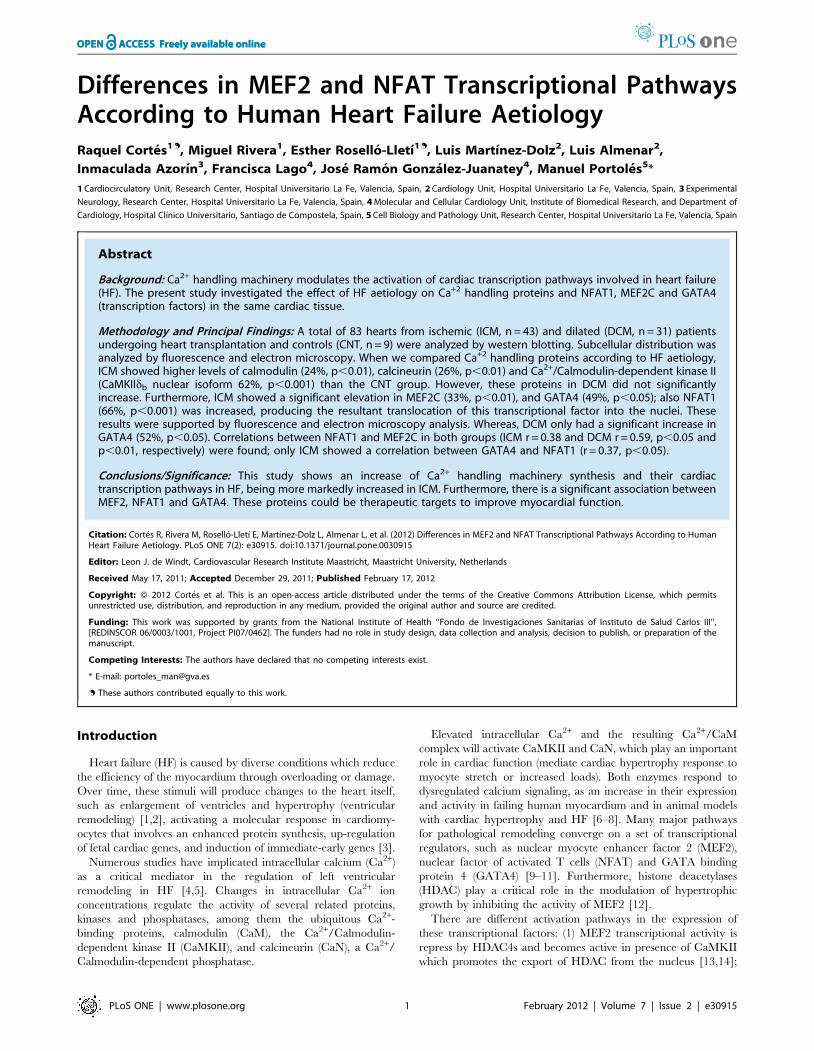
Figure 1. Western blots showing increased CaM, CaN and NCX1 levels, and decreased SERCA2 levels in human heart failure. CaM (A),CaN (B), NCX1 (C) and SERCA2 (D) in left ventricular myocardium from patients with ICM (n = 43) and DCM (n = 31) versus CNT group (n = 9). The dataare expressed as means 6 SEM of five independent experiments. Values are normalized to b-actin and finally to CNT group, which was alsonormalized to b-actin before. CaM, calmodulin; CaN, calcineurin; CNT, control; DCM, dilated cardiomyopathy; ICM, ischemic cardiomyopathy. *p,0.05versus control.doi:10.1371/journal.pone.0030915.g001
MEF2 and NFAT Pathways in Human Heart Failure
PLoS ONE | www.plosone.org 3 February 2012 | Volume 7 | Issue 2 | e30915
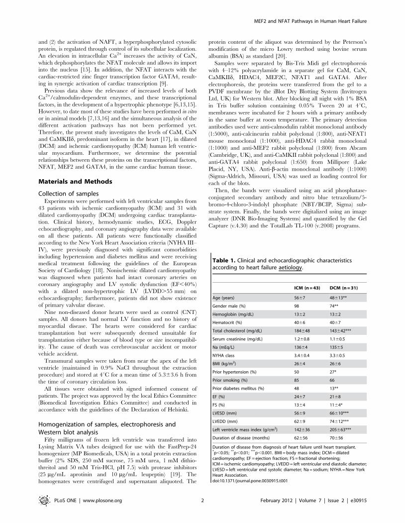
Figure 2. Detailed protein values of CaMKIId in cytosolic and nuclear fractions in human myocardium. Western blots for cytosolic (A)and nuclear (B) CaMKIId in controls, ischemic and dilated cardiomyopathies. The data are expressed as means 6 SEM of five independentexperiments. Values were normalized to b-actin and finally to control myocardium, which was also normalized to b-actin before. CaMKIId, Ca2+/calmodulin-dependent kinase II isoform delta; CNT, control, DCM, dilated cardiomyopathy; ICM, ischemic cardiomyopathy. **p,0.01 vs CNT.***p,0.001 vs. CNT.doi:10.1371/journal.pone.0030915.g002
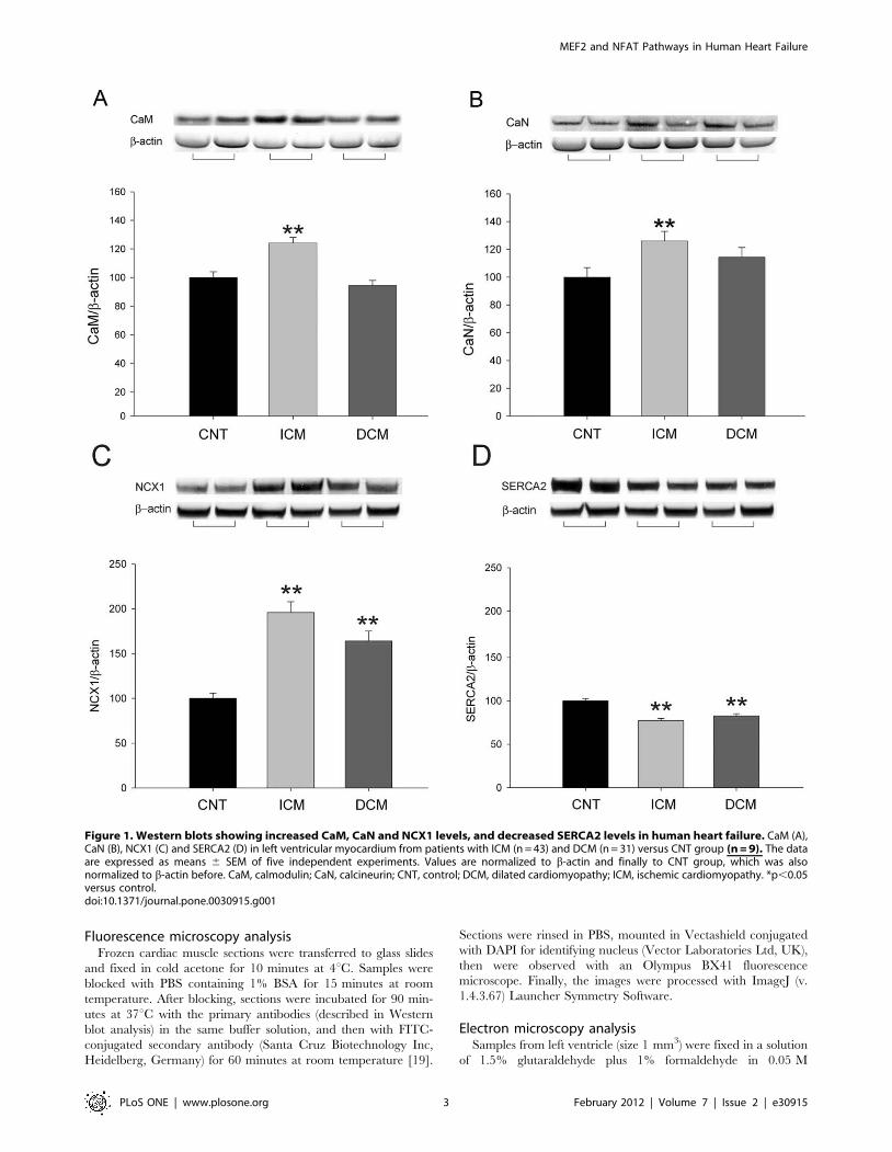
Figure 3. Influence of heart failure on the MEF2C and HDAC4 transcriptional factor levels. We determined the values of MEF2 and HDAC4by Western blots. In A, values of MEF2C were significantly increased in ICM samples (n = 43). In B, similar results were obtained in LV myocardium ofICM for HDAC4. Values are expressed as mean 6 SEM of five independent experiments and normalized to b-actin and finally to CNT myocardium,which was also normalized to b-actin before. ICM, ischemic cardiomyopathy; DCM, dilated cardiomyopathy; CNT, control. *p,0.05 versus CNT.doi:10.1371/journal.pone.0030915.g003
MEF2 and NFAT Pathways in Human Heart Failure
PLoS ONE | www.plosone.org 4 February 2012 | Volume 7 | Issue 2 | e30915
cacodylate buffer, pH 7.4, for 60 minutes at 4uC, and postfixed in
1% OsO4 for 60 minutes at 4uC, dehydrated in ethanol and
embedded in Epon 812. The 60 nm ultra-thin sections were
mounted on nickel grids and counter-stained with 2% uranyl
acetate for 20 minutes and 2.7% lead citrate for 3 minutes, for
electron microscopy observation, using a Philips CM-100, with
magnifications ranging from 4500 to 150006.
Statistical analysisData are presented as the mean 6 standard error mean. The
Kolmogorov-Smirnov test was used to analyze the distribution of
the variables. Comparisons of clinical characteristics were
achieved using Student’s t-test for continuous variables and Fisher
exact test for discrete variables. Comparisons for protein levels
between two groups were performed using the Mann-Whitney U
test and Spearman’s correlation coefficient was performed to
analyze the association between variables. Significance was
assumed as p,0.05. All statistical analyzes were performed using
SPSS software v. 15 for Windows (SPSS Inc., Chicago. IL, USA).
Results
Clinical characteristics of patientsMost of the patients were men (88%) with a mean age of 52611
years. The clinical characteristics of patients according to aetiology
of HF are summarized in Table 1. The ICM group showed a
significant increase in age (p,0.01), and total cholesterol
(p,0.001) compared with DCM group. Significant differences
were also found in left ventricular end-systolic diameter (LVESD)
(p,0.001), left ventricular end-diastolic diameter (LVEDD)
(p,0.001), and left ventricular mass index (LVMI) such as an
increase in the DCM group (p,0.001) compared with ICM
group. The percentage of hypertensive and diabetic patients
was also higher in ICM group (p,0.05 and p,0.01). Nine
non-diseased donor hearts were used as CNT samples (78% male,
mean age 5468 years, and EF.50).
Ca2+/CaM complex and Ca2+/calmodulin-dependentenzymes in heart failure
To investigate the effect of heart failure on several key Ca+2
handling proteins, we determined the levels of CaM and CaN in
human left ventricular myocardium by Western blot techniques.
When we compared protein levels between HF (n = 74) and CNT
(n = 9) hearts, the average of Ca+2 handling proteins (CaM and CaN)
was significantly increased in pathological samples (11263 vs.
10066; 12065 vs. 10063; p,0.05 in both, when normalized to b-
actin). Furthermore, Figure 1 shows that according to HF aetiology,
only in ICM (n = 43) CaM and CaN were significantly increased
(24%, p,0.001; and 26%, p,0.01, respectively). In addition, NCX1
and SERCA2 protein levels were also quantified. The results
obtained showed that NCX1 is significantly increased in ischemic
and dilated samples (96% and 64%, p,0.01, respectively) compared
to controls (Figure 1C), SERCA2 showed a similar decrease in both
aetiologies (23% and 17%, p,0.01, respectively) (Figure 1D).
On the other hand, we also quantify the total CaMKIId protein
amount, and its cytosolic and nuclear fractions (Figure 2). We
obtained a significant increase only in ICM group for total quantity
(29%, p,0.01), and for nuclear CaMKIIdb (62%, p,0.001)
(Figure 2B). There were significant differences for CaM and nuclear
CaMKIId fraction levels between these two aetiologies (p,0.01). In
addition, we obtained a significant correlation between CaM
protein with CaMKIId levels (r = 0.43, p,0.001).
Effect of heart failure and relationship between cardiactranscriptional pathways
We analyzed the influence of HF on the MEF2C transcriptional
factor, target of Ca2+/CaM signaling. We determined the values of
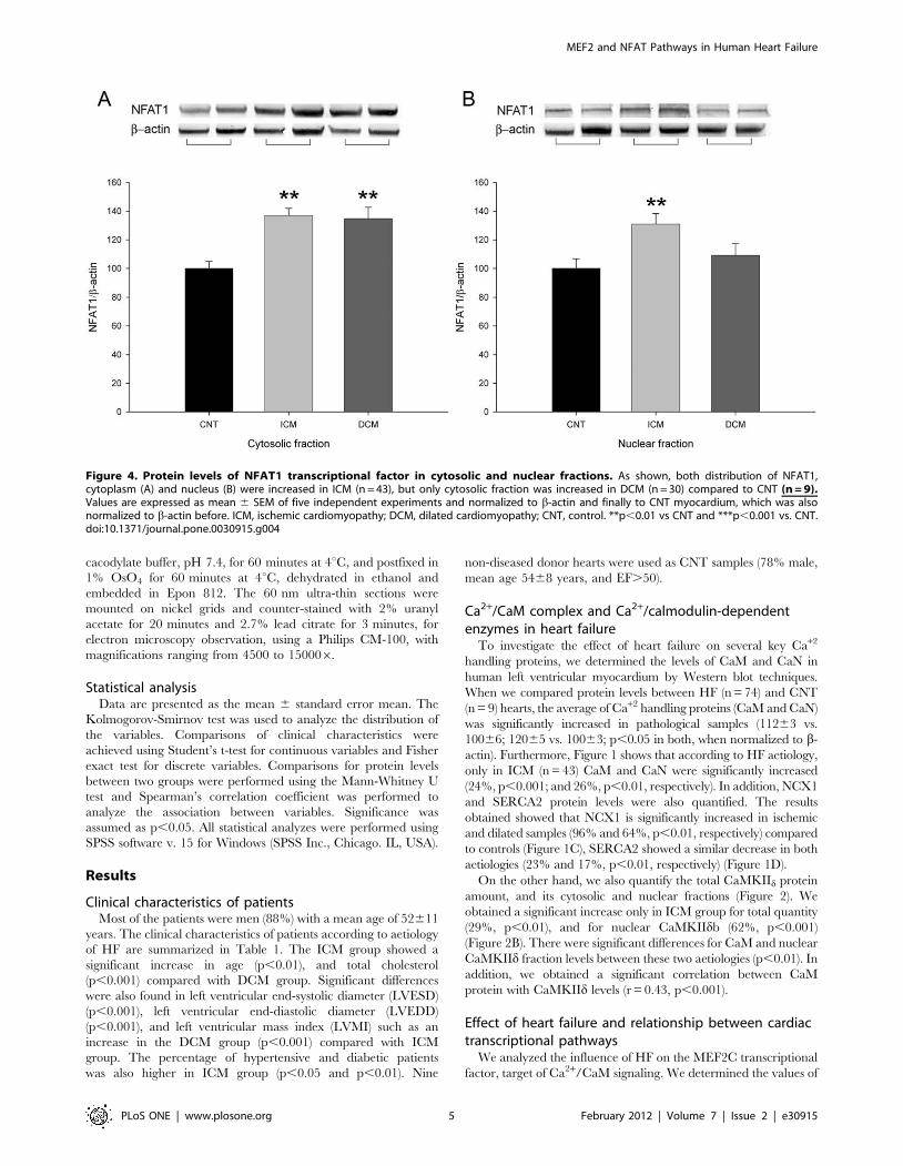
Figure 4. Protein levels of NFAT1 transcriptional factor in cytosolic and nuclear fractions. As shown, both distribution of NFAT1,cytoplasm (A) and nucleus (B) were increased in ICM (n = 43), but only cytosolic fraction was increased in DCM (n = 30) compared to CNT (n = 9).Values are expressed as mean 6 SEM of five independent experiments and normalized to b-actin and finally to CNT myocardium, which was alsonormalized to b-actin before. ICM, ischemic cardiomyopathy; DCM, dilated cardiomyopathy; CNT, control. **p,0.01 vs CNT and ***p,0.001 vs. CNT.doi:10.1371/journal.pone.0030915.g004
MEF2 and NFAT Pathways in Human Heart Failure
PLoS ONE | www.plosone.org 5 February 2012 | Volume 7 | Issue 2 | e30915
MEF2C and HDAC4, a histone deacetylase that interacts with this
factor. Pathological hearts had an increase in both proteins
(12663 vs. 100613, p,0.05; and 13364 vs. 100612, p,0.05,
respectively) compared to CNT samples. Then, only myocardium
from hearts with ICM showed higher MEF2C and HDAC4
protein levels (33% and 36%, p,0.01, respectively) (Figure 3).
When we analyzed the cytosolic and nuclear fractions of HDAC4,
ICM hearts only showed a significant increase in the cytosolic
fraction (45%, p,0.05) and 12% in the nuclei, but DCM did not
show significant differences (16% and 24%, respectively) com-
pared to CNT (data not shown). In addition, a statistical
correlation was found between MEF2C and HDAC4 in the
pathological human hearts (n = 74; r = 0.37, p,0.01). Finally,
HDAC4 also showed a significant direct correlation with CaN
expression (r = 0.25, p,0.05).
Furthermore, we also analyzed whether HF induced changes in
the NFAT1 transcriptional pathway. We observed a significant
increase in pathological myocardium (15267 vs 10068, p,0.01,
when normalized to b-actin). When we compared the NFAT1
according to aetiology of HF, only left ventricular myocardium
from ICM hearts showed a significant increase compared to CNT
hearts (66%, p,0.001) (Figure 4). Then, we quantified the protein
amount of NFAT1 in cytoplasm and nucleus, and we observed
that only ICM had a significant increase in nuclear NFAT1
(Figure 4B), and there were differences in nuclear NFAT1 between
HF etiologies (p,0.05).
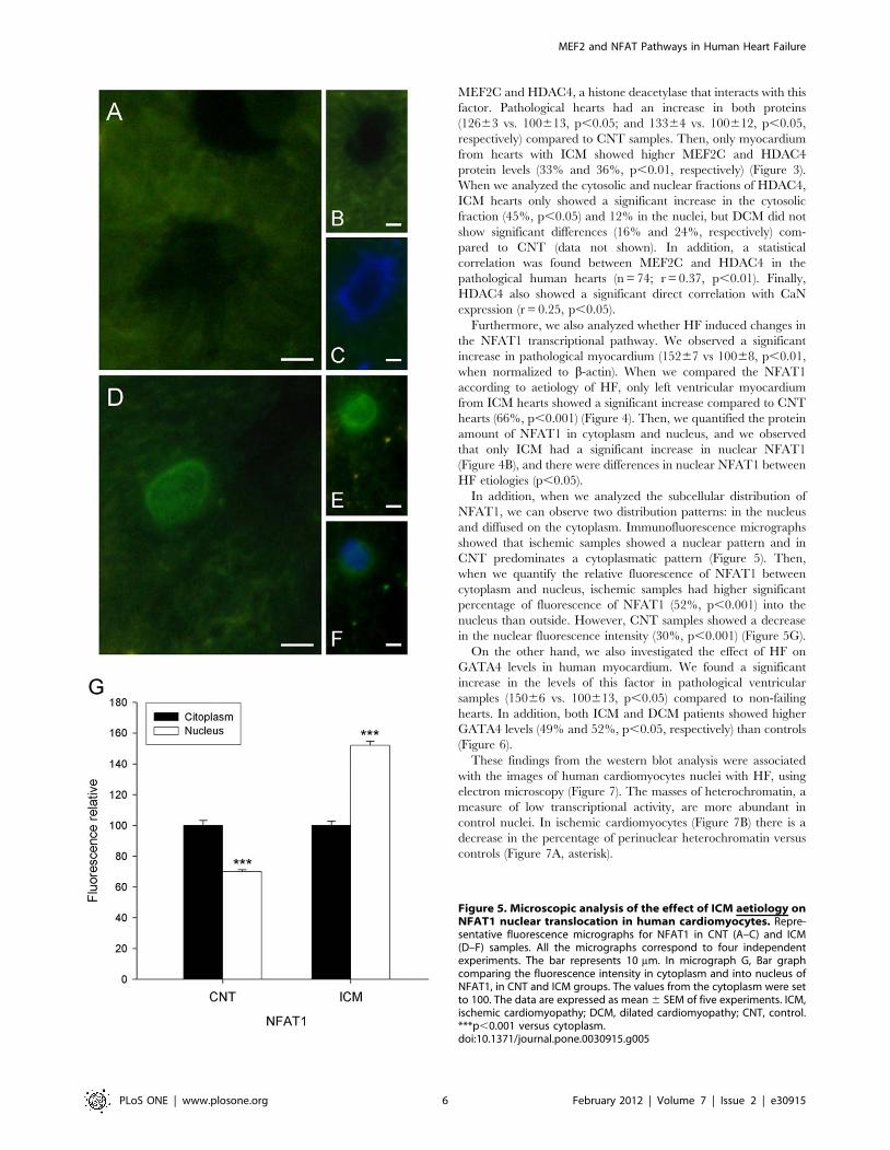
In addition, when we analyzed the subcellular distribution of
NFAT1, we can observe two distribution patterns: in the nucleus
and diffused on the cytoplasm. Immunofluorescence micrographs
showed that ischemic samples showed a nuclear pattern and in
CNT predominates a cytoplasmatic pattern (Figure 5). Then,
when we quantify the relative fluorescence of NFAT1 between
cytoplasm and nucleus, ischemic samples had higher significant
percentage of fluorescence of NFAT1 (52%, p,0.001) into the
nucleus than outside. However, CNT samples showed a decrease
in the nuclear fluorescence intensity (30%, p,0.001) (Figure 5G).
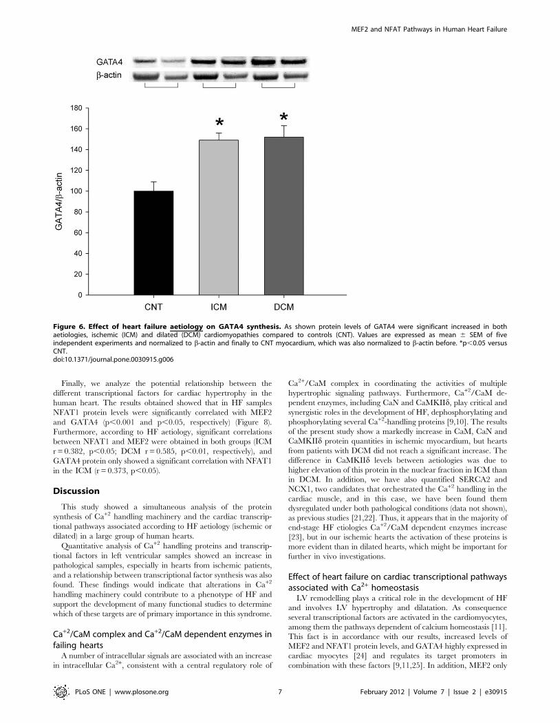
On the other hand, we also investigated the effect of HF on
GATA4 levels in human myocardium. We found a significant
increase in the levels of this factor in pathological ventricular
samples (15066 vs. 100613, p,0.05) compared to non-failing
hearts. In addition, both ICM and DCM patients showed higher
GATA4 levels (49% and 52%, p,0.05, respectively) than controls
(Figure 6).
These findings from the western blot analysis were associated
with the images of human cardiomyocytes nuclei with HF, using
electron microscopy (Figure 7). The masses of heterochromatin, a
measure of low transcriptional activity, are more abundant in
control nuclei. In ischemic cardiomyocytes (Figure 7B) there is a
decrease in the percentage of perinuclear heterochromatin versus
controls (Figure 7A, asterisk).
Figure 5. Microscopic analysis of the effect of ICM aetiology onNFAT1 nuclear translocation in human cardiomyocytes. Repre-sentative fluorescence micrographs for NFAT1 in CNT (A–C) and ICM(D–F) samples. All the micrographs correspond to four independentexperiments. The bar represents 10 mm. In micrograph G, Bar graphcomparing the fluorescence intensity in cytoplasm and into nucleus ofNFAT1, in CNT and ICM groups. The values from the cytoplasm were setto 100. The data are expressed as mean 6 SEM of five experiments. ICM,ischemic cardiomyopathy; DCM, dilated cardiomyopathy; CNT, control.***p,0.001 versus cytoplasm.doi:10.1371/journal.pone.0030915.g005
MEF2 and NFAT Pathways in Human Heart Failure
PLoS ONE | www.plosone.org 6 February 2012 | Volume 7 | Issue 2 | e30915
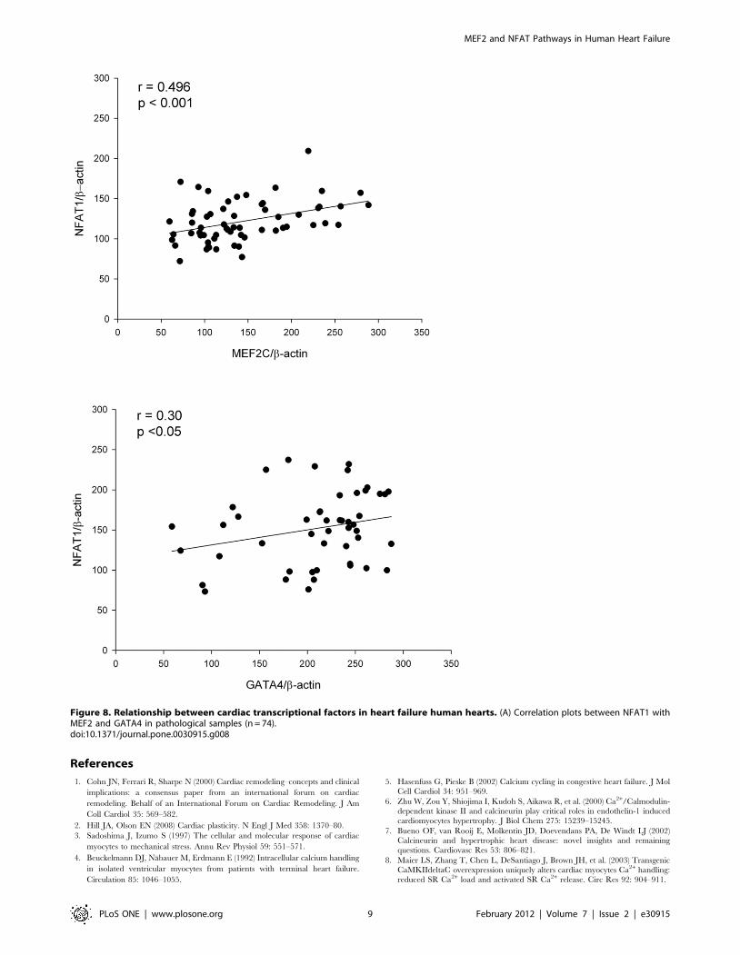
Finally, we analyze the potential relationship between the
different transcriptional factors for cardiac hypertrophy in the
human heart. The results obtained showed that in HF samples
NFAT1 protein levels were significantly correlated with MEF2
and GATA4 (p,0.001 and p,0.05, respectively) (Figure 8).
Furthermore, according to HF aetiology, significant correlations
between NFAT1 and MEF2 were obtained in both groups (ICM
r = 0.382, p,0.05; DCM r = 0.585, p,0.01, respectively), and
GATA4 protein only showed a significant correlation with NFAT1
in the ICM (r = 0.373, p,0.05).
Discussion
This study showed a simultaneous analysis of the protein
synthesis of Ca+2 handling machinery and the cardiac transcrip-
tional pathways associated according to HF aetiology (ischemic or
dilated) in a large group of human hearts.
Quantitative analysis of Ca+2 handling proteins and transcrip-
tional factors in left ventricular samples showed an increase in
pathological samples, especially in hearts from ischemic patients,
and a relationship between transcriptional factor synthesis was also
found. These findings would indicate that alterations in Ca+2
handling machinery could contribute to a phenotype of HF and
support the development of many functional studies to determine
which of these targets are of primary importance in this syndrome.
Ca+2/CaM complex and Ca+2/CaM dependent enzymes infailing hearts
A number of intracellular signals are associated with an increase
in intracellular Ca2+, consistent with a central regulatory role of
Ca2+/CaM complex in coordinating the activities of multiple
hypertrophic signaling pathways. Furthermore, Ca+2/CaM de-
pendent enzymes, including CaN and CaMKIId, play critical and
synergistic roles in the development of HF, dephosphorylating and
phosphorylating several Ca+2-handling proteins [9,10]. The results
of the present study show a markedly increase in CaM, CaN and
CaMKIId protein quantities in ischemic myocardium, but hearts
from patients with DCM did not reach a significant increase. The
difference in CaMKIId levels between aetiologies was due to
higher elevation of this protein in the nuclear fraction in ICM than
in DCM. In addition, we have also quantified SERCA2 and
NCX1, two candidates that orchestrated the Ca+2 handling in the
cardiac muscle, and in this case, we have been found them
dysregulated under both pathological conditions (data not shown),
as previous studies [21,22]. Thus, it appears that in the majority of
end-stage HF etiologies Ca+2/CaM dependent enzymes increase
[23], but in our ischemic hearts the activation of these proteins is
more evident than in dilated hearts, which might be important for
further in vivo investigations.
Effect of heart failure on cardiac transcriptional pathwaysassociated with Ca2+ homeostasis
LV remodelling plays a critical role in the development of HF
and involves LV hypertrophy and dilatation. As consequence
several transcriptional factors are activated in the cardiomyocytes,
among them the pathways dependent of calcium homeostasis [11].
This fact is in accordance with our results, increased levels of
MEF2 and NFAT1 protein levels, and GATA4 highly expressed in
cardiac myocytes [24] and regulates its target promoters in
combination with these factors [9,11,25]. In addition, MEF2 only
Figure 6. Effect of heart failure aetiology on GATA4 synthesis. As shown protein levels of GATA4 were significant increased in bothaetiologies, ischemic (ICM) and dilated (DCM) cardiomyopathies compared to controls (CNT). Values are expressed as mean 6 SEM of fiveindependent experiments and normalized to b-actin and finally to CNT myocardium, which was also normalized to b-actin before. *p,0.05 versusCNT.doi:10.1371/journal.pone.0030915.g006
MEF2 and NFAT Pathways in Human Heart Failure
PLoS ONE | www.plosone.org 7 February 2012 | Volume 7 | Issue 2 | e30915

become active with the phosphorylation and nuclear export of
HDAC4 [26] by CaN and CaMKIId [12]. Our data would
support this fact with a significant increase in CaMKIId and CaN
synthesis, but only in ICM hearts and mainly in the nuclear
fraction, with the subsequent nuclear export of HDAC4.
On the other hand we also observed an increase in the NFAT1
protein levels in the nuclear fraction and in the distribution pattern
into the nuclei by immunofluorescence only in ischemic aetiology.
These findings may be in concordance with a major activation of
calcineurin with the subsequent nuclear translocation of this
transcriptional factor in hearts from ischemic patients. These
results are supported by the electron microscopy analysis that
shows a high nuclear transcriptional activity (reduction of
heterochromatin masses) in ischemic hearts.
It has been argued that cardiac transcriptional pathways
dependent of Ca2+ homeostasis in human hearts would be an
important process of cardiac remodelling in HF. The present study is
in line with this idea, although our results show differences between
HF aetiology, a significant increase was detected only in ICM.
Perhaps, this fact could be related with the intrinsic variability of
the samples, given they originate from human hearts, whose
conditions (treatment they undergo) are not standardized. But it is
very unlikely because in our study almost all patients received
drugs like diuretics, ACE inhibitors and beta-blockers. In addition,
there is precedent for specific biochemical differences between
dilated and ischemic cardiomyopathies [27,28], and it has been
previously established that genes that cause DCM generally
encode cytoskeletal and sarcomeric (contractile apparatus) proteins
[29], although disturbance of calcium homeostasis also seems to be
important [25].
Association of MEF2C, NFAT1 and GATA4 cardiactranscriptional factors in human hearts
There is substantial evidence that transcriptional factors
function cooperatively with each other and with coactivators and
repressors in their regulation of gene expression. Specifically, Putt
et al. provided genomic evidence for coregulation of myocardial
gene expression by MEF2 and NFAT1 in advanced human HF
from patients with idiopathic DCM [30]. In the present study, we
determined the relationship between MEF2 and NFAT1 protein
levels in the same myocardium from patients with HF, revealing a
significant direct correlation in both cardiomyopathies (ischemic
and dilated). These data would show that coregulation of gene
expression may be also reflected at protein expression level in left
ventricular myocardium. We also observed, in ICM, the
correlation between the protein amounts of GATA4 with NFAT1
in ICM, a previous work where showed the interaction between
both factors in transgenic mice, resulting in synergistic activation
of cardiac transcription [9]. Furthermore, previous works have
shown that there is cross-talk between CaMKII and CaN signaling
pathways. Lu et al. [31] demonstrated that the transcriptional
upregulation of CaN is partially mediated by CaMKIId in rat
cardiomyocytes, and Khoo et al. [32] showed the role of CaMKII in
CaN cardiomyopathy. Our results would be in line with the theory
that there is a certain interaction between the two systems through
the relationship found between HDAC4 and CaN levels, as
previous reports [33].
The current study shows that the identification of an increase in
the synthesis of these proteins would show that these pathways
may be associated with a heart failure phenotype, especially in
ischemic hearts. Furthermore, significant correlation between
cardiac transcription factor protein levels, and a cross-talk between
CaMKIId and CaN signaling pathways, HDAC4 not only may
regulate MEF2 activation, would indicate the complexity of
calcium homeostasis in the development HF. Therefore, consid-
ering the important role of the Ca2+ dependent transcritpional
pathways in cardiac hypertrophy and heart failure, further studies
are necessary to determine which of these targets (Ca2+ handling
machinery and cardiac transcription factors) are of primary
importance in establishing therapeutic approaches to treat patients
with heart failure.
Acknowledgments
The authors thank the Transplant Coordination Unit (Hospital Uni-
versitario La Fe, Valencia, Spain) for their help in obtaining the samples.
Furthermore, we are grateful to Inmaculada Montserrat and Pilar Martın
(technicians at the Research Center, Hospital Universitario La Fe,
Valencia, Spain) for their assistance in sample procedures.
Author Contributions
Conceived and designed the experiments: RC MR ERL MP. Performed
the experiments: RC ERL IA LMD LA FL. Analyzed the data: RC ERL
FL MP. Contributed reagents/materials/analysis tools: RC ERL. Wrote
the paper: RC. Collection of data: RC ERL LMD LA IA. Revised the
paper critically: RC MR MP LMD LA JRGJ. Final approval of the
version: RC MR MP JRGJ. Funding: MR MP JRGJ..
Figure 7. Nuclear activity as heterochromatin mass by electronmicroscopy in human cardiomyocytes. Cross-sectional micro-graphs of a nucleus in control (A), ischemic (B) and dilated (C) samples,showing a more heterochromatin condensation (hc) in controls, overallperinuclear chromatin (asterisk). N indicates nucleus. Bar represents:400 nm.doi:10.1371/journal.pone.0030915.g007
MEF2 and NFAT Pathways in Human Heart Failure
PLoS ONE | www.plosone.org 8 February 2012 | Volume 7 | Issue 2 | e30915
References
1. Cohn JN, Ferrari R, Sharpe N (2000) Cardiac remodeling–concepts and clinical
implications: a consensus paper from an international forum on cardiac
remodeling. Behalf of an International Forum on Cardiac Remodeling. J Am
Coll Cardiol 35: 569–582.
2. Hill JA, Olson EN (2008) Cardiac plasticity. N Engl J Med 358: 1370–80.
3. Sadoshima J, Izumo S (1997) The cellular and molecular response of cardiac
myocytes to mechanical stress. Annu Rev Physiol 59: 551–571.
4. Beuckelmann DJ, Nabauer M, Erdmann E (1992) Intracellular calcium handling
in isolated ventricular myocytes from patients with terminal heart failure.
Circulation 85: 1046–1055.
5. Hasenfuss G, Pieske B (2002) Calcium cycling in congestive heart failure. J Mol
Cell Cardiol 34: 951–969.
6. Zhu W, Zou Y, Shiojima I, Kudoh S, Aikawa R, et al. (2000) Ca2+/Calmodulin-
dependent kinase II and calcineurin play critical roles in endothelin-1 induced
cardiomyocytes hypertrophy. J Biol Chem 275: 15239–15245.
7. Bueno OF, van Rooij E, Molkentin JD, Doevendans PA, De Windt LJ (2002)Calcineurin and hypertrophic heart disease: novel insights and remaining
questions. Cardiovasc Res 53: 806–821.
8. Maier LS, Zhang T, Chen L, DeSantiago J, Brown JH, et al. (2003) Transgenic
CaMKIIdeltaC overexpression uniquely alters cardiac myocytes Ca2+ handling:
reduced SR Ca2+ load and activated SR Ca2+ release. Circ Res 92: 904–911.
Figure 8. Relationship between cardiac transcriptional factors in heart failure human hearts. (A) Correlation plots between NFAT1 withMEF2 and GATA4 in pathological samples (n = 74).doi:10.1371/journal.pone.0030915.g008
MEF2 and NFAT Pathways in Human Heart Failure
PLoS ONE | www.plosone.org 9 February 2012 | Volume 7 | Issue 2 | e30915
9. Molkentin JD, Lu JR, Antos CL, Markham B, Richardson J, et al. (1998) A
calcineurin-dependent transcriptional pathway for cardiac hypertrophy. Cell 93:215–228.
10. Kapiloff MS, Mathis JM, Nelson CA, Lin CR, Rosenfeld MG (1991) Calcium/
calmodulin-dependent protein kinase mediates a pathway for transcriptionalregulation. Proc Natl Acad Sci U S A 88: 3710–3714.
11. Barry SP, Townsend PA (2010) What causes a broken heart–molecular insightsinto heart failure. Int Rev Cell Mol Biol 284: 113–179.
12. Zhang CL, McKinsey TA, Chang S, Antos CL, Hill JA, et al. (2002) Class II
histone deacetylases act as signal-responsive repressors of cardiac hypertrophy.Cell 110: 479–488.
13. Passier R, Zheng H, Frey N, Naya FJ, Nicol RL, et al. (2000) CaM Kinasesignaling induces cardiac hypertrophy and activates the MEF2 transcription
factor in vivo. J Clin Invest 105: 1395–1406.14. Little GH, Bai Y, Williams T, Poizat C (2007) Nuclear calcium/calmodulin-
dependent protein kinase II delta preferentially transmits signals to histone
deacetylase 4 in cardiac cells. J Biol Chem 282: 7219–7231.15. Wilkins BJ, Dai YS, Bueno OF, Parsons SA, Xu J, et al. (2004) Calcineurin/
NFAT coupling participates in pathological, but not physiological, cardiachypertrophy. Circ Res 94: 110–118.
16. Mishra S, Sabbah HN, Jain JC, Gupta RC (2003) Reduced Ca2+/calmodulin-
dependent protein kinase activy and expression in LV myocardium of dogs withheart failure. Am J Physiol Heart Circ Physiol 284: H876–H873.
17. Hoch B, Meyer R, Hetzer R, Krause EG, Karczewski P (1999) Identificationand expression of delta-isoforms of the multifunctional Ca2+/calmodulin-
dependent protein kinase in failing and nonfailing human myocardium. CircRes 84: 713–721.
18. Swedberg K, Cleland J, Dargie H, Drexler H, Follath F, et al. (2005) Guidelines
for the diagnosis and treatment of chronic heart failure: executive summary(update 2005): The Task Force for the Diagnosis and Treatment of Chronic
Heart Failure of the European Society of Cardiology. Eur Heart J 26:1115–1140.
19. Cortes R, Rosello-Lletı E, Rivera M, Martınez-Dolz L, Salvador A, et al. (2010)
Influence of heart failure on nucleocytoplasmic transport in human cardiomy-ocytes. Cardiovasc Res 85: 464–472.
20. Peterson GL (1977) A simplification of the protein assay method of Lowry et al.which is more generally applicable. Anal Biochem 83: 346–56.
21. Hasenfuss G (1998) Alterations of calcium-regulatory proteins in heart failure.
Cardiovasc Res 37: 279–289.22. Frank KF, Bolck B, Erdmann E, Schwinger RH (2003) Sarcoplasmic reticulum
Ca2+-ATPasa modulates cardiac contraction and relaxation. Cardiovasc Res 57:
20–27.23. Sossalla S, Fluschnik N, Schotola H, Ort KR, Neef S, et al. (2010) Inhibition of
elevated Ca2+/calmodulin-dependent protein kinase II improves contractility inhuman failing myocardium. Circ Res 107: 1150–1161.
24. Liang Q, De Windt LJ, Witt SA, Kimball TR, Markham BE, et al. (2001) The
transcription factors GATA4 and GATA6 regulate cardiomyocyte hypertrophyin vitro and in vivo. J Biol Chem 276: 30245–30253.
25. Ahmad F, Seidman JG, Seidman CE (2005) The genetic basis for cardiacremodeling. Annu Rev Genomics Hum Genet 6: 185–216.
26. Miska EA, Karlsson C, Langley E, Nielsen SJ, Pines J, et al. (1999) HDAC4deacetylase associates with and represses the MEF2 transcription factor. EMBO J
18: 5099–5107.
27. Bohm M, Gierschik P, Jakobs KH, Pieske B, Schnabel P, et al. (1990) Increase inGi alpha in human hearts with dilated but not ischemic cardiomyopathy.
Circulation 82: 1249–1265.28. Pauschinger M, Doerner A, Remppis A, Tannhauser R, Kuhl U, et al. (1998)
Differential myocardial abundance of collagen type I and type III mRNA in
dilated cardiomyopathy: effects of myocardial inflammation. Cardiovasc Res 37:123–9.
29. Jefferies JL, Towbin JA (2010) Dilated cardiomyopathy. Lancet 375: 752–762.30. Putt ME, Hannenhalli S, Lu Y, Haines P, Chandrupatla HR, et al. (2009)
Evidence for coregulation of myocardial gene expression by MEF2 and NFATin human heart failure. Circ Cardiovasc Genet 2: 212–219.
31. Lu YM, Shioda N, Yamamoto Y, Han F, Fukunaga K (2010) Transcriptional
upregulation of calcineurin Abeta by endothelin-1 is partially mediated bycalcium/calmodulin-dependent protein kinase IIdelta3 in rat cardiomyocytes.
Biochem Biophys Acta 1799: 429–441.32. Khoo MS, Li J, Singh MV, Yang Y, Kannankeril P, et al. (2006) Death, cardiac
dysfunction, and arrhythmias are increased by calmodulin kinase II in
calcineurin cardiomyopathy. Circulation 114: 1352–1359.33. Lynch J, Guo L, Gelebart P, Chilibeck K, Xu J, et al. (2005) Calreticulin signals
upstream of calcineurin and MEF2C in a critical Ca(2+)-dependent signalingcascade. J Cell Biol 170: 37–47.
MEF2 and NFAT Pathways in Human Heart Failure
PLoS ONE | www.plosone.org 10 February 2012 | Volume 7 | Issue 2 | e30915