Inhibition of renin angiotensin system decreases renal protein oxidative damage in diabetic rats
8
Inhibition of renin angiotensin system decreases renal protein oxidative damage in diabetic rats Manuel Portero-Otı ´n a,1 , Reinald Pamplona a,1 , Jordi Boada a , Mariona Jove ´ a , Hugo Gonzalo a , Marie Buleon b , Wolfgang Linz c , Stefan Scha ¨fer c , Ivan Tack b , Jean-Pierre Girolami b, * a Department of Experimental Medicine, Faculty of Medicine, University of Lleida-IRBLLEIDA-PCiTAL (Parc Cientı ´fic Agroalimentari de Lleida), 25008 Lleida, Spain b Institut National de la Sante ´ et de la Recherche Me ´dicale Unit 858 eq 5, IFR31 Institut Louis Bugnard, BP 84225, 31432 Toulouse Cedex 4, France c Therapeutic Department Cardiovascular Research, Sanofi Aventis, Frankfurt am Main, Germany Received 8 January 2008 Available online 1 February 2008 Abstract Renin angiotensin system (RAS) worsens diabetic nephropathy (DN) by increasing oxidative stress. We compared the effect of three different RAS inhibitors: the angiotensin converting enzyme inhibitor Ramipril, the vasopeptidase inhibitor AVE7688 and the angioten- sin receptor (AT1) antagonist Losartan on the formation of oxidative and carbonyl stress derived protein modifications in kidney from Zucker obese hyperglycemic rats (ZDFn Gm-fa/fa). Gas chromatography–mass spectrometry was used to measure representative mark- ers of several protein oxidative pathways: direct oxidation [dinitrophenylhydrazine reactive carbonyls (DNP), glutamic (GSA), and ami- noadipic (AASA) semialdehydes], mixed glyco- and lipoxidation [N e -carboxyethyl-lysine (CEL) and N e -(carboxymethyl)-lysine (CML)] and lipoxidation-[N e -(malondialdehyde)-lysine-(MDAL)], as well as renal fatty acid composition. Urinary albumin (a marker of DN), DNP, GSA, and MDAL levels, were increased in all obese rats and were dose dependently decreased by AVE7688 whereas Ramipril and Losartan were less efficient. These results show that RAS inhibition improves DN at several levels, independently of its effects on blood pressure and glycemic control, via mechanisms depending of renal oxidative stress. Ó 2008 Published by Elsevier Inc. Keywords: Zucker diabetic fatty acid rats; Diabetic nephropathy; Protein oxidation; Advanced glycated end products; Lipoxidation; Angiotensin con- verting enzyme inhibitor; AT1 receptor antagonist; Vasopeptidase inhibition; AVE7688; Losartan; Ramipril Diabetic nephropathy (DN) is a progressive and an irre- versible renal disease which has become the most common single cause of renal failure [1]. The onset of DN involves several additional mechanisms such as hemodynamic dys- functions, inflammatory reaction, growth factor and cyto- kine secretion and finally formation of glucose-derived toxic compounds. The knowledge and the control of these different mechanisms has become a fascinating therapeutic challenge, aimed to reduce the progression of DN. It is now generally admitted that advanced glycation end products (AGEs) play a pivotal role in the onset of detrimental side effects of diabetes including mainly diabetic neuropathy, retinopathy, and nephropathy [2–5]. Although no efficient therapy is yet available, an increasing number of reports indicate that blockade of the renin angiotensin system (RAS) is effective to delay the progression of DN and thereby to protect against end-stage renal failure. In addi- tion to inhibitors of the RAS, a new class of pharmaceuti- cal agents with high therapeutic potential, termed inhibitors of vasopeptidase (IVP) has emerged recently. The effect of IVP on the management of DN start being investigated in various models [6–9]. The overall effect of these pharmaceutical agents is a significant reduction of 0006-291X/$ - see front matter Ó 2008 Published by Elsevier Inc. doi:10.1016/j.bbrc.2008.01.101 * Corresponding author. Address: INSERM Unit 388, IFR31 Institut Louis Bugnard, BP 84225, 31432 Toulouse Cedex 4, France. E-mail address: [email protected] (J.-P. Girolami). 1 These authors contributed equally to the work. www.elsevier.com/locate/ybbrc Available online at www.sciencedirect.com Biochemical and Biophysical Research Communications 368 (2008) 528–535
-
Upload
independent -
Category
Documents
-
view
8 -
download
0
Transcript of Inhibition of renin angiotensin system decreases renal protein oxidative damage in diabetic rats
Available online at www.sciencedirect.com
www.elsevier.com/locate/ybbrc
Biochemical and Biophysical Research Communications 368 (2008) 528–535
Inhibition of renin angiotensin system decreases renal proteinoxidative damage in diabetic rats
Manuel Portero-Otın a,1, Reinald Pamplona a,1, Jordi Boada a, Mariona Jove a,Hugo Gonzalo a, Marie Buleon b, Wolfgang Linz c, Stefan Schafer c, Ivan Tack b,
Jean-Pierre Girolami b,*
a Department of Experimental Medicine, Faculty of Medicine, University of Lleida-IRBLLEIDA-PCiTAL (Parc Cientıfic Agroalimentari de Lleida),
25008 Lleida, Spainb Institut National de la Sante et de la Recherche Medicale Unit 858 eq 5, IFR31 Institut Louis Bugnard, BP 84225, 31432 Toulouse Cedex 4, France
c Therapeutic Department Cardiovascular Research, Sanofi Aventis, Frankfurt am Main, Germany
Received 8 January 2008Available online 1 February 2008
Abstract
Renin angiotensin system (RAS) worsens diabetic nephropathy (DN) by increasing oxidative stress. We compared the effect of threedifferent RAS inhibitors: the angiotensin converting enzyme inhibitor Ramipril, the vasopeptidase inhibitor AVE7688 and the angioten-sin receptor (AT1) antagonist Losartan on the formation of oxidative and carbonyl stress derived protein modifications in kidney fromZucker obese hyperglycemic rats (ZDFn Gm-fa/fa). Gas chromatography–mass spectrometry was used to measure representative mark-ers of several protein oxidative pathways: direct oxidation [dinitrophenylhydrazine reactive carbonyls (DNP), glutamic (GSA), and ami-noadipic (AASA) semialdehydes], mixed glyco- and lipoxidation [Ne-carboxyethyl-lysine (CEL) and Ne-(carboxymethyl)-lysine (CML)]and lipoxidation-[Ne-(malondialdehyde)-lysine-(MDAL)], as well as renal fatty acid composition. Urinary albumin (a marker of DN),DNP, GSA, and MDAL levels, were increased in all obese rats and were dose dependently decreased by AVE7688 whereas Ramipriland Losartan were less efficient. These results show that RAS inhibition improves DN at several levels, independently of its effectson blood pressure and glycemic control, via mechanisms depending of renal oxidative stress.� 2008 Published by Elsevier Inc.
Keywords: Zucker diabetic fatty acid rats; Diabetic nephropathy; Protein oxidation; Advanced glycated end products; Lipoxidation; Angiotensin con-verting enzyme inhibitor; AT1 receptor antagonist; Vasopeptidase inhibition; AVE7688; Losartan; Ramipril
Diabetic nephropathy (DN) is a progressive and an irre-versible renal disease which has become the most commonsingle cause of renal failure [1]. The onset of DN involvesseveral additional mechanisms such as hemodynamic dys-functions, inflammatory reaction, growth factor and cyto-kine secretion and finally formation of glucose-derivedtoxic compounds. The knowledge and the control of thesedifferent mechanisms has become a fascinating therapeuticchallenge, aimed to reduce the progression of DN. It is now
0006-291X/$ - see front matter � 2008 Published by Elsevier Inc.
doi:10.1016/j.bbrc.2008.01.101
* Corresponding author. Address: INSERM Unit 388, IFR31 InstitutLouis Bugnard, BP 84225, 31432 Toulouse Cedex 4, France.
E-mail address: [email protected] (J.-P. Girolami).1 These authors contributed equally to the work.
generally admitted that advanced glycation end products(AGEs) play a pivotal role in the onset of detrimental sideeffects of diabetes including mainly diabetic neuropathy,retinopathy, and nephropathy [2–5]. Although no efficienttherapy is yet available, an increasing number of reportsindicate that blockade of the renin angiotensin system(RAS) is effective to delay the progression of DN andthereby to protect against end-stage renal failure. In addi-tion to inhibitors of the RAS, a new class of pharmaceuti-cal agents with high therapeutic potential, termedinhibitors of vasopeptidase (IVP) has emerged recently.The effect of IVP on the management of DN start beinginvestigated in various models [6–9]. The overall effect ofthese pharmaceutical agents is a significant reduction of

M. Portero-Otın et al. / Biochemical and Biophysical Research Communications 368 (2008) 528–535 529
glomerular fibrosis progression. However, besides theirwell-documented efficiency, the precise mechanism and tar-gets of these inhibitors, with special attention to oxidativestress, remain to be elucidated.
The inhibition of the RAS could affect nephropathy bydifferent, non-excluding, mechanisms including regulationof hemodynamic factors, but also modulation of proteinkinase-C activation, inhibition of TGFb system and morerecently inhibition of AGEs formation and reduction ofgrowth factor activation [10,11]. Among those mecha-nisms, high-glucose induced reactive oxygen species(ROS) overproduction has recently received increasingattention [2–5]. In this line, angiotensin converting enzyme(ACE) inhibition leads to a diminished ROS productionthrough diminished angiotensin signaling and possiblyincreased activation of the B2-bradykinin receptor[11,12]. It is well admitted that AT1 receptors increase oxi-dative stress [5,13]. This is consistent with suggestions thatACE inhibition has a marked antioxidant activity[10,14,15].
However, direct evidences of oxidative damage to pro-teins have not been offered in these or similar diabeticnephropathy models. Usually, protein oxidative damageis assessed by examining 2,4-dinitrophenylhydrazine(DNP) reactivity, an assay which has been criticized dueto possibility of artefacts [16]. Because AGEs include a verylarge and heterogenous family resulting from multiplemechanisms of formation, direct in vivo measurement ofstructurally identified products could bring precise infor-mation regarding the preferential formation pathways ofAGEs. The major established pathway results from the ini-tial covalent bond between a reducing sugar (i.e. glucose)and an amino acid group, to produce an Amadori productwith subsequent reactions leading to the formation of irre-versible AGEs. In addition to glucose, a new pathwaytermed metal catalyzed oxidation (MCO) of lipids, orlipoxidation, has emerged as a new source of AGE forma-tion and is being currently explored as biomarker for spe-cific pathological processes [17,18].
In the present study we compared the effect of chronictreatment with three different classes of inhibitors i.e. Ram-ipril, an angiotensin converting enzyme inhibitor, Losar-tan, an AT1 receptor antagonist, and AVE7688, aninhibitor of vasopetidase, on the long term renal accumula-tion of five different species of AGEs in DN in obese hyper-glycaemic Zucker diabetic fatty rats, as well as in the fattyacid composition of renal cortex due to the new potentialimportance of lipids in AGE formation.
Materials and methods
The animal experiments were performed in accordance with the SanofiAventis Laboratory Animal Science and Welfare (LASW) guidelines, theGerman law for the protection of animals and the National Institute ofHealth (NIH) Guide for Care and Use of Laboratory Animals. Male ZDFrats (Gmi ZDF fa/fa) were purchased from Charles River (Sulzfeld,Germany). The animals were housed individually in standard cages with
tap water ad libitum and standard chow containing 0.2% sodium and 19%crude protein (Standard diet #1320, Altromin, Lage, Germany).
At age of 30 weeks, after baseline measurements, the animals wererandomly assigned to one of eight groups (n = 6 in each group) receivingeither no specific treatment (Placebo), the ACE inhibitor Ramipril (0.1 or1 mg/kg/day), the vasopeptidase inhibitor AVE7688 (0.3, 3, and 30 mg/kg/day) or the AT1 receptor antagonist Losartan (3 and 30 mg/kg/day).Ramipril and Losartan were administered via drinking water. AVE7688was administered orally pressed in chow [8]. The duration of treatmentwas 23 weeks. Heterozygous (Fa/fa) rats were used as non-diabetic control(n = 6). Urine and blood samples were taken to determine urinary albu-min/creatinine ratio, as an index of renal function, and blood glucose,respectively. Systolic arterial blood pressure and heart rate were measuredusing the tail-cuff method (TSE GmbH, BP system V2.2, Bad Homburg,Germany) in all animals. Kidneys were then carefully removed and storedfrozen until further measurements.
Biochemical studies
Gas chromatography-mass spectrometry (GC/MS) measurements of
glutamic semialdehyde (GSA), aminoadipic semialdehyde (AASA), Ne-
(carboxyethyl)-lysine (CEL), Ne-(carboxymethyl)-lysine (CML), and Ne-
(malondialdehyde)-lysine (MDAL). GSA, AASA, CML, CEL, andMDAL concentrations in total proteins from renal cortex homogenateswere measured by GC/MS as routinely performed in our laboratory[18,19]. Quantification was performed by external standardization usingstandard curves constructed from mixtures of deuterated and non-deu-terated standards. Analytes were detected by selected ion-monitoring GC/MS. The amounts of products were expressed as the ratio lmol glutamicsemialdehyde, aminoadipic semialdehyde, CML, CEL, or MDAL/mollysine.
Immunodetection of protein-bound 2,4-dinitrophenylhydrazones and the
glycoxidation product CEL. Prior to SDS electrophoresis, pools of sampleswere derivatized with DNP as previously described and routinely per-formed in the laboratory [19]. Immunodetection was performed using asprimary antibodies a rabbit anti-DNP antiserum (1:4.000, Dako V401,Carpenteria, CA) and a monoclonal anti-CEL antibody (1:2.000, Trans-genic Inc., Kumamoto, Japan). Peroxidase-coupled secondary antibodieswere used from the Tropix chemiluminescence kit (Bedford, MA). Signalquantification and recording was performed with a CCD camera-basedsystem (Lumi-Imager) from Boehringer Mannheim, scanning bandscomprised between 30 and 250 kDa.
Fatty acid analysis. Fatty acid analysis was performed as previouslydescribed [19]. Results are expressed as mol%. The following fatty acidindexes (FA) were calculated as previously described [19]: saturated fattyacids (SFA); unsaturated fatty acids (UFA); monounsaturated fatty acids(MUFA); polyunsaturated n � 3 fatty acids (PUFAn � 3); polyunsatu-rated n � 6 fatty acids (PUFAn � 6); average chain length (ACL);unsaturation index (UI); and peroxidizability index (PI).
Statistical analysis
All statistics calculations were performed using the SPSS software(SPSS, Chicago). Once normality of variable’s distribution was checked bythe Kolmogorov–Smirnoff test, differences between samples were analyzedby the ANOVA test, and by the DMS test for intra-group analyses. Ap < 0.05 level was selected as the point of minimal statistical significance inevery comparison.
Results
Blood pressure and albumin excretion
As shown in Table 1, no significant change in bloodpressure and heart rate can be found between the control
530 M. Portero-Otın et al. / Biochemical and Biophysical Research Communications 368 (2008) 528–535
and the diabetic placebo group. In contrast, a very sig-nificant increase in the albumin/creatinine ratio (an earlyeasy measurable and reliable biomarker of DN) wasfound in the diabetic untreated group (Placebo vs non-diabetic control). Interestingly, all the pharmacologicaltreatments decreased significantly the albumin/creatinineratio to various extents. A complete normalization tothe value of non-diabetic control was achieved with30 mg/kg/day of AVE7688, whereas blood glucose of
Table 1Effect of chronic treatments (23 weeks) with RAS inhibitors on systolic arteriamale ZDFn Gm-fa/fa rats
Group SAP (mm Hg) HR (b.p.m)
Ctl 154.5 ± 3.3 321 ± 10.6Plc 156 ± 3.5 311.1 ± 8.5Rmp 0.1 155.1 + 4 331.1 ± 12.9Rmp 1 145 ± 4.8 331.1 ± 6.5AVE7688 0.3 157.7 + 2.9 297.9 ± 6AVE7688 3 154.2 ± 3.4 321.1 ± 10.5AVE7688 30 149.4 ± 5.5 321 ± 10.5Lst 3 151 ± 2.7 317.1 ± 9.1Lst 30 141 ± 3.6* 327 ± 8.9
Values: mean ± SEM, n = 6 in each group. Abbreviations of treatments are as Fand **p < 0.01 diabetic treated vs value of diabetic placebo before treatment.
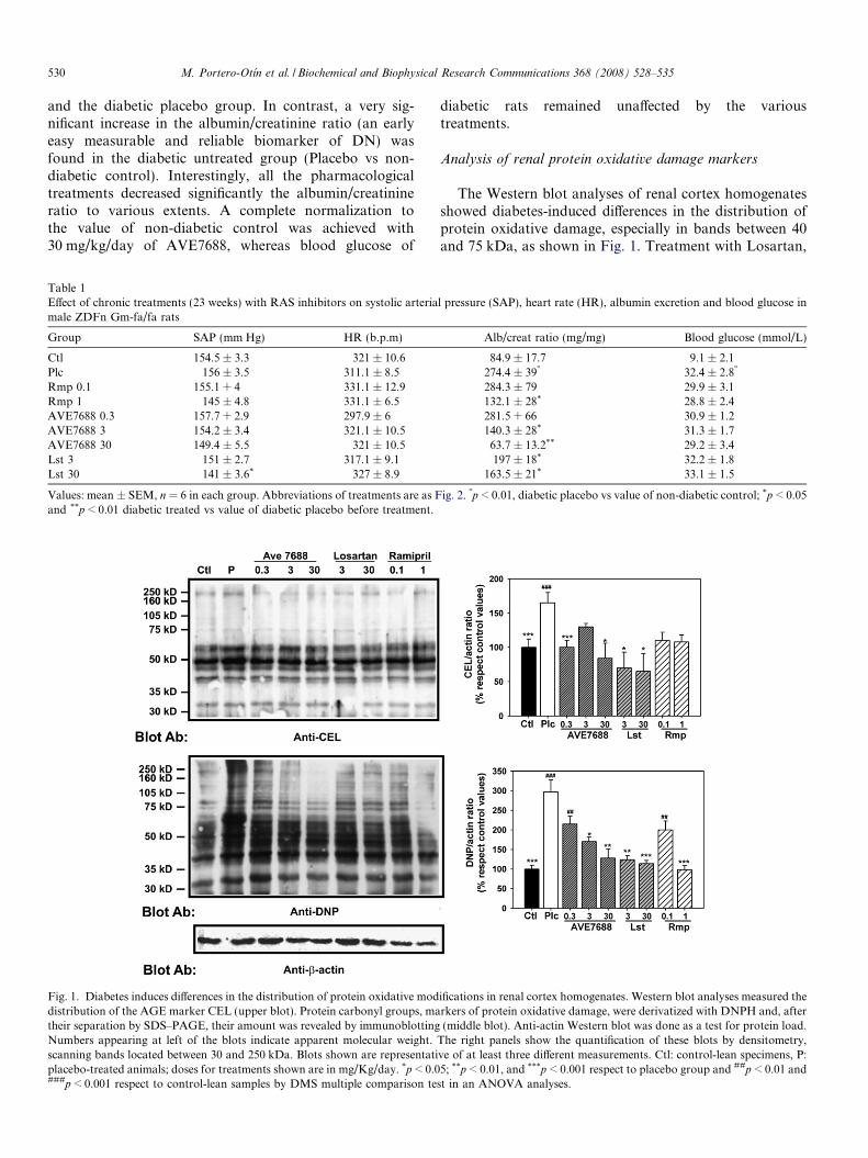
Fig. 1. Diabetes induces differences in the distribution of protein oxidative moddistribution of the AGE marker CEL (upper blot). Protein carbonyl groups, matheir separation by SDS–PAGE, their amount was revealed by immunoblottingNumbers appearing at left of the blots indicate apparent molecular weight.scanning bands located between 30 and 250 kDa. Blots shown are representatiplacebo-treated animals; doses for treatments shown are in mg/Kg/day. *p < 0.0###p < 0.001 respect to control-lean samples by DMS multiple comparison te
diabetic rats remained unaffected by the varioustreatments.
Analysis of renal protein oxidative damage markers
The Western blot analyses of renal cortex homogenatesshowed diabetes-induced differences in the distribution ofprotein oxidative damage, especially in bands between 40and 75 kDa, as shown in Fig. 1. Treatment with Losartan,
l pressure (SAP), heart rate (HR), albumin excretion and blood glucose in
Alb/creat ratio (mg/mg) Blood glucose (mmol/L)
84.9 ± 17.7 9.1 ± 2.1274.4 ± 39� 32.4 ± 2.8�
284.3 ± 79 29.9 ± 3.1132.1 ± 28* 28.8 ± 2.4281.5 + 66 30.9 ± 1.2140.3 ± 28* 31.3 ± 1.763.7 ± 13.2** 29.2 ± 3.4197 ± 18* 32.2 ± 1.8
163.5 ± 21* 33.1 ± 1.5
ig. 2. �p < 0.01, diabetic placebo vs value of non-diabetic control; *p < 0.05
ifications in renal cortex homogenates. Western blot analyses measured therkers of protein oxidative damage, were derivatized with DNPH and, after(middle blot). Anti-actin Western blot was done as a test for protein load.
The right panels show the quantification of these blots by densitometry,ve of at least three different measurements. Ctl: control-lean specimens, P:5; **p < 0.01, and ***p < 0.001 respect to placebo group and ##p < 0.01 and
st in an ANOVA analyses.

M. Portero-Otın et al. / Biochemical and Biophysical Research Communications 368 (2008) 528–535 531
Ramipril, and AVE7688 led to a dose-related decrease ofoxidative damage, as evidenced by densitometric analyses.To identify which DNP-reactive carbonyls were related toglycoxidation/lipoxidation, we performed Western blotwith an anti-CEL monoclonal antibody. CEL is a non-DNP-reactive carbonyl stress product resulting from mixedglycoxidation and lipoxidation reactions. These analyses,presented in Fig. 1, revealed only minor changes inducedby diabetes, suggesting that MCO is an important determi-nant for DNP-reactive increased carbonyl modifications inZDFn Gm-fa/fa rat kidneys. Therefore, to offer a moreaccurate quantitative measurement of protein oxidationin renal cortex proteins, we measured structurally definedMCO products by isotope-dilution GC/MS [19]. Proteinsfrom renal cortex samples contained oxidation productsresulting from MCO, glycoxidation and lipoxidation.Quantitative analyses revealed that the more abundant
Fig. 2. Proteins from placebo-treated diabetic rats (Plc) show significant increasthe concentrations of CML and CEL, arising from glycoxidation and lipoxidlipoxidation (lower graph). Values shown are % changes of mean ± SEM ovlysine; AASA: 278 ± 22 lmol/mol lysine; CEL: 211 ± 11 lmol/mol lysine; C*p < 0.05; **p < 0.01, and ***p < 0.001 respect to placebo group and #p < 0.0multiple comparison test in an ANOVA analyses. Values are also shown for AVshown in mg/Kg/day.
products were the amino acid derived oxidation productsderived from MCO, AASA, and GSA (almost 95% of mea-sured markers) (Fig. 2). GSA stood as the more MCOderived frequent modification (concentrations in the 30–35 mmol/mol lysine range), with levels being two ordersof magnitude higher than of those of AASA with concen-trations in the range between 250 and 290 lmol/mol lysine(Fig. 2). The mean concentrations of both GSA andAASA, presented in Fig. 2, were significantly higher(p < 0.01) in samples from diabetic rats than in controlage-matched lean, the change being more marked forGSA. Treatment with AVE7688 abolished completely theincreased GSA content, in a dose-related fashion, whereasonly higher doses of Ramipril were efficient in this sense.Regarding the AASA content, only the high dose of Losar-tan was able to maintain oxidative modification to controllevels.
es in the amounts of GSA and AASA, markers of MCO (upper graphs), ination (middle graphs), and in the concentration of MDAL, surrogate ofer values from control-lean (Ctl) samples (GSA: 33164 ± 1353 lmol/mol
ML: 589 ± 8 lmol/mol lysine; and MDAL: 127 ± 9 lmol/mol lysine).5; ##p < 0.01, and ###p < 0.001 respect to control-lean samples by DMSE7688, Losartan-treated (Lst), and Ramipril-treated (Rmp) animals, doses
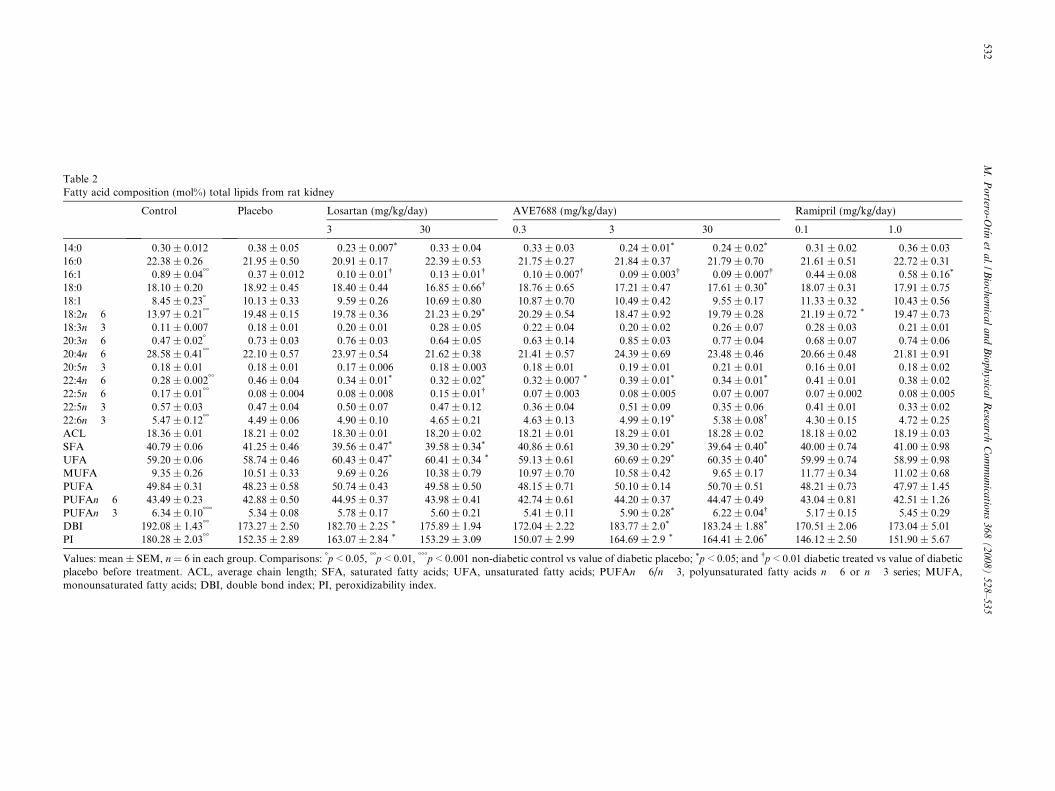
Table 2Fatty acid composition (mol%) total lipids from rat kidney
Control Placebo Losartan (mg/kg/day) AVE7688 (mg/kg/day) Ramipril (mg/kg/day)
3 30 0.3 3 30 0.1 1.0
14:0 0.30 ± 0.012 0.38 ± 0.05 0.23 ± 0.007* 0.33 ± 0.04 0.33 ± 0.03 0.24 ± 0.01* 0.24 ± 0.02* 0.31 ± 0.02 0.36 ± 0.0316:0 22.38 ± 0.26 21.95 ± 0.50 20.91 ± 0.17 22.39 ± 0.53 21.75 ± 0.27 21.84 ± 0.37 21.79 ± 0.70 21.61 ± 0.51 22.72 ± 0.3116:1 0.89 ± 0.04�� 0.37 ± 0.012 0.10 ± 0.01� 0.13 ± 0.01� 0.10 ± 0.007� 0.09 ± 0.003� 0.09 ± 0.007� 0.44 ± 0.08 0.58 ± 0.16*
18:0 18.10 ± 0.20 18.92 ± 0.45 18.40 ± 0.44 16.85 ± 0.66� 18.76 ± 0.65 17.21 ± 0.47 17.61 ± 0.30* 18.07 ± 0.31 17.91 ± 0.7518:1 8.45 ± 0.23� 10.13 ± 0.33 9.59 ± 0.26 10.69 ± 0.80 10.87 ± 0.70 10.49 ± 0.42 9.55 ± 0.17 11.33 ± 0.32 10.43 ± 0.5618:2n � 6 13.97 ± 0.21�� 19.48 ± 0.15 19.78 ± 0.36 21.23 ± 0.29* 20.29 ± 0.54 18.47 ± 0.92 19.79 ± 0.28 21.19 ± 0.72 * 19.47 ± 0.7318:3n � 3 0.11 ± 0.007 0.18 ± 0.01 0.20 ± 0.01 0.28 ± 0.05 0.22 ± 0.04 0.20 ± 0.02 0.26 ± 0.07 0.28 ± 0.03 0.21 ± 0.0120:3n � 6 0.47 ± 0.02� 0.73 ± 0.03 0.76 ± 0.03 0.64 ± 0.05 0.63 ± 0.14 0.85 ± 0.03 0.77 ± 0.04 0.68 ± 0.07 0.74 ± 0.0620:4n � 6 28.58 ± 0.41�� 22.10 ± 0.57 23.97 ± 0.54 21.62 ± 0.38 21.41 ± 0.57 24.39 ± 0.69 23.48 ± 0.46 20.66 ± 0.48 21.81 ± 0.9120:5n � 3 0.18 ± 0.01 0.18 ± 0.01 0.17 ± 0.006 0.18 ± 0.003 0.18 ± 0.01 0.19 ± 0.01 0.21 ± 0.01 0.16 ± 0.01 0.18 ± 0.0222:4n � 6 0.28 ± 0.002�� 0.46 ± 0.04 0.34 ± 0.01* 0.32 ± 0.02* 0.32 ± 0.007 * 0.39 ± 0.01* 0.34 ± 0.01* 0.41 ± 0.01 0.38 ± 0.0222:5n � 6 0.17 ± 0.01�� 0.08 ± 0.004 0.08 ± 0.008 0.15 ± 0.01� 0.07 ± 0.003 0.08 ± 0.005 0.07 ± 0.007 0.07 ± 0.002 0.08 ± 0.00522:5n � 3 0.57 ± 0.03 0.47 ± 0.04 0.50 ± 0.07 0.47 ± 0.12 0.36 ± 0.04 0.51 ± 0.09 0.35 ± 0.06 0.41 ± 0.01 0.33 ± 0.0222:6n � 3 5.47 ± 0.12�� 4.49 ± 0.06 4.90 ± 0.10 4.65 ± 0.21 4.63 ± 0.13 4.99 ± 0.19* 5.38 ± 0.08� 4.30 ± 0.15 4.72 ± 0.25ACL 18.36 ± 0.01 18.21 ± 0.02 18.30 ± 0.01 18.20 ± 0.02 18.21 ± 0.01 18.29 ± 0.01 18.28 ± 0.02 18.18 ± 0.02 18.19 ± 0.03SFA 40.79 ± 0.06 41.25 ± 0.46 39.56 ± 0.47* 39.58 ± 0.34* 40.86 ± 0.61 39.30 ± 0.29* 39.64 ± 0.40* 40.00 ± 0.74 41.00 ± 0.98UFA 59.20 ± 0.06 58.74 ± 0.46 60.43 ± 0.47* 60.41 ± 0.34 * 59.13 ± 0.61 60.69 ± 0.29* 60.35 ± 0.40* 59.99 ± 0.74 58.99 ± 0.98MUFA 9.35 ± 0.26 10.51 ± 0.33 9.69 ± 0.26 10.38 ± 0.79 10.97 ± 0.70 10.58 ± 0.42 9.65 ± 0.17 11.77 ± 0.34 11.02 ± 0.68PUFA 49.84 ± 0.31 48.23 ± 0.58 50.74 ± 0.43 49.58 ± 0.50 48.15 ± 0.71 50.10 ± 0.14 50.70 ± 0.51 48.21 ± 0.73 47.97 ± 1.45PUFAn � 6 43.49 ± 0.23 42.88 ± 0.50 44.95 ± 0.37 43.98 ± 0.41 42.74 ± 0.61 44.20 ± 0.37 44.47 ± 0.49 43.04 ± 0.81 42.51 ± 1.26PUFAn � 3 6.34 ± 0.10��� 5.34 ± 0.08 5.78 ± 0.17 5.60 ± 0.21 5.41 ± 0.11 5.90 ± 0.28* 6.22 ± 0.04� 5.17 ± 0.15 5.45 ± 0.29DBI 192.08 ± 1.43�� 173.27 ± 2.50 182.70 ± 2.25 * 175.89 ± 1.94 172.04 ± 2.22 183.77 ± 2.0* 183.24 ± 1.88* 170.51 ± 2.06 173.04 ± 5.01PI 180.28 ± 2.03�� 152.35 ± 2.89 163.07 ± 2.84 * 153.29 ± 3.09 150.07 ± 2.99 164.69 ± 2.9 * 164.41 ± 2.06* 146.12 ± 2.50 151.90 ± 5.67
Values: mean ± SEM, n = 6 in each group. Comparisons: �p < 0.05, ��p < 0.01, ���p < 0.001 non-diabetic control vs value of diabetic placebo; *p < 0.05; and �p < 0.01 diabetic treated vs value of diabeticplacebo before treatment. ACL, average chain length; SFA, saturated fatty acids; UFA, unsaturated fatty acids; PUFAn � 6/n � 3, polyunsaturated fatty acids n � 6 or n � 3 series; MUFA,monounsaturated fatty acids; DBI, double bond index; PI, peroxidizability index.
532M
.P
ortero
-Otın
eta
l./Bio
chem
ical
an
dB
iop
hy
sical
Resea
rchC
om
mu
nica
tion
s3
68
(2
00
8)
52
8–
53
5

M. Portero-Otın et al. / Biochemical and Biophysical Research Communications 368 (2008) 528–535 533
The mean concentrations of CEL and CML were alsosignificantly higher (p < 0.01) in diabetic rats than in con-trols, as shown in Fig. 2. Treatment with AVE7688 nor-malized CEL content, as Losartan, but not Ramipril. Alltreatments prevented the increase of CML induced by dia-betes (Fig. 2). The concentration of MDAL, a lipoxidationproduct, presented in Fig. 2, was also significantlyincreased in diabetic samples (p < 0.01). Notably,AVE7688 and Losartan treatments were able to normalizeincreased values of MDAL, but not Ramipril, which led tosignificant decreases with respect to placebo.
Since CML and CEL could arise from lipoxidation, anddue to the importance of MDAL as lipoxidation marker,we measured the fatty acid composition of renal cortex,to ascertain whether increased availability of peroxidizablesubstrate (i.e. increased amount of polyunsaturated fattyacids) could explain increases in carbonyl stress markers.In addition, indexes of fatty acid composition would alsoreflect increased lipoxidative consumption of polyunsatu-rated fatty acids. The analyses of fatty acids revealed signif-icant differences associated with diabetes in renal cortexsamples, both in individual fatty acids and in globalindexes (Table 2). Significant increases were noted for18:2n � 6, and decreased levels of 20:4n � 6 and22:5n � 6. The highly peroxidizable docosahexaenoic acid(DHA, 22:6n � 3) also showed a significant decrease. Withreference to the derived indexes, diabetes led to significantdecreases for DBI and PI indexes. In contrast with changesin protein oxidative modification, no major changesinduced by treatment were evident, with the exception ofsignificant normalization of PI and DBI indexes inducedby Losartan at lower doses and by AVE7688 in a dose-dependent fashion.
Discussion
The present work brings new information regardingthe pathways of MCO, AGE, and lipoxidation-derivedprotein modifications in the Zucker fatty rats andextends the knowledge of vasopeptidase inhibitor treat-ment on the accumulation of products derived from car-bonyl and oxidative stress. This is the first reportdemonstrating the presence of increased MCO in a dia-betic nephropathy model. GSA derives from the MCOof proline and arginine while AASA results from lysineoxidation [17]. These products are among the main car-bonyl products of MCO of proteins, thus represent spe-cific probes of oxidation of amino acids in protein.However, their presence and the factors affecting theirconcentrations in renal tissues were unknown up to date.Nevertheless, the chemical pathways linking increasedfree radical efflux and protein structural modificationalso involve third-party molecules, which may give riseto increased DNP-reactive carbonyls in proteins[16,17,19]. Particularly, carbohydrates, when reactingwith free radicals, generate highly reactive dicarbonylcompounds, such as glyoxal and methylglyoxal. In the
cellular context, these are mainly derived from glycolysisand triose phosphate metabolism, lipid peroxidation, andhypochlorite mediated reactions being also potentialsources. These compounds generate stable adducts react-ing with lysine, arginine, and cysteine residues in pro-teins. CEL and CML are two of these adducts, firstdescribed as AGE, later named glycoxidation productsand now recognized as mixed AGEs-advanced lipoxida-tion products. Despite CML has been thoroughlydetected in the DN lesions by immunohistochemicalanalyses [2], no chemical evidences have been reportedfor this product in renal tissues until very recently. Poly-unsaturated fatty acids are other third-party moleculesvery susceptible to the oxidative action of free radicals[20]. Free radical attack of those lipids generates specificreactive aldehydes, such as malondialdehyde or 4-hydroxynonenal, which in turn could react with proteins,generating also DNP-reactive moieties. However, therewere no chemical evidences for lipid peroxidative-induceddamage of proteins in DN. Using GC/MS, our resultsdemonstrate the presence of lipid peroxidation-derivedmarkers in renal tissues, and evidence increased modifica-tion in a type II diabetes mellitus model. Globally, thepresent work, evaluating the effects of different RASinhibitors (AVE7688, Losartan, and Ramipril) on therenal accumulation of different non-enzymatic proteinmodifications points out several differences reflecting thatmechanisms of action of these three compounds couldhave specific effects on each oxidative pathway. More-over, these data support the importance of the vasopep-tidase system (inhibited by AVE7688) in diabeticnephropathy.
Two previous works from our group described thebeneficial effect of AVE7688 on diabetic nephropathyin the diabetic and hypertensive models [6–8]. Wefound that AVE7688 ameliorates proteinuria withoutaffecting glycemia as sustained by steady level ofHb1Ac [6]. Reduction in proteinuria was also associ-ated with reduction in the severity of glomerular andtubulo-interstitial kidney damage, these effects beingpartly mediated by B2 kinin receptor activation [9].In addition, these nephroprotective effects, in the Zuc-ker fatty rat model, were not dependent of a bloodpressure lowering effect. This suggests that developmentof DN in this model may be dependent on peculiarmechanisms and especially on an exaggerated concen-tration of lipids resulting in an increased formationof AGEs and lipoxidation products.
Noteworthy, changes in fatty acid composition relatedto diabetes were not corrected by any treatment essayed,despite AVE7688 (and Losartan, in a lower extent)showed a trend to normalize both PI and DBI, mainlyby reversing the effects of diabetic status in the contentof PUFAn � 3. This suggests that RAS inhibition actson free radical generation [13], more than actuating ontissue susceptibility (i.e. fatty acid profile) to oxidativedamage. In this sense, AVE7688 also normalized the

534 M. Portero-Otın et al. / Biochemical and Biophysical Research Communications 368 (2008) 528–535
level of lipoxidation and MCO products namely GSAand MDAL, whereas it demonstrated a weaker effecton CEL, CML, and AASA. However, it is interestingto underline that AVE7688 is more efficient than thetwo other inhibitor on the products more sensible tohyperglycemia. With respect to the Zucker diabetic fattyrat model, this observation could be also of great inter-est, as the diabetes-induced increases in GSA andMDAL concentrations were much higher when com-pared to the three other measured products, whichshowed more moderate but significant increase. Thesefindings could reveal a major pathway—relying onMCO and lipoxidation—responsible for renal AGEsaccumulation.
When compared to the effects of the ACE inhibitorRamipril and the AT1 receptor antagonist Losartan,AVE7688 was more effective to reduce the concentrationof GSA and MDAL. This reduction has to be associ-ated with normalization of the albumin/creatinine ratio,only achieved with AVE7688 treatment. It has been pre-viously reported that both ACE inhibitors and AT1receptor blockers reduced AGEs formation[10,11,14,15]. However the relationship linking inhibitionof the RAS, AGE formation, and normalization of DNindex has not been extensively investigated. These previ-ous results demonstrate that AVE7688 has potent che-lating activity, and that by inhibiting metal-catalysedformation of AGE compounds, it decreases CML for-mation. The present results therefore suggest that inhibi-tion of vasopeptidases seems more efficient thanblockade of RAS alone to reduce renal AGEs formationin type II diabetes, an effect significantly associated withreduction of DN.
In summary, our work offers chemical evidences fordifferentially increased protein oxidative and carbonylstress in renal cortex of this model of DN, suggestingthe important role of lipid peroxidation and MCO. Nev-ertheless, RAS activity in diabetes underlies thesechanges, as different inhibitors, and specially IVP, areable to normalize the concentration and distribution ofoxidized proteins.
Acknowledgments
This study was supported in part by I+D grants fromthe Spanish Ministry of Science and Technology(HF2005-0082; BFU2006-14495/BFI, and AGL2006-12433), the Spanish Ministry of Health (P04-0355, P05-2241, and RD06/0013/0012), and the regional govern-ment of Catalonia (2005SGR00101) to R.P. andM.P.O. Marie Buleon is supported by a grant fromMRT. This work is also partly supported by funds fromINSERM to J.P.G and by a grant form the France/Spain collaborative program PICASSO and by theCOST B-35 Action ‘‘Lipid Peroxidation in Health andDisease”.
References
[1] P.A. McCullough, G.L. Bakris, W.F. Owen Jr., P.S. Klassen, R.M.Califf, Slowing the progression of diabetic nephropathy and itscardiovascular consequences, Am. Heart J. 148 (2004) 243–251.
[2] N. Tanji, G.S. Markowitz, C. Fu, T. Kislinger, A. Taguchi, M.Pischetsrieder, D. Stern, A.M. Schmidt, V.D. D’Agati, Expression ofadvanced glycation end products and their cellular receptor RAGE indiabetic nephropathy and nondiabetic renal disease, J. Am. Soc.Nephrol. 11 (2000) 1656–1666.
[3] M. Brownlee, Biochemistry and molecular cell biology of diabeticcomplications, Nature 414 (2001) 813–820.
[4] G. Jerums, S. Panagiotopoulos, J. Forbes, T. Osicka, M. Cooper,Evolving concepts in advanced glycation, diabetic nephropathy,and diabetic vascular disease, Arch. Biochem. Biophys. 419 (2003)55–62.
[5] H.B. Lee, M.R. Yu, Y. Yang, Z. Jiang, H. Ha, Reactive oxygenspecies-regulated signaling pathways in diabetic nephropathy, J. Am.Soc. Nephrol. 14 (2003) S241–S245.
[6] B.J. Davis, C.I. Johnston, L.M. Burrell, W.C. Burns, E. Kubota, Z.Cao, M.E. Cooper, T.J. Allen, Renoprotective effects of vasopepti-dase inhibition in an experimental model of diabetic nephropathy,Diabetologia 46 (2003) 961–971.
[7] S. Schafer, W. Linz, A. Bube, M. Gerl, J. Huber, G.U. Kurzel, M.Bleich, H.L. Schmidts, A.E. Busch, H. Rutten, Vasopeptidaseinhibition prevents nephropathy in Zucker diabetic fatty rats,Cardiovasc. Res. 60 (2003) 447–454.
[8] S. Schafer, W. Linz, H. Vollert, G. Biemer-Daub, H. Rutten, M.Bleich, A.E. Busch, The vasopeptidase inhibitor AVE7688 amelio-rates type 2 diabetic nephropathy, Diabetologia 47 (2004) 98–103.
[9] S. Schafer, H.L. Schmidts, M. Bleich, A.E. Busch, W. Linz,Nephroprotection in Zucker diabetic fatty rats by vasopeptidaseinhibition is partly bradykinin B2 receptor dependent, Br. J.Pharmacol. 143 (2004) 27–32.
[10] C. Wihler, S. Schafer, K. Schmid, E.K. Deemer, G. Munch, M.Bleich, A.E. Busch, T. Dingermann, V. Somoza, J.W. Baynes, J.Huber, Renal accumulation and clearance of advanced glycation end-products in type 2 diabetic nephropathy: effect of angiotensin-converting enzyme and vasopeptidase inhibition, Diabetologia 48(2005) 1645–1653.
[11] J. Allard, M. Buleon, E. Cellier, I.M. Renaud, C. Pecher, F.Praddaude, M. Conti, I. Tack, J.P. Girolami, ACE inhibitor reducesgrowth factor receptor expression and signaling but also albuminuriathrough B2-kinin glomerular receptor activation in diabetic rats, Am.J. Physiol. Renal Physiol. 293 (2007) F1083–F1092.
[12] R. Couture, J.P. Girolami, Putative roles of kinin receptors in thetherapeutic effects of angiotensin 1-converting enzyme inhibitors indiabetes mellitus, Eur. J. Pharmacol. 500 (2004) 467–485.
[13] G. Nickenig, D.G. Harrison, The AT(1)-type angiotensin receptor inoxidative stress and atherogenesis: part I: oxidative stress andatherogenesis, Circulation 105 (2002) 393–396.
[14] J.M. Forbes, M.E. Cooper, V. Thallas, W.C. Burns, M.C. Thomas,G.C. Brammar, F. Lee, S.L. Grant, L.A. Burrell, G. Jerums, T.M.Osicka, Reduction of the accumulation of advanced glycation endproducts by ACE inhibition in experimental diabetic nephropathy,Diabetes 51 (2002) 3274–3282.
[15] M. Nangaku, T. Miyata, T. Sada, M. Mizuno, R. Inagi, Y. Ueda, N.Ishikawa, H. Yuzawa, H. Koike, C. van Ypersele de Strihou, K.Kurokawa, Anti-hypertensive agents inhibit in vivo the formation ofadvanced glycation end products and improve renal damage in a type2 diabetic nephropathy rat model, J. Am. Soc. Nephrol. 14 (2003)1212–1222.
[16] G. Cao, R.G. Cutler, Protein oxidation and aging. I. Difficulties inmeasuring reactive protein carbonyls in tissues using 2,4-dinitro-phenylhydrazine, Arch. Biochem. Biophys. 320 (1995) 106–114.
[17] J.R. Requena, C.C. Chao, R.L. Levine, E.R. Stadtman ER, Glutamicand aminoadipic semialdehydes are the main carbonyl products of

M. Portero-Otın et al. / Biochemical and Biophysical Research Communications 368 (2008) 528–535 535
metal-catalyzed oxidation of proteins, Proc. Natl. Acad. Sci. USA 98(2001) 69–74.
[18] A.V. Cantero, M. Portero-Otin, V. Ayala, N. Auge, M. Sanson, M.Elbaz, J.C. Thiers, R. Pamplona, R. Salvayre, A. Negre-Salvayre,Methylglyoxal induces advanced glycation end product (AGEs)formation and dysfunction of PDGF receptor-beta: implications fordiabetic atherosclerosis, FASEB J. 21 (2007) 3096–3106.
[19] R. Pamplona, E. Dalfo, V. Ayala, M.J. Bellmunt, J. Prat, I. Ferrer,M. Portero-Otin, Proteins in human brain cortex are modified byoxidation, glycoxidation, and lipoxidation. Effects of Alzheimerdisease and identification of lipoxidation targets, J. Biol. Chem. 280(2005) 21522–21530.
[20] J.P. Cosgrove, D.F. Church, W.A. Pryor, The kinetics of the autox-idation of polyunsaturated fatty acids, Lipids 22 (1987) 299–304.