Localization and Distribution of Testicular Angiotensin I ... - MDPI
Upload
independentCategory
view
0download
0
Carcinogenesis vol.29 no.9 pp.1675–1684, 2008doi:10.1093/carcin/bgn171Advance Access publication July 16, 2008
REVIEW
The renin–angiotensin system and malignancy
Eleanor I.Ager�, Jaclyn Neo and Christopher Christophi
Department of Surgery, Austin Health, University of Melbourne, Heidelberg,Victoria 3084, Australia
�To whom correspondence should be addressed. Tel: þ61 3 9456 5734;Fax: þ61 3 9458 1650;Email: [email protected]
The renin–angiotensin system (RAS) is usually associated with itssystemic action on cardiovascular homoeostasis. However, recentstudies suggest that at a local tissue level, the RAS influencestumour growth. The potential of the RAS as a target for cancertreatment and the suggested underlying mechanisms of its para-crine effects are reviewed here. These include modulation ofangiogenesis, cellular proliferation, immune responses and extra-cellular matrix formation. Knowledge of the RAS has increaseddramatically in recent years with the discovery of new enzymes,peptides and feedback mechanisms. The local RAS appears toinfluence tumour growth and metastases and there is evidenceof tissue- and tumour-specific differences. Recent experimentalstudies provide strong evidence that drugs that inhibit the RAShave the potential to reduce cancer risk or retard tumour growthand metastases. Manipulation of the RAS may, therefore, providea safe and inexpensive anticancer strategy.
Introduction
Cancer is the leading cause of death worldwide (World HealthOrganization) (1). Therapeutic strategies usually involve a combi-nation of surgical ablation, radiotherapy and chemotherapy. Apartfrom conventional chemotherapy, targeting of the tumour vascula-ture by vascular disrupting agents or inhibitors of angiogenesis hasalso been used.
Recent evidence suggests an alternative pathway for targeted ther-apy in cancer. Several paracrine mechanisms existing at local tissuesites have been implicated in tumourigenesis. One such system is therenin–angiotensin system (RAS) that exists in several organs at a localtissue level. Epidemiological and experimental studies now suggestthat the RAS may contribute to the paracrine regulation of tumouri-genesis. Blockade of the RAS may, therefore, provide an alternative,adjunctive therapy for the treatment of solid tumours.
The RAS
Commonly, the RAS has been associated with the systemic regulationof cardiovascular homoeostasis. However, there is now increasingevidence that local RASs may influence tissue angiogenesis, cellularproliferation, apoptosis and inflammation (2). Components of theRAS are expressed in several adult organs including the liver, kidney,pancreas, brain and reproductive organs (3). It is the paracrine mech-anisms of locally expressed RASs, not its circulating counterpart, thatappear important for tumourigenesis.
A variety of physiological responses can be induced through acti-vation of the RAS, some of which can have antagonistic consequencesfor tumour growth. The physiological malleability of the RAS isachieved by alternative peptides and receptors. Angiotensin (ANG)
II is the main effector of the RAS (Figure 1). ANG II is an octapeptidecleaved from ANG I by the angiotensin I-converting enzyme (ACE).The majority of ANG II effects are mediated by the AT1 receptor(AT1R). The AT1R is expressed in many adult tissues, including bloodvessels, adrenal cortex, liver, kidney, and brain (4). A second receptorencoded by a different gene, the Angiotensin II type 2 receptor AT2R,is predominantly expressed during foetal life, but is present at a lowlevel in a few adult tissues such as the adrenal medulla, uterus andovarian follicles (5–7). Whereas the AT1R induces angiogenesis, cel-lular proliferation and inflammatory responses, as well as being anti-apoptotic (8,9), the AT2R appears to functionally antagonize many ofthese actions (4,6,10). There is some evidence, however, that signallingvia the AT2R can also be pro-angiogenic (11) and pro-inflammatory (12).
The Mas1 oncogene (MasR) represents a fifth RAS receptor andbinds the ANG-(1–7) peptide (13). ANG-(1–7) may be generated di-rectly from ANG II by the enzymatic activity of ACE2 or from ANGI, via ANG-(1–9), a pathway that utilizes both ACE2 and ACE (14,15).ACE2 is present in many tissues with high concentrations in the heart,kidney and gastrointestinal track (14). ACE2 expression is increasedin animal models of liver injury and in human cirrhosis and is asso-ciated with increasing plasma and tissue levels of ANG-(1–7) (16).
ANG-(1–7) appears to have an inhibitory influence on many of theevents induced by ANG II (15). ANG-(1–7) has depressor, vasodilator,apoptotic and anti-proliferative actions (17). ANG-(1–7) is suggested toinhibit angiogenesis (18,19), although further investigations are neededto confirm these effects in a wider range of pathological/physiologicalconditions. In contrast, ANG-(1–7) may also mimic some actions ofANG II. For example, ANG-(1–7) induces the release of prostanoids(17) and may increase proliferation of some cells, such as epidermalstem cells after injury (20) and haematopoietic progenitors in the bonemarrow of myelosuppressed mice (21).
The variety of physiological responses to the RAS reflects thealternative peptides and receptors and the different signalling path-ways they induce. The balance between these signalling events willinfluence the proliferative and angiogenic phenotype of cells that areeither directly or indirectly responsive to the RAS. Therefore, it can behypothesized that the balance between components of the RAS willcontribute to tumour growth, angiogenesis and metastatic potential(Figure 2).
Evidence for a RAS contribution to cancer development
Given the expression of local RASs in many tissues, it is perhaps notsurprising that many components of the RAS are also expressed inmalignant tissue. However, the RAS, in particular the AT1R, is oftenup-regulated during the progression from normal to malignant phe-notypes, indicating at the very least a correlation between the RASand tumour progression.
Components of the RAS are frequently differentially expressed invarious cancers including brain, lung, pancreatic, breast, prostate,colon, skin and cervical carcinomas in comparison with their corre-sponding non-malignant tissue (2). In particular, over-expression ofthe AT1R is common. Changes in the expression of RAS componentsappear to correlate with tumour grade (22,23). These changes, how-ever, are not consistent and vary for individual tumour types. Forexample, high levels of AT1R are found in breast hyperplasia butdecrease when breast cancer becomes invasive (24), while in ovariancarcinoma up-regulation of AT1R correlates with tumour invasiveness(25). These examples of AT1R expression in breast cancer suggestthat, while up-regulation of AT1R is common to abnormal breasttissue, whether this increase is associated with higher or lower gradesof tumour may depend on the expression of other components of theRAS.
Abbreviations: ACE, ANG I converting enzyme; ANG, Angiotensin; AT1R,Angiotensin II type 1 receptor; AT2R, Angiotensin II type 2 receptor; EMT,epithelial to mesenchymal transition; ET-1, endothelin-1; HSCs, hepatic stellatecells; MasR, mitochondrial assembly receptor; MCP, macrophage/monocytechemoattractant protein; PDGF, platelet-derived growth factor; RAS, renin-angiotensin system; VEGF, vascular endothelial growth factor.
� The Author 2008. Published by Oxford University Press. All rights reserved. For Permissions, please email: [email protected] 1675
by guest on January 13, 2016http://carcin.oxfordjournals.org/
Dow
nloaded from

Epidemiological studies provide further evidence that the RAS mayinfluence tumour progression. Drugs that target the RAS, in particularACE inhibitors and AT1R antagonists, are commonly used in thetreatment of hypertension. A retrospective cohort study based on5207 patients found that the incidence of fatal cancers was reducedin patients treated with ACE inhibitors for .3 years (26). A secondcohort study with nested case–control analysis found that captopril, anACE inhibitor, but not other classes of anti-hypertensive drugs, wasassociated with a lower risk of developing prostate cancer (27).A reduced risk of developing oesophageal (55%), pancreatic (48%)and colon cancer (47%) was observed in an assessment of 483 733veterans, 38% of which were taking ACE inhibitors (28). Other epi-demiological investigations, however, have failed to find a protective
effect of ACE inhibitors on the rates or development of some typesof cancers (29,30). The variable conclusions from these studies maybe due to differences in population profiles, the types of cancerexamined, the agents used and the dose and length of administrationof those agents. In an attempt to avoid these variables, otherepidemiological studies have utilized the ACE insertion (I)–deletion(D) polymorphism.
In humans, there are two ACE alleles; the I allele is associated withlower circulating and tissue levels of ACE compared with the D allele(31). Lower risks of breast cancer and a 50% reduced risk of advancedversus localized prostate cancer have been linked to the II genotype(32). Conversely, the DD genotype is associated with tumour pro-gression and lymph node metastases of gastric cancer (33). Most
Fig. 1. The RAS. Several enzymes catalyze the generation of angiotensinogen-derived peptides (thin black arrows). The most prominent of these, however, areACE (thick black) and ACE2 (thick grey). Similarly, there are several receptors, but most of the RAS effects are mediated by AT1R, AT2R and Mas receptor. Mas R,Mas receptor; BK1R and BK2R, bradykinin receptors; tPA, tissue plasminogen activator; CAGE, chymostatin-sensitive ANG II-generating enzyme. Thick whitearrows indicate receptor–ligand interactions and black/grey arrows indicate enzymatic conversion of RAS components. See References in text.
Fig. 2. The balancing effects of the RAS on tumourigenesis. The RAS can promote or inhibit angiogenesis and cellular proliferation, thus supporting or blockingtumour neovascularization, growth and metastasis.
E.I.Ager et al.
1676
by guest on January 13, 2016http://carcin.oxfordjournals.org/
Dow
nloaded from

recently, the Rotterdam Study, a population-based prospective cohortstudy with 6670 useful participants, found that the DD genotype hadan increased risk of breast cancer compared with the low-activity II/IDgenotypes, but no association was demonstrated for colorectal, lung orprostate cancer (34). Further complicating our understanding of thissystem, patients with the II/ID genotype on short term, high dosesof RAS blockers had an increased risk of progression of colorectalcancer.
The failure of epidemiological studies to conclusively show aneffect of either pharmacological RAS blockade or endogenous differ-ences in ACE level and activity with various cancers should not betaken a conclusive evidence that this system is not important fortumour growth, but rather that this is a complex system and that ifit is to be used in the treatment of cancer, we need a better under-standing of how it can regulate tumour growth and, importantly, howbest to manipulate the system to reduce tumour growth. This may in-volve a combination of RAS targeting agents, something that as yet tobe tested in in vivo experimental models, let alone in a clinical orepidemiological setting.
A range of RAS blockers (Table I) have, however, been used singlyto assess various cancer cell lines in rodent models of common humanprimary and metastatic carcinomas (Table II). These experiments pro-vide strong evidence for the possibility of RAS agents as anticancertreatments. In a murine model of hepatocellular carcinoma, the ACEinhibitors captopril and perindopril suppressed tumour growth (39).Perindopril has also been shown to decrease tumour growth and re-duce angiogenesis in head and neck squamous cell carcinoma (45). Incontrast, in renal cancer-bearing immunocompetent, but not immuno-compromised mice, captopril decreased survival and promoted immu-nogenic MethA sarcoma tumours (46). Similarly, patients on ACEinhibitors were found to have higher rates of kidney cancer (29).However, these were suggested to result from a correlation betweenhypertension and kidney cancer. Although, these results illustratepotentially important differences in the pathological/physiologicalrole of the local RAS in different host and neoplastic tissues, the vastmajority of experimental models have found a reduction in tumourgrowth following RAS blockade. Further, these models suggest pos-sible mechanisms by which these effects are achieved.
Candesartan, an AT1R antagonist, reduced tumour-related angio-genesis and the number of lung metastases in a murine Lewis lungcancer model (47) and, when explanted into nude mice, inhibited
tumour-associated angiogenesis (48). In a mouse model of colorectalcancer liver metastases, both captopril (an ACE inhibitor) and irbe-sartan (an AT1R antagonist) decreased tumour growth, the percentageof liver metastases and tumour-associated angiogenesis (43). A sig-nificant reduction in tumour growth and vascularization has also beenobserved in response to candesartan in mouse melanoma syngeneictumours (49) and in xenograft models of human prostate (50) andovarian cancer cells (25). These studies indicate that the RAS mayinfluence tumour neovascularization.
The RAS influences tumour angiogenesis
A major mechanism by which the RAS exerts its pro-tumour effectmay be through modulation of tumour angiogenesis, which is criticalfor tumour growth (51). ANG II stimulates the expression of severalpro-angiogenic agents and growth factors including vascular endo-thelial growth factor (VEGF) (9,44), angiopoietin 2 (45), basic fibro-blast growth factor (b-FGF) (46), and platelet-derived growth factor(PDGF) (47). RAS blockade is frequently associated with reducedexpression of the potent angiogenic factor VEGF (36,48–50). For ex-ample, a mouse xenograft model of human gastric cancer reducedtumour volume and a reduction in tumour-associated expression ofVEGF in candesartan-treated animals (51).
The pro-angiogenic effects of ANG II appear to be mediated by theAT1R. In models of ischemia-induced angiogenesis, ANG II pro-motes revascularization of damaged vessels by increasing VEGFand endothelial nitric oxide synthase levels via activation of theAT1R (57). In contrast, the AT2R appears to antagonise these actions.In a study using AT2R deficient mice, Silvestre et al. (2002)(53)confirmed that the ANG II-induced increases in VEGF and eNOSare regulated by the AT1R, since both responses were observed inAT2R gene-deleted mice. This study also illustrated that the AT2R cannegatively modulate ischaemia-induced angiogenesis by increasingapoptotic processes. The AT2R has also been shown to inhibit signalsfrom VEGFR2/Flk-1 and is suggested to reduce endothelial cell mi-gration and tube formation (59). However, high AT2R expression wasfound in intratumoural blood vessel of human pituitary adenomas (60)and blockade of the AT2R has been associated with inhibition ofangiogenesis (61), suggesting that the AT2R can also be pro-angio-genic. In contrast to ANG II, the ANG-(1–7) peptide appears to inhibitangiogenesis. ANG-(1–7) inhibited both angiogenesis and the pro-liferation of fibrovascular tissue in a murine sponge model of
Table I. Agents utilized to manipulate the RAS
Target inhibited or blocked Name Mechanism of action
ACE Enalapril (enalaprilate)a These drugs are competitive inhibitors of ACE.Some are active drugs, whereas others are admin-istered as prodrugs that are converted in vivo intotheir active metabolites. ACE inhibitors directlyblock the formation of ANG II (and also increasethe level of bradykinin). Captopril has a sulfhydrylgroup that can also inhibit matrixmetalloproteinases.
Captoprila
Lisinoprila
Ramiprila
Perindoprila
Benazeprila
Fosinoprila
Quinaprila
AT1R and AT2R Saralasin A peptide analogue capable of binding, and therebyblocking, both AT1R and AT2R.
AT1R Candesartan (TCV 116, CV 11974)a These drugs belong to a class of biphenylimidazoles.They can be competitive inhibitors (peptideanalogues) or insurmountable receptor antagonists(non-peptide). AT1R antagonists inhibit ANG IIreceptor binding and, therefore, prevent signaltransduction.
Losartan (DuP753)a
EXP3174 (active metabolite of losartan)Telmisartana
Irbesartana
L-158 809AT2R PD 123319 These drugs are tetrahydroimidazolepyridines.
AT2R antagonists inhibit ANG II receptor bindingand, therefore, prevent signal transduction.
PD 123177CGP 42114
AT4R Divalinal-ANG IV Peptide analogueMasR A-779 Peptide analogues
D-Pro7-ANG-(1–7)
aThere are several ACE inhibitors and AT1R antagonists that are currently in clinical use as anti-hypertensive agents.
The renin–angiotensin system and malignancy
1677
by guest on January 13, 2016http://carcin.oxfordjournals.org/
Dow
nloaded from

angiogenesis (18,19). Therefore, the balance between ANG-(1–7) andANG II as well as the AT1R and AT2R may be important in deter-mining if tumours gain an angiogenic phenotype.
These angiogenic effects of the RAS are also evident in severalmodels of malignancy. Ovarian cancer cells positive for AT1R secreteVEGF in response to ANG II stimulation (25) and AT1R antagonistsinhibit VEGF-induced affects on bovine retinal endothelial cells (62).Indicative of the angiogenic potential of ANG II, a reduction in tu-mour microvascular density is a common effect of ACE inhibitors(43). However, ACE inhibition has also been associated with pro-angiogenic outcomes in several models of vascular injury. In a clinicalstudy of congestive heart failure, ACE inhibition increased hepatocytegrowth factor (63), a potent growth and angiogenic factor (64), and ina mouse model of ischaemic injury, vessel density and capillary num-ber increased when treated with an ACE inhibitor (65).
While it is clear that the RAS can mediate angiogenic processes, thepro-angiogenic responses to ACE inhibitors appear to be, at least inpart, associated with the inhibition of ACE-mediated bradykinin deg-radation and the ensuing increased bradykinin levels (65–68). Indeed,many of the cardiovascular benefits resulting from treatment withACE inhibitors are now suggested to arise from the actions of theseinhibitors on blocking the production of ANG II in conjunction withthe increased activity of bradykinin (66). Given the potential pro-angiogenic responses of ACE inhibitors, AT1R blockade may providea more suitable option for the treatment of cancers. However, pro-angiogeneic responses to ACE inhibition have not been reported inexperimental models of cancer or in epidemiological investigations ofthe association between RAS blockade and tumour development.Moreover, it is unclear whether normalization of tumour vessels isin part responsible for the antitumour effects of ACE inhibitors, AT1Rblockers and other classes of anti-angiogenic agents (69).
The vasoactive properties of ANG II and other vasoactive peptidehormones such as endothelin (ET)-1 could also potentially be used toincrease blood flow to tumours (70–72). Increasing tumour blood flowwould presumably provide a mechanism to increase the efficacy ofradiotherapy as well as improving chemotherapeutic drug delivery.Lower expression of AT1Rs in many tumours compared with non-neoplastic tissue is suggested to result in a comparative hyporespon-siveness of tumours to ANG II and may provide an explanation for theobserved specific increase in tumour blood flow following ANG IIinfusion (73,74). The systemic delivery of ET-1 has also been shownto selectively increase tumour vasodilation in a rat model of breastcancer (75). However, others have failed to find a significant effecton tumour blood flow after treatment with ET-1 (71). Also, whereasANG II infusions have been shown to increase drug delivery to smalltumours (76–79), in larger tumours ANG II infusion did not altertumour blood flow (80). Alterations in tumour responsiveness toANG II may reflect changes in the expression of AT1R and AT2Ras tumours grow and/or gain a more aggressive phenotype. Moreover,it is unclear what overall effect ANG II infusion may have on tumourgrowth as the mitogenic and pro-angiogenic effects of increased ANG
II may counteract its potential benefit in increasing drug delivery.Bouzin et al. (73) and Sonveaux (81) present excellent reviews onthe potential of pro-vascular approaches in the treatment of cancer.
ANG II stimulates the synthesis of several vascular permeabilityfactors including prostaglandins, nitric oxide, nuclear factor-jß (NF-jß),VEGF, and endothelin. Interestingly, there are many similarities be-tween the RAS and the ET-1 system in addition to their vasoactiveproperties. Given the similarities between the RAS and the ET-1system, it is perhaps not surprising that the ET-1 system is also nowgaining interest as a potential target for anticancer treatments. LikeANG II, ET-1 effects are mediated via two subtypes of G-protein-coupled receptors (ETA and ETB) and ET-1 is generated by the actionof ET-1-converting enzyme, a metalloprotease belonging to the samefamily as ACE. ET-1, similar to ANG II, can stimulate hypertrophy,proliferation and pro-inflammatory responses in several pathological/physiological conditions (82,83). Although the interactions betweenANG II and ET-1 are incompletely understood, recent studies indicatethat ANG II can stimulate ET-1 generation in endothelial cells andincrease expression of ETB receptor in hepatic stellate cells (HSCs)(84,85). ET-1, acting via its ETB receptor, increases endothelial cellproliferation, migration and capillary-like tube formation in vitro andin vivo models of angiogenesis suggest that ET-1 augments the pro-angiogenic effects of VEGF (86). However, in a rat ischaemia-induced angiogenesis model, ET-1 infusion was not pro-angiogenic(87). In contrast, blockade of both ETA and ETB receptors by bosentanmarkedly increased vessel density, an effect apparently mediated byan increase in VEGF and endothelial nitric oxide synthase levels.These results suggest that, as with the RAS, there may be counter-regulatory mechanisms within the ET-1 system and that the balance ofthese determines the physiological outcomes.
Several studies have investigated the potential of inhibiting theET-1 system to reduce tumour growth and metastasis. Both AT1Rand ETA receptor are expressed in many human cancer cell linesand tumours and autocrine activation of these receptors is associatedwith increased tumour growth (25,75,83,88). Moreover, inhibition ofETA receptor was found to inhibit ovarian tumour growth in vivo andwas associated with a suppression of epithelial-to-mesenchymal tran-sition (EMT) (75). Parallels between the RAS and the ET-1 systemraise the possibility of potentially synergistic effects resulting fromthe therapeutic targeting of both systems simultaneously.
Of the varied angiogenic factors stimulated by ANG II, VEGF/VEGF-A is particularly important because of its potency and selec-tivity for vascular endothelial cells (89). VEGF is also over-expressedin many malignant carcinomas (90–93). The initial response to VEGFappears similar in many tissues (94). Microvascular permeability in-creases, extravascular fibrin is deposited and the extracellular matrixdegrades. Endothelial cells then migrate into surrounding tissuestroma, forming enlarged, thin-walled, pericyte-poor vessels, termedmother vessels. After these initial events, angiogenesis can proceeddifferently in different tissues with mother vessels differentiating intosmaller daughter vessels, disorganized tangle of vessels (glomeruloid
Table II. RAS blockade in vivo
Cell line/model Agent Tumour volume Metastases Reference
Lewis lung carcinoma 3LL Captopril Decreased Decreased Kowalski et al. (35)Rat fibrosarcoma Captropril Decreased N/A Volpert et al. (36)Renal carcinoma SN12K-1 Captopril Decreased N/A Hii et al. (37)Lewis lung carcinoma 3LL Captoril (alone and combined with batimastat) Decreased Decreased Prontera et al. (38)Murine hepatocellular carcinoma Captopril, perindopril and temocapril Decreased N/A Yoshiji et al. (39,40)Lung metastases of renal carcinoma Candesartan Decreased Decreased Miyajima et al. (41)Ovarian carcinoma SKOV-3 Candesartan Decreased N/A Suganuma et al. (25)Bladder cancer KU-19-19 Candesartan Decreased N/A Kosugi et al. (42)Mouse colorectal cancer liver metastases Captopril Decreased Decreased Neo et al. (43)MKN-28 human gastric cancer mouse xenograft Candesartan Decreased N/A Huang et al. (44)
N/A, not assessed. Several cancer cell lines have been used in xenograft or allograft animal models of metastases. These experiments have shown decreased tumourvolume and when assessed decreased metastases.
E.I.Ager et al.
1678
by guest on January 13, 2016http://carcin.oxfordjournals.org/
Dow
nloaded from
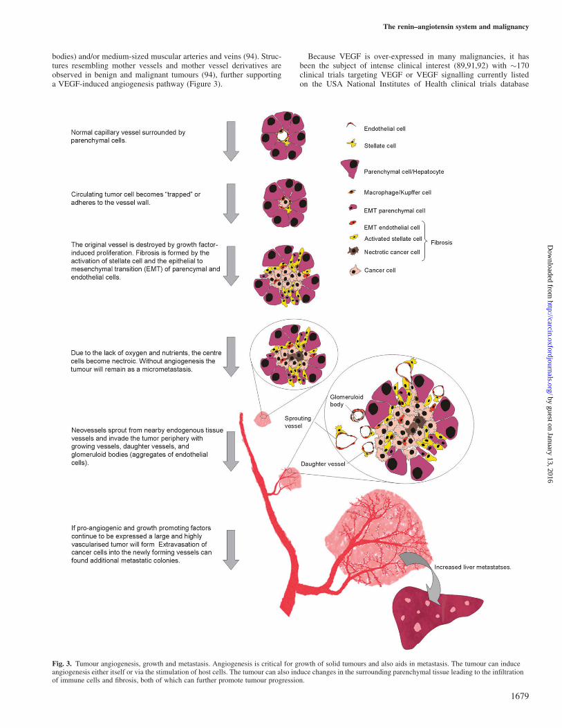
bodies) and/or medium-sized muscular arteries and veins (94). Struc-tures resembling mother vessels and mother vessel derivatives areobserved in benign and malignant tumours (94), further supportinga VEGF-induced angiogenesis pathway (Figure 3).
Because VEGF is over-expressed in many malignancies, it hasbeen the subject of intense clinical interest (89,91,92) with �170clinical trials targeting VEGF or VEGF signalling currently listedon the USA National Institutes of Health clinical trials database
Fig. 3. Tumour angiogenesis, growth and metastasis. Angiogenesis is critical for growth of solid tumours and also aids in metastasis. The tumour can induceangiogenesis either itself or via the stimulation of host cells. The tumour can also induce changes in the surrounding parenchymal tissue leading to the infiltrationof immune cells and fibrosis, both of which can further promote tumour progression.
The renin–angiotensin system and malignancy
1679
by guest on January 13, 2016http://carcin.oxfordjournals.org/
Dow
nloaded from

(ClinicalTrials.gov; http://clinicaltrials.gov/). Several anti-VEGFtherapies currently in clinical trials are listed in Table III. However,these anti-angiogenic agents are not without side effects. Bevacizumabhas been associated with proteinuria, bleeding and wound-healingcomplications, gastrointestinal perforation, thromboembolic eventsand, most commonly, hypertension (97). Indeed, for all anti-VEGFclinical trials listed on National Institutes of Health ClinicalTrials.gov, where data were available, hypertension was a listed side effect.Treatments with anti-hypertensive agents, including ACE inhibitors,are frequently described in clinical trials of anti-VEGF therapies, butwith no reference to the potential of these treatments to also influencetumourigenesis (98,99). Also, for tumours that express additionalangiogenic or proliferative factors, anti-VEGF therapies alone maynot provide an optimal strategy. Targeting multiple aspects of angio-genesis, including the VEGF pathway, may provide a more effectivetreatment. The RAS also contributes to cellular proliferation andtumour-associated fibrosis and blockade of these systems may provideadditional benefits beyond those predicted for anti-VEGF strategies.
Proposed effects of the RAS on cellular proliferation
The RAS can also effect the cell survival and or proliferation and may,therefore, have a direct effect on the number of live cancer cells withintumours. ANG II can stimulate or inhibit proliferation depending onwhether the AT1R or AT2R is activated. It is also now becomingevident that ANG-(1–7) also has a role in defining the proliferativepotential of some cells. ANG II is a mitogen for smooth muscle cells,fibroblasts and endothelial cells (6) and increases the expression ofgrowth-related oncogenes (100–102) and growth factors (2) in severalcell types. However, ANG II stimulation of the AT1R may increasesenescence of bone marrow-derived endothelial progenitor cells(103), which are important for tumour angiogenesis (104). Theseresults suggest that the effect of ANG II on proliferation may differfor different cell types, possibly due to the alternative physiologicalpathways that can be initiated by the RAS. Although AT2R is com-monly thought to mediate the anti-proliferative effects of ANG II(105), this may not always be the case. For example, in a normotensive
rat model, infusion of ANG II in conjunction with AT1R blockadeinduced aortic hypertrophy (106). In contrast, the number and size ofaortic smooth muscle cells remained normal in rats infused with ANGII in the presence of PD123319 (an AT2R-specific antagonist), sug-gesting that at least part of the vasotropic effects of ANG II weremediated by the AT2R.
ANG-(1–7) is generally thought to inhibit cellular proliferation(107). However, ANG-(1–7) also appears to increase proliferationof some cell types including fibroblasts, epidermal stem cells, kerati-nocytes and haematopoietic progenitor cells (20,21). ANG-(1–7)clearly has a complex role in regulating cellular proliferation. There-fore, whether ANG-(1–7) is pro- or anti-proliferative for a particulartumour/host cell may be an important consideration for the applica-bility of RAS blockade as a cancer treatment. However, at least forhuman lung cancer cells, it has been shown that ANG-(1–7) up-regulation can inhibit proliferation (108).
Immunomodulatory effects of the RAS
Tumour-infiltrating immune cells indicate an immune response by thehost to the developing tumour. ANG II can stimulate the release ofmacrophage/monocyte chemoattractant protein (MCP)-1, MCP-2 andgonocyte colony-stimulating factor, thus increasing macrophage in-filtration (42,49,109). In low-density lipoprotein receptor-deficientmice, ANG II infusion was found to promote macrophage infiltration,whereas treatment with valsartan, an AT1R inhibitor, reduced macro-phage accumulation and atherosclerosis (110). Similarly, in a rabbitaortic balloon injury model, AT1 blockade attenuated atherosclerosisin association with a reduction in plaque distribution and macrophageaccumulation (111). Surprisingly, however, in a rat model of nephritis,ANG II infusion induced a significant reduction in glomerular mono-cyte infiltration (112). Therefore, the overall effect of the RAS onmacrophage infiltration may be different in different tissues depend-ing on other immunomodulatory factors and the relative expression ofvarious RAS components.
AT1R is highly expressed in tumour-associated macrophages andmice mutant for AT1R have fewer infiltrating macrophages following
Table III. Inhibition of angiogenesis by blockade of the VEGF pathway
Drug Cancer Treatment Outcome
Bevacizumab (Avastin)—amonoclonal antibody that bindsand inhibits VEGF
Metastatic renal cell cancer Monotherapy Benefit in progression-free survival,but not in overall survival
Colorectal cancer In combination with chemotherapy(FOLFOX4) as a second-linetherapy
Benefit in progression-free survivaland overall survival
Non-small cell lung cancer In combination with chemotherapy(paclitaxel þ carboplatin) as afirst-line treatment
Benefit in progression-free andoverall survival
Metastatic breast cancer In combination with paclitaxel ina first-line treatment
Benefit in progression-free survival
Vatalanib—inhibits VEGF andplatelet-derived growth factorreceptors
Metastatic colorectal cancer In combination with chemotherapy(FOLFOX4) as a second-linetherapy
Benefit in progression-free survival,but not overall survival
VEGF-Trap (aflibercept)—a solubleVEGF receptor that binds andsequesters circulating VEGF
Non-small cell lung cancer Second-line monotherapy or incombination
In phase I trials, there was aradiographic improvement
Acute myeloid leukaemia Monotherapy Phase II trials are underwaySunitinib (Sutent)—binds to PTK
receptors, blocking signaltransduction. Includes VEGF andPCGF receptors and c-KIT andFlt-3 receptors
Cytokine-refractory metastaticrenal cell cancer
Monotherapy High rate of objective tumourresponses
Sorafenib (Nexavar)—inhibits RAFkinase (part of the RAS oncogenepathway) and also inhibitsplatelet-derived growth factor andVEGF receptors
Metastatic renal cell cancer Second-line monotherapy Improvement in progression-freesurvival, but low partial tumourresponses
PTK, protein tyrosine kinase receptors; RAF, product of v-raf-1 murine leukemia viral oncogene homolog 1; PCGF, Platelet derived growth factor.There are many phase II or phase III clinical trials currently underway. Examples of representative drugs, their mechanism of action and the cancers targeted areprovided [data cited in Kerbel (95), Cao (96), Los et al. (97) and table modified from Kerbel (95)].
E.I.Ager et al.
1680
by guest on January 13, 2016http://carcin.oxfordjournals.org/
Dow
nloaded from
tumour induction (49). Blockade of AT1R inhibits ANG II-stimulatedincreases in MCP-2. In contrast, AT2R antagonists enhanced ANGII-induced up-regulation of MCP-2 (109). Therefore, whereas AT1Rstimulation increases MCP-2 expression and presumably macrophageinfiltration, AT2R stimulation opposes this effect.
Classical macrophage activation is thought to eliminate microor-ganisms and kill tumour cells (113–116). However, the role of macro-phages in late-stage tumour development is less clearly defined. Thereis some evidence that tumour-infiltrating macrophages can promotegrowth and metastasis (117,118). The alternatively activated or M2macrophage pathway, which normally participates in debris salvagingand wound healing, is suggested to be associated with these pro-tumour roles. During the later stages of tumour metastases, hostdefences may no longer be capable of eliminating metastatic tumourcells due to rapid proliferation of cancer cells, angiogenesis and/orweakened host defences. For example, binding of circulating tumourcells by Kupffer cells, resident macrophages in the liver, is associatedwith promoting immune escape and facilitating the formation ofmetastatic colonies once the initial induction phase has past (113).Tumour-associated macrophages also release many cytokines that caninduce angiogenesis and these may facilitate tumour growth and me-tastases (49,118–120). In a B16-F1 melanoma model, sites of AT1Rexpression co-localized with VEGF protein and it is possible thatANG II may promote stimulation of macrophage infiltration and mac-rophage-mediated angiogenesis (49).
Macrophages themselves also release ACE and can participate inANG II synthesis. When macrophages were engineered to producehigh levels of ACE, by driving ACE expression from the macrophage-specific c-fms promoter, mice became resistant to melanoma growth(116). ACE inhibitors were capable of reversing this phenotype, butinhibition of ANG II or AT1R did not, suggesting that these effectswere not dependent on ACE–ANG II interactions. Instead, it wasfound that mice over-expressing ACE in their macrophages respondedto tumour challenge with an enhanced tumour-specific CD8þ T-cellresponse (121). Further, it was suggested that the increased peptidaseactivity of ACE by these macrophages may enhance the presentationof major histocompatibility class I-associated peptides to T cells.
Effects of the RAS on tumour-induced fibrosis
Fibro-proliferative diseases such as pulmonary fibrosis, liver cirrho-sis, cardiovascular disease, progressive kidney disease and maculardegeneration are typified by increased extracellular matrix formationand the differentiation of myofibroblasts. However, the process offibrosis is also closely associated with carcinogenesis (122,123).
The RAS, in particular activation of the AT1R by excess ANG II, isassociated with a number of renal and cardiac fibro-proliferative dis-eases (124–128). It is perhaps not surprising, therefore, that numerousexperimental and clinical investigations have demonstrated a benefitof ACE inhibitors and AT1R blockers in renal and cardiac disease.While some of the positive effects of ACE inhibitors and AT1R block-ers can be explained by the reduction in systemic blood pressure, thereis also substantial evidence for a direct anti-fibrotic effect.
A non-hypertensive mouse model of renal fibrosis, which allowsthe effects of RAS blockade on fibrosis to be assessed irrespective ofblood pressure involvement by the RAS, found that both ramipril (anACE inhibitor) and candesartan (an AT1R blocker) delayed the onsetand reduced the extent of proteinuria, postponed the onset of uraemiaand prolonged life (129). However, ramipril treatment was associatedwith a greater increase in life span and resulted in a more pronouncedanti-fibrotic effect. A mouse model of cyclosporine A-induced kidneydamage implicated the inhibition of bradykinin degradation by ACEinhibitors in its anti-fibrotic effects. Benazepril appeared to facilitatematrix degradation via activation of the bradykinin B2 receptor ontubular epithelial cells (130). Activation of the B2 receptor was asso-ciated with decreased plasminogen activator inhibitor-1 expression,which was suggested to increase production of plasmin and activationof matrix metalloproteinases. Indeed, B2 receptor activation has beendemonstrated to reduce renal fibrosis in unilateral ureteral obstruction
models by increasing extracellular matrix degradation through theactivation of plasminogen activator (124).
In the liver, HSCs are recognized as essential for fibrogenesis andthe RAS appears, at least in part, to mediate their fibrogenic role.Myofibroblasts can arise from activated HSCs, hepatocytes, bonemarrow-derived cells and possibly endothelial cells. Activated stellatecells secrete ANG II that can promote fibrosis via reduced nicotin-amide adenine dinucleotide phosphate oxidase, myofibroblast prolif-eration and differentiation and increased collagen synthesis (132).Also, the number of a-smooth muscle actin-positive cells (a markerof activated HSCs) was noticeably suppressed by candesartan andperindopril. Interestingly, ANG II activates NF-jB through bothAT1 and AT2Rs, in turn leading to increased expression of pro-inflammatory cytokines such as IL-6, TNFa, TGF-ß1 and intercellularadhesion molecule (12,126,127).
Fibrotic processes such as increased intratumoural collagen I orEMT of host and/or tumour cells are associated with increased tumourinvasiveness (135,136). EMT refers to a series of cellular and struc-tural changes, which allow cells to separate, lose apico-basal polarityand gain changeable cell adhesions, all of which potentially facilitatecell motility. In addition, the extracellular matrix can be induced toundergo changes that permit cell movement and the expression ofseveral growth and angiogenic factors, such as VEGF, can be inducedin response to EMT (137). A key inducer of EMT is TGF-ß1 whichhas tumour suppressor and oncogenic activities. In colorectal cancer,TGF-b1 changes from an inhibitor of proliferation to a stimulator ofgrowth and invasion at late stages of tumour progression (137). ANGII can increase TGF-b1 expression amongst other cytokines and this isassociated with an accumulation of fibrotic matrix proteins (119,138–141). ANG II also increases a-smooth muscle actin and decreases inE-cadherin, both of which regulate EMT (142,143).
Both ANG II and ANG-(1–7) influence the formation of fibrosisfollowing hepatic bile duct ligation (BDL) and other forms of tissueinjury (15,16,144). However, whereas plasma ANG II levels increasein the first week after BDL and then return to normal, ANG-(1–7)levels increase 3 weeks after BDL, but are normal prior to this point(15). ACE and AT1R are also up-regulated following BDL and local-ize to areas of active fibrogenesis (144). Changes in the RAS mayreflect a balance whereby up-regulation of hepatic ACE2 and ANG-(1–7) may provide a counter-regulatory response to ANG-II-mediatedacute responses. Therefore, it is possible that by manipulating theRAS tumour-induced fibrosis and EMT may also be reduced. Because
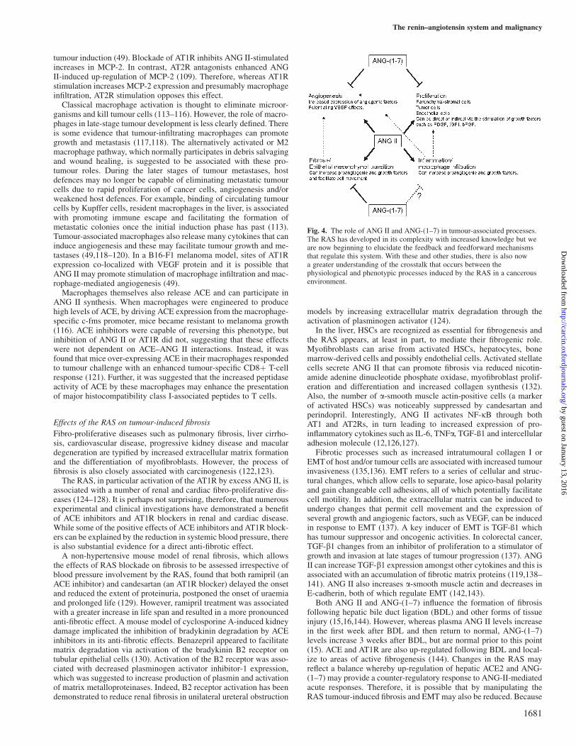
Fig. 4. The role of ANG II and ANG-(1–7) in tumour-associated processes.The RAS has developed in its complexity with increased knowledge but weare now beginning to elucidate the feedback and feedforward mechanismsthat regulate this system. With these and other studies, there is also nowa greater understanding of the crosstalk that occurs between thephysiological and phenotypic processes induced by the RAS in a cancerousenvironment.
The renin–angiotensin system and malignancy
1681
by guest on January 13, 2016http://carcin.oxfordjournals.org/
Dow
nloaded from
tumour-induced fibrosis can produce growth and angiogenic factors,its inhibition may initiate positive feedback mechanisms that furtherimpede tumour growth and metastases. However, in some instances,reduced fibrotic responses have been linked to more aggressive cancerdevelopment (145).
Conclusion
There is accumulating evidence that ACE inhibitors and AT1R antag-onists reduce tumour growth and metastatic potential. Our under-standing of the RAS in normal and pathological conditions,including cancer, has increased markedly over recent years, but hasalso demonstrated a complexity not initially evident. Newly discov-ered components of the RAS, such as the enzyme ACE2 and theANG-(1–7) peptide, may provide novel targets for the treatment ofcancer.
Not only does the RAS influence several important physiologicalprocesses such as angiogenesis, cellular proliferation, inflammationand fibrosis but also there is crosstalk between these processes thatwill further determine tumour potential (Figure 4). This complexityhighlights the need for greater knowledge of the role of the RAS inspecific tissues and tumour types. Nevertheless, anti-hypertensiveagents based on ACE inhibition and AT1R antagonism are already inclinical use without serious side effects and if these drugs can inhibittumour progression at comparable doses, then they may provide a usefuladjunctive therapeutic strategy in the treatment of cancer.
Funding
This work was funded by a Cancer Council of Victoria Grant.
Acknowledgements
Conflict of Interest Statement: None declared.
References
1.Mathers,C. et al. (2000) Global Burden of Cancer in the Year 2000:Version 1 Estimates. WHO, Oxford University Press, New York, NY.
2.Deshayes,F. et al. (2005) Angiotensin receptors: a new role in cancer?Trends Endocrinol. Metab., 16, 293–299.
3.Hunyady,L. et al. (2006) Pleiotropic AT1 receptor signaling pathwaysmediating physiological and pathogenic actions of angiotensin II. Mol.Endocrinol., 20, 953–970.
4.Smith,G.R. et al. (2004) Cancer, inflammation and the AT1 and AT2receptors. J. Inflamm. (Lond.), 1, 3.
5.Messerli,F.H. et al. (1996) Angiotensin II receptor inhibition. A newtherapeutic principle. Arch. Intern. Med., 156, 1957–1965.
6.Touyz,R.M. et al. (2000) Signal transduction mechanisms mediating thephysiological and pathophysiological actions of angiotensin II in vascularsmooth muscle cells. Pharmacol. Rev., 52, 639–672.
7.Velasquez,M.T. (1996) Angiotensin II receptor blockers. A new class ofantihypertensive drugs. Arch. Fam. Med., 5, 351–356.
8.Leung,P.S. et al. (2003) Expression and localization of AT1 receptors inhepatic Kupffer cells: its potential role in regulating a fibrogenic response.Regul. Pept., 116, 61–69.
9.Pupilli,C. et al. (1999) Angiotensin II stimulates the synthesis and secre-tion of vascular permeability factor/vascular endothelial growth factor inhuman mesangial cells. J. Am. Soc. Nephrol., 10, 245–255.
10.Tamarat,R. et al. (2002) Angiotensin II angiogenic effect in vivo involvesvascular endothelial growth factor- and inflammation-related pathways.Lab. Invest., 82, 747–756.
11.Sarlos,S. et al. (2003) Retinal angiogenesis is mediated by an interactionbetween the angiotensin type 2 receptor, VEGF, and angiopoietin. Am. J.Pathol., 163, 879–887.
12.Wolf,G. et al. (2002) Angiotensin II activates nuclear transcription factor-kappaB through AT1 and AT2 receptors. Kidney Int., 61, 1986–1995.
13.Santos,R.A. et al. (2003) Angiotensin-(1-7) is an endogenous ligand forthe G protein-coupled receptor Mas. Proc. Natl Acad. Sci. USA, 100,8258–8263.
14.Donoghue,M. et al. (2000) A novel angiotensin-converting enzyme-related carboxypeptidase (ACE2) converts angiotensin I to angiotensin1-9. Circ. Res., 87, E1–E9.
15.Herath,C.B. et al. (2007) Upregulation of hepatic angiotensin-convertingenzyme 2 (ACE2) and angiotensin-(1-7) levels in experimental biliaryfibrosis. J. Hepatol., 47, 387–395.
16.Paizis,G. et al. (2005) Chornic liver injury in rats and humans upregulatesthe novel enzyme angiotensin converting enzyme 2. Gut, 54, 1790–1796.
17.Ferrario,C.M. et al. (1997) Counterregulatory actions of angiotensin-(1-7).Hypertension, 30, 535–541.
18.Machado,R.D. et al. (2000) Opposing actions of angiotensins on angio-genesis. Life Sci., 66, 67–76.
19.Machado,R.D. et al. (2001) Mechanisms of angiotensin-(1-7)-inducedinhibition of angiogenesis. Am. J. Physiol. Regul. Integr. Comp. Physiol.,280, R994–R1000.
20.Rodgers,K. et al. (2001) Development of angiotensin (1-7) as an agent toaccelerate dermal repair. Wound Repair Regen., 9, 238–247.
21.Ellefson,D.D. et al. (2004) Synergistic effects of co-administration ofangiotensin 1-7 and Neupogen on hematopoietic recovery in mice. CancerChemother. Pharmacol., 53, 15–24.
22.Louis,S.N. et al. (2007) Appearance of angiotensin II expression in non-basal epithelial cells is an early feature of malignant change in humanprostate. Cancer Detect. Prev., 31, 391–395.
23.Sitzmann,J.V. et al. (1994) Altered angiotensin-II receptors in humanhepatocellular and hepatic metastatic colon cancers. Ann. Surg., 219,500–506; discussion 506–507.
24.De Paepe,B. et al. (2002) Increased angiotensin II type-2 receptor densityin hyperplasia, DCIS and invasive carcinoma of the breast is paralleledwith increased iNOS expression. Histochem. Cell Biol., 117, 13–19.
25.Suganuma,T. et al. (2005) Functional expression of the angiotensin II type1 receptor in human ovarian carcinoma cells and its blockade therapyresulting in suppression of tumor invasion, angiogenesis, and peritonealdissemination. Clin. Cancer Res., 11, 2686–2694.
26.Lever,A.F. et al. (1998) Do inhibitors of angiotensin-I-converting enzymeprotect against risk of cancer? Lancet, 352, 179–184.
27.Ronquist,G. et al. (2004) Association between captopril, other antihyper-tensive drugs and risk of prostate cancer. Prostate, 58, 50–56.
28.Lang,L. (2006) ACE inhibitors may reduce esophageal cancer incidence.Gastroenterology, 131, 343–344.
29.Friis,S. et al. (2001) Angiotensin-converting enzyme inhibitors and therisk of cancer: a population-based cohort study in Denmark. Cancer, 92,2462–2470.
30.Meier,C.R. et al. (2000) Angiotensin-converting enzyme inhibitors, calciumchannel blockers, and breast cancer. Arch. Intern. Med., 160, 349–353.
31.Rigat,B. et al. (1990) An insertion/deletion polymorphism in the angio-tensin I-converting enzyme gene accounting for half the variance of serumenzyme levels. J. Clin. Invest., 86, 1343–1346.
32.Koh,W.P. et al. (2003) Angiotensin I-converting enzyme (ACE) gene poly-morphism and breast cancer risk among Chinese women in Singapore.Cancer Res., 63, 573–578.
33.Rocken,C. et al. (2005) The number of lymph node metastases in gastriccancer correlates with the angiotensin I-converting enzyme gene insertion/deletion polymorphism. Clin. Cancer Res., 11, 2526–2530.
34.van der Knaap,R. et al. (2008) Renin-angiotensin system inhibitors, an-giotensin I-converting enzyme gene insertion/deletion polymorphism, andcancer: the Rotterdam Study. Cancer, 112, 748–757.
35.Kowalski,J. et al. (1995) Effects of thiorphan, bestatin and captopril on theLewis lung carcinoma metastases in mice. Pol. J. Pharmacol., 47, 423–427.
36.Volpert,O.V. et al. (1996) Captopril inhibits angiogenesis and slows thegrowth of experimental tumors in rats. J. Clin. Invest., 98, 671–679.
37.Hii,S.I. et al. (1998) Captopril inhibits tumour growth in a xenograftmodel of human renal cell carcinoma. Br. J. Cancer, 77, 880–883.
38.Prontera,C. et al. (1999) Inhibition of gelatinase A (MMP-2) by batimastatand captopril reduces tumor growth and lung metastases in mice bearingLewis lung carcinoma. Int. J. Cancer, 81, 761–766.
39.Yoshiji,H. et al. (2001) The angiotensin-I-converting enzyme inhibitorperindopril suppresses tumor growth and angiogenesis: possible role ofthe vascular endothelial growth factor. Clin. Cancer Res., 7, 1073–1078.
40.Yoshiji,H. et al. (2002) Perindopril: possible use in cancer therapy. Anti-cancer Drugs, 13, 221–228.
41.Miyajima,A. et al. (2002) Angiotensin II type I antagonist prevents pul-monary metastasis of murine renal cancer by inhibiting tumor angiogen-esis. Cancer Res., 62, 4176–4179.
42.Kosugi,M. et al. (2006) Angiotensin II type 1 receptor antagonist cande-sartan as an angiogenic inhibitor in a xenograft model of bladder cancer.Clin. Cancer Res., 12, 2888–2893.
E.I.Ager et al.
1682
by guest on January 13, 2016http://carcin.oxfordjournals.org/
Dow
nloaded from

43.Neo,J.H. et al. (2007) Effect of ACE inhibitors and angiotensin II re-ceptor antagonists in a mouse model of colorectal cancer liver metastases.J. Gastroenterol. Hepatol., 22, 577–584.
44.Huang,W. et al. (2007) Angiotensin II type 1 receptor antagonist suppressangiogenesis and growth of gastric cancer xenografts. Dig. Dis. Sci., 53,1206–1210.
45.Yasumatsu,R. et al. (2004) Effects of the angiotensin-I converting enzymeinhibitor perindopril on tumor growth and angiogenesis in head and necksquamous cell carcinoma cells. J. Cancer Res. Clin. Oncol., 130, 567–573.
46.Wysocki,P.J. et al. (2006) Captopril, an angiotensin-converting enzymeinhibitor, promotes growth of immunogenic tumors in mice. Clin. CancerRes., 12, 4095–4102.
47.Fujita,M. et al. (2002) Blockade of angiotensin AT1a receptor signalingreduces tumor growth, angiogenesis, and metastasis. Biochem. Biophys.Res. Commun., 294, 441–447.
48.Uemura,H. et al. (2003) Angiotensin II receptor blocker shows antiproli-ferative activity in prostate cancer cells: a possibility of tyrosine kinaseinhibitor of growth factor. Mol. Cancer Ther., 2, 1139–1147.
49.Egami,K. et al. (2003) Role of host angiotensin II type 1 receptor in tumorangiogenesis and growth. J. Clin. Invest., 112, 67–75.
50.Kosaka,T. et al. (2007) Angiotensin II type 1 receptor antagonist as anangiogenic inhibitor in prostate cancer. Prostate, 67, 41–49.
51.Folkman,J. (1975) Tumor angiogenesis: a possible control point in tumorgrowth. Ann. Intern. Med., 82, 96–100.
52.Williams,B. et al. (1995) Angiotensin II increases vascular permeabilityfactor gene expression by human vascular smooth muscle cells. Hyper-tension, 25, 913–917.
53.Fujiyama,S. et al. (2001) Angiotensin AT(1) and AT(2) receptors differ-entially regulate angiopoietin-2 and vascular endothelial growth factorexpression and angiogenesis by modulating heparin binding-epidermalgrowth factor (EGF)-mediated EGF receptor transactivation. Circ. Res.,88, 22–29.
54.Skaletz-Rorowski,A. et al. (2004) Angiotensin AT1 receptor upregulatesexpression of basic fibroblast growth factor, basic fibroblast growth factorreceptor and coreceptor in human coronary smooth muscle cells. BasicRes. Cardiol., 99, 272–278.
55. Ishizaka,N. et al. (2006) Expression and localization of PDGF-B, PDGF-D, and PDGF receptor in the kidney of angiotensin II-infused rat. Lab.Invest., 86, 1285–1292.
56.Rivera,E. et al. (2001) AT1 receptor is present in glioma cells; its blockagereduces the growth of rat glioma. Br. J. Cancer, 85, 1396–1399.
57.Tamarat,R. et al. (2002) Endothelial nitric oxide synthase lies downstreamfrom angiotensin II-induced angiogenesis in ischemic hindlimb. Hyper-tension, 39, 830–835.
58.Silvestre,J.S. et al. (2002) Antiangiogenic effect of angiotensin II type 2receptor in ischemia-induced angiogenesis in mice hindlimb. Circ. Res.,90, 1072–1079.
59.Benndorf,R. et al. (2003) Angiotensin II type 2 receptor inhibits vascularendothelial growth factor-induced migration and in vitro tube formation ofhuman endothelial cells. Circ. Res., 93, 438–447.
60.Pawlikowski,M. (2006) Immunohistochemical detection of angiotensinreceptors AT1 and AT2 in normal rat pituitary gland, estrogen-inducedrat pituitary tumor and human pituitary adenomas. Folia Histochem.Cytobiol., 44, 173–177.
61.Zhang,X. et al. (2004) Retinal expression of vascular endothelial growthfactor is mediated by angiotensin type 1 and type 2 receptors. Hyperten-sion, 43, 276–281.
62.Otani,A. et al. (2000) Angiotensin II-stimulated vascular endothelialgrowth factor expression in bovine retinal pericytes. Invest. Ophthalmol.Vis. Sci., 41, 1192–1199.
63.Yasuda,S. et al. (1999) Angiotensin-converting enzyme inhibition restoreshepatocyte growth factor production in patients with congestive heartfailure. Hypertension, 33, 1374–1378.
64.Van Belle,E. et al. (1998) Potentiated angiogenic effect of scatter factor/hepatocyte growth factor via induction of vascular endothelial growthfactor: the case for paracrine amplification of angiogenesis. Circulation,97, 381–390.
65.Silvestre,J.S. et al. (2001) Proangiogenic effect of angiotensin-convertingenzyme inhibition is mediated by the bradykinin B(2) receptor pathway.Circ. Res., 89, 678–683.
66.Madeddu,P. et al. (2007) Mechanisms of disease: the tissue kallikrein-kinin system in hypertension and vascular remodeling. Nat. Clin. Pract.Nephrol., 3, 208–221.
67.Yayama,K. et al. (2007) Angiotensin-converting enzyme inhibitor enhancesliver regeneration following partial hepatectomy: involvement of bradyki-nin B2 and angiotensin AT1 receptors. Biol. Pharm. Bull., 30, 591–594.
68.Schachter,M. (2000) ACE inhibitors, angiotensin receptor antagonists andbradykinin. J. Renin Angiotensin Aldosterone Syst., 1, 27–29.
69. Jain,R.K. (2005) Normalization of tumor vasculature: an emerging con-cept in antiangiogenic therapy. Science, 307, 58–62.
70.Bell,K.M. et al. (1996) Tumour blood flow modification by endothelin-related peptides in the rat HSN fibrosarcoma. Br. J. Cancer Suppl., 27,S161–S163.
71.Bell,K.M. et al. (1996) A comparative study of tumour blood flow mod-ification in two rat tumour systems using endothelin-1 and angiotensin II:influence of tumour size on angiotensin II response. Int. J. Cancer, 67,730–738.
72.Rai,A. et al. (2003) Evidence for the involvement of ET(B) receptors inET-1-induced changes in blood flow to the rat breast tumor. Cancer Che-mother. Pharmacol., 51, 21–28.
73.Bouzin,C. et al. (2007) Targeting tumor stroma and exploiting maturetumor vasculature to improve anti-cancer drug delivery. Drug Resist.Updat., 10, 109–120.
74.Kohzuki,M. et al. (1998) Endothelin receptors and angiotensin IIreceptors in tumor tissue. J. Cardiovasc. Pharmacol., 31 (suppl. 1),S531–S533.
75.Rosano,L. et al. (2005) Endothelin-1 promotes epithelial-to-mesenchymaltransition in human ovarian cancer cells. Cancer Res., 65, 11649–11657.
76.Goldberg,J.A. et al. (1990) Regional chemotherapy for colorectal livermetastases: a phase II evaluation of targeted hepatic arterial 5-fluorouracilfor colorectal liver metastases. Br. J. Surg., 77, 1238–1240.
77.Goldberg,J.A. et al. (1991) Angiotensin II as a potential method of target-ing cytotoxic-loaded microspheres in patients with colorectal liver metas-tases. Br. J. Cancer, 64, 114–119.
78.Kerr,D.J. et al. (1992) The effect of angiotensin II on tumor blood flow andthe delivery of microparticulate cytotoxic drugs. EXS, 61, 339–345.
79.Trotter,M.J. et al. (1991) Effect of angiotensin II on intermittent tumourblood flow and acute hypoxia in the murine SCCVII carcinoma. Eur. J.Cancer, 27, 887–893.
80.Thews,O. et al. (2000) Disparate responses of tumour vessels to angio-tensin II: tumour volume-dependent effects on perfusion and oxygenation.Br. J. Cancer, 83, 225–231.
81.Sonveaux,P. (2008) Provascular strategy: targeting functional adaptationsof mature blood vessels in tumors to selectively influence the tumor vas-cular reactivity and improve cancer treatment. Radiother. Oncol.
82.Lopez Farre,A. et al. (1993) Effect of endothelin-1 on neutrophil adhesionto endothelial cells and perfused heart. Circulation, 88, 1166–1171.
83.Bagnato,A. et al. (1998) Endothelins as autocrine regulators of tumor cellgrowth. Trends Endocrinol. Metab., 9, 378–383.
84.Bataller,R. et al. (2003) Activated human hepatic stellate cells express therenin-angiotensin system and synthesize angiotensin II. Gastroenterology,125, 117–125.
85.Cheng,Z.J. et al. (2005) Angiotensin II and vascular inflammation. Med.Sci. Monit., 11, RA194–RA205.
86.Ribatti,D. et al. (2007) Nonclassic endogenous novel regulators of angio-genesis. Pharmacol. Rev., 59, 185–205.
87. Iglarz,M. et al. (2001) Chronic blockade of endothelin receptors improvesischemia-induced angiogenesis in rat hindlimbs through activation ofvascular endothelial growth factor-no pathway. Arterioscler. Thromb.Vasc. Biol., 21, 1598–1603.
88.Zhang,W.M. et al. (2008) Endothelin-1 enhances proliferation oflung cancer cells by increasing intracellular free Ca2þ. Life Sci., 82,764–771.
89.Dvorak,H.F. (2002) Vascular permeability factor/vascular endothelialgrowth factor: a critical cytokine in tumor angiogenesis and a potentialtarget for diagnosis and therapy. J. Clin. Oncol., 20, 4368–4380.
90.Barr,M.P. et al. (2008) Vascular endothelial growth factor is an autocrinesurvival factor for breast tumour cells under hypoxia. Int. J. Oncol., 32,41–48.
91.Delongchamps,N.B. et al. (2007) The role of vascular endothelial growthfactor in kidney and prostate cancer. Can. J. Urol., 14, 3669–3677.
92.Holtz,D.O. et al. (2008) Should tumor VEGF expression influence deci-sions on combining low-dose chemotherapy with antiangiogenic therapy?Preclinical modeling in ovarian cancer. J. Transl. Med., 6, 2.
93.Kamat,A.A. et al. (2007) Clinical and biological significance of vascularendothelial growth factor in endometrial cancer. Clin. Cancer Res., 13,7487–7495.
94.Pettersson,A. et al. (2000) Heterogeneity of the angiogenic response in-duced in different normal adult tissues by vascular permeability factor/vascular endothelial growth factor. Lab. Invest., 80, 99–115.
95.Kerbel,R.S. (2006) Antiangiogenic therapy: a universal chemosensitiza-tion strategy for cancer? Science, 312, 1171–1175.
The renin–angiotensin system and malignancy
1683
by guest on January 13, 2016http://carcin.oxfordjournals.org/
Dow
nloaded from

96.Cao,Y. (2004) Antiangiogenic cancer therapy. Semin. Cancer Biol., 14,139–145.
97.Los,M. et al. (2007) Target practice: lessons from phase III trials withbevacizumab and vatalanib in the treatment of advanced colorectal cancer.Oncologist, 12, 443–450.
98.Hurwitz,H. et al. (2004) Bevacizumab plus irinotecan, fluorouracil, andleucovorin for metastatic colorectal cancer. N. Engl. J. Med., 350, 2335–2342.
99.Miller,K.D. et al. (2005) Randomized phase III trial of capecitabine com-pared with bevacizumab plus capecitabine in patients with previouslytreated metastatic breast cancer. J. Clin. Oncol., 23, 792–799.
100.Bell,L. et al. (1992) Autocrine angiotensin system regulation of bovine aorticendothelial cell migration and plasminogen activator involves modulation ofproto-oncogene pp60c-src expression. J. Clin. Invest., 89, 315–320.
101.Kawahara,Y. et al. (1988) Angiotensin II induces expression of the c-fosgene through protein kinase C activation and calcium ion mobilization incultured vascular smooth muscle cells. Biochem. Biophys. Res. Commun.,150, 52–59.
102.Nogueira,E.F. et al. (2007) Angiotensin-II acute regulation of rapid re-sponse genes in human, bovine, and rat adrenocortical cells. J. Mol. En-docrinol., 39, 365–374.
103.Kobayashi,K. et al. (2006) Endothelial progenitor cell differentiation andsenescence in an angiotensin II-infusion rat model. Hypertens. Res., 29,449–455.
104.Shaked,Y. et al. (2006) Therapy-induced acute recruitment of circulatingendothelial progenitor cells to tumors. Science, 313, 1785–1787.
105.Tea,B.S. et al. (2000) Proapoptotic and growth-inhibitory role of angio-tensin II type 2 receptor in vascular smooth muscle cells of spontaneouslyhypertensive rats in vivo. Hypertension, 35, 1069–1073.
106.Levy,B.I. et al. (1996) Chronic blockade of AT2-subtype receptors pre-vents the effect of angiotensin II on the rat vascular structure. J. Clin.Invest., 98, 418–425.
107.Freeman,E.J. et al. (1996) Angiotensin-(1-7) inhibits vascular smoothmuscle cell growth. Hypertension, 28, 104–108.
108.Gallagher,P.E. et al. (2006) Distinct roles for ANG II and ANG-(1-7) inthe regulation of angiotensin-converting enzyme 2 in rat astrocytes. Am. J.Physiol. Cell Physiol., 290, C420–C426.
109.Tone,A. et al. (2007) Changes of gene expression profiles in macrophagesstimulated by angiotensin II–angiotensin II induces MCP-2 through AT1-receptor. J. Renin Angiotensin Aldosterone Syst., 8, 45–50.
110.Miyazaki,M. et al. (2002) Anti-atherosclerotic efficacy of olmesartan.J. Hum. Hypertens., 16 (suppl. 2), S7–S12.
111. Johnstone,M.T. et al. (2004) Angiotensin receptor blockade with candesar-tan attenuates atherosclerosis, plaque disruption, and macrophage accumu-lation within the plaque in a rabbit model. Circulation, 110, 2060–2065.
112.Wenzel,U.O. et al. (2002) Angiotensin II infusion ameliorates the earlyphase of a mesangioproliferative glomerulonephritis. Kidney Int., 61,1020–1029.
113.Bayon,L.G. et al. (1996) Role of Kupffer cells in arresting circulatingtumor cells and controlling metastatic growth in the liver. Hepatology,23, 1224–1231.
114.Heuff,G. et al. (1993) Enhanced tumour growth in the rat liver afterselective elimination of Kupffer cells. Cancer Immunol. Immunother.,37, 125–130.
115.Pearson,H.J. et al. (1986) The effect of Kupffer cell stimulation or de-pression on the development of liver metastases in the rat. Cancer Immu-nol. Immunother., 23, 214–216.
116.Shen,X.Z. et al. (2007) Mice with enhanced macrophage angiotensin-converting enzyme are resistant to melanoma.Am. J. Pathol., 170, 2122–2134.
117.Leek,R.D. et al. (1994) Cytokine networks in solid human tumors: regu-lation of angiogenesis. J. Leukoc. Biol., 56, 423–435.
118.van der Bij,G.J. et al. (2005) Therapeutic potential of Kupffer cells inprevention of liver metastases outgrowth. Immunobiology, 210, 259–265.
119.De Bleser,P.J. et al. (1997) Transforming growth factor-beta gene expres-sion in normal and fibrotic rat liver. J. Hepatol., 26, 886–893.
120.Nishie,A. et al. (1999) Macrophage infiltration and heme oxygenase-1expression correlate with angiogenesis in human gliomas. Clin. CancerRes., 5, 1107–1113.
121.Shen,X.Z. et al. (2008) Expression of angiotensin converting enzymechanges MHC class I peptide presentation by modifying C-termini ofpeptide precursors. J. Biol. Chem.
122.Guarino,M. (2007) Epithelial-mesenchymal transition and tumour inva-sion. Int. J. Biochem. Cell Biol., 39, 2153–2160.
123.Kwon,S. et al. (2007) Expression of connective tissue growth factor inpancreatic cancer cell lines. Int. J. Oncol., 31, 693–703.
124.Bascands,J.L. et al. (2005) Obstructive nephropathy: insights from genet-ically engineered animals. Kidney Int., 68, 925–937.
125.Klahr,S. et al. (2000) The role of vasoactive compounds, growth factorsand cytokines in the progression of renal disease. Kidney Int. Suppl., 75,S7–S14.
126.Ozawa,Y. et al. (2007) Sustained renal interstitial macrophage infiltrationfollowing chronic angiotensin II infusions. Am. J. Physiol. Renal Physiol.,292, F330–F339.
127.Ruiz-Ortega,M. et al. (2006) Angiotensin II: a key factor in the inflam-matory and fibrotic response in kidney diseases. Nephrol. Dial. Trans-plant., 21, 16–20.
128.Sakai,N. et al. (2008) The renin-angiotensin system contributes to renalfibrosis through regulation of fibrocytes. J. Hypertens., 26, 780–790.
129.Gross,O. et al. (2004) Antifibrotic, nephroprotective potential of ACEinhibitor vs AT1 antagonist in a murine model of renal fibrosis. Nephrol.Dial. Transplant., 19, 1716–1723.
130.Okada,H. et al. (2004) Bradykinin decreases plasminogen activator in-hibitor-1 expression and facilitates matrix degradation in the renal tubu-lointerstitium under angiotensin-converting enzyme blockade. J. Am. Soc.Nephrol., 15, 2404–2413.
131.Bascands,J.L. et al. (2004) Bradykinin and renal fibrosis: have we ACE’dit? J. Am. Soc. Nephrol., 15, 2504–2506.
132.Bataller,R. et al. (2005) Liver fibrogenesis: a new role for the renin-angiotensin system. Antioxid. Redox Signal., 7, 1346–1355.
133.Davis,P.A. et al. (2006) Early markers of inflammation in a high angio-tensin II state–results of studies in Bartter’s/Gitelman’s syndromes. Neph-rol. Dial. Transplant., 21, 1697–1701.
134.Liu,H.Q. et al. (2006) Angiotensin II stimulates intercellular adhesionmolecule-1 via an AT1 receptor/nuclear factor-kappaB pathway in brainmicrovascular endothelial cells. Life Sci., 78, 1293–1298.
135.Gressner,O.A. et al. (2007) Evolving concepts of liver fibrogenesis pro-vide new diagnostic and therapeutic options. Comp. Hepatol., 6, 7.
136.Lockwood,D.S. et al. (2003) Tumor progression in hepatocellular carci-noma: relationship with tumor stroma and parenchymal disease. J. Gastro-enterol. Hepatol., 18, 666–672.
137.Bates,R.C. et al. (2007) Epithelial-mesenchymal transition and colorectalcancer: gaining insights into tumor progression using LIM 1863 cells.Cells Tissues Organs, 185, 29–39.
138.Weber,K.T. et al. (1999) Angiotensin II and extracellular matrix homeo-stasis. Int. J. Biochem. Cell Biol., 31, 395–403.
139.Wei,Y.H. et al. (2004) Effect of losartan, an angiotensin II antagonist, onhepatic fibrosis induced by CCl4 in rats. Dig. Dis. Sci., 49, 1589–1594.
140.Yang,L. et al. (2005) Attenuated hepatic inflammation and fibrosis inangiotensin type 1a receptor deficient mice. J. Hepatol., 43, 317–323.
141.Yoshiji,H. et al. (2001) Angiotensin-II type 1 receptor interaction is amajor regulator for liver fibrosis development in rats. Hepatology, 34,745–750.
142.Chen,F. et al. (1999) New insights into the role of nuclear factor-kappaB,a ubiquitous transcription factor in the initiation of diseases. Clin. Chem.,45, 7–17.
143.Liu,B.C. et al. (2007) Influence of irbesartan on expression of ILK and itsrelationship with epithelial-mesenchymal transition in mice with unilat-eral ureteral obstruction. Acta Pharmacol. Sin., 28, 1810–1818.
144.Paizis,G. et al. (2002) Up-regulation of components of the renin-angiotesin system in the bile duct-ligated rat liver. Gastroenterology,123, 1667–1676.
145.Ueno,H. et al. (2004) Histological categorisation of fibrotic cancer stromain advanced rectal cancer. Gut, 53, 581–586.
Received April 23, 2008; revised July 10, 2008; accepted July 10, 2008
E.I.Ager et al.
1684
by guest on January 13, 2016http://carcin.oxfordjournals.org/
Dow
nloaded from
Copyright © 2022 FDOKUMEN