Predictors of pulmonary complications after bariatric surgery
Upload
independentCategory
view
3download
0
Angiotensin Converting Enzyme, Angiotensinogen, and Diabetic Nephropathy
1585
J. Clin. Invest.© The American Society for Clinical Investigation, Inc.0021-9738/97/04/1585/11 $2.00Volume 99, Number 7, April 1997, 1585–1595
Contribution of Genetic Polymorphism in the Renin–Angiotensin System to the Development of Renal Complications in Insulin-dependent Diabetes
Génétique de la Néphropathie Diabétique (GENEDIAB) Study Group
Michel Marre,*
iii
Xavier Jeunemaitre,
‡
Yves Gallois,* Michel Rodier,
§
Gilles Chatellier,
i
Caroline Sert,
¶
Laurent Dusselier,** Zoubida Kahal,
‡‡
Lucy Chaillous,
§§
Serge Halimi,
ii
Anne Muller,
¶¶
Henry Sackmann,*** Bernard Bauduceau,
‡‡‡
Françoise Bled,* Philippe Passa,
§§§
and François Alhenc-Gelas
iii
*
University Hospital, 49033 Angers, France;
‡
Molecular Genetics Laboratory, Broussais Hospital, 75014 Paris, France;
§
University Hospital, 30900 Nîmes, France;
i
Medical Informatics, Broussais Hospital, 75014 Paris, France;
¶
La Pitié Hospital, 75013 Paris, France;
**
University Hospital, 54201 Nancy, France;
‡‡
University Hospital, 94000 Créteil, France;
§§
University Hospital, 44000 Nantes, France;
ii
University Hospital, 38043 Grenoble, France;
¶¶
University Hospital, 86021 Poitiers, France; ***Rangueil Hospital, 31054 Toulouse, France;
‡‡‡
Bégin Hospital, 94160 Saint-Mandé, France;
§§§
Saint-Louis Hospital, 75010 Paris, France; and
iii
Institut National de laSanté et de la Recherche Médicale U367, 75005 Paris, France
Abstract
Diabetic nephropathy is a glomerular disease due to uncon-
trolled diabetes and genetic factors. It can be caused by
glomerular hypertension produced by capillary vasodila-
tion, due to diabetes, against constitutional glomerular re-
sistance. As angiotensin II increases glomerular pressure,
we studied the relationship between genetic polymorphisms
in the renin-angiotensin system—angiotensin I converting
enzyme (ACE), angiotensinogen (AGT), and angiotensin II,
subtype 1, receptor—and the renal involvement of insulin-
dependent diabetic subjects with proliferative retinopathy:
those exposed to the risk of nephropathy due to diabetes. Of
494 subjects recruited in 17 centers in France and Belgium
(GENEDIAB Study), 157 (32%) had no nephropathy, 104
(21%) incipient (microalbuminuria), 126 (25 %) established
(proteinuria), and 107 (22%) advanced (plasma creatinine
$
150
m
mol/liter or renal replacement therapy) nephropa-
thy. The severity of renal involvement was associated with
ACE insertion/deletion (I/D) polymorphism:
x
2
for trend
5.135,
P
5
0.023; adjusted odds ratio attributable to the D
allele 1.889 (95% CI 1.209–2.952,
P
5
0.0052). Renal in-
volvement was not directly linked to other polymorphisms.
However, ACE I-D and AGT M235T polymorphisms inter-
acted significantly (
P
5
0.0166): in subjects with ACE ID
and DD genotypes, renal involvement increased from the
AGT MM to TT genotypes. Thus, genetic determinants that
affect renal angiotensin II and kinin productions are risk
factors for the progression of glomerular disease in uncon-
trolled insulin-dependent diabetic patients. (
J. Clin. Invest.
1997. 99:1585–1595.) Key words: angiotensin I converting
enzyme
•
angiotensinogen
•
diabetes mellitus
•
diabetic ne-
phropathy
•
glomerular disease
Introduction
Diabetic nephropathy is a glomerular disease that accounts formost of the reduced life expectancy of insulin-dependent dia-betic patients (1). The main causes of premature death in insu-lin-dependent subjects with diabetic nephropathy are cardio-vascular events (primarily coronary heart disease), and notrenal failure (2, 3). The development of diabetic nephropathydepends mostly on the duration of diabetes and on how well itis controlled, as demonstrated by studies on animals (4) andcohort (5) and intervention (6) studies in humans. However,diabetic nephropathy develops in only a fraction of insulin-dependent diabetic subjects, while nearly all of them can de-velop other manifestations of diabetic microangiopathy like di-abetic retinopathy (2). The fact that some insulin-dependentdiabetic subjects are protected against diabetic nephropathyprovides the impetus to search for nonglycemic factors thatmodulate the risk of renal complications in diabetes. Such fac-tors can be genetically transmitted, because several studies in-dicate a familial clustering of diabetic nephropathy, togetherwith cardiovascular disease (7–9).
Studying the renin–angiotensin system is of special interestin insulin-dependent diabetes. Experimental (10) and clinical(11) studies suggest that an increase in intraglomerular capil-lary pressure, a well documented effect of angiotensin II (12),can cause diabetic glomerulosclerosis. Some components ofthe renin–angiotensin system, angiotensin I converting enzyme(ACE)
1
and angiotensinogen (AGT), display a genetic poly-morphism in their circulatory or cellular levels (13–15), whichcan provide a basis for a relationship between constitutive ac-tivity of the renin–angiotensin system and the development ofvascular or renal damage in individual subjects. Genomicmarkers for these polymorphisms have been identified (15–17). An insertion/deletion (I/D) polymorphism in intron 16 ofthe ACE gene is strongly associated with the plasma and cellu-lar ACE levels (14, 17). Several studies suggest that this poly-morphism is an independent risk factor for myocardial infarc-tion and coronary artery disease (18–24). Associations havealso been described between AGT gene polymorphism, plasmaAGT levels (15), and essential hypertension (15, 16). Polymor-phisms have also been described in the gene encoding subtype
Address correspondence to Michel Marre, Médecine B—CentreHospitalier Universitaire, 49033 Angers, France. Phone: 33-02-41-35-44-99; FAX: 33-02-41-73-82-30; E-mail: [email protected]
Received for publication 6 August 1996 and accepted in revised
form 16 January 1997.
1.
Abbreviations used in this paper:
ACE, angiotensin I converting en-zyme; ACEI, angiotensin I converting enzyme inhibitor; AGT, angio-tensinogen; AT1R, subtype 1 of the angiotensin II receptor; I/D, in-sertion/deletion.
1586
Marre et al.
1 of the angiotensin II receptor (AT1R), but these mutationshave not yet been linked to any biological phenotype (25).
We and another group reported that the polymorphism ofthe ACE gene is associated with diabetic nephropathy in sin-gle-center case-control studies (26, 27), but this association hassince been disputed (28–34). We have now undertaken a large-scale, multicenter study on insulin-dependent diabetic subjectsat risk of kidney complications due to long-term exposure tohyperglycemia, i.e., those who have developed proliferative di-abetic retinopathy, a severe complication of diabetic microan-giopathy, to test the relationship between genetic factors andrenal involvement in insulin-dependent diabetes. This study,called GENEDIAB (GEnetique de la NEphropathie DIABé-tique) was conducted prospectively over one year. The degreeof renal involvement of the patients was classified according tothe genetic polymorphism of ACE, AGT, and AT1R. Thisstudy allowed to test the association of the ACE gene poly-morphism with the presence of the disease and its evolution,establishing that the ACE gene is involved in both the suscep-tibility to the disease and its progression toward renal failure.Regarding other genes of the renin–angiotensin system, theAGT and AT1R polymorphisms were not contributive alone,but we found an interaction between the ACE I/D and AGTM235T polymorphisms that can account for the degree of re-nal involvement in the patients studied.
Methods
Study protocol
patient selection
A cross-sectional study was conducted prospectively in 17 diabeticclinics in France and Belgium (see list in Acknowledgments) fromMay 1994 to April 1995. All inpatients and outpatients attendingthese clinics were asked to participate in the study if they fullfilledtwo criteria: (
a
) insulin-dependent diabetes before the age of 35 yr,and (
b
) past or present proliferative diabetic retinopathy. In France,20% of patients with insulin-dependent diabetes attending diabeticclinics (35) display proliferative retinopathy. Exclusion criteria wereterminal cancer and personal disability. Patients gave their writtenconsents to participate in the study after appropriate information.Within each clinic, the mean acceptance rate of this protocol was 86%(range 59–100%) by eligible patients. The study protocol was ap-proved by the Ethics Committee of the Centre Hospitalier Universi-taire of Angers (Angers, France).
Insulin-dependent diabetes was defined according to WorldHealth Organization criteria (36); subjects who started insulin
.
1 yrafter diagnosis of diabetes were excluded. Proliferative diabetic ret-inopathy was documented by a description of the history of diabeticretinal disease made by a specialized ophthalmologist within eachcenter. The calendar year of the first retinal proliferation was re-ported. For 76 subjects who first underwent laser panphotocoagula-tion because of severe preproliferative lesions, the calendar year oflaser panphotocoagulation was taken as the year of the first retinalproliferation (37). Subjects given focal laser treatment were excluded.The time to retinopathy onset was calculated as the calendar year ofthe first retinal proliferation minus the calendar year of insulin-depen-dent diabetes onset. Control subjects were 346 age- and sex-matchedhealthy persons participating in a prospective study in the Frenchgeneral population (38).
collection of patient characteristics
The following information was collected anonymously:
Demographic data.
Age, sex, ethnicity (Caucasian/Black/Asian).
Clinical characteristics.
Weight (in kg); height (in m; body massindex was calculated as weight
3
height
2
); systolic and diastolic blood
pressure (BP; mean of two consecutive measurements made in supineposition with a mercury manometer to the nearest 2 mmHg).
Insulin-dependent diabetes.
Calendar year of onset (diabetes du-ration was calculated from the calendar year of data collection minusthe calendar year of diabetes onset); current treatment type (1, 2, 3,or 4 daily insulin injections or insulin pump) and doses (expressed asIU/Kg body wt/d); current glycated hemoglobin value measured lo-cally by HPLC (39; not obtained in 32 cases because of pancreatictransplantation, hemoglobinopathies, or other sources of interpreta-tion error).
Renal involvement.
In addition to BP values, all types of antihy-pertensive treatment currently taken by the patients; for angiotensin Iconverting enzyme inhibitors (ACEI), the calendar year of first treat-ment with ACEI was noted. All available results of urinary albuminmeasured locally during the past 5 yr were recorded. For patients onrenal replacement therapy, the type of treatment and the year of firsttreatment were recorded.
Cardiovascular complications.
Past or present myocardial infarc-tion, cerebral stroke, and lower limb arteriopathy (indicated by a lackof foot pulse, or leg amputation). Current resting electrocardiogram(ECG) was attached for centralized, anonymous reading (see below).
Prescriptions.
All current medications were noticed.
classification of patients
A review committee (see composition in Acknowledgments), notaware of patients’ origin, reviewed all case records to validate patientselection criteria and to grade the renal involvement of each patient.The review committee worked in three sessions; it reviewed therecords of 50 randomly chosen patients at two separate sessions toanalyze the variation in grading patients’ renal involvement (these 50patients were graded identically in both sessions). The stages of renalinvolvement were assessed from patient records and from the urinaryalbumin and plasma creatinine concentrations at the time of selection(these variables were measured for all patients in a central lab-oratory, Biochemistry Department, University Hospital, Angers,France). The following stages of renal involvement were used: (
a
) nonephropathy—normal kidney function (urinary albumin
,
30 mg/24 h, 20
m
g/min, or 20 mg/liter) and plasma creatinine
,
150
m
mol/literwithout antihypertensive treatment); (
b
) incipient nephropathy—microalbuminuria (30–300 mg/24 h, 20–200
m
g/min, or 20–200
m
g/li-ter) without antihypertensive treatment, and plasma creatinine
,
150
m
mol/liter; (
c
) established nephropathy—past or present macroalbu-minuria (
.
300 mg/24 h, 200
m
g/min, or 200 mg/liter) in patients onantihypertensive treatment or macroalbuminuria without antihyper-tensive treatment, and plasma creatinine
,
150
m
mol/liter; (
d
) ad-vanced nephropathy—past or present macroalbuminuria with orwithout antihypertensive treatment and plasma creatinine
$
150mmol/liter, or renal replacement therapy.
assessment of cardiovascular complications
The presence or absence of cerebral stroke or lower limb arteriop-athy was taken from patient records. The presence or absence of myo-cardial infarction was documented by the patient records and/or bythe presence of pathological Q wave on current ECG. All currentECG were analyzed blindly by an ECG committee (composition inAcknowledgments) using Minnesota code 1.1.1. to 1.3.6. (40) for Qwave identification.
Determinations
38 ml of blood were collected from each patient (21 ml on EDTA, 10ml on lithium heparin and 7 ml without preservative) in the fasting(
n
5
170) or nonfasting (
n
5
324) state, as well as a random urinesample; they reached the central laboratory (Biochemisty Depart-ment, University Hospital) via express mail within 24 h of collection.
A 500-
m
g sample of DNA was extracted from a 15-ml blood sam-ple from each patient by the standard method. The genotypes of thecomponents of the renin–angiotensin system were determined using100 ng DNA for each PCR reaction. The ACE I/D polymorphismwas determined by the original method (41) and by two other meth-

Angiotensin Converting Enzyme, Angiotensinogen, and Diabetic Nephropathy
1587
ods, because it has been reported that the original PCR method canfail to extend the long allele across the insertion in some samples (42).The first method used an allele-specific oligonucleotide (ASO) hy-bridization technique in which labeled, specific oligonucleotides forthe I or D allele were hybridized to a filter containing the correspond-ing amplified sequence as described by Ludwig et al. (23). The secondtechnique was nested PCR performed in a single tube that directlyextended the insertion. The first step of this new method amplified in-tron 16 across the insertion using flanking primers GIIS (5
9
-CTC-AAGCACGCCCCTCACAGGACTG-3
9
) and GAS (5
9
-GATGTG-GCCATCACATTCGTCAGAT-3
9
) by heating for 1.5 min at 95
8
Cfollowed by 15 cycles of amplification (95
8
C, 1 min; 62
8
C, 1 min; 72
8
C,1 min) in 50-
m
l buffer containing 2 mM MgCl
2
and 0.25% DMSO, us-ing 2.5 U of GOLDSTAR DNA polymerase (Eurogentec, Seraing,Belgium), and 400-pM primer concentration. The tubes were cooledbriefly at 4
8
C and the primer FYM, corresponding to the insertion se-quence (5
9
-ATC ACG AGG TCA GGA GAT CGA GAC-3
9
) andGIIS were added (each 20 pmol/tube) and PCR continued for an ad-ditional 15 cycles in the same conditions. The extension betweenGAS and GIIS generated a 561-bp PCR product for the I allele, and a274-bp product for the D allele. Further amplification between FYMand GIIS generated a 376-bp products for the I allele only (Fig. 1).This method relies on prior amplification of intron 16 to increase thesensitivity and specificity of insertion amplification, which is an Alurepeat-type sequence. The first technique was performed in the Mo-lecular Genetics Laboratory (Broussais Hospital, Paris, France), andthe second, independently in the Biochemistry Department (Univer-sity Hospital, Angers). The techniques generated identical results.These results differed in only four cases from the genotypes deter-mined with the single step PCR method (41).
The M235T and T174M variants of the AGT gene and theA1166C and C573T forms of the AT1R receptor gene were deter-mined by the allele-specific oglionucleotide technique (15, 25).
The plasma (collected on lithium heparin) ACE concentrationwas measured in patients not taking ACEI and in the control subjectsby an enzymatic method using Hippuryl-glycyl-glycine as syntheticsubstrate (43; intra- and interassay coefficient of variation of 5%each). Urinary albumin was measured by nephelometry (44; assaysensitivity 1 mg/liter; intra- and interassay coefficient of variation of 2and 4%, respectively). The use of a threshold value for albumin con-centration of 20 mg/liter to predict persistent micro- or macroalbu-minuria was validated earlier (45). Plasma creatinine was measuredby a modification of Jaffe’s method (46).
Statistical analysis
All case record files were read by two independent secretaries andanalyzed using the Statview IV.5 program (Abacus Concepts, Inc.,Berkeley, CA) running on an Apple II Macintosh. Logistic regres-sions were determined using the Proc Logistic procedure of SASsoftware (SAS Statistical Software, version G; SAS Institute Inc.,Cary, NC).
Data are presented as means
6
SD, or medians (ranges) when dis-tributions were skewed. Groups were compared using the
x
2
test fortrend for categorical variables (47), or parametric (if normally distrib-uted, ANOVA or Student’s
t
test) or nonparametric (if not normallydistributed, Kruskal-Wallis or Mann-Whitney U tests) tests for con-tinuous variables. Spearman’s rank coefficient was used for correla-tion analysis. Logistic regression analysis was used for calculating ad-justed odds ratio, and a polytomous logistic regression model wasused for ordinal-scaled outcome variable. The outcome variable wasthe nephropathy stage. The independent variables were either geno-types and their interaction. Our main hypothesis examined the possi-ble role of ACE I/D polymorphism as a risk factor of nephropathyamong diabetic subjects. Because we had no specific hypothesis forthe interactions of ACE, AGT, and AT1R polymorphism, only theinteraction of ACE I/D with each of the four other tested genotypeswas analyzed. The population attributable risk was calculated fromthe prevalence of diabetic nephropathy (obtained by grouping stages2 [incipient] to 4 [advanced nephropathy]), the relative risk for ne-phropathy due to one given genetic trait, and the prevalence of thegenetic trait (48).
Results
Patients’ description.
A total of 551 patients were recruited in 1yr; 57 patients were excluded because the characteristics oftheir diabetes or the severity of their diabetic retinopathy didnot fulfill the inclusion criteria. Of the 494 remaining patients,484 were Caucasians, 9 were Blacks, and 1 Asian. 157 patientshad no nephropathy (32%), 104 had incipient nephropathy(21%), 126 established nephropathy (25 %), and 107 advancednephropathy (22%) (of them, 46 [9%] were on renal replace-ment therapy). The daily insulin doses were 0.63
6
0.21 IU/kg/d (not different according to the stage of nephropathy;ANOVA,
P
5
0.2953), and the proportions of patients on one,two, three, or four daily insulin injections or portable insulinpumps were 1, 36, 30, 15, and 18%, respectively (not differentaccording to the stage of nephropathy;
x
2
test
5
11.8,
P
5
0.4612). Patient characteristics are given in Table I; those whohad no nephropathy were older, had had diabetes for longer,and their time to retinopathy onset was longer and glycatedhemoglobin lower than the others. Systolic and diastolic BP,plasma creatinine, urinary albumin, the number of antihyper-tensive treatments, and the proportion of patients on ACEI in-creased with the stage of renal involvement. The prevalence ofcardiovascular complications increased with the stage renal in-volvement regarding cerebral stroke (3 patients [1.9%] with-out nephropathy, one [1.0%] with incipient, 6 [4.8%] with es-tablished, and 9 [8.6%] with advanced nephropathy [
x
2
fortrend
5
8.18;
P
5
0.004]) and lower limb arteriopathy (20 pa-tients [12.7%] without nephropathy, 15 [14.4%] with incipient,21 [16.7%] with established, and 30 [28.0%] with advancednephropathy [
x
2
for trend
5
9.2 ;
P
5
0.002]), but not regard-ing myocardial infarction (12 patients [7.6%] without nephrop-athy, 4 [3.9%] with incipient, 9 [7.1%] with established, and 12[11.2%] with advanced nephropathy [
x
2
for trend
5
1.24 ;
P
5
0.25]).Fig. 2 illustrates the cumulated frequency of subjects ac-
Figure 1. Nested PCR technique for determining ACE gene I/D polymorphism. (Top) Diagram of intron 16 and 59 part of exon 17 with positions of primers used for the first (GIIS and GAS) and the second (GIIS and FYM) round of amplification. Hatched segment is the insertion. (Bottom) Results of genotyping in individuals of the three genotypes. Numbers on the right indicate size (in bp) of ampli-fied fragments.

1588
Marre et al.
cording to the time to retinopathy onset (
top
) and the relation-ship between renal involvement and diabetes duration (
bot-
tom
). The median time to retinopathy onset was 23 yr; 75% ofthe patients had developed proliferative retinopathy beforethey had had diabetes for 29 yr. Conversely, the proportion ofpatients without nephropathy reached a nadir (21%) in pa-tients who have had diabetes for 16–20 yr; then this proportionincreased to 63% for patients suffering from diabetes for over45 yr.
The amount of glycated hemoglobin was negatively corre-lated with the diabetes duration (Spearman’s rank coefficient
5
20.162; P 5 0.0005) and with the time to retinopathy onset(Spearman’s rank coefficient 5 20.124; P 5 0.0079).
The 346 control subjects were 180 men and 166 women,aged 4469 yr, with 24.464.2 kg/m2 body mass index, and130616/79610 mmHg systolic/diastolic BP.
Relationships between renal involvement and angiotensin I
converting enzyme. The distribution of ACE I/D polymor-phism in patients was in Hardy-Weinberg equilibrium and notdifferent from that of age- and sex-matched controls: DD ge-notype 167 (34%) vs. 117 (34%), ID genotype 237 (48%) vs.154 (44%), II genotype 90 (18%) vs. 75 (22%), (x2 5 1.25, P .0.50). There was a significant association between ACE I/Dpolymorphism and the stage of renal involvement in the dia-betic patients (x2 for trend 5 5.135; P 5 0.023; Table II). TheD allele frequency increased with nephropathy stages: 1,0.5255; 2, 0.5865; 3, 0.5992; 4, 0.6215 (x2 for trend 5 5.22; P 50.022). This association was due to a reduced proportion of pa-
Table I. Patient Characteristics
Stage of nephropathyNo nephropathy
(n 5 157)
Incipient nephropathy
(n 5 104)
Establishednephropathy
(n 5 126)
Advancednephropathy
(n 5 107)All patients(n 5 494) P value
Age (yr) 46613 43613 42612 44611 44612 0.0051*
Sex (M/F) 84/73 62/42 66/60 65/42 277/217 0.458‡
Age at diabetes onset (yr) 1669 16610 1568 1568 1569 0.6976*
Diabetes duration (yr) 31610 2769 2769 2869 29610 0.0011*
Time to retinopathy onset (yr) 2669 2368 2368 2368 2468 0.0088*
Body mass index (kg/m2) 23.862.9 23.762.9 23.663.3 23.363.4 23.663.1 0.7012*
Glycated hemoglobin (%) 8.461.6 8.661.9 9.162.1 8.561.6 8.661.8 0.0149§
Systolic/diastolic BP (mmHg) 129614/7469 137618/79612 141618/82611 148620/85611 138619/79611 0.0001/0.0001*
Plasma creatinine (mM) 80 84 103 193 95 0.0001§
(35–134) (53–144) (35–148) (79–1047) (35–1047)
Urinary albumin 6 32 585 501 36 0.0001§
concentration (mg/liter) (1–60) (1–204) (2–7032) (1–7883) (1–7883)
Percentage of patients
treated with 0/1/2/3/4/5
antihypertensive agents 76/18/5/1/0/0 47/35/12/6/0/0 22/32/33/11/2/0 15/22/30/21/10/2 43/26/19/9/2/1 0.0001‡
Percentage of patients
on ACEI 22 51 72 69 51 0.0001‡
Data are means6SD, or medians (ranges). Glycated hemoglobin was obtained from only 462 participants for technical reasons. Urinary albumin and
plasma creatinine concentrations are those measured in patients on treatment at the time of study. *ANOVA; ‡x2 test; §Kruskall-Wallis test.
Figure 2. Cumulative frequency of patients in study according to the time to retinopathy onset (see Methods) (top) and proportion of dia-
betic nephropathy stages according to diabetes duration (bottom): u no nephropathy, incipient, established, and j advanced nephrop-athy. Numbers between parentheses indicate number of patients.

Angiotensin Converting Enzyme, Angiotensinogen, and Diabetic Nephropathy 1589
tients with the II genotype according to the stage of renal in-volvement (comparison to patients with the ID or DD geno-types [x2 for trend 5 8.364; P 5 0.038]), and not to anincreased proportion of patients with the DD genotype (com-parison to patients with the ID or II genotypes [x2 for trend 51.021; P 5 0.3123]). There was no difference for the stage ofrenal involvement between patients with the DD or ID geno-types (x2 for trend 5 0.001; P 5 0.9702; Table II).
Patient characteristics according to ACE I/D genotypes areshown in Table III. The BP, plasma creatinine, and the num-ber of antihypertensive agents used by patients were associ-ated to the ACE I/D genotype. Patients with II genotypes hadlower systolic (P 5 0.0258; Student’s t test) and diastolic (P 50.0135; Student’s t test) BP and lower plasma creatinine (P 50.0056; Mann-Whitney U test) than those with ID or DD geno-types and they were given fewer antihypertensive agents (P 5
0.0321; Mann-Whitney U test) than those with ID or DD ge-notypes. There was no difference between patients with IDand those with the DD genotype regarding BP, plasma creati-nine, and antihypertensive agents.
The possible influence of ACE I/D polymorphism on therisk of a diabetic nephropathy constitution was assessed bypolytomous logistic regression analysis. Gender, age at diabe-tes onset, time to retinopathy onset, and diabetes duration (thelast three variables were divided into tertiles of equal magni-tude) were used as covariates. When the I and the D alleles ofACE were introduced into this model, the D allele was an in-dependent contributor to diabetic nephropathy: adjusted oddsratio 1.889 (95% CI 1.209–2.952, P 5 0.0052). The I allele wasnot contributive: odds ratio 1.046 (95% CI 0.731–1.496; TableIV). This suggests that the ACE D allele has a dominant effecton the risk of diabetic nephropathy. The concordance betweenthe observed and predicted severity of diabetic nephropathywas 56.9%, using this model. In this population, the attribut-able risk for diabetic nephropathy due to the D allele was 42%(95% CI 15–61).
Among the 37 patients with myocardial infarction, 11 werehomozygotes DD (6.6% of patients with DD genotype), 25were ID (10.6% of those with ID genotype), and 1 was II(1.1% of those with II genotype) (x2 test 5 8.68; P 5 0.013).As myocardial infarction is more prevalent in insulin-depen-dent diabetic patients with nephropathy than in those without(2, 3), and as ACE polymorphism is associated with myocar-dial infarction (18), we repeated the logistic regression analysisdescribed above after excluding the 37 patients with myocar-dial infarction. The D allele remained a significant contributorto diabetic nephropathy: adjusted odds ratio 1.755 (95% CI1.117–2.756, P 5 0.0146), population attributable risk 38%(95% CI 9–59).
There was no detectable relationship between ACE I/Dpolymorphism and cerebral stroke (6 patients with the DDgenotype [3.6%], 11 with the ID genotype [4.6%], and 2 withthe II genotype [2.2%] [x2 test 5 1.08; P 5 0.588]) or lower
Table II. ACE I/D Polymorphism and the Stages ofRenal Involvement
ACE I/Dpolymorphism
No nephropathy(n 5 157)
Incipientnephropathy
(n 5 104)
Establishednephropathy
(n 5 126)
Advancednephropathy
(n 5 107)
DD 48 36 44 39
n (%) (31%) (35%) (35%) (37%)
ID 69 50 63 55
n (%) (44%) (48%) (50%) (51%)
II 40 18 19 13
n (%) (25%) (17%) (15%) (12%)
Relationship between ACE I/D polymorphism and the stages of renal
involvement: x2 for trend 5 5.135; P 5 0.023. Comparison of patients
with the II genotype to the others: x2 for trend 5 8.364; P 5 0.0038. Com-
parison of patients with the DD genotype to the others: x2 for trend 5
1.021; P 5 0.3123. Comparison between patients with the DD or ID ge-
notypes; x2 for trend 5 0.001; P 5 0.9702.
Table III. Patient Characteristics According to ACE I/D Genotypes
ACE I/Dgenotype
DD(n 5 167)
ID(n 5 237)
II(n 5 90) P value
Age (yr) 43613 44612 44613 0.7802*
Sex (M/F) 86/81 143/94 48/42 0.1787‡
Age at diabetes onset (yr) 1669 1569 1568 0.8784*
Diabetes duration (yr) 28610 29610 29610 0.4362*
Time to retinopathy onset (yr) 3268 2568 2468 0.0637*
Body mass index (kg/m2) 23.363.1 23.963.2 23.562.8 0.1608*
Glycated hemoglobin (%) 8.561.9 8.861.9 8.361.5 0.0913§
Systolic/diastolic BP (mmHg) 139619/81611 138620/80612 134616/77611 0.0804/0.0302*
Plasma creatinine 129 130 113 0.0079§
(mM) (35–1047) (35–874) (35–526)
Urinary albumin concentration 507 460 409 0.3474§
(mg/liter) (1–7032) (1–7883) (1–5254)
Percentage of patients treated
with 0/1/2/3/4/5 antihypertensive agents (46/25/18/9/1/1) (37/27/22/9/4/1) (52/26/13/9/0/0) 0.0221§
Percentage of patients on ACEI 38 45 34 0.1773‡
Data are means6SD, or medians (ranges). Glycated hemoglobin was obtained from only 462 participants for technical reasons. Urinary albumin and
plasma creatinine concentrations are those measured in patients on treatment at the time of study. *ANOVA; ‡x2 test; §Kruskall-Wallis test.
1590 Marre et al.
limb arteriopathy (28 patients with the DD genotype [16.8%],45 with the ID genotype [19%] and 13 with the II genotype[14.4%] [x2 test 5 1.01; P 5 0.604]).
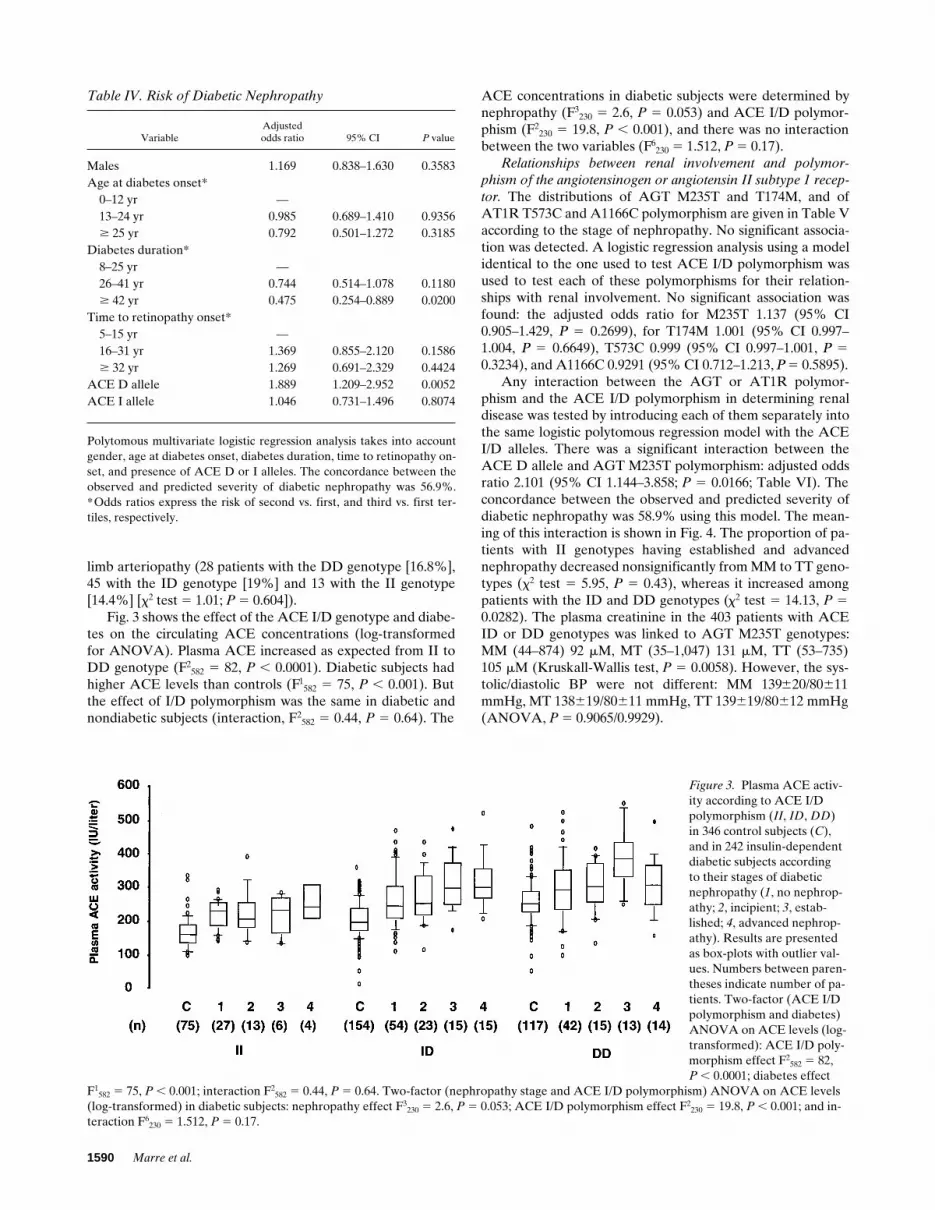
Fig. 3 shows the effect of the ACE I/D genotype and diabe-tes on the circulating ACE concentrations (log-transformedfor ANOVA). Plasma ACE increased as expected from II toDD genotype (F2
582 5 82, P , 0.0001). Diabetic subjects hadhigher ACE levels than controls (F1
582 5 75, P , 0.001). Butthe effect of I/D polymorphism was the same in diabetic andnondiabetic subjects (interaction, F2
582 5 0.44, P 5 0.64). The
ACE concentrations in diabetic subjects were determined bynephropathy (F3
230 5 2.6, P 5 0.053) and ACE I/D polymor-phism (F2
230 5 19.8, P , 0.001), and there was no interactionbetween the two variables (F6
230 5 1.512, P 5 0.17).Relationships between renal involvement and polymor-
phism of the angiotensinogen or angiotensin II subtype 1 recep-
tor. The distributions of AGT M235T and T174M, and ofAT1R T573C and A1166C polymorphism are given in Table Vaccording to the stage of nephropathy. No significant associa-tion was detected. A logistic regression analysis using a modelidentical to the one used to test ACE I/D polymorphism wasused to test each of these polymorphisms for their relation-ships with renal involvement. No significant association wasfound: the adjusted odds ratio for M235T 1.137 (95% CI0.905–1.429, P 5 0.2699), for T174M 1.001 (95% CI 0.997–1.004, P 5 0.6649), T573C 0.999 (95% CI 0.997–1.001, P 5
0.3234), and A1166C 0.9291 (95% CI 0.712–1.213, P 5 0.5895).Any interaction between the AGT or AT1R polymor-
phism and the ACE I/D polymorphism in determining renaldisease was tested by introducing each of them separately intothe same logistic polytomous regression model with the ACEI/D alleles. There was a significant interaction between theACE D allele and AGT M235T polymorphism: adjusted oddsratio 2.101 (95% CI 1.144–3.858; P 5 0.0166; Table VI). Theconcordance between the observed and predicted severity ofdiabetic nephropathy was 58.9% using this model. The mean-ing of this interaction is shown in Fig. 4. The proportion of pa-tients with II genotypes having established and advancednephropathy decreased nonsignificantly from MM to TT geno-types (x2 test 5 5.95, P 5 0.43), whereas it increased amongpatients with the ID and DD genotypes (x2 test 5 14.13, P 50.0282). The plasma creatinine in the 403 patients with ACEID or DD genotypes was linked to AGT M235T genotypes:MM (44–874) 92 mM, MT (35–1,047) 131 mM, TT (53–735)105 mM (Kruskall-Wallis test, P 5 0.0058). However, the sys-tolic/diastolic BP were not different: MM 139620/80611mmHg, MT 138619/80611 mmHg, TT 139619/80612 mmHg(ANOVA, P 5 0.9065/0.9929).
Table IV. Risk of Diabetic Nephropathy
VariableAdjustedodds ratio 95% CI P value
Males 1.169 0.838–1.630 0.3583
Age at diabetes onset*
0–12 yr —
13–24 yr 0.985 0.689–1.410 0.9356
$ 25 yr 0.792 0.501–1.272 0.3185
Diabetes duration*
8–25 yr —
26–41 yr 0.744 0.514–1.078 0.1180
$ 42 yr 0.475 0.254–0.889 0.0200
Time to retinopathy onset*
5–15 yr —
16–31 yr 1.369 0.855–2.120 0.1586
$ 32 yr 1.269 0.691–2.329 0.4424
ACE D allele 1.889 1.209–2.952 0.0052
ACE I allele 1.046 0.731–1.496 0.8074
Polytomous multivariate logistic regression analysis takes into account
gender, age at diabetes onset, diabetes duration, time to retinopathy on-
set, and presence of ACE D or I alleles. The concordance between the
observed and predicted severity of diabetic nephropathy was 56.9%.
*Odds ratios express the risk of second vs. first, and third vs. first ter-
tiles, respectively.
Figure 3. Plasma ACE activ-ity according to ACE I/D polymorphism (II, ID, DD) in 346 control subjects (C), and in 242 insulin-dependent diabetic subjects according to their stages of diabetic nephropathy (1, no nephrop-athy; 2, incipient; 3, estab-lished; 4, advanced nephrop-athy). Results are presented as box-plots with outlier val-ues. Numbers between paren-theses indicate number of pa-tients. Two-factor (ACE I/D polymorphism and diabetes) ANOVA on ACE levels (log-transformed): ACE I/D poly-morphism effect F2
582 5 82,P , 0.0001; diabetes effect
F1582 5 75, P , 0.001; interaction F2
582 5 0.44, P 5 0.64. Two-factor (nephropathy stage and ACE I/D polymorphism) ANOVA on ACE levels (log-transformed) in diabetic subjects: nephropathy effect F3
230 5 2.6, P 5 0.053; ACE I/D polymorphism effect F2230 5 19.8, P , 0.001; and in-
teraction F6230 5 1.512, P 5 0.17.

Angiotensin Converting Enzyme, Angiotensinogen, and Diabetic Nephropathy 1591
No interaction was found in the model including ACE I/Dpolymorphism and AGT T174M (adjusted odds ratio 0.558[95% CI 0.239–1.304, P 5 0.1778]), AT1R T573 C (adjustedodds ratio 0.994 [95% CI 0.953–1.038, P 5 0.7874]), or A1166C(adjusted odds ratio 0.687 [95% CI 0.386–1.223, P 5 0.2019])polymorphisms.
Discussion
This study indicates that genetic polymorphisms of ACE andAGT potentially affecting the levels of angiotensins and kininsin the kidney can affect the development of renal disease in in-sulin-dependent diabetic subjects whose diabetes is inade-quately controlled. Brenner proposed that elevated hydraulicpressure within the glomerular capillaries can cause glomeru-lar damage (49), especially in diabetes (10). As uncontrolleddiabetes causes overall capillary vasodilation (50), a relativepostglomerular vasoconstriction (as produced by angiotensinII) can lead to elevated intraglomerular capillary pressure(10). However, not all insulin-dependent diabetic subjects de-velop diabetic glomerulopathy (1, 2). Thus, the physiologicalfactors that affect glomerular capillary pressure are potentialrisk factors for susceptibility to glomerulopathy due to diabe-tes. The present data are consistent with the hypothesis thatconstitutive levels of angiotensin II can affect the developmentof diabetic glomerulosclerosis. Subjects with the ACE II geno-type have the lowest plasma and cellular ACE levels (14, 17).Because the conversion of angiotensin I is low in the kidney(51), it has been proposed that the plasma ACE circulatingthrough the kidney is an important contributor but yet a limit-ing factor in angiotensin II production within the renal circula-tion (52). Upstream angiotensin II production, angiotensin Iproduction can be limited by the amount of substrate for renin,which explains the interaction with the M235T polymorphism,because circulating AGT is partly determined by the AGT
Table V. Distributions of Substitution Polymorphisms M235T and T174M in Angiotensinogen, and T573C and A1166C in Angiotensin II Subtype 1 Receptor Gene According to Patient Stage of Nephropathy
Stage ofnephropathy No nephropathy Incipient nephropathy Established nephropathy Advanced nephropathy All patients P value
M235T
MM 49 38 46 25 158 0.2441
MT 78 49 49 56 232
TT 29 17 31 26 103
T174M
TT 117 85 94 86 382 0.6741
TM 35 19 26 20 100
MM 4 0 5 1 10
T573C
TT 34 19 27 18 98 0.9846
TC 67 51 54 53 225
CC 53 34 44 36 167
A1166C
AA 75 59 64 54 252 0.6776
AC 64 39 57 45 205
CC 14 6 5 8 33
M235T genotypes were determined in only 493 patients, T174M in 492 patients, T573C in 490 patients, and A1166C in 490 patients. P values were ob-
tained from x2 tests for trend.
Table VI. Risk of Diabetic Nephropathy
VariableAdjustedodds ratio 95% CI P value
Males 1.127 0.806–1.575 0.4859
Age at diabetes onset*
0–12 yr —
13–24 yr 0.967 0.675– 1.385 0.8557
$ 25 yr 0.819 0.516–1.299 0.3956
Diabetes duration*
8–25 yr —
26–41 yr 0.733 0.506–1.061 0.0999
$ 42 yr 0.496 0.264–0.932 0.0293
Time to retinopathy onset*
5–15 yr —
16–31 yr 1.346 0.869–2.086 0.1833
$ 32 yr 1.230 0.668–2.263 0.5058
ACE D allele 0.465 0.137–1.574 0.2183
ACE I allele 1.025 0.715–1.468 0.8939
AGT M235T 0.625 0.359–1.089 0.0974
Interaction between ACE
I/D alleles and AGT
polymorphism 2.101 1.144–3.858 0.0166
Polytomous multivariate logistic regression analysis taking into account
gender, age at diabetes onset, diabetes duration, time to retinopathy on-
set, ACE I/D alleles, and AGT M235T polymorphism. Due to interac-
tion between ACE I/D alleles and AGT polymorphism, no independent
effect was observed of ACE I/D alleles or AGT polymorphism alone on
diabetic nephropathy severity, as illustrated by Fig. 4. The concordance
between the observed and predicted severity of diabetic nephropathy
was 58.9%. *Odds ratios express the risk of second vs. first, and third vs.
first tertile, respectively.

1592 Marre et al.
M235T polymorphism (15). Also, the vasodilator kinins, whichare produced in the renal circulation and metabolized by ACE(53), may participate in the control of glomerular hemodynam-ics in diabetes and their inactivation can mediate the deleteri-ous effect of ACE on kidney function (54).
This study was done on insulin-dependent diabetic subjectswho had already expressed their risk of renal complicationsdue to diabetes, because they had proliferative diabetic reti-nopathy. Patients with proliferative diabetic retinopathy are 20and 24%, respectively, of all patients with insulin-dependentdiabetes attending a French (35) or a Danish (55) diabeticclinic, and 37% of those with insulin-dependent diabetes for 30yr or more attending diabetic clinics in Europe (56). The age atonset of diabetes, the diabetes duration, and the time to prolif-erative diabetic retinopathy in the studied patients all fit withprevious descriptions (2, 5, 35, 55). The degree of renal in-volvement in these patients was graded using a classificationderived from the one proposed by Mogensen et al. (1). Theproportion of patients without nephropathy, or with incipient,established, or advanced nephropathy is consistent with previ-ous cross-sectional studies (1, 35, 55). The relationship be-tween the severity of clinical renal involvement and diabetesduration (Fig. 1) is in keeping with previous follow-up studies,that indicated a peak incidence of diabetic nephropathy (iden-tified by proteinuria) after 10–30 yr of insulin-dependent dia-betes (2, 57). Lastly, the blood glucose control of the studiedpatients (as assessed by glycated hemoglobin at time of datacollection) was linked to the time required to develop diabeticretinal disease, and to diabetes duration. These data are con-sistent with previous studies showing that the degree of bloodglucose control affects the incidence of diabetic retinal and re-nal complications and the vital prognosis of insulin-dependentdiabetic patients (5, 6).
The possible role of genetic factors in the development ofdiabetic nephropathy could be tested using the present patient
cohort, because there was no uncertainty in these patients onthe role of insulin-dependent diabetes in diabetic nephropathyconstitution. Genetic determinants of the renin-angiotensinsystem were the first candidates to be tested. Taking the ad-justed odds ratio as an estimate of relative risk (value 1.889[95% CI 1.209–2.952]), the population risk for diabetic ne-phropathy attributable to ACE D allele was 42% (95% CI 15–61), because the ACE D allele is frequent in the population. Instudies examining diabetic nephropathy in families with multi-ple siblings with insulin-dependent diabetes, the relative riskfor siblings to develop diabetic nephropathy was reported tobe 2.2, 3.3, and 4.8, respectively (7, 58, 59), if the proband haddeveloped diabetic nephropathy. The familial component in-creased by 50% the absolute risk for diabetic nephropathy inthe most recent study (59). Thus, the contribution of ACEgene polymorphism to the heritable part of risk for diabeticnephropathy seems important, and further suggests that mostof the familial component of the risk for diabetic nephropathyis due to genetic factors, as supported by familial studies per-formed in patients with insulin-dependent diabetes living inNew England (59) and in Pima Indians with noninsulin-depen-dent diabetes (8). However, we cannot reject from this studythat nonglycemic, nongenetic variables can also affect the de-velopment of diabetic nephropathy, like smoking habits (60),although we found in the present study no relationship be-tween past or current tabacco use and renal disease (data notshown), or the nongenetic components of BP.
The ACE D allele had a dominant effect, while the risk ofnephropathy was relatively reduced in the insulin-dependentdiabetic subjects with the II genotype. These data from thepresent multicenter study confirm those we reported in a pre-vious monocenter case-control study (26) and those obtainedin other studies (27, 28), but differ from two other publishedstudies (30, 31). However, the controls used in these latterstudies were not matched with cases for diabetic retinopathyseverity and/or diabetes control (30, 31). As controls in thesestudies were less exposed to hyperglycemia than cases, then aneffect of candidate genes able to modulate renal risk may havebeen masked.
There was also an association between ACE I/D polymor-phism and myocardial infarction, in agreement with previousreports (18–24), including on insulin-dependent diabetic sub-jects with nephropathy (61), and in noninsulin-dependent dia-betic subjects (19, 24). Insulin-dependent diabetic subjectswith nephropathy are especially prone to coronary heart dis-ease (2, 3). The coexistence of myocardial infarction with dia-betic nephropathy can therefore be a source of confusion forassessing the role of the ACE gene polymorphism in the con-stitution of diabetic nephropathy. This study shows that the as-sociation between the ACE D allele and nephropathy is stillpresent after subjects with myocardial infarction are excluded,indicating an association between this polymorphism and dia-betic nephropathy that is independent of any coexisting coro-nary heart disease.
There was an effect of the diabetes state on the plasmaACE that was independent of diabetic nephropathy and of I/Dpolymorphism. This confirms earlier reports (26, 62, 63). As aconsequence, the relationship between plasma ACE levels andACE I/D polymorphism was shifted upwards by the diabetesstate. The effect of diabetes state on plasma ACE levels is cur-rently not explained. It seems to be independent of the degreeof diabetes control (26, 30). It may be due to alterations in
Figure 4. Proportions of patients with various stages of diabetic nephropathy according to their ACE II (left) or ID and DD (right) genotypes, and according to AGT M235T polymorphism (MM, MT, TT): u no nephropathy, incipient, established, and j ad-vanced nephropathy. Adjusted odds ratio of risk for diabetic ne-phropathy due to interaction between ACE D/I alleles and AGT M235T genotypes: 2.101 (95% CI 1.144–3.858, P 5 0.0166). Associa-tion between AGT M235T genotypes and stages of diabetic nephrop-athy in 90 subjects with ACE II genotype x2 5 5.95, P 5 0.43 and in 403 subjects with ACE ID and DD genotypes: x2 5 14.13, P 5 0.0282.
Angiotensin Converting Enzyme, Angiotensinogen, and Diabetic Nephropathy 1593
ACE metabolism, because ACE can be released from endo-thelial cells and trapped by the reticuloendothelial system, andbecause both endothelial cells and reticuloendothelial systemcan be altered by diabetes (50). If we accept that the risk of re-nal damage is linked to ACE levels, which are increased in dia-betes, then the subjects with the lowest ACE levels, i.e., mostlythose with the II genotype, would indeed appear to be at re-duced risk of diabetic nephropathy.
As the plasma ACE levels were weakly associated to theseverity of renal disease (P 5 0.053), independently of I/Dpolymorphism, increments in plasma ACE levels can also besecondary to renal disease, although previous reports give con-flicting results on the relationship between kidney functionand plasma ACE levels (26, 30).
Thus, the ACE II genotype seems to reduce risk for renaldisease in insulin-dependent diabetes, which is consistent withour first report (26) and with others about insulin-dependent(27, 28) and noninsulin-dependent diabetic patients (32–34).Pharmacological blockade of ACE protects kidney function ofinsulin-dependent diabetic patients with incipient or estab-lished diabetic nephropathy, and seems to do so independentlyof its blood pressure lowering effect (64–66). Thus, ACE maywell be a risk factor for the progression of diabetic renal dis-ease. Longitudinal studies of renal function and the responseto ACEI should now be done on insulin-dependent diabeticpatients according to their ACE I/D genotypes. Until then, it isstill premature to formulate recommendations for the care ofpatients with insulin-dependent diabetes according to ACEI/D genotypes. Recommendations for the use of ACEI muststill rely on identification of microalbuminuria or proteinuriain these patients.
The suggestion of a role for ACE and its inhibition in theprogression of renal disease was first done for insulin-depen-dent diabetic subjects (26, 64, 66). Since then, similar observa-tions have been reported in nondiabetic renal diseases, dealingwith progression to renal failure according to ACE I/D poly-morphism (67–69), and with retardation of renal failure byACEI (70, 71). Constitutional ACE levels seem to be criticalfor the susceptibility of the glomerular circulation to externalinjury, whatever the nature of the injury (52).
The AGT polymorphism does not appear to be an inde-pendent risk factor for diabetic nephropathy, in agreementwith Tarnow et al. (72) and Doria et al. (73), but in contrastwith Fogarty et al. (74) who found a direct association betweenAGT M235T polymorphism and diabetic nephropathy. Butthere was in the present study a significant interaction betweenACE I/D and AGT M235T polymorphisms. As the AGTM235T polymorphism can partly determine the AGT level(15), this interaction suggests that the level of AGT, the pre-cursor of angiotensin I, can also affect the glomerular circula-tion of insulin-dependent diabetic subjects, and that this effectwould only be apparent in subjects in whom angiotensin I con-version is not strongly limited by low ACE availability. This in-teraction between ACE and AGT polymorphisms now needsto be tested in nondiabetic renal diseases. The AGT geneticpolymorphism has also been linked to familial hypertension(15, 16) which may share genetic determinants with diabeticnephropathy (75).
No relationship was found between AT1R polymorphismand diabetic nephropathy, directly or through interaction withthe ACE I/D polymorphism, although an association betweenAT1R polymorphism and blood pressure has been reported
(25), and previous studies reported alterations in sensitivity toangiotensin II in diabetes (76, 77). The physiological signifi-cance of the AT1R polymorphism is unknown, and the presentdata do not rule out a role for the AT1R gene in susceptibilityto diabetic nephropathy.
Finally, this study strongly suggests that genetic polymor-phisms in the renin–angiotensin and kallikrein–kinin systemscan affect the progression to renal failure of patients with inad-equately controlled insulin-dependent diabetes. This possibil-ity now needs to be confirmed by prospective studies. The ef-fects of other candidate genes or acquired factors should alsobe tested in these patients (78).
Acknowledgments
We thank the insulin-dependent diabetic subjects who took part inthis study. Thanks to Franck Pean, Vincent Benoit, Gaëlle Jouet-Pas-tre, Gwenaëlle Renoult, Cécile Dumont, and Philippe Coudol fortechnical assistance. Thanks to Laëtitia Martin and Line Godiveaufor secretarial assistance.
The GENEDIAB (GEnétique de la NEphropathie DIABétique)Study Group consists of the following: (Investigators in Clinical Cen-
ters [number of recruited/selected patients]) Philippe Passa, Abdera-mane Bouallouche in Hôpital Saint-Louis, Paris (82/72); Michel Rod-ier, Florence Galtier, Jacques Bringer in Nîmes-Montpellier (64/59);Michel Marre, Béatrice Bouhanick in Angers (coordinating center)(67/54); Caroline Sert, André Grimaldi in Hôpital La Pitié, Paris (57/51); Laurent Dusselier, Thérèse Crea, Pierre Drouin in Nancy (47/35); Zoubida Kahal, Dominique Simon in Créteil (31/29); Lucy Chail-lous, Bernard Charbonnel in Nantes (27/27); Anne Muller, RichardMarechaud in Poitiers (25/25); Serge Halimi in Grenoble (27/25);Henry Sackmann, Jean-Pierre Tauber in Toulouse (23/22); HervéMayaudon, Bernard Bauduceau in Saint-Mandé (21/20); Nicolas Pa-quot, André Scheen in Liège (19/18); Patrick Miossec, Jean-Ray-mond Attali in Bondy (21/18); Isabelle Cerf, Guillaume Charpentierin Corbeil-Essonnes (13/12); Charles Thivolet in Lyon (11/11); Domi-nique Tielmans, Pierre-Jean Guillausseau in Hôpital Lariboisière,Paris (11/11); Jean-Yves Poirier, Hubert Allannic in Rennes (5/5); allcenters are in France, except for Liège in Belgium. (Review commit-tee) Philippe Passa (President), Jean-Raymond Attali, Bernard Bau-duceau, André Grimaldi, Pierre-Jean Guillausseau, Michel Marre,Michel Rodier. (ECG committee) François Alhenc-Gelas (InstitutNational de la Santé et de la Recherche Médicale [INSERM] U367,Paris), Bernard Bauduceau, Michel Rodier, Philippe Pezard (An-gers). (Laboratories) Françoise Bled, Yves Gallois, Andrée Girault-Louvel in Biochemistry Department, Angers; François Alhenc-Gelas,Joël Menard in INSERM U367, Paris; Xavier Jeunemaitre, PierreCorvol in INSERM U36, Collège de France, Paris. (Statistical analy-sis) Gilles Chatellier, Hôpital Broussais, Paris.
This work was supported by grants from Juvenile Diabetes For-mation International (grant 194/55), by INSERM research network(grant 94/325), by CNAM-INSERM research grant 4 AIC18, by aBristol-Myers-Squibb-INSERM grant (86-002) and by a DRC-AP re-search grant (931007). The English text was checked by Dr. OwenParkes.
References
1. Mogensen, C.E. 1994. Definition of diabetic renal disease in insulin-dependent diabetes mellitus based on renal function tests. In The Kidney andHypertension in Diabetes Mellitus. C.E. Mogensen, editor. Kluwer AcademicPublishers, Dordrecht, The Netherlands. 1–14.
2. Krolewski, A.J., J.H. Warram, L.I. Rand, and C.R. Kahn. 1987. Epidemi-ologic approach to the etiology of type I diabetes mellitus and its complications.N. Engl. J. Med. 317:1390–1398.
3. Borch-Johnsen, K., and S. Kreiner. 1987. Proteinuria: value as predictorof cardiovascular mortality in insulin-dependent diabetes. BMJ (Br. Med. J.).294:1651–1654.

1594 Marre et al.
4. Mauer, M.S., M.W. Steffes, D.E.R. Sutherland, J.S. Najarian, A.F.Michael, and M. Brown. 1975. Studies of the rate of regression of the glomeru-lar lesions in diabetic rats treated with pancreatic islet transplantation. Diabe-
tes. 24:250–255.5. Pirart, J. 1977. Diabète et complications dégénératives. Présentation
d’une étude prospective portant sur 4400 cas observés entre 1947 et 1973. Dia-
bete Metab. 3:97–107, 173–182, 245–256.6. The Diabetes Control and Complications Trial Reseach Group. 1993.
The effect of intensive treatment of diabetes on the development and progres-sion of long-term complications in insulin-dependent diabetes mellitus. N. Engl.
J. Med. 329:977–986.7. Seaquist, E.R., F.C. Goetz, S. Rich, and J. Barbosa. 1989. Familial clus-
tering of diabetic kidney disease: evidence for genetic susceptibility to diabeticnephropathy. N. Engl. J. Med. 320:1161–1165.
8. Pettit, D.J., M.F. Saad, P.H. Bennett, R.G. Nelson, and W.C. Knowler.1990. Familial predisposition to renal disease in two generations of Pima Indi-ans with 2 (non-insulin-dependent) diabetes mellitus. Diabetologia. 33:438–443.
9. Earle, K., J. Walker, C. Hill, and G.C. Viberti. 1992. Familial clustering ofcardiovascular disease in patients with insulin-dependent diabetes and ne-phropathy. N. Engl. J. Med. 326:673–677.
10. Hostetter, T.H., J.L. Troy, and B.M. Brenner. 1981. Glomerular hemo-dynamics in experimental diabetes mellitus. Kidney Int. 19:410–415.
11. Marre, M., H. Leblanc, L. Suarez , T.T. Guyenne, J. Menard, and P.Passa. 1987. Converting enzyme inhibition and kidney function in normotensivediabetic patients with persistent microalbuminuria. BMJ (Br. Med. J.). 294:1448–1452.
12. Hall, J.E., A.C. Guyton, T.E. Ackson, T.G. Coleman, T.E. Lohmeier,and N.C. Tripoddo. 1977. Control of glomerular filtration rate by renin-angio-tensin system. Am. J. Physiol. 233:F366–F372.
13. Cambien, F., F. Alhenc-Gelas, B. Herberth, J.L. Andre, R. Rakotovao,M.F. Gonzales, J. Allegrini, and C. Bloch. 1988. Familial ressemblance ofplasma angiotensin converting enzyme level: the Nancy study. Am. J. Hum.
Genet. 43:774–780.14. Costerousse, O., J. Allegrini, M. Lopez, and F. Alhenc-Gelas. 1993. An-
giotensin I converting enzyme in human circulating mononuclear cells: geneticpolymorphism of expression in T-lymphocytes. Biochem. J. 290:33–40.
15. Jeunemaitre, X., F. Soubrier, Y. Kotelevtsev, R. Lifton, C. Williams, A.Charru, S. Hunt, P. Hopkins, R. Williams, J.M. Lalouel, et al. 1992. Molecularbasis of human hypertension: role of angiotensinogen. Cell. 71:169–180.
16. Caulfield, M., P. Lavender, M. Farrall, P. Munroe, M. Lawson, P.Turner, and A.J.L. Clark. 1994. Linkage of the angiotensinogen gene to essen-tial hypertension. N. Engl. J. Med. 330:1629–1633.
17. Rigat, B., C. Hubert, F. Alhenc-Gelas, F. Cambien, P. Corvol, and F.Soubrier. 1990. An insertion deletion polymorphism in angiotensin I conversionenzyme gene accounting for half of the variance of serum enzyme levels. J. Clin.
Invest. 86:1343–1346.18. Cambien, F., O. Poirier, L. Lecerf, A. Evans, J.P. Cambou, D. Arveiler,
G. Luc, J.M. Bard, L. Bara, S. Ricard, et al. 1992. Deletion polymorphism in thegene for angiotensin converting enzyme is a potent risk factor for myocardialinfarction. Nature (Lond.). 359:641–644.
19. Ruiz, J., H. Blanche, N. Cohen, G. Velho, F. Cambien, D. Cohen, P.Passa, and P. Froguel. 1994. Insertion/deletion polymorphism of the angio-tensin converting enzyme gene is strongly associated with coronary heart dis-ease in non-insulin-dependent diabetes mellitus. Proc. Natl. Acad. Sci. USA. 91:3662–3665.
20. Nakai, K., C. Itoh, Y. Miura, K. Hotta, T. Musha, T. Itoh, T. Miyakawa,R. Iwasaki, and K. Hiramori. 1994. Deletion polymorphism of the angiotensin Iconverting enzyme gene is associated with serum ACE concentration and in-creased risk for CAD in the Japanese. Circulation. 90:2199–2202.
21. Mattu, R.K., E.W.A. Needham, D.J. Galton, E. Frangos, A.J.L. Clark,and M. Caulfield. 1995. A DNA variant at the angiotensin-converting enzymegene locus associates with coronary artery disease in the caerphilly heart study.Circulation. 91:270–274.
22. Badenhop, R.F., X.L. Wang, and D.E.L. Wilcken. 1995. Angiotensinconverting enzyme genotype in children and coronary events in their grandpar-ents. Circulation. 91:1655–1658.
23. Ludwig, E., P.S. Corneli, J.L. Anderson, H.W. Marshall, J.M. Lalouel,and R.H. Ward. 1995. Angiotensin converting enzyme gene polymorphism isassociated with myocardial infarction but not with development of coronarystenosis. Circulation. 91:2120–2124.
24. Keavney, B.D., C.R.K. Dudley, I.M. Stratton, R.R. Holman, D.R. Mat-thews, P.J. Ratcliffe, and R.C. Turner. 1995. UK Prospective Diabetes Study(UKPDS) 14: association of angiotensin-converting-enzyme insertion/deletionpolymorphism with myocardial infarction in NIDDM. Diabetologia. 38:948–952.
25. Bonnardeaux, A., E. Davies, X. Jeunemaitre, I. Fery, A. Charru, E.Clauser, L. Tiret, F. Cambien, P. Corvol, and F. Soubrier. 1994. Angiotensin IItype 1 receptor gene polymorphisms in human essential hypertension. Hyper-
tension. 24:63–69.26. Marre, M., P. Bernadet, Y. Gallois, F. Savagner, T.T. Guyene, M.
Hallab, F. Cambien, P. Passa, and F. Alhenc-Gelas. 1994. Relationships be-tween angiotensin I converting enzyme gene polymorphism, plasma levels, and
diabetic retinal and renal complications. Diabetes. 43:384–388.27. Doria, A., J.H. Warram, and A.S. Krolewski. 1994. Genetic predisposi-
tion to diabetic nephropathy: evidence for a role of the angiotensin I convertingenzyme gene. Diabetes. 43:690–695.
28. Fogarty, D.G., A.P. Maxwell, A. Hughes, N.C. Nevin, and C.C. Doher-thy. 1994. A deletion polymorphism in the angiotensin-converting-enzyme geneis a risk factor for diabetic nephropathy. European Dialysis Transplantation As-
sociation (EDTA), proceedings of XXIst Congress. Abstract volume. 117.29. Powrie, J.K., G.F. Watts, J.N. Ingham, N.A. Taub, P.J. Talmud, and
K.M. Shaws. 1994. Role of glycæmic control in development of microalbumin-uria in patients with insulin-dependent diabetes. BMJ (Br. Med. J.) 309:1608–1612.
30. Tarnow, L., F. Cambien, P. Rossing, F.S. Nielsen, B.V. Hansen, L.Lecerf, O. Poirier, S. Danilov, and H.H. Parving. 1995. Lack of relationship be-tween an insertion/deletion polymorphism in the angiotensin I-converving en-zyme gene and diabetic nephropathy and proliferative retinopathy in IDDMpatients. Diabetes. 44:489-494.
31. Schmidt, S., N. Schone, E. Ritz, and the Diabetic Nephropathy StudyGroup. 1995. Association of ACE gene polymorphism and diabetic nephropa-thy? Kidney Int. 47:1176–1181.
32. Mizuiri, S., H. Hemmi, A. Inoue, H. Yoshikawa, M. Tanegashima, T.Fushimi, M. Ishigami, Y. Amagasaki, T. Ohara, H. Shimatake, and A. Hase-gawa. 1995. Angiotensin-converting-enzyme polymorphism and developmentof diabetic nephropathy in non-insulin-dependent diabetes mellitus. Nephron.
70:455–459.33. Doi, Y., H. Yoshizumi, K. Lino, M. Yamamoto, K. Ichikawa, M. Iwase,
and M. Fujishima. 1996. Association between a polymorphism in the angio-tensin-converting-enzyme gene and microvascular complications in Japanesepatients with NIDDM. Diabetologia. 39:97–102.
34. Dudley, C.R.K., B. Keavney, I.M. Stratton, R.C. Turner, and P.J. Rat-cliffe. 1995. U.K. prospective diabetes study XV: relationship of renin-angio-tensin system gene polymorphisms with microalbuminuria in NIDDM. Kidney
Int. 48:1907–1911.35. Marre, M., H. Leblanc, D. Bruel, M. Leroy, and P. Passa, 1986. Rela-
tions entre excrétion urinaire d’albumine et rétinopathie chez les diabétiquesinsulino-dépendants. Presse Med. 15:1621–1624.
36. WHO Expert Committee on Diabetes Mellitus. 1980. 2nd report.Geneva. 1980. (WHO technical report series No. 646).
37. Kohner, E.M. 1991. The lesions and natural history of diabetic retinopa-thy, chapter 58. In Textbook of Diabetes. J. Pickup and G. Williams, editors.Blackwell Scientific Publications, Oxford. 575–588.
38. Gallois, Y., S. Vol, E. Caces, B. Balkau, and the DESIR Study Group.1996. Distribution of fasting serum insulin, measured by enzymoimmunoassay,in an unselected population of 4032 individuals, references values, according toage and sex. Diabetes Metab. 22:427–431.
39. Goldstein, D.E., R.R. Little, J.D. England, H.M. Wiedmeyer, and E.M.McKenzie. 1986. Methods for quantitating glycosylated hemoglobins: high-per-formance liquid chromatography and thiobarbituric acid colorimetry. In Meth-ods in diabetes research. Vol. 2: Clinical methods. W.L. Clarke, J. Larner, andS.L. Pohl, editors. John Wiley & Sons, New York. 475–504.
40. Blackburn, H. 1969. Classification of the electrocardiogram for popula-tion studies: Minnesota code. J. Electrocardiol. 2:305–310.
41. Rigat, B., C. Hubert, P. Corvol, and F. Soubrier. 1992. PCR detection ofthe insertion/deletion polymorphism of the human angiotensin converting en-zyme gene (DCP1) dipeptidyl-carboxy peptidase. Nucleic Acids Res. 20:1433.
42. Shanmugam, V., K.W. Sell, and B.K. Saha. 1993. Mistyping ACE het-erozygotes. PCR Methods Applications. 3:120–121.
43. Neels, H.M., M.E. Van Sande, and S.L. Scharpe. 1983. Sensitive colori-metric assay for angiotensin converting enzyme in serum. Clin. Chem. 29:1399–1403.
44. Marre, M., J.P. Claudel, P. Ciret, N. Luis, L. Suarez, and P. Passa. 1987.Laser immunonephelometry for routine quantification of urinary albumin ex-cretion. Clin. Chem. 33:209–213.
45. Bouhanick, B., G. Berrut, A.M. Chameau, M. Hallab, F. Bled, B. Che-vet, J. Vergely, V. Rohmer, P. Fressinaud, and M. Marre. 1992. Predictive valueof testing random urine sample to detect microalbuminuria in diabetic subjectsduring outpatient visits. Diabete Metab. 18:54–58.
46. Fabiny, D.L., and G. Ertingshausen. 1971. Automated reaction-rate-method for determination of serum creatinine with the Centrichem. Clin.
Chem. 17:696–700.47. Armitage, P., and G. Berry. 1990. Components of x2. In Statistical Meth-
ods in Medical Research. 2nd ed. Blackwell Scientific Publications, Oxford.371–380.
48. Armitage, P., and G. Berry. 1990. Attributable risk. In Statistical Meth-ods in Medical Research. 2nd ed. Blackwell Scientific Publications, Oxford.469–472.
49. Brenner, B.M. 1983. Hemodynamically mediated glomerular injury andthe progressive nature of kidney disease. Kidney Int. 23:647–655.
50. Williamson, J.R., K. Chang, M. Frangos, K.S. Hasan, Y. Ido, F. Kawa-mura, J.R. Nyengaard, M. Van Den Enden, C. Kilo, and R.G. Tilton. 1993. Hy-perglycemic pseudohypoxia and diabetic complications. Diabetes. 42:801–813.
51. Vane, G.R. 1972. Sites of conversion of angiotensin I. In Hypertension.

Angiotensin Converting Enzyme, Angiotensinogen, and Diabetic Nephropathy 1595
J. Genest and E. Koine, editors. Springer-Verlag New York Inc., New York.523–532.
52. Alhenc-Gelas, F., T. Baussant, C. Hubert, F. Soubrier, and P. Corvol.1989. The angiotensin I converting enzyme in the kidney. J. Hypertens. 7:S9–S13.
53. Erdos, E.G. 1990. Angiotensin I-converting enzyme and the changes inour concepts through the years. Hypertension (Dallas). 16:363–370.
54. Jaffa, A.A., P.F. Rust, and R.K. Mayfield. 1995. Kinin, a mediator of di-abetes—induced glomerular hyperfiltration. Diabetes. 44:156–160.
55. Parving, H.H., E. Hommel, E. Mathiesen, P. Skott, B. Edsberg, M.Bahnsen, M. Lauritzen, P. Hougaard, and E. Lauritzen. 1988. Prevalence of mi-croalbuminuria, arterial hypertension, retinopathy and neuropathy in patientswith insulin-dependent diabetes. BMJ (Br. Med. J.). 296:156–160.
56. The EURODIAB IDDM Complications Study Group. 1994. Microvas-cular and acute complications in IDDM subjects: the EURODIAB IDDMComplications Study. Diabetologia. 37:278–285.
57. Kofoed-Enevoldsen, A., K. Borch-Johnsen, S. Kreiner, J. Nerup, and T.Deckert. 1987. Declining incidence of persistent proteinuria in type 1 (insulin-dependent) diabetic patients in Denmark. Diabetes. 36:205–209.
58. Borch-Johnsen, K., K. Norgaard, E. Hommel, E.R. Mathiesen, J.S.Jensen, T. Deckert, and H.H. Parving. 1992. Is diabetic nephropathy an inher-ited complication? Kidney Int. 41:719–722.
59. Quinn, M., M.C. Angelico, J.H. Warram, and A.S. Krolewski. 1996. Fa-milial factors determine the development of diabetic nephropathy in patientswith IDDM. Diabetologia. 39:940–945.
60. Chase, H.P., S.K. Garg, G. Marshall, C.L. Berg, S. Harris, W.E. Jackson,and R.E. Hamman. 1991. Cigarette smoking increases the risk of albuminuriaamong subjects with type I diabetes. JAMA (J. Am. Med. Assoc.). 265:614–617.
61. Tarnow, L., F. Cambien, P. Rossing, F.S. Nielsen, B.V. Hansen, L.Lecerf, O. Poirier, S. Danilov, S. Boelskifte, K. Borch-Johnsen, and H.H. Par-ving. 1995. Insertion/deletion polymorphism in the angiotensin-I-converting en-zyme gene is associated with coronary heart disease in IDDM patients with dia-betic nephropathy. Diabetologia. 38:798–803.
62. Lieberman, J., and A. Sastre. 1980. Serum angiotensin-converting en-zyme: elevations in diabetes mellitus. Ann. Intern. Med. 93:825–826.
63. Hallab, M., F. Bled, J.M. Ebran, S. Suraniti, A. Girault, P. Fressinaud,and M. Marre. 1992. Elevated serum angiotensin I converting enzyme activityin type I, insulin-dependent diabetic subjects with persistent microalbuminuria.Acta Diabetologica. 29:82–85.
64. Marre, M., G. Chatellier, H. Leblanc, T.T. Guyenne, J. Menard, and P.Passa. 1988. Prevention of diabetic nephropathy with enalapril in normotensivediabetics with microalbuminuria. BMJ (Br. Med. J.). 297:1092–1095.
65. Hallab, M., Y. Gallois, G. Chatellier, V. Rohmer, P. Fressinaud, and M.Marre. 1993. Comparison of reduction in microalbuminuria by enalapril andhydrochlorothiazide in normotensive patients with insulin dependent diabetes.BMJ (Br. Med. J.). 306:175–182.
66. Lewis, E.J., L.G. Hunsiker, R.P. Bain, and R.D. Rohde. 1993. The effectof angiotensin-converting-enzyme inhibition on diabetic nephropathy. N. Engl.
J. Med. 329:1456–1462.67. Harden, P.N., C. Geddes, P.A. Rowe, J.H. Mcllroy, M. Boulton-Jones,
R.S.C. Rodger, B.J.R. Junor, J.D. Briggs, J.M.C. Connell, and A.G. Jardine.1995. Polymorphisms in angiotensin-converting-enzyme gene and progressionof IgA nephropathy. Lancet. 345:1540–1542.
68. Yoshida, H., T. Mitarai, T. Kawamura, T. Kitajima, Y. Miyasaki, R. Na-gasawa, Y. Kawaguchi, H. Kubo, I. Ichikawa, and O. Sakai. 1995. Role of thedeletion polymorphism of the angiotensin-converting-enzyme gene in the pro-gression and therapeutic responsiveness of IgA nephropathy. J. Clin. Invest. 96:2162–2169.
69. Van Essen, G.G., P.L. Rensma, D. de Zeeuw, W.J. Sluiter, H. Scheffer,A.J. Apperloo, and P.E. de Jong. 1996. Association between angiotensin-con-verting-enzyme gene polymorphism and failure of renoprotective therapy. Lan-
cet. 347:94–95.70. Hannedouche, T., P. Landais , B. Goldfard, N. El Esper, A. Fournier,
M. Godin, D. Durand, J. Chanard, F. Mignon, J.M. Suc, et al. 1994. Ran-domised controlled trial of enalapril and b blockers in non-diabetic chronic re-nal failure. BMJ (Br. Med. J.). 309:833–837.
71. Mashio, G., D. Alberti, G. Janin, F. Locatelli, J.F.E. Mann, M. Mo-tolese, C. Ponticelli, E. Ritz, P. Ziuchelli, and the Angiotensin-Converting-Enzyme Inhibition in Progressive Renal Insufficiency Study Group. 1996. Effectof the angiotensin-converting-enzyme inhibitor benazepril on the progressionof chronic renal insufficiency. N. Engl. J. Med. 334:939–945.
72. Tarnow, L., F. Cambien, P. Rossing, F.S. Nielsen, B.V. Hansen, S.Ricard, O. Poirier, and H.H. Parving. 1996. Angiotensinogen gene polymor-phisms in IDDM patients with diabetic nephropathy. Diabetes. 45:367–369.
73. Doria, A., T. Onuma, G. Gearin, M.B.S. Freire, J.H. Warram, and A.S.Krolewski. 1996. Angiotensinogen polymorphism M235T, hypertension, andnephropathy in insulin-dependent diabetes. Hypertension. 27:1134–1139.
74. Fogarty, D.G., J.C. Harron, A.E. Hughes, N.C. Nevin, C.C. Doherty,and A.P. Maxwell. 1996. A molecular variant of angiotensinogen is associatedwith diabetic nephropathy in IDDM. Diabetes. 45:1204–1208.
75. Viberti, G.C., H. Keen, and M.J. Wiseman. 1987. Raised arterial pres-sure in parents of proteinuric insulin-dependent diabetics. BMJ (Br. Med. J.).295:515–517.
76. Christlieb, A.R., H.U. Janka, B. Kraus, R.E. Gleason, E.A. Icasas-Cabral,L.M. Aiello, B.V. Cabral, and A. Solano. 1976. Vascular reactivity to angio-tensin II and to norepinephrine in diabetic subjects. Diabetes. 25:268–274.
77. Ballerman, B., K.L. Skorecki, and B.M. Brenner. 1984. Reduced glom-erular angiotensin II receptor density in early untreated diabetes mellitus in therat. Am. J. Physiol. 247:F110–F116.
78. Rossing, P., L. Tarnow, F.S. Nielsen, S. Boelskifte, B.M. Brenner, andH.H. Parving. 1995. Short stature and diabetic nephropathy. BMJ (Br. Med. J.).
310:296–297.
Copyright © 2022 FDOKUMEN