Porcine dentin matrix protein 1: gene structure, cDNA sequence, and expression in teeth
9
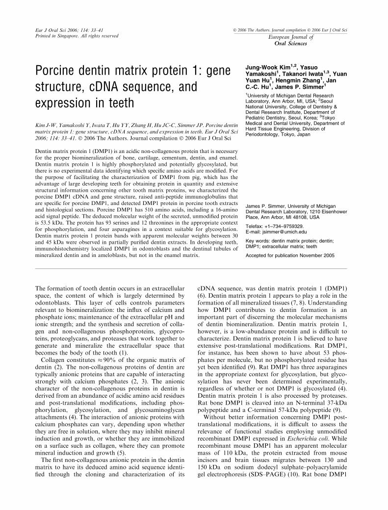
Porcine dentin matrix protein 1: gene structure, cDNA sequence, and expression in teeth Jung-Wook Kim 1,2 , Yasuo Yamakoshi 1 , Takanori Iwata 1,3 , Yuan Yuan Hu 1 , Hengmin Zhang 1 , Jan C.-C. Hu 1 , James P. Simmer 1 1 University of Michigan Dental Research Laboratory, Ann Arbor, MI, USA; 2 Seoul National University, College of Dentistry & Dental Research Institute, Department of Pediatric Dentistry, Seoul, Korea; 3 Tokyo Medical and Dental University, Department of Hard Tissue Engineering, Division of Periodontology, Tokyo, Japan The formation of tooth dentin occurs in an extracellular space, the content of which is largely determined by odontoblasts. This layer of cells controls parameters relevant to biomineralization: the influx of calcium and phosphate ions; maintenance of the extracellular pH and ionic strength; and the synthesis and secretion of colla- gen and non-collagenous phosphoproteins, glycopro- teins, proteoglycans, and proteases that work together to generate and mineralize the extracellular space that becomes the body of the tooth (1). Collagen constitutes 90% of the organic matrix of dentin (2). The non-collagenous proteins of dentin are typically anionic proteins that are capable of interacting strongly with calcium phosphates (2, 3). The anionic character of the non-collagenous proteins in dentin is derived from an abundance of acidic amino acid residues and post-translational modifications, including phos- phorylation, glycosylation, and glycosaminoglycan attachments (4). The interaction of anionic proteins with calcium phosphates can vary, depending upon whether they are free in solution, where they may inhibit mineral induction and growth, or whether they are immobilized on a surface such as collagen, where they can promote mineral induction and growth (5). The first non-collagenous anionic protein in the dentin matrix to have its deduced amino acid sequence identi- fied through the cloning and characterization of its cDNA sequence, was dentin matrix protein 1 (DMP1) (6). Dentin matrix protein 1 appears to play a role in the formation of all mineralized tissues (7, 8). Understanding how DMP1 contributes to dentin formation is an important part of discerning the molecular mechanisms of dentin biomineralization. Dentin matrix protein 1, however, is a low-abundance protein and is difficult to characterize. Dentin matrix protein 1 is believed to have extensive post-translational modifications. Rat DMP1, for instance, has been shown to have about 53 phos- phates per molecule, but no phosphorylated residue has yet been identified (9). Rat DMP1 has three asparagines in the appropriate context for glycosylation, but glyco- sylation has never been determined experimentally, regardless of whether or not DMP1 is glycosylated (4). Dentin matrix protein 1 is also processed by proteases. Rat bone DMP1 is cleaved into an N-terminal 37-kDa polypeptide and a C-terminal 57-kDa polypeptide (9). Without better information concerning DMP1 post- translational modifications, it is difficult to assess the relevance of functional studies employing unmodified recombinant DMP1 expressed in Escherichia coli. While recombinant mouse DMP1 has an apparent molecular mass of 110 kDa, the protein extracted from mouse incisors and brain tissues migrates between 130 and 150 kDa on sodium dodecyl sulphate–polyacrylamide gel electrophoresis (SDS–PAGE) (10). Rat bone DMP1 Kim J-W, Yamakoshi Y, Iwata T, Hu YY, Zhang H, Hu JC-C, Simmer JP. Porcine dentin matrix protein 1: gene structure, cDNA sequence, and expression in teeth. Eur J Oral Sci 2006; 114: 33–41. ȑ 2006 The Authors. Journal compilation ȑ 2006 Eur J Oral Sci Dentin matrix protein 1 (DMP1) is an acidic non-collagenous protein that is necessary for the proper biomineralization of bone, cartilage, cementum, dentin, and enamel. Dentin matrix protein 1 is highly phosphorylated and potentially glycosylated, but there is no experimental data identifying which specific amino acids are modified. For the purpose of facilitating the characterization of DMP1 from pig, which has the advantage of large developing teeth for obtaining protein in quantity and extensive structural information concerning other tooth matrix proteins, we characterized the porcine DMP1 cDNA and gene structure, raised anti-peptide immunoglobulins that are specific for porcine DMP1, and detected DMP1 protein in porcine tooth extracts and histological sections. Porcine DMP1 has 510 amino acids, including a 16-amino acid signal peptide. The deduced molecular weight of the secreted, unmodified protein is 53.5 kDa. The protein has 93 serines and 12 threonines in the appropriate context for phosphorylation, and four asparagines in a context suitable for glycosylation. Dentin matrix protein 1 protein bands with apparent molecular weights between 30 and 45 kDa were observed in partially purified dentin extracts. In developing teeth, immunohistochemistry localized DMP1 in odontoblasts and the dentinal tubules of mineralized dentin and in ameloblasts, but not in the enamel matrix. James P. Simmer, University of Michigan Dental Research Laboratory, 1210 Eisenhower Place, Ann Arbor, MI 48108, USA Telefax: +1–734–9759329. E-mail: [email protected] Key words: dentin matrix protein; dentin; DMP1; extracellular matrix; teeth Accepted for publication November 2005 Eur J Oral Sci 2006; 114: 33–41 Printed in Singapore. All rights reserved ȑ 2006 The Authors. Journal compilation ȑ 2006 Eur J Oral Sci European Journal of Oral Sciences
-
Upload
independent -
Category
Documents
-
view
3 -
download
0
Transcript of Porcine dentin matrix protein 1: gene structure, cDNA sequence, and expression in teeth
Porcine dentin matrix protein 1: genestructure, cDNA sequence, andexpression in teeth
Jung-Wook Kim1,2, YasuoYamakoshi1, Takanori Iwata1,3, YuanYuan Hu1, Hengmin Zhang1, JanC.-C. Hu1, James P. Simmer1
1University of Michigan Dental ResearchLaboratory, Ann Arbor, MI, USA; 2SeoulNational University, College of Dentistry &Dental Research Institute, Department ofPediatric Dentistry, Seoul, Korea; 3TokyoMedical and Dental University, Department ofHard Tissue Engineering, Division ofPeriodontology, Tokyo, Japan
The formation of tooth dentin occurs in an extracellularspace, the content of which is largely determined byodontoblasts. This layer of cells controls parametersrelevant to biomineralization: the influx of calcium andphosphate ions; maintenance of the extracellular pH andionic strength; and the synthesis and secretion of colla-gen and non-collagenous phosphoproteins, glycopro-teins, proteoglycans, and proteases that work together togenerate and mineralize the extracellular space thatbecomes the body of the tooth (1).Collagen constitutes � 90% of the organic matrix of
dentin (2). The non-collagenous proteins of dentin aretypically anionic proteins that are capable of interactingstrongly with calcium phosphates (2, 3). The anioniccharacter of the non-collagenous proteins in dentin isderived from an abundance of acidic amino acid residuesand post-translational modifications, including phos-phorylation, glycosylation, and glycosaminoglycanattachments (4). The interaction of anionic proteins withcalcium phosphates can vary, depending upon whetherthey are free in solution, where they may inhibit mineralinduction and growth, or whether they are immobilizedon a surface such as collagen, where they can promotemineral induction and growth (5).The first non-collagenous anionic protein in the dentin
matrix to have its deduced amino acid sequence identi-fied through the cloning and characterization of its
cDNA sequence, was dentin matrix protein 1 (DMP1)(6). Dentin matrix protein 1 appears to play a role in theformation of all mineralized tissues (7, 8). Understandinghow DMP1 contributes to dentin formation is animportant part of discerning the molecular mechanismsof dentin biomineralization. Dentin matrix protein 1,however, is a low-abundance protein and is difficult tocharacterize. Dentin matrix protein 1 is believed to haveextensive post-translational modifications. Rat DMP1,for instance, has been shown to have about 53 phos-phates per molecule, but no phosphorylated residue hasyet been identified (9). Rat DMP1 has three asparaginesin the appropriate context for glycosylation, but glyco-sylation has never been determined experimentally,regardless of whether or not DMP1 is glycosylated (4).Dentin matrix protein 1 is also processed by proteases.Rat bone DMP1 is cleaved into an N-terminal 37-kDapolypeptide and a C-terminal 57-kDa polypeptide (9).Without better information concerning DMP1 post-
translational modifications, it is difficult to assess therelevance of functional studies employing unmodifiedrecombinant DMP1 expressed in Escherichia coli. Whilerecombinant mouse DMP1 has an apparent molecularmass of 110 kDa, the protein extracted from mouseincisors and brain tissues migrates between 130 and150 kDa on sodium dodecyl sulphate–polyacrylamidegel electrophoresis (SDS–PAGE) (10). Rat bone DMP1
Kim J-W, Yamakoshi Y, Iwata T,HuYY, ZhangH,Hu JC-C, Simmer JP. Porcine dentinmatrix protein 1: gene structure, cDNA sequence, and expression in teeth. Eur J Oral Sci2006; 114: 33–41. � 2006 The Authors. Journal compilation � 2006 Eur J Oral Sci
Dentin matrix protein 1 (DMP1) is an acidic non-collagenous protein that is necessaryfor the proper biomineralization of bone, cartilage, cementum, dentin, and enamel.Dentin matrix protein 1 is highly phosphorylated and potentially glycosylated, butthere is no experimental data identifying which specific amino acids are modified. Forthe purpose of facilitating the characterization of DMP1 from pig, which has theadvantage of large developing teeth for obtaining protein in quantity and extensivestructural information concerning other tooth matrix proteins, we characterized theporcine DMP1 cDNA and gene structure, raised anti-peptide immunoglobulins thatare specific for porcine DMP1, and detected DMP1 protein in porcine tooth extractsand histological sections. Porcine DMP1 has 510 amino acids, including a 16-aminoacid signal peptide. The deduced molecular weight of the secreted, unmodified proteinis 53.5 kDa. The protein has 93 serines and 12 threonines in the appropriate contextfor phosphorylation, and four asparagines in a context suitable for glycosylation.Dentin matrix protein 1 protein bands with apparent molecular weights between 30and 45 kDa were observed in partially purified dentin extracts. In developing teeth,immunohistochemistry localized DMP1 in odontoblasts and the dentinal tubules ofmineralized dentin and in ameloblasts, but not in the enamel matrix.
James P. Simmer, University of MichiganDental Research Laboratory, 1210 EisenhowerPlace, Ann Arbor, MI 48108, USA
Telefax: +1–734–9759329.E-mail: [email protected]
Key words: dentin matrix protein; dentin;DMP1; extracellular matrix; teeth
Accepted for publication November 2005
Eur J Oral Sci 2006; 114: 33–41Printed in Singapore. All rights reserved
� 2006 The Authors. Journal compilation � 2006 Eur J Oral Sci
European Journal ofOral Sciences

runs as a doublet at 150 kDa, while rat dentin DMP1can have an apparent molecular mass as high as200 kDa. It is evident that better structural characteri-zation is necessary before meaningful structure/functionrelationships can be established for DMP1.Pigs have large developing teeth, which offer obvious
advantages for the biochemical characterization of low-abundance proteins such as DMP1. Furthermore, the pigmodel has the advantage of having a large body ofinformation concerning the biochemical structures andproperties of other enamel (11–13) and dentin (14,15)proteins. In this study we expand the investigation ofDMP1 to the pig animal model. We report the isolationand characterization of the porcine DMP1 cDNA, theDMP1 gene structure and deduced amino acid sequence,and generate porcine DMP1 anti-peptide immunoglob-ulins that specifically detect DMP1 protein in proteinextracts and histological sections of developing teeth.These data are analyzed in the light of what has beenlearned about DMP1 from other organisms.
Material and methodsAll experimental procedures involving the use of animalswere reviewed and approved by the Institutional AnimalCare and Use Program at the University of Michigan.
Cloning of porcine DMP1 cDNA (method 1)
RNA from porcine dental pulp tissue was isolated by usingthe RNeasy Protect Maxi kit and protocol (Invitrogen, LaJolla, CA, USA), and converted to cDNA by reverse tran-scription. The human (16), bovine (17), and mouse (18)DMP1 cDNA sequences were aligned to identify regionsof maximum homology. Degenerate primers (pDMP1tF1,5¢-AGYGGAGATGACACCTTTGG; and pDMP1tR1,5¢-CTGCCRCCTCCCACCC) were synthesized and used toamplify a segment of porcine DMP1 cDNA by polymerasechain reaction (PCR) using Platinum Taq DNA polymeraseHigh Fidelity (Invitrogen). The PCR conditions used a5-min denaturation at 94�C, followed by 40 cycles each ofdenaturation at 94�C for 30 s, primer annealing at 55�C for30 s, and product extension at 72�C for 30 s, with a finalextension at 72�C for 7 min. The amplification product wasligated into pCR2.1-TOPO (Invitrogen) and transfectedinto E. coli. DNA minipreps of the recombinant plasmidcontaining the 257-bp insert were characterized by DNAsequencing using ABI Big Dye Terminator chemistry and anABI 3100 automated DNA sequencer at the University ofMichigan’s DNA Sequencing Core. This sequence wasanalyzed, and porcine DMP1-specific oligonucleotideprimers were designed for rapid amplification of cDNAends (RACE). The DMP1-specific primers used for5¢-RACE were pDMP1RACE5R (5¢-GCCTGCTGTCC-ACCTCATCCTCGT) and pDMP1RACE5R1 (5¢-GGC-CCCTGCTCTCACTGGTGGTATC). The 3¢-RACEprimers were pDMP1RACE3F (5¢-GTGGCCCAGGAC-CTGAAGAGAGCA) and pDMP1RACE3F1 (5¢-GGG-GAAGTCAGTGCCCAAGATACCA). The RACE wasaccomplished using the BD SMART RACE cDNAAmplification Kit (BD Bioscience Clontech, San Jose, CA,USA). The 5¢- and 3¢-RACE products were ligated intopCR2.1-TOPO (Invitrogen) and transfected into E. coli.
Cloning full-length porcine DMP1 cDNA (method 2)
RNA was isolated from dental mesenchyme using TRIzol(Invitrogen) reagent and converted to cDNA in three sep-arate experiments using Superscript� reverse transcriptase(Invitrogen). Each cDNA preparation was amplified inthree separate PCR reactions (nine reactions in total) usingAccu Pfx Polymerase SuperMix (Invitrogen) and theprimerpair 5¢-CAGCTATGAAGACCAGCATCC and5¢-CTAGTAGCCGTCCTGGCAGT. The PCR conditionsused a 5-min denaturation at 95�C, followed by 35 cycleseach of denaturation at 95�C for 15 s, primer annealing at60�C for 30 s, and product extension at 68�C for 105 s, witha final extension at 68�C for 7 min. To minimize potentialerrors caused by infidelity in the PCR and reverse tran-scription (RT) reactions, three DMP1 cDNAs from eachreaction (27 PCR products) were characterized by directDNA sequencing using the two PCR oligonucleotides andtwo internal oligonucleotides (5¢-TGTCCTGTGCTCTCC-CAGT and 5¢-TCGCCCAAGGTGTCATCTCC) as prim-ers. Direct DNA sequencing of amplified porcine genomicDNA verified the entire porcine DMP1 cDNA sequence.The oligonucleotides and PCR conditions are provided inTable 1.
Determination of intron–exon boundaries
Primers were designed from each exon after sequencecomparison with human, mouse, and bovine DMP1. Usingthe porcine genomic DNA as a template, DMP1 genomicsegments were amplified using Platinum Taq DNA polym-erase High Fidelity (Invitrogen), cloned and characterizedby DNA sequencing. The oligonucleotide primer pairs andPCR conditions are shown in Table 1.
Genome walking
Genomic sequences outside the 5¢ and 3¢ ends of the porcineDMP1 gene were obtained using the DNA Walking Spee-dUp premix kit (Seegene, Seoul, Korea) and protocol. Forthe 5¢ end, oligonucleotides pDMP1TSP6 (5¢-CAGTG-GTCTTAGCATCTCCT) and pDMP1TSP5 (5¢-CTGGC-TCTACTGAAGTCACA), and for the 3¢ end,oligonucleotides pDMP1TSPF4 (5¢-ACATGGTGAGACT-AGAAGC) and pDMP1TSP F2 (5¢-GCACAGTGA-GGTGTGTTGGATTAG), were used according to theprotocol.
Sequence analyses
The signal peptide cleavage site was predicted using Signal P3.0 (19) at the Center for Biological Sequence AnalysisWebsite (http://www.cbs.dtu.dk/services/SignalP/). Themolecular weight and isoelectric point of porcine DMP1was predicted using the Compute pI/Mw tool (http://c.expasy.org/tools/pi_tool.html) on the Expasy molecularbiology server (http://expasy.cbr.nrc.ca/tools/scnpsit1.html)at the Swiss Institute of Bioinformatics. Searches forpotential phosphorylation, and O- and N-linked glycosyla-tion sites were performed at the Center for BiologicalSequence Analysis at the Technical University of DenmarkWebsite (http://www.cbs.dtu.dk/) using the NetNGlyc 1.0(20), NetOGlyc 2.0 (21), and NetPhos 2.0 (22) predictionservers. Percentage identities of DMP amino acid sequencesfrom porcine, human, bovine, and mouse were calculated
34 Kim et al.

from pairwise aligments using SIM (at http://www.expasy.org/tools/sim-prot.html).
Preparation of porcine tooth powder and extraction ofDMP1
Tooth germs of permanent molars were surgically extractedfrom the maxillae and mandibles of 6-month-old pigs at theMichigan State University Meat Laboratory (East Lansing,MI, USA), as described previously (14). Soft tissue wasremoved and the hard tissue was quickly frozen in dry ice.Secretory stage dental enamel was removed by scraping witha curette. The remaining hard tissue was reduced to apowder using a jaw crusher (Retsch, Newtown, PA, USA).All extraction steps were carried out at 4�C or on ice. Pro-tease Inhibitor Cocktail Set III (1 mM 4-[Ami-noethyl]benzenesulfonyl fluoxide hydrochloride [AEBSF],0.8 lM aprotinin, 50 lM bestatin, 15 lM E-64, 20 lMleupeptin and 10 lM pepstatin) (EMD Biosciences, SanDiego, CA, USA) and 1 mM 1,10-phenanthroline (Sigma,St Louis, MO, USA) were added into the buffer during theextraction. Dentin powder was sequentially extracted with50 mM Tris-HCl/4 M guanidine buffer (pH 7.4), then with50 mM Tris-HCl/4 M guanidine/0.5 M EDTA buffer(pH 7.4). These extracts were centrifuged at 7000 g, desaltedand concentrated using the combination of a Spectra/Por 3membrane (Spectrum Laboratories, Rancho Dominguez,CA, USA) with a YM-3 membrane (Amicon, Beverly, MA,USA), and were lyophilized and stored at )80�C.
Partial purification of porcine DMP1
The following fractionation of dentin proteins was carriedout as previously described (14). In brief, the lyophilized
EDTA extract was dissolved in 10 ml of 50 mM Tris-HCl/6 M urea buffer (pH 7.4) and fractionated by anion-exchange chromatography using a Q-Sepharose Fast Flowcolumn (1.6 cm · 20 cm; GE Healthcare Biosciences, LittleChalfont, Bucks., UK). Dentin matrix protein 1 eluted inthe third peak (Q3), which was concentrated with a YM-3membrane and further fractionated over a hydroxyapatite(HA) column. The HA column divided Q3 into three parts.The contents of the second peak (H2) were fractionated bysize-exclusion chromatography on a Superdex 200 column(1.6 cm · 60 cm; GE Healthcare Biosciences). Dentin mat-rix protein 1-positive bands were identified among thecontents of the second chromatographic peak (S2). Samplesfrom the guanidine supernatant, guanidine pellet, theguanidine/EDTA extracts and S2 were incubated withProtein G–Sepharose (GE Healthcare Biosciences) andcentrifuged to remove immunoglobulins.
Antibody production
A 14-amino acid segment (Gln173–Asp186; Cys-QEEGV-GEPRGDNPD) from the porcine DMP1 deduced aminoacid sequence was selected based upon overall antigenicindex, favorable secondary structure, peptide location, andcross-species reactivity. The peptide was conjugated to thecarrier peptide-KLH (keyhole limpet hemocyanin). Anti-bodies were generated in rabbits using a 2-month protocolthat included three immunizations, one test bleed, a fourthimmunization, and a final bleed. Specific anti-DMP1immunoglobulin was purified from the final bleed using anaffinity column containing the immobilized unconjugatedDMP1 peptide, and tested using enzyme-linked immuno-sorbent assay (ELISA) before being used for western blot-ting and immunohistochemistry.
Table 1
Oligonucleotide primers for dentin matrix protein 1 (DMP1) gene characterization
Name AnnealExtensiontemperature
Extensiontime
A. Primers used to amplify porcine DMP1 genomic sequencespDMP1gF1 5¢-ATCCACACGAGAGTGGCTTC 60�C 68�C 5 minpDMP1gR1 5¢-TGGGAGAGCACAGGACAACpDMP1gF2 5¢-GAGGTGACGTATCCAGCTATGA 60�C 68�C 3 minpDMP1gR2 5¢-CAAAGGTGGTGCTGGTGTCpDMP1gF3 5¢-GTCAGTTGGCTCAGACACCA 60�C 68�C 3 minpDMP1gR3 5¢-TTTTCCTCCACTCACAGCAAB. Primers used for direct PCR sequencing of the porcine DMP1 genepDMP1x1F 5¢-AGTGTGGGTGTCCTGGAGTC 55�C 72�C 30 spDMP1x1R 5¢-TTTTCCAAACCACACAGCAGpDMP1x23F 5¢-TCAGTTGACGGACTTTTGGA 55�C 72�C 45 spDMP1x23R 5¢-CATTTGCAGTGAAGCCAGTGpDMP1x45F 5¢-AGAGATTTGGGTCACCATGC 55�C 72�C 30 spDMP1x45R 5¢-GCCGGGGAATCTAATCTAGCpDMP1x45R1 5¢-TGGCCTAAGAGGCTGAGAAGpDMP1x6F 5¢-GGGCCTTAGGTAGTGCCTTC 55�C 72�C 30 spDMP1x6R 5¢-GTCCACCTCATCCTCGTTGTpDMP1g6F1 5¢-AGTGCCCAAGATACCACCAG 60�C 68�C 60 spDMP1g6R1 5¢-ACTTGAGGTTGGCAGTGCTTpDMP1g6F2 5¢-CAGAGTCCACGGAAGAGGAG 60�C 68�C 60 spDMP1g6R2 5¢-TTGATTTGCTGCTGTCTTGGpDMP1g6F3 5¢-CACGAACACTGACTCCCAGA 55�C 72�C 30 spDMP1x6cR 5¢-CTTCTGTTTAATGGGAGGAAAG
PCR, polymerase chain reaction.
Porcine DMP1 35

Sodium dodecyl sulphate–polyacrylamide gelelectrophoresis
Sodium dodecyl sulphate–polyacrylamide gel electrophor-esis was performed using PAGEr Gold Precast Gels (4–20%gradient, 10% or 7.5% Tris-Glycine gels) (Cambrex BioScience, Rockland, ME, USA). Samples were dissolved inLaemmli sample buffer (Bio-Rad, Hercules, CA, USA) andelectrophoresis was carried out using a current of 20 mA for1.5 h. The gels were stained with Bio-Safe Coomassie (Bio-Rad) or Stains-all (Sigma). The apparent molecular weightsof protein bands were estimated by comparison with See-Blue Plus2 Pre-Stained Standard (Invitrogen).
Western immunoblots
After SDS–PAGE, proteins were electrotransferred onto aHybond-P membrane (GE Healthcare Biosciences). PorcineDMP1 antibodies were used at dilution of 1 : 50,000. Themembrane was immunostained by chemiluminescent detec-tion with the ECL Advance Western Blotting Detection Kit(Amersham Pharmacia, Piscataway, NJ, USA).
Preparation of tissues for immunostaining
Developing second molars of 6-month-old pigs were fixed,demineralized, and embedded in paraffin. In brief, fixationwas in 4% paraformaldehyde/DEPC at 4�C for 2 d. Thefixed teeth were demineralized for 3 wk using 10% EDTA/diethylpyrocarbonate (DEPC) at pH 7.4. Histology pro-cessing used washes at 4�C in phosphate-buffered saline(PBS) (pH 7.4) for 6–8 h, 70% (v/v) ethanol overnight, 95%(v/v) ethanol the second day, 100% (v/v) ethanol overnight,and xylene for 2 h at room temperature, followed byembedding by paraffin perfusion for 3–4 h. The molars wereembedded with the mesial surface down and sectioned at6-lm thickness.
Immunohistochemistry
The AEC rabbit primary staining kit (Zymed, San Fran-cisco, CA, USA) was used. Rabbit anti-porcine amelogeninimmunoglobulin was used as a positive control and thenegative control was rabbit immunoglobulin G (IgG) at adilution of 1 : 100. To enhance antigen–antibody bindingand reduce non-specific reaction, an antigen retrieval pro-cedure was conducted prior to immunohistochemistry(IHC) experiments (23). The affinity-purified porcine DMP1antibodies were dissolved at a concentration of 1 mg ml)1
and used at a dilution of 1 : 100. Tissue sections were an-alyzed and photographed using a Nikon Eclipse Microscopeand Act1 software (Mager Scientific, Dexter, MI, USA).
Results
The full-length porcine dentin matrix acidic phospho-protein (DMP1) cDNA (2681 bp) was cloned and char-acterized, and its amino acid sequence was deduced(Fig. 1). The pig protein has 494 amino acids followingthe removal of a 16-amino acid signal peptide. Thededuced molecular weight of the secreted protein is53.5 kDa, although the actual molecular weight is likelyto be much larger owing to post-translational modifica-tions. Porcine DMP1 has 99 serines (20%), 93 of which
are in the appropriate context for phosphorylation. Itsamino acid composition is also notable because of itsmany acidic residues [89 (or 18%) glutamic acids; 51 (or10.3%) aspartic acids]. Overall, negatively charged sidechains outnumber positively charged groups by 140 to42, giving the protein an acidic calculated isoelectricpoint of 4.1. Porcine DMP1 also has four asparagines inthe appropriate context for glycosylation (Asn341,Asn360, Asn416, and Asn464).To confirm the cDNA sequence and to determine the
positions of intervening sequences in the porcine DMP1gene, we characterized the DMP1 coding region andintron/exon borders by direct sequencing of porcinegenomic DNA (Tables 2 and 3). The sequences at theintron/exon borders are shown in Table 3. Full-lengthporcine DMP1 cDNAs were independently cloned and
Fig. 1. Analysis of porcine dentin matrix protein 1 (DMP1)cDNA and deduced amino acid sequences. The porcine DMP1coding region from exons 2 to 6 are shown. On the right are thenumbers of the last amino acid in each row. In the nucleotiderows, numbers in bold indicate the positions of introns in theporcine Dmp1 gene. The Signal P predicted signal peptidecleavage site is after Ala16, suggesting that Leu17 is at the Nterminus of the secreted protein. NetNGlyc identified fourasparagines (shown in bold and underlined) in the appropriatecontext for glycosylation: Asn341, Asn360, Asn416, andAsn464, with Asn360 being in the most favorable context.NetPhos indicated that 93 out of 99 serines (bold) and 12 of 19threonines (not shown) are in the appropriate contexts forphosphorylation. The amino acid segment used to raise DMP1antibodies in rabbit is boxed. The translation initiation (ATG)and termination (TAG) codons are in bold and underlined. Thepositions of the five introns in porcine Dmp1 are indicated bynumbers within the nucleotide sequence. Seven nucleotidepolymorphisms were identified, four of which changed thededuced amino acid sequence: Met76Leu, Val231Ala, Glu312-Lys, and Leu326Pro (underlined, not bold).
36 Kim et al.

five nucleotide polymorphisms were identified, four ofwhich changed the deduced amino acid sequence(Met76Leu, Leu90Leu, Val231Ala, Glu312Lys, andLeu326Pro). This indicates that DMP1 protein sequencesvary within species.The porcine DMP1 deduced amino acid sequence was
aligned to human (16), bovine (17), and mouse (18)DMP1 sequences (Fig. 2). Among the 93 serinesappropriate for phosphorylation, 72 were found to beconserved in all four DMP1 sequences. Asn464 is theonly potentially glycosylated asparagine that is con-served in all four DMP1s. The predicted integrin-bindingsite (RGD) is conserved in all of the four known mam-malian DMP1 sequences. Pairwise alignments of the fourDMP1 sequences gave the following percentage identi-ties: porcine–human, 67%; porcine–bovine, 73%; por-cine–murine, 62%; human–bovine, 70%; human–murine64%; and bovine–murine 64%.We prepared an anti-peptide immunoglobulin against
the conserved segment of porcine DMP1 containing theRGD integrin-binding motif (QEEGVGEPRGDNPD),which contained no residues that were predicted to bepost-translationally modified. The affinity-purified anti-peptide immunoglobulin was used to detect DMP1 inporcine dentin extracts (Fig. 3). Dentin matrix protein 1was detected as four bands, which are presumably clea-vage products, migrating between 30 and 45 kDa onSDS–PAGE. In rat, DMP1 cleavage products at 37 and57 kDa have been characterized (9). Full-length recom-binant DMP1 protein, which lacks post-translationalmodifications, migrates at 110 kDa (10). Negative con-trols demonstrated the necessity of removing naturallyoccurring immunoglobulins from dentin extracts prior toperforming western blot analyses. We did not detectlarger immunopositive bands following the removal ofcontaminating immunoglobulins The largest DMP1immunopositive bands in western blots of bovine and
mouse dentin are at 150 kDa (10), while a band as highas 200 kDa was detected in rat (24). As in studies ofother organisms, the quantity of DMP1 in dentinextracts was very low. Future studies of porcine DMP1will need to be scaled up significantly to determinewhether DMP1 components having higher molecularweights are present.The porcine DMP1 anti-peptide immunoglobulin
was used to detect the presence of DMP1 protein in
Table 2
Porcine dentin matrix protein 1 (DMP1) gene structure
Exon Length bp Coding bp Intron Length
1 104 0 1 � 6 kb2 75 54 2 475 bp3 48 48 3 � 2.7 kb4 33 33 4 189 bp5 48 48 5 � 2.6 kb6 2,373 1,347
Table 3
Porcine dentin matrix protein 1 (DMP1) exon/intron boundaries
Ex/In/Ex Exon Intron Exon
1/1/2 GAACTGAAGAGG/GTAAGACATTTT...CTTTCCCCACAG/GTAGAGGTGACG2/2/3 TGTGCTCTCCCA/GTAAGTATCAGG…TAAATTTTCTAG/GTAGCCAGGTAT3/3/4 GAAGAACGGAAG/GTAAGTAGAAAT…TCCTTTTCACAG/GGTCAGTTGGCT4/4/5 GCACCACCTTTG/GTAACTACCTTA…TTTTCTTTTTAG/GAGAGCAGTGAG5/6/6 TCAGAGGAACAG/GTAATTAAGCCG…CCCAAACTCCAG/GCACATGAAGAC
Fig. 2. Analysis of aligned dentin matrix protein 1 (DMP1)sequences. The deduced porcine DMP1 amino acid sequence(Por; Sus scrofa; acc. no.: AY963261) was aligned with thehomologous sequences Hum (human; Homo sapiens; acc. no.:U89012), Bov (bovine; Bos taurusk; acc. no.: U47636), and Mse(mouse; Mus musculus; acc. no.: U65020). The number of thelast amino acid in each row is indicated on the right. All of theintrons in the porcine Dmp1 gene are phase 0, that is, they occurbetween codons. The numbers and positions of the four porcineDmp1 introns (2 to 5) interrupting coding segments are indi-cated by the underlined numbers in the first row. Most of theDMP1 protein is encoded by exon 6. The 93 serines in porcineDMP1 that are in the appropriate context for phosphorylationare in bold. The four asparagines in porcine DMP1 that arepotentially glycosylated are in bold and underlined. The con-served RGD integrin-binding site is boxed. A solid bar abovethe alignment marks the three peptide segments conserved in allknown DMP1 sequences, inclusive of birds. Hollow bars belowthe alignment mark segments of recombinant rat DMP1 thatbound collagen (41). A downward pointing arrowhead in thefirst row indicates the signal peptide cleavage site. Four putativecleavage sites assigned from studies of rat DMP1 extractedfrom bone are indicated by upward pointing arrowheads belowthe alignment in row 4.
Porcine DMP1 37

developing pig molars (Fig. 4). Dentin matrix protein1-immunopositive signals were observed in secretory-stage ameloblasts, especially at their proximal ends nearthe stratum intermedium, in odontoblasts, and in thedentinal tubules of mineralized dentin. No positive signalwas observed in forming predentin, or in the nuclearcompartments of any cells. The strength of the positivereaction was not intense and was sensitive to theexperimental conditions used.
Discussion
Dentin matrix acidic phosphoprotein 1 was originallydiscovered by deducing the amino acid sequence of arat cDNA that was obtained by screening a LambdacDNA library with polyclonal antibodies raised againsta mixture of dentin phosphoproteins (6). This was thefirst cDNA to be characterized that encoded a dentin-specific acidic phosphoprotein. Shortly after thisdiscovery, it was determined that the gene encodinghuman DMP1 localizes to a region on chromosome4q12-q21 that is linked to dentinogenesis imperfecta(DGI) (25, 26).Subsequent studies have determined that DMP1 is
not dentin specific, but is expressed in bone (27), inmineralized tissues in general (28), and even in non-mineralized tissues (including liver, muscle, brain, pan-creas, and kidney) (10). Dmp1 knockout mice displayedno dental phenotype in the heterozygous condition(+/–) or in the homozygous condition (–/–) in embryosand newborns. After birth, defects were observed in thematuration of predentin to dentin, which was associatedwith increased accumulation (but not expression) ofbiglycan in the expanded predentin, and a generalreduction in the expression of dentin sialophosphopro-tein (DSPP) (7). Other mineralized tissues, such asbone, enamel, and cementum, were affected and thirdmolars were either missing or retarded in some Dmp1null mice. Severe defects in cartilage formation werealso observed (8).Because DMP1 functions in many tissues, genetic
defects in human DMP1 probably do not contribute tothe etiology of inherited defects of dentin, such as DGIand dentin dysplasia (DD), as these phenotypes arerestricted to dentin specifically. Mutations in the DMP1coding region have been ruled out in some kindreds withDGI (16), while an increasing number of DSPP muta-tions have been associated with inherited defects ofdentin (29–34).The DMP1 gene appears to evolve rapidly in verte-
brates. The various porcine DMP1 cDNAs we charac-terized showed five variations among their deducedamino acid sequences. The DMP1 coding sequence fromexon 6, which comprises all but the first 60 codons, hasbeen determined for a broad range of mammalian spe-cies, including monotremes (platypus) and marsupials(wallaby and opossum) (35), as well as from 19 species ofbat (36). DMP1 sequences have also been determined forreptiles (caiman) (37) and birds (chicken and pheasant)(38). Alignments of all known DMP1 sequences identi-
250
A1 2 3 4 1 2 3 4
148
98645036
22166
CBB Stains-all
C1 2 3 4 1 2 3 4
250148
98645036
22166
+2° Ab 2° Ab Only
B1 2 3 4 1 2 3 4
250148
98645036
22166
2°1°+2° Ab
Fig. 3. Detection of dentin matrix protein 1 (DMP1) in porcinedentin extracts. Selected fractions in the extraction of DMP1from developing porcine molars are shown on 4–20% gradientsodium dodecyl sulphate–polyacrylamide gels (A) stained withCoomassie Brilliant Blue (CBB, left) and Stains-all (right) andby western blot analyses (B and C), with (left), and without(right), the porcine DMP1 primary antibody. The four lanes arethe guanidine extract supernatant (lane 1), the guanidine extractpellet (lane 2), the guanidine/EDTA extract (lane 3) and thefinal DMP1-containing fraction that was partially purified fromthe guanidine/EDTA extract by ion exchange, hydroxyapatiteaffinity, and size exclusion chromatography (lane 4). The initialwestern blot analysis of replica gels of those shown in (A) hadmultiple positive bands in the negative control using only thesecondary antibody (B), presumably as a result of the presenceof porcine immunoglobulins in the dentin extracts. All fractionswere then cleared of immunoglobulins using a protein G affinitycolumn, which eliminated the positive reactions associated withthe 2� antibody (C). Four DMP1-positive bands were identifiedas having apparent molecular weights ranging from 30 to45 kDa (lane 4), in a fraction that contained a Stains-all pos-itive smear that was most intense between 45 and 100 kDa.1�Ab is the DMP1 antibody produced in this study. 2�Ab is agoat-anti-rabbit antibody.
38 Kim et al.

fied three short conserved segments of the DMP1 pri-mary structure (38). According to the numbering forporcine DMP1, these conserved areas are Asp104–Leu114, Glu209–Pro216, and Asp502–Tyr510. The uni-versal importance of these DMP1 segments is unknown.The RGD sequence associated with integrin binding isconserved in all mammalian species, but is absent fromall non-mammalian DMP1 sequences characterized todate.Dentin matrix protein 1 is proposed to be a SIBLING
(small integrin-binding ligand N-linked glycoprotein).The five proteins in this family each have an integrin-binding motif and conserved phosphorylation andN-glycosylation sites, and have proposed roles as acti-vators of specific matrix metalloproteinase �partners�,with DMP1 activating proMMP-3 (39). Although thefunction of DMP1 is not known, its functional propertiesare likely to be influenced by its post-translationalmodifications. For instance, with respect to the ability ofDMP1 to nucleate hydroxyapatite formation, the non-phosphorylated recombinant protein acted as an HAnucleator, whereas the same protein phosphorylatedin vitro had no detectable effect on HA formation andgrowth (40). Non-phosphorylated, recombinant ratDMP1 bound collagen, but the three DMP1 segmentsimplicated in collagen binding (DSESSEEDR, SEENR,and DSDSQDSSR) (41) are all predicted to be phos-phorylated (underlined), suggesting that the nativeDMP1 protein may not show the same tendency to bindcollagen.It has been proposed that non-phosphorylated DMP1
is taken up into the nucleus, where it acts as a tran-
scription factor that drives the differentiation of precur-sor cells into osteoblasts. Having accomplished this,DMP1 is proposed to be phosphorylated by a nuclearkinase and transported out of the cell where it nucleatesHA formation (42). Such a scenario raises issues as tohow the DMP1 signal peptide is cleaved and how theprotein might be glycosylated without passing throughthe endoplasmic reticulum. The post-translational mod-ifications of DMP1 may vary in different tissues. Ratbone DMP1 differs in apparent molecular weight fromrat dentin DMP1 (24).We tested our porcine DMP1 anti-peptide immuno-
globulin in western blot and immunohistochemical ana-lyses (Figs 3 and 4). The antibody gave a positive andspecific signal on western blots of dentin extracts, butonly after immunoglobulins were removed from theprotein samples. We suspect that contaminating immu-noglobulins may partially explain the detection ofDMP1-positive bands of various sizes in different tissues(10, 24). In the rat, two excellent studies have combinedin situ hybridization and immunohistochemistry to clar-ify the expression of DMP1 during tooth formation atmultiple developmental stages (43, 44). Both of thesestudies identified DMP1 in dentinal tubules, which isconsistent with our findings in pig. We also observed aDMP1 signal in secretory ameloblasts, but not in dentalenamel, and we could not detect DMP1 protein inenamel extracts by western blot analyses (data notshown). Dentin matrix protein 1 was not immunodetec-ted in ameloblasts in the rat studies, so our positiveDMP1 immunostaining of porcine ameloblasts requiresconfirmation by other methods.
A B C
D E F
Fig. 4. Immunodetection of dentin matrix protein 1 (DMP1) in developing porcine molars. All sections (A–F) are of developingmolars from 6-month-old pigs at the secretory stage of amelogenesis. The top row (A–C) shows negative controls in which pre-immune serum was used instead of the affinity-purified DMP1 antibody. The sections on the left (A, D) are lower-magnification viewsshowing the spatial relationship between ameloblasts (left) and odontoblasts (right). The bottom row (D–F) shows equivalentsections using the DMP1 antibody. Ameloblasts (Am) were immunopositive, as were odontoblasts (Od) and the dentinal tubules ofthe mineralized dentin matrix (Dm). Predentin (Pd) and the enamel matrix (Em) were negative.
Porcine DMP1 39

By opening the investigation of DMP1 in the porcineanimal model, we hope to facilitate studies that will al-low us to characterize the structural features of DMP1 sothat we can better understand its functional properties.Characterization of the porcine cDNA sequence andgene structure has allowed us to deduce the porcineDMP1 primary structure, assess its potential for post-translational modifications, develop DMP1-specificantibodies, and identify DMP1 in tooth extracts and inhistological sections of porcine molars. These studies willfacilitate the isolation of DMP1 from porcine teeth toadvance our understanding of its structural features andthe mechanisms of its functional roles in tooth develop-ment and mineralization.
Acknowledgements – We thank Mr Tom Forton, Manager ofthe Michigan State University Meat Laboratory and membersof the Michigan State University Department of Animal Sci-ence for their kind assistance in obtaining fresh developingmolars from pigs slaughtered at their facility. We thankDr Robert H. Lyons, director of the University of Michigan’sDNA Sequencing Core. We thank Zymed Laboratories (SanFrancisco, CA, USA) for generation of the rabbit anti-peptideimmunoglobulin against porcine DMP1. This investigation wassupported by USPHS Research Grants DE12769, DE15846,and DE11301 from the National Institute of Dental and Cra-niofacial Research, National Institutes of Health, Bethesda,MD 29892.
References1. Lundquist P, Ritchie HH, Moore K, Lundgren T, Linde A.
Phosphate and calcium uptake by rat odontoblast-like MRPC-1 cells concomitant with mineralization. J Bone Miner Res2002; 17: 1801–1813.
2. Linde A. Dentin matrix proteins: composition and possiblefunctions in calcification. Anat Rec 1989; 224: 154–166.
3. Boskey AL. The role of extracellular matrix components indentin mineralization. Crit Rev Oral Biol Med 1991; 2: 369–387.
4. Qin C, Baba O, Butler WT. Post-translational modificationsof sibling proteins and their roles in osteogenesis and dentino-genesis. Crit Rev Oral Biol Med 2004; 15: 126–136.
5. Linde A, Lussi A, Crenshaw MA. Mineral induction byimmobilized polyanionic proteins. Calcif Tissue Int 1989; 44:286–295.
6. George A, Sabsay B, Simonian PA, Veis A. Characterizationof a novel dentin matrix acidic phosphoprotein. Implicationsfor induction of biomineralization. J Biol Chem 1993; 268:12624–12630.
7. Ye L,MacDougallM, Zhang S, Xie Y, Zhang J, Li Z, Lu Y,Mishina Y, Feng JQ. Deletion of dentin matrix protein-1 leadsto a partial failure of maturation of predentin into dentin,hypomineralization, and expanded cavities of pulp and rootcanal during postnatal tooth development. J Biol Chem 2004;279: 19141–19148.
8. Ye L, Mishina Y, Chen D, Huang H, Dallas SL, Dallas M,Kunieda TW, Tsutsui T, Boskey A, Bonewald LF, Feng JQ.Dentin matrix protein 1 (Dmp1) deficient mice displaysevere defects in cartilage formation responsible for a chon-drodysplasia-like phenotype. J Biol Chem 2004; 280: 6197–6203.
9. Qin C, Brunn JC, Cook RG, Orkiszewski RS, Malone JP,Veis A, Butler WT. Evidence for the proteolytic processing ofdentin matrix protein 1. Identification and characterization ofprocessed fragments and cleavage sites. J Biol Chem 2003; 278:34700–34708.
10. Terasawa M, Shimokawa R, Terashima T, Ohya K, TakagiY, Shimokawa H. Expression of dentin matrix protein 1
(DMP1) in nonmineralized tissues. J Bone Miner Metab 2004;22: 430–438.
11. Fincham AG, Moradian-Oldak J, Simmer JP. The structuralbiology of the developing dental enamel matrix. J Struct Biol1999; 126: 270–299.
12. Hu JC-C, Yamakoshi Y. Enamelin and autosomal-dominantamelogenesis imperfecta. Crit Rev Oral Biol Med 2003; 14: 387–398.
13. Yamakoshi Y, Hu JC-C, Ryu OH, Tanabe T, Oida S, FukaeM, Simmer JP. A comprehensive strategy for purifying pigenamel proteins. In: Kobayashi I Ozawa H, eds. Biominerali-zation: formation, diversity, evolution and application. Hadano:Tokai University Press, 2004; 326–332.
14. Yamakoshi Y, Hu JC, FukaeM, Iwata T,Kim JW, ZhangH,Simmer JP. Porcine dentin sialoprotein is a proteoglycan withglycosaminoglycan chains containing chondroitin 6-sulfate.J Biol Chem 2005; 280: 1552–1560.
15. Yamakoshi Y, Hu JC-C, Fukae M, Zhang H, Simmer JP.Dentin glycoprotein: The protein in the middle of the dentinsialophosphoprotein chimera. J Biol Chem 2005; 280: 17472–17479.
16. Hirst KL, Simmons D, Feng J, Aplin H, Dixon MJ,MacDougall M. Elucidation of the sequence and the genomicorganization of the human dentin matrix acidic phosphoprotein1 (DMP1) gene: exclusion of the locus from a causative role inthe pathogenesis of dentinogenesis imperfecta type II. Genomics1997; 42: 38–45.
17. HirstKL, Ibaraki OCK, YoungMF,DixonMJ. Cloning andexpression analysis of the bovine dentin matrix acidic phos-phoprotein gene. J Dent Res 1997; 76: 754–760.
18. MacDougall M, Gu TT, Luan X, Simmons D, Chen J.Identification of a novel isoform of mouse dentin matrix pro-tein 1: spatial expression in mineralized tissues. J Bone MinerRes 1998; 13: 422–431.
19. Bendtsen JD, Nielsen H, Von Heijne G, Brunak S. Im-proved prediction of signal peptides: SignalP 3.0. J Mol Biol2004; 340: 783–795.
20. Blom N, Sicheritz-Ponten T, Gupta R, Gammeltoft S,Brunak S. Prediction of post-translational glycosylation andphosphorylation of proteins from the amino acid sequence.Proteomics 2004; 4: 1633–1649.
21. Hansen JE, Lund O, TolstrupN,Gooley AA,WilliamsKL,Brunak S. NetOglyc: prediction of mucin type O-glycosylationsites based on sequence context and surface accessibility. Gly-coconj J 1998; 15: 115–130.
22. Blom N, Gammeltoft S, Brunak S. Sequence and structure-based prediction of eukaryotic protein phosphorylation sites.J Mol Biol 1999; 294: 1351–1362.
23. Shi SR, Key ME, Kalra KL. Antigen retrieval in formalin-fixed, paraffin-embedded tissues: an enhancement method forimmunohistochemical staining based on microwave ovenheating of tissue sections. J Histochem Cytochem 1991; 39: 741–748.
24. Qin C, Brunn JC, Jones J, George A, Ramachandran A,Gorski JP, ButlerWT. A comparative study of sialic acid-richproteins in rat bone and dentin. Eur J Oral Sci 2001; 109: 133–141.
25. George A, Gui J, Jenkins NA, Gilbert DJ, Copeland NG,Veis A. In situ localization and chromosomal mapping ofthe AG1 (Dmp1) gene. J Histochem Cytochem 1994; 42: 1527–1531.
26. Aplin HM, Hirst KL, Crosby AH, Dixon MJ. Mapping ofthe human dentin matrix acidic phosphoprotein gene (DMP1)to the dentinogenesis imperfecta type II critical region atchromosome 4q21. Genomics 1995; 30: 347–349.
27. D’Souza RN, Cavender A, Sunavala G, Alvarez J,Ohshima T, Kulkarni AB, MacDougall M. Gene expres-sion patterns of murine dentin matrix protein 1 (Dmp1) anddentin sialophosphoprotein (DSPP) suggest distinct develop-mental functions in vivo. J Bone Miner Res 1997; 12: 2040–2049.
28. Feng JQ, Huang H, Lu YYeL, Xie Y, Tsutsui TW, Kunieda
T, Castranio T, Scott G, Bonewald LB, Mishina Y. TheDentin matrix protein 1 (Dmp1) is specifically expressed in
40 Kim et al.

mineralized, but not soft, tissues during development. J DentRes 2003; 82: 776–780.
29. Xiao S, Yu C, Chou X, Yuan W, Wang Y, Bu L, Fu G, Qian
M, Yang J, Shi Y, Hu L, Han B, Wang Z, Huang W, Liu J,Chen Z, Zhao G, Kong X. Dentinogenesis imperfecta 1 withor without progressive hearing loss is associated with distinctmutations in DSPP. Nat Genet 2001; 27: 201–204.
30. Zhang X, Zhao J, Li C,Gao S, Qiu C, Liu P,WuG, Qiang B,Lo WH, Shen Y. DSPP mutation in dentinogenesis imperfectaShields type II. Nat Genet 2001; 27: 151–152.
31. Rajpar MH, Koch MJ, Davies RM, Mellody KT, Kielty
CM, Dixon MJ. Mutation of the signal peptide region of thebicistronic gene DSPP affects translocation to the endoplasmicreticulum and results in defective dentine biomineralization.Hum Mol Genet 2002; 11: 2559–2565.
32. Kim JW,Hu JC, Lee JI,Moon SK,Kim YJ, JangKT, Lee SH,Kim CC, Hahn SH, Simmer JP. Mutational hot spot in theDSPP gene causing dentinogenesis imperfecta type II. HumGenet 2005; 116: 186–191.
33. Kim JW, Nam SH, Jang KT, Lee SH, Kim CC, Hahn SH, Hu
JC, Simmer JP. A novel splice acceptor mutation in the DSPPgene causing dentinogenesis imperfecta type II. Hum Genet2004; 115: 248–254.
34. Dong J, Gu T, Jeffords L, MacDougall M. Dentin phos-phoprotein compound mutation in dentin sialophosphoproteincauses dentinogenesis imperfecta type III. Am J Med Genet A2005; 132: 305–309.
35. Toyosawa S, O’Huigin C, Klein J. The dentin matrix protein1 gene of prototherian and metatherian mammals. J Mol Evol1999; 48: 160–167.
36. Van Den Bussche RA, Reeder SA, Hansen EW, Hoofer SR.Utility of the dentin matrix protein 1 (DMP1) gene for resol-
ving mammalian intraordinal phylogenetic relationships. MolPhylogenet Evol 2003; 26: 89–101.
37. Toyosawa S, O’’Huigin C, TichyH,Klein J. Characterizationof dentin matrix protein 1 gene in crocodilia. Gene 1999; 234:307–314.
38. Toyosawa S, Sato A, O’’Huigin C, Tichy H, Klein J.Expression of the dentin matrix protein 1 gene in birds. J MolEvol 2000; 50: 31–38.
39. Ogbureke KU, Fisher LW. Expression of SIBLINGs andtheir partner MMPs in salivary glands. J Dent Res 2004; 83:664–670.
40. Tartaix PH, Doulaverakis M, George A, Fisher LW,Butler WT, Qin C, Salih E, Tan M, Fujimoto Y, Spevak L,Boskey AL. In vitro effects of dentin matrix protein-1 on hy-droxyapatite formation provide insights into in vivo functions.J Biol Chem 2004; 279: 18115–18120.
41. He G, George A. Dentin matrix protein 1 immobilized on typeI collagen fibrils facilitates apatite deposition in vitro. J BiolChem 2004; 279: 11649–11656.
42. Narayanan K, Ramachandran A, Hao J, He G, Park KW,Cho M, George A. Dual functional roles of dentin matrixprotein 1. Implications in biomineralization and gene tran-scription by activation of intracellular Ca2+ store. J Biol Chem2003; 278: 17500–17508.
43. Baba O, Qin C, Brunn JC, Wygant JN, McIntyre BW,Butler WT. Colocalization of dentin matrix protein 1 anddentin sialoprotein at late stages of rat molar development.Matrix Biol 2004; 23: 371–379.
44. Toyosawa S, Okabayashi K, Komori T, Ijuhin N. mRNAexpression and protein localization of dentin matrix protein 1during dental root formation. Bone 2004; 34: 124–133.
Porcine DMP1 41