Consumption of wheat bran modified by autoclaving reduces fat mass in hamsters
Upload
independentCategory
view
2download
0
Dh
RKMa
b
c
d
e
f
U
a
ARRAA
KHLVSDP
1
cSmrSi
3m
P4
0d
Acta Tropica 120 (2011) 185– 190
Contents lists available at SciVerse ScienceDirect
Acta Tropica
journa l h o me pa g e: www.elsev ier .com/ locate /ac ta t ropica
NA vaccination with KMP11 and Lutzomyia longipalpis salivary protein protectsamsters against visceral leishmaniasis
obson A.A. da Silvaa,b,c, Natália M. Tavaresa, Dirceu Costaa, Maiana Pitomboa, Larissa Barbosaa,yioshi Fukutania, Jose C. Mirandaa, Camila I. de Oliveiraa, Jesus G. Valenzuela f, Aldina Barrala,anuel Sotoe, Manoel Barral-Nettoa, Cláudia Brodskynd,∗
Centro de Pesquisas Gonc alo Moniz (CPqGM), FIOCRUZ, Salvador, Bahia, BrazilUniversidade Federal da Bahia - UFBA (Instituto Multidisciplinar em Saúde, Vitória da Conquista, Bahia, BrazilInstituto de Ciências da Saúde e Faculdade de Medicina, BrazilInstituto Nacional de Ciência e Tecnologia de Investigac ão em Imunologia (iii-INCT), São Paulo, BrazilCentro de Biologia Molecular Severo Ochoa (SCIC-UAM), Departamento de Biologia Molecular, Universidad Autónoma de Madrid, SpainVector Molecular Biology Section, Laboratory of Malaria and Vector Research, National Institute of Allergy and Infectious Diseases, National Institutes of Health,nited States of America
r t i c l e i n f o
rticle history:eceived 10 March 2011eceived in revised form 2 August 2011ccepted 12 August 2011vailable online 22 August 2011
eywords:amster
eishmania chagasi
a b s t r a c t
It was recently shown that immunization of hamsters with DNA plasmids coding LJM19, a sand fly salivaryprotein, partially protected against a challenge with Leishmania chagasi, whereas immunization withKMP11 DNA plasmid, a Leishmania antigen, induced protection against L. donovani infection. In the presentstudy, we evaluated the protective effect of immunization with both LJM19 and KMP11 DNA plasmidtogether. Concerning the protection against an infection by L. chagasi, immunization with DNA plasmidscoding LJM19 or KMP11, as well as with both plasmids combined, induced IFN-� production in draininglymph nodes at 7, 14 and 21 days post-immunization. Immunized hamsters challenged with L. chagasiplus Salivary Gland Sonicate (SGS) from Lutzomyia longipalpis showed an enhancement of IFN-�/IL-10
isceral leishmaniasisalivaNA plasmidsrotection
and IFN-�/TGF-� in draining lymph nodes after 7 and 14 days of infection. Two and five months afterchallenge, immunized animals showed reduced parasite load in the liver and spleen, as well as increasedIFN-�/IL-10 and IFN-�/TGF-� ratios in the spleen. Furthermore, immunized animals remained with anormal hematological profile even five months after the challenge, whereas L. chagasi in unimmunizedhamsters lead to a significant anemia. The protection observed with LJM19 or KMP11 DNA plasmids usedalone was very similar to the protection obtained by the combination of both plasmids.
. Introduction
Leishmania are transmitted by sand flies and are the etiologi-al agents of cutaneous, mucocutaneous or visceral leishmaniasis.aliva of sand flies and other blood feeders contains potent phar-acologic components that facilitate blood meals and also plays a
ole in pathogen transmission (Andrade et al., 2007; Ribeiro, 1995).mall amount of vector saliva can also exacerbate parasite infectiv-ty (Belkaid et al., 1998; Lima and Titus, 1996; Theodos et al., 1991;
Abbreviations: DTH, delayed-type hypersensitivity; GAPDH, glyceraldehyde-phosphate dehydrogenase; SGH, salivary gland homogenate; VL, visceral leish-aniasis; i.d., intradermal.∗ Corresponding author at: Laboratório de Imuno-regulac ão (LIMI), Centro de
esquisas Gonc alo Moniz, FIOCRUZ. Rua Waldemar Falcão, 121, Salvador, Bahia0296-710, Brazil. Tel.: +55 71 3176 2211, fax: +55 71 3176 2279.
E-mail address: [email protected] (C. Brodskyn).
001-706X/$ – see front matter © 2011 Elsevier B.V. All rights reserved.oi:10.1016/j.actatropica.2011.08.007
© 2011 Elsevier B.V. All rights reserved.
Titus and Ribeiro, 1988). On the other hand, immune responseto arthropod bites or to its saliva precludes establishment of thepathogen in the vertebrate host (Belkaid et al., 1998; Silva et al.,2005). Recent reports have shown the importance of salivary pro-teins from sand flies as potential targets for vaccine development tocontrol Leishamania infection (Kamhawi et al., 2000; Morris et al.,2001; Valenzuela et al., 2001).
L. chagasi causes VL in Latin America and Lutzomyia longipalpisis its natural vector. Recently, we developed a model for VL in ham-sters infecting the animals intradermally in the ear, with parasitesplus SGS of Lu. longipalpis. In this model, hamsters developed themain symptoms of the disease such as visceral parasite burden,massive splenomegaly, bone marrow dysfunction, cachexia, pancy-topenia, hypergammaglobulinaemia, and ultimately death (Melby
et al., 2001). However, hamsters immunized with DNA plasmidcoding LJM19, a Lu. longipalpis salivary protein, protected themfrom disease development and the fatal outcome of visceral leish-maniasis (Gomes et al., 2008). Immunization of hamsters with
1 a Trop
DiboCprKaatwKi
2
2
PwNgusMf
2
iU(fpr
2s
weCnppmdiwmiTpiietwic(
86 R.A.A. da Silva et al. / Act
NA plasmids coding KMP11 induced a mixed Th1/Th2 T cellularmmune response, with high levels of IFN-�, TNF-�, IL-4 and IL-12ut a lack of IL-10 and protected the animals against disease devel-pment (Basu et al., 2005). It was also demonstrated that humanD8 T cells could recognize KMP11epitopes associated to MCH I,ointing out the importance of this protein in the human immuneesponse (Basu et al., 2007). Immunization with either LJM19 orMP11 DNA plasmid led to a marked reduction in parasite loadnd to a less severe disease, but neither one induced parasite cures some parasites remained during the whole period of observa-ion (Basu et al., 2005; Gomes et al., 2008). In the present study,e evaluated the combination of DNA plasmids coding LJM19 andMP11 as potent inducers of protective immunity against L. chagasi
nfection in hamsters.
. Methods
.1. Sand flies and salivary gland lysates
Laboratory colonies of Lu. longipalpis were reared at Centro deesquisa Gonc alo Moniz. Salivary gland from adult female fliesere dissected and transferred to 10 or 20 �l Hepes 10 mM pH 7.0aCl 0.15 in 1.5 polypropilene vials, usually in groups of 20 pairs oflands in 20 �l of Hepes saline. Salivary glands were kept at −70 ◦Cntil used, when they were disrupted by sonication using a Bran-on Sonifier 450 homogenizer (Branson, Danbury, CT) (Ribeiro andodi, 2001). Salivary homogenates were centrifuged at 10,000 × g
or 2 min and the supernatants were used in the experiments.
.2. Leishmania parasites
L. chagasi (MCAN/BR/00/BA262) promastigotes were cultivatedn Schineider’s insect Medium (Sigma Chemical Co., St Louis, MO,SA) supplemented, with 20% of inactivated FCS, l-glutamine
2 mM), penicillin (100 U/ml), streptomycin (100 �g/ml) at 23 ◦Cor 5–7 days when parasites reached the stationary-phase. Thesearasites were washed 3 times with saline at 3000 rpm for 10 min,esuspended in saline and adjusted to 5 × 10⁷–108 per ml.
.3. Construction of DNA plasmids coding for Lu. longipalpisalivary proteins and KMP11. Immunization of hamsters
Male Golden Syrian Hamsters (Mesocricetus auratus) at 10–12eeks-old from Centro de Pesquisa Gonc alo Moniz were used for
xperimental purposes with previous approval of the Animal Ethicsommittee of the Fundac ão Oswaldo Cruz-Fiocruz, Bahia- Brazilumber 17/2007. DNA plasmids coding for Lu. longipalpis salivaryroteins were cloned into the VR2001-TOPO vector and purified asreviously described (Oliveira et al., 2006). KMP-11 encoding plas-ids were provided by Dr. Manoel Soto of Universidad Autónona
e Madrid, Spain (Fuertes et al., 2001). Groups of hamsters weremmunized intradermally (i.d.) (Belkaid et al., 1998) in the left ear
ith 10 �g/animal of LJM19 and/or 100 �g/animal of KMP11 plas-ids for three times at 14-day intervals. Control hamsters were
mmunized with saline or empty plasmid constructions VR2001-OPO for LJM19 and pcDNA3 for KMP11. Each hamster receivedlasmid constructions in 20 �l of saline in different situations: I –
njected with saline; II – injected with DNA containing LJM19, III –njected with DNA containing KMP11, IV – injected with two differ-nt plasmids of DNA containing LJM19 and KMP11 and V – injected
ogether the combination of two different empty plasmid (CT). Twoeeks after the last immunization, hamsters were challenged i.d.n the right ear with 105 stationary phase of promastigotes of L.hagasi plus equivalent of 0.5 salivary gland pairs in 20 �l of salineGomes et al., 2008).
ica 120 (2011) 185– 190
2.4. Anti-saliva and anti-Leishmania antigen serology by ELISA
ELISA plates were coated with 5 pairs of salivary glands/ml(approximately 5 �g protein/ml) or 10 �g/ml soluble Leish-mania antigen (SLA) overnight at 4 ◦C. After three washeswith PBS-0.05% Tween 20, the plates were blocked for 1 hat 37 ◦C with PBS-0.1% Tween 20 plus 0.05% BSA. Sera werediluted with 1:100 with PBS-0.05%Tween 20, then incubatedovernight at 4 ◦C. After further washes, the wells were incubatedwith alkaline-phosphatase-conjugated anti-hamster IgG (JacksonImmuno Research) at a 1:1000 dilution for 45 min at 37 ◦C. Fol-lowing another washing cycle, the color was developed for 30 minwith p-nitrophenylphosphate in sodium carbonate buffer pH 9.6with 0.2 mM of MgCl2. The absorbance was recorded at 405 nm.
2.5. Limiting dilution assay to determine parasites loads ininfected tissues
Parasite load was determined using the quantitative limitingdilution assay as described by Lima et al. (1997). Briefly, infectedears, lymph nodes, liver and spleen were aseptically removed fromeach hamster at the completion of the experiments. Tissues werehomogenized and diluted in Schneider’s insect cell culture medium(Sigma, St. Louis, MO) supplemented with 10% heat-inactivatedfetal bovine serum, 100 U/ml of penicillin and 100 �g/ml of strep-tomycin. Homogenate samples were serially diluted in microtiter96-wells and incubated for one week at 23 ◦C. Wells with positivegrowth were noted at specific dilutions and these results were ana-lyzed by ELIDA (Lima et al., 1997) to determine the parasite burdenin the samples. Results were expressed as mean parasite titer ± SD.
2.6. RNA isolation and quantitative real-time PCR of cytokine
Total RNA was extracted from the spleen and liver of infectedhamsters using Trizol reagent (Invitrogen). First strand of cDNAsynthesis was performed with approximately 1–2 �g of RNAin a total volume of 20 �l using the SuperScriptTM III reversetranscriptase. DNA was amplified adding 2 �g of RNA in 30 �lof a mix containing primers oligo (dT), 2.5 �M, dNTPs, 1 mM(Invitrogen), buffer 1× (Tris–HCl 20 mM, pH 8.4, KCl 50 mM,MgCl2 2 mM), 20 U of ribonuclease inhibitor and 50 U of Super-script II reverse transcriptase (Gibco). Amplification conditionsconsisted of an initial pre-incubation at 42 ◦C for 50 min, fol-lowed by amplification of the target DNA for 40 cycles at95 ◦C for 5 min. A standard curve was generated for each setof primers and the efficiency of each reaction was determined.The expression levels of the genes were normalized to GAPDHlevels. The results are expressed in fold change over control.Oligonucleotide primers used for real time PCR were: GAPDH(5′CTGACATGCCGCCCTGGAG; 3′TCAGTGTAGCCCAGGATGCC); IFN-� (5′GAAGCTCACCAAGATTCCGGTAA; 3′TTTTCGTGACAGGTGAGGCAT); IL-10 5′AGACGCCTTTCTCTTGGAGCTTAT; 3′GGCAACTGCAGCGCTGTC); TGF-� (5′GCTACCACGCCAACTTCTGTC; 3′TGTTGGTAGAGGGCAAGG). Primers for IL10, TGF-�, IFN-� and GAPDH wasobtained from Applied Biosystem, EUA. The real time reaction wasperformed in 96 well plates using SYBER-Green PCR Master Mixand Perkin-Elmer ABI Prism 7500 sequence detection system. Fortycycle reactions with 15 s at 94 ◦C and 1 min at 60 ◦C were performedaccording with ABI-Prism 7500 manufacturater’s instructions.
2.7. Hematological analyses
Blood samples were collected from different animal groupsfor hematological evaluation, two and five months after chal-lenge. Blood smears were stained by Giemsa and cell counts were
R.A.A. da Silva et al. / Acta Tropica 120 (2011) 185– 190 187
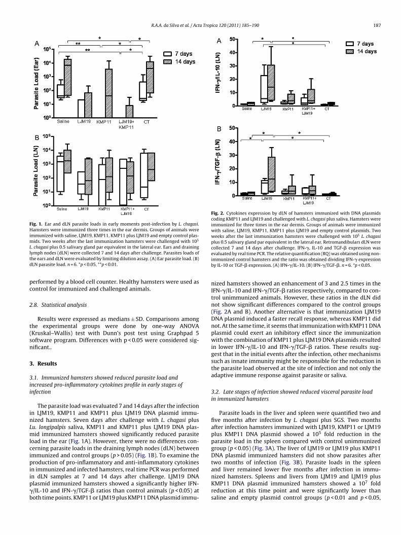
Fig. 1. Ear and dLN parasite loads in early moments post-infection by L. chagasi.Hamsters were immunized three times in the ear dermis. Groups of animals wereimmunized with saline, LJM19, KMP11, KMP11 plus LJM19 and empty control plas-mids. Two weeks after the last immunization hamsters were challenged with 105
L. chagasi plus 0.5 salivary gland par equivalent in the lateral ear. Ears and drainingltd
pc
2
t(sn
3
3ii
inLmlcipiip�b
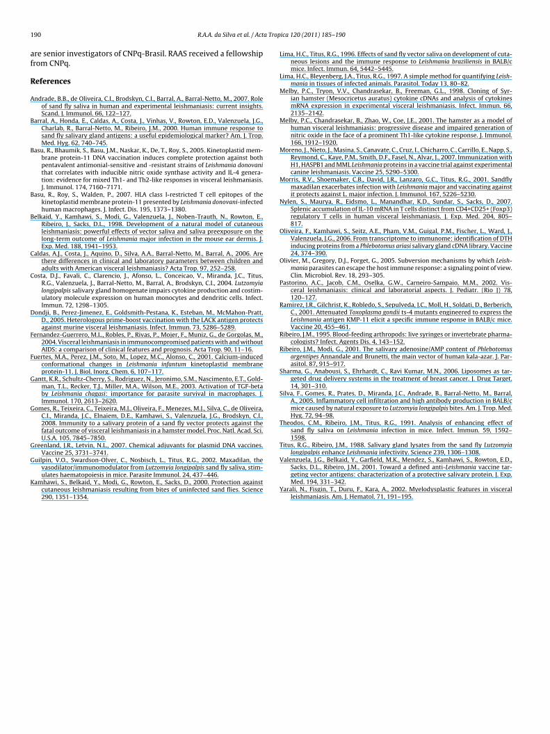
Fig. 2. Cytokines expression by dLN of hamsters immunized with DNA plasmidscoding KMP11 and LJM19 and challenged with L. chagasi plus saliva. Hamsters wereimmunized for three times in the ear dermis. Groups of animals were immunizedwith saline, LJM19, KMP11, KMP11 plus LJM19 and empty control plasmids. Twoweeks after the last immunization hamsters were challenged with 105 L. chagasiplus 0.5 salivary gland par equivalent in the lateral ear. Retromandibulars dLN werecollected 7 and 14 days after challenge. IFN-�, IL-10 and TGF-� expression wasevaluated by real time PCR. The relative quantification (RQ) was obtained using non-
ymph nodes (dLN) were collected 7 and 14 days after challenge. Parasites loads ofhe ears and dLN were evaluated by limiting dilution assay. (A) Ear parasite load. (B)LN parasite load. n = 6. *p < 0.05, **p < 0.01.
erformed by a blood cell counter. Healthy hamsters were used asontrol for immunized and challenged animals.
.8. Statistical analysis
Results were expressed as medians ± SD. Comparisons amonghe experimental groups were done by one-way ANOVAKruskal–Wallis) test with Dunn’s post test using Graphpad 5oftware program. Differences with p < 0.05 were considered sig-ificant..
. Results
.1. Immunized hamsters showed reduced parasite load andncreased pro-inflammatory cytokines profile in early stages ofnfection
The parasite load was evaluated 7 and 14 days after the infectionn LJM19, KMP11 and KMP11 plus LJM19 DNA plasmid immu-ized hamsters. Seven days after challenge with L. chagasi plusu. longipalpis saliva, KMP11 and KMP11 plus LJM19 DNA plas-id immunized hamsters showed significantly reduced parasite
oad in the ear (Fig. 1A). However, there were no differences con-erning parasite loads in the draining lymph nodes (dLN) betweenmmunized and control groups (p > 0.05) (Fig. 1B). To examine theroduction of pro-inflammatory and anti-inflammatory cytokines
n immunized and infected hamsters, real time PCR was performed
n dLN samples at 7 and 14 days after challenge. LJM19 DNAlasmid immunized hamsters showed a significantly higher IFN-/IL-10 and IFN-�/TGF-� ratios than control animals (p < 0.05) atoth time points. KMP11 or LJM19 plus KMP11 DNA plasmid immu-immunized control hamsters and the ratio was obtained dividing IFN-� expressionby IL-10 or TGF-� expression. (A) IFN-�/IL-10. (B) IFN-�/TGF-�. n = 6. *p < 0.05.
nized hamsters showed an enhancement of 3 and 2.5 times in theIFN-�/IL-10 and IFN-�/TGF-� ratios respectively, compared to con-trol unimmunized animals. However, these ratios in the dLN didnot show significant differences compared to the control groups(Fig. 2A and B). Another alternative is that immunization LJM19DNA plasmid induced a faster recall response, whereas KMP11 didnot. At the same time, it seems that immunization with KMP11 DNAplasmid could exert an inhibitory effect since the immunizationwith the combination of KMP11 plus LJM19 DNA plasmids resultedin lower IFN-�/IL-10 and IFN-�/TGF-� ratios. These results sug-gest that in the initial events after the infection, other mechanismssuch as innate immunity might be responsible for the reduction inthe parasite load observed at the site of infection and not only theadaptive immune response against parasite or saliva.
3.2. Late stages of infection showed reduced visceral parasite loadin immunized hamsters
Parasite loads in the liver and spleen were quantified two andfive months after infection by L. chagasi plus SGS. Two monthsafter infection hamsters immunized with LJM19, KMP11 or LJM19plus KMP11 DNA plasmid showed a 105 fold reduction in theparasite load in the spleen compared with control unimmunizedgroup (p < 0.05) (Fig. 3A). The liver of LJM19 or LJM19 plus KMP11DNA plasmid immunized hamsters did not show parasites aftertwo months of infection (Fig. 3B). Parasite loads in the spleenand liver remained lower five months after infection in immu-
nized hamsters. Spleens and livers from LJM19 and LJM19 plusKMP11 DNA plasmid immunized hamsters showed a 107 foldreduction at this time point and were significantly lower thansaline and empty plasmid control groups (p < 0.01 and p < 0.05,
188 R.A.A. da Silva et al. / Acta Tropica 120 (2011) 185– 190
Fig. 3. Liver and spleen parasite loads of hamsters immunized with DNA plasmidscoding KMP11 and LJM19 and challenged with L. chagasi plus saliva. Hamsters wereimmunized for three times in the ear dermis. Groups of animals were immunizedwith saline, LJM19, KMP11, KMP11 plus LJM19 and control plasmids. Two weeksafter the last immunization hamsters were challenged with 105 L. chagasi plus 0.5salivary gland par equivalent in the lateral ear. Two and five months after challengeswl
rdnKii�iha
3a
dHdioetgamahdsm
Fig. 4. Spleen cytokine expression in immunized hamsters challenged with L. cha-gasi plus saliva. Hamsters were immunized for three times in the ear dermis. Groupsof animals were immunized with saline, LJM19, KMP11, KMP11 plus LJM19 andempty control plasmids. Two weeks after the last immunization hamsters werechallenged with 105 L. chagasi plus 0.5 salivary gland par equivalent in the lat-eral ear. Two and five months after challenge spleen samples were collected. IFN-�,IL-10 and TGF-� production was evaluated by real time PCR. The relative quantifi-cation (RQ) was obtained using non-immunized control hamsters and the ratio was
pleen and liver were collected and weighted. Parasites loads of the spleen and liverere evaluated by limiting dilution assay. (A). Spleen parasite load. (B). Liver parasite
oad. n = 5–9. *p < 0.05.
espectively) (Fig. 3). At two months post-infection no significantifferences were observed in IFN-�/IL-10 ratio in immunized andon-immunized hamster groups (Fig. 4A). However, the group ofMP11 DNA plasmid immunized hamsters showed a significant
ncreased IFN-�/TGF-� ratio (Fig. 4B). At five months post-infectionmmunized groups showed an increase in IFN-�/IL-10 and IFN-/TGF-� ratio. However, control group (saline) also showed an
ncrease in this ratio as well as empty vectors, suggesting that theigh number of parasites at this point could be responsible for thislteration in cytokine profile.
.3. Immunized hamsters do not develop hematological disordersfter challenge with L. chagasi
It is well known that patients with visceral leishmaniasisevelop hematological disorders (Fernandez-Guerrero et al., 2004).amsters can reproduce many of these clinical features andevelop anemia, leukopenia and pancytopenia. To examine if
mmunization with DNA plasmids could preclude anemia devel-pment, blood samples from different groups of hamsters werevaluated two and five months after challenge. We observedhat immunization of hamsters with DNA coding plasmids abro-ated the development of anemia. Healthy hamsters were useds controls. Compared with healthy hamsters, our control ani-als showed a decreased number of red blood cells (p < 0.05),
decreased hematocrit (p < 0.05) and a reduced amount of
emoglobin (p < 0.05). However, immunized hamsters did notevelop reduction in these hematological parameters and did nothow significant differences compared to the healthy control ani-al group (p > 0.05) (Table 1 ).obtained dividing IFN-� expression by IL-10 or TGF-� expression. (A) IFN-�/IL-10.(B) IFN-�/TGF-�. n = 5–9. *p < 0.05, **p < 0.01.
4. Discussion
Previous studies from other groups as well as ours have demon-strated protection against experimental visceral leishmaniasisusing plasmids coding KMP11, a parasite product, or LJM19, a prod-uct from the vector saliva (Basu et al., 2005; Gomes et al., 2008). Inthis report, we show that such effects are not addictive as the pro-tection obtained with a combination of KMP11 + LJM19 was similarto the levels obtained with either product used separately. Accord-ingly, the production of IFN-�, TGF-� as well as IL-10 was similar inall immunized groups. Although no significant differences in para-site load were found in the draining lymph nodes in the early stagesof infection (7 and 14 days), hamsters immunized with KMP11 andLJM19 plus KMP11, did not showed a significant reduction in para-site load at 7 and 14 days after infection in the ears. This reductionpersisted in the spleen and liver in later stages of infection, possi-bly, reflecting the initial control in the number of parasites throughinnate immunity and/or increase in levels of IFN-� produced in theearly stages of the infection.
An enhancement of IFN-� early production in lymph nodescould be important for parasite control at the inoculation siteand could be related with lower visceralization in immunizedgroups. Many studies show the important role of IL-10 as anti-inflammatory cytokine, deactivating macrophages, diminishing NOproduction and facilitating Leishmania survival inside phagocytes(Olivier et al., 2005). Similarly, TGF-� is an important cytokinefor parasite survival and disease progression (Gantt et al., 2003).Interestingly, our control hamsters showed higher ratios of IFN-�/IL-10 and IFN-�/TGF-� in the spleen five months after challenge.
These ratios are in agreement with previous studies using cellsfrom human VL patients, which show elevated parasite load evenin a pro-inflammatory environment (Nylen et al., 2007). The level
R.A.A. da Silva et al. / Acta Trop
Tab
le
1Er
itro
gram
from
ham
ster
s
imm
un
ized
wit
h
DN
A
pla
smid
s
cod
ing
KM
P11
and
LJM
19
and
chal
len
ged
wit
h
L.
infa
ntum
chag
asi p
lus
sali
va
of
Lutz
omyi
a
long
ipal
pis.
Hea
lth
ySa
lin
eLj
m19
Km
p11
Km
p11
+
Ljm
19C
T
2
Mon
ths
5
mon
ths
2
Mon
ths
5
mon
ths
2
Mon
ths
5
mon
ths
2
Mon
ths
5
mon
ths
2 M
onth
s
5
mon
ths
Red
bloo
d
cell
s
(106
)
8.5
±
0.1
8.1
±
0.1
6.8
±
0.4*
8.4
±
0.2
8.4
±
0.1
8.2
±
0.1
7.7
±
0.4
7.8
±
0.2
8.5
±
0.3
7.8
±
0.2
6.9
±
0.4*
Hem
oglo
bin
(g%
)16
.1±
0.4
14.9
±
0.3
13.2
±
1.1*
16.2
±
0.4
14.9
±
0.5
16.1
±
0.2
14.1
±
0.7
14.4
±
0.4
15.8
± 0.
4 14
.5
±
0.7
13.1
±
0.6*
Hem
atoc
rit
(%)
47.5
±
0.6
45.1
±
0.5
37.3
±
1.7*
45.8
±
1.0
47.6
±
1.6
46.4
±
0.6
47.7
±
2.1
43.7
±
1.2
47.9
± 1.
9
43.6
±
1.2
37.6
±
2.2*
n
=
6.*
p
<
0.05
.
ica 120 (2011) 185– 190 189
of IFN-�/TGF-� probably increased as a response to an excessivepathology caused by sustained infection (Gomes et al., 2008). Inter-estingly, immunized hamsters did not show sterilizing infection,however they did not develop clinical manifestations of diseaseneither hematological alterations, The role of phlebotomine sandfly saliva has been shown both in animal and human models (Barralet al., 2000; Costa et al., 2004; Guilpin et al., 2002) The formaldemonstration that mice immunized with DNA plasmids codingSP15, a P. papatasi salivary component, and hamsters immunizedwith DNA plasmid coding LJM19, a Lu. longipalpis salivary pro-tein, protect these animals against L. major and L. chagasi infectionrespectively, pointing out for new topics in vaccination againstLeishmania infection (Gomes et al., 2008; Valenzuela et al., 2001).These studies showed that DTH development at the site bites pro-mote protective immunity against Leishmania.
Recently, it was demonstrated that immunization with DNAplasmid coding KMP11, a conserved protein in different parasitespecies, protected hamsters against L. donovani infection (Basuet al., 2005; Fuertes et al., 2001; Ramirez et al., 2001). This pro-tection was related with an enhancement of inducible nitric oxidesynthetase, IFN-� and IL-4 production, a reduction of IL-10 andanti-Leishmania antibodies production (Basu et al., 2005). Our dataconfirmed the protection conferred by KMP11, using L. chagasi plussaliva as challenge.
Blood disorders are frequently observed both in humans andanimal models for visceral leishmaniasis (Caldas et al., 2006;Fernandez-Guerrero et al., 2004; Moreno et al., 2007). Similarly tohuman patients, hamsters develop anemia and leucopenia (Melbyet al., 1998). Parasitism and higher cytokine production can inducealterations in hematopoetic cell behavior conducting to pancytope-nia (Pastorino et al., 2002; Yarali et al., 2002). Interestingly, KMP11and/or LJM19 DNA plasmid immunized hamsters did not showhematological disorders even 5 months after challenge when com-pared with healthy controls, suggesting that the control of parasitenumber also correlated to the better condition of the animals.
Interestingly, in our study the combination of DNA plasmidscoding KMP11 and LJM19 did not enhance the protection observedin immunized hamsters. Different immunization protocols canbe used to reach higher protection. Heterelogous prime/boosterimmunization, using DNA and protein, has shown excellent resultsin vaccine development protocols (Dondji et al., 2005). Similarly,Leishmania antigens entrapment in liposomes and nanoparticleshas demonstrated to induce Th1 profiles in different animal mod-els, contributing to the control of intracellular microoganisms(Greenland and Letvin, 2007; Sharma et al., 2006).
In summary, we have confirmed the ability of KMP11 DNA plas-mid to induce a protective immune response against L. chagasiinfection in the hamster model. We confirm the previous resultsshowing protection against this Leishmania specie in hamstersimmunized with LJM19 DNA plasmid. This protection is followedby an enhancement of IFN-� production in the dLN and a reductionin parasites load in the liver and spleen. Interestingly, we demon-strated for the first time that the immunization with these plasmidscan abrogate hematological disorders. However, the combinationof DNA plasmids coding KMP11 or LJM19 does not enhance theseprotective effects probably because the immune response elicitedby these plasmid immunization could be acting more in the initialphase of infection (presence of saliva) and in promastigotes that aremore abundant in the early infection stages, conferring protectionin this model.
Acknowledgements
We thank Edivaldo Passos for technical assistance. This researchwas supported by FAPESB, CNPq and CAPES. CB, AB, CIO and MB-N
1 a Trop
af
R
A
B
B
B
B
C
C
D
F
F
G
G
G
G
K
90 R.A.A. da Silva et al. / Act
re senior investigators of CNPq-Brasil. RAAS received a fellowshiprom CNPq.
eferences
ndrade, B.B., de Oliveira, C.I., Brodskyn, C.I., Barral, A., Barral-Netto, M., 2007. Roleof sand fly saliva in human and experimental leishmaniasis: current insights.Scand. J. Immunol. 66, 122–127.
arral, A., Honda, E., Caldas, A., Costa, J., Vinhas, V., Rowton, E.D., Valenzuela, J.G.,Charlab, R., Barral-Netto, M., Ribeiro, J.M., 2000. Human immune response tosand fly salivary gland antigens: a useful epidemiological marker? Am. J. Trop.Med. Hyg. 62, 740–745.
asu, R., Bhaumik, S., Basu, J.M., Naskar, K., De, T., Roy, S., 2005. Kinetoplastid mem-brane protein-11 DNA vaccination induces complete protection against bothpentavalent antimonial-sensitive and -resistant strains of Leishmania donovanithat correlates with inducible nitric oxide synthase activity and IL-4 genera-tion: evidence for mixed Th1- and Th2-like responses in visceral leishmaniasis.J. Immunol. 174, 7160–7171.
asu, R., Roy, S., Walden, P., 2007. HLA class I-restricted T cell epitopes of thekinetoplastid membrane protein-11 presented by Leishmania donovani-infectedhuman macrophages. J. Infect. Dis. 195, 1373–1380.
elkaid, Y., Kamhawi, S., Modi, G., Valenzuela, J., Noben-Trauth, N., Rowton, E.,Ribeiro, J., Sacks, D.L., 1998. Development of a natural model of cutaneousleishmaniasis: powerful effects of vector saliva and saliva preexposure on thelong-term outcome of Leishmania major infection in the mouse ear dermis. J.Exp. Med. 188, 1941–1953.
aldas, A.J., Costa, J., Aquino, D., Silva, A.A., Barral-Netto, M., Barral, A., 2006. Arethere differences in clinical and laboratory parameters between children andadults with American visceral leishmaniasis? Acta Trop. 97, 252–258.
osta, D.J., Favali, C., Clarencio, J., Afonso, L., Conceicao, V., Miranda, J.C., Titus,R.G., Valenzuela, J., Barral-Netto, M., Barral, A., Brodskyn, C.I., 2004. Lutzomyialongipalpis salivary gland homogenate impairs cytokine production and costim-ulatory molecule expression on human monocytes and dendritic cells. Infect.Immun. 72, 1298–1305.
ondji, B., Perez-Jimenez, E., Goldsmith-Pestana, K., Esteban, M., McMahon-Pratt,D., 2005. Heterologous prime-boost vaccination with the LACK antigen protectsagainst murine visceral leishmaniasis. Infect. Immun. 73, 5286–5289.
ernandez-Guerrero, M.L., Robles, P., Rivas, P., Mojer, F., Muniz, G., de Gorgolas, M.,2004. Visceral leishmaniasis in immunocompromised patients with and withoutAIDS: a comparison of clinical features and prognosis. Acta Trop. 90, 11–16.
uertes, M.A., Perez, J.M., Soto, M., Lopez, M.C., Alonso, C., 2001. Calcium-inducedconformational changes in Leishmania infantum kinetoplastid membraneprotein-11. J. Biol. Inorg. Chem. 6, 107–117.
antt, K.R., Schultz-Cherry, S., Rodriguez, N., Jeronimo, S.M., Nascimento, E.T., Gold-man, T.L., Recker, T.J., Miller, M.A., Wilson, M.E., 2003. Activation of TGF-betaby Leishmania chagasi: importance for parasite survival in macrophages. J.Immunol. 170, 2613–2620.
omes, R., Teixeira, C., Teixeira, M.J., Oliveira, F., Menezes, M.J., Silva, C., de Oliveira,C.I., Miranda, J.C., Elnaiem, D.E., Kamhawi, S., Valenzuela, J.G., Brodskyn, C.I.,2008. Immunity to a salivary protein of a sand fly vector protects against thefatal outcome of visceral leishmaniasis in a hamster model. Proc. Natl. Acad. Sci.U.S.A. 105, 7845–7850.
reenland, J.R., Letvin, N.L., 2007. Chemical adjuvants for plasmid DNA vaccines.Vaccine 25, 3731–3741.
uilpin, V.O., Swardson-Olver, C., Nosbisch, L., Titus, R.G., 2002. Maxadilan, the
vasodilator/immunomodulator from Lutzomyia longipalpis sand fly saliva, stim-ulates haematopoiesis in mice. Parasite Immunol. 24, 437–446.amhawi, S., Belkaid, Y., Modi, G., Rowton, E., Sacks, D., 2000. Protection againstcutaneous leishmaniasis resulting from bites of uninfected sand flies. Science290, 1351–1354.
ica 120 (2011) 185– 190
Lima, H.C., Titus, R.G., 1996. Effects of sand fly vector saliva on development of cuta-neous lesions and the immune response to Leishmania braziliensis in BALB/cmice. Infect. Immun. 64, 5442–5445.
Lima, H.C., Bleyenberg, J.A., Titus, R.G., 1997. A simple method for quantifying Leish-mania in tissues of infected animals. Parasitol. Today 13, 80–82.
Melby, P.C., Tryon, V.V., Chandrasekar, B., Freeman, G.L., 1998. Cloning of Syr-ian hamster (Mesocricetus auratus) cytokine cDNAs and analysis of cytokinesmRNA expression in experimental visceral leishmaniasis. Infect. Immun. 66,2135–2142.
Melby, P.C., Chandrasekar, B., Zhao, W., Coe, J.E., 2001. The hamster as a model ofhuman visceral leishmaniasis: progressive disease and impaired generation ofnitric oxide in the face of a prominent Th1-like cytokine response. J. Immunol.166, 1912–1920.
Moreno, J., Nieto, J., Masina, S., Canavate, C., Cruz, I., Chicharro, C., Carrillo, E., Napp, S.,Reymond, C., Kaye, P.M., Smith, D.F., Fasel, N., Alvar, J., 2007. Immunization withH1, HASPB1 and MML Leishmania proteins in a vaccine trial against experimentalcanine leishmaniasis. Vaccine 25, 5290–5300.
Morris, R.V., Shoemaker, C.B., David, J.R., Lanzaro, G.C., Titus, R.G., 2001. Sandflymaxadilan exacerbates infection with Leishmania major and vaccinating againstit protects against L. major infection. J. Immunol. 167, 5226–5230.
Nylen, S., Maurya, R., Eidsmo, L., Manandhar, K.D., Sundar, S., Sacks, D., 2007.Splenic accumulation of IL-10 mRNA in T cells distinct from CD4+CD25+ (Foxp3)regulatory T cells in human visceral leishmaniasis. J. Exp. Med. 204, 805–817.
Oliveira, F., Kamhawi, S., Seitz, A.E., Pham, V.M., Guigal, P.M., Fischer, L., Ward, J.,Valenzuela, J.G., 2006. From transcriptome to immunome: identification of DTHinducing proteins from a Phlebotomus ariasi salivary gland cDNA library. Vaccine24, 374–390.
Olivier, M., Gregory, D.J., Forget, G., 2005. Subversion mechanisms by which Leish-mania parasites can escape the host immune response: a signaling point of view.Clin. Microbiol. Rev. 18, 293–305.
Pastorino, A.C., Jacob, C.M., Oselka, G.W., Carneiro-Sampaio, M.M., 2002. Vis-ceral leishmaniasis: clinical and laboratorial aspects. J. Pediatr. (Rio J) 78,120–127.
Ramirez, J.R., Gilchrist, K., Robledo, S., Sepulveda, J.C., Moll, H., Soldati, D., Berberich,C., 2001. Attenuated Toxoplasma gondii ts-4 mutants engineered to express theLeishmania antigen KMP-11 elicit a specific immune response in BALB/c mice.Vaccine 20, 455–461.
Ribeiro, J.M., 1995. Blood-feeding arthropods: live syringes or invertebrate pharma-cologists? Infect. Agents Dis. 4, 143–152.
Ribeiro, J.M., Modi, G., 2001. The salivary adenosine/AMP content of Phlebotomusargentipes Annandale and Brunetti, the main vector of human kala-azar. J. Par-asitol. 87, 915–917.
Sharma, G., Anabousi, S., Ehrhardt, C., Ravi Kumar, M.N., 2006. Liposomes as tar-geted drug delivery systems in the treatment of breast cancer. J. Drug Target.14, 301–310.
Silva, F., Gomes, R., Prates, D., Miranda, J.C., Andrade, B., Barral-Netto, M., Barral,A., 2005. Inflammatory cell infiltration and high antibody production in BALB/cmice caused by natural exposure to Lutzomyia longipalpis bites. Am. J. Trop. Med.Hyg. 72, 94–98.
Theodos, C.M., Ribeiro, J.M., Titus, R.G., 1991. Analysis of enhancing effect ofsand fly saliva on Leishmania infection in mice. Infect. Immun. 59, 1592–1598.
Titus, R.G., Ribeiro, J.M., 1988. Salivary gland lysates from the sand fly Lutzomyialongipalpis enhance Leishmania infectivity. Science 239, 1306–1308.
Valenzuela, J.G., Belkaid, Y., Garfield, M.K., Mendez, S., Kamhawi, S., Rowton, E.D.,
Sacks, D.L., Ribeiro, J.M., 2001. Toward a defined anti-Leishmania vaccine tar-geting vector antigens: characterization of a protective salivary protein. J. Exp.Med. 194, 331–342.Yarali, N., Fisgin, T., Duru, F., Kara, A., 2002. Myelodysplastic features in visceralleishmaniasis. Am. J. Hematol. 71, 191–195.
Copyright © 2022 FDOKUMEN