Phylogeography of the Lutzomyia gomezi (Diptera: Phlebotominae) on the Panama Isthmus
11
RESEARCH Open Access Phylogeography of the Lutzomyia gomezi (Diptera: Phlebotominae) on the Panama Isthmus Anayansi Valderrama 1* , Mara Garcia Tavares 2 and Jose Dilermando Andrade Filho 3 Abstract Background: Lutzomyia gomezi (Nitzulescu, 1931) is one of the main Leishmania ( Vianna) panamensis vectors in Panama, and despite its medical significance, there are no population genetic studies regarding this species. In this study, we used the sequences of the mitochondrial gene cytochrome b/start of NADH1 and the nuclear elongation gene α-1 in order to analyze genetic variation and phylogeographic structure of the Lu. gomezi populations. Methods: A total of 86 Lu. gomezi individuals were captured in 38 locations where cutaneous leishmaniasis occurred. DNA was extracted with phenol/chloroform methods and amplification of genes was performed using PCR primers for mitochondrial and nuclear markers. Results: We found a total of 37 and 26 haplotypes of mitochondrial and nuclear genes, high haplotype diversity (h) for all three populations were detected with both molecular markers. Nucleotide diversity (π) was estimated to be high for all three populations with the mitochondrial marker, which was opposite to the estimate with the nuclear marker. In the AMOVA Φst recorded moderate (mitochondrial) and small (nuclear) population structure with statistical significance among populations. The analysis of the fixation index (Fst) used to measure the differentiation of populations showed that with the exception of the population located in the region of Bocas del Toro, the other populations presented with minor genetic differentiation. The median-Joining network of the mitochondrial marker reveled three clusters and recorded four haplotypes exclusively of localities sampled from Western Panama, demonstrating strong divergence. We found demographic population expansion with Fu´s Fs neutrality test. In the analysis mismatch distribution was observed as a bimodal curve. Conclusion: Lu. gomezi is a species with higher genetic pool or variability and mild population structure, due to possible capacity migration and local adaptation to environmental changes or colonization potential. Thus, knowledge of the genetic population and evolutionary history is useful to understand the implications of different population genetic structures for cutaneous leishmaniasis epidemiology. Keywords: Panamá, Sandflies, Leishmania panamensis, Cutaneous leishmaniasis, Genetic variability and phylogeographic Background Cutaneous leishmaniasis is the most common form of leishmaniasis reported in the Republic of Panama; its clinical manifestations range from minor lesions to severe skin ulcers [1-3]. In Panama it was first recorded in 1910 [4], an overall of 15 cases occurred during 1910–1944; but there has been a sharp increase since 2000 [5]. According to the Epidemiology Department of the Health Ministry of Panama, a total of 26,163 cases of cutaneous leishmaniasis occurred during 2000–2010 and the distribu- tion of occurrence of CL was in regions of the provinces of Bocas de Toro (28%), Panama West (20%), Coclé (17%), Colón (11%), Panama East (5%), Veraguas (3%). The disease is mainly caused by Leishmania (Vianna) panamensis and Lutzomyia ylephiletor , Lu. sanguinaria, Lu. panamensis, Lu. trapidoi, Lu. gomezi were identified as the transmission vectors [6-8]. In Panama, recent research has show that Lutzomyia (Lutzomyia) gomezi (Nitzulescu, 1931) is the most abun- dant species with wide geographical distribution and their abundance has been associated with cases of clinical * Correspondence: [email protected] 1 Department of Medical Entomology, Instituto Conmemorativo Gorgas de Estudios de la Salud, Panama, Panama Full list of author information is available at the end of the article © 2014 Valderrama et al.; licensee BioMed Central Ltd. This is an open access article distributed under the terms of the Creative Commons Attribution License (http://creativecommons.org/licenses/by/2.0), which permits unrestricted use, distribution, and reproduction in any medium, provided the original work is properly cited. Valderrama et al. Parasites & Vectors 2014, 7:9 http://www.parasitesandvectors.com/content/7/1/9
Transcript of Phylogeography of the Lutzomyia gomezi (Diptera: Phlebotominae) on the Panama Isthmus

Valderrama et al. Parasites & Vectors 2014, 7:9http://www.parasitesandvectors.com/content/7/1/9
RESEARCH Open Access
Phylogeography of the Lutzomyia gomezi(Diptera: Phlebotominae) on the Panama IsthmusAnayansi Valderrama1*, Mara Garcia Tavares2 and Jose Dilermando Andrade Filho3
Abstract
Background: Lutzomyia gomezi (Nitzulescu, 1931) is one of the main Leishmania (Vianna) panamensis vectors inPanama, and despite its medical significance, there are no population genetic studies regarding this species. In thisstudy, we used the sequences of the mitochondrial gene cytochrome b/start of NADH1 and the nuclear elongationgene α-1 in order to analyze genetic variation and phylogeographic structure of the Lu. gomezi populations.
Methods: A total of 86 Lu. gomezi individuals were captured in 38 locations where cutaneous leishmaniasisoccurred. DNA was extracted with phenol/chloroform methods and amplification of genes was performed usingPCR primers for mitochondrial and nuclear markers.
Results: We found a total of 37 and 26 haplotypes of mitochondrial and nuclear genes, high haplotype diversity (h)for all three populations were detected with both molecular markers. Nucleotide diversity (π) was estimated to behigh for all three populations with the mitochondrial marker, which was opposite to the estimate with the nuclearmarker. In the AMOVA Φst recorded moderate (mitochondrial) and small (nuclear) population structure withstatistical significance among populations. The analysis of the fixation index (Fst) used to measure thedifferentiation of populations showed that with the exception of the population located in the region of Bocas delToro, the other populations presented with minor genetic differentiation. The median-Joining network of themitochondrial marker reveled three clusters and recorded four haplotypes exclusively of localities sampled fromWestern Panama, demonstrating strong divergence. We found demographic population expansion with Fu´s Fsneutrality test. In the analysis mismatch distribution was observed as a bimodal curve.
Conclusion: Lu. gomezi is a species with higher genetic pool or variability and mild population structure, due topossible capacity migration and local adaptation to environmental changes or colonization potential. Thus,knowledge of the genetic population and evolutionary history is useful to understand the implications of differentpopulation genetic structures for cutaneous leishmaniasis epidemiology.
Keywords: Panamá, Sandflies, Leishmania panamensis, Cutaneous leishmaniasis, Genetic variability andphylogeographic
BackgroundCutaneous leishmaniasis is the most common form ofleishmaniasis reported in the Republic of Panama; itsclinical manifestations range from minor lesions to severeskin ulcers [1-3]. In Panama it was first recorded in 1910[4], an overall of 15 cases occurred during 1910–1944;but there has been a sharp increase since 2000 [5].According to the Epidemiology Department of the HealthMinistry of Panama, a total of 26,163 cases of cutaneous
* Correspondence: [email protected] of Medical Entomology, Instituto Conmemorativo Gorgas deEstudios de la Salud, Panama, PanamaFull list of author information is available at the end of the article
© 2014 Valderrama et al.; licensee BioMed CenCreative Commons Attribution License (http:/distribution, and reproduction in any medium
leishmaniasis occurred during 2000–2010 and the distribu-tion of occurrence of CL was in regions of the provinces ofBocas de Toro (28%), Panama West (20%), Coclé (17%),Colón (11%), Panama East (5%), Veraguas (3%). The diseaseis mainly caused by Leishmania (Vianna) panamensis andLutzomyia ylephiletor, Lu. sanguinaria, Lu. panamensis,Lu. trapidoi, Lu. gomezi were identified as the transmissionvectors [6-8].In Panama, recent research has show that Lutzomyia
(Lutzomyia) gomezi (Nitzulescu, 1931) is the most abun-dant species with wide geographical distribution andtheir abundance has been associated with cases of clinical
tral Ltd. This is an open access article distributed under the terms of the/creativecommons.org/licenses/by/2.0), which permits unrestricted use,, provided the original work is properly cited.
Valderrama et al. Parasites & Vectors 2014, 7:9 Page 2 of 11http://www.parasitesandvectors.com/content/7/1/9
CL acquired in households of rural communities [9].Alongside Lu. gomezi other Lutzomyia species vectorshave been caught in the same place and associated with afocus of cutaneous leishmaniasis infection in otherAmerican countries, incriminating this species with highpotential in the transmission of the disease [2,10-14].Lutzomyia (Lutzomia) gomezi in Central America is
present from Mexico to Panama and Trinidad Tobago(Caribbean), also recently reported in Guatemala [14]. InSouth America, it has been reported in Colombia,Venezuela, Ecuador, Peru, French Guiana and Brazil,specifically in Amapá, Acre, Pará, Mata Grosso, Goiás,Bahia, Maranhão and Rondônia [15-17].In general, its natural environment is a humid and
dark place, such as nests, rock crevices, animal burrowsand tree bark in the tropical rainforest [2]. However, it isalso reported in forest gaps and the canopies were lightavailability produces humid change [2,18]. The foragingand blood feeding behavior occurs at twilight and atnight from 18:00–20:00 hours [18]. Nevertheless, thedeforestation and loss of natural habitat have caused itsadaptation to peridomestic areas, perhaps changing thehourly activity and feeding on a large variety of domesticanimals [19,20]. In the focus of leishmaniasis, this spe-cies is predominant near peridomestic or outdoor areasmore than indoors households, which is difficult to con-trol [2,19,21-23].Despite of the potential significance of Lu. gomezi as a
Leishmania vector, few researches have targeted geneticaspects or interrelationship of host-vector species. Forinstance, [24] detected a natural infection of Lu. gomeziwith the Le. braziliensis in Venezuela, in order to establisha methods for determine the circulation of Leishmaniaparasites in leishmaniasis endemics areas. On the otherhand, [25] analyzed the changes to the primary andsecondary structures of tRNAser of the species; Lu.trinidadensis (Oswaldoi group), Lu. (Psychodopygus)panamensis, Lu. (Micropygomyia) cayennensis cayennensis,Lu. dubitans (Migonei group), Lu. (Lutzomyia) gomezi, Lu.rangeliana (ungrouped) and Lu. evansi (Verrucarumgroup) for taxonomic purposes, considering that morpho-logical identification can be difficult. Also, [26] detectedwith ITS-1 a pool of Lu. gomezi infected with Le. naiffi inPanama, the first report for the country which promptedseveral hypotheses on the introduction of this parasite intothis country.Attempts to understand the role that arthropod vectors
play in disease dynamics and pathogen transmission ofleishmaniasis, several studies over genetic population ofsandfly vectors has been performance in the Latin America,Iran, Turkey, Palestine, Israel and Egypt [27-30]. Many ofthese studies are focused in two principal species, Lu.longipalpis and Phlebotomus papatasi to determinatetheir genetic variation, structures and differentiation of
populations [27,28,31,32]. Thereby, molecular evidence fordivergence of vectors has been found and an assessment ofthe impact on leishmaniasis epidemiology. For instance,the taxonomic status of the Lu. longipalpis complex hasbeen fundamental to the understanding of leishmaniasisepidemiology. Symptoms observed for transmission ofLeishmania infantum chagasi by Lu. longipalpis in Braziland Colombia results in visceral infections, whereas thetransmission of the same parasite by Lu. longipalpis inCosta Rica results in non-ulcerative lesions [33,34].Thus, it becomes important to analyze the population
genetics and demographic history of this vector speciesto assess the genetic pool, gene flow and colonization po-tential, consequently known as the population changesand genetic variation affecting the vector competenceand resistance to insecticides [35,36]. On the other hand,the population genetics analysis can provide informationabout migration capacity, reproduction, and adaptationto the conditions of the new habitat, favoring the emer-gence of vector diseases [37]. In addition to this, thedetermination of cryptic vector species and their ability totransmit pathogens are even more relevant to understandtheir implications in the epidemiology of vector diseasesand to suggest appropriate and effective prevention andcontrol programs without any environmental risks [36].Due to the lack of population genetics information of Lu.
gomezi species in Panama and the Americas as a whole,the goal of this research was to evaluate and compare theintra- and inter-population variability of Lu. gomezi fromlocalities with a high incidence of cutaneous leishmaniasisin Panama. Also to identify the barriers that may influencegene flow among Lu. gomezi populations. Moreover, toinfer the historical processes that defines actual geographicdistributions of the species. These results may contributeon the knowledge of leishmaniasis epidemiology and im-prove the development of focal or large-scale programscontrol for leishmaniasis vectors in Panama.
MethodsStudy sitesThe study was performed in the Republic of Panama be-tween the coordinates 7º11′-9°39′N and 77º10′-83º03′W. The most prevalent climatic regime in Panama istropical humid, with the dry season (January-March) pre-senting an average temperature of 31.5ºC and relativehumidity of 75%. In other months (April-December), theaverage temperature is 27ºC and the relative humidityaverages 90%. The vegetation in Panama varies accordingto climate zones and consists mainly of tropical humidforest or savannahs resulting from agricultural activity.
Sandfly collections and identificationLu. gomezi samples were collected from the thirty-eightlocalities in Panama, during the dry season (January-April)
Valderrama et al. Parasites & Vectors 2014, 7:9 Page 3 of 11http://www.parasitesandvectors.com/content/7/1/9
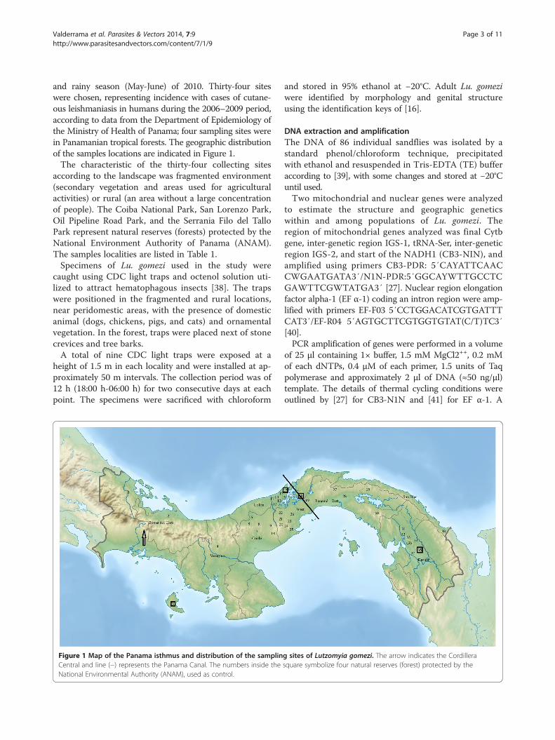
and rainy season (May-June) of 2010. Thirty-four siteswere chosen, representing incidence with cases of cutane-ous leishmaniasis in humans during the 2006–2009 period,according to data from the Department of Epidemiology ofthe Ministry of Health of Panama; four sampling sites werein Panamanian tropical forests. The geographic distributionof the samples locations are indicated in Figure 1.The characteristic of the thirty-four collecting sites
according to the landscape was fragmented environment(secondary vegetation and areas used for agriculturalactivities) or rural (an area without a large concentrationof people). The Coiba National Park, San Lorenzo Park,Oil Pipeline Road Park, and the Serrania Filo del TalloPark represent natural reserves (forests) protected by theNational Environment Authority of Panama (ANAM).The samples localities are listed in Table 1.Specimens of Lu. gomezi used in the study were
caught using CDC light traps and octenol solution uti-lized to attract hematophagous insects [38]. The trapswere positioned in the fragmented and rural locations,near peridomestic areas, with the presence of domesticanimal (dogs, chickens, pigs, and cats) and ornamentalvegetation. In the forest, traps were placed next of stonecrevices and tree barks.A total of nine CDC light traps were exposed at a
height of 1.5 m in each locality and were installed at ap-proximately 50 m intervals. The collection period was of12 h (18:00 h-06:00 h) for two consecutive days at eachpoint. The specimens were sacrificed with chloroform
Figure 1 Map of the Panama isthmus and distribution of the samplinCentral and line (−) represents the Panama Canal. The numbers inside theNational Environmental Authority (ANAM), used as control.
and stored in 95% ethanol at −20°C. Adult Lu. gomeziwere identified by morphology and genital structureusing the identification keys of [16].
DNA extraction and amplificationThe DNA of 86 individual sandflies was isolated by astandard phenol/chloroform technique, precipitatedwith ethanol and resuspended in Tris-EDTA (TE) bufferaccording to [39], with some changes and stored at −20°Cuntil used.Two mitochondrial and nuclear genes were analyzed
to estimate the structure and geographic geneticswithin and among populations of Lu. gomezi. Theregion of mitochondrial genes analyzed was final Cytbgene, inter-genetic region IGS-1, tRNA-Ser, inter-geneticregion IGS-2, and start of the NADH1 (CB3-NIN), andamplified using primers CB3-PDR: 5′CAYATTCAACCWGAATGATA3′/N1N-PDR:5′GGCAYWTTGCCTCGAWTTCGWTATGA3′ [27]. Nuclear region elongationfactor alpha-1 (EF α-1) coding an intron region were amp-lified with primers EF-F03 5′CCTGGACATCGTGATTTCAT3′/EF-R04 5′AGTGCTTCGTGGTGTAT(C/T)TC3′[40].PCR amplification of genes were performed in a volume
of 25 μl containing 1× buffer, 1.5 mM MgCl2++, 0.2 mMof each dNTPs, 0.4 μM of each primer, 1.5 units of Taqpolymerase and approximately 2 μl of DNA (≈50 ng/μl)template. The details of thermal cycling conditions wereoutlined by [27] for CB3-N1N and [41] for EF α-1. A
g sites of Lutzomyia gomezi. The arrow indicates the Cordillerasquare symbolize four natural reserves (forest) protected by the
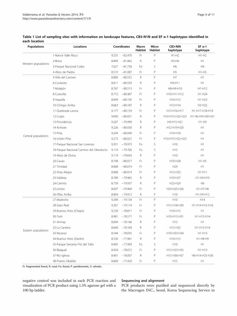
Table 1 List of sampling sites with information on landscape features, CB3-N1N and EF α-1 haplotypes identified ineach location
Populations Locations Coordinates MacroHabitat
MicroHabitat
CB3-NINhaplotype
EF α-1haplotype
Western populations
1-Nance Valle Risco 9.255 −82.470 Fr P H1-H2 H1-H2
2-Bisira 8.899 −81.862 R P H3-H4 H1
3-Parque Nacional Coiba 7.627 −81.730 Fo S H6 H4
4-Altos de Piedra 8.515 −81.087 Fr P H5 H1-H3
Central populations
5-Villa del Carmen 8.800 −80.552 R P H7 H1
6-Coclesito 8.811 −80.550 R P H9-H11 H1
7-Molejón 8.767 −80.513 Fr P H8-H9-H10 H1-H12
8-Cutevilla 8.772 −80.487 Fr P H10-H11-H12 H1-H24
9-Vaquilla 8.699 −80.195 Fr P H10-H12 H1-H23
10-Chirigui Arriba 8.663 −80.187 R P H13-H14 H3-H22
11-Quebrada Leona 9.177 −80.139 Fr P H15-H16-H17 H1-H17-H18-H19
12-Cuipo 9.090 −80.051 R P H10-H15-H22-H23 H1-H6-H9-H20-H21
13-Providencia 9.207 −79.999 R P H9-H15-H21 H1-H9
14-Achiote 9.226 −80.030 R P H12-H19-H20 H1
15-Piña 9.244 −80.040 Fr P H10-H18 H1
16-Unión Piña 9.273 −80.021 Fr P H10-H15-H22-H23 H1
17-Parque Nacional San Lorenzo 9.351 −79.973 Fo S H10 H1
18-Parque Nacional Camino del Oleoducto 9.119 −79.700 Fo S H15 H1
19-Altos de Divisa 9.119 −79.693 R P H10 H1
20-Cacao 8.748 −80.017 Fr P H10-H28 H1-H5
21-Trinidad 8.808 −80.019 Fr P H29 H1
22-Vista Alegre 8.808 −80.014 Fr P H15-H23 H1-H11
23-Valdeza 8.789 −79.965 R P H10-H27 H1-H9-H10
24-Caimito 8.739 −79.937 R P H23-H29 H6
25-Limón 8.697 −79.904 Fr P H24-H25-H26 H1-H7-H8
26-Ollas Arriba 8.804 −79.912 R P H10 H1-H9-H12
Eastern populations
27-Madroño 9.284 −79.134 Fr P H10 H14
28-Gato Real 9.267 −79.119 Fr P H15-H18-H30 H1-H14-H15-H16
29-Buenos Aires (Chepo) 9.239 −78.817 Fr P H10-H15 H1
30-Torti 8.981 −78.571 Fr P H10-H15-H31 H1-H13-H14
31-Arimay 8.694 −78.146 R P H15 H1
32-La Cantera 8.640 −78.169 R P H15-H32 H1-H13-H14
33-Nicanor 8.544 −78.035 Fr P H10-H33-H34 H1-H13
34-Buenos Aires (Darién) 8.530 −77.961 R P H10-H15 H1-H8-H9
35-Parque Serranía Filo del Tallo 8.465 −77.993 Fo S H10 H1
36-Bijagual 8.459 −78.012 Fr P H15-H23-H35 H1-H13
37-Rio Iglesia 8.401 −78.007 R P H15-H36-H37 H8-H14-H25-H26
38-Puerto Obaldía 8.669 −77.429 Fr P H15 H1
Fr: fragmented forest; R: rural; Fo: forest; P: peridomestic; S: selvatic.
Valderrama et al. Parasites & Vectors 2014, 7:9 Page 4 of 11http://www.parasitesandvectors.com/content/7/1/9
negative control was included in each PCR reaction andvisualization of PCR product using 1.5% agarose gel with a100 bp ladder.
Sequencing and alignmentPCR products were purified and sequenced directly bythe Macrogen INC., Seoul, Korea Sequencing Service in

Valderrama et al. Parasites & Vectors 2014, 7:9 Page 5 of 11http://www.parasitesandvectors.com/content/7/1/9
both directions with the same primers used in the amplifi-cations. The sequences were compared to the nucleotideBLAST (Basic Local Alignment Search Tool) tool availablein GenBank NCBI (National Center for BiotechnologyInformation) in order to verify the similarities between thesequences of Lu. gomezi stored in the database. TheCodonCode Aligner software was used to edit the nucleo-tide sequences and identify the heterozygous as doublepeaks on the chromatograms of the nuclear region. Thesequences were aligned with the Muscle option includedin the MEGA 5.05 software [42]. The polymorphic sites,single substitution (singleton) and average base frequen-cies were computed with MEGA 5.05 software.
Data analysisTo carry out an analysis of population genetics (geneticdiversity, structure and differentiation) to specimenscollected in different localities, we clustered and definedsamples in western, central and eastern populations fromPanama (Table 1). Thus, the specimens collected in Bocasdel Toro, Veraguas and Coiba represented the westernpopulations, those collected from Coclé, Colon, and westof the Panama canal were central populations, whereasspecimens from east of the Panama canal, Darién and SanBlas corresponding to the eastern population (Figure 1).
Genetic diversityWe emphasized that heterozygotes detected in the EF α-1were treated with recombination model in PHASE option,incorporated in the DnaSP v.5. To estimate the geneticdiversity we used the following indexes: haplotypes num-ber, haplotype diversity (h), and nucleotide diversity (π)for each population. DnaSP v.5 [43] and Arlequin 3.11[44] software was used for this purpose.
Genetic structure and differentiationThe population structure was determined performing anAnalysis of Molecular Variance (AMOVA) [45,46]. TheArlequin 3.11 [44] software analyzed the variance compo-nents and significance levels p = 0.01 using non-parametricpermutations of 1,000 times. To estimate levels of geneticdifferentiation among the populations a pairwise compari-son test was performed. Non-parametric permutations of1,000 times and a significance p = 0.01 was used to esti-mate Fst-value [45] in Arlequin 3.11 [44] software.
Network haplotypesA haplotype network for both genes was inferredthrough the median-joining network method [47], usingthe Network v.4.5 software. In order to obtain the valuesof posteriori probabilities among the clades, a Bayesiananalysis was calculated using MrBayes 3.1 [48] program.MrModelTest [49] software was used to choose the bestmodel of nucleotide substitution according to the AIC
(Akaike Information Criterion). The model obtained byMrModeltest for CB3-N1N Bayesian inference analysiswas HKY + I, with the proportion of invariable places (I) =0.7594. For EF α-1, the model obtained was K80 + I, with aproportion of invariable places (I) = 0.6725. 10 million gen-erations of Markov Chain Monte Carlo (MCMC) andburn-in of 5 million were used.
Demographic history of populationIn addition to this, Tajima’s D [50] was used for estimatedeviations from neutrality mutation or selective neutral-ity of these populations, P-value 0.01 was consideredstatistically significant. Also Fu’s Fs [51] were calculatedfor neutrality of mutation against population growth,hitchhiking and background selection, P-value 0.02 assignificant. Both values Tajima’s D and Fu’s Fs werecalculated using the software Arlequin 3.11 [44] withpermutations of 1,000 times.The mismatch distribution was calculated for CB3-N1N
and EF α-1 as supplementary test to evidence of demo-graphic expansion of the population, through suddenexpansion model [52]. In Arlequin 3.11 [44] we computedthe sum of square deviations (SSD) and Harpending´sraggedness index to test the goodness-of-fit of the modelto the data. The time since population expansion wasinferred using CB3-NIN region by means of the equationt = τ /2 μ, where tau (τ) was obtained on the mismatchdistribution outputs and a mutation rate (μ) of 1.1-1.2%per million years in Heliconius erato [53].
ResultsSequence characterizationOverall, 86 sequences isolated of CB3-NIN and EF α-1 ofLu. gomezi individuals from 38 selected localities fromPanama Isthmus were analyzed. Of these, an alignment of501 nucleotides was obtained of CB3-N1N with thirty-nine polymorphic sites and nineteen single substitutions(singleton). The average base frequencies were A = 37%,C = 15%, G = 10%, and T = 38% recording a sequence richin A-T (75%). A fragmented size of EF α-1 was 597 bp, inthis case 166 sequences were obtained by clone generatingwith the recombination model algorithm of DnaSP v.5considering the heterozygous individuals. Of thosesequences, thirty-three polymorphic sites, six singletonsand a larger proportion of G-C (53%): 23%T, 27%C, 23%A,27%G, were detected.
Genetic diversityFor the analysis of genetic diversity indexes both CB3-NINand EF α-1 region demonstrated high haplotype diversity(h) for all three populations [Table 2]. While the nucleotidediversity (π) for the mitochondrial region was estimated tobe high for all three populations, nuclear nucleotide diver-sity was opposite to the estimate with mitochondrial,
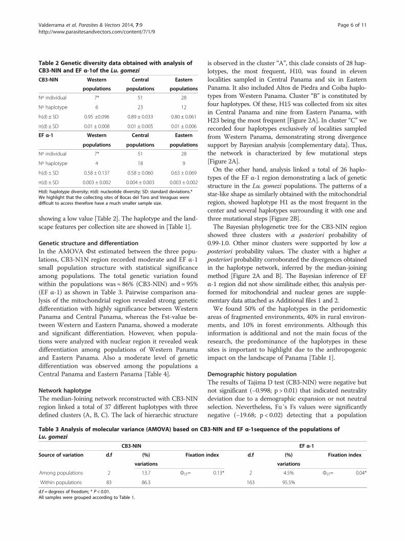
Table 2 Genetic diversity data obtained with analysis ofCB3-NIN and EF α-1of the Lu. gomezi
CB3-NIN Western Central Eastern
populations populations populations
Nº individual 7* 51 28
Nº haplotype 6 23 12
h(d) ± SD 0.95 ±0.096 0.89 ± 0.033 0.80 ± 0.061
π(d) ± SD 0.01 ± 0.008 0.01 ± 0.005 0.01 ± 0.006
EF α-1 Western Central Eastern
populations populations populations
Nº individual 7* 51 28
Nº haplotype 4 18 9
h(d) ± SD 0.58 ± 0.137 0.58 ± 0.060 0.63 ± 0.069
π(d) ± SD 0.003 ± 0.002 0.004 ± 0.003 0.003 ± 0.002
H(d): haplotype diversity; π(d): nucleotide diversity; SD: standard deviations.*We highlight that the collecting sites of Bocas del Toro and Veraguas weredifficult to access therefore have a much smaller sample size.
Valderrama et al. Parasites & Vectors 2014, 7:9 Page 6 of 11http://www.parasitesandvectors.com/content/7/1/9
showing a low value [Table 2]. The haplotype and the land-scape features per collection site are showed in [Table 1].
Genetic structure and differentiationIn the AMOVA Φst estimated between the three popu-lations, CB3-N1N region recorded moderate and EF α-1small population structure with statistical significanceamong populations. The total genetic variation foundwithin the populations was ≈ 86% (CB3-NIN) and ≈ 95%(EF α-1) as shown in Table 3. Pairwise comparison ana-lysis of the mitochondrial region revealed strong geneticdifferentiation with highly significance between WesternPanama and Central Panama, whereas the Fst-value be-tween Western and Eastern Panama, showed a moderateand significant differentiation. However, when popula-tions were analyzed with nuclear region it revealed weakdifferentiation among populations of Western Panamaand Eastern Panama. Also a moderate level of geneticdifferentiation was observed among the populations aCentral Panama and Eastern Panama [Table 4].
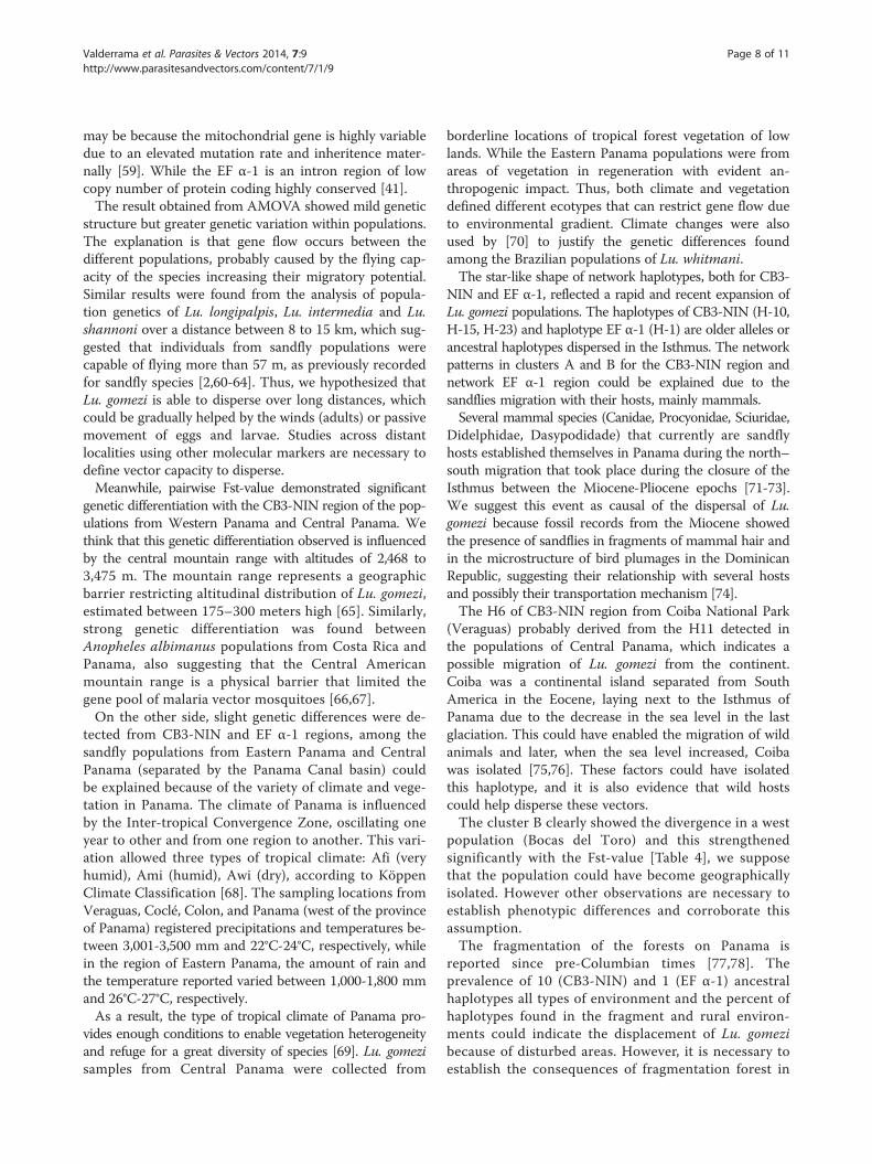
Network haplotypeThe median-Joining network reconstructed with CB3-NINregion linked a total of 37 different haplotypes with threedefined clusters (A, B, C). The lack of hierarchic structure
Table 3 Analysis of molecular variance (AMOVA) based on CBLu. gomezi
CB3-NIN
Source of variation d.f (%) Fixation
variations
Among populations 2 13.7 ΦST=
Within populations 83 86.3
d.f = degrees of freedom; * P < 0.01.All samples were grouped according to Table 1.
is observed in the cluster “A”, this clade consists of 28 hap-lotypes, the most frequent, H10, was found in elevenlocalities sampled in Central Panama and six in EasternPanama. It also included Altos de Piedra and Coiba haplo-types from Western Panama. Cluster “B” is constituted byfour haplotypes. Of these, H15 was collected from six sitesin Central Panama and nine from Eastern Panama, withH23 being the most frequent [Figure 2A]. In cluster “C” werecorded four haplotypes exclusively of localities sampledfrom Western Panama, demonstrating strong divergencesupport by Bayesian analysis [complementary data]. Thus,the network is characterized by few mutational steps[Figure 2A].On the other hand, analysis linked a total of 26 haplo-
types of the EF α-1 region demonstrating a lack of geneticstructure in the Lu. gomezi populations. The patterns of astar-like shape as similarly obtained with the mitochondrialregion, showed haplotype H1 as the most frequent in thecenter and several haplotypes surrounding it with one andthree mutational steps [Figure 2B].The Bayesian phylogenetic tree for the CB3-NIN region
showed three clusters with a posteriori probability of0.99-1.0. Other minor clusters were supported by low aposteriori probability values. The cluster with a higher aposteriori probability corroborated the divergences obtainedin the haplotype network, inferred by the median-joiningmethod [Figure 2A and B]. The Bayesian inference of EFα-1 region did not show similitude either, this analysis per-formed for mitochondrial and nuclear genes are supple-mentary data attached as Additional files 1 and 2.We found 50% of the haplotypes in the peridomestic
areas of fragmented environments, 40% in rural environ-ments, and 10% in forest environments. Although thisinformation is additional and not the main focus of theresearch, the predominance of the haplotypes in thesesites is important to highlight due to the anthropogenicimpact on the landscape of Panama [Table 1].
Demographic history populationThe results of Tajima D test (CB3-NIN) were negative butnot significant (−0.998; p > 0.01) that indicated neutralitydeviation due to a demographic expansion or not neutralselection. Nevertheless, Fu´s Fs values were significantlynegative (−19.68; p < 0.02) detecting that a population
3-NIN and EF α-1sequence of the populations of
EF α-1
index d.f (%) Fixation index
variations
0.13* 2 4.5% ΦST= 0.04*
163 95.5%

Table 4 Estimates of pairwise Fst of CB3-N1N and EF α-1between Lu. gomezi populations
CB3-NIN Western populations Central populations
EF α-1
Western populations *
Central populations 0.23 *
0.02
Eastern populations 0.21 0.10
0.04 0.12
Significance (p < 0.05)*.
Valderrama et al. Parasites & Vectors 2014, 7:9 Page 7 of 11http://www.parasitesandvectors.com/content/7/1/9
expanded in the past. The assessment of demographicchange with EF α-1 region using both Tajima D and Fu´sFs was significantly negative (−2.08; p < 0.01 and −13.65;p < 0.02) respectively.Considering that Fu´s Fs are sensitive estimators for de-
tecting demographic population expansion, we includedthe analysis of mismatch distribution for characterization ofexpansion detected with both genes. A bimodal curve wasobserved with CB3-NIN region and smoother curves withEF α-1 region. SSD value (0.011; p > 0.05) for CB3-NIN didnot reject the sudden expansion model, while SSD value(0.049; p > 0.05) for EF α-1 region was observed. TheHarpending´s raggedness index determined a good fit of
Figure 2 A haplotype network inferred by a median-joining method uLutzomyia gomezi. Circles represent different haplotypes and sizes that arindicate number of mutations among haplotypes. Each color of circles shoEastern (blue). Values in parentheses showed the a posteriori probability of
data (Additional file 3). The time since expansion ofthe population was estimated to be approximately4,223,825 years ago corresponding to the Pliocene Epoch.
DiscussionThe use of the mitochondrial and nuclear marker in ecol-ogy has provided enough information to allow contrast ofthe past population processes of species [54]. The highcontent of A-T (74%) of the mitochondrial gene analyzedin Lu. gomezi is similar to that detected in other sandflyspecies: Phlebotomus papatasi and Lu. evansi [30,55]. Thissuggests a high rate of substitutions as a result of naturalselection, favorable or unfavorable to populations [56]. Ahigher proportion of G-C (53%) observed in the nucleargene in Lu. gomezi was comparable to the proportion veri-fied in Anopheles gambiae (45%) and Culex pipiens (63%),characteristically of intron region [57,58].The high haplotype diversity detected among popula-
tions indicated a large polymorphism or gene pool of theLu. gomezi. The existence of this polymorphism is im-portant for the survival and adaptability, considering thatthe natural habitat of Phlebotominae sandflies isdestroyed because of anthropogenic factors [20]. How-ever, we observed discordance in the level of nucleotidediversity between CB3-NINI and EF α-1 sequences. This
sing CB3-NIN sequences (A) and EF α-1 sequences (B) ofe proportional to haplotype frequencies. The length of the traceswed the geographical populations: Western (red), Central (yellow),Bayesian inference.
Valderrama et al. Parasites & Vectors 2014, 7:9 Page 8 of 11http://www.parasitesandvectors.com/content/7/1/9
may be because the mitochondrial gene is highly variabledue to an elevated mutation rate and inheritence mater-nally [59]. While the EF α-1 is an intron region of lowcopy number of protein coding highly conserved [41].The result obtained from AMOVA showed mild genetic
structure but greater genetic variation within populations.The explanation is that gene flow occurs between thedifferent populations, probably caused by the flying cap-acity of the species increasing their migratory potential.Similar results were found from the analysis of popula-tion genetics of Lu. longipalpis, Lu. intermedia and Lu.shannoni over a distance between 8 to 15 km, which sug-gested that individuals from sandfly populations werecapable of flying more than 57 m, as previously recordedfor sandfly species [2,60-64]. Thus, we hypothesized thatLu. gomezi is able to disperse over long distances, whichcould be gradually helped by the winds (adults) or passivemovement of eggs and larvae. Studies across distantlocalities using other molecular markers are necessary todefine vector capacity to disperse.Meanwhile, pairwise Fst-value demonstrated significant
genetic differentiation with the CB3-NIN region of the pop-ulations from Western Panama and Central Panama. Wethink that this genetic differentiation observed is influencedby the central mountain range with altitudes of 2,468 to3,475 m. The mountain range represents a geographicbarrier restricting altitudinal distribution of Lu. gomezi,estimated between 175–300 meters high [65]. Similarly,strong genetic differentiation was found betweenAnopheles albimanus populations from Costa Rica andPanama, also suggesting that the Central Americanmountain range is a physical barrier that limited thegene pool of malaria vector mosquitoes [66,67].On the other side, slight genetic differences were de-
tected from CB3-NIN and EF α-1 regions, among thesandfly populations from Eastern Panama and CentralPanama (separated by the Panama Canal basin) couldbe explained because of the variety of climate and vege-tation in Panama. The climate of Panama is influencedby the Inter-tropical Convergence Zone, oscillating oneyear to other and from one region to another. This vari-ation allowed three types of tropical climate: Afi (veryhumid), Ami (humid), Awi (dry), according to KöppenClimate Classification [68]. The sampling locations fromVeraguas, Coclé, Colon, and Panama (west of the provinceof Panama) registered precipitations and temperatures be-tween 3,001-3,500 mm and 22°C-24°C, respectively, whilein the region of Eastern Panama, the amount of rain andthe temperature reported varied between 1,000-1,800 mmand 26°C-27°C, respectively.As a result, the type of tropical climate of Panama pro-
vides enough conditions to enable vegetation heterogeneityand refuge for a great diversity of species [69]. Lu. gomezisamples from Central Panama were collected from
borderline locations of tropical forest vegetation of lowlands. While the Eastern Panama populations were fromareas of vegetation in regeneration with evident an-thropogenic impact. Thus, both climate and vegetationdefined different ecotypes that can restrict gene flow dueto environmental gradient. Climate changes were alsoused by [70] to justify the genetic differences foundamong the Brazilian populations of Lu. whitmani.The star-like shape of network haplotypes, both for CB3-
NIN and EF α-1, reflected a rapid and recent expansion ofLu. gomezi populations. The haplotypes of CB3-NIN (H-10,H-15, H-23) and haplotype EF α-1 (H-1) are older alleles orancestral haplotypes dispersed in the Isthmus. The networkpatterns in clusters A and B for the CB3-NIN region andnetwork EF α-1 region could be explained due to thesandflies migration with their hosts, mainly mammals.Several mammal species (Canidae, Procyonidae, Sciuridae,
Didelphidae, Dasypodidade) that currently are sandflyhosts established themselves in Panama during the north–south migration that took place during the closure of theIsthmus between the Miocene-Pliocene epochs [71-73].We suggest this event as causal of the dispersal of Lu.gomezi because fossil records from the Miocene showedthe presence of sandflies in fragments of mammal hair andin the microstructure of bird plumages in the DominicanRepublic, suggesting their relationship with several hostsand possibly their transportation mechanism [74].The H6 of CB3-NIN region from Coiba National Park
(Veraguas) probably derived from the H11 detected inthe populations of Central Panama, which indicates apossible migration of Lu. gomezi from the continent.Coiba was a continental island separated from SouthAmerica in the Eocene, laying next to the Isthmus ofPanama due to the decrease in the sea level in the lastglaciation. This could have enabled the migration of wildanimals and later, when the sea level increased, Coibawas isolated [75,76]. These factors could have isolatedthis haplotype, and it is also evidence that wild hostscould help disperse these vectors.The cluster B clearly showed the divergence in a west
population (Bocas del Toro) and this strengthenedsignificantly with the Fst-value [Table 4], we supposethat the population could have become geographicallyisolated. However other observations are necessary toestablish phenotypic differences and corroborate thisassumption.The fragmentation of the forests on Panama is
reported since pre-Columbian times [77,78]. Theprevalence of 10 (CB3-NIN) and 1 (EF α-1) ancestralhaplotypes all types of environment and the percent ofhaplotypes found in the fragment and rural environ-ments could indicate the displacement of Lu. gomezibecause of disturbed areas. However, it is necessary toestablish the consequences of fragmentation forest in

Valderrama et al. Parasites & Vectors 2014, 7:9 Page 9 of 11http://www.parasitesandvectors.com/content/7/1/9
the population genetics by increasing sampling in localforests.The neutrality tests Fu´s Fs results were significantly
negative, and mismatch distribution statistic and networkhaplotype support the assumption of population expansionin the past. Moreover, high haplotype diversity but low nu-cleotide diversity in EF α- region also suggests populationexpansion.Is it the fact that the closure of the Isthmus of Panama
affected by climate change in the world was the main fac-tor that enabled the establishment of several species inAmerica, allowing for faunal exchanges between Northand South [79]. An alternative explanation of the popula-tion expansion is that conditions later to closure of theisthmus may have been favorable to its adaptation to thesuitable habitat and different ecological conditions inPanama territory. According to our estimated date thePliocene climate was much cooler, allowing the expan-sion of plant and animal species into new habitats [80].The colonization and adaptation into new habitat can beobserved in the star-like shape of network haplotype.
ConclusionsIn conclusion, Lu. gomezi is a species with a higher geneticpool or variability and mild population structure, due topossible capacity migration and local adaptation to envir-onmental changes or colonization potential. However theexistence of geographic barriers, such as the centralmountain range separates a subpopulation. The establish-ment of Lu. gomezi in the Isthmus of Panama could beinterpreted by the exchange of mammals in the Americancontinent, vegetation and climate conditions of the epoch.Their relevance as a carrier of cutaneous leishmaniasis inPanama is important for health schemes, especially if weconsider that sandflies have a wide-spread geographicdistribution in Panama, and this distribution is possiblyassociated to the landscape changes caused by deforest-ation [20]. In this study, mitochondrial and nuclear analysishas provided important information to allow assemblesabout genetic population and evolutionary history useful tounderstand the implications of different populationgenetic structures for cutaneous leishmaniasis epidemi-ology. Thereby, proposing new perspectives for thecontrol of leishmaniasis vectors.
Additional files
Additional file 1: Bayesian tree based on mitochondrial CB3-N1N ofpopulations of Lu. gomezi.
Additional file 2: Bayesian tree based on nuclear EF α-1 ofpopulations of Lu. gomezi.
Additional file 3: Mismatch distribution of Lu. gomezi speciesbased on (A) mitochondrial sequence CB3-N1N and (B) nucleotidesequences EF α-1.
AbbreviationsCL: Cutaneous leishmaniasis; CB3-NIN: Cytb gene, inter-genetic regionIGS-1; tRNA-Ser: Inter-genetic region IGS-2, and start of the NADH1; EFα-1: Elongation factor alpha-1.
Competing interestsThe authors declare that they have no competing interests.
Authors’ contributionsConceived and designed the experiments: AVC, MGT. Performed theexperiments: AVC. Analyzed the data: AVC. Contributed reagents/materials/analysis tools/specimens identifications: MGT, JDAF. Wrote the paper: AVC,JDAF and MGT. All authors read and approved the final version of themanuscript.
AcknowledgmentsWe are grateful to Maykon Cristiano Passos, for helping with laboratory andanalyses data; Jorge Edergarm Santos, Gustavo Martins and Juliana LopesFietto for comments in previous drafts of this manuscript; furthermore wethank the anonymous reviewers and editor for their comments.
FundingThis research was supported by the Secretaria Nacional de Ciencias yTecnología Innovadora (SENACYT) Grant Col09-008.
Author details1Department of Medical Entomology, Instituto Conmemorativo Gorgas deEstudios de la Salud, Panama, Panama. 2Department of Biology, UniversidadeFederal de Viçosa, Viçosa, MG, Brasil. 3Centro de Referência Nacional eInternacional para Flebotomíneos/Coleção de Flebotomíneos, Instituto RenéRachou-Fiocruz, Belo Horizonte, MG, Brasil.
Received: 12 July 2013 Accepted: 3 January 2014Published: 8 January 2014
References1. Christensen HA, Herrer A, Telford SR: Enzootic cutaneous leishmaniasis in
eastern Panama. II. Entomological investigations. Ann Trop Med Parasitol1972, 66:55–66.
2. Christensen H, Fairchild GB, Herrer A, Johnson C, Young D, de Vásquez A:The ecology of cutaneous, leishmaniasis in the Republic of Panama.J Med Entomol 1983, 20:463–484.
3. Christensen H, Johnson C, Vasquez AM: Leishmaniasis cutánea en Panamá:Un breve resumen. Rev Med Panama 1984, 9:182–1987.
4. Darling ST: Oriental sore in Panama. Proc Canal Zone Med Assn 1910,2:7–20.
5. Miranda A, Carrasco R, Paz H, Pascale JM, Samudio F, Saldaña A, Santamaría G,Mendoza Y, Calzada JE: Molecular epidemiology of American tegumentaryleishmaniasis in Panama. Am J Trop Med Hyg 2009, 81:565–571.
6. Herrer A, Telford S, Christensen HA: Enzootic cutaneous leishmaniasis ineastern Panama I: investigation of the infection among forest mammals.Ann Trop Med Parasitol 1971, 65:349–358.
7. Tesh RB, Chaniotis BN, Aronson MD, Johnson KM: Natural host preferencesof Panamanian Phlebotominae sandflies as determined by precipitintest. Am J Trop Med Hyg 1971, 20:150–156.
8. Herrer A, Christensen HA: Epidemiological patterns of cutaneousleishmaniasis in Panama. Am J Trop Med Hyg 1973, 25:54–58.
9. Saldaña A, Chaves LF, Rigg CA, Wald C, Smucker JE, Calzada JE: Clinicalcutaneous leishmaniasis rates are associated with household Lutzomyiagomezi, Lu. Panamensis, and Lu. trapidoi Abundance in Trinidad de LasMinas, Western Panama. Am J Trop Med Hyg 2013, 88(3):579.
10. Jaramillo C, Travi BL, Montoya J: Vector competence of some neotropicalsandflies for the Leishmania (Viannia) braziliensis complex. Med VetEntomol 1994, 8:1–7.
11. Jiménez AE, Rojas JC, Vargas F, Herrero MV: Temporal and spatial variationof phlebotomine (Diptera: Psychodidae) community diversity in acutaneous leishmaniasis endemic area of Costa Rica. J Med Entomol 2000,37:216–221.
12. Bejarano EE, Uribe S, Rojas W, Vélez ID: Phlebotomine sand flies(Diptera: Psychodidae) associated with the appearance of urban
Valderrama et al. Parasites & Vectors 2014, 7:9 Page 10 of 11http://www.parasitesandvectors.com/content/7/1/9
leishmaniasis in the city of Sincelejo, Colombia. Mem Inst Oswaldo Cruz2002, 97:645–647.
13. Santamaría E, Ponce N, Zipa Y, Ferro C: Presencia en el peridomestic devectores infectados con Leishmania (Viannia) panamensis en dos focosendémicos en el occidente de Boyacá, Piedemonte del valle delMagdalena medio, Colombia. Biomedica 2006, 26:82–94.
14. Vivero RJ, Contreras MA, Cadena H, Acosta LA, Mondragon K, Vélez A,Uribe SI, Vélez ID: Sand flies Phlebotominae (Diptera: Psychodidae) in fiveCentral American countries. In International Symposium on PhlebotominaeSandflies ISOPS VII: 25–30 april. Kudasy-Turkey; 2011:95.
15. Martins AV, Williams P, Falcão AL: American sand flies (Diptera: Psychodidae:Phlebotominae). Rio de Janeiro: Acad Bras Ciênc; 1978:195.
16. Young DG, Duncan MA: Guide to the identification and geographicdistribution of Lutzomyia sandflies in Mexico, the West Indies, Centraland South America (Diptera: Psychodidae). Mem Amer Ent Inst 1994,54:1–881.
17. Galati EAB: Classificação de Phlebotominae. In Flebotomíneos do Brasil.Edited by Rangel ER, Lainson R. Rio de Janeiro: Editora Fiocruz Brazil;2003:23–52.
18. Chaniotis BN, Correa MA, Tesh RB, Jonhson KM: Daily and seasonalman-biting activity of Phlebotominae sandflies in Panama. J Med Entomol1971, 8:415–420.
19. Travi BL, Alder GH, Lozano M, Cadena H, Montoya-Lerma J: Impact ofhabitat degradation on Phlebotominae (Diptera: Psychodidae) of tropicaldry forest in northern Colombia. J Med Entomol 2002, 39:451–456.
20. Valderrama A, Tavares MG, Andrade Filho DJ: Anthropogenic influence onthe distribution, abundance and diversity of sandfly species (Diptera:Phlebotominae: Psychodidae), vectors of cutaneous leishmaniasis inPanama. Mem Inst Oswaldo Cruz 2011, 106:1024–1031.
21. Feliciangeli MD, Rabinovich J: Abundance of Lutzomyia ovallesi but notLu. gomezi (Diptera: Psychodidae) correlated with cutaneousleishmaniasis incidence in north-central Venezuela. Med Vet Entomol1988, 12:121–131.
22. Duque P, Vélez ID, Morales M, Sierra D: Sand flies fauna involved in thetransmission of cutaneous leishmaniasis in Afro-Colombian andAmerindian communities of Choco, Pacific Coast of Colombia.Neotrop Entomol 2004, 33:255–264.
23. Cortés LA, Fernández JJ: Especies de Lutzomyia en un foco urbano deleishmaniasis visceral y cutánea en El Carmen de Bolívar, Bolívar,Colombia. Biomedica 2008, 28:433–440.
24. Jorquera A, González R, Marchán-Marcano E, Oviedo M, Matos M: Multiplex-PCR for detection of natural Leishmania infection in Lutzomyia sppcaptured in an endemic region for cutaneous leishmaniasis in state ofSucre, Venezuela. Mem Inst Oswaldo Cruz 2005, 100:43–46.
25. Vivero RJ, Contreras-Gutiérrez MA, Bejarano EE: Análisis de la estructuraprimaria y secundaria del ARN de transferencia mitocondrial para serinaen siete especies de Lutzomyia. Biomedica 2007, 27:429–438.
26. Azpurua J, de la Cruz D, Valderrama A, Windsor D: Lutzomyia sandflydiversity and rates of infection by Wolbachia and an exotic Leishmaniaspecies on Barro Colorado Island, Panama. PLoS Negl Trop Dis 2010, 4:e627.
27. Ready PD, Day JC, de Souza AA, Rangel EF, Davies CR: Mitochondrial DNAcharacterization of populations of Lutzomyia whitmani (Diptera:Psychodidae) incriminated in the peridomestic and silvatic transmissionof Leishmania species. Bull Entomol Res 1997, 87:187–195.
28. Soto SI, Lehmann T, Rowton ED, Velez BID, Porter CH: Speciation andpopulation structure in the morphospecies Lutzomyia longipalpis (Lutz& Neiva) as derived from the mitochondrial ND4 gene. Mol Phylogen Evol2001, 18:84–93.
29. Hodgkinson VH, Birungi J, Quintana M, Deitze R, Munstermann LE:Mitochondrial Cytocrome B variation in population of the visceralleishmanisis vector Lutzomyia longipalpis across eastern Brazil. Am J TropMed Hyg 2003, 69:386–392.
30. Hamarsheh O, Presber W, Abdeen Z, Sawalha S, Al-Lahem A, Schoenian G:Genetic structure of mediterranean populations of the sandflyPhlebotomus papatasi by mitochondrial cytochrome b haplotypeanalysis. Med Vet Entomol 2007, 21:270–277.
31. Esseghir S, Ready PD, Killick-Kendrick R, Ben-Ismail R: Mitochondrialhaplotypes and phylogeography of Phlebotomus vectors of Leishmaniamajor. Insect Mol Biol 1997, 6:211–225.
32. Depaquit J, Lienard E, Verzeaux-Griffon A, Ferte A, Bounamous H, Gantier JC,Hanafi HA, Jacobson RL, Maroli M, Moin-Vaziri V, Muller F, Ozbel Y,
Svobodova M, Volf P, Leger N: Molecular homogeneity in diversegeographical populations of Phlebotomus papatasi (Diptera, Psychodidae)inferred from ND4 mtDNA and ITS2 rDNA Epidemiological consequences.Infect Genet Evol 2008, 8:159–170.
33. Zeledón R, Hidalgo H, Víquez A, Urbina A: Atypical cutaneousleishmaniasis in a semiarid region of north-west Costa Rica. Trans R SocTrop Med Hyg 1989, 83:786.
34. Warburg A, Saraiva E, Lanzaro GC, Titus R, Neva F: Saliva of Lutzomyialongipalpis sibling species differs in its composition and propensity toenhance leishmaniasis. Phil Trans Roy Soc Lond 1994, 345:223–230.
35. Lourenço-de-Oliveira R, Vazeille M, Fillipis AMB, Failloux A: Aedes aegypti inBrazil: genetically differentiated populations with high susceptibility todengue and yellow fever viruses. Tran R Soc Trop Med Hyg 2004, 98:43–54.
36. Black WC IV, Tabachnick WJ: Population genetics in vector biology. In TheBiology of Disease Vectors. Edited by Beaty BJ, Marquardt WC. Colorado:University Press of Colorado, Niwot; 2004:417–437.
37. Reisen WK, Lothrop HD, Presser SB, Hardy JL, Gordon EW: Landscapeecology of arboviruses in southeastern California: temporal and spatialpatterns of enzootic activity in Imperial Valley, 1991–94. J Med Entomol1997, 34:179–188.
38. Sudia WD, Chamberlain RW: Battery operated light trap, an improvedmodel. Mosq News 1962, 22:126–129.
39. Michalsky EM, Fortes-Dias CL, Pimenta PFP, Secundino NFC, Dias ES:Assessment of PCR in the detection of Leishmania spp in experimentallyinfected individual phlebotomine sandflies (Diptera: Psychodidae:Plebotominae). Rev Inst Med Trop Sao Paulo 2002, 44:255–259.
40. Testa JM, Montoya-Lerna J, Cadena H, Oviedo M, Ready PD: Molecularidentification of vectors of Leishmania in Colombia: mitochondrialintrogression in the Lutzomyia townsendi series. Acta Trop 2002,84:205–218.
41. Parvizi P, Assmar M: Nuclear elongation factor 1-α gene, A molecularmarker for Iranian sandfly identification. Iranian J Publ Health 2007,36:25–37.
42. Tamura K, Peterson D, Peterson N, Stecher G, Nei M, Kumar S: MEGA5:Molecular evolutionary genetics analysis using maximum likelihood,evolutionary distance, and maximum parsimony methods. Mol Biol Evol2011, 28:2731–2739.
43. Librado P, Rozas J: DnaSP v5: A software for comprehensive analysis ofDNA polymorphism data. Bioinformatics 2009, 25:1451–1452.
44. Excoffier L, Laval G, Schneider S: Arlequin ver. 3.0: An integrated softwarepackage for population genetics data analysis. Evol Bioinform Online 2005,1:47–50.
45. Weir BS, Cockerham C: Estimating F-statistics for the analysis ofpopulation structure. Evolution 1984, 38:1358–1370.
46. Excoffier L, Smouse PE, Quattro JM: Analysis of molecular variance inferredfrom metric distances among DNA haplotypes: application to humanmitochondrial DNA restriction data. Genetics 1992, 131:479–491.
47. Bandelt HJ, Forster P, Röhl A: Median-joining networks for inferringintraspecific phylogenies. Mol Biol Evol 1999, 16:37–48.
48. Huelsenbecr JP, Ronquist, MrBayes F: Bayesian inference of phylogeneticstree. Bioinformatics 2001, 17:754–755.
49. Nylander JAA, Ronquist F, Huelsenbeck JP, Nieves-Aldrey JL: Bayesianphylogenetic analysis of combined data. System Biol 2004, 53:47–67.
50. Tajima F: Evolutionary relationship of DNA sequences in finitepopulations. Genetics 1983, 105:437–460.
51. Fu YX: Statistical tests of neutrality of mutations against populationgrowth, hitchhiking and background selection. Genetics 1997,147:915–925.
52. Rogers AR, Harpending HC: Population growth makes waves in thedistribution of pairwise genetic differences. Mol Biol Evol 1992,9:552–569.
53. Brower AVZ: Phylogeny of Heliconius butterflies inferred frommitochondrial DNA sequences (Lepidoptera: Nymphalidae). MolPhylogenet Evol 1994, 3:159–174.
54. Galtier N, Nabholz B, Glémin S, Hurst GD: Mitochondrial DNA as a markerof molecular diversity: a reappraisal. Mol Ecol 2009, 18:4541–4550.
55. Bejarano EE, Rojas W, Uribe S, Vélez ID: Genetic analysis of a recentlydetected urban population of Lutzomyia evansi (Diptera: Psychodidae) inColombia. Rev Soc Entomol Argent 2009, 68:135–141.
56. Hedrick PW: Sex differences in mutation, recombination, selection, geneflow, and genetic drift. Evolution 2007, 61(12):2750–2771.
Valderrama et al. Parasites & Vectors 2014, 7:9 Page 11 of 11http://www.parasitesandvectors.com/content/7/1/9
57. Calcagnotto D: Taxa de evolução e o relógio molecular. In BiologiaMolecular e Evolução. Edited by Matioli SR. Ribeirão Preto São Paulo: HolosEditora; 2001:52–63.
58. Nahum LA: Evolução dos genomas. In Biologia Molecular e Evolução. Editedby Matioli SR. Ribeirão Preto SP: Holos Editora; 2011:82–96.
59. Simon C, Frati F, Beckenbach A, Crespi B, Liu H, Flook P: Evolution,weighting and phylogenetic utility of mitochondrial gene sequencesand a compilation of conserved polymerase chain reaction primers.Ann Entomol Soc Am 1994, 87:651–701.
60. Alexander JB, Young DG: Dispersal of phlebotomine sand flies (Diptera:Psychodidae) in a Colombian focus of Leishmania (Viannia) braziliensis.Mem Inst Oswaldo Cruz 1992, 87:397–403.
61. Morrison AC, Ferro C, Morales A, Tesh RB, Wilson ML: Dispersal of the sandfly Lutzomyia longipalpis (Diptera: Psychodidae) at an endemic focus ofvisceral leishmaniasis in Colombia. J Med Entomol 1993, 30:427–435.
62. Márquez LM, Lampo M, Rinaldi M, Lau P: Gene flow natural and domesticpopulations of Lutzomyia longipalpis (Diptera: Psychodidae) in arestricted focus of American visceral leishmaniasis in Venezuela.J Med Entomol 2001, 38:12–16.
63. De Queiroz BV, Coutinho-Abreu IV, Sonoda IV, Melo MA, de Andrade PP,de Castro JA, Rebelo JM, Carvalho SM, Ramalho-Ortigao M: Geneticstructure of natural populations of the sand fly Lutzomyia longipalpis(Diptera: Psychodidae) from the Brazilian northeastern region. Acta Trop2006, 98(1):15–24.
64. Rocha LS, Falqueto A, dos Santos CB, Ferreira AL, da Graça CC, Grimaldi G,Cupolillo E: Survey of natural infection by Leishmania in sand fly speciescollected in southeastern Brazil. Trans R Soc Trop Med Hyg 2010,104:461–466.
65. Añez N, Cazorla D, Nieves E, Chataing B, Castro M, de Yarbuh AL:Epidemiología de la leishmaniasis tegumentaria en Mérida, Venezuela. I.Diversidad y dispersión de especies flebotominas en tres pisosaltitudinales y su posible rol en la transmisión de la enfermedad.Mem Inst Oswaldo Cruz 1988, 83:455–463.
66. Molina-Cruz A, de Mérida AM, Mills K, Rodríguez F, Schoua C, Yurrita MM,Molina E, Palmieri M, Black WC IV: Gene Flow Among Anopheles albimanuspopulations in Central América, South América and the CaribbeanAssessed by microsatellites and mitochondrial DNA. Am J Trop Med Hyg2004, 71:350–359.
67. Loaiza JR, Bermingham E, Scott ME, Rovira JR, Conn JE: Speciescomposition and distribution of adult Anopheles (Diptera: Culicidae) inPanama. J Med Entomol 2008, 45:841–851.
68. ETESA. http://www.hidromet.com.pa/.69. Condit R, Hubbell SP, Foster RB: Changes in a tropical forest with a
shifting climate: results from a 50 ha permanent census plot in Panama.J Trop Ecol 1996, 12:231–256.
70. Ready PD, de Souza AA, Macario Rebelo JM, Day JC, Silveira FT,Campbell-Lendrum D, Davies CR, Costa JML: Phylogenetic species anddomesticity of Lutzomyia whitmani at the southeast boundary ofAmazonian Brazil. Trans R Soc Trop Med Hyg 1998, 92:159–160.
71. Coates AG, Obando JA: Geological evolution of the Central AmericanIsthmus. In Evolution and environment in tropical America. Edited byJackson JBC, Budd AF, Coates AG. Chicago: University of Chicago Press;1996:21–56.
72. Cox CB: Plate tectonics, seaways and climate in the historicalbiogeography of mammals. Mem Inst Oswaldo Cruz 2000, 95:509–516.
73. Webb SD: The great American biotic interchange: patterns andprocesses. Ann Mo Bot Gard 2006, 93:245–257.
74. Peñalver E, Grimaldi D: Assemblages of mammalian hair andblood-feeding midges (Insecta: Diptera: Psychodidae: Phlebotominae) inMiocene amber. Trans R Soc Edin: Earth Sci 2005, 96:177–195.
75. Heckadon MS: Panamá: Puente biológico. Panama. Panamá: SmithsonianTropical Research Institute; 2001:233.
76. Hoernle KA, Bogaard PVD, Werner R, Lissinna B, Hauff G: Missing history(16–71 Ma) of the Galapagos hotspot: Implications for the tectonic andbiological evolution of the Americas. Geology 2002, 30:795–798.
77. Linares OF, Sheets PD, Rosenthal EJ: Prehistoric agriculture in tropicalhighlands. Science 1975, 187:137–145.
78. Piperno DR, Jones J: Paleoecological and archaeological implications of alate pleistocene/early holocene record of vegetation and climate fromthe Pacific coastal plain of Panama. Quaternary Res 2003, 59:79–87.
79. Marshall LG: Land Mammals and the Great American Interchange. Am Sci1988, 76(4):380–388.
80. Ravelo AC, Andreasen DH, Lyle M, Lyle AO, Wara MW: Regional climateshifts caused by gradual global cooling in the Pliocene epoch.Nature 2004, 429:263–267.
doi:10.1186/1756-3305-7-9Cite this article as: Valderrama et al.: Phylogeography of the Lutzomyiagomezi (Diptera: Phlebotominae) on the Panama Isthmus. Parasites &Vectors 2014 7:9.
Submit your next manuscript to BioMed Centraland take full advantage of:
• Convenient online submission
• Thorough peer review
• No space constraints or color figure charges
• Immediate publication on acceptance
• Inclusion in PubMed, CAS, Scopus and Google Scholar
• Research which is freely available for redistribution
Submit your manuscript at www.biomedcentral.com/submit