Ascidiidae Herdman, 1882 (Tunicata: Ascidiacea) on the Pacific coast of Panama
14
Accepted by R. Brunetti: 11 Jun. 2013; published: 22 Jul. 2013 ZOOTAXA ISSN 1175-5326 (print edition) ISSN 1175-5334 (online edition) Copyright © 2013 Magnolia Press Zootaxa 3691 (3): 351–364 www.mapress.com/ zootaxa/ Article 351 http://dx.doi.org/10.11646/zootaxa.3691.3.4 http://zoobank.org/urn:lsid:zoobank.org:pub:855A00F4-2E4B-4D0C-A458-B8111BFB7762 Ascidiidae Herdman, 1882 (Tunicata: Ascidiacea) on the Pacific coast of Panama NADIA Y. K. BONNET 1,4 , ROSANA M. ROCHA 2 & MARY R. CARMAN 3 1 Universidade Federal do Paraná, graduate program in Zoology, CP 19020, 81.531-980, Curitiba, Paraná, Brazil. E-mail: [email protected] 2 Universidade Federal do Paraná, Departamento de Zoologia, CP 19020, 81.531-980, Curitiba, Paraná, Brazil. E-mail: [email protected] 3 Biology Department, Woods Hole Oceanographic Institution, Woods Hole, MA 02543, USA. E-mail: [email protected] 4 Present address: Universidade Federal do Ceará, graduate program in Ciência Marinhas Tropicais, Av. da Abolição 3207, Meireles, 60.165-081, Fortaleza, Ceará, Brazil Abstract The ascidian fauna of the Pacific coast of Panama is poorly known and only recently four species in the family Ascidiidae were reported on. Ascidia is the only known genus of Ascidiidae in Pacific Panama waters. In the present research, we describe a new species, Ascidia sideralis sp. nov., and we document the new occurrence of A. cf. gemmata and A. cf. lib- erata (both previously known to the West Pacific), A. archaia (a cosmopolitan species elsewhere in the Pacific), A. cera- todes (previously documented in the eastern N. Pacific), and A. sydneiensis (an Atlantic species on the east coast of Panama) in Pacific Panama waters. A tabular key for the identification of Ascidiidae on the American Pacific coast com- plements this study. Key words: Ascidia, biodiversity, taxonomy Introduction The ascidian fauna of the Americas includes a large diversity of species (Van Name 1945) and this knowledge has been improved on the last decades. Rocha et al. (2012) reported 26 species of Ascidiidae from the shallow waters of the Atlantic coast of the Americas, while only nine species were reported on the Pacific coast (Van Name 1945; Lambert & Lambert 1998; Lambert & Sanamyan 2001; Monniot 2007; Nova-Bustos et al. 2010; Carman et al. 2011). New efforts to survey the ascidian diversity in Panama were recently done, both on the east (Rocha et al. 2005; Bonnet & Rocha 2011) and the west coast (Carman et al. 2011). These studies are important, because the intense traffic of ships through the Panama Canal makes this region more susceptible to the introduction and establishment of exotic species. Here we report on six species of the genus Ascidia, among which four have already been reported in Carman et al. (2011). Ascidia sp. is now formally described and given the name A. sideralis. The report of Ascidia incrassata is corrected, and the assigned species (A. cf. liberata) is considered an introduction to the region. The identifications of A. ceratodes and A. sydneiensis remain unchanged. The two additional species reported here are A. archaia and A. cf. gemmata. Material and methods Specimens were collected between December 2008 and November 2011, from natural and artificial substrates on the Pacific coast of Panama up to 13.0 m deep. Tidal exchange in the area is about 3.0 m. Rocky substrates predominated in the natural environment which were well exposed during low tide. Thus, animals were usually subtidal and deeper than 5.0 m. The coordinates of each collecting site are listed in Table 1. TERMS OF USE This pdf is provided by Magnolia Press for private/research use. Commercial sale or deposition in a public library or website is prohibited.
Transcript of Ascidiidae Herdman, 1882 (Tunicata: Ascidiacea) on the Pacific coast of Panama

TERMS OF USE This pdf is provided by Magnolia Press for private/research use. Commercial sale or deposition in a public library or website is prohibited.
ZOOTAXAISSN 1175-5326 (print edition)
ISSN 1175-5334 (online edition)Copyright © 2013 Magnolia Press
Zootaxa 3691 (3): 351–364 www.mapress.com/zootaxa/ Article
http://dx.doi.org/10.11646/zootaxa.3691.3.4http://zoobank.org/urn:lsid:zoobank.org:pub:855A00F4-2E4B-4D0C-A458-B8111BFB7762
Ascidiidae Herdman, 1882 (Tunicata: Ascidiacea) on the Pacific coast of Panama
NADIA Y. K. BONNET1,4, ROSANA M. ROCHA2 & MARY R. CARMAN3
1Universidade Federal do Paraná, graduate program in Zoology, CP 19020, 81.531-980, Curitiba, Paraná, Brazil. E-mail: [email protected] Federal do Paraná, Departamento de Zoologia, CP 19020, 81.531-980, Curitiba, Paraná, Brazil. E-mail: [email protected] Department, Woods Hole Oceanographic Institution, Woods Hole, MA 02543, USA. E-mail: [email protected] address: Universidade Federal do Ceará, graduate program in Ciência Marinhas Tropicais, Av. da Abolição 3207, Meireles, 60.165-081, Fortaleza, Ceará, Brazil
Abstract
The ascidian fauna of the Pacific coast of Panama is poorly known and only recently four species in the family Ascidiidae were reported on. Ascidia is the only known genus of Ascidiidae in Pacific Panama waters. In the present research, we describe a new species, Ascidia sideralis sp. nov., and we document the new occurrence of A. cf. gemmata and A. cf. lib-erata (both previously known to the West Pacific), A. archaia (a cosmopolitan species elsewhere in the Pacific), A. cera-todes (previously documented in the eastern N. Pacific), and A. sydneiensis (an Atlantic species on the east coast of Panama) in Pacific Panama waters. A tabular key for the identification of Ascidiidae on the American Pacific coast com-plements this study.
Key words: Ascidia, biodiversity, taxonomy
Introduction
The ascidian fauna of the Americas includes a large diversity of species (Van Name 1945) and this knowledge has been improved on the last decades. Rocha et al. (2012) reported 26 species of Ascidiidae from the shallow waters of the Atlantic coast of the Americas, while only nine species were reported on the Pacific coast (Van Name 1945; Lambert & Lambert 1998; Lambert & Sanamyan 2001; Monniot 2007; Nova-Bustos et al. 2010; Carman et al. 2011).
New efforts to survey the ascidian diversity in Panama were recently done, both on the east (Rocha et al. 2005; Bonnet & Rocha 2011) and the west coast (Carman et al. 2011). These studies are important, because the intense traffic of ships through the Panama Canal makes this region more susceptible to the introduction and establishment of exotic species. Here we report on six species of the genus Ascidia, among which four have already been reported in Carman et al. (2011). Ascidia sp. is now formally described and given the name A. sideralis. The report of Ascidia incrassata is corrected, and the assigned species (A. cf. liberata) is considered an introduction to the region. The identifications of A. ceratodes and A. sydneiensis remain unchanged. The two additional species reported here are A. archaia and A. cf. gemmata.
Material and methods
Specimens were collected between December 2008 and November 2011, from natural and artificial substrates on the Pacific coast of Panama up to 13.0 m deep. Tidal exchange in the area is about 3.0 m. Rocky substrates predominated in the natural environment which were well exposed during low tide. Thus, animals were usually subtidal and deeper than 5.0 m. The coordinates of each collecting site are listed in Table 1.
Accepted by R. Brunetti: 11 Jun. 2013; published: 22 Jul. 2013 351
TERMS OF USE This pdf is provided by Magnolia Press for private/research use. Commercial sale or deposition in a public library or website is prohibited.
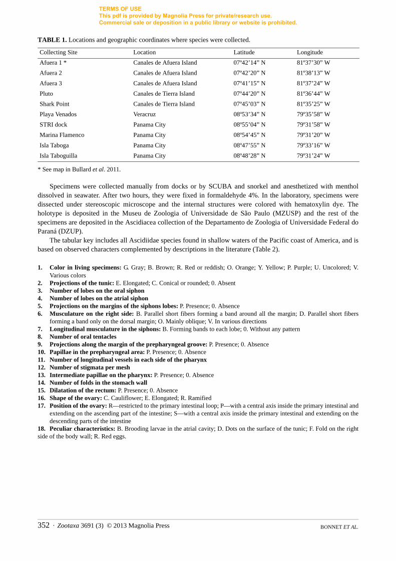
TABLE 1. Locations and geographic coordinates where species were collected.
* See map in Bullard et al. 2011.
Specimens were collected manually from docks or by SCUBA and snorkel and anesthetized with menthol dissolved in seawater. After two hours, they were fixed in formaldehyde 4%. In the laboratory, specimens were dissected under stereoscopic microscope and the internal structures were colored with hematoxylin dye. The holotype is deposited in the Museu de Zoologia of Universidade de São Paulo (MZUSP) and the rest of the specimens are deposited in the Ascidiacea collection of the Departamento de Zoologia of Universidade Federal do Paraná (DZUP).
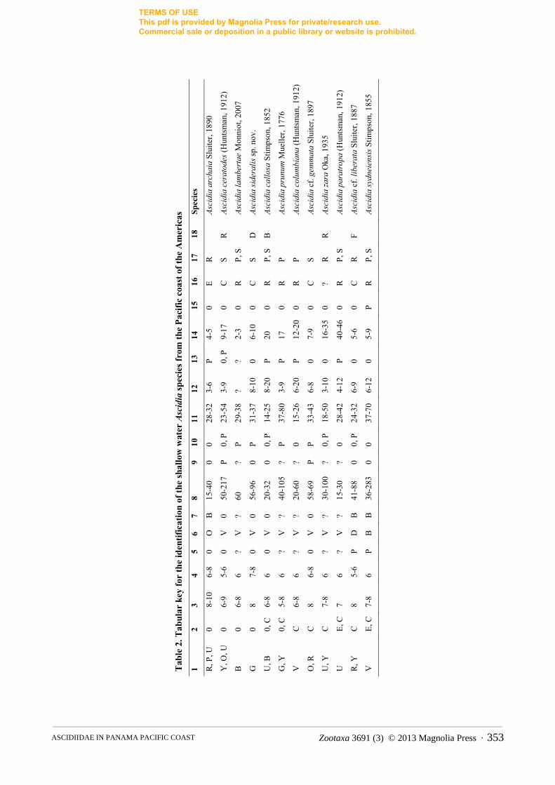
The tabular key includes all Ascidiidae species found in shallow waters of the Pacific coast of America, and is based on observed characters complemented by descriptions in the literature (Table 2).
1. Color in living specimens: G. Gray; B. Brown; R. Red or reddish; O. Orange; Y. Yellow; P. Purple; U. Uncolored; V. Various colors
2. Projections of the tunic: E. Elongated; C. Conical or rounded; 0. Absent3. Number of lobes on the oral siphon 4. Number of lobes on the atrial siphon 5. Projections on the margins of the siphons lobes: P. Presence; 0. Absence6. Musculature on the right side: B. Parallel short fibers forming a band around all the margin; D. Parallel short fibers
forming a band only on the dorsal margin; O. Mainly oblique; V. In various directions 7. Longitudinal musculature in the siphons: B. Forming bands to each lobe; 0. Without any pattern8. Number of oral tentacles 9. Projections along the margin of the prepharyngeal groove: P. Presence; 0. Absence10. Papillae in the prepharyngeal area: P. Presence; 0. Absence11. Number of longitudinal vessels in each side of the pharynx12. Number of stigmata per mesh13. Intermediate papillae on the pharynx: P. Presence; 0. Absence 14. Number of folds in the stomach wall15. Dilatation of the rectum: P. Presence; 0. Absence16. Shape of the ovary: C. Cauliflower; E. Elongated; R. Ramified17. Position of the ovary: R—restricted to the primary intestinal loop; P—with a central axis inside the primary intestinal and
extending on the ascending part of the intestine; S—with a central axis inside the primary intestinal and extending on the descending parts of the intestine
18. Peculiar characteristics: B. Brooding larvae in the atrial cavity; D. Dots on the surface of the tunic; F. Fold on the right side of the body wall; R. Red eggs.
Collecting Site Location Latitude Longitude
Afuera 1 * Canales de Afuera Island 07º42’14” N 81º37’30” W
Afuera 2 Canales de Afuera Island 07º42’20” N 81º38’13” W
Afuera 3 Canales de Afuera Island 07º41’15” N 81º37’24” W
Pluto Canales de Tierra Island 07º44’20” N 81º36’44” W
Shark Point Canales de Tierra Island 07º45’03” N 81º35’25” W
Playa Venados Veracruz 08º53’34” N 79º35’58” W
STRI dock Panama City 08º55’04” N 79º31’58” W
Marina Flamenco Panama City 08º54’45” N 79º31’20” W
Isla Taboga Panama City 08º47’55” N 79º33’16” W
Isla Taboguilla Panama City 08º48’28” N 79º31’24” W
BONNET ET AL. 352 · Zootaxa 3691 (3) © 2013 Magnolia Press
TERMS OF USE This pdf is provided by Magnolia Press for private/research use. Commercial sale or deposition in a public library or website is prohibited.
Tab
le 2
. Tab
ular
key
for
the
iden
tific
atio
n of
the
shal
low
wat
er Ascidia
spec
ies f
rom
the
Paci
fic c
oast
of t
he A
mer
icas
1 2
3 4
5 6
7 8
9 10
11
12
13
14
15
16
17
18
Sp
ecie
s
R, P
, U
0 8-
10
6-8
0 O
B
15
-40
0 0
28-3
2 3-
6 P
4-5
0 E
R
As
cidi
a ar
chai
a Sl
uite
r, 18
90
Y, O
, U
0 6-
9 5-
6 0
V
0 50
-217
P
0, P
23
-54
3-9
0, P
9-
17
0 C
S
R
Asci
dia
cera
tode
s (H
unts
man
, 191
2)
B
0 6-
8 6
? V
?
60
? P
29-3
8 ?
? 2-
3 0
R
P, S
Asci
dia
lam
bert
ae M
onni
ot, 2
007
G
0 8
7-8
0 V
0
56-9
6 0
P 31
-37
8-10
0
6-10
0
C
S D
As
cidi
a si
dera
lis sp
. nov
.
U, B
0,
C
6-8
6 0
V
0 20
-32
0 0,
P
14-2
5 8-
20
P 20
0
R
P, S
B
As
cidi
a ca
llosa
Stim
pson
, 185
2
G, Y
0,
C
5-8
6 ?
V
? 40
-105
?
P 37
-80
3-9
P 17
0
R
P
Asci
dia
prun
um M
uelle
r, 17
76
V
C
6-8
6 ?
V
? 20
-60
? 0
15-2
6 6-
20
P 12
-20
0 R
P
As
cidi
a co
lum
bian
a (H
unts
man
, 191
2)
O, R
C
8
6-8
0 V
0
58-6
9 P
P 33
-43
6-8
0 7-
9 0
C
S
Asci
dia
cf. g
emm
ata
Slui
ter,
1897
U, Y
C
7-
8 6
? V
?
30-1
00
? 0,
P
18-5
0 3-
10
0 16
-35
0 ?
R
R
Asci
dia
zara
Oka
, 193
5
U
E, C
7
6 ?
V
? 15
-30
? 0
28-4
2 4-
12
P 40
-46
0 R
P,
S
As
cidi
a pa
ratr
opa
(Hun
tsm
an, 1
912)
R, Y
C
8
5-6
P D
B
41
-88
0 0,
P
24-3
2 6-
9 0
5-6
0 C
R
F
Asci
dia
cf. l
iber
ata
Slui
ter,
1887
V
E, C
7-
8 6
P B
B
36
-283
0
0 37
-70
6-12
0
5-9
P R
P,
S
As
cidi
a sy
dnei
ensi
s Stim
pson
, 185
5
Zootaxa 3691 (3) © 2013 Magnolia Press · 353ASCIDIIDAE IN PANAMA PACIFIC COAST
TERMS OF USE This pdf is provided by Magnolia Press for private/research use. Commercial sale or deposition in a public library or website is prohibited.
Descriptions
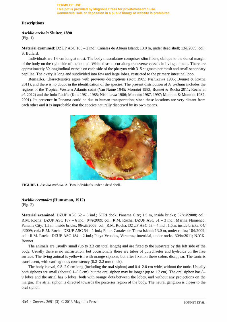
Ascidia archaia Sluiter, 1890 (Fig. 1)
Material examined: DZUP ASC 185 – 2 ind.; Canales de Afuera Island; 13.0 m, under dead shell; 13/i/2009; col.: S. Bullard.
Individuals are 1.6 cm long at most. The body musculature comprises slim fibers, oblique to the dorsal margin of the body on the right side of the animal. White discs occur along transverse vessels in living animals. There are approximately 30 longitudinal vessels on each side of the pharynx with 3–5 stigmata per mesh and small secondary papillae. The ovary is long and subdivided into few and large lobes, restricted to the primary intestinal loop.
Remarks. Characteristics agree with previous descriptions (Kott 1985; Nishikawa 1986; Bonnet & Rocha 2011), and there is no doubt in the identification of the species. The present distribution of A. archaia includes the regions of the Tropical Western Atlantic coast (Van Name 1945; Monniot 1983; Bonnet & Rocha 2011; Rocha et al. 2012) and the Indo-Pacific (Kott 1981, 1985; Nishikawa 1986; Monniot 1987, 1997; Monniot & Monniot 1987, 2001). Its presence in Panama could be due to human transportation, since these locations are very distant from each other and it is improbable that the species naturally dispersed by its own means.
FIGURE 1. Ascidia archaia. A. Two individuals under a dead shell.
Ascidia ceratodes (Huntsman, 1912) (Fig. 2)
Material examined. DZUP ASC 52 – 5 ind.; STRI dock, Panama City; 1.5 m, inside bricks; 07/xii/2008; col.: R.M. Rocha; DZUP ASC 187 – 6 ind.; 04/i/2009; col.: R.M. Rocha. DZUP ASC 51 – 3 ind.; Marina Flamenco, Panama City; 1.5 m, inside bricks; 06/xii/2008; col.: R.M. Rocha; DZUP ASC 53 – 4 ind.; 1.5m, inside bricks; 04/i/2009; col.: R.M. Rocha. DZUP ASC 54 – 1 ind.; Pluto, Canales de Tierra Island; 13.0 m, under rocks; 10/i/2009; col.: R.M. Rocha. DZUP ASC 184 – 2 ind.; Playa Venados, Veracruz; intertidal, under rocks; 30/ix/2011; N.Y.K. Bonnet.
The animals are usually small (up to 3.3 cm total length) and are fixed to the substrate by the left side of the body. Usually there is no incrustation, but occasionally there are tubes of polychaetes and hydroids on the free surface. The living animal is yellowish with orange siphons, but after fixation these colors disappear. The tunic is translucent, with cartilaginous consistency (0.2–2.2 mm thick).
The body is oval, 0.8–2.0 cm long (including the oral siphon) and 0.4–2.0 cm wide, without the tunic. Usually both siphons are small (about 0.1–0.5 cm), but the oral siphon may be longer (up to 1.2 cm). The oral siphon has 8–9 lobes and the atrial has 6 lobes; both with orange dots between the lobes, and without any projections on the margin. The atrial siphon is directed towards the posterior region of the body. The neural ganglion is closer to the oral siphon.
BONNET ET AL. 354 · Zootaxa 3691 (3) © 2013 Magnolia Press

TERMS OF USE This pdf is provided by Magnolia Press for private/research use. Commercial sale or deposition in a public library or website is prohibited.
A net of 0.03–0.15 mm thick fibers forms the musculature on the right side. On the left side, the longitudinal muscles extending from the oral siphon are short and a band of short parallel transverse fibers is found on the dorsal region. The longitudinal fibers in the siphons are not organized in bands.
There are 83–217 oral tentacles, of three sizes; the largest is 1.3–2.1 mm long. The prepharyngeal groove is double and the anterior membrane of the prepharyngeal groove usually has projections. The distance between the line of tentacles and the prepharyngeal groove is short (up to 0.3–1.1 mm), with or without papillae. The peritubercular area is short and the dorsal tubercle aperture is U-shaped, with or without enrolled ends. The dorsal lamina is double anteriorly; when it merges, the margin is toothed (teeth not related with the ends of the transverse vessels). The dorsal lamina passes by the left of the esophagus aperture to the end of the pharynx, close to the stomach; absence of papillae on the right side of the dorsal lamina at the level of the esophagus. There is a narrow lamina on the right of the esophageal aperture. The pharynx has 24–38 longitudinal vessels on the right side, 23–35 longitudinal vessels on the left, and 47–81 transverse vessels; it is pleated, with 5–7 stigmata per mesh. The primary papillae in the pharynx could be simple, bi or trilobed. There are secondary papillae in some parts of the pharynx. Parastigmatic vessels are absent.
The alimentary canal occupies half or more of the left side of the body. The stomach is oval and large, with 9–11 internal longitudinal folds. The isodiametric intestine forms a primary and a secondary loop. The anus is located approximately 3.0–8.9 mm from the oral tentacles; it has a smooth or bilobed rim. Renal vesicles are very conspicuous (0.1–0.25 mm in diameter) and cover the stomach and the ascendant loop of the intestine.
The ovary is compact and localized inside the primary intestinal loop, where it is visible both from the outside and atrial cavity side. Oocytes are approximately 0.1 mm in diameter. The testis follicles are elongated and overlying part of the stomach and intestine. Gonoducts open just posterior to the anus aperture.
Remarks. Van Name (1945) described A. ceratodes as similar to juveniles of A. interrupta Heller, 1978. We do not agree because the absence of projections in the tunic surface, the larger number of oral tentacles, the smaller number of longitudinal vessels in the pharynx, the isodiametric intestine and the conspicuous renal vesicles are characteristics which distinguish A. ceratodes from A. interrupta. Van Name (1945) cited the geographical distribution of this species from British Columbia south to California and comments about doubtful specimens from Ecuador and north Chile. Later, Tokioka (1972) reported the species in Costa Rica and Carman et al. (2011) extended its range to Panama. It is uncertain whether these tropical populations are in their native region and the species is expected to be found along all Central America Pacific coast or whether the southern populations were introduced by human transport.
FIGURE 2. Ascidia ceratodes. A. Right side of body (without tunic). B. Left side of body (without tunic). Scale bar: A, B = 0.5 cm.
Zootaxa 3691 (3) © 2013 Magnolia Press · 355ASCIDIIDAE IN PANAMA PACIFIC COAST
TERMS OF USE This pdf is provided by Magnolia Press for private/research use. Commercial sale or deposition in a public library or website is prohibited.
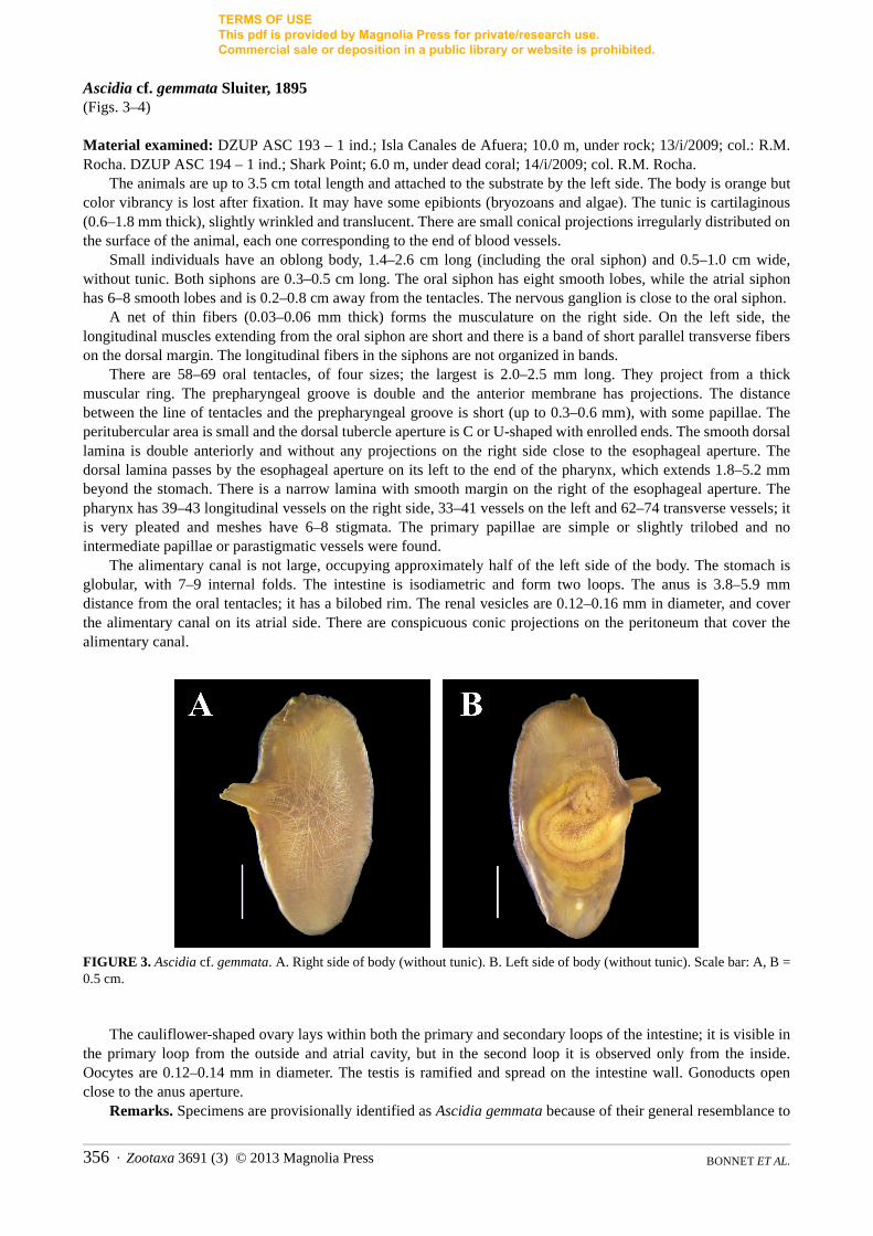
Ascidia cf. gemmata Sluiter, 1895 (Figs. 3–4)
Material examined: DZUP ASC 193 – 1 ind.; Isla Canales de Afuera; 10.0 m, under rock; 13/i/2009; col.: R.M. Rocha. DZUP ASC 194 – 1 ind.; Shark Point; 6.0 m, under dead coral; 14/i/2009; col. R.M. Rocha.
The animals are up to 3.5 cm total length and attached to the substrate by the left side. The body is orange but color vibrancy is lost after fixation. It may have some epibionts (bryozoans and algae). The tunic is cartilaginous (0.6–1.8 mm thick), slightly wrinkled and translucent. There are small conical projections irregularly distributed on the surface of the animal, each one corresponding to the end of blood vessels.
Small individuals have an oblong body, 1.4–2.6 cm long (including the oral siphon) and 0.5–1.0 cm wide, without tunic. Both siphons are 0.3–0.5 cm long. The oral siphon has eight smooth lobes, while the atrial siphon has 6–8 smooth lobes and is 0.2–0.8 cm away from the tentacles. The nervous ganglion is close to the oral siphon.
A net of thin fibers (0.03–0.06 mm thick) forms the musculature on the right side. On the left side, the longitudinal muscles extending from the oral siphon are short and there is a band of short parallel transverse fibers on the dorsal margin. The longitudinal fibers in the siphons are not organized in bands.
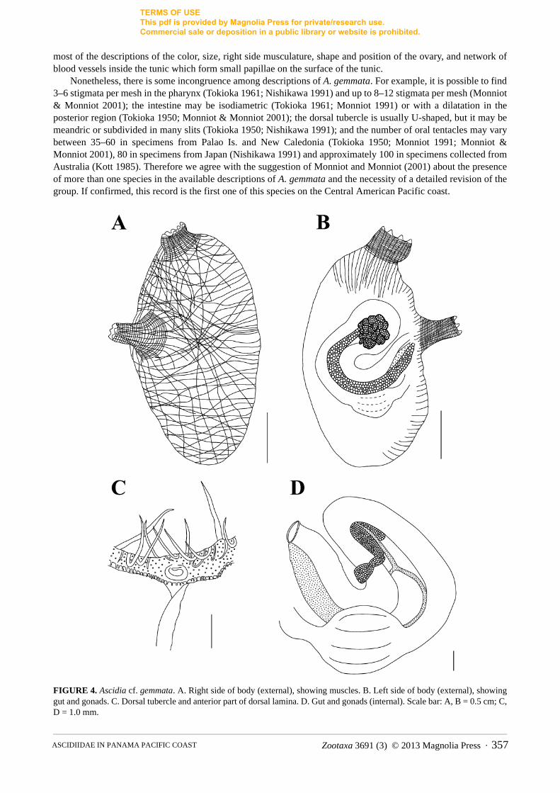
There are 58–69 oral tentacles, of four sizes; the largest is 2.0–2.5 mm long. They project from a thick muscular ring. The prepharyngeal groove is double and the anterior membrane has projections. The distance between the line of tentacles and the prepharyngeal groove is short (up to 0.3–0.6 mm), with some papillae. The peritubercular area is small and the dorsal tubercle aperture is C or U-shaped with enrolled ends. The smooth dorsal lamina is double anteriorly and without any projections on the right side close to the esophageal aperture. The dorsal lamina passes by the esophageal aperture on its left to the end of the pharynx, which extends 1.8–5.2 mm beyond the stomach. There is a narrow lamina with smooth margin on the right of the esophageal aperture. The pharynx has 39–43 longitudinal vessels on the right side, 33–41 vessels on the left and 62–74 transverse vessels; it is very pleated and meshes have 6–8 stigmata. The primary papillae are simple or slightly trilobed and no intermediate papillae or parastigmatic vessels were found.
The alimentary canal is not large, occupying approximately half of the left side of the body. The stomach is globular, with 7–9 internal folds. The intestine is isodiametric and form two loops. The anus is 3.8–5.9 mm distance from the oral tentacles; it has a bilobed rim. The renal vesicles are 0.12–0.16 mm in diameter, and cover the alimentary canal on its atrial side. There are conspicuous conic projections on the peritoneum that cover the alimentary canal.
FIGURE 3. Ascidia cf. gemmata. A. Right side of body (without tunic). B. Left side of body (without tunic). Scale bar: A, B = 0.5 cm.
The cauliflower-shaped ovary lays within both the primary and secondary loops of the intestine; it is visible in the primary loop from the outside and atrial cavity, but in the second loop it is observed only from the inside. Oocytes are 0.12–0.14 mm in diameter. The testis is ramified and spread on the intestine wall. Gonoducts open close to the anus aperture.
Remarks. Specimens are provisionally identified as Ascidia gemmata because of their general resemblance to
BONNET ET AL. 356 · Zootaxa 3691 (3) © 2013 Magnolia Press
TERMS OF USE This pdf is provided by Magnolia Press for private/research use. Commercial sale or deposition in a public library or website is prohibited.
most of the descriptions of the color, size, right side musculature, shape and position of the ovary, and network of blood vessels inside the tunic which form small papillae on the surface of the tunic.
Nonetheless, there is some incongruence among descriptions of A. gemmata. For example, it is possible to find 3–6 stigmata per mesh in the pharynx (Tokioka 1961; Nishikawa 1991) and up to 8–12 stigmata per mesh (Monniot & Monniot 2001); the intestine may be isodiametric (Tokioka 1961; Monniot 1991) or with a dilatation in the posterior region (Tokioka 1950; Monniot & Monniot 2001); the dorsal tubercle is usually U-shaped, but it may be meandric or subdivided in many slits (Tokioka 1950; Nishikawa 1991); and the number of oral tentacles may vary between 35–60 in specimens from Palao Is. and New Caledonia (Tokioka 1950; Monniot 1991; Monniot & Monniot 2001), 80 in specimens from Japan (Nishikawa 1991) and approximately 100 in specimens collected from Australia (Kott 1985). Therefore we agree with the suggestion of Monniot and Monniot (2001) about the presence of more than one species in the available descriptions of A. gemmata and the necessity of a detailed revision of the group. If confirmed, this record is the first one of this species on the Central American Pacific coast.
FIGURE 4. Ascidia cf. gemmata. A. Right side of body (external), showing muscles. B. Left side of body (external), showing gut and gonads. C. Dorsal tubercle and anterior part of dorsal lamina. D. Gut and gonads (internal). Scale bar: A, B = 0.5 cm; C, D = 1.0 mm.
Zootaxa 3691 (3) © 2013 Magnolia Press · 357ASCIDIIDAE IN PANAMA PACIFIC COAST

TERMS OF USE This pdf is provided by Magnolia Press for private/research use. Commercial sale or deposition in a public library or website is prohibited.
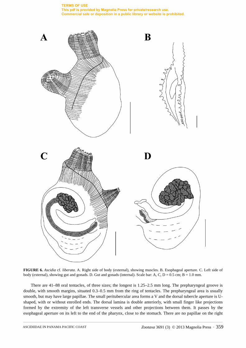
Ascidia cf. liberata Sluiter, 1887 (Figs. 5–6)
Material examined: DZUP ASC 55 – 2 ind.; STRI dock, Panama City; 1.5 m, inside bricks; 07/xii/2008; col.: R.M. Rocha. DZUP ASC 56 – 2 ind.; Marina Flamenco, Panama City; 1.5m, inside bricks; 04/i/2009; col.: R.M. Rocha. DZUP ASC 57 – 2 ind.; Isla Canales de Afuera; 5.0 m, under rocks; 13/i/2009; col.: R.M. Rocha.
Individuals are attached to the substrate by the left side of the body; there are no incrustations or epibionts. The color of living animals is red or yellow. The tunic is cartilaginous (0.6–1.5 mm thick), translucent and with numerous conic projections around the siphons; it may also have elongated projections on the ventral margin of the body.
The largest specimen measured 3.2 cm total length. The body is oblong, 1.3–3.2 cm long (including the oral siphon) and 0.9–1.2 cm wide, without tunic. Siphons are short (0.3–0.7 cm each one), the oral siphon is apical with eight lobes, while the atrial is displaced posteriorly 0.5–1.2 cm from the ring of tentacles and has 5–6 lobes. Lobes in both siphons have indented margins. The nervous ganglion is closer to the oral siphon. On the right side of the body there is a fold in the antero-dorsal region; in some individuals it is also present on the left side.
On both sides, the body wall musculature is formed by many short strong fibers (0.1–0.25 mm thick) perpendicular to the dorsal margin, forming a compact layer of muscles. These fibers extend posteriorly, but do not reach the ventral margin of the body. The longitudinal fibers in the siphons are organized in bands (one to each lobe).
FIGURE 5. Ascidia cf. liberata. A. Fresh animal. B. Right side of body (without tunic). C. Left side of body (without tunic). Scale bar: B, C = 0.5 cm.
BONNET ET AL. 358 · Zootaxa 3691 (3) © 2013 Magnolia Press
TERMS OF USE This pdf is provided by Magnolia Press for private/research use. Commercial sale or deposition in a public library or website is prohibited.
FIGURE 6. Ascidia cf. liberata. A. Right side of body (external), showing muscles. B. Esophageal aperture. C. Left side of body (external), showing gut and gonads. D. Gut and gonads (internal). Scale bar: A, C, D = 0.5 cm; B = 1.0 mm.
There are 41–88 oral tentacles, of three sizes; the longest is 1.25–2.5 mm long. The prepharyngeal groove is double, with smooth margins, situated 0.3–0.5 mm from the ring of tentacles. The prepharyngeal area is usually smooth, but may have large papillae. The small peritubercular area forms a V and the dorsal tubercle aperture is U-shaped, with or without enrolled ends. The dorsal lamina is double anteriorly, with small finger like projections formed by the extremity of the left transverse vessels and other projections between them. It passes by the esophageal aperture on its left to the end of the pharynx, close to the stomach. There are no papillae on the right
Zootaxa 3691 (3) © 2013 Magnolia Press · 359ASCIDIIDAE IN PANAMA PACIFIC COAST

TERMS OF USE This pdf is provided by Magnolia Press for private/research use. Commercial sale or deposition in a public library or website is prohibited.
side of the dorsal lamina at the level of the esophageal aperture. There is a row of long languets on the right side of the esophageal aperture, formed by the elongation of the right transverse vessels of the pharynx. The pharynx has 26–32 longitudinal vessels on the right side, 24–31 on the left side and 33–73 transverse vessels. It is strongly pleated and there are 6–9 stigmata per mesh; the primary papillae are bi or trilobed. In some parts of the pharynx parastigmatic vessels are found, but secondary papillae are absent.
The alimentary canal is large, occupying more than half of the left side of the body. The stomach is rounded and wide, with 5–6 internal longitudinal folds. The isodiametric intestine forms two loops and the bilobed anus is located approximately 4.0–10.5 mm from the oral tentacles. Small renal vesicles (0.04–0.08 mm diameter) cover the digestive tract. The peritoneum that covers the stomach and ascending portion of the intestine has irregular papillae, but on the rectum there are conical projections.
The ovary is compact, included in the primary intestinal loop, while the testis is ramified, with elongated follicles. Both gonads are visible from the outside and atrial cavity. The gonoducts open close to anus aperture, just posterior to it.
Remarks. The dissected specimens are in agreement with previous descriptions of A. liberata collected in Australia and Solomon Is.: small size (up to 2.0 cm total length), tunic with conical projections around the siphons, siphon lobes with indented margins, body wall with a fold on the right side, corporal musculature restricted to perpendicular fibers in the dorsal margin, longitudinal musculature in the siphons organized in bands, 6–8 stigmata per mesh and isodiametric intestine (Kott 1985; Nishikawa 1986). Nevertheless, there is some doubt about the identification because of differences in the descriptions and distribution. Living specimens are maroon-purple color and have 30–50 oral tentacles (Kott 1985; Nishikawa 1986), the ovary is ramified on the secondary intestinal loop in Australian specimens (Kott 1985), and there is a large geographical distance between previous records and this one.
Similar to A. liberata, Ascidia incrassata Heller, 1878 has projections on the margin of the siphonal lobes, body musculature on the right side formed by short fibers perpendicular to the dorsal margin, indented lamina on the right side of the esophageal aperture and the cauliflower-shaped ovary restricted to the primary intestinal loop. A. incrassata may be distinguished from the present specimens of A. cf. liberata by the large size (up to 10.0 cm total length and tunic 2.0 cm thick); absence of conical projections on the tunic, close to the siphons; body musculature only in the anterior region of the body (not posterior to the atrial siphon); and 7–12 stigmata per mesh in the pharynx (Monniot et al. 2001).
Recently, Carman et al. (2011) reported the presence of A. incrassata near the Pacific entrance of the Panama Canal. However, this identification should be corrected because the specimens better correspond to A. cf. liberata.
Ascidia sideralis sp. nov. Bonnet & Rocha (Figs. 7–8)
Material examined. Holotype: MZUSP 00034 – 1 ind.; Pluto, Isla Canales de Tierra; 5.0 m; 12/i/2009; col. J. Dijkstra.
Paratypes: DZUP ASC 148 – 1 ind.; Isla Taboguilla, Panama City; 09/xii/2008; col. R.M. Rocha. DZUP ASC 149 – 6 ind.; Isla Taboga, Panama City; floating dock; 05/xii/2008; col. R.M. Rocha.
Etymology. The name is in reference to the white dots present on the entire surface of the tunic in living animals, giving the impression of a stellated night.
Diagnosis. Living animals are grayish with conspicuous white dots on the surface of the tunic; body musculature as a net is on the right side of the body, with complete transverse fibers close to the oral siphon; oral tentacles are connected by a thick membrane; large papillae are in the prebranchial area; 8–10 stigmata per mesh; primary papillae are trilobed in the pharynx; the intestine is isodiametric; and the ovary is lobed inside both intestinal loops.
Individuals are up to 5.7 cm total length and attached to the substrate by the left side of the body. The free surface usually has some epibionts (hydrozoans and bryozoans). Living specimens are grayish with numerous white dots on the entire surface of the tunic; the dots correspond to the terminations of blood vessels, which do not project from the surface of the animal. In formaldehyde, the gray coloration disappears, but the white dots are visible at least after ten months in preservative. The tunic is translucent and slightly wrinkled, with rigid or cartilaginous consistency (0.6–2.5 mm thick). Siphons are side-by-side and apical.
BONNET ET AL. 360 · Zootaxa 3691 (3) © 2013 Magnolia Press

TERMS OF USE This pdf is provided by Magnolia Press for private/research use. Commercial sale or deposition in a public library or website is prohibited.
The body is oblong, 2.0–3.5 cm long (without the oral siphon) and 1.1–1.9 cm wide. The oral siphon is 0.4–1.0 cm long and has eight lobes, while the atrial siphon is 0.4–1.6 cm long and has 7–8 lobes. The atrial siphon is displaced posteriorly 0.7–1.5 cm away from the ring of oral tentacles. Lobes on both siphons have smooth margins. The neural ganglion is closer to the oral siphon.
A dense net of fibers (0.08–0.16 mm thick) forms the body wall musculature on the right side, but close to the oral siphon horizontal fibers predominate. On the left side of the body there are short longitudinal fibers extending out of the oral siphon and some short fibers on the dorsal margin. The longitudinal muscle fibers in the siphons are not organized in bands.
FIGURE 7. Ascidia sideralis sp. nov. A, B. Animal in field. C. Right side of body (without tunic). D. Left side of body (without tunic). Scale bar: C, D = 0.5 cm.
There are 56–96 oral tentacles, of four sizes, and the largest is 1.7–5.3 mm long, with a thick membrane connecting them. The smooth prepharyngeal groove is double; the distance between the line of tentacles and the prepharyngeal groove is short (0.4–0.8 mm long), with large but not abundant papillae (0.04–0.06 mm in diameter). The dorsal tubercle is U-shaped, with enrolled ends; the peritubercular area is U-shaped. The dorsal lamina is double and smooth anteriorly, but it is toothed posteriorly due the elongation of the left transverse vessels plus small projections between them. The dorsal lamina passes by the esophageal aperture on its left to the end of the pharynx, close to the stomach. There are no papillae on the right side of the dorsal lamina near the esophageal aperture, but there is a wide lamina on the right side of the esophageal aperture. The pharynx has 35–47 longitudinal vessels on the right side, 31–41 on the left side and 86–142 transverse vessels; it is very pleated and there are 8–10 stigmata per mesh. The primary papillae are slightly trilobed and no intermediate papillae or parastigmatic vessels were found.
Zootaxa 3691 (3) © 2013 Magnolia Press · 361ASCIDIIDAE IN PANAMA PACIFIC COAST

TERMS OF USE This pdf is provided by Magnolia Press for private/research use. Commercial sale or deposition in a public library or website is prohibited.
The alimentary canal occupies half or more of the left side of the body. The stomach is elongated, with 6–10 internal longitudinal folds. The isodiametric intestine forms two loops and the bilobed anus is located 6.2–12.5 mm from the oral tentacles. Renal vesicles that are 0.1–0.16 mm in diameter cover the intestine and stomach. There are conspicuous conical projections on the peritoneum covering the alimentary canal.
The ovary is lobed, included in the primary and secondary intestinal loops, and visible mostly from the external side of the body wall. The oocytes are 0.15–0.18 mm in diameter. The testis follicles are elongated, but not very ramified, and overlie the stomach and most of the intestine. Gonoducts open just posterior to the anus aperture.
FIGURE 8. Ascidia sideralis sp. nov. A. Right side of body (external), showing muscles. B. Papillae in the prepharyngeal area. C. Left side of body (external), showing gut and gonads. D. Gut and gonads (internal). Scale bar: A, C, D = 0.5 cm; B = 1.0 mm.
BONNET ET AL. 362 · Zootaxa 3691 (3) © 2013 Magnolia Press

TERMS OF USE This pdf is provided by Magnolia Press for private/research use. Commercial sale or deposition in a public library or website is prohibited.
Remarks. The only species of Ascidia whose living specimens have brilliant dots in the tunic are A. deceptaKott, 1985, A. latesiphonica Hartmeyer, 1922 and A. ornata Monniot & Monniot, 2001. Ascidia decepta also has eight lobes on the oral siphon and 6–7 lobes on the atrial siphon; body wall musculature as a net, 60–80 oral tentacles, 8–10 stigmata per mesh, and an isodiametric intestine. However, A. decepta can be differentiated from A. sideralis sp. nov. by the presence of sand embedded in the tunic, pigmented stripes in the siphon lining in some individuals, and the ovary ramified and restricted to the primary intestinal loop (Kott 1985). Similar to A. sideralis, A. latesiphonica has 40–100 oral tentacles, a very pleated pharynx, isodiametric intestine and lobed ovary (Hartmeyer 1922; Kott 1985). The differences with A. sideralis sp. nov. are the yellow dots in the tunic, body wall musculature on the right side that is formed by long longitudinal fibers in the middle of the body, short perpendicular fibers along the dorsal and ventral margins, longitudinal fibers in the siphons that are organized in bands, 6–8 stigmata per mesh, and ovary restricted to the primary intestinal loop (Hartmeyer 1922; Kott 1985). Similar to A. sideralis sp. nov., A. ornata has a net of muscular fibers on the right side of the body, papillae covering the prepharyngeal area, 6–10 stigmata per mesh and a lobed ovary (Monniot & Monniot 2001). However, the presence of 20 lobes on the oral siphon, white pigment lines in the margin of both siphons, 30 oral tentacles, descending limb of the intestine dilated, and ovary restricted to the primary intestinal loop (Monniot & Monniot 2001) distinguish A. ornata from A. sideralis sp. nov.
Phallusia julinea Sluiter, 1915, a common species in the Pacific Ocean, is characterized by the conspicuous dots on the surface of the tunic in living animals and, although classified in the genus Phallusia, the absence of accessory openings in some individuals has been reported (Monniot 1987). However, the presence of 10–13 toothed lobes on the siphons, complete transverse muscle fibers close to the oral siphon on the left side of the body, a toothed lamina on the right side of the esophageal aperture, 70–90 longitudinal vessels in each side of the pharynx, intestine with the descending part dilated, and opening in the multilobed anus, are characteristics that distinguish P. julinea from A. sideralis sp. nov.
If the living aspect is disregarded, two other species of Ascidia also have a similar network pattern of body musculature and an isodiametric intestine. The dorsal tubercle in Ascidia occidentalis Kott, 1985, is also close to the neural gland and it has 8–10 stigmata per mesh (Kott 1985). The differences are the toothed lobes on the siphons, the reduced number of oral tentacles (approximately 30) and the few internal folds in the stomach (Kott 1985). Ascidia melanostoma Sluiter, 1885 also has 36–90 oral tentacles, papillae in the prepharyngeal area, 26–48 longitudinal vessels on each side of the pharynx and a bilobed anus (Kott 1981; Nishikawa 1986). Nevertheless, the smaller number of stigmata per mesh (6–8) and the ramified ovary (Kott 1981; Nishikawa 1986) distinguish A. melanostoma from A. sideralis sp. nov.
Ascidia sydneiensis Stimpson, 1855
Material examined: DZUP ASC 120 – 2 ind.; STRI dock, Panama City; 1.0 m, inside bricks; 07/xii/2008; col. R.M. Rocha; DZUP ASC 122 – 4 ind.; 0.5 m, inside bricks; 04/i/2009; col. R.M. Rocha. DZUP ASC 119 – 2 ind.; Marina Flamenco, Panama City; 1.0 m, inside bricks; 06/xii/2008; col. R.M. Rocha; DZUP ASC 121 – 1 ind.; 04/i/2009; col. R.M. Rocha.
Tunic forms longitudinal ridges along the siphons; siphons are lobed with indented margins; body musculature on the right side is formed by short fibers perpendicular to the margin of the body; dorsal tubercle is meandering; rectum as a sac-like pouch; anus is multilobed; and the ovary is ramified.
Remarks. The characteristics observed in the present specimens are similar to specimens from the Atlantic coast of Panama (Bonnet & Rocha 2011).
Acknowledgments
We thank Stephan Bullard, Jennifer Dijkstra, James Roper, Mimi Carman and Zachary Carman for helping with fieldwork. We would like to thank Dr. Rachel Collin for having accepted NYKB as an intern at the Collin Lab, in the Smithsonian Tropical Research Station (STRI), Panama, in 2011. We thank Dr. Mark Torchin and Javiar Jara, STRI for logistical assistance in Panama City and Dr. Luis Camilli and the staff of the Liquid Jungle Lab at Isla Canales de Tierra for logistical assistance. We also thank Gretchen Lambert for helping with the key for species and two anonimous reviewers. CNPq supplied Master scholarship to NYKB and a sabbatical grant to RMR. MRC
Zootaxa 3691 (3) © 2013 Magnolia Press · 363ASCIDIIDAE IN PANAMA PACIFIC COAST
TERMS OF USE This pdf is provided by Magnolia Press for private/research use. Commercial sale or deposition in a public library or website is prohibited.
received funding from Woods Hole Oceanographic Institution, Ocean Life Institute. This paper is contribution 1891 of Departamento de Zoologia, Universidade Federal do Paraná.
References Bonnet, N.Y.K. & Rocha, R.M. (2011) The family Ascidiidae Herdman (Tunicata: Ascidiacea) in Bocas del Toro, Panama.
Description of six new species. Zootaxa, 2864, 1–33.Bullard, S., Carman, M., Rocha, R.M., Dijkstra, J.A., Goodwin, A. (2011) Abundance and diversity of ascidians in the southern
Gulf of Chiriquí, Pacific Panama. Aquatic Invasions, 6, 381–390. http://dx.doi.org/10.3391/ai.2011.6.4.03
Carman, M.R., Bullard, S.G., Rocha, R.M., Lambert, G., Dijkstra, J.A., Roper, J.J., Goodwin, A., Carman, M.M. & Vail, E.M. (2011) Ascidians at the Pacific and Atlantic entrances to the Panama Canal. Aquatic Invasions, 6 (4), 371–380. http://dx.doi.org/10.3391/ai.2011.6.4.02
Hartmeyer, R. (1922) Miscellanea ascidiologica. Mitteilungen aus dem Zoologischen Museum in Berlin, 10, 299–323.Kott, P. (1981) The ascidians of the Reef Flats of Fiji. Proceedings of the Linnean Society of New South Wales, 105 (3), 147–
212.Kott, P. (1985) The Australian Ascidiacea. Part 1. Phlebobranchia and Stolidobranchia. Memories of the Queensland Museum,
23, 1–440.Lambert, C.C. & Lambert, G. (1998) Non-indigenous ascidians in southern California harbors and marinas. Marine Biology,
130, 675–688. http://dx.doi.org/10.1007/s002270050289
Lambert, G. & Sanamyan, K. (2001) Distaplia alaskensis sp. nov. (Ascidiacea, Aplousobranchia) and other new ascidian records from south-central Alaska, with a redescription of Ascidia columbiana (Huntsman, 1912). Canadian Journal of Zoology, 79, 1766–1781.
Monniot, C. (1983) Ascidies littorales de Guadeloupe II. Phlebobranches. Bulletin du Muséum Nationale d’Histoire Naturelle, 4 ser. Zool., 5 (1), 51–71.
Monniot, C. (1987) Ascidies de Nouvelle-Calédonie. I. Phlébobranches du lagon. Bulletin du Muséum National d’Histoire Naturelle, 4 ser. Zool., 9 (1), 3–31.
Monniot, C. (1991) Ascidies de Nouvelle-Calédonie. VIII. Phlébobranches (suite). Bulletin du Muséum National d’Histoire Naturelle, 4 ser. Zool., 3–4, 491–515.
Monniot, C. (1997) Ascidies phlébobranches du canal du Mozambique. Zoosystema, 19 (4), 557–571.Monniot, C. & Monniot, F. (1987) Les ascidies de Polynésie Française. Mémoires du Muséum Nationale d’Histoire Naturelle,
ser. A, Zool., 136, 1–155.Monniot, C., Monniot, F., Griffiths, C.L. & Schleyer, M. (2001) South African ascidians. Annals of the South African Museum,
108 (1), 1–141.Monniot, F. (2007) Some ascidians (Tunicata) from the Clipperton Island. Cahier de Biologie Marine, 48, 303–310.Monniot, F. & Monniot, C. (2001) Ascidians from the tropical western Pacific. Zoosystema, 23 (2), 201–383.
http://dx.doi.org/10.1046/j.1096-3642.2002.00017.xNishikawa, T. (1986) Ascidians from the Gilbert and Solomon Islands and Nauru. I. Perophoridae, Ascidiidae and Corellidae.
Proceedings of the Japanese Society of Systematic Zoology, 32, 30–78.Nishikawa, T. (1991) The ascidians of the Japan Sea. II. Publications of the Seto Marine Biological Laboratory, 35 (1/3), 25–
170.Nova-Bustos, N., Hernández-Zanuy, A.C. & Viquez-Portuguez, R. (2010) Distribución y abundancia de las ascidias de los
fondos rocosos de la Bahía de Cuajiniquil, Costa Rica. Boletín de Investigaciones Marinas y Costeras, 39 (1), 57–66.Rocha, R.M., Moreno, T.R. & Faria, S.B. (2005) Ascidians from Bocas del Toro, Panamá. I. Biodiversity. Caribbean Journal of
Science, 41 (3), 600–612. Rocha, R.M., Zanata, T.B. & Moreno, T.R. (2012) Keys for the identification of families and genera of Atlantic shallow water
ascidians. Biota Neotropica, 12 (1), http://www.biotaneotropica.org.br/v12n1/en/abstract?identification-ey+bn01712012012 http://dx.doi.org/10.1590/s1676-06032012000100022
Tokioka, T. (1950) Ascidians from the Palao Island. I. Publications of the Seto Marine Biological Laboratory, I (3), 115–153.Tokioka, T. (1961) Ascidians collected during the Melanesia Expedition of the Osaka Museum of Natural History. I. Ascidians
presented by Dr. R.L.A. Catala of the Aquarium of Noumea. Publications of the Seto Marine Biological Laboratory, IX (1), 103–139.
Tokioka, T. (1972) On a small collection of ascidians from the Pacific coast of Costa Rica. Publications of the Seto Marine Biological Laboratory, XIX (6), 383–408.
Van Name, W.G. (1945) The North and South American ascidians. Bulletin of the American Museum of Natural History, 84, 1–476.
BONNET ET AL. 364 · Zootaxa 3691 (3) © 2013 Magnolia Press