Discovery of Markers of Exposure Specific to Bites of Lutzomyia longipalpis, the Vector of...
10
Discovery of Markers of Exposure Specific to Bites of Lutzomyia longipalpis, the Vector of Leishmania infantum chagasi in Latin America Clarissa Teixeira 1. , Regis Gomes 1. , Nicolas Collin 1 , David Reynoso 1 , Ryan Jochim 1 , Fabiano Oliveira 1 , Amy Seitz 1 , Dia-Eldin Elnaiem 2 , Arlene Caldas 3 , Ana Paula de Souza 4 , Cla ´ udia I. Brodskyn 4 , Camila Indiani de Oliveira 4 , Ivete Mendonca 5 , Carlos H. N. Costa 5 , Petr Volf 6 , Aldina Barral 4 , Shaden Kamhawi 1 , Jesus G. Valenzuela 1 * 1 Vector Molecular Biology Section, Laboratory of Malaria and Vector Research, National Institute of Allergy and Infectious Diseases, National Institutes of Health, Rockville, Maryland, United States of America, 2 Department of Natural Sciences, University of Maryland Eastern Shore, Princess Anne, Maryland, United States of America, 3 Universidade Federal do Maranha ˜o, Sa ˜o Luis, Maranha ˜o, Brazil, 4 Centro de Pesquisas Goncalo Moniz–FIOCRUZ, Salvador, Bahia, Brazil, 5 Laboratorio de Leishmanioses, Instituto de Doencas Tropicais Natan Portella and Universidade Federal do Piaui, Teresina, Piaui, Brazil, 6 Department of Parasitology, Faculty of Science, Charles University in Prague, Prague, Czech Republic Abstract Background: Sand flies deliver Leishmania parasites to a host alongside salivary molecules that affect infection outcomes. Though some proteins are immunogenic and have potential as markers of vector exposure, their identity and vector specificity remain elusive. Methodology/Principal Findings: We screened human, dog, and fox sera from endemic areas of visceral leishmaniasis to identify potential markers of specific exposure to saliva of Lutzomyia longipalpis. Human and dog sera were further tested against additional sand fly species. Recombinant proteins of nine transcripts encoding secreted salivary molecules of Lu. longipalpis were produced, purified, and tested for antigenicity and specificity. Use of recombinant proteins corresponding to immunogenic molecules in Lu. longipalpis saliva identified LJM17 and LJM11 as potential markers of exposure. LJM17 was recognized by human, dog, and fox sera; LJM11 by humans and dogs. Notably, LJM17 and LJM11 were specifically recognized by humans exposed to Lu. longipalpis but not by individuals exposed to Lu. intermedia. Conclusions/Significance: Salivary recombinant proteins are of value as markers of vector exposure. In humans, LJM17 and LJM11 emerged as potential markers of specific exposure to Lu. longipalpis, the vector of Leishmania infantum chagasi in Latin America. In dogs, LJM17, LJM11, LJL13, LJL23, and LJL143 emerged as potential markers of sand fly exposure. Testing these recombinant proteins in large scale studies will validate their usefulness as specific markers of Lu. longipalpis exposure in humans and of sand fly exposure in dogs. Citation: Teixeira C, Gomes R, Collin N, Reynoso D, Jochim R, et al. (2010) Discovery of Markers of Exposure Specific to Bites of Lutzomyia longipalpis, the Vector of Leishmania infantum chagasi in Latin America. PLoS Negl Trop Dis 4(3): e638. doi:10.1371/journal.pntd.0000638 Editor: Genevieve Milon, Institut Pasteur, France Received October 9, 2009; Accepted February 2, 2010; Published March 23, 2010 This is an open-access article distributed under the terms of the Creative Commons Public Domain declaration which stipulates that, once placed in the public domain, this work may be freely reproduced, distributed, transmitted, modified, built upon, or otherwise used by anyone for any lawful purpose. Funding: This work was supported by the Intramural Research Program of the Division of Intramural Research, National Institute of Allergy and Infectious Diseases, US National Institutes of Health. The funders had no role in study design, data collection and analysis, decision to publish, or preparation of the manuscript. Competing Interests: The authors have declared that no competing interests exist. * E-mail: [email protected] . These authors contributed equally to this work. Introduction Sand fly salivary proteins play a major role in blood feeding and Leishmania transmission [1–3]. Exposure to sand fly salivary proteins induces both cellular immunity and specific antibodies [3,4]. A relationship between the level of specific antibodies to saliva, vector exposure and risk of contracting disease has been demonstrated for different vector-host models [5–9]. Production of antibodies against mosquito and tick saliva not only contributed to development of host allergic reactions but was strongly related to risk of disease development [5,10]. Similarly, in an endemic area of Senegal, production of antibodies against Anopheles gambiae salivary proteins was identified as an indicator of the risk of malaria [10]. This correlation was also observed for tick exposure, where antibody production against tick saliva was associated with self-reported tick exposure and Lyme disease [11]. Recently, saliva of Triatoma infestans was shown to be a potential marker for vector infestation in domestic animals [12]. Therefore, the detection of antibodies against the saliva of hematophagous insect vectors could be used as an indicator of vector exposure and in some instances as an indicator for risk of contracting disease. Previous work shows that humans and animals exposed to sand fly bites or immunized with saliva can develop antibodies that recognize specific salivary proteins [4,7,13–15]. In Sa ˜ o Luis, an area of endemic visceral leishmaniasis (VL) in Maranha ˜o, Brazil, the www.plosntds.org 1 March 2010 | Volume 4 | Issue 3 | e638
Transcript of Discovery of Markers of Exposure Specific to Bites of Lutzomyia longipalpis, the Vector of...
Discovery of Markers of Exposure Specific to Bites ofLutzomyia longipalpis, the Vector of Leishmaniainfantum chagasi in Latin AmericaClarissa Teixeira1., Regis Gomes1., Nicolas Collin1, David Reynoso1, Ryan Jochim1, Fabiano Oliveira1,
Amy Seitz1, Dia-Eldin Elnaiem2, Arlene Caldas3, Ana Paula de Souza4, Claudia I. Brodskyn4, Camila
Indiani de Oliveira4, Ivete Mendonca5, Carlos H. N. Costa5, Petr Volf6, Aldina Barral4, Shaden Kamhawi1,
Jesus G. Valenzuela1*
1 Vector Molecular Biology Section, Laboratory of Malaria and Vector Research, National Institute of Allergy and Infectious Diseases, National Institutes of Health, Rockville,
Maryland, United States of America, 2 Department of Natural Sciences, University of Maryland Eastern Shore, Princess Anne, Maryland, United States of America,
3 Universidade Federal do Maranhao, Sao Luis, Maranhao, Brazil, 4 Centro de Pesquisas Goncalo Moniz–FIOCRUZ, Salvador, Bahia, Brazil, 5 Laboratorio de Leishmanioses,
Instituto de Doencas Tropicais Natan Portella and Universidade Federal do Piaui, Teresina, Piaui, Brazil, 6 Department of Parasitology, Faculty of Science, Charles University
in Prague, Prague, Czech Republic
Abstract
Background: Sand flies deliver Leishmania parasites to a host alongside salivary molecules that affect infection outcomes.Though some proteins are immunogenic and have potential as markers of vector exposure, their identity and vectorspecificity remain elusive.
Methodology/Principal Findings: We screened human, dog, and fox sera from endemic areas of visceral leishmaniasis toidentify potential markers of specific exposure to saliva of Lutzomyia longipalpis. Human and dog sera were further testedagainst additional sand fly species. Recombinant proteins of nine transcripts encoding secreted salivary molecules of Lu.longipalpis were produced, purified, and tested for antigenicity and specificity. Use of recombinant proteins correspondingto immunogenic molecules in Lu. longipalpis saliva identified LJM17 and LJM11 as potential markers of exposure. LJM17 wasrecognized by human, dog, and fox sera; LJM11 by humans and dogs. Notably, LJM17 and LJM11 were specificallyrecognized by humans exposed to Lu. longipalpis but not by individuals exposed to Lu. intermedia.
Conclusions/Significance: Salivary recombinant proteins are of value as markers of vector exposure. In humans, LJM17 andLJM11 emerged as potential markers of specific exposure to Lu. longipalpis, the vector of Leishmania infantum chagasi inLatin America. In dogs, LJM17, LJM11, LJL13, LJL23, and LJL143 emerged as potential markers of sand fly exposure. Testingthese recombinant proteins in large scale studies will validate their usefulness as specific markers of Lu. longipalpis exposurein humans and of sand fly exposure in dogs.
Citation: Teixeira C, Gomes R, Collin N, Reynoso D, Jochim R, et al. (2010) Discovery of Markers of Exposure Specific to Bites of Lutzomyia longipalpis, the Vector ofLeishmania infantum chagasi in Latin America. PLoS Negl Trop Dis 4(3): e638. doi:10.1371/journal.pntd.0000638
Editor: Genevieve Milon, Institut Pasteur, France
Received October 9, 2009; Accepted February 2, 2010; Published March 23, 2010
This is an open-access article distributed under the terms of the Creative Commons Public Domain declaration which stipulates that, once placed in the publicdomain, this work may be freely reproduced, distributed, transmitted, modified, built upon, or otherwise used by anyone for any lawful purpose.
Funding: This work was supported by the Intramural Research Program of the Division of Intramural Research, National Institute of Allergy and InfectiousDiseases, US National Institutes of Health. The funders had no role in study design, data collection and analysis, decision to publish, or preparation of themanuscript.
Competing Interests: The authors have declared that no competing interests exist.
* E-mail: [email protected]
. These authors contributed equally to this work.
Introduction
Sand fly salivary proteins play a major role in blood feeding and
Leishmania transmission [1–3]. Exposure to sand fly salivary proteins
induces both cellular immunity and specific antibodies [3,4]. A
relationship between the level of specific antibodies to saliva, vector
exposure and risk of contracting disease has been demonstrated for
different vector-host models [5–9]. Production of antibodies against
mosquito and tick saliva not only contributed to development of host
allergic reactions but was strongly related to risk of disease development
[5,10]. Similarly, in an endemic area of Senegal, production of
antibodies against Anopheles gambiae salivary proteins was identified as an
indicator of the risk of malaria [10]. This correlation was also observed
for tick exposure, where antibody production against tick saliva was
associated with self-reported tick exposure and Lyme disease [11].
Recently, saliva of Triatoma infestans was shown to be a potential marker
for vector infestation in domestic animals [12]. Therefore, the detection
of antibodies against the saliva of hematophagous insect vectors could
be used as an indicator of vector exposure and in some instances as an
indicator for risk of contracting disease.
Previous work shows that humans and animals exposed to sand
fly bites or immunized with saliva can develop antibodies that
recognize specific salivary proteins [4,7,13–15]. In Sao Luis, an area
of endemic visceral leishmaniasis (VL) in Maranhao, Brazil, the
www.plosntds.org 1 March 2010 | Volume 4 | Issue 3 | e638
presence of anti-saliva antibodies in humans strongly correlated
with protection and the development of anti-Leishmania delayed-type
hypersensitivity response [7]. Furthermore, individuals that poorly
recognized salivary proteins developed anti-Leishmania antibodies
associated with disease progression [7]. In contrast, in areas
endemic for cutaneous leishmaniasis (CL)—such as Canoa (Bahia,
Brazil) and Sanliurfa (Turkey)—the presence of anti-saliva antibod-
ies correlated with risk of contracting disease [16,17].
The presence of antibodies to sand fly salivary proteins has also
been demonstrated in animal reservoirs of leishmaniasis. In
canines, two sand fly salivary proteins were recognized by sera
of infected dogs from an endemic VL area in Brazil [18].
Hostomska et al. [14] reported the presence of anti-saliva
antibodies to six different sand fly proteins in dogs experimentally
exposed to Lutzomyia longipalpis bites. Importantly, foxes captured
in Teresina, an endemic VL area in Brazil, also showed high levels
of anti-saliva antibodies, particularly to a 44-kDa salivary protein
from Lu. longipalpis, suggesting exposure to bites of this vector [19].
Hence, vector salivary proteins also represent a potential tool as
markers of exposure to important reservoirs of disease.
Identification of the sand fly salivary proteins recognized by the
mammalian host will not only increase our understanding of
vector-host interactions but will also aid in developing new
epidemiological tools to correlate host exposure to vector sand flies
with immunity or susceptibility to leishmaniasis. It will also help
identify potential reservoirs of Leishmania. Here we describe a
practical functional transcriptomic approach for the identification
of the Lu. longipalpis salivary proteins most recognized by humans
and canids (dogs and foxes) using sera from Sao Luis and Teresina,
endemic areas for VL in Brazil [15,20].
Methods
Sand flies and preparation of salivary gland homogenate(SGH)
Lu. longipalpis (Jacobina strain) were reared at LMVR, NIAID,
USA; Lu. verrucarum (Peru strain) and Phlebotomus perniciosus (Italy
strain) at WRAIR, USA; Lu. intermedia (Corte de Pedra strain) were
obtained from CPqGM (FIOCRUZ, Bahia, Brazil). Females were
used for dissection of salivary glands 5–8 days post-eclosion; SGH
was prepared as described elsewhere [21]. Briefly, salivary glands
were dissected and stored in sterile PBS (pH 7.4) at 270uC. To
obtain the homogenate, salivary glands were disrupted by
ultrasonication and the supernatant collected after centrifugation
at 15,000g for 2 minutes.
Serum samplesA total of 14 human sera from from a VL-endemic region in
Sao Luis (Maranhao, Brazil) [15] and 6 from a CL-endemic region
in Canoa (Bahia, Brazil) [22] were used in this study. Informed
written consent was obtained from parents or legal guardians of
minors. The project was approved by the institutional review
board from the Federal University of Bahia (1993) and the Federal
University of Maranhao (1996). Dog and fox (Cerdocyon thous) sera
(total of 8 and 11, respectively) were from animals captured in a
VL-endemic area around Teresina (Piaui, Brazil) [19]. Fox and
dog studies were approved in 2000 by the Brazilian agency for
protection of the wildlife (IBAMA/PI) and in 2005 by the Federal
University of Piaui. Sera were also obtained from dogs (total of 6)
experimentally exposed to Lu. longipalpis [14]. Dog studies were
approved by Bayer Health Care AG (Leverkusen, Germany) and
handled in accordance with the European guidelines for animal
husbandry.
Cloning of Lu. longipalpis salivary transcripts into aVR2001-TOPO vector
DNA was amplified by polymerase chain reaction (PCR) using a
forward primer deduced from the amino-terminus and a reverse
primer encoding a hexhistidine motif. PCR amplification
conditions were: one hold of 94uC 5 min, two cycles of 94uC30 s, 48uC 1 min, 72uC 1 min, 23 cycles of 94uC 30 s, 58uC1 min, 72uC 1 min, and one hold of 72uC 7 min. The PCR
product was cloned into the VR2001-TOPO vector and
sequenced [23].
Polyclonal antibodies against Lu. longipalpis salivaryprotein
A plasmid encoding a distinct salivary protein (1 mg/ml) was
injected intradermally into female Swiss Webster mice in 10 ml,
three times at two-week intervals to generate polyclonal antibodies
for each of the nine selected candidates [23].
Expression and high-performance liquidchromatography (HPLC) purification of His-tagged Lu.longipalpis salivary proteins
Recombinant proteins were produced by transfecting 293-F
cells (Invitrogen) with plasmid following the manufacturer’s
recommendations. After 72 h, the supernatant was recovered,
filtered and concentrated to 30 ml in an Amicon concentrator
device (Millipore) in the presence of Buffer A (20 mM NaH2PO4,
20 mM Na2HPO4, pH 7.4, 500 mM NaCl). A HiTrap chelating
HP column (GE Healthcare) was charged with 5 ml of 0.1M
Ni2SO4. The concentrated protein was added to the HiTrap
chelating HP column that was then connected to a Summit station
HPLC system (Dionex, Sunnyvale, CA) consisting of a P680
HPLC pump and a PDA-100 detector. The column was
equilibrated for 30 min with Buffer A at 1 ml/min. Elution
conditions were: 0-5 min, 100% Buffer A; 5-15 min, a gradient of
0% to 100% Buffer B ( Buffer A+50 mM imidazole); 15-20 min, a
gradient of 0% C (Buffer A+500 mM imidazole) to 10% C (90%
Author Summary
Leishmania parasites are transmitted by the bite of aninfected vector sand fly that injects salivary molecules intothe host skin during feeding. Certain salivary moleculescan produce antibodies and can be used as an indicator ofexposure to a vector sand fly and potentially the disease ittransmits. Here we identified potential markers of specificexposure to the sand fly Lutzomyia longipalpis, the vectorof visceral leishmaniasis in Latin America. Initially, wedetermined which of the salivary proteins produceantibodies in humans, dogs, and foxes from areas endemicfor the disease. To identify potential specific markers ofvector exposure, we produced nine different recombinantsalivary proteins from Lu. longipalpis and tested for theirrecognition by individuals exposed to another human-biting sand fly, Lu. intermedia, that transmits cutaneousleishmaniasis and commonly occurs in the same endemicareas as Lu. longipalpis. Two of the nine salivary proteinswere recognized only by humans exposed to Lu. long-ipalpis, suggesting they are immunogenic proteins andmay be useful in epidemiological studies. The identifica-tion of specific salivary proteins as potential markers ofexposure to vector sand flies will increase our understand-ing of vector–human interaction, bring new insights tovector control, and in some instances act as an indicatorfor risk of acquiring disease.
Markers of Exposure to Lutzomyia longipalpis
www.plosntds.org 2 March 2010 | Volume 4 | Issue 3 | e638
B); 20-25 min, 90% B and 10% C; min 25-30, a gradient of 10%
C to 20% C (80% B); 30-35 min, 80% B and 20% C; 35-40 min, a
gradient of 20% C to 100%C; and 40-50 min, 100% C. Eluted
proteins were detected at 280 nm and collected every minute on a
96-well microtiter plate using a Foxy 200 fraction collector
(Teledyne ISCO).
Five-microliter aliquots of all fractions were blotted on
nitrocellulose and blocked with TBS-tween 3% non-fat milk for
1 h and then incubated for 1 h with anti-saliva antibodies, washed,
and incubated for 1 h with an anti-mouse IgG (H+L) alkaline
phosphatase-conjugated secondary antibody (Promega). Positive
fractions were developed with Western Blue stabilized substrate for
alkaline phosphatase (Promega). Positive fractions were run on
sodium dodecyl sulfate (SDS-PAGE) and silver stained using
SilverQuest (Invitrogen). Imidazole was removed from positive
fractions by dialysis overnight against PBS, pH 7.4.
Western blotting using human, dog, and fox seraSalivary glands (40 pairs approximately equivalent to 40 mg
total protein) or soluble recombinant sand fly salivary proteins
(20 mg) were run on a 4–20% Tris-glycine gel or on a 4–12%
NuPAGE gel. After transfer to a nitrocellulose membrane using
the iBlot device (Invitrogen), the membrane was blocked with 3%
(w/v) nonfat dry milk in Tris-buffered saline (TBS)-0.05%
Tween, pH 8.0, overnight at 4uC. After washing with TBS-T,
pH 8.0, the membrane was placed on a mini-protean II
multiscreen apparatus (Bio-Rad, Hercules, CA), and different
lanes were incubated with various sera (1:80 dilution, human and
dog sera; 1:50 dilution, fox sera) for 3 h at room temperature.
After washing with TBS-T, pH 8.0, three times for 5 min, the
membrane was incubated with either anti-dog IgG (H+L)
alkaline phosphate-conjugated antibody (1:10,000) (Jackson
Immuno Research) for 1 h at room temperature for dog and
fox sera or with anti-human IgG alkaline phosphate-conjugated
antibody (1:8,000) (Sigma) for human sera. Membranes were
developed by addition of Western Blue stabilized substrate for
alkaline phosphatase (Promega), and the reaction was stopped by
washing the membrane with deionized water.
Results
Recognition of salivary proteins from the sand fly Lu.longipalpis by sera from different hosts
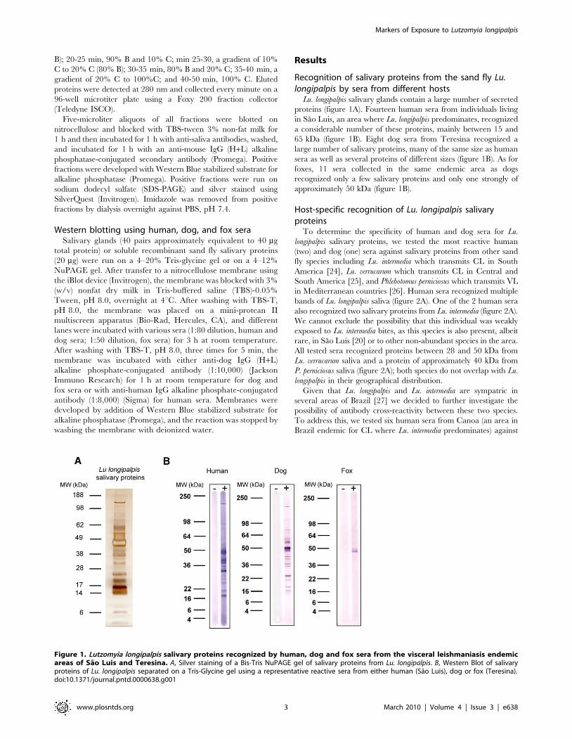
Lu. longipalpis salivary glands contain a large number of secreted
proteins (figure 1A). Fourteen human sera from individuals living
in Sao Luis, an area where Lu. longipalpis predominates, recognized
a considerable number of these proteins, mainly between 15 and
65 kDa (figure 1B). Eight dog sera from Teresina recognized a
large number of salivary proteins, many of the same size as human
sera as well as several proteins of different sizes (figure 1B). As for
foxes, 11 sera collected in the same endemic area as dogs
recognized only a few salivary proteins and only one strongly of
approximately 50 kDa (figure 1B).
Host-specific recognition of Lu. longipalpis salivaryproteins
To determine the specificity of human and dog sera for Lu.
longipalpis salivary proteins, we tested the most reactive human
(two) and dog (one) sera against salivary proteins from other sand
fly species including Lu. intermedia which transmits CL in South
America [24], Lu. verrucarum which transmits CL in Central and
South America [25], and Phlebotomus perniciosus which transmits VL
in Mediterranean countries [26]. Human sera recognized multiple
bands of Lu. longipalpis saliva (figure 2A). One of the 2 human sera
also recognized two salivary proteins from Lu. intermedia (figure 2A).
We cannot exclude the possibility that this individual was weakly
exposed to Lu. intermedia bites, as this species is also present, albeit
rare, in Sao Luis [20] or to other non-abundant species in the area.
All tested sera recognized proteins between 28 and 50 kDa from
Lu. verrucarum saliva and a protein of approximately 40 kDa from
P. perniciosus saliva (figure 2A); both species do not overlap with Lu.
longipalpis in their geographical distribution.
Given that Lu. longipalpis and Lu. intermedia are sympatric in
several areas of Brazil [27] we decided to further investigate the
possibility of antibody cross-reactivity between these two species.
To address this, we tested six human sera from Canoa (an area in
Brazil endemic for CL where Lu. intermedia predominates) against
Figure 1. Lutzomyia longipalpis salivary proteins recognized by human, dog and fox sera from the visceral leishmaniasis endemicareas of Sao Luis and Teresina. A, Silver staining of a Bis-Tris NuPAGE gel of salivary proteins from Lu. longipalpis. B, Western Blot of salivaryproteins of Lu. longipalpis separated on a Tris-Glycine gel using a representative reactive sera from either human (Sao Luis), dog or fox (Teresina).doi:10.1371/journal.pntd.0000638.g001
Markers of Exposure to Lutzomyia longipalpis
www.plosntds.org 3 March 2010 | Volume 4 | Issue 3 | e638
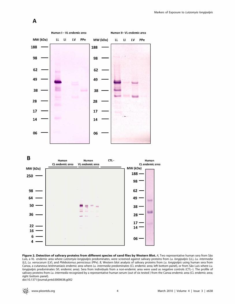
Figure 2. Detection of salivary proteins from different species of sand flies by Western Blot. A, Two representative human sera from SaoLuis, a VL- endemic area where Lutzomyia longipalpis predominates, were screened against salivary proteins from Lu. longipalpis (LL), Lu. intermedia(LI), Lu. verrucarum (LV), and Phlebotomus perniciosus (PPe). B, Western blot analysis of salivary proteins from Lu. longipalpis using human sera fromCanoa, a cutaneous leishmaniasis endemic area where Lu. intermedia predominates (CL endemic area; left bottom panel), or from Sao Luis where Lu.longipalpis predominates (VL endemic area). Sera from individuals from a non-endemic area were used as negative controls (CTL–). The profile ofsalivary proteins from Lu. intermedia recognized by a representative human serum (out of six tested ) from the Canoa endemic area (CL endemic area;right bottom panel).doi:10.1371/journal.pntd.0000638.g002
Markers of Exposure to Lutzomyia longipalpis
www.plosntds.org 4 March 2010 | Volume 4 | Issue 3 | e638
Lu. longipalpis saliva. These sera did not recognize any of the
salivary proteins of Lu. longipalpis but recognized those of Lu.
intermedia (figure 2B). Together, these results suggest an overall low
level of cross-reactivity between Lu. longipalpis and Lu. intermedia
salivary proteins.
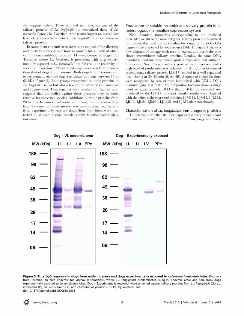
Because in an endemic area there is no control of the diversity
and intensity of exposure of hosts to sand fly bites—both of which
can influence antibody response [14]—we compared dogs from
Teresina, where Lu. longipalpis is prevalent, with dogs experi-
mentally exposed to Lu. longipalpis bites. Overall, the reactivity of
sera from experimentally exposed dogs was considerably lower
than that of dogs from Teresina. Both dogs from Teresina and
experimentally exposed dogs recognized proteins between 15 to
65 kDa (figure 3). Both groups recognized multiple proteins in
Lu. longipalpis saliva but also a few in the saliva of Lu. verrucarum
and P. perniciosus. This, together with results from human sera,
suggests that antibodies against these proteins may be cross
reactive for these two species. Additionally, while proteins from
28 to 50 kDa from Lu. intermedia were recognized by sera of dogs
from Teresina, only one protein was poorly recognized by sera
from experimentally exposed dogs. Sera from foxes were also
tested but showed no cross-reactivity with the other species (data
not shown).
Production of soluble recombinant salivary protein in aheterologous mammalian expression system
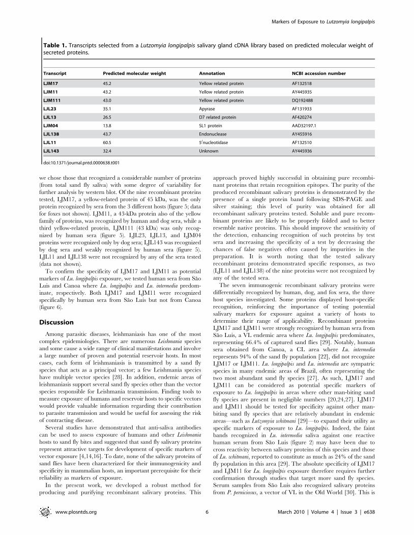
Nine abundant transcripts corresponding to the predicted
molecular weight of the most antigenic salivary proteins recognized
by human, dog, and fox sera within the range of 15 to 65 kDa
(figure 1) were selected for expression (Table 1). Figure 4 shows a
flow diagram of the approach used to express and purify the nine
chosen recombinant salivary proteins. Notably, the same DNA
plasmid is used for recombinant protein expression and antibody
production. Nine different salivary proteins were expressed and a
high level of purification was achieved by HPLC. Purification of
recombinant salivary protein LJM17 resulted in a well separated
peak eluting at 35–40 min (figure 4B). Aliquots of eluted fractions
were recognized by sera of mice immunized with LJM17 DNA
plasmid (figure 4C). SDS-PAGE of positive fractions shows a single
band of approximately 50 kDa (figure 4D), the expected size
predicted by the LJM17 transcript. Similar results were obtained
with the other eight expressed proteins: LJM111, LJM11, LJL143,
LJL13, LJL23, LJM04, LJL138, and LJL11 (data not shown).
Characterization of Lu. longipalpis immunogenic proteinsTo determine whether the nine expressed salivary recombinant
proteins were recognized by sera from humans, dogs, and foxes,
Figure 3. Total IgG response in dogs from endemic areas and dogs experimentally exposed to Lutzomyia longipalpis bites. Dog serafrom Teresina, an area endemic for visceral leishmaniasis where Lu. longipalpis predominates, (Dog-VL endemic area) and sera from dogsexperimentally exposed to Lu. longipalpis bites (Dog – Experimentally exposed) were screened against salivary proteins from Lu. longipalpis (LL), Lu.intermedia (LI), Lu. verrucarum (LV), and Phlebotomus perniciosus (PPe) by Western Blot.doi:10.1371/journal.pntd.0000638.g003
Markers of Exposure to Lutzomyia longipalpis
www.plosntds.org 5 March 2010 | Volume 4 | Issue 3 | e638
we chose those that recognized a considerable number of proteins
(from total sand fly saliva) with some degree of variability for
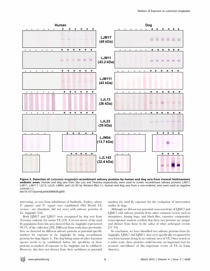
further analysis by western blot. Of the nine recombinant proteins
tested, LJM17, a yellow-related protein of 45 kDa, was the only
protein recognized by sera from the 3 different hosts (figure 5; data
for foxes not shown). LJM11, a 43-kDa protein also of the yellow
family of proteins, was recognized by human and dog sera, while a
third yellow-related protein, LJM111 (43 kDa) was only recog-
nized by human sera (figure 5). LJL23, LJL13, and LJM04
proteins were recognized only by dog sera; LJL143 was recognized
by dog sera and weakly recognized by human sera (figure 5).
LJL11 and LJL138 were not recognized by any of the sera tested
(data not shown).
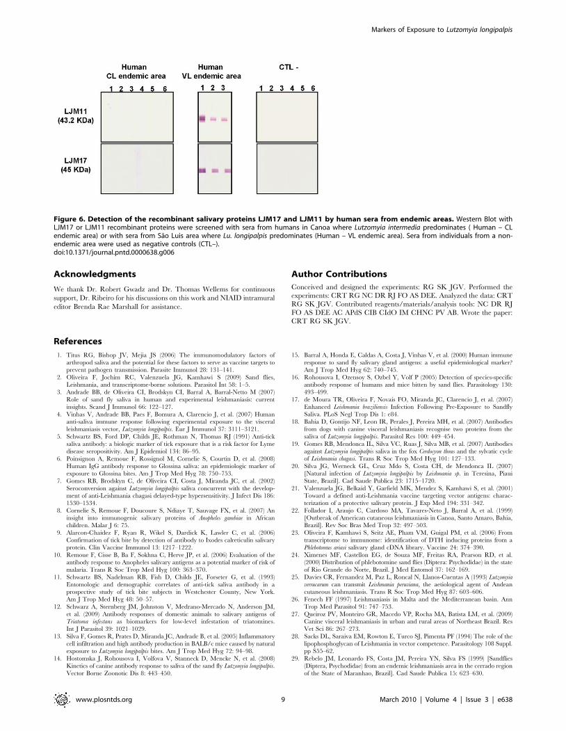
To confirm the specificity of LJM17 and LJM11 as potential
markers of Lu. longipalpis exposure, we tested human sera from Sao
Luis and Canoa where Lu. longipalpis and Lu. intermedia predom-
inate, respectively. Both LJM17 and LJM11 were recognized
specifically by human sera from Sao Luis but not from Canoa
(figure 6).
Discussion
Among parasitic diseases, leishmaniasis has one of the most
complex epidemiologies. There are numerous Leishmania species
and some cause a wide range of clinical manifestations and involve
a large number of proven and potential reservoir hosts. In most
cases, each form of leishmaniasis is transmitted by a sand fly
species that acts as a principal vector; a few Leishmania species
have multiple vector species [28]. In addition, endemic areas of
leishmaniasis support several sand fly species other than the vector
species responsible for Leishmania transmission. Finding tools to
measure exposure of humans and reservoir hosts to specific vectors
would provide valuable information regarding their contribution
to parasite transmission and would be useful for assessing the risk
of contracting disease.
Several studies have demonstrated that anti-saliva antibodies
can be used to assess exposure of humans and other Leishmania
hosts to sand fly bites and suggested that sand fly salivary proteins
represent attractive targets for development of specific markers of
vector exposure [4,14,16]. To date, none of the salivary proteins of
sand flies have been characterized for their immunogenicity and
specificity in mammalian hosts, an important prerequisite for their
reliability as markers of exposure.
In the present work, we developed a robust method for
producing and purifying recombinant salivary proteins. This
approach proved highly successful in obtaining pure recombi-
nant proteins that retain recognition epitopes. The purity of the
produced recombinant salivary proteins is demonstrated by the
presence of a single protein band following SDS-PAGE and
silver staining; this level of purity was obtained for all
recombinant salivary proteins tested. Soluble and pure recom-
binant proteins are likely to be properly folded and to better
resemble native proteins. This should improve the sensitivity of
the detection, enhancing recognition of such proteins by test
sera and increasing the specificity of a test by decreasing the
chances of false negatives often caused by impurities in the
preparation. It is worth noting that the tested salivary
recombinant proteins demonstrated specific responses, as two
(LJL11 and LJL138) of the nine proteins were not recognized by
any of the tested sera.
The seven immunogenic recombinant salivary proteins were
differentially recognized by human, dog, and fox sera, the three
host species investigated. Some proteins displayed host-specific
recognition, reinforcing the importance of testing potential
salivary markers for exposure against a variety of hosts to
determine their range of applicability. Recombinant proteins
LJM17 and LJM11 were strongly recognized by human sera from
Sao Luis, a VL endemic area where Lu. longipalpis predominates,
representing 66.4% of captured sand flies [29]. Notably, human
sera obtained from Canoa, a CL area where Lu. intermedia
represents 94% of the sand fly population [22], did not recognize
LJM17 or LJM11. Lu. longipalpis and Lu. intermedia are sympatric
species in many endemic areas of Brazil, often representing the
two most abundant sand fly species [27]. As such, LJM17 and
LJM11 can be considered as potential specific markers of
exposure to Lu. longipalpis in areas where other man-biting sand
fly species are present in negligible numbers [20,24,27]. LJM17
and LJM11 should be tested for specificity against other man-
biting sand fly species that are relatively abundant in endemic
areas—such as Lutzomyia whitmani [29]—to expand their utility as
specific markers of exposure to Lu. longipalpis. Indeed, the faint
bands recognized in Lu. intermedia saliva against one reactive
human serum from Sao Luis (figure 2) may have been due to
cross reactivity between salivary proteins of this species and those
of Lu. whitmani, reported to constitute as much as 24% of the sand
fly population in this area [29]. The absolute specificity of LJM17
and LJM11 for Lu. longipalpis exposure therefore requires further
confirmation through studies that target more sand fly species.
Serum samples from Sao Luis also recognized salivary proteins
from P. perniciosus, a vector of VL in the Old World [30]. This is
Table 1. Transcripts selected from a Lutzomyia longipalpis salivary gland cDNA library based on predicted molecular weight ofsecreted proteins.
Transcript Predicted molecular weight Annotation NCBI accession number
LJM17 45.2 Yellow related protein AF132518
LJM11 43.2 Yellow related protein AY445935
LJM111 43.0 Yellow related protein DQ192488
LJL23 35.1 Apyrase AF131933
LJL13 26.5 D7 related protein AF420274
LJM04 13.8 SL1 protein AAD32197.1
LJL138 43.7 Endonuclease AY455916
LJL11 60.5 5/nucleotidase AF132510
LJL143 32.4 Unknown AY445936
doi:10.1371/journal.pntd.0000638.t001
Markers of Exposure to Lutzomyia longipalpis
www.plosntds.org 6 March 2010 | Volume 4 | Issue 3 | e638
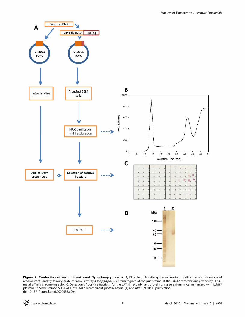
Figure 4. Production of recombinant sand fly salivary proteins. A, Flowchart describing the expression, purification and detection ofrecombinant sand fly salivary proteins from Lutzomyia longipalpis. B, Chromatogram of the purification of the LJM17 recombinant protein by HPLC-metal affinity chromatography. C, Detection of positive fractions for the LJM17 recombinant protein using sera from mice immunized with LJM17plasmid. D, Silver-stained SDS-PAGE of LJM17 recombinant protein before (1) and after (2) HPLC purification.doi:10.1371/journal.pntd.0000638.g004
Markers of Exposure to Lutzomyia longipalpis
www.plosntds.org 7 March 2010 | Volume 4 | Issue 3 | e638
interesting, as sera from inhabitants of Sanliurfa, Turkey, where
P. papatasi and P. sergenti—two established Old World CL
vectors—are abundant, did not react with salivary proteins of
Lu. longipalpis [16].
Both LJM17 and LJM11 were recognized by dog sera from
Teresina, endemic for canine VL [19]. A recent survey of the sand
fly population from this area showed that Lu. longipalpis represented
99.7% of the collection [20]. Different from work done previously,
here we detected six different salivary proteins as potential specific
markers for exposure to Lu. longipalpis by using recombinant
proteins for dogs (figure 5). The dog-biting status of other Lutzomyia
species needs to be established before the specificity of these
proteins as markers of exposure to Lu. longipalpis can be validated.
However, this does not detract from their usefulness as potential
markers for sand fly exposure for the evaluation of intervention
studies in dogs.
Although we did not test potential cross-reactivity of LJM17 and
LJM11 with salivary proteins from other common vectors such as
mosquitoes, kissing bugs, and black flies, extensive comparative
transcriptomic analysis confirm that these two proteins are unique
and distinct from those in the saliva of other arthropod vectors
[31–35].
In conclusion, we have identified two salivary proteins from Lu.
longipalpis, LJM17 and LJM11, that were specifically recognized by
sera from humans living in an endemic area of VL. Once tested on
a wider scale, these proteins could become an important tool for
accurate surveillance of this important vector of VL in Latin
America.
Figure 5. Detection of Lutzomyia longipalpis recombinant salivary proteins by human and dog sera from visceral leishmaniasisendemic areas. Human and dog sera from Sao Luis and Teresina respectively, were used to screen recombinant salivary proteins LJM17,LJM11, LJM111, LJL13, LJL23, LJM04, and LJL143 by Western Blot (+). Human and dog sera from a non-endemic area were used as negativecontrols (–).doi:10.1371/journal.pntd.0000638.g005
Markers of Exposure to Lutzomyia longipalpis
www.plosntds.org 8 March 2010 | Volume 4 | Issue 3 | e638
Acknowledgments
We thank Dr. Robert Gwadz and Dr. Thomas Wellems for continuous
support, Dr. Ribeiro for his discussions on this work and NIAID intramural
editor Brenda Rae Marshall for assistance.
Author Contributions
Conceived and designed the experiments: RG SK JGV. Performed the
experiments: CRT RG NC DR RJ FO AS DEE. Analyzed the data: CRT
RG SK JGV. Contributed reagents/materials/analysis tools: NC DR RJ
FO AS DEE AC APdS CIB CIdO IM CHNC PV AB. Wrote the paper:
CRT RG SK JGV.
References
1. Titus RG, Bishop JV, Mejia JS (2006) The immunomodulatory factors of
arthropod saliva and the potential for these factors to serve as vaccine targets toprevent pathogen transmission. Parasite Immunol 28: 131–141.
2. Oliveira F, Jochim RC, Valenzuela JG, Kamhawi S (2009) Sand flies,Leishmania, and transcriptome-borne solutions. Parasitol Int 58: 1–5.
3. Andrade BB, de Oliveira CI, Brodskyn CI, Barral A, Barral-Netto M (2007)
Role of sand fly saliva in human and experimental leishmaniasis: current
insights. Scand J Immunol 66: 122–127.
4. Vinhas V, Andrade BB, Paes F, Bomura A, Clarencio J, et al. (2007) Humananti-saliva immune response following experimental exposure to the visceral
leishmaniasis vector, Lutzomyia longipalpis. Eur J Immunol 37: 3111–3121.
5. Schwartz BS, Ford DP, Childs JE, Rothman N, Thomas RJ (1991) Anti-tick
saliva antibody: a biologic marker of tick exposure that is a risk factor for Lymedisease seropositivity. Am J Epidemiol 134: 86–95.
6. Poinsignon A, Remoue F, Rossignol M, Cornelie S, Courtin D, et al. (2008)Human IgG antibody response to Glossina saliva: an epidemiologic marker of
exposure to Glossina bites. Am J Trop Med Hyg 78: 750–753.
7. Gomes RB, Brodskyn C, de Oliveira CI, Costa J, Miranda JC, et al. (2002)
Seroconversion against Lutzomyia longipalpis saliva concurrent with the develop-ment of anti-Leishmania chagasi delayed-type hypersensitivity. J Infect Dis 186:
1530–1534.
8. Cornelie S, Remoue F, Doucoure S, Ndiaye T, Sauvage FX, et al. (2007) An
insight into immunogenic salivary proteins of Anopheles gambiae in Africanchildren. Malar J 6: 75.
9. Alarcon-Chaidez F, Ryan R, Wikel S, Dardick K, Lawler C, et al. (2006)
Confirmation of tick bite by detection of antibody to Ixodes calreticulin salivaryprotein. Clin Vaccine Immunol 13: 1217–1222.
10. Remoue F, Cisse B, Ba F, Sokhna C, Herve JP, et al. (2006) Evaluation of theantibody response to Anopheles salivary antigens as a potential marker of risk of
malaria. Trans R Soc Trop Med Hyg 100: 363–370.
11. Schwartz BS, Nadelman RB, Fish D, Childs JE, Forseter G, et al. (1993)
Entomologic and demographic correlates of anti-tick saliva antibody in aprospective study of tick bite subjects in Westchester County, New York.
Am J Trop Med Hyg 48: 50–57.
12. Schwarz A, Sternberg JM, Johnston V, Medrano-Mercado N, Anderson JM,
et al. (2009) Antibody responses of domestic animals to salivary antigens ofTriatoma infestans as biomarkers for low-level infestation of triatomines.
Int J Parasitol 39: 1021–1029.
13. Silva F, Gomes R, Prates D, Miranda JC, Andrade B, et al. (2005) Inflammatory
cell infiltration and high antibody production in BALB/c mice caused by naturalexposure to Lutzomyia longipalpis bites. Am J Trop Med Hyg 72: 94–98.
14. Hostomska J, Rohousova I, Volfova V, Stanneck D, Mencke N, et al. (2008)
Kinetics of canine antibody response to saliva of the sand fly Lutzomyia longipalpis.
Vector Borne Zoonotic Dis 8: 443–450.
15. Barral A, Honda E, Caldas A, Costa J, Vinhas V, et al. (2000) Human immune
response to sand fly salivary gland antigens: a useful epidemiological marker?Am J Trop Med Hyg 62: 740–745.
16. Rohousova I, Ozensoy S, Ozbel Y, Volf P (2005) Detection of species-specificantibody response of humans and mice bitten by sand flies. Parasitology 130:
493–499.
17. de Moura TR, Oliveira F, Novais FO, Miranda JC, Clarencio J, et al. (2007)Enhanced Leishmania braziliensis Infection Following Pre-Exposure to Sandfly
Saliva. PLoS Negl Trop Dis 1: e84.
18. Bahia D, Gontijo NF, Leon IR, Perales J, Pereira MH, et al. (2007) Antibodies
from dogs with canine visceral leishmaniasis recognise two proteins from thesaliva of Lutzomyia longipalpis. Parasitol Res 100: 449–454.
19. Gomes RB, Mendonca IL, Silva VC, Ruas J, Silva MB, et al. (2007) Antibodies
against Lutzomyia longipalpis saliva in the fox Cerdocyon thous and the sylvatic cycleof Leishmania chagasi. Trans R Soc Trop Med Hyg 101: 127–133.
20. Silva JG, Werneck GL, Cruz Mdo S, Costa CH, de Mendonca IL (2007)[Natural infection of Lutzomyia longipalpis by Leishmania sp. in Teresina, Piaui
State, Brazil]. Cad Saude Publica 23: 1715–1720.
21. Valenzuela JG, Belkaid Y, Garfield MK, Mendez S, Kamhawi S, et al. (2001)Toward a defined anti-Leishmania vaccine targeting vector antigens: charac-
terization of a protective salivary protein. J Exp Med 194: 331–342.
22. Follador I, Araujo C, Cardoso MA, Tavares-Neto J, Barral A, et al. (1999)
[Outbreak of American cutaneous leishmaniasis in Canoa, Santo Amaro, Bahia,Brazil]. Rev Soc Bras Med Trop 32: 497–503.
23. Oliveira F, Kamhawi S, Seitz AE, Pham VM, Guigal PM, et al. (2006) From
transcriptome to immunome: identification of DTH inducing proteins from a
Phlebotomus ariasi salivary gland cDNA library. Vaccine 24: 374–390.
24. Ximenes MF, Castellon EG, de Souza MF, Freitas RA, Pearson RD, et al.(2000) Distribution of phlebotomine sand flies (Diptera: Psychodidae) in the state
of Rio Grande do Norte, Brazil. J Med Entomol 37: 162–169.
25. Davies CR, Fernandez M, Paz L, Roncal N, Llanos-Cuentas A (1993) Lutzomyia
verrucarum can transmit Leishmania peruviana, the aetiological agent of Andean
cutaneous leishmaniasis. Trans R Soc Trop Med Hyg 87: 603–606.
26. Fenech FF (1997) Leishmaniasis in Malta and the Mediterranean basin. Ann
Trop Med Parasitol 91: 747–753.
27. Queiroz PV, Monteiro GR, Macedo VP, Rocha MA, Batista LM, et al. (2009)Canine visceral leishmaniasis in urban and rural areas of Northeast Brazil. Res
Vet Sci 86: 267–273.
28. Sacks DL, Saraiva EM, Rowton E, Turco SJ, Pimenta PF (1994) The role of the
lipophosphoglycan of Leishmania in vector competence. Parasitology 108 Suppl.pp S55–62.
29. Rebelo JM, Leonardo FS, Costa JM, Pereira YN, Silva FS (1999) [Sandflies
(Diptera, Psychodidae) from an endemic leishmaniasis area in the cerrado region
of the State of Maranhao, Brazil]. Cad Saude Publica 15: 623–630.
Figure 6. Detection of the recombinant salivary proteins LJM17 and LJM11 by human sera from endemic areas. Western Blot withLJM17 or LJM11 recombinant proteins were screened with sera from humans in Canoa where Lutzomyia intermedia predominates ( Human – CLendemic area) or with sera from Sao Luis area where Lu. longipalpis predominates (Human – VL endemic area). Sera from individuals from a non-endemic area were used as negative controls (CTL–).doi:10.1371/journal.pntd.0000638.g006
Markers of Exposure to Lutzomyia longipalpis
www.plosntds.org 9 March 2010 | Volume 4 | Issue 3 | e638
30. Maroli M, Rossi L, Baldelli R, Capelli G, Ferroglio E, et al. (2008) The
northward spread of leishmaniasis in Italy: evidence from retrospective andongoing studies on the canine reservoir and phlebotomine vectors. Trop Med Int
Health 13: 256–264.
31. Anderson JM, Oliveira F, Kamhawi S, Mans BJ, Reynoso D, et al. (2006)Comparative salivary gland transcriptomics of sandfly vectors of visceral
leishmaniasis. BMC Genomics 7: 52.32. Ribeiro JM, Arca B, Lombardo F, Calvo E, Phan VM, et al. (2007) An
annotated catalogue of salivary gland transcripts in the adult female mosquito,
Aedes aegypti. BMC Genomics 8: 6.
33. Assumpcao TC, Francischetti IM, Andersen JF, Schwarz A, Santana JM, et al.
(2008) An insight into the sialome of the blood-sucking bug Triatoma infestans, a
vector of Chagas’ disease. Insect Biochem Mol Biol 38: 213–232.
34. Andersen JF, Pham VM, Meng Z, Champagne DE, Ribeiro JM (2009) Insight
into the sialome of the Black Fly, Simulium vittatum. J Proteome Res 8: 1474–1488.
35. Calvo E, Pham VM, Marinotti O, Andersen JF, Ribeiro JM (2009) The salivary
gland transcriptome of the neotropical malaria vector Anopheles darlingi reveals
accelerated evolution of genes relevant to hematophagy. BMC Genomics 10: 57.
Markers of Exposure to Lutzomyia longipalpis
www.plosntds.org 10 March 2010 | Volume 4 | Issue 3 | e638