Reversal of photoschedule in spring does not prevent photorefractoriness in Siberian hamsters

BASIC RESEARCH
Vimentin and laminin are altered on cheek pouchmicrovessels of streptozotocin-induced diabetichamstersJemima Fuentes R. Silva,I Fatima Z. G. A. Cyrino,III Marisa M. D. Breitenbach,II Eliete Bouskela,III Jorge Jose
CarvalhoI
I Universidade do Estado do Rio de Janeiro, Laboratory of Cellular Ultrastructure and Tissue Biology, Biomedical Center, Institute of Biology, Rio de
Janeiro/RJ, Brazil. II Universidade do Estado do Rio de Janeiro, Department of Physiological Sciences, Biomedical Center, Rio de Janeiro/RJ, Brazil.III Universidade do Estado do Rio de Janeiro, Clinical and Experimental Research Laboratory on Vascular Biology (BioVasc), Biomedical Center, Rio de
Janeiro/RJ, Brazil.
OBJECTIVES: Normal endothelial cells respond to shear stress by elongating and aligning in the direction of fluidflow. Hyperglycemia impairs this response and contributes to microvascular complications, which result indeleterious effects to the endothelium. This work aimed to evaluate cheek pouch microvessel morphologicalcharacteristics, reactivity, permeability, and expression of cytoskeleton and extracellular matrix components inhamsters after the induction of diabetes with streptozotocin.
METHODS: Syrian golden hamsters (90–130 g) were injected with streptozotocin (50 mg/kg, i.p.) or vehicle either 6(the diabetes mellitus 6 group) or 15 (the diabetes mellitus 15 group) days before the experiment. Vasculardimensions and density per area of vessels were determined by morphometric and stereological measurements.Changes in blood flow were measured in response to acetylcholine, and plasma extravasation was measured by thenumber of leakage sites. Actin, talin, a-smooth muscle actin, vimentin, type IV collagen, and laminin were detectedby immunohistochemistry and assessed through a semiquantitative scoring system.
RESULTS: There were no major alterations in the lumen, wall diameters, or densities of the examined vessels.Likewise, vascular reactivity and permeability were not altered by diabetes. The arterioles demonstrated increasedimmunoreactivity to vimentin and laminin in the diabetes mellitus 6 and diabetes mellitus 15 groups.
DISCUSSION: Antibodies against laminin and vimentin inhibit branching morphogenesis in vitro. Therefore, lamininand vimentin participating in the structure of the focal adhesion may play a role in angiogenesis.
CONCLUSIONS: Our results indicated the existence of changes related to cell–matrix interactions, which maycontribute to the pathological remodeling that was already underway one week after induction of experimentaldiabetes.
KEYWORDS: Diabetes Mellitus; Microcirculation; Cytoskeleton; Extracellular Matrix; Immunohistochemistry.
Silva JFR, Cyrino FZGA, Breitenbach MMD, Bouskela E, Carvalho JJ. Vimentin and laminin are altered on cheek pouch microvessels of streptozotocin-induced diabetic hamsters. Clinics. 2011;66(11):1961-1968.
Received for publication on April 2, 2011; First review completed on May 19, 2011; Accepted for publication on July 11, 2011
E-mail: [email protected]
Tel.: 55 21 2587-6135
INTRODUCTION
The microcirculation represents a unique environmentthat acts as the site of gas and nutrient exchange betweenblood and tissues.1 Microvessel networks are often idealizedas a set of vessels of different categories that are connectedin a series with the parallel vessels in each category
exhibiting identical properties.1 In reality, however, thestructures of microvessel networks are highly heteroge-neous. This heterogeneity has important effects on thefunctional behavior of the microcirculation.2 Consequently,the physiology, biochemistry, and pharmacology varyaccording to the portion of the network that is studiedand the location within different tissues.1
Diabetes mellitus is caused by physical or functional lossof beta-cell mass, which results in hyperglycemia. In turn,hyperglycemia contributes to microvascular complications,which result in deleterious effects on the endothelium.Endothelial and smooth muscle cells are largely responsiblefor regulating vascular tone and permeability, and theymake up the subendothelial matrix. Endothelial dysfunction
Copyright � 2011 CLINICS – This is an Open Access article distributed underthe terms of the Creative Commons Attribution Non-Commercial License (http://creativecommons.org/licenses/by-nc/3.0/) which permits unrestricted non-commercial use, distribution, and reproduction in any medium, provided theoriginal work is properly cited.
No potential conflict of interest was reported.
CLINICS 2011;66(11):1961-1968 DOI:10.1590/S1807-59322011001100018
1961

is regarded as an important factor in the pathogenesis ofmicroangiopathy.3
Numerous hyperglycemia-related mechanisms have beenhypothesized to mediate micro- and macrovascular compli-cations. These mechanisms include the polyol and hexo-samine pathways, protein kinase C (PKC) activation,generation of reactive oxygen species, poly(ADP-ribose)polymerase (PARP) activation, and the accumulation ofadvanced glycoxidation or glycation end-products (AGEs).4
Microvascular deterioration in diabetes has been asso-ciated with increased blood flow, venular dilation, basementmembrane thickening, increased exudation of plasmaproteins, decreased density of microvessels, and decreasedoxygen supply to the tissues.5
Several investigators have described an increase inmicrovascular permeability6 and changes in endothelium-dependent relaxation7 in streptozotocin (STZ)-induceddiabetic animals during the prediabetic period (six–eightweeks). These changes could be due to increased intravas-cular pressure, which occurs as a result of the increasedshear stress on the vascular luminal surface,8 and theaccumulation of extracellular matrix and cytoskeletonproteins glycated by AGEs.9 The regulation of theseprocesses occurs via the mechanically stimulated releaseof potent, shear-responsive, endothelial-derived factors,such as nitrovasodilators, prostaglandins, lipoxygenases,hyperpolarizing factors, growth factors.10
In contrast to vessel changes observed during acutevasoregulation, sustained changes in the local hemody-namics promote the adaptive structural remodeling of thearterial wall through endothelium-dependent regulation ofgene and protein expression.11 Moreover, morphologicalteration of arterioles, capillaries, and venules has beendemonstrated after chronic hyperglycemia in diabetichuman subjects, rats, and mice.12
Prolonged hyperglycemic states result in the generation ofreactive oxygen species, which leads to morphologicalchanges in response to endothelial dysfunction.13 Thesemorphological changes ultimately require the dynamiccytoskeleton and its interaction with the extracellular matrix(ECM).14
Most previous analyses of microvessels in the hamstercheek pouch model and other animal models have beenperformed after a period of two weeks from the develop-ment of diabetes.5,6 Early alterations (first two weeks) in themicrovasculature that may contribute to the onset andprogression of type 1 diabetes, however, remain elusive.Thus, an integrated analysis of the structural and physio-logical parameters of the microcirculation during the firsttwo weeks of diabetes development is necessary.
The main purpose of this work was to qualitativelydescribe the structural alterations that occur in microvesselsand in the cytoskeleton and ECM components during theearly stage of diabetes mellitus development. In addition,we wanted to study the sensitivity of the microvascularmembrane to histamine and the endothelium-dependentrelaxation induced by acetylcholine.
MATERIALS AND METHODS
Animals and Induction of Diabetes MellitusThe study protocol was approved by the Ethics
Committee for Experimental Animals, no. CEA 004/2009,of the State University of Rio de Janeiro, Brazil. All
procedures were carried out in accordance with theconventional guidelines for experimentation with animals.Twenty-four male Syrian golden hamsters (Mesocricetusauratus), seven to ten weeks old and weighing 90-130 g,were maintained in separate cages under controlledtemperature and humidity and a 12/12 h light/dark cycle.Animals had free access to food and water.
Syrian golden hamsters were randomly divided into acontrol group (CG) and a diabetic group. Experimental type1 diabetes mellitus was induced by intraperitoneal (i.p.)injections of STZ (50 mg/kg body weight/day, SigmaChemical Co., St. Louis, MO, USA) dissolved in 50 mmol/l sodium citrate buffer (pH 4.5) for three consecutive days (atotal dose of 150 mg/kg) at intervals of 24 h. Hamsters inthe CG (n = 8) received sodium citrate buffer alone. Diabeticanimals were further divided according to the duration afterSTZ administration: 6 days (n = 8; DM6) or 15 days (n = 8;DM15) of diabetes development. Before the STZ injectionand at 1 h, 24 h, and 3, 6, and 15 days after the last injection,postprandial blood glucose and body mass were measuredto confirm diabetes ($210 mg/dl). Three days after the lastSTZ injection, the diabetic condition was confirmed. Onlyanimals showing weight loss (due to uncontrolled diabetes)and severe hyperglycemia were considered for the analyses.
Intravital Microscopy of the Hamster Cheek PouchHamsters from each group were prepared for intravital
microscopy according to the methods described in Svensjo(1999)6 at 6 or 15 days after the STZ treatment. Animals wereanesthetized with an injection of sodium pentobarbital(60 mg/ml, i.p.) supplemented with i.p. dose of a-chloralose(100 mg/kg) through a femoral vein catheter, and bodytemperature was kept at 36.5 C. To facilitate spontaneousbreathing, a tracheostomy was performed. The right femoralvein was cannulated for the administration of fluorescein-dextran (FITC–dextran, MW 150,000, TdB Consultancy,Uppsala, Sweden), which was used to measure the restric-tiveness of the microvascular membrane permeability.Alterations in the postcapillary venular permeability weremeasured using semiquantitative methods by counting siteswith extravasation of fluorescent plasma (leaky sites = leaks)at 0, 2, and 5 min after the topical application of histamine(10-6 mol). To determine the vascular reactivity, the luminaldiameter of selected arterioles and venules (40-90 mm) wasmeasured both before and 10 min after the microapplicationof acetylcholine (10-4 mol, micropipette 10-12 mm andconstant volume of 200 ml/min). Microvessels were ana-lyzed using a millimetric ocular lens of a light microscopeand observed with a computer-assisted method on analog–digital-converted, video-recorded images. To avoid bias dueto single-operator measurements, two independent blindedoperators measured vessel diameters.
Morphometric and Stereologic AnalysisTo reduce measurement errors, intimal (IL) and media
layers (ML) were analyzed by different techniques. Todetermine the thickness of the IL, the cheek pouch was fixedin 2.5% glutaraldehyde and 2.0% paraformaldehyde in0.15 M cacodylate buffer (pH 7.2) for 2 h. The IL was thenpostfixed in 1% osmium tetroxide, stained in 2% uranylacetate, dehydrated in acetone, and embedded in epoxyresin (Epon-812, SEM, Hatfield, PA, USA). Thin sections(1 mm thick) were stained with 0.25% toluidine blue in 1%acetic acid (Vetec, Brazil) and observed under a light
Vimentin and laminin in diabetic hamstersSilva JF et al.
CLINICS 2011;66(11):1961-1968
1962

microscope (Axiophot, Carl Zeiss, Germany). Media layerthickness was determined using light microscopy withimmunolabeling for a-smooth muscle actin (a-SMA). Fordetails on immunohistochemistry, please see section‘‘Immunohistochemistry Assay’’.
The digital images of 20 vessels for each hamster (TIFFformat, 36-bit color, 1,280 X 1,024 pixels) were acquiredusing a 100X oil-immersion objective lens and manuallyquantified with the Image-Pro Plus 4.5 program (Image ProPlus 4.5, Media Cybernetics, Bethesda USA). This samplesize was based on previous studies and was designed toavoid biases related to inter-animal variability.15,16
Vascular dimensions were measured taking into accountthree parameters: lumen diameter, wall thickness and wall/lumen ratio. We obtained four measurements of IL and MLthickening (i.e., at 0 C, 90 C, 180 C and 270 C). The luminal areawas measured from the internal circumferential line, and thelumen diameter was calculated from the area. The percentageof thickening was measured by the lumen/wall ratio.17 It isimportant to highlight that the lumen diameter, wall thicknessand lumen/wall ratio measurements in hamsters wereobtained from a previous work,5 and the measures from theprevious study were used as a pattern for the species.
The volume density was analyzed by stereology using atest system composed of 36 test points (PT)15 and estimatedby the following equation: Vv [structure] = Pp [structure]/Pt, where Pp is the number of points that hit the structure,and Pt is the total number of test points inside the testsystem. The density per area was estimated for vessels(arterioles or venules) with the following equation:QA[structure] = N[structure]/AT (1 mm2), where N is thenumber of profiles counted in the test area (i.e., the frameAT), considering the forbidden line and its extensions.15
Immunohistochemistry AssayParaffin sections of cheek pouches were deparaffinized,
rehydrated, and incubated (10 min) with 0.3% H2O2 inmethanol to block endogenous peroxidase. Nonspecificprotein binding was blocked by incubation with 1% bovineserum albumin diluted in phosphate-buffered saline (PBS/BSA). Antigen retrieval for ECM components was per-formed with trypsin (3%) diluted in distilled water for10 min at 37 C and the sections were incubated with theprimary antibody diluted 15100 with 1% PBS/BSA over-night (4 C, in a humid atmosphere). Cytoskeleton proteinsections were incubated in 10 mM sodium citrate buffer(pH 6.0, Antigen Unmasking Solution, H-3300; VectorLaboratories, CA, USA) at 98 C for 20 min and subsequentlyincubated with the primary antibody diluted 1525. Afterwashing with PBS, slides were incubated with reagent fromthe Labeled Streptavidin-Biotin System (LSAB) immunohis-tochemical kit (LSAB-kit, Dako, Carpinteria, CA). Sectionswere then counterstained with hematoxylin and mountedusing an aqueous mounting medium. Control sections wereprocessed after the omission of the primary antibody.Immunohistochemistry staining was evaluated based on asemiquantitative scoring system: score 0, no label; score 1,poor label; score 2, medium label; score 3, strong label.16
Data AnalysisAll data are presented as the mean ¡ the standard error
of the mean (SEM) and tested with one-way ANOVAfollowed by Tukey’s test, using the GraphPad Prismprogram (5.0 version, GraphPad Software San Diego,
USA). In all cases, p,0.05 was considered statisticallysignificant.
RESULTS
Effects of STZ-Induced Diabetes on Body Weightand Morphofunctional Parameters
As expected, glycemia was extremely high in thehamsters in the diabetic groups. The hamsters were onlypersistently hyperglycemic after the third day after STZadministration. Compared with the CG, glycemia wasincreased two-fold in diabetic hamsters 6 days (DM6) afterSTZ administration and increased almost three-fold indiabetic hamsters 15 days (DM15) after the induction ofdiabetes (p,0.001 for the CG vs. DM6 and DM15). Thedegree of glycemia was also different between DM6 andDM15 (p,0.05, Figure 1A). In addition, diabetic hamsterssuffered a significant reduction in body weight. The controlgroup showed a weight gain of approximately 5% from day0 to day 6, whereas the DM6 hamsters displayed a loss inbody weight of around 15% for the same time period(p,0.05 for the CG vs. DM6). DM15 hamsters lost 20%compared with the control (p,0.001 for the control groupvs. DM15) (Figure 1B).
There were significant differences (p,0.05) in the micro-vascular permeabilities of both the diabetic subgroups(105¡3 and 112¡8 leakage sites/cm2 for the DM6 andDM15 groups, respectively) compared with the controlgroup (158¡13 leakage sites/cm2) 2 min after the end of thetopical application of histamine. The peak vascularresponse, however, usually occurred at 3 to 5 min afterthe application and dissipated within 10 or 20 min (data notshown). Responses were not significantly different betweenany two groups at 5 min (p.0.05, Figure 1C). For acetylcho-line, there were no significant differences between thediabetic and control hamsters in any determination for anyvessel tested (arterioles or venules) (p.0.05 for comparisonbetween groups, Figure 1D).
Table 1 shows that no significant differences betweengroups were observed for morphometric or stereologicalparameters. Microvessel density per cheek pouch area (QA)and volume density (Vv) were estimated to determine theamount of cheek pouch vascularization in age-matchedcontrol and diabetic hamsters. For both parameters, thenumber of venules was two times greater than the numberof arterioles, but no significant difference between groupswas observed in any analysis (p.0.05).
Immunohistochemistry ScoreActin expression increased in arterioles of the DM6 hamsters
compared with the CG. DM15 hamsters do not alter actinexpression compared with the CT but showed decreasecompared with the DM6 hamsters (CG, score 1; DM6, score2; DM15, score 1). Actin immunoreactivity was seen in allendothelial and smooth muscle cells (data not shown). There isno difference in actin or talin immunostaining in venulesbetween the groups. The a-SMA immunostaining in arteriolesdo not change between the groups. In arterioles, talinimmunostaining showed an increase in DM6 hamsters whencompared to control hamsters. Talin-positive vessels decreasedin DM15 hamsters when compared to DM6 hamsters and thereis no different relationship with the control group (CG, score 1;DM6, score 3; DM15, score 1). Immunoreactivity for talin waspositive in endothelial cells and smooth muscle cells (data not
CLINICS 2011;66(11):1961-1968 Vimentin and laminin in diabetic hamstersSilva JF et al.
1963

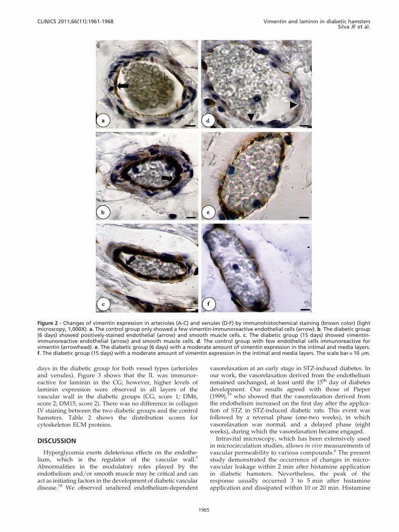
shown). a-SMA expression in venules was increased in theDM6 hamsters compared with the control and the DM15hamsters (CG, score 1; DM6, score 3; DM15, score 1). Vimentin-positive arterioles and venules increased in DM6 and DM15hamsters when compared to control hamsters (CG, score 1;DM6, score 2; DM15, score 2). In the CG, the anti-vimentinantibody only reacted with some endothelial cells, but
immunoreactivity was also apparent in smooth muscle cellsin both of the diabetic groups. Furthermore, a greater numberof cells stained positively for vimentin in the DM6 and DM15groups compared with the CG (Figure 2).
The expression of laminin was more apparent in hamstersin the first week after STZ induction of diabetes than in thecontrol hamsters. Laminin labeling was maintained for 15
Figure 1 - The mean ¡ the standard error of the mean (SEM) for glycemia, body weight, and microvascular responses to histamine andacetylcholine in age-matched controls and hamsters at 6 (DM6) and 15 days (DM15) after the induction of diabetes with streptozotocin(STZ, 50 mg/kg). Statistical significance was determined using one-way ANOVA followed by Tukey’s post-hoc analysis. A. Glycemia (mg/dl): p,0.001 for the control group vs. both DM6 and DM15, and p,0.05 for the DM6 group compared with the DM15 group. B. Bodyweight (g): p,0.05 for the control group vs. the DM6 group, p,0.001 for the control group vs. the DM15 group, and p,0.05 for theDM6 group vs. the DM15 group. C. Microvascular responses (number of dextran 150kDa leakage sites per cm2) to histamine (10-6 M).There were no significant differences between the groups (p.0.05). D. Responses (as percent increase in lumen diameter) of arteriolesand venules to topical application of acetylcholine. There were no significant differences between the groups (p.0.05).
Table 1 - Morphological and stereological measurements of microcirculatory vessels of age-matched control anddiabetic hamsters.
Measurements/Groups/Vessels Control 6 days of diabetes 15 days of diabetes
Arterioles Venules Arterioles Venules Arterioles Venules
Thickness of the intimal and media layers (mm)
Lumen diameter 31¡2.7 40¡3 33¡1.2 42¡2.4 25¡2.5 35¡3.1
Wall thickness 5.7¡0.7 3.6¡0.6 5.4¡0.9 2.7¡0.4 3.8¡0.4 2.4¡0.2
Wall/lumen ratio 0.2 0 0.1 0 0.2 0 0.1 0 0.2 0 0.1 0
Thickness of the media layer (mm)
Lumen diameter 38¡3.7 41¡2.4 30¡3.8 32¡2.1 28¡7.4 39¡5.4
Wall thickness 5.3¡0.3 2.6¡0.2 5.4¡0.2 2.5¡0.1 5¡0.8 2.4¡0.1
Wall/lumen ratio 0.1 0 0.1 0 0.2 0 0.1 0 0.2 0 0.1 0
Density per cheek pouch area (QA) (mm2.102)
QA 3.2¡0.7 8.8¡1.3 3.2¡0.5 7.2¡1 3.5¡0.7 9.7¡1
Volume density (Vv) (%)
Vv 5.5¡1.3 11¡1.5 5.3¡1.1 11¡1.6 5.2¡1.4 10¡1.9
Values are the mean¡the standard error of the mean. p-values are not significant for any data measured.
Vimentin and laminin in diabetic hamstersSilva JF et al.
CLINICS 2011;66(11):1961-1968
1964
days in the diabetic group for both vessel types (arteriolesand venules). Figure 3 shows that the IL was immunor-eactive for laminin in the CG; however, higher levels oflaminin expression were observed in all layers of thevascular wall in the diabetic groups (CG, score 1; DM6,score 2; DM15, score 2). There was no difference in collagenIV staining between the two diabetic groups and the controlhamsters. Table 2 shows the distribution scores forcytoskeleton ECM proteins.
DISCUSSION
Hyperglycemia exerts deleterious effects on the endothe-lium, which is the regulator of the vascular wall.3
Abnormalities in the modulatory roles played by theendothelium and/or smooth muscle may be critical and canact as initiating factors in the development of diabetic vasculardisease.18 We observed unaltered endothelium-dependent
vasorelaxation at an early stage in STZ-induced diabetes. Inour work, the vasorelaxation derived from the endotheliumremained unchanged, at least until the 15th day of diabetesdevelopment. Our results agreed with those of Pieper(1999),19 who showed that the vasorelaxation derived fromthe endothelium increased on the first day after the applica-tion of STZ in STZ-induced diabetic rats. This event wasfollowed by a reversal phase (one-two weeks), in whichvasorelaxation was normal, and a delayed phase (eightweeks), during which the vasorelaxation became engaged.
Intravital microscopy, which has been extensively usedin microcirculation studies, allows in vivo measurements ofvascular permeability to various compounds.6 The presentstudy demonstrated the occurrence of changes in micro-vascular leakage within 2 min after histamine applicationin diabetic hamsters. Nevertheless, the peak of theresponse usually occurred 3 to 5 min after histamineapplication and dissipated within 10 or 20 min. Histamine
Figure 2 - Changes of vimentin expression in arterioles (A-C) and venules (D-F) by immunohistochemical staining (brown color) (lightmicroscopy, 1,000X). a. The control group only showed a few vimentin-immunoreactive endothelial cells (arrow). b. The diabetic group(6 days) showed positively-stained endothelial (arrow) and smooth muscle cells. c. The diabetic group (15 days) showed vimentin-immunoreactive endothelial (arrow) and smooth muscle cells. d. The control group with few endothelial cells immunoreactive forvimentin (arrowhead). e. The diabetic group (6 days) with a moderate amount of vimentin expression in the intimal and media layers.f. The diabetic group (15 days) with a moderate amount of vimentin expression in the intimal and media layers. The scale bar = 10 mm.
CLINICS 2011;66(11):1961-1968 Vimentin and laminin in diabetic hamstersSilva JF et al.
1965

binding to cell-surface receptors triggers subtle or rapidchanges in the contractility of endothelial cells thatdepends on the cytoskeletal organization and cellularadhesion. Therefore, we can suggest that hyperglycemiainfluences vascular function at 6 and 15 days of diabetesdevelopment and that this change may be associated withsubtle changes in the expression and distribution ofendothelial cell cytoskeleton components. No significant
changes were observed after 5 min of histamine applica-tion between the two diabetes groups and the controls.These data agreed with the results of Llorach and co-workers (1976),20 which did not show alterations in one-two-week diabetic animal models.
Many reports have been published about functional andmorphological alterations that occur during the early stage ofdiabetic disease in multiple animal models. In the present
Figure 3 - Changes of laminin expression in arterioles (A-C) and venules (D-F) by immunohistochemical staining (light microscopy,1000X). a. The intimal layers of the hamsters in the control group were immunoreactive for laminin. b and c. All layers showed positivelaminin staining for both diabetic groups. d. The control group showed a poorly labeled intimal layer. e. The diabetic group (6 days)with all layers staining positive. f. The diabetic group (15 days) with all layers staining positive. The scale bar = 10 mm.
Table 2 - Distribution of actin, talin, a-SMA, vimentin, laminin, and type-IV collagen immunohistochemistry scores inage-matched control and 6- and 15-day diabetic hamsters.
Antibodies Control 6 days of diabetes 15 days of diabetes
Arteriole Venule Arteriole Venule Arteriole Venule
Actin + + ++ + + +Talin + + +++ + + +a-SMA ++ + ++ +++ ++ +Vimentin + + ++ ++ ++ ++Laminin ++ + +++ ++ +++ ++Type-IV collagen ++ ++ ++ ++ ++ ++
Vimentin and laminin in diabetic hamstersSilva JF et al.
CLINICS 2011;66(11):1961-1968
1966

study, no significant changes were observed in morpho-metric or stereological measurements, in wall thickening ormicrovessel lumen diameters, in the density of the vessels percheek pouch area (QA) or in volume density (Vv) during theearly stage of disease development. Morphological changesrequire the accumulation of ECM proteins glycated by AGEs9
and are associated with basement membrane thickening.14 Inpart, this thickening may occur when cells synthesizeincreased amounts of collagen IV.21 The expression of type-IV collagen, however, did not change in cheek pouchmicrovessels, which may have resulted from a compensatorymechanism to hyperglycemia in vascular cells that promoteda change in the metabolism of type-IV collagen and increasedits turnover. An increase in the turnover of type-IV collagenwould avoid accumulation of type-IV collagen and result in athickening of the basement membrane.
There is some clinical and experimental evidence ofaugmented blood flow at the early stage of diabetes.22 Smallarteries have the most important influence on local bloodflow and are subjected to chemical and neurohormonalinfluences as well as the continuous effect of mechanicalfactors generated by the blood stream.8 The endotheliumresponds to mechanical stresses through mechanotransduc-tion to regulate the vasomotion and function of the vascularwall. Focal adhesions, which significantly contribute toendothelial cell tethering, mediate the attachment of theactin cytoskeleton to the ECM through a cytoplasmicprotein complex (i.e., the focal adhesion plaque).23 Bloodvessel endothelial cells produce focal adhesion molecules,including focal adhesion kinase, vinculin, talin, and cytos-keletal b-actin.
In arterioles, we found that several proteins involved infocal adhesion, such as actin, talin, vimentin, and laminin,were increased in the DM6 group; however, the immuno-labels for actin and talin in arterioles were decreased in theDM15 hamsters.
Talin is part of the cytoskeletal protein complex that bindsthe cytoplasmic tail of integrins to the b-actin cytoskeletonin focal adhesions and is essential for focal adhesionfunction.24 Talin is essential for integrin activation, and itis the initial weak link between integrins and the actincytoskeleton.25
Talin,26 actin and tubulin readily undergo nonenzymaticglycosylation.27 Loufrani and Henrion (2008)28 reported thatAGEs promote an initial depolymerization of actin filaments(actin F) and a subsequent decrease in actin G expression,which corresponds with a reduction in actin microfilamentsand the disruption of cell-cell and cell-matrix junctions. Inour study, we used a single antibody to immunolabel bothforms of actin; this method could explain the increasedexpression of actin at 6 days after the onset of diabetes. Talinand actin are key proteins in focal adhesion and represent amechanical interaction between the cell and the ECM. Theforce of contraction of the cell is related to focal adhesion.29
Chronic hyperglycemia produces a nonenzymatic glycationof talin and actin, which may contribute to reduced steady-state adhesion strength and mechanosensing responses. Inaddition, hyperglycemia can also promote further changesin vascular permeability.
Diabetic hamsters presented increased labeling for lami-nin in both vessel types. Because laminin is the primarydeterminant of basement membrane assembly,30 increasedlaminin expression may suggest a structural remodeling ofvessel basement membranes.
We also found that vimentin was concomitantlyincreased with laminin expression in blood vessels. Allthree major cytoskeletal networks have been shown toadapt their structures by elongating their cell shapes andreorienting parallel to the flow direction.31 Intermediatefilament cables, which are initially not under tension,contribute to cell stiffness during large shear deformations.This strain-hardening behavior in the intermediate cytos-keleton filament likely prevents the excessive deformationof the cell at finite strains that would otherwise rupture theactin cytoskeleton.32 Intermediate filaments may alsoindirectly interact with focal adhesion sites. Indeed,vimentin intermediate filaments are anchored to a6b4-integrins in microvascular endothelial cells by plectin.Because a6b4 binds to laminin, these interactions areconsistent with the hypothesis that intermediate filamentsare recruited to provide mechanical stability duringphysiological adaptation of the microfilament network tohemodynamic shear stress.31 Thus, the increased expres-sion of vimentin is associated with a proliferative pheno-type, possibly due to hyperglycemia.33 Antibodies againstlaminin and vimentin inhibit branching morphogenesis invitro. Therefore, this adhesion structure may play a role inangiogenesis.34 Like the endothelial cell cytoskeleton,smooth muscle cells are important in maintaining endothe-lial structural integrity and in regulating endothelialrepair.33
The expression of a-SMA in venules was enhanced in theDM6 hamsters, and increased expression of a-SMA hasbeen shown to result in increased contractile force genera-tion.35 In addition, increased expression of a-SMA has beenshown to be associated with vessel stability.32 The primaryfunction of smooth muscle cells (SMC) is the maintenanceof blood flow and pressure through their contractility, anda-SMA is necessary for SMC and pericytes to interact withendothelial cells to form a fully functional blood–tissuebarrier.35
Our results suggest that changes related to cell-matrixinteractions occur in response to a new metabolic condition(6 days of a diabetic state). These changes may contribute topathological remodeling of the microvessels and persist forat least 15 days after the induction of diabetes.
AUTHOR CONTRIBUTIONS
Silva JF was responsible for the morphometric and stereologic analysis.
Carvalho JJ was responsible for the immunohistochemistry assay.
Breitenbach MMD was responsible for the induction of diabetes mellitus
and data analysis. Cyrino FZGA was responsible for the intravital
microscopy of the hamster cheek pouch. Bouskela E participated in the
intravital microscopy technique, interpretation of intravital microscopy,
immunohistochemistry, analysis of morphometric and stereological
techniques, and was responsible for the draft of the introduction,
discussion, and conclusions sections.
REFERENCES
1. Aguiar LG, Villela NR, Bouskela E. [Microcirculation in diabetes:implications for chronic complications and treatment of the disease].Arq Bras Endocrinol Metabol. 2007;51:204-11.
2. Duling BR, Damon DH. An examination of the measurement of flowheterogeneity in striated muscle. Circ Res. 1987;60:1-13.
3. Schalkwijk CG, Stehouwer CD. Vascular complications in diabetesmellitus: the role of endothelial dysfunction. Clin Sci (Lond).2005;109:143-59.
4. Meerwaldt R, Links T, Zeebregts C, Tio R, Hillebrands JL, Smit A. Theclinical relevance of assessing advanced glycation endproducts accumu-lation in diabetes. Cardiovasc Diabetol. 2008;7:29.
CLINICS 2011;66(11):1961-1968 Vimentin and laminin in diabetic hamstersSilva JF et al.
1967

5. Joyner WL, Mayhan WG, Johnson RL, Phares CK. Microvascularalterations develop in Syrian hamsters after the induction of diabetesmellitus by streptozotocin. Diabetes. 1981;30:93-100.
6. Svensjo E, Cyrino F, Michoud E, Ruggiero D, Bouskela E, Wiernsperger N.Vascular permeability increase as induced by histamine or bradykinin isenhanced by advanced glycation endproducts (AGEs). J DiabetesComplications. 1999;13:187-90.
7. Sotnikova R, Skalska S, Okruhlicova L, Navarova J, Kyselova Z, Zurova J,et al. Changes in the function and ultrastructure of vessels in the ratmodel of multiple low dose streptozotocin-induced diabetes. GenPhysiol Biophys. 2006;25:289-302.
8. Davies PF. Hemodynamic shear stress and the endothelium incardiovascular pathophysiology. Nat Clin Pract Cardiovasc Med.2009;6:16-26.
9. Mott RE, Helmke BP. Mapping the dynamics of shear stress-inducedstructural changes in endothelial cells. Am J Physiol Cell Physiol.2007;293:C1616-26.
10. Moncada S. Adventures in vascular biology: a tale of two mediators.Philos Trans R Soc Lond B Biol Sci. 2006;361:735-59.
11. Zhang H, Sunnarborg SW, McNaughton KK, Johns TG, Lee DC, Faber JE.Heparin-binding epidermal growth factor-like growth factor signaling inflow-induced arterial remodeling. Circ Res. 2008;102:1275-85.
12. Jarvisalo MJ, Raitakari M, Toikka JO, Putto-Laurila A, Rontu R, Laine S,et al. Endothelial dysfunction and increased arterial intima-mediathickness in children with type 1 diabetes. Circulation. 2004;109:1750-5.
13. Singh R, Barden A, Mori T, Beilin L. Advanced glycation end-products: areview. Diabetologia. 2001;44:129-46.
14. Liu Y, Li H, Bubolz AH, Zhang DX, Gutterman DD. Endothelialcytoskeletal elements are critical for flow-mediated dilation in humancoronary arterioles. Med Biol Eng Comput. 2008;46:469-78.
15. Mandarim-de-Lacerda CA. Stereological tools in biomedical research.An Acad Bras Cienc. 2003;75:469-86.
16. Mendonca Lde S, Fernandes-Santos C, Mandarim-de-Lacerda CA.Cardiac and aortic structural alterations due to surgically-inducedmenopause associated with renovascular hypertension in rats. Int J ExpPathol. 2007;88:301-9.
17. Bonnet F, Cao Z, Cooper ME, Cox AJ, Kelly DJ, Gilbert RE. Tranilastattenuates vascular hypertrophy, matrix accumulation and growth factoroverexpression in experimental diabetes. Diabetes Metab. 2003;29:386-92.
18. Matsumoto T, Oda SI, Kobayashi T, Kamata K. Flow-induced endothe-lium-dependent vasoreactivity in rat mesenteric arterial bed. J SmoothMuscle Res. 2004;40:1-14.
19. Pieper GM. Enhanced, unaltered and impaired nitric oxide-mediatedendothelium-dependent relaxation in experimental diabetes mellitus:importance of disease duration. Diabetologia. 1999;42:204-13.
20. Llorach MA, Bohm GM, Leme JG. Decreased vascular reactions topermeability factors in experimental diabetes. Br J Exp Pathol.1976;57:747-54.
21. Iglesias-de la Cruz MC, Ziyadeh FN, Isono M, Kouahou M, Han DC,Kalluri R, et al. Effects of high glucose and TGF-beta1 on the expression
of collagen IV and vascular endothelial growth factor in mousepodocytes. Kidney Int. 2002;62:901-13.
22. Kobayashi T, Kaneda A, Kamata K. Possible involvement of IGF-1receptor and IGF-binding protein in insulin-induced enhancement ofnoradrenaline response in diabetic rat aorta. Br J Pharmacol.2003;140:285-94.
23. Harrington MA, Daub R, Song A, Stasek J, Garcia JG. Interleukin 1 alphamediated inhibition of myogenic terminal differentiation: increasedsensitivity of Ha-ras transformed cultures. Cell Growth Differ.1992;3:241-8.
24. Weber E, Rossi A, Solito R, Sacchi G, Agliano M, Gerli R. Focal adhesionmolecules expression and fibrillin deposition by lymphatic and bloodvessel endothelial cells in culture. Microvasc Res. 2002;64:47-55.
25. Wegener KL, Partridge AW, Han J, Pickford AR, Liddington RC,Ginsberg MH, et al. Structural basis of integrin activation by talin. Cell.2007;128:171-82.
26. Lee TY, Gotlieb AI. Early stages of endothelial wound repair: conversionof quiescent to migrating endothelial cells involves tyrosine phosphor-ylation and actin microfilament reorganization. Cell Tissue Res.1999;297:435-50.
27. Pekiner C, Cullum NA, Hughes JN, Hargreaves AJ, Mahon J, Casson IF,et al. Glycation of brain actin in experimental diabetes. J Neurochem.1993;61:436-42.
28. Loufrani L, Henrion D. Role of the cytoskeleton in flow (shear stress)-induced dilation and remodeling in resistance arteries. Med Biol EngComput. 2008;46:451-60.
29. Lee SE, Kamm RD, Mofrad MR. Force-induced activation of talin and itspossible role in focal adhesion mechanotransduction. J Biomech.2007;40:2096-106.
30. Sasaki T, Fassler R, Hohenester E. Laminin: the crux of basementmembrane assembly. J Cell Biol. 2004;164:959-63.
31. Helmke BP. Molecular control of cytoskeletal mechanics by hemody-namic forces. Physiology (Bethesda). 2005;20:43-53.
32. Schopferer M, Bar H, Hochstein B, Sharma S, Mucke N, Herrmann H,et al. Desmin and vimentin intermediate filament networks: theirviscoelastic properties investigated by mechanical rheometry. J MolBiol. 2009;388:133-43.
33. Skalli O, Bloom WS, Ropraz P, Azzarone B, Gabbiani G. Cytoskeletalremodeling of rat aortic smooth muscle cells in vitro: relationships toculture conditions and analogies to in vivo situations. J SubmicroscCytol. 1986;18:481-93.
34. Gonzales M, Weksler B, Tsuruta D, Goldman RD, Yoon KJ, HopkinsonSB, et al. Structure and function of a vimentin-associated matrix adhesionin endothelial cells. Mol Biol Cell. 2001;12:85-100.
35. Tomasek JJ, Haaksma CJ, Schwartz RJ, Vuong DT, Zhang SX, Ash JD,et al. Deletion of smooth muscle alpha-actin alters blood-retina barrierpermeability and retinal function. Invest Ophthalmol Vis Sci.2006;47:2693-700.
Vimentin and laminin in diabetic hamstersSilva JF et al.
CLINICS 2011;66(11):1961-1968
1968
Copyright © 2022 FDOKUMEN