Cytokine expression during the outcome of canine experimental infection by Leishmania infantum

RESEARCH ARTICLE
Vaccination with Leishmania infantum AcidicRibosomal P0 but Not with NucleosomalHistones Proteins Controls Leishmaniainfantum Infection in HamstersLais Pereira1, Melissa Abbehusen1, Clarissa Teixeira1, Jurema Cunha1, IvanP. Nascimento2, Kyioshi Fukutani1, Washington dos-Santos1, Aldina Barral1,3,4, CamilaIndiani de Oliveira1,4, Manoel Barral-Netto1,3,4, Manoel Soto5, Cláudia Ida Brodskyn1,4,6*
1 Centro de Pesquisa Gonçalo Moniz, FIOCRUZ-BA, Bahia, Brazil, 2 Centro de Biotecnologia, InstitutoButantan, São Paulo, Brazil, 3 Faculdade de Medicina, Universidade Federal da Bahia, Bahia, Brazil, 4Instituto de Investigação em Imunologia, São Paulo, Brazil, 5 Centro de Biología Molecular "Severo Ochoa,"Universidad Autónoma de Madrid, Madrid, Spain, 6 Instituto de Ciências da Saúde, Universidade Federal daBahia, Bahia, Brazil
Abstract
Background
Several intracellular Leishmania antigens have been identified in order to find a potential
vaccine capable of conferring long lasting protection against Leishmania infection. Histonesand Acid Ribosomal proteins are already known to induce an effective immune response
and have successfully been tested in the cutaneous leishmaniasis mouse model. Here, we
investigate the protective ability of L. infantum nucleosomal histones (HIS) and ribosomal
acidic protein P0 (LiP0) against L. infantum infection in the hamster model of visceral leish-
maniasis using two different strategies: homologous (plasmid DNA only) or heterologous
immunization (plasmid DNA plus recombinant protein and adjuvant).
Methodology/Principal Findings
Immunization with both antigens using the heterologous strategy presented a high antibody
production level while the homologous strategy immunized group showed predominantly a
cellular immune response with parasite load reduction. The pcDNA-LiP0 immunized group
showed increased expression ratio of IFN-γ/IL-10 and IFN-γ/TGF-β in the lymph nodes be-
fore challenge. Two months after infection hamsters immunized with the empty plasmid pre-
sented a pro-inflammatory immune response in the early stages of infection with increased
expression ratio of IFN-γ/IL-10 and IFN-γ/TGF-β, whereas hamsters immunized with
pcDNA-HIS presented an increase only in the ratio IFN-γ/ TGF-β. On the other hand, ham-
sters immunized with LiP0 did not present any increase in the IFN-γ/TGF-β and IFN-γ/IL-10
ratio independently of the immunization strategy used. Conversely, five months after in-
fection, hamsters immunized with HIS maintained a pro-inflammatory immune response
PLOS Neglected Tropical Diseases | DOI:10.1371/journal.pntd.0003490 February 2, 2015 1 / 16
OPEN ACCESS
Citation: Pereira L, Abbehusen M, Teixeira C, CunhaJ, Nascimento IP, Fukutani K, et al. (2015)Vaccination with Leishmania infantum AcidicRibosomal P0 but Not with Nucleosomal HistonesProteins Controls Leishmania infantum Infection inHamsters. PLoS Negl Trop Dis 10(2): e0003490.doi:10.1371/journal.pntd.0003490
Editor: Hechmi Louzir, Institut Pasteur de Tunis,TUNISIA
Received: July 4, 2014
Accepted: December 18, 2014
Published: February 2, 2015
Copyright: © 2015 Pereira et al. This is an openaccess article distributed under the terms of theCreative Commons Attribution License, which permitsunrestricted use, distribution, and reproduction in anymedium, provided the original author and source arecredited.
Data Availability Statement: All relevant data arewithin the paper and its Supporting Information files.
Funding: This study was funded by CNPq andCYTED. The funders had no role in study design,data collection and analysis, decision to publish, orpreparation of the manuscript.
Competing Interests: The authors have declaredthat no competing interests exist.

(ratio IFN-γ/ IL-10) while pcDNA-LiP0 immunized hamsters continued showing a balanced
cytokine profile of pro and anti-inflammatory cytokines. Moreover we observed a significant
reduction in parasite load in the spleen, liver and lymph node in this group compared
with controls.
Conclusions/Significance
Our results suggest that vaccination with L. infantum LiP0 antigen administered in a DNA
formulation could be considered a potential component in a vaccine formulation against
visceral leishmaniasis.
Author Summary
Visceral leishmaniasis caused by Leishmania infantum is the most severe form of leish-maniasis. The disease is fatal if not treated and there is no vaccine available for human use.In the search for potential antigens, the protective ability of conserved parasite proteinfamilies such as L. infantum histones (HIS) and acidic ribosomal (LiP0) antigens were suc-cessfully tested in the mouse model of cutaneous leishmaniasis. Here, we evaluate HIS andLiP0 antigens using two different immunization strategies in the hamster model of visceralleishmaniasis. Hamsters are highly susceptible to L. infantum infection and we demon-strate that immunization with LiP0, but not HIS, protects against the fatal outcome of vis-ceral leishmaniasis. Immunization with LiP0 was able to induce an increased expressionof IFN-γ in detriment of IL-10 and TGF-β in the draining lymph node before infectioncreating an inhospitable environment for parasite growth. Following challenge, a reducedparasite load in the lymph node, spleen and liver of LiP0 immunized hamsters wasdetected five months after challenge. These findings suggest that LiP0 used in a DNA for-mulation could be considered a potential component in a vaccine formulation againstvisceral leishmaniasis.
IntroductionLeishmaniasis is a parasitic disease caused by protozoan from the genus Leishmania transmit-ted by the bite of infected sand flies. Leishmaniasis is one of the six major tropical diseases tar-geted by the World Health Organization [1]. The disease has a broad spectrum of clinicalmanifestations, from cutaneous, self-limited skin lesions to a visceral form of the disease. Vis-ceral leishmaniasis (VL) caused by Leishmania infantum in the NewWorld is the most severeform of disease characterized by hepatosplenomegaly, fever with a high mortality rate if nottreated [2].
Although extensive research has been performed to identify an antigen able to elicit a longlasting protection against infection, there is still no successful vaccine available for humanleishmaniasis. The majority of Leishmania vaccine candidates tested are able to induce humor-al and/or cellular immune responses. However, the immune response derived is not able to in-duce protection and may contribute to pathology exacerbation [1].
Several secreted and surface Leishmania antigens have been tested, targeting virulence fac-tors or molecules important for parasite invasion but most of these candidates resulted in ashort-lived or partial protection [3]. On the other hand, intracellular house-keeping proteinsare able to modulate the host immune response because they do not undergo selective pressure
LiP0 Vaccine Against Visceral Leishmaniasis
PLOS Neglected Tropical Diseases | DOI:10.1371/journal.pntd.0003490 February 2, 2015 2 / 16

by the immune response [4]. During infection, these molecules are released after the destruc-tion of intracellular amastigotes by activated macrophages but they can be also excreted bynon-classical secretion pathways [5,6]. Several intracellular antigens such as heat shock pro-teins, ribosomal proteins and histones have been investigated as potential vaccine candidatesagainst different species of Leishmania [3]. Histones (HIS) are important structural proteins inthe organization and regulation of genes. There are four main classes of histones (H2A, H2B,H3, and H4) that are responsible for the composition of the Leishmania nucleosome [7]. Iborraet al demonstrated that immunization with DNA plasmid coding for nucleosomal histonesplus CpG adjuvant was able to significantly reduce lesion size after challenge with L.major inBALB/c mice [8]. More recently, Carneiro et al. observed that mice immunized with either aplasmid DNA cocktail composed of four different Leishmania histones or with a combinationof the DNA cocktail followed by the corresponding recombinant proteins, resulted in the ab-sence of infected macrophages at the site of challenge with L. braziliensis in the presence ofsand fly saliva. The protective response was associated with increased expression of IFN-γ anddown regulation of IL-4 at the infection site [9].
Another immunodominant antigen is the Leishmania infantum acidic ribosomal protein(LiP0), a structural component of the large ribosome subunit that has been recognized bysera from both patients and dogs infected by L. infantum [10,11]. Also, it has been demon-strated that ribosomal protein (P0) was able to stimulate proliferation and IFN-γ secretion ofa T-cell clone established from a human donor by stimulation with paraformaldehyde fixedpromastigotes [12]. Using an immunization strategy that included LiP0 plasmid DNA vacci-nation and/or with LiP0 recombinant protein plus CpG, Iborra et al. showed the induction ofpartial protection against L.major infection in BALB/c mice [13]. However, C57BL/6 miceimmunized with the same strategy was able to significantly reduce parasite load controllinglesion development [14]. The induction of IFN-γ was related with protection against Leish-mania infection in these models. Although not demonstrated in Leishmania, it has beenshown that P0 can be located in the surface of some protozoan parasites from the genusPlasmodium and Toxoplasma [15]. Its location on the cell surface of these organismsunderlies the protective responses elicited by different vaccines based on P0 antigen. Thus,the generation of humoral responses to Neospora caninum P0 induces protection againstneosporosis and toxoplasmosis [16]. Similarly, an experimental vaccine based on the car-boxy-terminal domain of Plasmodium falciparum P0 was able to induce humoral responsesthat protects mice against malaria [17].
However, the immunization strategy used can be imperative to improve immunogenicity.The heterologous prime-boost strategy is used combining different formulations of the sameantigen and has been effective in immunizations against cutaneous and visceral leishmania-sis. Dogs immunized with LACK using the heterologous strategy (DNA/protein) presentedprotection against VL caused by L. infantum [18]. Similarly, Iborra et al. (2005) used thesame strategy with pcDNA3-LiP0 followed by recombinant protein (rLiP0) that protectedC5BL/6 mice against challenge with L.major [14]. Moreover, administration of CpG ODNwith antigens from different pathogens has been shown to induce a strong Th1 immune re-sponse [19].
Therefore, based on the protective potential of antigens belonging to conserved proteinfamilies used in the cutaneous leishmaniasis mouse model, we hypothesized that immunizationwith HIS and LiP0 antigens could also protect hamsters against the fatal outcome of L. infan-tum infection. In addition, we also compared HIS and LiP0 antigens using two differentimmunization strategies: plasmid DNA only (homologous) or plasmid DNA and recombinantprotein plus CpG ODN (heterologous).
LiP0 Vaccine Against Visceral Leishmaniasis
PLOS Neglected Tropical Diseases | DOI:10.1371/journal.pntd.0003490 February 2, 2015 3 / 16

Methods
AnimalsMale Syrian golden hamsters (Mesocricetus auratus) six to eight weeks old were obtained fromCentro de Pesquisa Gonçalo Moniz/ Fundação Oswaldo Cruz (FIOCRUZ) animal facility. Allanimal work was conducted according to the Guidelines for Animal Experimentation of theColégio Brasileiro de Experimentação Animal and of the Conselho Nacional de Controle deExperimentação Animal. The local Ethics Committee on Animal Care and Utilization (CEUA)approved all procedures involving animals (CEUA—Centro de Pesquisas Gonçalo Muniz—CPqGM/FIOCRUZ—L-IGM-011/09 and 005/2011.
DNA plasmids coding for LiP0 and nucleossomal histonesDNA plasmid coding for Leishmania infantum acid ribosomal protein (pcDNALiP0) and thehistones pcDNAHIS (pcDNA3LiH2-H3 and pcDNA3LiH2B-H4) were cloned and purified aspreviously described [13,14].
Immunization strategiesImmunization experiments were carried out in groups of 15 hamsters. In the homologous im-munization experiments, hamsters were inoculated three times intramuscularly (i.m.) with100 μg of DNA of pcDNA3-LiP0, pcDNA3 HIS (50 μg of each plasmid) or pcDNA3 plus 1nMof CpG ODN 1826 (18–24 pb—5´TCC ATGACG TTC CTG ACG TT-3´ mol wt 6364,1g/mol)(empty plasmid) in a total volume of 50 μL. In the heterologous strategy, hamsters receivedtwo inoculations of DNA followed by one intradermal (i.d.) inoculation of recombinant pro-tein (5 μg of each rHIS or 10 μg rLiP0 protein) plus 1 nM of CpG ODN 1826 in the right ear[20]. In all groups, hamsters were inoculated at 2-week intervals. Two weeks after the last im-munization 15 hamsters per group were euthanized to collect draining lymph nodes and serato evaluate the cellular and humoral response induced by the immunization.
Determination of antibody titersFifteen days after last immunization and two and 5 months after infection a sample of bloodwas collected from 6–8 hamsters per group by retrorbital plexus and sera was obtained aftercentrifugation for 1500 rpm for 5 minutes and stored at -4°C until use. Briefly, to measure spe-cific antibody responses by ELISA, standard ELISA plates were coated overnight at room tem-perature with 100μL of SLA (10 μg/mL), rLiP0 (2μg/mL) or rHIS (1 μg/mL) in PBS. Serumsamples were added at dilutions of 1:100. Following a washing step, a goat anti-hamster IgG al-kaline phosphatase conjugate (1:1,000 and 1:2,000, Sigma, Missouri, USA) was added and incu-bated for one hour. The wells were then re-washed, substrate and chromogen (p-nitrophenylphosphate; Sigma, USA) were added, and absorbance was recorded at 405 nm on a SpectraMax190 spectrophotometer (Molecular Devices, USA) automatic micro plate reader.
Intradermal challenge with Leishmania infantumLeishmania infantum (MCAN/BR/00/BA262) promastigotes isolated from a naturally infecteddog (Bahia State, Brazil) were cultured in Schneider’s medium (LGC, Brazil) supplementedwith 10% of inactivated FBS (fetal bovine serum) (Gibco, USA), 2 mM L-glutamine, 100 IU/mlpenicillin, 1% streptomycin (Gibco, USA).
Fifteen days after the last immunization, hamsters were inoculated by intradermal route, inthe left ear with 105 stationary phase promastigotes plus 0.5 pair of sonicated salivary glands(SGH) from female Lutzomyia longipalpis, using a 29-gauge needle (BD Ultra-Fine) in 20uL of
LiP0 Vaccine Against Visceral Leishmaniasis
PLOS Neglected Tropical Diseases | DOI:10.1371/journal.pntd.0003490 February 2, 2015 4 / 16

saline. Salivary glands were dissected from 5- to 7-day-old females and stored in endotoxin-free PBS at −70°C. Salivary glands were sonicated (Sonifer 450 homogenizer, Branson, Danbury,Connecticut), and afterwards centrifuged at 12,000 g for 5 minutes. Supernatant was collectedand used immediately.
Limiting Dilution Assay (LDA)Parasite load was determined 2 and 5 months post-infection using the quantitative LimitingDilution Assay (LDA) as described by Titus et al [21]. Briefly, infected liver, spleen and retro-maxillar draining lymph nodes were aseptically removed from individual hamsters. Tissueswere homogenized and diluted in Schneider’s Insect Medium (Sigma, St. Louis, MO) supple-mented with 10% heat inactivated fetal bovine serum (Gibco, USA), 100 U of penicillin/ml and100 mg/ml of streptomycin. Homogenate samples were serially diluted into 96-wells platescontaining biphasic blood agar (Novy-Nicolle-McNeal) medium and incubated for one weekat 23°C when the presence of viable parasites was determined. Parasite burden in the tissue wascalculated applying ELIDA software [21].
RNA isolation and quantitative Real-Time PCRUsing Trizol reagent (Invitrogen, USA), total RNA was extracted from the draining retromaxillarlymph nodes two weeks after the last immunization, and from the spleen obtained two and fiveweeks after infection. First-strand cDNA synthesis was performed with 1–2 μg of RNA in a totalvolume of 25 μL using Super Script II (Gibco, Carlsbad, CA, USA). DNA was amplified in thethermocycler (Mastercycler gradient—Eppendorf, USA) with an initial pre- incubation at 72°Cfor 5 minutes, followed by amplification of the target DNA at 42°C for 50 minutes. A standardcurve was generated for each set of primers and efficiency of each reaction was determined. Theexpression levels of genes were normalized to GAPDH levels. Results are expressed in foldchange over control. Oligonucleotide primers used were: GAPDH (reverse 5’- CTGACATGCCGCCCTGGAG-3’ and forward 3’-TCAGTG- TAGCCCAGGATGCC-5’); IFN-γ (reverse 5’-GAAGCTCAC- CAAGATTCCGGTAA-3’ and forward 3’-TTTTCGTGACA- GGTGAGGCAT-5’); IL-10 (reverse 5’-AGACGCCTTTCTC- TTGGAGCTTAT-3’ and forward 39-GGCAACTGCAGCGC- TGTC-5’); and TGF-β (reverse 5’-GCTACCACGCCAACTTC- TGTC-3’ and for-ward 3’-TGTTGGTAGAGGGCAAGG-5’).
HistologyFor histology, five months after infection spleen and liver fragments of four hamsters fromeach group were fixed in 10% phosphate-buffered formalin and embedded in paraffin. Four-micrometer sections were stained with hematoxylin–eosin and studied by optical microscopy.
Statistical analysisExperiments were repeated three times with five hamsters per group per time point. Compari-sons among immunized and non-immunized control groups were done by one-way ANOVA(Kruskal-Wallis) analysis with Dunn’s post-test. Results were considered statistically signifi-cant when p�0.05. All statistical analysis was done using Graph Pad 5.0 software program.
LiP0 Vaccine Against Visceral Leishmaniasis
PLOS Neglected Tropical Diseases | DOI:10.1371/journal.pntd.0003490 February 2, 2015 5 / 16

Results
Heterologous immunization strategy using HIS or LiP0 antigens inducesproduction of specific antibodiesTo evaluate production of anti-HIS or anti-LiP0 IgG antibodies we collected sera from immu-nized hamsters fifteen days after the last immunization. We observed that only hamsters thatwere immunized with heterologous strategy showed significantly higher titers of antibodies.There was a significant increase of IgG in the group that received DNA plasmid coding for HISfollowed by booster of nucleosomal histone recombinant protein plus CpG adjuvant (pcDNAHIS-rHIS+CpG) compared to non-immunized hamsters (Fig. 1A). Similar results were ob-served regarding antibody production in hamsters immunized with LiP0, using the same heter-ologous strategy. There was a significantly higher production of anti-LiP0 IgG in vaccinatedhamsters (pcDNA-LiP0/ rLiP0+CpG) compared to the animals that received empty plasmidand saline, the control groups (Fig. 1B).
Immunization with DNA plasmid coding HIS and LiP0 proteins increaseexpression of IFN-γ in the lymph nodeIn order to verify the cellular immune response induced by the immunization with HIS andLiP0, expression of IFN-γ, IL-10, and TGF-β was evaluated in retromaxillar draining lymphnodes 15 days after immunization. We observe a significant increase in the ratio of IFN-γ/IL-10expression in the lymph nodes of animals immunized with HIS and LiP0 using the homolo-gous immunization strategy with DNA plasmids (Fig. 2A). Conversely, there was no differencein the IFN-γ/TGF-β ratio in the HIS immunized group using both immunization strategies(Fig. 2B). Interestingly, we observed that the IFN-γ/TGF-β ratio was significantly higher inhamsters immunized with DNA plasmid (pcDNA-LiP0) using homologous strategy comparedwith the control group (Fig. 2B).
Fig 1. Humoral immune response after immunization with nucleosomal histones (HIS) or acidic ribosomal protein (LiP0) antigens.Hamsters(n = 6–10) were bled 2 weeks after immunization and sera were tested by ELISA to determine specific total IgG levels. (A) Anti-HIS total IgG in sera fromhamsters immunized with recombinant DNA coding for HIS (pcDNA HIS) or with recombinant DNA followed by recombinant nucleosomal histones+CpG(pcDNA HIS-rHIS+CpG). (B) Anti-LiP0 total IgG in sera from hamsters immunized with recombinant DNA coding for LiP0 (pcDNA LiP0) or with recombinantDNA followed by recombinant protein LiP0+CpG (pcDNA LiP0- rLiP0+CpG). Control groups were immunized with saline or empty plasmid. *p<0.05;**p<0.01; ***p<0.001, compared with controls.
doi:10.1371/journal.pntd.0003490.g001
LiP0 Vaccine Against Visceral Leishmaniasis
PLOS Neglected Tropical Diseases | DOI:10.1371/journal.pntd.0003490 February 2, 2015 6 / 16

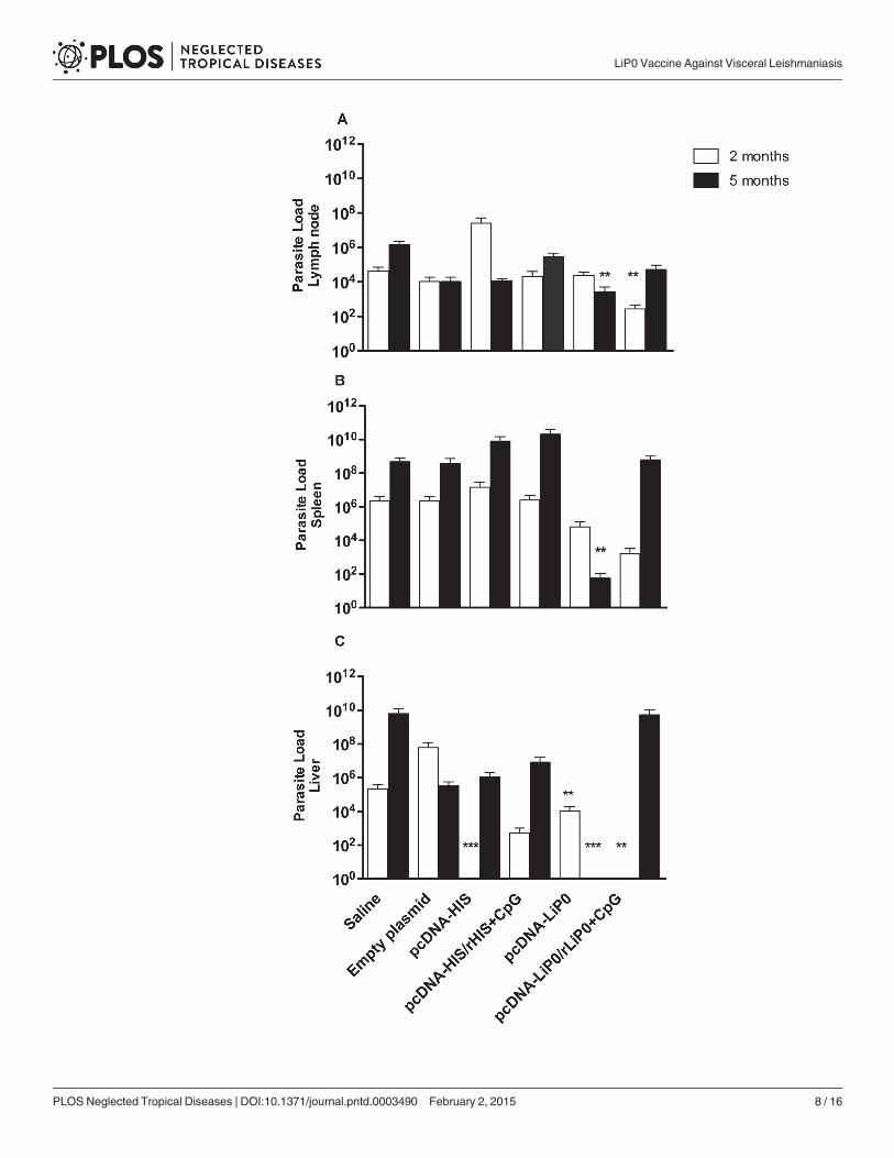
Immunization with LiP0 protects hamsters against the fatal outcome ofvisceral leishmaniasisIn order to verify if the immune responses resulting from immunization with acidic ribosomalprotein and nucleosomal histone antigens using homologous and heterologous immunizationstrategies, are able to protect against L. infantum infection, immunized hamsters were chal-lenged with 105 L. infantum plus SGH, trying to mimic natural transmission [22].
Two months after challenge, hamsters immunized with HIS using the homologous strategyshowed a significant parasite load reduction in the liver (Fig. 3C) but not in the retromaxillarlymph nodes or spleen compared to control groups (Fig. 3A and B).
On the other hand, we observed a significant reduction in the lymph node, spleen and liverparasite loads in LiP0 DNA plasmid (pcDNA3-LiP0) immunized group, compared with con-trols five months after challenge (Fig. 3A, B and C). Interestingly, parasite burden in the spleen(Fig. 3B) (p = 0.0016) and liver (Fig. 3C) (p = 0.0001) of hamsters immunized with the LiP0-based DNA vaccine were lower at 5 months than 2 months after infection, indicating that thevaccine was able to induce a long term leishmanicidal response.
Antibody levels in HIS and LiP0 immunized hamsters after L. infantuminfectionTo access the humoral immune response after infection, we measured production of anti-LiP0,HIS and SLA IgG antibodies. For this purpose, we collected sera from infected hamsters at twoand five months following challenge with L. infantum plus SGS.
We observed an increase in anti-HIS IgG in all groups immunized with HIS especially atfive months after infection (Fig. 4A). Conversely, LiP0 immunized groups did not present anysignificant levels of anti-LiP0 IgG levels two and five months following infection when com-pared with controls (Fig. 4B).
Interestingly, animals immunized with LiP0 homologous strategy showed a significantlylower anti-SLA IgG levels at five months after infection compared to HIS immunized and con-trols groups (Fig. 4C).
Cytokines detection in HIS and LiP0 immunized hamsters after L.infantum infectionTo evaluate the cellular immune response of hamsters challenged with L. infantum, we investi-gated the expression of cytokines in the spleen two and five months following infection.
Fig 2. Cytokine expression following immunization with nucleosomal histones (HIS) or acidic ribosomal protein (LiP0) antigens.Hamsters(n = 6–10) were immunized with HIS or LiP0 antigens using the homologous (DNA only) or heterologous (DNA-recombinant protein plus CpG) andretromaxillar lymph nodes collected for evaluation of cytokines expression by Real-Time PCR, two weeks after the last immunization. (A) IFN-γ/IL-10mRNA expression ratio and (B) IFN-γ/TGF-βmRNA expression ratio. *p<0.05; **p<0.01; ***p<0.001, compared with controls.
doi:10.1371/journal.pntd.0003490.g002
LiP0 Vaccine Against Visceral Leishmaniasis
PLOS Neglected Tropical Diseases | DOI:10.1371/journal.pntd.0003490 February 2, 2015 7 / 16
LiP0 Vaccine Against Visceral Leishmaniasis
PLOS Neglected Tropical Diseases | DOI:10.1371/journal.pntd.0003490 February 2, 2015 8 / 16

Two months after challenge hamsters immunized with DNA-HIS (homologous strategy)presented an increased expression ratio of IFN-γ/IL-10 when compared with the non-immu-nized control group (p = 0.0253) (Fig. 5A). Expression ratio of IFN-γ/TGF-β was increased inthe pcDNA-HIS (p = 0.0003), and pcDNA-HIS/rHIS+CpG (p = 0.018) immunized groupscompared with saline control suggesting that DNA-HIS immunized hamsters presented a pro-inflammatory immune response in the early stages of infection (Fig. 5B). On the other hand,hamsters immunized with LiP0 using independent of strategy employed did not present anyincrease in the expression ratio of IFN-γ/IL-10 or IFN-γ/TGF-β (Fig. 5A and B).
Fig 3. Parasite load in hamsters immunized with HIS or LiP0 antigens following L. infantum infection.Hamsters (n = 15) were immunized with HIS or LiP0 antigens using the homologous (DNA only) orheterologous (DNA-recombinant protein plus CpG). Two weeks after the last immunization, hamsters werechallenged in the left ear with 105 L. infantum in the presence of Lu. longipalpis SGH. Draining retromaxillarlymph node (A), Spleen (B) and Liver (C) parasite loads per total organ were determined two and five monthsafter challenge by limiting dilution assay (LDA). *p<0.05; **p<0.01; ***p<0.001, compared with controls.
doi:10.1371/journal.pntd.0003490.g003
Fig 4. Humoral immune response in HIS or LiP0 immunized hamsters following L. infantum infection. Hamsters (n = 15) were immunized with HIS orLiP0 antigens using the homologous (DNA only) or heterologous (DNA-recombinant protein plus CpG). Two weeks after the last immunization, hamsterswere challenged in the left ear with 105 L. infantum in the presence of Lu. longipalpis SGH. Two and five months after challenge sera were tested by ELISA todetermine specific total IgG levels. (A) Anti-HIS total IgG in sera from hamsters immunized with pcDNA HIS or with pcDNA HIS-rHIS+CpG. (B) Anti-LiP0 totalIgG in sera from hamsters immunized with DNA LiP0 or DNA LiP0-rLiP0+CpG. (C) Anti-SLA total IgG in sera from hamsters immunized with HIS or LiP0.*p<0.05; **p<0.01; ***p<0.001, compared with controls.
doi:10.1371/journal.pntd.0003490.g004
LiP0 Vaccine Against Visceral Leishmaniasis
PLOS Neglected Tropical Diseases | DOI:10.1371/journal.pntd.0003490 February 2, 2015 9 / 16

Five months after infection, hamsters immunized with HIS using both immunization strate-gies showed an increased expression ratio of IFN-γ/IL-10 (p<0.05) suggesting a pro-inflamma-tory immune response (Fig. 5C). Concerning LiP0 immunization, there was an increase in theexpression ratio of IFN-γ/TGF-β in the spleen of pcDNA-LiP0/rLiP0+CpG immunized ham-sters (heterologous strategy) compared with the control group (p<0.0101) (Fig. 5D) suggestingthat this strategy induce a delayed cellular immune response.
The spleen and liver of LiP0 immunized hamsters present fewergranulomas and macrophage aggregatesFive months after infection spleen and liver samples were collected from four hamsters pergroup for histopathological analysis. In the spleen, we observed disorganized follicles with orwithout germinal center, macrophage aggregates with L. infantum amastigotes inclusions, epi-thelioid cells present in the red and white pulp, as well as granulomas in the groups immunizedwith HIS or LiP0. However, hamsters immunized with HIS displayed a higher loss of splenicstructure. . . Animals immunized with LiP0 presented smaller macrophage aggregates. Fig. 6shows the granulomas present in the control (A) and LiP0 (B) immunized hamsters after infec-tion (arrows).
At the same time point, liver from control and HIS immunized groups showed accentuatedintrasinusoidal leukocytosis with portal mononuclear infiltrate. Areas of necrosis and granulo-mas containing giant cells filled with inclusions of amorphous material, few Schaumann bodies
Fig 5. Cytokine expression in the spleen of HIS or LiP0 immunized hamsters following L. infantum infection.Hamsters (n = 4–11) were immunizedwith HIS or LiP0 antigens using the homologous (DNA only) or heterologous (DNA-recombinant protein plus CpG). Two weeks after the last immunization,hamsters were challenged in the left ear with 105 L. infantum in the presence of Lu. longipalpis SGH. Spleen samples were collected for evaluation of mRNAexpression of IFN-γ, IL-10 and TGF-β by Real-Time PCR. IFN-γ/IL-10mRNA expression ratio at two (A) and five (C) months after challenge; and IFN-γ /TGF-βmRNA expression ratio at two (B) and five (D) months after challenge. *p<0.05; **p<0.01; ***p<0.001, compared with controls.
doi:10.1371/journal.pntd.0003490.g005
LiP0 Vaccine Against Visceral Leishmaniasis
PLOS Neglected Tropical Diseases | DOI:10.1371/journal.pntd.0003490 February 2, 2015 10 / 16

were observed (arrows in Fig. 6C). Although HIS and LiP0 immunized groups presented com-parable intense inflammatory infiltrate the majority of hamsters immunized with LiP0 antigenspresented fewer granulomas (arrows in Fig 6D). These data were summarized in Table 1, show-ing the number and percentage of animals displaying the histopathological alterations.
DiscussionLeishmania nucleosomal histones (HIS) and acidic ribosomal protein P0 (LiP0) elicit definedimmune responses and have been the focus of investigation as potential vaccines against cuta-neous leishmaniasis [3]. Herein, we further explore their antigenic potential against the visceralform of leishmaniasis. Using the susceptible hamster model of VL we show that both moleculesare immunogenic but only immunization with LiP0 is able to control disease progression.
Following immunization a notable difference was already observed on the profile of cyto-kine expression in the draining lymph nodes. The expression ratio of IFN-γ/IL-10 of hamsters
Fig 6. Histological aspects of hamsters immunized with LiP0 antigen following L. infantum infection. Hamsters (n = 4) were immunized with HIS orLiP0 antigens using the homologous (DNA only) or heterologous (DNA-recombinant protein plus CpG) immunization strategies. Two weeks after the lastimmunization, hamsters were challenged in the left ear with 105 L. infantum in the presence of Lu. longipalpis SGS. Five months after challenge,spleen (A and B) and liver (C and D) were obtained, from control (A and C) and LiP0 (B and D) immunized hamsters, fixed and stained with H&E forhistological evaluation.
doi:10.1371/journal.pntd.0003490.g006
LiP0 Vaccine Against Visceral Leishmaniasis
PLOS Neglected Tropical Diseases | DOI:10.1371/journal.pntd.0003490 February 2, 2015 11 / 16

immunized with pcDNA-LiP0 and pcDNA-HIS was significantly higher compared with con-trol groups. Interestingly, significant differences in the expression ratio of IFN-γ/TGF-β wasobserved only in hamsters immunized with LiP0 using the homologous strategy. Similar resultswere observed previously where production of IFN-γ but not IL-4 was detected in the spleen ofmice immunized with nucleosomal HIS antigens [8]. Another study also demonstrated IFN-γproduction upon stimulation of splenocytes and lymph node cells in vitro with rLiP0 after im-munizations with pcDNA-LiP0 [13].
In the group immunized with empty vector we detected higher ratios of IFN-γ /IL-10 andIFN-γ/TGF-β two months after infection, suggesting that the infection could be responsible forthis increase. Additionally, we did not detect the same levels for the control group (saline). Theempty vector contains CpG motifs that inespecifically increase production of inflammatory cy-tokines, such as IFN-γ. It is important to emphasize that evaluation of cytokines in the spleenwas done ex vivo that does not comprise specific in vitro restimulation. Therefore, we can spec-ulate that the higher number of parasites present in the spleen could contribute to the inflam-matory pattern observed. However, IFN-γ expression was not sustained, five months afterinfection cytokines ratios returned to basal levels that correlated with a high parasite load inthe liver and spleen. The same interpretation could be applied to the group immunized withHIS, concerning the restimulation in vitro, where at two months, immunization with HIS in-duce a lower IFN-γ/IL-10 ratio. However, five months after infection, the number of parasiteswas high in the spleen and possibly, in an attempt to control parasite growth, there was an in-crease in IFN-γ expression as observed in other reports in the literature [22]. Interestingly, theratio of IFN-γ/TGF-β presented opposite results, where we observed an increase in this ratio attwo months post-infection and a decrease five months after infection. We know that IL-10 andTGF-β play similar roles in the inhibitory responses and we can speculate these cytokinesmight alternate in the balance of pro and anti-inflammatory responses after infection [23,24].
Moreover, the expression ratio of IFN-γ/IL-10 and IFN-γ/TGF-β in the animals immunizedwith LiP0 antigens did not present any significant increase, independently of the immunization
Table 1. Spleen and liver histopatholgical alteration after five months of infection in hamsters immunized with different strategies.
Spleen Folicles without germinalcenter
Folicles with germinalcenter
Follicles with atrophy Granuloma Macrophageaggregates
Saline 1/4 (25%) 0/4 (0%) 2/4 (50%) 0/4 (0%) 0/4 (0%)
Empty Plasmid 1/4 (25%) 1/4 (25%) 0/4 (0%) 1/4 (25%) 0/4 (0%)
pcDNA-HIS 0/4 (0%) 2/4 (50%) 0/4 (0%) 2/4 (50%) 0/4 (0%)
pcDNA-HIS/rHIS+CpG
0/4 (0%) 4/4 (100%) 0/4 (0%) 1/4 (25%) 1/4 (25%)
pcDNA-LiP0 0/4 (0%) 4/4 (100%) 0/4 (0%) 2/4 (50%) 0/4 (0%)
pcDNA-LiP0/rLiP0+CpG
0/4 (0%) 3/4 (75%) 0/4 (0%) 3/4 (75%) 0/4 (0%)
Liver Leukocytosis Portal mononuclearinfiltrate
Inclusions of amorphousmaterial
Granuloma Macrophageaggregates
Saline 2/4 (50%) 2/4 (50%) 0/4 (0%) 1/4 (25%) 1/4 (25%)
Empty Plasmid 3/4 (75%) 3/4 (75%) 0/4 (0%) 3/4 (75%) 0/4 (0%)
pcDNA-HIS 2/4 (50%) 0/4 (0%) 2/4 (50%) 2/4 (50%) 1/4 (25%)
pcDNA-HIS/rHIS+CpG
3/4 (75%) 3/4 (75%) 0/4 (0%) 2/4 (50%) 0/4 (0%)
pcDNA-LiP0 2/4 (50%) 3/4 (75%) 0/4 (0%) 1/4 (25%) 0/4 (0%)
pcDNA-LiP0/rLiP0+CpG
1/4 (25%) 3/4 (75%) 0/4 (0%) 1/4 (25%) 0/4 (0%)
doi:10.1371/journal.pntd.0003490.t001
LiP0 Vaccine Against Visceral Leishmaniasis
PLOS Neglected Tropical Diseases | DOI:10.1371/journal.pntd.0003490 February 2, 2015 12 / 16

strategy used. Interestingly, these animals displayed a significant reduction in the parasite loadin lymph nodes, spleen and liver five months after infection, suggesting that this modulation inthe immune response is important for infection control. Hamsters immunized with LiP0 alsopresented fewer infected granulomas and macrophage aggregates in the spleen and liver andlower anti-SLA IgG levels after infection compared to the HIS immunized group indicatingcontrol of parasite replication.
A significant production of IgG antibodies was also detected following immunization withboth antigens (LiP0 or HIS). Similarly, Iborra et al. also detected specific levels of IgG andIgG2a in mice immunized with LiP0 and HIS antigens. Antibodies against a LiP0 ribosomalprotein epitope were also detected in dogs naturally infected with L. infantum [8,11,14]. Specif-ic total IgG production was only detected when the heterologous immunization strategy wasemployed. Indeed, vaccination strategies using exclusively DNA are known to elicit low IgGproduction as previously demonstrated [25]. Interestingly, five months after infection, anti-HIS and anti-LiP0 IgG levels increased in all groups with no significant differences betweenimmunization strategies.
The significant parasite load reduction observed in the pcDNA-LiP0 immunized groupafter five months could be a result of the increased expression ratio of IFN-γ/IL-10 andIFN-γ/TGF-β in the lymph nodes before challenge. The induction of a predominant Th1immune response following immunization could have an impact on parasite establishmentand disease development. The importance of a Th1 environment established before challengewas clearly demonstrated in hamsters immunized with LJM19, a vector salivary molecule,Protection was associated with a considerably higher expression of IFN-γ/TGF-β ratio in thespleen compared with controls [22].
Using the mouse model, Carrion et al. demonstrated that immunization with a pcDNA-HISvaccine and subsequently challenged with L. infantum did not result in parasite load reduction[26]. Another study demonstrated different results with parasite load reduction in the spleenand liver of mice immunized with HIS pulsed dendritic cells associated with CpG after L. infan-tum infection. Protection was associated with a potent polarized Th1 type immune responseelicited by the immunostimulatory ability of HIS pulsed dendritic cells [27]. More recently, im-munization with recombinant histones proteins from L. donovani was able to confer protectionagainst L. donovani infection [28].
In the cutaneous leishmaniasis model, the protective ability of HIS was demonstrated usingboth homologous and heterologous immunization strategies. BALB/c mice immunized withHIS, using both experimental approaches and challenged with L. braziliensis, were able to con-trol lesion development, decreased parasite burden in lymph nodes and absence of infectedmacrophages at site of infection [9]. In another study, DNA-HIS vaccines were able to reducethe number of L.major parasites in the draining lymph node and spleen compared to controls[8]. Contrasting results between different animal models and clinical forms were also observedafter immunization with KMP11, a different parasite antigen. Immunization of hamsters withKMP11 was able to confer protection against L. donovani and L. infantum infection. The sameprotective response was not observed when encapsulated KMP11 was used to immunizeBALB/c mice against L. braziliensis infection [29–31]. These contradictory findings concerningvisceral and cutaneous leishmaniasis models could be explained by the remarkable differencesin disease development and the host’s immune response depending on the animal model used.Visceral leishmaniasis is a systemic chronic disease that affects many organs while cutaneousleishmaniasis is characterized by a restricted skin lesion. Although the mouse model has beenextensively used to study cutaneous disease, infection with L. infantum is self-limiting. Thehamster model of VL is a well described model of susceptibility, with progressive disease thatmore closely mimics the severity of infection in humans and dogs [32,33]. Assessment of long
LiP0 Vaccine Against Visceral Leishmaniasis
PLOS Neglected Tropical Diseases | DOI:10.1371/journal.pntd.0003490 February 2, 2015 13 / 16

term memory is important when evaluating a potential vaccine candidate. Unfortunately, thereare no immunological tools available to investigate and characterize memory cells in hamsters.
Vaccines based on combination of different antigen candidates have been shown to improveprotection. In fact, a recombinant Q protein formed by genetic fusion of five parasite intracel-lular antigens has been successfully tested in dogs [34–36]. Although this is imperative whenconsidering a new vaccine, the focus of this study was to initially test HIS and LiP0 antigens in-dependently. Besides that, combination of vector salivary antigens, such as LJM19, and parasiteantigens could improve protection compared to immunization with isolated antigens. In orderto select the antigens that could be used in combination its necessary to initially evaluate theirimmunogenicity and protection potential independently. Interestingly, more recent data pub-lished by our group have showed that combination of LJM19 plasmid with KMP11, a parasiteantigen, was not able to improve protection resulting from immunization with LJM19 orKMP11 alone in the hamster model[37].
There are two licensed vaccines for canine VL available in Brazil. The fucose mannose li-gand of Leishmania donovani (Leishmune) [38,39] and the Leishmania amastigote recombi-nant A2-antigen (Leish-Tec) [40]. In a recent study, no significant differences in the rate ofseroconversion, clinical signs, parasitism and parasite transmission to the vector were observedin dogs vaccinated with any of the two vaccines [41]. Although both vaccines showed promis-ing results, large-scale field studies are necessary to validate their inclusion in a mass controlstrategy for canine VL.
Taken together, the data shown herein indicates that although L. infantum LiP0 and HIS an-tigens were immunogenic in hamsters, only LiP0 antigen was able to confer a significant degreeof protection against L. infantum using the susceptible hamster model of VL. Importantly, wealso noted that the immunization strategy used is critical when a potential vaccine candidate isbeing tested. Indeed, immunization with pcDNA-LiP0 followed by rLiP0 boost (heterologousstrategy) resulted only in short-term protection after two months. Our results suggest that L.infantum LiP0 antigen administered in a DNA formulation (homologous strategy) is a poten-tial vaccine candidate and further investigation of the protective mechanism could improve L.infantum LiP0 protective effect against visceral leishmaniasis.
Author ContributionsConceived and designed the experiments: CIB MS MA LP. Performed the experiments: MA LPJC KFWdS. Analyzed the data: MA LP CT CIdOMS AB CIB. Contributed reagents/materials/analysis tools: MS IPN MBN. Wrote the paper: CT MA LP CIB CIdOMBNMS.
References1. Handman E (2001) Leishmaniasis: current status of vaccine development. Clin Microbiol Rev 14: 229–
243. PMID: 11292637
2. Pearson RD, Sousa AQ (1996) Clinical spectrum of Leishmaniasis. Clin Infect Dis 22: 1–13. PMID:8824958
3. Santarem N, Silvestre R, Tavares J, Silva M, Cabral S, et al. (2007) Immune response regulation byleishmania secreted and nonsecreted antigens. J Biomed Biotechnol 2007: 85154. PMID: 17710243
4. Requena JM, Alonso C, Soto M (2000) Evolutionarily conserved proteins as prominent immunogensduring Leishmania infections. Parasitol Today 16: 246–250. PMID: 10827430
5. Chang KP, Reed SG, McGwire BS, Soong L (2003) Leishmania model for microbial virulence: the rele-vance of parasite multiplication and pathoantigenicity. Acta Trop 85: 375–390. PMID: 12659975
6. Silverman JM, Chan SK, Robinson DP, Dwyer DM, Nandan D, et al. (2008) Proteomic analysis of thesecretome of Leishmania donovani. Genome Biol 9: R35. doi: 10.1186/gb-2008-9-2-r35 PMID:18282296
LiP0 Vaccine Against Visceral Leishmaniasis
PLOS Neglected Tropical Diseases | DOI:10.1371/journal.pntd.0003490 February 2, 2015 14 / 16

7. MacFarlane J, Blaxter ML, Bishop RP, Miles MA, Kelly JM (1990) Identification and characterisation ofa Leishmania donovani antigen belonging to the 70-kDa heat-shock protein family. Eur J Biochem 190:377–384. PMID: 2163842
8. Iborra S, Soto M, Carrion J, Alonso C, Requena JM (2004) Vaccination with a plasmid DNA cocktail en-coding the nucleosomal histones of Leishmania confers protection against murine cutaneous leishma-niosis. Vaccine 22: 3865–3876. PMID: 15364433
9. Carneiro MW, Santos DM, Fukutani KF, Clarencio J, Miranda JC, et al. (2012) Vaccination withL. infantum chagasi nucleosomal histones confers protection against new world cutaneous leishmania-sis caused by Leishmania braziliensis. PLoS One 7: e52296. doi: 10.1371/journal.pone.0052296PMID: 23284976
10. Skeiky YA, Benson DR, Elwasila M, Badaro R, Burns JM, et al. (1994) Antigens shared by Leishmaniaspecies and Trypanosoma cruzi: immunological comparison of the acidic ribosomal P0 proteins. InfectImmun 62: 1643–1651. PMID: 7513304
11. Soto M, Requena JM, Quijada L, Guzman F, Patarroyo ME, et al. (1995) Identification of the Leishman-ia infantum P0 ribosomal protein epitope in canine visceral leishmaniasis. Immunol Lett 48: 23–28.PMID: 8847086
12. Arora SK, Pal NS, Mujtaba S (2005) Leishmania donovani: identification of novel vaccine candidatesusing human reactive sera and cell lines. Exp Parasitol 109: 163–170. PMID: 15713447
13. Iborra S, Soto M, Carrion J, Nieto A, Fernandez E, et al. (2003) The Leishmania infantum acidic ribo-somal protein P0 administered as a DNA vaccine confers protective immunity to Leishmania major in-fection in BALB/c mice. Infect Immun 71: 6562–6572. PMID: 14573678
14. Iborra S, Carrion J, Anderson C, Alonso C, Sacks D, et al. (2005) Vaccination with the Leishmania in-fantum acidic ribosomal P0 protein plus CpG oligodeoxynucleotides induces protection against cutane-ous leishmaniasis in C57BL/6 mice but does not prevent progressive disease in BALB/c mice. InfectImmun 73: 5842–5852. PMID: 16113303
15. Singh S, Sehgal A, Waghmare S, Chakraborty T, Goswami A, et al. (2002) Surface expression of theconserved ribosomal protein P0 on parasite and other cells. Mol Biochem Parasitol 119: 121–124.PMID: 11755193
16. Zhang H, Lee EG, Liao M, Compaore MK, Zhang G, et al. (2007) Identification of ribosomal phospho-protein P0 of Neospora caninum as a potential common vaccine candidate for the control of both neos-porosis and toxoplasmosis. Mol Biochem Parasitol 153: 141–148. PMID: 17412435
17. Rajeshwari K, Patel K, Nambeesan S, Mehta M, Sehgal A, et al. (2004) The P domain of the P0 proteinof Plasmodium falciparum protects against challenge with malaria parasites. Infect Immun 72: 5515–5521. PMID: 15322057
18. Ramos I, Alonso A, Marcen JM, Peris A, Castillo JA, et al. (2008) Heterologous prime-boost vaccinationwith a non-replicative vaccinia recombinant vector expressing LACK confers protection against caninevisceral leishmaniasis with a predominant Th1-specific immune response. Vaccine 26: 333–344.PMID: 18093705
19. Oxenius A, Martinic MM, Hengartner H, Klenerman P (1999) CpG-containing oligonucleotides are effi-cient adjuvants for induction of protective antiviral immune responses with T-cell peptide vaccines. JVirol 73: 4120–4126. PMID: 10196308
20. Sane SA, Shakya N, HaqW, Gupta S (2010) CpG oligodeoxynucleotide augments the antileishmanialactivity of miltefosine against experimental visceral leishmaniasis. J Antimicrob Chemother 65: 1448–1454. doi: 10.1093/jac/dkq164 PMID: 20495208
21. Titus RG, Marchand M, Boon T, Louis JA (1985) A limiting dilution assay for quantifying Leishmaniamajor in tissues of infected mice. Parasite Immunol 7: 545–555. PMID: 3877902
22. Gomes R, Teixeira C, Teixeira MJ, Oliveira F, Menezes MJ, et al. (2008) Immunity to a salivary proteinof a sand fly vector protects against the fatal outcome of visceral leishmaniasis in a hamster model.Proc Natl Acad Sci U S A 105: 7845–7850. doi: 10.1073/pnas.0712153105 PMID: 18509051
23. Taylor A, Verhagen J, Blaser K, Akdis M, Akdis CA (2006) Mechanisms of immune suppression by in-terleukin-10 and transforming growth factor-beta: the role of T regulatory cells. Immunology 117: 433–442. PMID: 16556256
24. Alves CF, de Amorim IFG, Moura EP, Ribeiro RR, Alves CF, et al. (2009) Expression of IFN-gamma,TNF-alpha, IL-10 and TGF-beta in lymph nodes associates with parasite load and clinical form of dis-ease in dogs naturally infected with Leishmania (Leishmania) chagasi. Vet Immunol Immunopathol128: 349–358. doi: 10.1016/j.vetimm.2008.11.020 PMID: 19124159
25. Aguilar-Be I, da Silva Zardo R, Paraguai de Souza E, Borja-Cabrera GP, Rosado-Vallado M, et al.(2005) Cross-protective efficacy of a prophylactic Leishmania donovani DNA vaccine against visceraland cutaneous murine leishmaniasis. Infect Immun 73: 812–819. PMID: 15664920
LiP0 Vaccine Against Visceral Leishmaniasis
PLOS Neglected Tropical Diseases | DOI:10.1371/journal.pntd.0003490 February 2, 2015 15 / 16

26. Carrion J, Nieto A, Soto M, Alonso C (2007) Adoptive transfer of dendritic cells pulsed with Leishmaniainfantum nucleosomal histones confers protection against cutaneous leishmaniosis in BALB/c mice.Microbes Infect 9: 735–743. PMID: 17400015
27. Agallou M, Smirlis D, Soteriadou KP, Karagouni E (2012) Vaccination with Leishmania histone H1-pulsed dendritic cells confers protection in murine visceral leishmaniasis. Vaccine 30: 5086–5093. doi:10.1016/j.vaccine.2012.05.075 PMID: 22704924
28. Baharia RK, Tandon R, Sahasrabuddhe AA, Sundar S, Dube A (2014) Nucleosomal histone proteins ofL. donovani: a combination of recombinant H2A, H2B, H3 and H4 proteins were highly immunogenicand offered optimum prophylactic efficacy against Leishmania challenge in hamsters. PLoS One 9:e97911. doi: 10.1371/journal.pone.0097911 PMID: 24926878
29. Basu R, Bhaumik S, Basu JM, Naskar K, De T, et al. (2005) Kinetoplastid membrane protein-11 DNAvaccination induces complete protection against both pentavalent antimonial-sensitive and-resistantstrains of Leishmania donovani that correlates with inducible nitric oxide synthase activity and IL-4 gen-eration: ev. J Immunol 174: 7160–7171. PMID: 15905560
30. Da Silva RA, Tavares NM, Costa D, Pitombo M, Barbosa L, et al. (2011) DNA vaccination with KMP11and Lutzomyia longipalpis salivary protein protects hamsters against visceral leishmaniasis. Acta Trop120: 185–190. doi: 10.1016/j.actatropica.2011.08.007 PMID: 21875567
31. Santos DM, Carneiro MW, de Moura TR, Fukutani K, Clarencio J, et al. (2012) Towards development ofnovel immunization strategies against leishmaniasis using PLGA nanoparticles loaded with kinetoplas-tid membrane protein-11. Int J Nanomedicine 7: 2115–2127. doi: 10.2147/IJN.S30093 PMID:22619548
32. Melby PC, Yang J, ZhaoW, Perez LE, Cheng J (2001) Leishmania donovani p36(LACK) DNA vaccineis highly immunogenic but not protective against experimental visceral leishmaniasis. Infect Immun 69:4719–4725. PMID: 11447143
33. Dea-Ayuela MA, Rama-Iniguez S, Bolas-Fernandez F (2007) Contrasting effects of Trichinella spiralisand Trichuris muris antigens on the infection by Leishmania infantum in BALB/c mice. Acta Trop 103:212–221. PMID: 17679099
34. Soto M, Requena JM, Quijada L, Alonso C (1998) Multicomponent chimeric antigen for serodiagnosisof canine visceral leishmaniasis. J Clin Microbiol 36: 58–63. PMID: 9431920
35. Molano I, Alonso MG, Mirón C, Redondo E, Requena JM, et al. (2003) A Leishmania infantummulti-component antigenic protein mixed with live BCG confers protection to dogs experimentally infectedwith L. infantum. Vet Immunol Immunopathol 92: 1–13. PMID: 12628759
36. Carcelén J, Iniesta V, Fernández-Cotrina J, Serrano F, Parejo JC, et al. (2009) The chimerical multi-component Q protein from Leishmania in the absence of adjuvant protects dogs against an experimen-tal Leishmania infantum infection. Vaccine 27: 5964–5973. doi: 10.1016/j.vaccine.2009.07.069 PMID:19666153
37. Da Silva RAA, Tavares NM, Costa D, Pitombo M, Barbosa L, et al. (2011) DNA vaccination withKMP11 and Lutzomyia longipalpis salivary protein protects hamsters against visceral leishmaniasis.Acta Trop 120: 185–190. doi: 10.1016/j.actatropica.2011.08.007 PMID: 21875567
38. Borja-Cabrera GP, Correia Pontes NN, da Silva VO, Paraguai de Souza E, SantosWR, et al. (2002)Long lasting protection against canine kala-azar using the FML-QuilA saponin vaccine in an endemicarea of Brazil (São Gonçalo do Amarante, RN). Vaccine 20: 3277–3284. PMID: 12213397
39. Nogueira FS, Moreira MAB, Borja-Cabrera GP, Santos FN, Menz I, et al. (2005) Leishmune vaccineblocks the transmission of canine visceral leishmaniasis: absence of Leishmania parasites in blood,skin and lymph nodes of vaccinated exposed dogs. Vaccine 23: 4805–4810. PMID: 16011864
40. Fernandes AP, Costa MMS, Coelho EAF, Michalick MSM, de Freitas E, et al. (2008) Protective immuni-ty against challenge with Leishmania (Leishmania) chagasi in beagle dogs vaccinated with recombi-nant A2 protein. Vaccine 26: 5888–5895. doi: 10.1016/j.vaccine.2008.05.095 PMID: 18786587
41. Fernandes CB, Junior JTM, de Jesus C, Souza BMP da S, Larangeira DF, et al. (2014) Comparison oftwo commercial vaccines against visceral leishmaniasis in dogs from endemic areas: IgG, and sub-classes, parasitism, and parasite transmission by xenodiagnosis. Vaccine 32: 1287–1295. doi: 10.1016/j.vaccine.2013.12.046 PMID: 24406392
LiP0 Vaccine Against Visceral Leishmaniasis
PLOS Neglected Tropical Diseases | DOI:10.1371/journal.pntd.0003490 February 2, 2015 16 / 16
Copyright © 2022 FDOKUMEN