The Nucleosomal Surface as a Docking Station for Kaposi's Sarcoma Herpesvirus LANA
19
DOI: 10.1126/science.1120541 , 856 (2006); 311 Science et al. Andrew J. Barbera, Kaposi's Sarcoma Herpesvirus LANA The Nucleosomal Surface as a Docking Station for This copy is for your personal, non-commercial use only. . clicking here colleagues, clients, or customers by , you can order high-quality copies for your If you wish to distribute this article to others . here following the guidelines can be obtained by Permission to republish or repurpose articles or portions of articles (this information is current as of February 19, 2010 ): The following resources related to this article are available online at www.sciencemag.org http://www.sciencemag.org/cgi/content/full/311/5762/856 version of this article at: including high-resolution figures, can be found in the online Updated information and services, http://www.sciencemag.org/cgi/content/full/311/5762/856/DC1 can be found at: Supporting Online Material http://www.sciencemag.org/cgi/content/full/311/5762/856#otherarticles , 16 of which can be accessed for free: cites 37 articles This article 59 article(s) on the ISI Web of Science. cited by This article has been http://www.sciencemag.org/cgi/content/full/311/5762/856#otherarticles 32 articles hosted by HighWire Press; see: cited by This article has been http://www.sciencemag.org/cgi/collection/virology Virology : subject collections This article appears in the following registered trademark of AAAS. is a Science 2006 by the American Association for the Advancement of Science; all rights reserved. The title Copyright American Association for the Advancement of Science, 1200 New York Avenue NW, Washington, DC 20005. (print ISSN 0036-8075; online ISSN 1095-9203) is published weekly, except the last week in December, by the Science on February 19, 2010 www.sciencemag.org Downloaded from
-
Upload
independent -
Category
Documents
-
view
1 -
download
0
Transcript of The Nucleosomal Surface as a Docking Station for Kaposi's Sarcoma Herpesvirus LANA

DOI: 10.1126/science.1120541 , 856 (2006); 311Science
et al.Andrew J. Barbera,Kaposi's Sarcoma Herpesvirus LANAThe Nucleosomal Surface as a Docking Station for
This copy is for your personal, non-commercial use only.
. clicking herecolleagues, clients, or customers by , you can order high-quality copies for yourIf you wish to distribute this article to others
. herefollowing the guidelines can be obtained byPermission to republish or repurpose articles or portions of articles
(this information is current as of February 19, 2010 ):The following resources related to this article are available online at www.sciencemag.org
http://www.sciencemag.org/cgi/content/full/311/5762/856version of this article at:
including high-resolution figures, can be found in the onlineUpdated information and services,
http://www.sciencemag.org/cgi/content/full/311/5762/856/DC1 can be found at: Supporting Online Material
http://www.sciencemag.org/cgi/content/full/311/5762/856#otherarticles, 16 of which can be accessed for free: cites 37 articlesThis article
59 article(s) on the ISI Web of Science. cited byThis article has been
http://www.sciencemag.org/cgi/content/full/311/5762/856#otherarticles 32 articles hosted by HighWire Press; see: cited byThis article has been
http://www.sciencemag.org/cgi/collection/virologyVirology
: subject collectionsThis article appears in the following
registered trademark of AAAS. is aScience2006 by the American Association for the Advancement of Science; all rights reserved. The title
CopyrightAmerican Association for the Advancement of Science, 1200 New York Avenue NW, Washington, DC 20005. (print ISSN 0036-8075; online ISSN 1095-9203) is published weekly, except the last week in December, by theScience
on
Feb
ruar
y 19
, 201
0 w
ww
.sci
ence
mag
.org
Dow
nloa
ded
from
they will seem to display regarding their
musical preferences; thus the characteristics of
success will seem predictable in retrospect. On
the other hand, looking across different realiza-
tions of the same process, we see that as social
influence increases (i.e., from experiment 1 to
experiment 2), which particular products turn
out to be regarded as good or bad becomes
increasingly unpredictable, whether unpre-
dictability is measured directly (Fig. 2) or in
terms of quality (Fig. 3). We conjecture, there-
fore, that experts fail to predict success not
because they are incompetent judges or mis-
informed about the preferences of others, but
because when individual decisions are subject
to social influence, markets do not simply
aggregate pre-existing individual preferences.
In such a world, there are inherent limits on the
predictability of outcomes, irrespective of how
much skill or information one has.
Although Web-based experiments of the
kind used here are more difficult to control in
some respects than are experiments conducted in
physical laboratories (18), they have an impor-
tant methodological advantage for studying
collective social processes like cultural market
formation. Whereas experimental psychology,
for example, tends to view the individual as the
relevant unit of analysis, we are explicitly in-
terested in the relationship between individu-
al (micro) and collective (macro) behavior;
thus we need many more participants. In or-
der to ensure that our respective worlds had
reached reasonably steady states, we required
over 14,000 participants—a number that can be
handled easily in a Web-based experiment, but
which would be impractical to accommodate
in a physical laboratory. Because this Bmicro-
macro[ feature of our experiment is central to
all collective social dynamics (23), we antic-
ipate that Web-based experiments will become
increasingly useful to the study of social pro-
cesses in general.
References and Notes1. H. L. Vogel, Entertainment Industry Economics
(Cambridge Univ. Press, Cambridge, UK, 2004).2. A. B. Krueger, J. Labor Econ. 23, 1 (2005).3. K. H. Chung, R. A. K. Cox, Rev. Econ. Stat. 76, 771
(1994).4. A. De Vany, Hollywood Economics (Routledge, London,
2004).5. P. M. Hirsch, Am. J. Sociology 77, 639 (1972).6. W. T. Bielby, D. D. Bielby, Am. J. Sociology 99, 1287
(1994).7. R. E. Caves, Creative Industries (Harvard Univ. Press,
Cambridge, MA, 2000).8. R. A. Peterson, D. G. Berger, Admin. Sci. Quart. 16, 97
(1971).9. S. Rosen, Am. Econ. Rev. 71, 845 (1981).10. R. H. Frank, P. J. Cook, The Winner-Take-All Society
(Free Press, New York, NY, 1995).11. R. Bond, P. B. Smith, Psychol. Bull. 119, 111 (1996).12. R. B. Cialdini, N. J. Goldstein, Annual Rev. Psych. 55, 591
(2004).13. D. J. Watts, Proc. Natl. Acad. Sci. U.S.A. 99, 5766 (2002).
14. P. Hedstrom, in Social Mechanisms: An AnalyticalApproach to Social Theory, P. Hedstrom, R. Swedberg,Eds. (Cambridge Univ. Press, Cambridge, UK, 1998),pp. 306–327.
15. M. Adler, Am. Econ. Rev. 75, 208 (1985).16. Available at (http://musiclab.columbia.edu).17. Available at (http://bolt.com).18. Materials and methods are available as supporting
material on Science Online.19. P. D. Allison, Am. Sociol. Rev. 43, 865 (1978).20. S. Bikhchandani, D. Hirshleifer, I. Welch, J. Pol. Econ.
100, 992 (1992).21. L. R. Anderson, C. A. Holt, Am. Econ. Rev. 87, 847
(1997).22. D. Kubler, G. Weizsacker, Rev. Econ. Stud. 71, 425
(2004).23. J. S. Coleman, Foundations of Social Theory (Harvard
Univ. Press, Cambridge, MA, 1990).24. We thank P. Hausel for developing the MusicLab
Web site; J. Booher-Jennings for design work; S. Haskerfor helpful conversations; and A. Cohen, B. Thomas,and D. Arnold at Bolt Media for their assistance inrecruiting participants. Supported in part by an NSFGraduate Research Fellowship (to M.J.S.), NSF grantsSES-0094162 and SES-0339023, the McDonnellFoundation, and Legg Mason Funds.
Supporting Online Materialwww.sciencemag.org/cgi/content/full/311/5762/854/DC1Materials and MethodsSOM TextFigs. S1 to S10Tables S1 to S4References
6 October 2005; accepted 22 December 200510.1126/science.1121066
The Nucleosomal Surface asa Docking Station for Kaposi’sSarcoma Herpesvirus LANAAndrew J. Barbera,1* Jayanth V. Chodaparambil,2* Brenna Kelley-Clarke,1 Vladimir Joukov,3
Johannes C. Walter,4 Karolin Luger,2 Kenneth M. Kaye1†
Kaposi’s sarcoma–associated herpesvirus (KSHV) latency-associated nuclear antigen (LANA)mediates viral genome attachment to mitotic chromosomes. We find that N-terminal LANA docksonto chromosomes by binding nucleosomes through the folded region of histones H2A-H2B. Thesame LANA residues were required for both H2A-H2B binding and chromosome association.Further, LANA did not bind Xenopus sperm chromatin, which is deficient in H2A-H2B; chromatinbinding was rescued after assembly of nucleosomes containing H2A-H2B. We also describe the2.9-angstrom crystal structure of a nucleosome complexed with the first 23 LANA amino acids.The LANA peptide forms a hairpin that interacts exclusively with an acidic H2A-H2B region that isimplicated in the formation of higher order chromatin structure. Our findings present a paradigmfor how nucleosomes may serve as binding platforms for viral and cellular proteins and reveal apreviously unknown mechanism for KSHV latency.
Kaposi_s sarcoma–associated herpes-
virus (KSHV) has an etiological role
in Kaposi_s sarcoma (KS), the pre-
dominant AIDS malignancy; primary effusion
lymphoma (PEL); and multicentric Castleman_sdisease (1–4). KSHV persists as a multicopy
episome in latently infected tumor cells (5, 6).
Viral genomes lack centromeres, which govern
faithful DNA partitioning in eukaryotic cells,
and use a distinct segregation mechanism in
which the 1162–amino acid KSHV latency-
associated nuclear antigen (LANA) tethers
episomes to mitotic chromosomes. LANA is
required for episome persistence, and interac-
tion with mitotic chromosomes is essential for
its function. The first 22 residues comprise the
dominant LANA chromosome-association re-
gion, because the C-terminal chromosome tar-
geting domain is unable to rescue chromosome
association in mutants that are deleted for or
contain specific mutations within the N-terminal
region (7–10). We therefore sought to deter-
mine the chromosome docking partner of the
LANA N terminus.
Genetic analysis of LANA_s chromosome
binding region was central to our strategy for
characterization of putative docking partners.
Transient assays have shown that alanine sub-
stitutions at LANA residues 5 to 7 Eoriginalamino acids were GMR (11)^, 8 to 10 (origi-
nally LRS), or 11 to 13 (originally GRS)
(termed LANA5GMR
7, LANA
8LRS
10, and
LANA11GRS
13, respectively) (Fig. 1A) lack
chromosome association, whereas LANA with
alanine substitutions at amino acids 17 to 19
(originally PLT) or 20 to 22 (originally RGS)
(termed LANA17PLT
19and LANA
20RGS
22,
1Channing Laboratory, Department of Medicine, Brighamand Women’s Hospital, Harvard Medical School, Boston,MA 02115, USA. 2Howard Hughes Medical Institute andDepartment of Biochemistry and Molecular Biology,Colorado State University, Fort Collins, CO 80523–1870,USA. 3Department of Cancer Biology, Dana-Farber CancerInstitute and Harvard Medical School, Boston, MA 02115,USA. 4Department of Biological Chemistry and MolecularPharmacology, Harvard Medical School, Boston, MA 02115,USA.
*These authors contributed equally to this work.†To whom correspondence should be addressed. E-mail:[email protected]
REPORTS
10 FEBRUARY 2006 VOL 311 SCIENCE www.sciencemag.org856
on
Feb
ruar
y 19
, 201
0 w
ww
.sci
ence
mag
.org
Dow
nloa
ded
from

respectively) associates with chromosomes
(Fig. 1A). LANA with alanine substitutions at
residues14TG
15(termed LANA
14TG
15) may
have reduced affinity for chromosomes (7). To
further investigate LANA14TG
15, we stably
expressed these mutants in uninfected BJAB
cells at amounts similar to those of LANA in
infected PEL cells. LANA (green) tightly as-
sociated with chromosomes (red) (overlay
generates yellow), whereas LANA5GMR
7,
LANA8LRS
10, and LANA
11GRS
13(green)
did not (Fig. 1B). LANA14TG
15(green) as-
sociated with chromosomes (red) (overlay
generates yellow) but also distributed between
chromosomes, indicating weak association. We
also investigated LANA14TG
15chromosome
association in cells with KSHV episomes. In
contrast to its broad distribution over chromo-
somes in the absence of KSHV episomes,
LANA concentrates to dots along mitotic
chromosomes at sites of episomes, consistent
with its role in tethering KSHV DNA to chro-
mosomes (5, 12). Although LANA dots always
tightly associated with chromosomes, È30% of
mitotic cells had LANA14TG
15dots that were
detached from chromosomes (Fig. 1B, arrows).
Because LANA dots are sites of KSHV DNA,
LANA14TG
15dots not associated with chromo-
somes indicate inefficient episome partitioning.
This finding follows our previous observation
that LANA14TG
15is deficient in supporting
episome persistence (7).
To identifyN-terminal LANA_smitotic chro-
mosome binding partner, we affinity-purified
interacting proteins. BJAB cells stably express-
ing green fluorescent protein (GFP) fused to
LANA residues 1 to 32 (GFP LANA 1-32), or
GFP fused with a nuclear localization signal
(GFP NLS), were generated (Fig.1C). GFP does
not affect LANA_s chromosome localization (7)
or negate its ability to mediate episome persist-
ence (13). Proteins that interacted specifically
with GFP LANA 1-32 were identified by co-
immunoprecipitation followed by mass spectrom-
etry (Fig. 1D). These included large amounts of
core histones H2A, H2B, H3, and H4, as well as
Ku70, Ku80, poly(adenosine diphosphate-ribose)
polymerase 1 (PARP1), and BAB14565, a pro-
tein with high homology to the histone variant
macroH2A. We determined with the use of
knockout mouse embryo fibroblasts (MEFs)
that Ku70, Ku80, and PARP1 do not mediate
LANA chromosome association Efig. S1 and
Supporting Online Material (SOM) Text^.The diffuse distribution of the LANA N
terminus over mitotic chromosomes and the
efficient precipitation of core histones strongly
suggested that core histones mediate LANA
chromosome docking. To further investigate
this possibility, we assayed whether N-terminal
LANA bound histones during mitosis. GFP
LANA 1-32 was immunoprecipitated from
asynchronous cells (È5% mitotic) (Fig. 2A,
lane 2) or from metaphase-arrested cells
(È85% mitotic) (Fig. 2A, lane 5). Despite the
17-fold difference in mitotic index, core his-
tones precipitated similarly from asynchronous
and metaphase-arrested cells. These results in-
dicate that LANA associates with core histones
throughout most or all of the cell cycle.
We determined whether full-length LANA
also associated with core histones. GFP LANA
1-32 and GFP LANA, but not GFP NLS, ef-
ficiently precipitated core histones after expres-
sion inCOS cells (Fig. 2B). We also investigated
LANA_s association with core histones in
KSHV-infected BCBL-1 PEL cells. After in-
cubation with a monoclonal antibody against
LANA or with polyclonal serum, histone
H2B was precipitated from BCBL-1 cells but
not uninfected BJAB cells (Fig. 2C). Therefore,
LANA interacts with core histones in KSHV-
infected tumor cells.
We investigated whether the LANA N
terminus directly binds nucleosome core par-
ticles (NCPs), which consist of two copies each
of core histones H2A, H2B, H3, and H4,
organizing È147 base pairs (bp) of DNA (14).
Glutathione S-transferase (GST) LANA 1-23,
but not GST, directly bound and precipitated
purified nucleosomes (Fig. 2D). Further, GST
LANA 1-23 supershifted recombinant nucleo-
somes in a native gel (Fig. 2E). Because GST
LANA 1-23 does not interact with purified
DNA (15), binding was specific to the histone
component of nucleosomes.
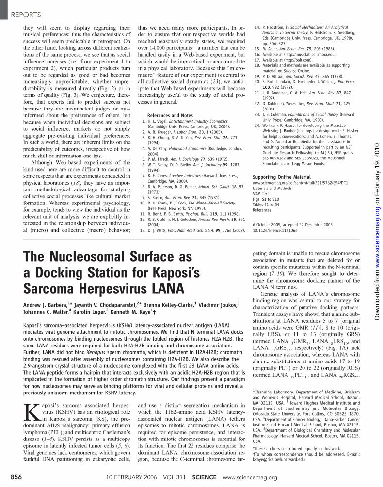
Fig. 1. LANA N terminuschromosome binding. (A)LANA scanning alanine mu-tants with summaries forchromosome binding, epi-some persistence (7), andH2A-H2B binding. nd, notdetermined. (B) Metaphasespreads of BJAB cells andBJAB cells stably express-ing LANA, LANA5GMR7,LANA 8LRS10, LANA 11GRS13,or LANA 14TG15. Overlay ofLANA (green) and chromo-somes (red) generates yel-low. Cells containing KSHVepisomes are indicated. Ar-rows denote LANA 14TG15dots that have detachedfrom chromosomes. Magni-fication is 630�. (C) Meta-phase BJAB cells stablyexpressing GFP NLS orGFP LANA 1-32 at 630�magnification. (D) Proteinsco-precipitating with GFPLANA 1-32 (lane 2) wereidentified after resolutionin a 4 to 16% gradientgel. HC, heavy chain; LC,light chain; asterisk, GFP;
&, GFP LANA 1-32. The stoichiometry of histones within nucleosomes and their arginine-rich nature contribute to the intense histone Coomassie staining.Numbers on the left-hand side of the gel are size markers (kD).
REPORTS
www.sciencemag.org SCIENCE VOL 311 10 FEBRUARY 2006 857
on
Feb
ruar
y 19
, 201
0 w
ww
.sci
ence
mag
.org
Dow
nloa
ded
from
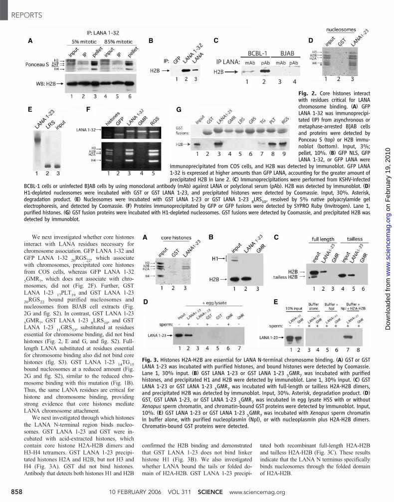
We next investigated whether core histones
interact with LANA residues necessary for
chromosome association. GFP LANA 1-32 and
GFP LANA 1-3220RGS
22, which associate
with chromosomes, precipitated core histones
from COS cells, whereas GFP LANA 1-32
5GMR
7, which does not associate with chro-
mosomes, did not (Fig. 2F). Further, GST
LANA 1-2317PLT
19and GST LANA 1-23
20RGS
22bound purified nucleosomes and
nucleosomes from BJAB cell extracts (Fig.
2G and fig. S2). In contrast, GST LANA 1-23
5GMR
7, GST LANA 1-23
8LRS
10, and GST
LANA 1-2311GRS
13, substituted at residues
essential for chromosome binding, did not bind
histones (Fig. 2, E and G, and fig. S2). Full-
length LANA substituted at residues essential
for chromosome binding also did not bind core
histones (fig. S3). GST LANA 1-2314TG
15
bound nucleosomes at a reduced amount (Fig.
2G and fig. S2), similar to the reduced chro-
mosome binding with this mutation (Fig. 1B).
Thus, the same LANA residues are critical for
histone and chromosome binding, providing
strong evidence that core histones mediate
LANA chromosome attachment.
We next investigated through which histones
the LANA N-terminal region binds nucleo-
somes. GST LANA 1-23 and GST were in-
cubated with acid-extracted histones, which
contain core histone H2A-H2B dimers and
H3-H4 tetramers. GST LANA 1-23 precipi-
tated histones H2A and H2B, but not H3 and
H4 (Fig. 3A). GST did not bind histones.
Antibody that detects both histones H1 and H2B
confirmed the H2B binding and demonstrated
that GST LANA 1-23 does not bind linker
histone H1 (Fig. 3B). We also investigated
whether LANA bound the tails or folded do-
main of H2A-H2B. GST LANA 1-23 precipi-
tated both recombinant full-length H2A-H2B
and tailless H2A-H2B (Fig. 3C). These results
indicate that the LANA N terminus specifically
binds nucleosomes through the folded domain
of H2A-H2B.
Fig. 2. Core histones interactwith residues critical for LANAchromosome binding. (A) GFPLANA 1-32 was immunoprecipi-tated (IP) from asynchronous ormetaphase-arrested BJAB cellsand proteins were detected byPonceau S (top) or H2B immu-noblot (bottom). Input, 3%;pellet, 10%. (B) GFP NLS, GFPLANA 1-32, or GFP LANA were
immunoprecipitated from COS cells, and H2B was detected by immunoblot. GFP LANA1-32 is expressed at higher amounts than GFP LANA, accounting for the greater amount ofprecipitated H2B in lane 2. (C) Immunoprecipitations were performed from KSHV-infected
BCBL-1 cells or uninfected BJAB cells by using monoclonal antibody (mAb) against LANA or polyclonal serum (pAb). H2B was detected by immunoblot. (D)H1-depleted nucleosomes were incubated with GST or GST LANA 1-23, and precipitated histones were detected by Coomassie. Input, 30%. Asterisk,degradation product. (E) Nucleosomes were incubated with GST LANA 1-23 or GST LANA 1-23 8LRS10, resolved by 5% native polyacrylamide gelelectrophoresis, and detected by Coomassie. (F) Proteins immunoprecipitated by GFP or GFP fusions were detected by SYPRO Ruby (Invitrogen). Lane 1,purified histones. (G) GST fusion proteins were incubated with H1-depleted nucleosomes. GST fusions were detected by Coomassie, and precipitated H2B wasdetected by immunoblot.
Fig. 3. Histones H2A-H2B are essential for LANA N-terminal chromosome binding. (A) GST or GSTLANA 1-23 was incubated with purified histones, and bound histones were detected by Coomassie.Lane 1, 30% input. (B) GST LANA 1-23 or GST LANA 1-23 5GMR7 was incubated with purifiedhistones, and precipitated H1 and H2B were detected by immunoblot. Lane 1, 30% input. (C) GSTLANA 1-23 or GST LANA 1-23 5GMR7 was incubated with full-length or tailless H2A-H2B dimers,and precipitated H2B was detected by immunoblot. Input, 30%. Asterisk, degradation product. (D)GST, GST LANA 1-23, or GST LANA 1-23 5GMR7 was incubated in egg lysate HSS with or withoutXenopus sperm chromatin, and chromatin-bound GST proteins were detected by immunoblot. Input,10%. (E) GST LANA 1-23 or GST LANA 1-23 5GMR7 was incubated with Xenopus sperm chromatinin buffer alone, with purified nucleoplasmin (Npl), or with nucleoplasmin plus H2A-H2B dimers.Chromatin-bound GST proteins were detected.
REPORTS
10 FEBRUARY 2006 VOL 311 SCIENCE www.sciencemag.org858
on
Feb
ruar
y 19
, 201
0 w
ww
.sci
ence
mag
.org
Dow
nloa
ded
from

We wished to demonstrate directly that the
LANA N terminus uses H2A-H2B to bind chro-
mosomes. We used Xenopus laevis sperm chro-
matin, which is naturally deficient in H2A-H2B
and instead contains sperm-specific basic pro-
teins X and Y. In addition, Xenopus sperm lack
H1 (16–18). Upon incubation with high-speed
supernatant (HSS) from Xenopus egg lysate, egg
cell–derived nucleoplasmin protein mediates
sperm chromatin decondensation and replace-
ment of X and Y with egg H2A-H2B dimers.
To verify LANA chromosome binding in this
system, we incubated HSS-treated chromatin,
which contains wild-type H2A-H2B dimers,
with GST fusions. GST LANA 1-23 bound sperm
chromatin that had undergone H2A-H2B deposi-
tion through HSS treatment, but GST LANA 1-23
5GMR
7and GST did not (Fig. 3D). No LANA
protein precipitated in the absence of chroma-
tin. Therefore, N-terminal LANA binds Xeno-
pus chromosomes after H2A-H2B deposition.
Fig. 4. Structure of the LANA-nucleosomecomplex. (A) Stereoview of a section of thefinal 2Fo–Fc electron density map calculated at2.9 A and contoured at 2s, depicting theLANA peptide. Intramolecular hydrogen bondsare shown as red dashes. (B) Space-fillingrepresentation of the nucleosome-LANA com-plex. H2A is shown in yellow, H2B in red, H3in light blue, H4 in green, and LANA in darkblue. DNA is silver. (C) Overview of LANAinteraction with the H2A-H2B dimer within theNCP. Only H2A (yellow ribbon), H2B (redribbon), and LANA (blue sticks) are shown.Intramolecular and intermolecular bonds areshown as red and blue dashes, respectively.Secondary structural elements in the histonesare indicated. (D) Crystal contact between theH4 tail of the neighboring nucleosome andthe H2A-H2B dimer. Orientation and coloringof H2A and H2B is shown as in (C); the H4 tailis shown in green. (E) LANA recognizes distinctfeatures of the nucleosomal surface. Chargedsurfaces (red, negatively charged; blue, posi-tively charged) were calculated with GRASP(37). The H2A-H2B dimer (left) and LANA areshown individually; LANA has been rotated by90- along the y axis. The H2A-H2B dimer is inabout the same conformation as in (C). (F) Topview of LANA bound to the histone dimerwithin the NCP [rotation by 90- around y and180- around x with respect to the view in (E)].Only the H2A-H2B dimer (charged surface)and LANA (stick model) are shown.
REPORTS
www.sciencemag.org SCIENCE VOL 311 10 FEBRUARY 2006 859
on
Feb
ruar
y 19
, 201
0 w
ww
.sci
ence
mag
.org
Dow
nloa
ded
from

We stringently assayed whether H2A-H2B
were required for LANA chromosome binding.
HSS contains other factors in addition to H2A-
H2B and nucleoplasmin. We therefore used a
purified system with nucleoplasmin and recom-
binant H2A-H2B dimers in place of HSS. GST
LANA 1-23 did not bind H2A-H2B-deficient
sperm chromatin that had been treated with
buffer or with purified nucleoplasmin alone.
However, after incubation with nucleoplasmin
and recombinant histone H2A-H2B dimers,
which allows for deposition of histones H2A-
H2B into sperm chromatin, GST LANA 1-23
specifically bound sperm chromatin (Fig. 3E).
Thus, H2A-H2B is essential for LANA chro-
mosome binding.
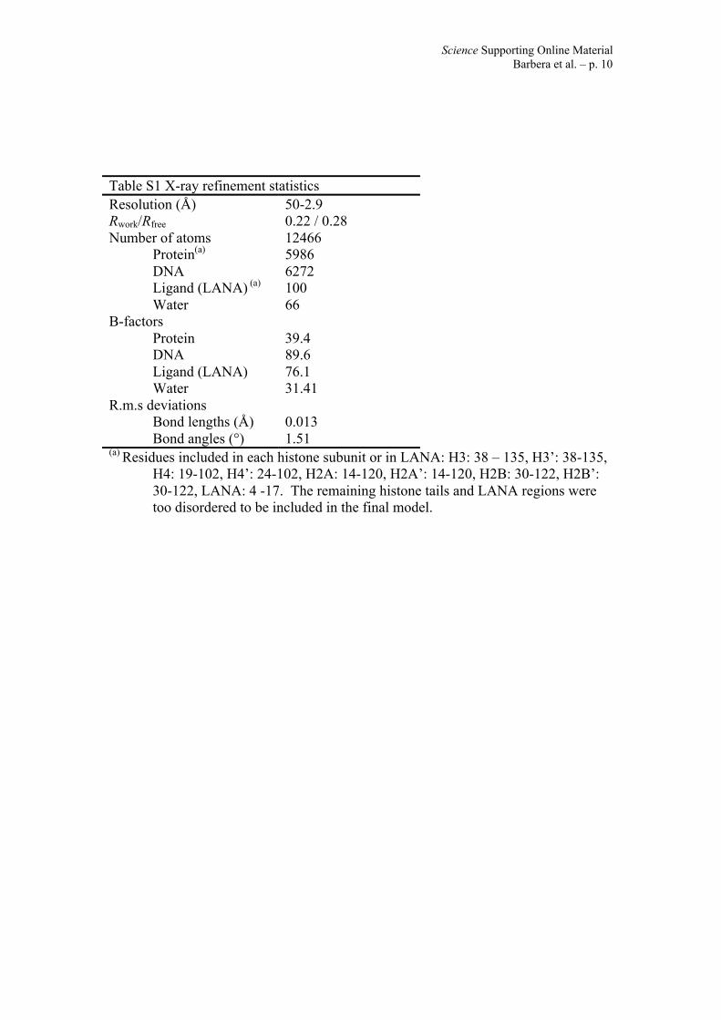
We solved the x-ray crystal structure of
LANA residues 1 to 23 complexed with the
NCP. Data collection and refinement statistics
are summarized in table S1. Figure 4A shows a
2Fo–F
cmap of the final model of the LANA
peptide, contoured at 2s. LANA forms a tight
hairpin that is stabilized by five intramolecular
hydrogen bonds (three b-type interactions and
two side-chain or main-chain interactions) (Fig.
4, A and C) and by numerous hydrogen bonds
and Van der Waals contacts with the nucleoso-
mal surface.
Consistent with the biochemical experiments
(Fig. 3, A to C), the LANA peptide interacts
exclusively with the H2A-H2B dimer within
the nucleosome (Fig. 4B). Histone fold regions
and extensions of H2A and H2B are implicated
in the interaction, but not the flexible histone
tails. The hairpin is wedged between the aCand a1 helix of H2B (Fig. 4C); the turn of the
hairpin abuts the H2A docking domain that
forms a major interaction interface between the
H2A-H2B dimer and the (H3-H4)2tetramer
(19). The L1 loop of H2B as well as the a2 anda3 helices of H2A are also involved in LANA
binding, consistent with the requirement for a
folded H2A-H2B dimer for LANA binding.
Molecular details of the interactions between
LANA and the nucleosome are shown in fig.
S4 (SOM Text). Substitution of individual
LANA amino acids 5 to 16 demonstrated that
residues important for chromosome association
(fig. S5 and SOM Text) have critical roles in
the interaction between LANA and the NCP.
Of note, the overall structure of the nucleosome
is maintained upon LANA binding (Fig. 4B).
Interactions of LANA with the NCP resem-
ble those between the NCP and the H4 N-
terminal tail from a neighboring nucleosome
within the crystal lattice (Fig. 4D) (20). Both
peptides interact with the same conserved
acidic patch composed of several residues from
H2A and H2B on the highly contoured nucleo-
somal surface (21). Despite a lack of sequence
homology between the LANA peptide and the
N-terminal tail, many of the targeted residues in
H2A and H2B are the same (see, for example,
LANA R9and H4 R
19in Fig. 4, C and D,
respectively). The interaction shown in Fig. 4D
is essential for nucleosome crystallization (14),
and biophysical experiments have indicated a
unique role for the H4 tail and acidic patch
interaction in the formation of chromatin higher
order structure (22, 23).
Analysis of the molecular surfaces of both
the LANA peptide and the H2A-H2B dimer
demonstrates excellent shape and charge com-
plementarity (Fig. 4E), indicating that the
LANA N-terminal region has evolved to rec-
ognize this region within the NCP with high
specificity. LANA R9and Ser
10point into the
acidic pocket formed by H2A and H2B, and
hydrophobic LANA residues are inserted deep
into a cleft delineated by the aC helix of H2B
(Fig. 4F). The LANA peptide interaction buries
1340 )2, well within the range that is con-
sidered to be a stable interaction (24), which is
notable considering that only 14 residues of
LANA contribute to the interaction. For com-
parison, the molecular surface buried by the H4
tail–NCP interaction (Fig. 4D) is only 680 )2
and contains larger cavities.
This work demonstrates that LANA_s N-
terminal chromosome association is mediated by
H2A-H2B and not by the earlier proposed
candidates methyl-CpG binding protein 2
(MeCP2) or H1 (8, 12, 25). It was previously
reported that LANA did not associate with
murine chromosomes unless human MeCP2
was co-expressed (8). In contrast, we found that
LANA bound murine chromosomes (fig. S1);
further, MeCP2 was not identified from our
affinity purification. Histone H1 did not bind
the LANA N terminus and was not required for
LANA to bind Xenopus chromatin (Fig. 3, B
and E). These results also differ from proposed
chromosome binding mechanisms for other
episome maintenance proteins: Epstein-Barr
virus EBNA1 binds chromosomes through the
nucleolar EBP2 protein or AT hooks, and
bovine papillomavirus E2 binds through the
bromodomain protein Brd4 (26–28).
This work may also link H2A-H2B binding
to LANA_s transcriptional regulatory effects
(29, 30). In fact, LANA transcriptional activity
can be dependent on N-terminal chromosome
association (31). An intriguing possibility is that
LANA may affect transcription by regulating
transient H2A-H2B removal from nucleosomes
through complexes such as FACT or nucleo-
some assembly protein 1 (32,33). Histone mod-
ifications regulate transcription and may also
affect LANA_s affinity for nucleosomes and
effects on chromatin, although experiments with
bacterially expressed protein (Figs. 2 to 4) in-
dicate that histone modifications are not re-
quired for binding.
This work indicates a role for H2A-H2B in
LANA-mediated DNA replication and episome
persistence, because these functions are depen-
dent on N-terminal LANA chromosome binding
(7). Interestingly, histone fusions have been used
as an alternative method of targeting LANA and
EBNA1 to chromosomes (10, 27, 34, 35). Link-
er histone H1 in place of the LANA or EBNA1
chromosome association region permits epi-
some persistence, whereas core histones (H2B
and H3, respectively) do not, perhaps because
of positional restrictions related to the covalent
linkages. Of note, LANA has a C-terminal
chromosome association domain, but it cannot
rescue chromosome binding of N-terminal
mutated LANA (Fig. 1B) (7–10); its role in
episome persistence is currently under investi-
gation. The distribution of H2A-H2B through-
out chromosomes provides a platform through
which LANA tethered episomes can efficiently
segregate to progeny nuclei. Strategies that
interrupt the interaction between LANA and
H2A-H2B may provide effective treatment and
prevention of KSHV-associated diseases.
The x-ray crystal structure shows that a
hairpin formed by KSHV LANA residues 5 to
13 interacts with eukaryotic chromatin by
binding to an acidic patch formed by H2A-H2B
within the nucleosome. Thus, LANAhas evolved
to use the differentially charged and contoured
surface of the nucleosome as a Bdocking station[for episome attachment. The concept of the
nucleosomal surface (as opposed to the flex-
ible histone tails) as an interaction platform has
been proposed earlier (14, 22, 36–38); we now
report the structure of a protein complexed with
the nucleosome core. It appears that an im-
portant function of histones, in addition to
maintaining interaction with other histones to
form the octamer and compacting genomic
DNA, is to maintain a distinct surface land-
scape that is used as a docking platform by
cellular and viral factors. Such interactions may
locally affect nucleosome dynamics and/or alter
chromatin higher order structure, with profound
implications for transcription of underlying
DNA regions.
References and Notes1. Y. Chang et al., Science 266, 1865 (1994).2. P. S. Moore, Y. Chang, N. Engl. J. Med. 332, 1181
(1995).3. E. Cesarman, Y. Chang, P. S. Moore, J. W. Said,
D. M. Knowles, N. Engl. J. Med. 332, 1186 (1995).4. J. Soulier et al., Blood 86, 1276 (1995).5. M. E. Ballestas, P. A. Chatis, K. M. Kaye, Science 284,
641 (1999).6. L. L. Decker et al., J. Exp. Med. 184, 283 (1996).7. A. J. Barbera, M. E. Ballestas, K. M. Kaye, J. Virol. 78, 294
(2004).8. A. Krithivas, M. Fujimuro, M. Weidner, D. B. Young,
S. D. Hayward, J. Virol. 76, 11596 (2002).9. T. Piolot, M. Tramier, M. Coppey, J. C. Nicolas,
V. Marechal, J. Virol. 75, 3948 (2001).10. H. Shinohara et al., J. Virol. 76, 12917 (2002).11. Single-letter abbreviations for the amino acid residues
are as follows: A, Ala; C, Cys; D, Asp; E, Glu; F, Phe;G, Gly; H, His; I, Ile; K, Lys; L, Leu; M, Met; N, Asn; P, Pro;Q, Gln; R, Arg; S, Ser; T, Thr; V, Val; W, Trp; and Y, Tyr.
12. M. A. Cotter 2nd, E. S. Robertson, Virology 264, 254(1999).
13. T. Tetsuka et al., Virus Genes 29, 175 (2004).14. K. Luger, A. W. Mader, R. K. Richmond, D. F. Sargent,
T. J. Richmond, Nature 389, 251 (1997).15. A. J. Barbera et al., unpublished data.16. A. Philpott, G. H. Leno, R. A. Laskey, Cell 65, 569 (1991).17. A. Philpott, G. H. Leno, Cell 69, 759 (1992).
REPORTS
10 FEBRUARY 2006 VOL 311 SCIENCE www.sciencemag.org860
on
Feb
ruar
y 19
, 201
0 w
ww
.sci
ence
mag
.org
Dow
nloa
ded
from

18. A. W. Murray, Methods Cell Biol. 36, 581 (1991).19. K. Luger, T. J. Richmond, Curr. Opin. Genet. Dev. 8, 140
(1998).20. C. A. Davey, D. F. Sargent, K. Luger, A. W. Maeder,
T. J. Richmond, J. Mol. Biol. 319, 1097 (2002).21. K. Luger, T. J. Richmond, Curr. Opin. Struct. Biol. 8, 33
(1998).22. B. Dorigo, T. Schalch, K. Bystricky, T. J. Richmond, J. Mol.
Biol. 327, 85 (2003).23. J. Y. Fan, D. Rangasamy, K. Luger, D. J. Tremethick,
Mol. Cell 16, 655 (2004).24. S. Jones, J. M. Thornton, Proc. Natl. Acad. Sci. U.S.A. 93,
13 (1996).25. S. C. Verma, E. S. Robertson, FEMS Microbiol. Lett. 222,
155 (2003).26. J. You, J. L. Croyle, A. Nishimura, K. Ozato, P. M. Howley,
Cell 117, 349 (2004).27. J. Sears et al., J. Virol. 78, 11487 (2004).28. P. Kapoor, K. Shire, L. Frappier, EMBO J. 20, 222 (2001).29. A. Krithivas, D. B. Young, G. Liao, D. Greene,
S. D. Hayward, J. Virol. 74, 9637 (2000).30. R. Renne et al., J. Virol. 75, 458 (2001).
31. L. Y. Wong, G. A. Matchett, A. C. Wilson, J. Virol. 78,10074 (2004).
32. G. Orphanides, G. LeRoy, C. H. Chang, D. S. Luse,D. Reinberg, Cell 92, 105 (1998).
33. Y. J. Park, J. V. Chodaparambil, Y. Bao, S. J. McBryant,K. Luger, J. Biol. Chem. 280, 1817 (2004).
34. J. L. Yates, N. Warren, B. Sugden, Nature 313, 812(1985).
35. S. C. Hung, M. S. Kang, E. Kieff, Proc. Natl. Acad. Sci.U.S.A. 98, 1865 (2001).
36. R. K. Suto, M. J. Clarkson, D. J. Tremethick, K. Luger,Nat. Struct. Biol. 7, 1121 (2000).
37. F. van Leeuwen, P. R. Gafken, D. E. Gottschling, Cell 109,745 (2002).
38. J. H. Park, M. S. Cosgrove, E. Youngman, C. Wolberger,J. D. Boeke, Nat. Genet. 32, 273 (2002).
39. A. Nicholls, K. A. Sharp, B. Honig, Proteins 11, 281 (1991).40. HeLa nucleosomes were a gift of X. He, P. Pascual-Ahuir
Giner, and R. Kingston. PARP1 and Ku80 knockout MEFswere generously provided by J. Jung and A. Nussenzweig,respectively. We thank P. Yiu for assistance with spermchromatin experiments; E. Kieff for helpful discussions;
P. Dyer, O. Peersen, and V. Srinivasan for technicalassistance; R. Edayathumangalam and S. Chakravarthyfor help with data collection and structure determination;and A. Straight and T. Mitchison for the plasmid encodingH2A-H2B used in the chromatin binding experiments.This work was supported by grant CA82036 (to K.M.K.)from the National Cancer Institute and grants GM067777(to K.L.) and GM62267 (to J.C.W.) from the NationalInstitute of General Medical Sciences. Coordinates havebeen deposited at the Protein Data Bank database (entrycode 1ZLA).
Supporting Online Materialwww.sciencemag.org/cgi/content/full/311/5762/856/DC1Materials and MethodsSOM TextFigs. S1 to S5Table S1References and Notes
26 September 2005; accepted 12 January 200610.1126/science.1120541
Neurochemical Modulation ofResponse Inhibition and ProbabilisticLearning in HumansSamuel R. Chamberlain,1,3* Ulrich Muller,1,2,3 Andrew D. Blackwell,1,3 Luke Clark,2,3
Trevor W. Robbins,2,3 Barbara J. Sahakian1,3
Cognitive functions dependent on the prefrontal cortex, such as the ability to suppress behavior(response inhibition) and to learn from complex feedback (probabilistic learning), play critical rolesin activities of daily life. To what extent do different neurochemical systems modulate these twocognitive functions? Here, using stop-signal and probabilistic learning tasks, we show a doubledissociation for the involvement of noradrenaline and serotonin in human cognition. In healthyvolunteers, inhibition of central noradrenaline reuptake improved response inhibition but had noeffect on probabilistic learning, whereas inhibition of central serotonin reuptake impairedprobabilistic learning with no effect on response inhibition.
Ascending monoamine projections play
important neuromodulatory roles in
high-level cognition through actions
upon the prefrontal cortex (PFC), a major
brain structure with considerable functional
heterogeneity in humans (1). Dysfunction in
these neurochemical systems is implicated in
the etiology and psychopathology of psychiatric
illnesses associated with cognitive deficits and
PFC abnormalities, including depression, atten-
tion deficit–hyperactivity disorder (ADHD),
obsessive-compulsive disorder (OCD), and drug
addiction (2–7). Dopamine regulates executive
functions dependent on the dorsolateral PFC,
including working memory and attentional set-
shifting, but the role of noradrenaline (NA) and
serotonin E5-hydroxytryptamine (5-HT)^ in cog-
nition is less well characterized (8). The
orbitofrontal cortex (OFC) is involved in
emotion-cognition interactions, and 5-HT
drugs modulate response to feedback and
decision-making within this region (9–15).
5-HT and NA have both been implicated in
response inhibition (16, 17), a function that has
been linked to the right inferior frontal gyrus
(RIFG) (18).
We investigated the differential involvement
of NA and 5-HT transmitter systems in these
processes in humans, using the selective NA
reuptake inhibitor (SNRI) atomoxetine and
the selective 5-HT reuptake inhibitor (SSRI)
citalopram. These agents are among the most
selective inhibitors for brain NA and 5-HT
reuptake transporters available for human
use, according to in vitro and in vivo findings
(19–21). Microdialysis studies in experimental
animals have shown that acute systemic ad-
ministration of atomoxetine rapidly increases
PFC NA but not 5-HT and that the administra-
tion of citalopram rapidly increases PFC 5-HT
but not NA (19, 22). As such, these agents rep-
resent useful neurochemical tools for inves-
tigating the differential involvement of NA and
5-HT in human cognition.
Response inhibition, the ability to exert high-
level inhibitory control over motor responses so
as to suppress unwanted actions, can be assessed
with the stop-signal procedure (6, 23). In this
procedure, volunteers are required to make
rapid motor responses on Go trials but to inhibit
responses if an auditory stop-signal occurs. By
the infrequent nature of Stop trials, motor
responses are made Bprepotent.[ Response
inhibition can be quantified by the stop-signal
reaction time (SSRT), an estimate of the time
taken to inhibit the prepotent motor response
(18, 23). Probabilistic learning refers to the abil-
ity to develop cognitive associations between
stimuli and outcomes on the basis of punishing
and rewarding feedback, and to modify these
associations as appropriate (12). On probabilis-
tic learning tasks, volunteers are required to
select which of two stimuli they believe to be
correct over a series of trials. After each choice,
the computer provides punishing or rewarding
feedback that is Bdegraded[ (i.e., misleading on
a subset of trials) (12).
The aim of the present study was to delineate
the precise differential contribution of NA and
5-HT neurochemical systems to response inhi-
bition and probabilistic learning. Sixty healthy
male participants were recruited from the local
community on the basis of being free from
medical or psychiatric disorders according to
assessment by a psychiatrist (mean age 25.7 TSD 4.7 years, range 20 to 35) (24). Participants
received single clinically relevant oral doses of
atomoxetine (60 mg), citalopram (30 mg), or
placebo in a double-blind parallel-groups design
(24). Groups were matched for demographic
characteristics (table S1). After spending 1.5
hours in a quiet waiting area to ensure drug
1Department of Psychiatry, University of Cambridge Schoolof Clinical Medicine, Addenbrooke’s Hospital, Box 189,Cambridge CB2 2QQ, UK. 2Department of ExperimentalPsychology, 3Behavioural and Clinical Neuroscience Insti-tute, University of Cambridge, Cambridge CB2 3EB, UK.
*To whom correspondence should be addressed. E-mail:[email protected]
REPORTS
www.sciencemag.org SCIENCE VOL 311 10 FEBRUARY 2006 861
on
Feb
ruar
y 19
, 201
0 w
ww
.sci
ence
mag
.org
Dow
nloa
ded
from

www.sciencemag.org/cgi/content/full/311/5762/856/DC1
Supporting Online Material for
The Nucleosomal Surface as a Docking Station for Kaposi’s Sarcoma Herpesvirus LANA
Andrew J. Barbera, Jayanth V. Chodaparambil, Brenna Kelley-Clarke, Vladimir Joukov, Johannes C. Walter, Karolin Luger, Kenneth M. Kaye*
*To whom correspondence should be addressed. E-mail: [email protected]
Published 10 February 2006, Science 311, 856 (2006)
DOI: 10.1126/science.1120541
This PDF file includes:
Materials and Methods SOM Text Figs. S1 to S5 Table S1 References and Notes

Science Supporting Online MaterialBarbera et al. – p. 1
Supporting Online Material
Materials and Methods
Plasmids, cell lines, and microscopy. GFP NLS(1) has the green fluorescent protein(GFP) gene fused to a nuclear localization signal (NLS). GFP LANA 1-32 and GFPLANA contain alanine substitutions as indicated (2). GST LANA 1-23 has theindicated LANA residues downstream of GST in the vector pGEX-KG(3). Scanningalanine substitutions within GST LANA 1-23 were generated by PCR mutagenesisand sequence confirmed. Point mutations within LANA 1-32 were generated byQuickChange Mutagenesis II kit (Stratagene) and sequence confirmed. BJAB Blymphoma cells stably expressing GFP NLS or GFP LANA 1-32 were generatedusing neomycin. BJAB cells stably express FLAG epitope tagged wild-typeLANA(4) or LANA with alanine substitution mutants(2). Ku80+/-, Ku80-/-(5),PARP1 -/- and PARP1 +/+(6) MEFs were described. BJAB cells were transfected asdescribed(2) and adherent cells were transfected with Lipofectamine 2000 reagent(Invitrogen). Metaphase spreads and confocal microscopy were performed asdescribed(2) except cells were cytospun onto glass slides. For immune fluorescence,LANA was detected with anti-LANA monoclonal antibody (ABI).
Immune precipitations and protein detection. For affinity purification, 1 x 10⁹
BJAB cells expressing GFP LANA 1-32 or control GFP NLS were lysed in 40 mL ofIP buffer (50 mM HEPES pH 7.2, 250mM NaCl, 10% glycerol, 2mM EDTA, 1% NP-40, 0.1 mM PMSF, 1µg/mL aprotinin, 1 µg/mL leupeptin, and 0.7 µg/mL pepstatin)at 4°C with brief sonication. Protein was precipitated with anti-GFP polyclonal serum(Clontech) followed by protein G bead capture. Gel slices were analyzed by tandemmass spectrometry at Partners HealthCare Center for Genetics and Genomics(HPCGG).
Lysis for smaller scale immune precipitations was performed with IP buffer(or RIPA buffer (phosphate-buffered saline, 0.5% NP-40, 0.25% sodiumdeoxycholate, 0.05% SDS, 0.1 mM PMSF, 1µg/mL aprotinin, 1 µg/mL leupeptin, and0.7 µg/mL pepstatin) for anti-LANA polyclonal serum) and lysates weresupplemented with 5mM MgSO4 and 2mM CaCl2 and incubated with 50 µg DNAse I(Sigma) at 4°C x 45 min, prior to the addition of antibody. DNAse I treatment wasagain performed on collected protein G beads. DNAse I cleaves DNA betweennucleosomes but does not disrupt mononucleosomes, and limits nonspecific co-precipitations bridged by DNA. Anti-H2B (and anti-H1) antibody (Upstate) at 1:1000dilution was used in conjunction with HRP-conjugated secondary antibodies(Southern Biotechnology) and ECL reagents (Perkin Elmer). Alternatively, proteinwas detected with Coomassie Brilliant Blue G250 or Sypro RUBY (MolecularProbes).
GST fusion protein binding assays. GST fusion proteins were expressed in E. coliand collected on glutathione sepharose beads (Amersham). Full-length H2A-H2Bdimers and tailless H2A-H2B dimers (H2A, residues 13-118; H2B, residues 27-122)were purified from E. coli (7). GST precipitations were performed in precipitationbuffer (150 mM NaCl, 50mM Tris-pH 7.5, 10% glycerol, 0.1% NP-40, 0.5 mM DTT,

Science Supporting Online MaterialBarbera et al. – p. 2
1µg/mL aprotinin, 1 µg/mL leupeptin, and 0.7 µg/mL pepstatin) overnight at 4°C, andwashed in above buffer supplemented to 250mM NaCl. For GST precipitations fromcell extracts, BJAB cells were lysed in IP lysis buffer, treated with DNAse, pre-cleared with GST beads, and incubated overnight at 4°C with GST fusion proteins.For gel shift assays, NCPs generated from recombinant full length Xenopus histoneswere prepared as described (8), and incubated with 5-fold excess GST fusion proteinsfor 10 hours at 4o C in buffer (20mM Tris pH 7.5, 1mM EDTA, 1mM DTT) andanalyzed by 5% native PAGE.
Xenopus laevis sperm chromatin binding assays. Xenopus laevis sperm wereobtained and demembranated nuclei prepared (9, 10). Xenopus eggs were lysed andhigh speed supernatant (HSS) generated. Adenosine triphosphate (ATP)-regenerationsystem(11), nocodazole (3µg/mL), and GST fusion proteins (30 ng/µl) were added toHSS and the extract was then clarified by centrifugation at 6000 x g for 5 minutes.Sperm chromatin was added to 10 µl of the clarified extract at a concentration of 5000nuclei equivalents per µl and incubated for 30 minutes at 22°C. Chromatin wasisolated through a sucrose cushion as described (12). GST was detected byimmunoblot with anti-GST antibody (Covance).
For decondensation reactions using purified factors, sperm chromatin wasincubated in Buffer D (50mM HEPES-KOH pH 7.8, 75 mM K-Acetate, 0.5 mMspermidine, 0.15 mM spermine, 1 mM EGTA)(13) for 30 min. at 22°C. Whereindicated, histone H2A-H2B dimers (40ng/µl) and nucleoplasmin (900ng/µl) purifiedfrom Xenopus laevis eggs were added. H2A-H2B dimers were expressed from abicistronic vector in E. coli and purified using HiTrap SP FF columns (Pharmacia).
X-ray crystal structure. Crystals of the NCP reconstituted with a palindromic146mer DNA fragment derived from human a-satellite DNA(14) were incubated withchemically synthesized LANA 1-23 (1MAPPGMRLRSGRSTGAPLTRGS23) (TuftsUniversity Core Facility). Nucleosomes were crystallized using salting in vapordiffusion at NCP concentrations ranging from 5-6 mg/ml with salt concentrations of27.5 mM KCl and 35 mM of MnCl2. The crystals were soaked in 24% 2-methyl, 2,4-pentadiol (MPD) containing 5% trehalose(14) and 5 mg/ml of LANA peptide. X-raydata were collected on a Rigaku RU-H3R rotating anode generator (1.5418Å Cu-Karadiation) with osmic confocal multilayer optics system, R-axis IV++ image platedetector and an X-stream cryocooling system. The data was processed with Denzoand Scalepack(15). PDB entry 1AOI was used as a search model for molecularreplacement. Molecular replacement and further refinements were done usingCNS(16), model building was done using O(17). The fidelity of the model waschecked using simulated annealing omit maps during early stages of the refinement.The geometry of the final model was checked using a Ramachandran plot with 90%of the amino acids in the most favored region, 8.8% of the amino acids in theadditionally allowed region, and 1.2% of the model lying in the generously allowedregion. Figures 4A-D were made using PyMOL molecular graphics system(www.pymol.org). Figure 4E, F and occluded surface areas were done usingGRASP(18).

Science Supporting Online MaterialBarbera et al. – p. 3
Supporting Online Text
Ku70, Ku80, and PARP1 do not mediate LANA chromosome association. A rolefor Ku70, Ku80 and PARP1 in mediating LANA chromosome association wasassessed using knockout mouse embryo fibroblasts (MEFs). In the absence of itsmitotic docking partner, LANA chromosome association is expected to be lost. GFPLANA 1-32 (green) tightly associated with chromosomes (red) (overlay generatesyellow) both in the absence (Ku80-/-) and presence (Ku80+/-) of Ku80 (Fig. S1). Thedramatic reduction of Ku70 expression in these cells(5) also did not affect GFPLANA 1-32’s chromosome targeting. Further, GFP LANA 1-32 associated withchromosomes in the absence (PARP-/-) and presence (PARP+/+) of PARP1 (Fig S1).Therefore, neither Ku70, Ku80 nor PARP1 is the LANA N-terminal mitotic dockingpartner.
Interactions between LANA and the nucleosome. Details of the interactionbetween LANA and the nucleosome are shown in Fig. S4. The main chain of oneside of the LANA hairpin (amino acids 5-8) makes three hydrogen bonds with threedifferent amino acid side chains in H2B aC (Fig. S4A). The complex is furtherstabilized by the insertion of LANA Met 6 and Leu 8 into a hydrophobic regionformed by the long a2 helix of H2A (Fig. S4B and Fig. 4F respectively). Of allLANA residues, Arg 9 makes the most interactions with the H2A-H2B dimer: wecount a total of five hydrogen bonds between the side chain of LANA Arg 9 andresidues in H2A that form the acidic patch (H2A Glu 92, Glu 61, and Asp 90; Fig.S4C). Interactions with this particular region are enforced by a hydrogen bondbetween LANA Ser 10 and H2A Glu 64 (Fig. S4C), and between the main chain ofLANA Arg 7 and H2B Glu 110. Thus, the LANA peptide interacts with five of theseven amino acids that form the acidic patch on the nucleosomal surface.
Interactions between the other side of the LANA hairpin and the H2A-H2Bdimer are less abundant, and engage the side chains of LANA residues Ser 10, Arg12, and the main chain of Ser 13, Gly 15 (Fig. S4D). A glycine in position 11maintains the conformation of the ß-turn. LANA residue Arg 12 assumes twoalternative side chain conformations (Fig. S4D) that are both clearly visible in theoriginal electron density map; both are involved in hydrogen bonding with the mainchain of the a1 helix of H2B. Nevertheless, this residue can be replaced with analanine without significant reduction in LANA chromosome association, unlike otherLANA residues involved in interactions with the nucleosome (Fig. S5).
Individual LANA residues critical for chromosome association. We investigatedthe importance of individual LANA residues for chromosome association sinceprevious experiments used triple alanine substitutions. Residues Gly 5, Met 6, Leu 8,Arg 9, Ser 10, and Gly 11 were each essential for chromosome association since GFPfused to N-terminal LANA (GFP LANA 1-32 (green)) with alanine substitutions atthese sites did not localize to chromosomes (red) (Fig. S5). In contrast, Arg 7, Arg12, Ser 13, Gly 15, and Ala 16 were each dispensable for chromosome associationsince GFP LANA 1-32 (green) mutated at each residue diffusely paintedchromosomes (red) (overlay of green and red generates yellow). Thr 14 had a role in

Science Supporting Online MaterialBarbera et al. – p. 4
chromosome association since GFP LANA 1-32 mutated at this site diffusely paintedchromosomes but also distributed between them. These results are consistent withthose of others who used point mutations to investigate LANA chromosomeassociation(19, 20). This work demonstrates that with the exception of Arg 7 whichpoints outwards, away from the H2A-H2B dimer, LANA residues 5 – 11 are eachessential for binding of LANA to chromatin. The presence of the two glycineresidues at strategic places appears to be essential to maintain the hairpin structure.The moderate importance of Thr 14 is consistent with its role in stabilizing the lowerportion of the hairpin via a hydrogen bond (Fig. 4A).

Figure S1. Ku70, Ku80, and PARP1 do not mediate LANA N-terminal chromosome association. MEF cells expressing GFP LANA 1-32 (green) were metaphase arrested and DNA counterstained with propidium iodide (red) (overlay of green and red generates yellow). (630X)
Science Supporting Online MaterialBarbera et al. – p. 5

Figure S2. Bound histones were detected by Coomassie after incubation of bacterially expressed GST fusions with cell extract. Greater histone signal in lanes 9, 10 are due to ~50% more input GST fusion. (*), GST or GST fusion degradation products.
Science Supporting Online MaterialBarbera et al. – p. 6

Figure S3. Core histones interact with LANA residues necessary for chromosome binding. After expression in COS cells, GFP LANA or GFP LANA 5GMR7 were immune precipitated, and co-precipitated H2B detected by immunoblot. Input, 5%.
Science Supporting Online MaterialBarbera et al. – p. 7

Figure S4. Details of the LANA – nucleosome interactions. Coloring of histones and LANA as in Fig. 4. Hydrogen bonds between LANA and histones are indicated by blue dashed lines. Intramolecular hydrogen bonds are shown as red dashed lines.
Science Supporting Online MaterialBarbera et al. – p. 8

Figure S5. Point mutation analysis of the KSHV LANA N-terminal region. Residues essential for chromosome association are underlined at the top. Residues 5 through 15 were individually mutated to alanine. Alanine 16 was mutated to glycine. Panels are denoted by the mutated residue. Mutations were generated in the context of GFP LANA 1-32. Each mutant was expressed in BJAB cells. Arrows indicate GFP LANA 1-32 T14 dispersed between chromosomes. Overlay of GFP LANA 1-32 (green) and chromosomes (red) generates yellow. (630X)
Science Supporting Online MaterialBarbera et al. – p. 9
Science Supporting Online MaterialBarbera et al. – p. 10
Table S1 X-ray refinement statisticsResolution (Å) 50-2.9Rwork/Rfree 0.22 / 0.28Number of atoms 12466
Protein(a) 5986 DNA 6272
Ligand (LANA) (a) 100Water 66
B-factorsProtein 39.4
DNA 89.6Ligand (LANA) 76.1Water 31.41
R.m.s deviationsBond lengths (Å) 0.013Bond angles (°) 1.51
(a) Residues included in each histone subunit or in LANA: H3: 38 – 135, H3’: 38-135,H4: 19-102, H4’: 24-102, H2A: 14-120, H2A’: 14-120, H2B: 30-122, H2B’:30-122, LANA: 4 -17. The remaining histone tails and LANA regions weretoo disordered to be included in the final model.

Science Supporting Online MaterialBarbera et al. – p. 11
Supporting References
1. S. C. Hung, M. S. Kang, E. Kieff, Proc Natl Acad Sci U S A 98, 1865-70 (Feb13, 2001).
2. A. J. Barbera, M. E. Ballestas, K. M. Kaye, J Virol 78, 294-301 (Jan, 2004).3. K. L. Guan, J. E. Dixon, Anal Biochem 192, 262-7 (Feb 1, 1991).4. M. E. Ballestas, P. A. Chatis, K. M. Kaye, Science 284, 641-4 (1999).5. A. Nussenzweig et al., Nature 382, 551-5 (Aug 8, 1996).6. Y. Gwack et al., Mol Cell Biol 23, 8282-94 (Nov, 2003).7. K. Luger, T. J. Rechsteiner, T. J. Richmond, Methods Mol Biol 119, 1-16
(1999).8. P. N. Dyer et al., Methods Enzymol 375, 23-44 (2004).9. J. Walter, J. W. Newport, Science 275, 993-5 (Feb 14, 1997).10. J. C. Walter, J Biol Chem 275, 39773-8 (Dec 15, 2000).11. C. Smythe, J. W. Newport, Methods Cell Biol 35, 449-68 (1991).12. T. S. Takahashi, P. Yiu, M. F. Chou, S. Gygi, J. C. Walter, Nat Cell Biol 6,
991-6 (Oct, 2004).13. A. Philpott, G. H. Leno, Cell 69, 759-67 (May 29, 1992).14. K. Luger, A. W. Mader, R. K. Richmond, D. F. Sargent, T. J. Richmond,
Nature 389, 251-60 (Sep 18, 1997).15. Z. Otwinowski, W. Minor, in Methods in Enzymology C. W. Carter, R. M.
Sweet, Eds. (Academic Press, New York, 1997), vol. 276, pp. 307-26.16. L. M. Rice, Y. Shamoo, A. T. Brunger, Journal of Applied Crystallography
31, 798 (1998).17. T. A. Jones, J. Y. Zou, S. W. Cowan, Kjeldgaard, Acta Crystallogr A 47 ( Pt
2), 110-9 (Mar 1, 1991).18. A. Nicholls, K. A. Sharp, B. Honig, Proteins 11, 281-96 (1991).19. L. Y. Wong, G. A. Matchett, A. C. Wilson, J Virol 78, 10074-85 (Sep, 2004).20. C. Lim, C. Choi, J. Choe, J Virol 78, 7248-56 (Jul, 2004).