Histopathological analysis of initial cellular response in TLR-2 deficient mice experimentally...
9
ORIGINAL ARTICLE Histopathological analysis of initial cellular response in TLR-2 deficient mice experimentally infected by Leishmania (L.) amazonensis Camila Silva Guerra*, Roger Magno Macedo Silva , Luı ´s Ota ´ vio Pereira Carvalho à , Ka ´tia da Silva Calabrese à , Patrı ´cia Torres Bozza § and Suzana Co ˆ rte-Real* [Correction added after online publication, 25 June 2010: First author’s sequence of names re-ordered.] *Laboratory of Structural Biology, Oswaldo Cruz Institute, FIOCRUZ – Rio de Janeiro ⁄ RJ-Brazil, Department of Biological Science, Public Health National School, FIOCRUZ – Rio de Janeiro ⁄ RJ-Brazil, à Laboratory of Immunomodulation and Protozoology, Oswaldo Cruz Institute, FIOCRUZ – Rio de Janeiro ⁄ RJ-Brazil and § Laboratory of Immuno pharmacology, Oswaldo Cruz Institute, FIOCRUZ, Rio de Janeiro ⁄ RJ-Brazil Introduction Leishmaniasis is an anthropozoonosis widely distributed worldwide. As a result of a multiplicity of agents, of insect vectors and animal reservoirs, this disease occurs in different clinical modalities. In South America, Brazil is the country with the highest occurrence of American tegumentary leish- maniasis (ATL), with more than 25,000 cases annually (Ministe ´rio da Sau ´ de 2007). Among various parasites of the genus Leishmania, L. (L.) amazonensis is the causative agent of cutaneous leishmaniasis and cutaneous diffuse leishmania- sis (Almeida et al. 1996), characterized by the appearance of chronic lesions and disseminated through the skin, being a rare and disabling disease and with difficult treatment. The severity and the clinical form of the illness are directly related with the parasite as well as with the genetic and immunological factors of the host (Kane & Mosser 2000). According to the genotype of the mouse, L. (L.) major infec- tion leads to the development of polarized Th1 or Th2 responses: where BALB ⁄ c mice represent a susceptibility model with a Th2 response that results in increased injury and the number of parasites, and C57BL ⁄ 6 mice represent a resistant model, with a Th1 response with inhibition of par- asite proliferation and healing of the lesion (McMahon-Pratt INTERNATIONAL JOURNAL OF EXPERIMENTAL PATHOLOGY doi: 10.1111/j.1365-2613.2010.00717.x Received for publication: 26 August 2009 Accepted for publication: 7 March 2010 Correspondence: Dra. Suzana Co ˆ rte-Real Laborato ´ rio de Biologia Estrutural Instituto Oswaldo Cruz Fundac ¸a ˜o Oswaldo Cruz Pavilha ˜ o Carlos Chagas Av. Brasil 4365, 21040-361 – Rio de Janeiro Brazil Tel.: +55 21 2498 4413 Fax: +55 21 2260 4434 E-mail: scrf@ioc.fiocruz.br Summary Tegumentary leishmaniasis is an important public health problem in several coun- tries. The capacity of the Leishmania species, at the initial moments of the infec- tion, to invade and survive inside the host cells involves the interaction of surface molecules that are crucial in determining the evolution of the disease. Using C57BL ⁄ 6 wild-type and TLR-2 ) ⁄ ) mice infected with L. (L.) amazonensis, we demonstrated that TLR-2 ) ⁄ ) mice presented eosinophilic granuloma in the ear der- mis, different from C57BL ⁄ 6 wild-type mice that presented a cellular profile char- acterized mainly by mononuclear cell infiltrates, besides neutrophils and eosinophils, during the two first week of infection. When the parasite load was evaluated, we found that the absence of TLR-2 lead to a significant reduction of the infection in deficient mice, when compared with C57BL ⁄ 6 mice which were more susceptible to the infection. Using TLR-2 deficient mice, it was possible to show that the absence of this receptor determined the reduction of the parasite load and the recruitment of inflammatory cells during the two first weeks after L. (L.) amazonensis infection. Keywords C57BL ⁄ 6 wild-type, cellular profile, histopathology, Leishmania (L.) amazonensis, TLR-2 deficient mice, Toll-‘like’ receptor-2 Int. J. Exp. Path. (2010), 91, 451–459 ȑ 2010 The Authors. Journal compilation ȑ 2010 Blackwell Publishing Ltd 451
-
Upload
independent -
Category
Documents
-
view
0 -
download
0
Transcript of Histopathological analysis of initial cellular response in TLR-2 deficient mice experimentally...
ORIG INAL ART ICLE
Histopathological analysis of initial cellular response in TLR-2deficient mice experimentally infected by Leishmania (L.)amazonensisCamila Silva Guerra*, Roger Magno Macedo Silva�, Luıs Otavio Pereira Carvalho�, Katia da SilvaCalabrese�, Patrıcia Torres Bozza§ and Suzana Corte-Real* [Correction added after online publication, 25 June 2010:
First author’s sequence of names re-ordered.]
*Laboratory of Structural Biology, Oswaldo Cruz Institute, FIOCRUZ – Rio de Janeiro ⁄ RJ-Brazil,�Department of Biological
Science, Public Health National School, FIOCRUZ – Rio de Janeiro ⁄ RJ-Brazil,�Laboratory of Immunomodulation and
Protozoology, Oswaldo Cruz Institute, FIOCRUZ – Rio de Janeiro ⁄ RJ-Brazil and§Laboratory of Immuno pharmacology,
Oswaldo Cruz Institute, FIOCRUZ, Rio de Janeiro ⁄ RJ-Brazil
Introduction
Leishmaniasis is an anthropozoonosis widely distributed
worldwide. As a result of a multiplicity of agents, of insect
vectors and animal reservoirs, this disease occurs in different
clinical modalities. In South America, Brazil is the country
with the highest occurrence of American tegumentary leish-
maniasis (ATL), with more than 25,000 cases annually
(Ministerio da Saude 2007). Among various parasites of the
genus Leishmania, L. (L.) amazonensis is the causative agent
of cutaneous leishmaniasis and cutaneous diffuse leishmania-
sis (Almeida et al. 1996), characterized by the appearance of
chronic lesions and disseminated through the skin, being a
rare and disabling disease and with difficult treatment. The
severity and the clinical form of the illness are directly
related with the parasite as well as with the genetic and
immunological factors of the host (Kane & Mosser 2000).
According to the genotype of the mouse, L. (L.) major infec-
tion leads to the development of polarized Th1 or Th2
responses: where BALB ⁄ c mice represent a susceptibility
model with a Th2 response that results in increased injury
and the number of parasites, and C57BL ⁄ 6 mice represent a
resistant model, with a Th1 response with inhibition of par-
asite proliferation and healing of the lesion (McMahon-Pratt
INTERNATIONAL
JOURNAL OF
EXPERIMENTAL
PATHOLOGY
doi: 10.1111/j.1365-2613.2010.00717.x
Received for publication: 26 August2009Accepted for publication: 7 March2010
Correspondence:Dra. Suzana Corte-RealLaboratorio de Biologia EstruturalInstituto Oswaldo CruzFundacao Oswaldo CruzPavilhao Carlos ChagasAv. Brasil4365, 21040-361 – Rio de JaneiroBrazilTel.: +55 21 2498 4413Fax: +55 21 2260 4434E-mail: [email protected]
Summary
Tegumentary leishmaniasis is an important public health problem in several coun-
tries. The capacity of the Leishmania species, at the initial moments of the infec-
tion, to invade and survive inside the host cells involves the interaction of surface
molecules that are crucial in determining the evolution of the disease. Using
C57BL ⁄ 6 wild-type and TLR-2) ⁄ ) mice infected with L. (L.) amazonensis, we
demonstrated that TLR-2) ⁄ ) mice presented eosinophilic granuloma in the ear der-
mis, different from C57BL ⁄ 6 wild-type mice that presented a cellular profile char-
acterized mainly by mononuclear cell infiltrates, besides neutrophils and
eosinophils, during the two first week of infection. When the parasite load was
evaluated, we found that the absence of TLR-2 lead to a significant reduction of
the infection in deficient mice, when compared with C57BL ⁄ 6 mice which were
more susceptible to the infection. Using TLR-2 deficient mice, it was possible to
show that the absence of this receptor determined the reduction of the parasite
load and the recruitment of inflammatory cells during the two first weeks after L.
(L.) amazonensis infection.
Keywords
C57BL ⁄ 6 wild-type, cellular profile, histopathology, Leishmania (L.) amazonensis,
TLR-2 deficient mice, Toll-‘like’ receptor-2
Int. J. Exp. Path. (2010), 91, 451–459
� 2010 The Authors. Journal compilation � 2010 Blackwell Publishing Ltd 451
& Alexander 2004). In contrast, in the L. (L.) amazonensis
infection the host can display an intermediate phenotype,
where there is a balance between the Th1 and Th2 responses
leading to the susceptibility of most mice strains (Ji et al.
2003).
So, the initial moments of the infection are crucial to
determine the evolution of this disease (De Almeida et al.
2003). Skin is the main organ involved in the infection,
where the resident and inflammatory cells play a crucial role
in the initial immune response to the pathogens through the
release of cytokines, chemokines and growth factors (Wil-
liams & Kupper 1996; Fuhlbrigge & Kupper 2004).
The capacity of the Leishmania species to invade and sur-
vive in the host cells involves complex mechanisms, with the
participation of parasite and surface components of host
cells. The TLR has been described as a family of Pattern
Recognition Receptors (PRR), used by several cell types in
the recognition, internalization and processing of antigens,
acting as a molecule in the central link between innate and
adaptive immune responses (Medzhitov & Janeway 2000;
Akira & Hemmi 2003). Several studies have demonstrated
the role of TLR in immunity against parasites through the
recognition of molecules such as lysophosphatidylserine of
S. mansoni (van der Kleij. 2002), anchor of GPI and GIPL
of T. cruzi (Campos et al. 2001; Ouaissi et al. 2002).
Recent data have shown the involvement of TLR-2 by
macrophages and NK cells in the recognition of the LPG of
Leishmania spp. (Becker et al. 2003; De Veer et al. 2003).
On the other hand, TLR-4 deficient mice infected by L. (L.)
major presented an increase in the synthesis of IL-10 and
the expression of the receptor for IL-4, besides the increased
activity of arginase promoting parasite proliferation (Kropf
et al. 2004a,b). Several questions about the initial response
to infection caused by L. (L.) amazonensis should be eluci-
dated. Thus, in this work we studied the influence of TLR-2
in cellular recruitment and parasite load during the initial
stages of L. (L.) amazonensis infection.
Materials and methods
Mice
Female C57BL ⁄ 6 mice were obtained from the Animal Facil-
ity (CECAL) of the Fundacao Oswaldo Cruz (CECAL ⁄ FIO-
CRUZ). Toll-Like Receptor 2 deficient mice (TLR-2) ⁄ )) in a
homogeneous C57BL ⁄ 6 background (Takeuchi et al. 1999)
were kindly donated by Dr. Shizuo Akira (Osaka University,
Japan). Animals were bred and maintained under standard
conditions at the breeding unit of the Fundacao Oswaldo
Cruz, Brazil. The animals were used according to the rules
set out by the Ethics Committee of FIOCRUZ for use of ani-
mals under the Protocol No p024705.
Parasite culture
Promastigote forms of Leishmania (Leishmania) amazonen-
sis of MHOM ⁄ BR ⁄ 77 ⁄ LTB0016 strain, provided by Dr.
Gabriel Grimaldi of the Center for Reference Laboratory of
Leishmaniasis, Department of Immunology – IOC ⁄ FIO-
CRUZ, RJ, were used in all experiments. Parasites were
incubated at 25 �C in BHI (Brain Heart Infusion) supple-
mented with 10% foetal bovine serum (FBS) and used in the
stationary phase of growth until the third in vitro passage.
(a) (b)
(c) (d)
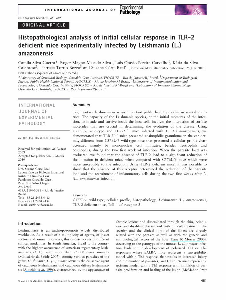
Figure 1 Ear lesions in C57BL ⁄ 6 WT and TLR-2) ⁄ ) mice 1 and2 weeks following intradermal inoculation of 2.5 · 105 Leish-mania (L.) amazonensis promastigotes. After the first week,C57BL ⁄ 6 WT (a) and TLR-2) ⁄ ) mice (c) present increased vas-cularization of the inoculation site. After the second week ofinfection, C57BL ⁄ 6 WT mice (b) presented lesions with littleulceration, while in TLR-2) ⁄ ) mice (d) the formation of smallnon-ulcerative nodular lesions were observed.
Figure 2 Diameter of induration following intradermal inocula-tion of 2.5 · 105 Leishmania (L.) amazonensis promastigotes inC57BL ⁄ 6 WT (¤) and TLR-2) ⁄ ) mice ( ). Values represent themean induration (millimetres) ±1 SD (10 mice ⁄ group). Kruskal–Wallis statistical test and a *P < 0.05 was considered signifi-cant.
452 C. S. Guerra et al.
� 2010 The Authors. Journal compilation � 2010 Blackwell Publishing Ltd, International Journal of Experimental Pathology, 91, 451–459
Intradermal inoculation and lesion measurement
Mice were sedated by an intraperitoneal injection with Com-
paz� (Cristalia, Sao Paulo, SP, Brasil) (Diazepam 5 mg ⁄ ml)
at a dosage of 5 mg ⁄ kg and Fentanyl� (Janssen-Cilag, Sao
Paulo, SP, Brasil) (Fentanyl citrate 78.5 lg ⁄ 10 ml) at a dos-
age of 0.02 mg ⁄ kg. A total of 250,000 metacyclic promastig-
otes of L. (L.) amazonensis were inoculated intradermally
into the ears of the animals in 10 ll of PBS. A group of mice
of each strain was inoculated only with 10 ll of PBS as con-
trol. The lesion developments were measured with a calliper
(Schnelltaster, HC Kroplin, GMBH, Hessen, Germany) and
ear thicknesses given in millimetres. After 1, 7 and 15 days
of intradermal inoculation, mice were killed in a CO2 cham-
ber and their ears were collected. Each experiment was car-
ried out three times and the same results were obtained.
Histological Analysis
Ears of control and infected animals were washed in PBS
and fixed in 10% buffered formalin. After fixation, samples
were routinely processed for paraffin embedding in an Auto-
matic Tissue Processor (Leica TP1020, Wetzlar, Germany).
Five micrometre thick sections were obtained in a Rotary
Microtome (Micron HM 360, Walldorf, Germany). The sec-
tions were stained with haematoxylin-eosin, differentiated
into 1% hydrochloric alcohol, stained with alcoholic eosin
1%, assembled with Entelan and analysed in a light micro-
scope (Zeiss Axioplan 2; Zeiss Inc., Thornwood, NY, USA).
Transmission Electron Microscopy
Ears were removed after 1, 7 and 15 days of infection, fixed in
2.5% glutaraldehyde in the buffer cacodylate sodium 0.1 M,
pH 7.2 with 3.5% sucrose and postfixed with 1% of osmium
tetroxide (OsO4) for 1 h at 4 �C. Then, they were dehydrated
in an acetone series and embedded in resin PolyBed 812. After
polymerization, semi-thin sections (0.5 lm) were stained with
toluidine blue and eosin and observed under a light micro-
scope (Zeiss Axioplan 2). The quantification of cellular profile
was made in five semi-thin sections with an area of 60 mm2
per ear in an average of 3–5 mice ⁄ group. After the choice of
(d)(a)
(b) (e)
(f)(c)
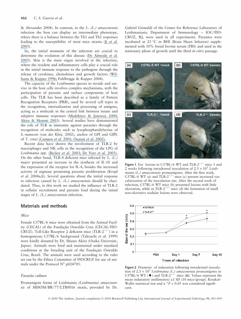
Figure 3 (a–f) – Histological analysis ofsections from the ear region ofC57BL ⁄ 6 WT mice and TLR-2) ⁄ ) miceafter 1, 7 and 15 days of Leishmania(L.) amazonensis infection stained withhaematoxylin-eosin (bar = 20lm). Onthe first day of infection the presence ofcongested blood vessels with marginali-zation and diapedesis of inflammatorycells ( ) in C57BL ⁄ 6 WT (a) andTLR-2) ⁄ ) mice (d) were seen. After firstweek of infection, the C57BL ⁄ 6 WTmice showed mononuclear ( ) andpolymorphonuclear ( ) cell infiltrationsin ear dermis (b) and second week afterinfection the presence of epidermalalterations such as exocytosis (inset)and dermis with an increase of intracel-lular amastigotes ( ) (c). In TLR-2) ⁄ )
mice during the first week of infectionsome polymorphonuclear cells ( ) wereseen (e). After the second week of infec-tion organized granulomas ( ) formedby eosinophils and without parasites(inset) were observed (f). The experi-ment is representative of three separateexperiments.
Leishmania infection in TLR-2) ⁄ ) mice 453
� 2010 The Authors. Journal compilation � 2010 Blackwell Publishing Ltd, International Journal of Experimental Pathology, 91, 451–459

(a) (d)
(e)(b)
(c) (f)
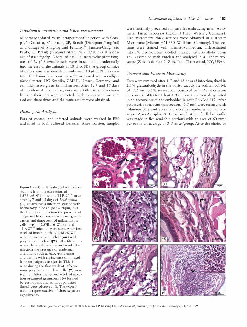
Figure 4 (a–f) – Semi-thin sections from the ear lesion of C57BL ⁄ 6 WT mice and TLR-2) ⁄ ) mice after 1, 7 and 15 days of Leish-mania (L.) amazonensis infection, stained with toluidine blue and eosin (bar = 20lm). On the first day of infection inflammatoryinfiltrates composed of neutrophils (inset), macrophages (MØ), degranulated mast cells in C57BL ⁄ 6 WT (a) and TLR-2) ⁄ ) mice (d)were observed. In the first week of infection, in C57BL ⁄ 6 WT mice (b) showed immature macrophages (iMØ) and eosinophils (Eos)composing the inflammatory infiltrate (inset) and free parasites in dermal ear (b). In the second week (c) an increase of inflammatoryinfiltrate predominantly composed of macrophages (MØ), as well neutrophils (Neu) and eosinophil (Eos) populations were seen. Inaddition to the presence of free amastigotes and a large amount of macrophages (MØ) containing amastigotes within large parasi-tophorous vacuoles (PV) (inset) and numerous free amastigotes in the matrix were observed. In TLR-2) ⁄ ) mice, during the first weekof infection (e), mast cells were observed between parasitized macrophages (MØ) and some free amastigotes in the matrix. In the sec-ond week of infection (f), organized granulomas formed predominantly of eosinophils (Eos), a large amount of macrophages (MØ),some mast cells and fibroblasts were seen. Also, few macrophages (MØ) with amastigotes in parasitophorous vacuoles (PV) werefound. The experiment is representative of three separate experiments.
454 C. S. Guerra et al.
� 2010 The Authors. Journal compilation � 2010 Blackwell Publishing Ltd, International Journal of Experimental Pathology, 91, 451–459
areas, the ultra-thin sections were prepared (ultramicrotome
Reichert OmU3), collected on copper grids of 300 mesh, con-
trasted with 5% uranyl acetate and lead citrate and observed
by a transmission electron microscope (Zeiss EM10C) of the
Oswald Cruz Institute electron microscopy Platform. Data
were obtained from three independent experiments.
Statistical analysis
The significance of the results was calculated by a non-para-
metric one-way analysis of variance test (Kruskal–Wallis)
and a P-value of <0.05 was considered significant.
(a) (d)
(b) (e)
(c) (f)
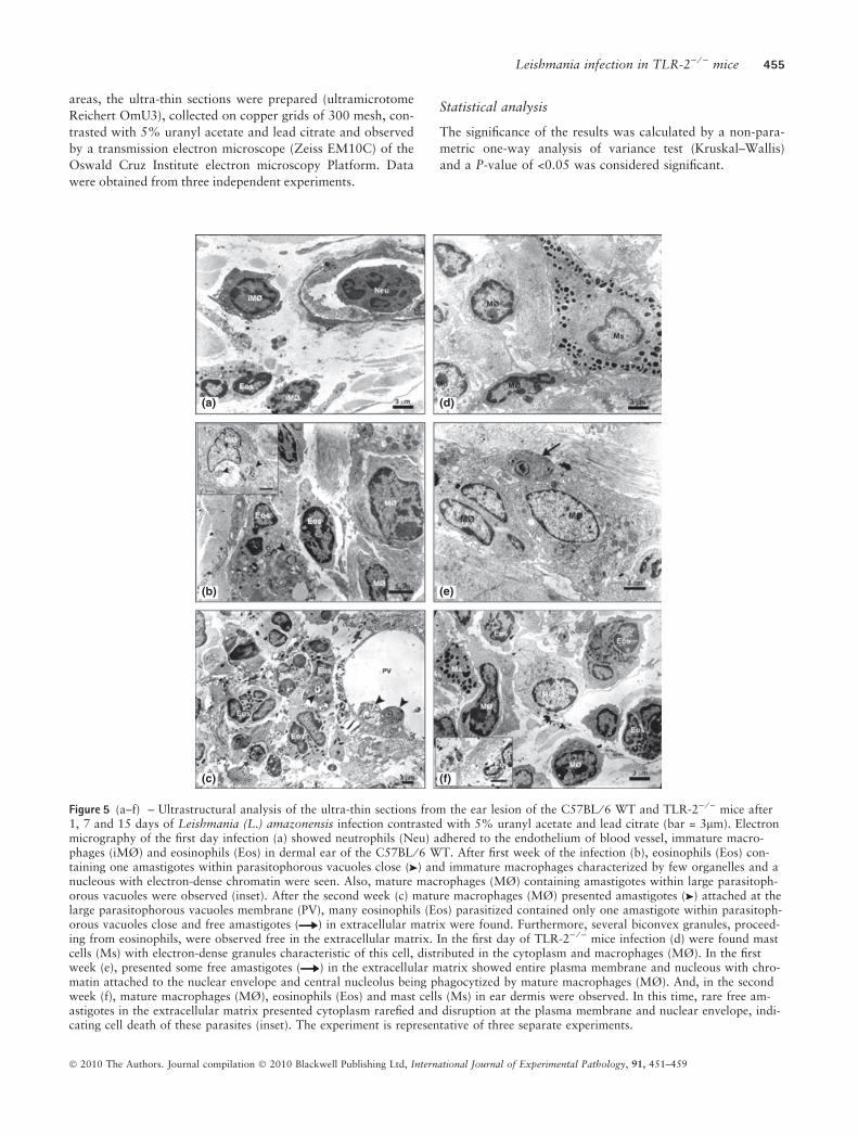
Figure 5 (a–f) – Ultrastructural analysis of the ultra-thin sections from the ear lesion of the C57BL ⁄ 6 WT and TLR-2) ⁄ ) mice after1, 7 and 15 days of Leishmania (L.) amazonensis infection contrasted with 5% uranyl acetate and lead citrate (bar = 3lm). Electronmicrography of the first day infection (a) showed neutrophils (Neu) adhered to the endothelium of blood vessel, immature macro-phages (iMØ) and eosinophils (Eos) in dermal ear of the C57BL ⁄ 6 WT. After first week of the infection (b), eosinophils (Eos) con-taining one amastigotes within parasitophorous vacuoles close ( ) and immature macrophages characterized by few organelles and anucleous with electron-dense chromatin were seen. Also, mature macrophages (MØ) containing amastigotes within large parasitoph-orous vacuoles were observed (inset). After the second week (c) mature macrophages (MØ) presented amastigotes ( ) attached at thelarge parasitophorous vacuoles membrane (PV), many eosinophils (Eos) parasitized contained only one amastigote within parasitoph-orous vacuoles close and free amastigotes ( ) in extracellular matrix were found. Furthermore, several biconvex granules, proceed-ing from eosinophils, were observed free in the extracellular matrix. In the first day of TLR-2) ⁄ ) mice infection (d) were found mastcells (Ms) with electron-dense granules characteristic of this cell, distributed in the cytoplasm and macrophages (MØ). In the firstweek (e), presented some free amastigotes ( ) in the extracellular matrix showed entire plasma membrane and nucleous with chro-matin attached to the nuclear envelope and central nucleolus being phagocytized by mature macrophages (MØ). And, in the secondweek (f), mature macrophages (MØ), eosinophils (Eos) and mast cells (Ms) in ear dermis were observed. In this time, rare free am-astigotes in the extracellular matrix presented cytoplasm rarefied and disruption at the plasma membrane and nuclear envelope, indi-cating cell death of these parasites (inset). The experiment is representative of three separate experiments.
Leishmania infection in TLR-2) ⁄ ) mice 455
� 2010 The Authors. Journal compilation � 2010 Blackwell Publishing Ltd, International Journal of Experimental Pathology, 91, 451–459
Results
Evolution of the dermal lesion in TLR-2) ⁄ )
Macroscopic analysis of C57BL ⁄ 6 wild-type (WT) and TLR-
2) ⁄ ) mice showed increased vascularization of the inocula-
tion site after the first week of L. (L.) amazonensis promasti-
gote infection. After the second week of infection, C57BL ⁄ 6WT mice presented lesions with little ulceration, while in
TLR-2) ⁄ ) mice the formation of a small nodule at the inocu-
lation site was observed (Figure 1). The evaluations of the
thickness of the inoculation site, after different times of
infection, are represented in the Figure 2.
Evolution of the cellular profile of dermal lesion inTLR-2) ⁄ )
Analysis of the control mice ears showed the presence of res-
ident cells of the dermis, whereas in PBS inoculated mice
only a mild inflammatory infiltrate was observed (data not
shown). On the first day of infection the presence of con-
gested blood vessels with marginalization and diapedesis of
inflammatory cells (Figure 3a,d) were seen in both C57BL ⁄ 6WT and TLR-2) ⁄ ) mice. In the dermal ear, inflammatory
infiltrates composed of neutrophils, macrophages, degranu-
lated mast cells were observed in both C57BL ⁄ 6 WT (Fig-
ures 4a and 5a) and TLR-2) ⁄ ) mice (Figures 4d and 5d).
In the first week of infection, the analysis of the inocula-
tion site in the C57BL ⁄ 6 WT mice showed inflammatory
infiltrates composed of mononuclear and polymorphonuclear
cells in the dermal ear (Figure 3b). In this infiltrate, many
eosinophils and neutrophils with a preponderance of mono-
cytes, immature and mature macrophages were observed.
Moreover, in these mice large amounts of parasitized cells
and free parasites in the extracellular matrix were observed
(Figure 4b). Ultrastructural analysis showed the presence of
eosinophils containing generally one amastigote in parasi-
tophorous vacuoles close in the dermal ear. Also, Immature
macrophages characterized by cytoplasm with few organelles
and nucleus with electron-dense chromatin and macrophages
containing several amastigotes within large parasitophorous
vacuoles were observed (Figure 5b). Moreover, in the TLR-
2) ⁄ ) mice a reduced infiltrate of inflammatory cells com-
posed mostly of immature and mature macrophages and
large amount of mast cells and fibroblasts were observed
(Figure 3e). Also a small amount of parasitized cells such as
macrophages, fibroblasts and rare free amastigotes in extra-
cellular matrix (Figures 4e and 5e) were seen.
During the second week of infection there was a signifi-
cant increase in the influx of inflammatory cell infiltrates in
the dermal ear in both mice strains, thus the cellular profile
for the initial response to infection by L. (L.) amazonensis
could be defined. In the C57BL ⁄ 6 WT mice, we observed
the presence of disorganized granuloma predominantly com-
posed of macrophages, as well as the presence of neutrophils
and eosinophils (Figures 3c and 4c). Also, a large amount of
macrophages containing amastigotes in large parasitophor-
ous vacuoles and eosinophils with only one amastigote in
the parasitophorous vacuoles close were observed. Further-
more, free amastigotes and biconvex eosinophils granules in
extracellular matrix were seen (Figure 5c). In these mice the
number of parasitized cells was significantly higher when
compared with the number of free amastigotes in extracellu-
lar matrix (Figure 6). In the ear dermis of TLR-2) ⁄ ) mice
there were organized granulomas (Figure 3f) formed pre-
dominantly of eosinophils and a large amount of macro-
phages and mast cells (Figures 4f and 5f). Differently to
what was found in WT mice, the TLR-2) ⁄ ) mice showed a
low parasite load, few eosinophils and parasitized macro-
phages and rare amastigotes free in extracellular matrix (Fig-
ures 4f and 6). An important observation was the presence
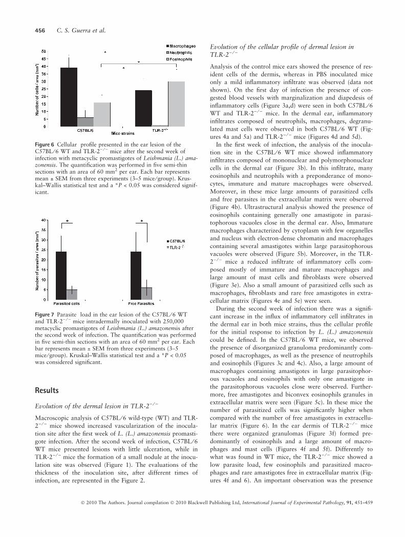
Figure 6 Cellular profile presented in the ear lesion of theC57BL ⁄ 6 WT and TLR-2) ⁄ ) mice after the second week ofinfection with metacyclic promastigotes of Leishmania (L.) ama-zonensis. The quantification was performed in five semi-thinsections with an area of 60 mm2 per ear. Each bar representsmean ± SEM from three experiments (3–5 mice ⁄ group). Krus-kal–Wallis statistical test and a *P < 0.05 was considered signif-icant.
Figure 7 Parasite load in the ear lesion of the C57BL ⁄ 6 WTand TLR-2) ⁄ ) mice intradermally inoculated with 250,000metacyclic promastigotes of Leishmania (L.) amazonensis afterthe second week of infection. The quantification was performedin five semi-thin sections with an area of 60 mm2 per ear. Eachbar represents mean ± SEM from three experiments (3–5mice ⁄ group). Kruskal–Wallis statistical test and a *P < 0.05was considered significant.
456 C. S. Guerra et al.
� 2010 The Authors. Journal compilation � 2010 Blackwell Publishing Ltd, International Journal of Experimental Pathology, 91, 451–459
of eosinophils near the infected macrophages in both mice
strains.
A quantitative evaluation of the cellular profile and the
parasite load in the second week of L. (L.) amazonensis
infection in C57BL ⁄ 6 and TLR-2) ⁄ ) mice is represented in
Figures 6 and 7 using the Kruskal–Wallis statistical test and
a P-value of <0.05 was considered significant.
Discussion
The first moments of infection by Leishmania are crucial to
drive the progress of the disease. There is evidence that the
phenotype of the leishmaniasis can be determined in the first
hours after infection, starting with the recognition of PAMPs
of the parasite by PRRs present on the surface of host cells,
such as TLRs (Launois et al. 1995; Sacks & Noben-Trauth
2002). The involvement of TLRs in the initial response to
infection with Leishmania have been described in recent
years, through various in vivo and in vitro studies (Hawn
et al. 2002; De Veer et al. 2003; Debus et al. 2003; De Trez
et al. 2004).
So, to evaluate the involvement of TLR-2 during in vivo
L. (L.) amazonensis infection TLR-2 deficient mice were
used. The ear dermis was chosen as the inoculation site
because, in addition to being a common transmission site in
rodent reservoirs, it offers the advantage that all the dynamic
events occur at the infection site, facilitating the study of the
initial inflammatory response (Belkaid et al. 1996, 1998).
The control group of mice, inoculated only with PBS,
showed a small inflammatory infiltrate composed of neu-
trophils, increase of vascularization and the presence of
oedema at the inoculation site. This initial inflammatory
response, generated by mechanical disruption of cells in the
epidermis and dermis was consistent with the results pre-
sented by Grimaldi and Moriearty (1981). Nonetheless, the
histological analysis of inoculation site after the first day of
L. (L.) amazonensis infection showed that the inflammatory
infiltrate in the ear dermis was higher when compared with
mice inoculated only with PBS. This fact is related to the
presence of free parasites in the extracellular matrix of the
ear dermis leading to activation of resident cells, through
recognition of parasite surface molecules by receptors pres-
ent in these cells (De Almeida et al. 2003).
Since the first days of infection, we observed the neutroph-
ils infiltration at the inoculation site in both mice strains,
being able to occur because of the disruption of the skin
caused by either the insect vector or the needle. This
mechanical injury induces the recruitment of these cells to
participate in the process of tissue repair, even in the
absence of the parasite (Peters et al. 2008).
The initial recruitment of neutrophils to the inoculation
site is supported by several studies that demonstrate the role
of these cells in the first line of defence against infection by
protozoa of the Leishmania genus, acting directly on the
endocytosis and destruction of the parasite by proteolytic
enzymes, production of reactive oxygen intermediates,
inflammatory mediators and cellular recruitment to the
infection site (Chang. 1981; Pimenta et al. 1987; Awasthi
et al. 2004).
C57BL ⁄ 6 WT mice showed significant increase of imma-
ture and mature macrophages at the infection site in first
week of infection. This high concentration of macrophages
at the inoculation site may be corroborated by Soong et al.
(1996, 1997) who showed that mice infected with L. (L.)
amazonensis are able to recruit a large quantity of immature
macrophages, but they are unable to eliminate the parasite
facilitating the spread of infection. Thus, the increased num-
ber of immature and mature macrophages found in
C57BL ⁄ 6 mice during the first week of infection may be
related to the maturation of recruited monocytes in the early
stages of the infection. These observations could explain the
presence of many macrophages parasitized with numerous
amastigotes within large parasitophorous vacuoles and the
presence of free amastigotes in the tissue from the disruption
of these cells.
Using TLR-2) ⁄ ) mice, we showed that the absence of this
receptor leads to an alteration of the cellular profile and an
expressive reduction in the susceptibility of these animals to
L. (L.) amazonensis infection, which are capable of control-
ling the parasite load during the first 2 weeks of infection.
TLR-2) ⁄ ) mice presented a lower infiltrate of inflammatory
cells at the inoculation site forming organized granulomas
mainly made up of eosinophils, unlike the C57BL/6 mice,
which had infiltrated more expressive, but no organized
granulomas were observed.
The eosinophils constitutively express few TLRs on their
surface and the direct activation of eosinophils through
TLR-2 is controversial (Sabroe et al. 2002; Nagase et al.
2003; D’Avila et al. 2007; Driss et al. 2009). Although a
role for TLR-2 in regulating eosinophils recruitment and
activation through direct and indirect mechanisms has been
described in bacterial and parasitic infections (D’Avila et al.
2007; Driss et al. 2009), similar to our observations, a lack
of impairment or increased recruitment of eosinophils in
TLR-2 deficient animals has been reported following ocular
filarial infection and also in a ear model of contact dermati-
tis (Daehnel et al. 2007; Jin et al. 2009). The tissue eosino-
philia presented in TLR-2) ⁄ ) mice is characteristic of a Th2
response, as observed with asthma and helminthic infections
(Del Prete. 1992; Mehlotra et al. 1998). However, a Th2
response would be favourable for parasite proliferation,
which was not observed in TLR-2 deficient mice. Of note,
eosinophils may participate in the process of killing parasites
through their ability to phagocytize, mount a respiratory
burst and mobilize cytotoxic proteins from specific granules
after infection, suggesting an immunomodulatory and in
some conditions protective role of eosinophils in infections
(Akuthota et al. 2008; Blanchard & Rothenberg 2009).
On the other hand, TLR-2) ⁄ ) mice showed a significant
reduction in the number of neutrophils during the second
week of infection when compared with C57BL ⁄ 6 WT mice.
This reduction can be related to the involvement of TLR-2
in the recruitment, activation and apoptosis of these cells
(Sabroe et al. 2005; Jablonska et al. 2006).
Leishmania infection in TLR-2) ⁄ ) mice 457
� 2010 The Authors. Journal compilation � 2010 Blackwell Publishing Ltd, International Journal of Experimental Pathology, 91, 451–459
Furthermore, we observed that these mice showed a low
parasite load from the first day of infection when compared
with C57BL ⁄ 6 WT mice. This fact indicates that these
receptors have an effective participation in the adhesion and
internalization of the parasite in the host cells present in the
initial stages of infection. The difference in the parasite load
observed in the second week of infection in TLR-2) ⁄ ) and
C57BL ⁄ 6 WT mice may be related to the association of
macrophages with eosinophils present at the inoculation site.
This association was described by Grimaldi et al. (1984)
who found that the eosinophils could serve as donors of per-
oxidase for mature macrophages. Moreover, these eosinoph-
ils may be acting in direct control of amastigotes free
through the release of extracellular peroxidase, can then be
adsorbed at plasma membrane of Leishmania by making it
more susceptible to death after phagocytosis by macrophages
as suggested by Grimaldi et al. (1984) and described by Pi-
menta et al. (1987).
The presence of mast cells in TLR-2) ⁄ ) and C57BL ⁄ 6 WT
mice from the first day of infection is supported by data
from the literature that show the presence of these cells in
the dermis during infections caused by protozoa of the genus
Leishmania and their direct participation in the initial
immune response through the production of several inflam-
matory mediators (Bidri et al. 1997; Saha et al. 2004). So,
we can suggest that mast cell degranulation, mainly seen in
TLR-2) ⁄ ) mice infected with L. (L.) amazonensis, is helping
to reduce the number of free amastigotes in the extracellular
matrix.
With these studies, we have demonstrated the importance
of TLR-2 in the initial response to L. (L.) amazonensis infec-
tion, where the absence of these receptors in initial stages of
infection favours the control of the parasite load. Thus, we
suggest the study of TLR pathways as an alternative for the
development of new medicines for the treatment of infec-
tions caused by L. (L.) amazonensis.
Acknowledgements
The authors would like to thank Andrea Henriques Pons for
helpful discussions. We thank Generval Luciano Batista,
Renata Correa Hespanhol and Vanessa de Souza Vaz for
their technical assistance.
This work was supported by grants from Instituto
Oswaldo Cruz-IOC ⁄ FIOCRUZ, Conselho Nacional de De-
senvolvimento Cientifico e Tecnologico (48.0629 ⁄ 2004-8)
and PAPES V-CNPq ⁄ FIOCRUZ (403642 ⁄ 2008-6).
References
Akira S. & Hemmi H. (2003) Recognition of pathogen-associated
molecular patterns by TLR family. Immunol. Lett. 85, 85–95.
Akuthota P., Wang H.B., Spencer L.A., Weller P.F. (2008) Immuno-
regulatory roles of eosinophils: a new look at a familiar cell. Clin.
Exp. Allergy 38, 1254–1263.
Almeida R.P., Barral-Netto M., De Jesus A.M., De Freitas L.A.,
Carvalho E.M., Barral A. (1996) Biological behavior of Leish-
mania amazonensis isolated from humans with cutaneous, muco-
sal, or visceral leishmaniasis in BALB ⁄ c mice. Am. J. Trop. Med.
Hyg. 54, 178–184.
Awasthi A., Mathur R.K., Saha B. (2004) Immune response to
Leishmania infection. Indian J. Med. Res. 119, 238–258.
Becker I., Salaiza N., Aguirre M. et al. (2003) Leishmania lipophos-
phoglycan (LPG) activates NK cells through Toll-like receptor 2.
Mol. Biochem. Parasitol. 130, 65–74.
Belkaid Y., Jouin H., Milon G. (1996) A method to recover, enu-
merate and identify lymphomyeloid cells present in an inflamma-
tory dermal site: a study in laboratory mice. J. Immunol.
Methods 199, 5.
Belkaid Y., Kamhawi S., Modi G. et al. (1998) Development of a
natural model of cutaneous leishmaniasis: powerful effects of vec-
tor saliva and saliva preexposure on the long-term outcome of
Leishmania major infection in the mouse ear dermis. J. Exp.
Med. 188, 1941.
Bidri M., Vouldoukis I., Mossalayi M.D. et al. (1997) Evidence for
direct interaction between mast cells and Leishmania parasites.
Parasite Immunol. 19, 475–483.
Blanchard C. & Rothenberg M.E. (2009) Biology of the eosinophil.
Adv. Immunol. 101, 81–121.
Campos M.A., Almeida I.C., Takeuchi O. et al. (2001) Activation
of toll-like receptor-2 by glycosylphosphatidylinositol anchors
from a protozoan parasite. J. Immunol. 167, 416–423.
Chang K.P. (1981) Leishmanicidal mechanisms of human polymor-
phonuclear phagocytes. Am. J. Trop. Med. Hyg. 30(2), 322–333.
Daehnel K., Gillette-Ferguson I., Hise A.G. et al. (2007)
Filaria ⁄ Wolbachia activation of dendritic cells and development
of Th1-associated responses is dependent on Toll-like receptor 2
in a mouse model of ocular onchocerciasis (river blindness). Para-
site Immunol. 29, 455–465.
D’Avila H., Almeida P.E., Roque N.R., Castro-Faria-Neto H.C., Bo-
zza P.T. (2007) Toll-like receptor-2-mediated C-C chemokine
receptor 3 and eotaxin-driven eosinophil influx induced by Myco-
bacterium bovis BCG pleurisy. Infect. Immun. 75, 1507–1511.
De Almeida M.C., Vilhena V., Barral A., Barral-Netto M. (2003)
Leishmanial infection: analysis of its first steps. A review. Mem.
Inst. Oswaldo Cruz 98, 861–870.
De Trez C., Brait M., Leo O. et al. (2004) Myd88-dependent in vivo
maturation of splenic dendritic cells induced by Leishmania dono-
vani and other Leishmania species. Infect. Immun. 72, 824–832.
De Veer M.J., Curtis J.M., Baldwin T.M. et al. (2003) MyD88 is
essential for clearance of Leishmania major: possible role for lipo-
phosphoglycan and Toll-like receptor 2 signaling. Eur. J. Immu-
nol. 33, 2822–2831.
Debus A., Glasner J., Rollinghoff M., Gessner A. (2003) High levels
of susceptibility and T helper 2 response in MyD88-deficient mice
infected with Leishmania major are interleukin-4 dependent.
Infect. Immun. 71, 7215–7218.
Del Prete G. (1992) Human Th1 and Th2 lymphocytes: their role in
the pathophysiology of atopy. Allergy 47(5), 450–455.
Driss V., Legrand F., Hermann E. et al. (2009) TLR2-dependent
eosinophil interactions with mycobacteria: role of alpha-defensins.
Blood 113, 3235–3244.
Fuhlbrigge R.C. & Kupper T.S. (2004) Immune surveillance in the
skin: mechanisms and clinical consequences. Nat. Rev. Immunol.
4, 211–222.
Grimaldi Jr G. & Moriearty P.L. (1981) Kinetics and histopathology
of the ear thickness test for delayed hypersensitivity in murine
leishmaniasis. Rev. Inst. Med. Trop. Sao Paulo 23, 127–132.
458 C. S. Guerra et al.
� 2010 The Authors. Journal compilation � 2010 Blackwell Publishing Ltd, International Journal of Experimental Pathology, 91, 451–459
Grimaldi Jr G., Soares M.J., Moriearty P.L. (1984) Tissue eosino-
philia and Leishmania mexicana mexicana eosinophil interactions
in murine cutaneous leishmaniasis. Parasite Immunol. 6, 397–408.
Hawn T.R., Ozinsky A., Underhill D.M., Buckner F.S., Akira S.,
Aderem A. (2002) Leishmania major activates IL-1a expression in
macrophages through a MyD88-dependent pathway. Microbes
Infect. 4(8), 763–771.
Jablonska E., Marcinczyk M., Jablonski J. (2006) Toll-like receptors
types 2 and 6 and the apoptotic process in human neutrophils.
Arch. Immunol. Ther. Exp. (Warsz) 54, 137–142.
Ji J., Sun J., Soong L. (2003) Impaired expression of inflammatory
cytokines and chemokines at early stages of infection with Leish-
mania amazonensis. Infect. Immun. 71, 4278–4288.
Jin H., Kumar L., Mathias C. et al. (2009) Toll-like receptor 2 is
important for the T(H)1 response to cutaneous sensitization. J.
Allergy Clin. Immunol. 123, 875–882.
Kane M.M. & Mosser D.M. (2000) Leishmania parasites and their
ploys to disrupt macrophage activation. Curr. Opin. Hematol. 7,
26–31.
Kropf P., Freudenberg M.A., Modolell M. et al. (2004a) Toll-like
receptor 4 contributes to efficient control of infection with the pro-
tozoan parasite Leishmania major. Infect. Immun. 72, 1920–1928.
Kropf P., Freudenberg N., Kalis C. et al. (2004b) Infection of
C57BL ⁄ 10ScCr and C57BL ⁄ 10ScNCr mice with Leishmania
major reveals a role for Toll-like receptor 4 in the control of par-
asite replication. J. Leukoc. Biol. 76, 48–57.
Launois P., Ohteki T., Swihart K., MacDonald H.R., Louis J.A.
(1995) In susceptible mice, Leishmania major induce very rapid
interleukin-4 production by CD4 + T cells which are NK1.1).
Eur. J. Immunol. 25, 3298–3307.
McMahon-Pratt D. & Alexander J. (2004) Does the Leishmania
major paradigm of pathogenesis and protection hold for New
World cutaneous leishmaniases or the visceral disease? Immunol.
Rev. 201, 206–224.
Medzhitov R. & Janeway C.A. (2000) The Toll receptor family and
microbial recognition. Trends Microbiol. 8, 452–456.
Mehlotra R.K., Hall L.R., Higgins A.W. et al. (1998) Interleukin-12
suppresses filaria-induced pulmonary eosinophilia, deposition of
major basic protein and airway hyperresponsiveness. Parasite
Immunol. 20, 455–462.
Ministerio da Saude - Secretaria de Vigilancia em Saude (2007)
Manual de Vigilancia da Leishmaniose Tegumentar Americana.
Brasılia, Brasil: Ministerio da Saude. pp. 17.
Nagase H., Okugawa S., Ota Y. et al. (2003) Expression and func-
tion of Toll-like receptors in eosinophils: activation by Toll-like
receptor 7 ligand. J. Immunol. 171, 3977–3982.
Ouaissi A., Guilvard E., Delneste Y. et al. (2002) The Trypanosoma
cruzi Tc-52-released protein induces human dendritic cell matura-
tion, signals via toll-like receptor 2, and confers protection against
lethal infection. J. Immunol. 168, 6366–6374.
Peters N.C., Egen J.G., Secundino N. et al. (2008). In vivo imaging
reveals an essential role for neutrophils in leishmaniasis transmit-
ted by sand flies. Science. 321: 970–974. Erratum in: Science 12;
322 (5908):1634.
Pimenta P.F.P., Dos Santos M.A.V., De Souza W. (1987) Fine
estructure and cytochemistry of the interaction between Leish-
mania mexicana amazonensis and rat neutrophils and eosinophils.
J. Submicrosc. Cytol. 19, 387–395.
Sabroe I., Jones E.C., Usher L.R., Whyte M.K., Dower S.K. (2002)
Toll-like receptor (TLR)2 and TLR4 in human peripheral blood
granulocytes: a critical role for monocytes in leukocyte lipopoly-
saccharide responses. J. Immunol. 168, 4701–4710.
Sabroe I., Dower S.K., Whyte M.K. (2005) The role of Toll-like
receptors in the regulation of neutrophil migration, activation,
and apoptosis. Clin. Infect. Dis. 41(Suppl 7), S421–S426.
Sacks D.L. & Noben-Trauth N. (2002) The immunology of suscep-
tibility and resistance to Leishmania major mice. Nat. Rev.
Immunol. 2, 845–858.
Saha B., Tonkal A.M., Croft S., Roy S. (2004) Mast cells at the
host-pathogen interface: host-protection versus immune evasion in
leishmaniasis. Clin. Exp. Immunol. 137, 19–23.
Soong L., Xu J.C., Grewal I.S. et al. (1996) Disruption of CD40-
CD40 ligand interactions results in an enhanced susceptibility to
Leishmania amazonensis infection. Immunity 4, 263–273.
Soong L., Chang C.H., Sun J. et al. (1997) Role of CD4 + T cells
in pathogenesis associated with Leishmania amazonensis infec-
tion. J. Immunol. 158, 5374–5383.
Takeuchi O., Hoshino K., Kawai T. et al. (1999) Differential roles
of TLR2 and TLR4 in recognition of gram-negative and gram-
positive bacterial cell wall components. Immunity 11, 443–451.
van der Kleij D., Latz E., Brouwers J.F., Kruize Y.C., Schmitz M.,
Kurt-Jones E.A., Espevik T., de Jong E.C., Kapsenberg M.L., Go-
lenbock D.T., Tielens A.G., Yazdanbakhsh M. (2002) A novel
host-parasite lipid cross talk: schistosomal lyso-phosphatidylserine
activates Toll-like receptor-2 and effects immune polarization. J.
Biol. Chem. 277, 48122–48129.
Williams I.R. & Kupper T.S. (1996) Immunity at the surface:
homeostatic mechanisms of the skin immune system. Life Sci. 58,
1485–1507.
Leishmania infection in TLR-2) ⁄ ) mice 459
� 2010 The Authors. Journal compilation � 2010 Blackwell Publishing Ltd, International Journal of Experimental Pathology, 91, 451–459