Leishmania/HIV co-infections in the second decade
32
Leishmania/HIV co-infections in the second decade Israel Cruz, Javier Nieto, Javier Moreno , Carmen Cañavate, Philippe Desjeux* & Jorge Alvar** WHO Collaborating Centre for Leishmaniasis, National Centre of Microbiology, Institute of Health Carlos III, Madrid, Spain; Centre for Biological Research, CSIC, Madrid, Spain, *One World Health, San Francisco, USA & **Neglected Tropical Diseases, World Health Organization, Geneva, Switzerland Received May 25, 2005 Leishmania-HIV co-infection has been globally controlled in Southern Europe since 1997 because of highly active anti retroviral therapy (HAART), but it appears to be an increasing problem in other countries such as Ethopia, Sudan, Brazil or India where both infections are becoming more and more prevalent. Most of the scientific background on Leishmania/HIV co-infection has been dropped from the Mediterranean experience and although the situations among countries are not fully comparable, it is of high importance to take advantage of this knowledge. In this review several aspects of the Leishmania/ HIV co-infection are emphasized viz., epidemiological features, new ways of transmission, pathogenesis, clinical outcome, diagnosis, treatment and secondary prohylaxis. An extensive review of the literature on Leishmania/HIV co-infection has allowed the inclusion of a comprehensive and updated list of bibliographical references. Key words Co-infection - HIV - immunotherapy - Leishmania - pathogenesis The development of the HIV/AIDS pandemic during the last 20 yr has modified the spectrum of leishmaniasis in both the clinical and epidemiological fields. From middle of the 1980s, when the first case of leishmaniasis associated with human immunodeficiency virus (HIV) infection was published 1 , an increase in the cases of co-infection has been registered; to date cases in 35 countries have been detected 2 (Fig. 1). The increase in the number of cases of co- infection arises from the overlap between the AIDS epidemic, basically from urban transmission, and the areas in which leishmaniasis is endemic, Indian J Med Res 123, March 2006, pp 357-388 357 fundamentally from rural transmission. Epidemiological changes, such as the increase of the population in suburban areas where the vector and the reservoir abound, have increased this overlap. To date, the greatest prevalence of Leishmania/ HIV co-infection has been in the Mediterranean basin. Of more than 2,000 cases notified to the WHO, 90 per cent of them come from Spain, Italy, France and Portugal 2-5 . From the beginning of the AIDS epidemic up to the era of highly active antiretroviral therapy (HAART), between 25-70 per cent of all the cases of visceral leishmaniasis (VL) in these countries were associated with HIV infection, and it Review Article
Transcript of Leishmania/HIV co-infections in the second decade

Leishmania/HIV co-infections in the second decade
Israel Cruz, Javier Nieto, Javier Moreno , Carmen Cañavate, Philippe Desjeux* & Jorge Alvar**
WHO Collaborating Centre for Leishmaniasis, National Centre of Microbiology, Institute ofHealth Carlos III, Madrid, Spain; Centre for Biological Research, CSIC, Madrid, Spain, *One World Health,San Francisco, USA & **Neglected Tropical Diseases, World Health Organization, Geneva, Switzerland
Received May 25, 2005
Leishmania-HIV co-infection has been globally controlled in Southern Europe since 1997 becauseof highly active anti retroviral therapy (HAART), but it appears to be an increasing problem inother countries such as Ethopia, Sudan, Brazil or India where both infections are becoming moreand more prevalent. Most of the scientific background on Leishmania/HIV co-infection has beendropped from the Mediterranean experience and although the situations among countries are notfully comparable, it is of high importance to take advantage of this knowledge. In this reviewseveral aspects of the Leishmania/HIV co-infection are emphasized viz., epidemiological features,new ways of transmission, pathogenesis, clinical outcome, diagnosis, treatment and secondaryprohylaxis. An extensive review of the literature on Leishmania/HIV co-infection has allowed theinclusion of a comprehensive and updated list of bibliographical references.
Key words Co-infection - HIV - immunotherapy - Leishmania - pathogenesis
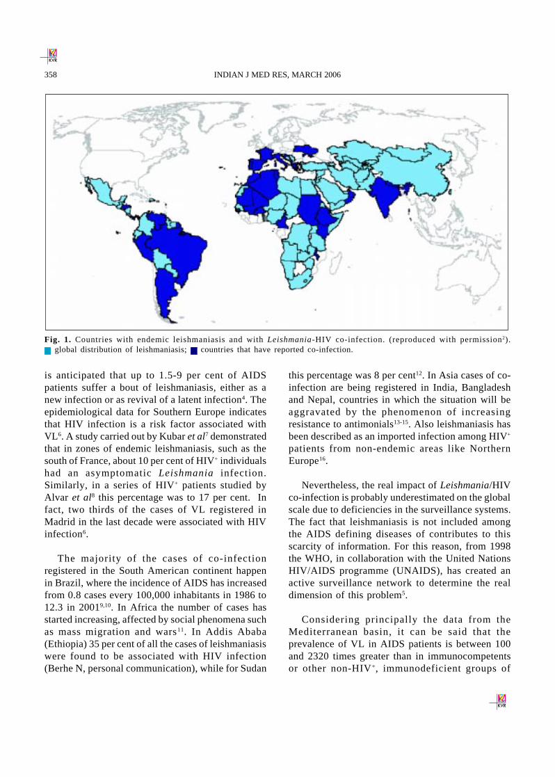
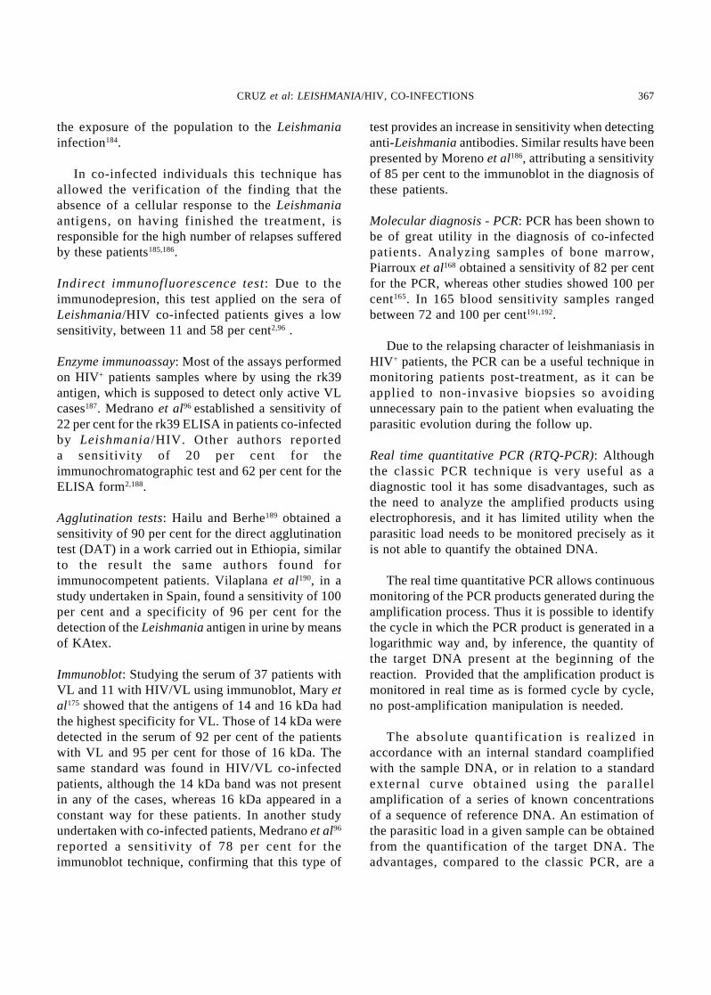
The development of the HIV/AIDS pandemicduring the last 20 yr has modified the spectrum ofleishmaniasis in both the clinical and epidemiologicalfields. From middle of the 1980s, when the first caseof leishmaniasis associated with humanimmunodeficiency virus (HIV) infection waspublished1, an increase in the cases of co-infectionhas been registered; to date cases in 35 countries havebeen detected2 (Fig. 1).
The increase in the number of cases of co-infection arises from the overlap between the AIDSepidemic, basically from urban transmission, and theareas in which leishmaniasis is endemic,
Indian J Med Res 123, March 2006, pp 357-388
357
fundamentally from rural transmission.Epidemiological changes, such as the increase of thepopulation in suburban areas where the vector andthe reservoir abound, have increased this overlap.
To date, the greatest prevalence of Leishmania/HIV co-infection has been in the Mediterraneanbasin. Of more than 2,000 cases notified to the WHO,90 per cent of them come from Spain, Italy, Franceand Portugal2-5. From the beginning of the AIDSepidemic up to the era of highly active antiretroviraltherapy (HAART), between 25-70 per cent of all thecases of visceral leishmaniasis (VL) in thesecountries were associated with HIV infection, and it
Review Article
is anticipated that up to 1.5-9 per cent of AIDSpatients suffer a bout of leishmaniasis, either as anew infection or as revival of a latent infection4. Theepidemiological data for Southern Europe indicatesthat HIV infection is a risk factor associated withVL 6. A study carried out by Kubar et al7 demonstratedthat in zones of endemic leishmaniasis, such as thesouth of France, about 10 per cent of HIV+ individualshad an asymptomatic Leishmania infection.Similarly, in a series of HIV+ patients studied byAlvar et al8 this percentage was to 17 per cent. Infact, two thirds of the cases of VL registered inMadrid in the last decade were associated with HIVinfection6.
The majority of the cases of co-infectionregistered in the South American continent happenin Brazil, where the incidence of AIDS has increasedfrom 0.8 cases every 100,000 inhabitants in 1986 to12.3 in 20019,10. In Africa the number of cases hasstarted increasing, affected by social phenomena suchas mass migration and wars11. In Addis Ababa(Ethiopia) 35 per cent of all the cases of leishmaniasiswere found to be associated with HIV infection(Berhe N, personal communication), while for Sudan
this percentage was 8 per cent12. In Asia cases of co-infection are being registered in India, Bangladeshand Nepal, countries in which the situation will beaggravated by the phenomenon of increasingresistance to antimonials13-15. Also leishmaniasis hasbeen described as an imported infection among HIV+
patients from non-endemic areas like NorthernEurope16.
Nevertheless, the real impact of Leishmania/HIVco-infection is probably underestimated on the globalscale due to deficiencies in the surveillance systems.The fact that leishmaniasis is not included amongthe AIDS defining diseases of contributes to thisscarcity of information. For this reason, from 1998the WHO, in collaboration with the United NationsHIV/AIDS programme (UNAIDS), has created anactive surveillance network to determine the realdimension of this problem5.
Considering principally the data from theMediterranean basin, it can be said that theprevalence of VL in AIDS patients is between 100and 2320 times greater than in immunocompetentsor other non-HIV+, immunodeficient groups of
358 INDIAN J MED RES, MARCH 2006
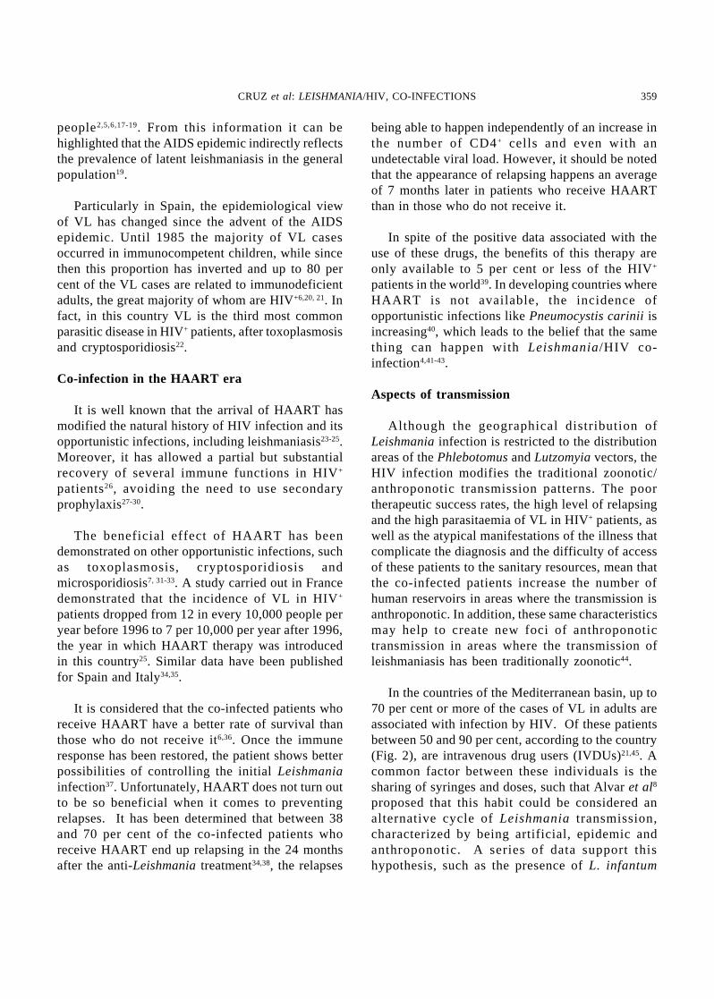
Fig. 1. Countries with endemic leishmaniasis and with Leishmania-HIV co-infection. (reproduced with permission2). global distribution of leishmaniasis; countries that have reported co-infection.
people2,5,6,17-19. From this information it can behighlighted that the AIDS epidemic indirectly reflectsthe prevalence of latent leishmaniasis in the generalpopulation19.
Particularly in Spain, the epidemiological viewof VL has changed since the advent of the AIDSepidemic. Until 1985 the majority of VL casesoccurred in immunocompetent children, while sincethen this proportion has inverted and up to 80 percent of the VL cases are related to immunodeficientadults, the great majority of whom are HIV+6,20, 21. Infact, in this country VL is the third most commonparasitic disease in HIV+ patients, after toxoplasmosisand cryptosporidiosis22.
Co-infection in the HAART era
It is well known that the arrival of HAART hasmodified the natural history of HIV infection and itsopportunistic infections, including leishmaniasis23-25.Moreover, it has allowed a partial but substantialrecovery of several immune functions in HIV+
patients26, avoiding the need to use secondaryprophylaxis27-30.
The beneficial effect of HAART has beendemonstrated on other opportunistic infections, suchas toxoplasmosis, cryptosporidiosis andmicrosporidiosis7, 31-33. A study carried out in Francedemonstrated that the incidence of VL in HIV+
patients dropped from 12 in every 10,000 people peryear before 1996 to 7 per 10,000 per year after 1996,the year in which HAART therapy was introducedin this country25. Similar data have been publishedfor Spain and Italy34,35.
It is considered that the co-infected patients whoreceive HAART have a better rate of survival thanthose who do not receive it6,36. Once the immuneresponse has been restored, the patient shows betterpossibilities of controlling the initial Leishmaniainfection37. Unfortunately, HAART does not turn outto be so beneficial when it comes to preventingrelapses. It has been determined that between 38and 70 per cent of the co-infected patients whoreceive HAART end up relapsing in the 24 monthsafter the anti-Leishmania treatment34,38, the relapses
being able to happen independently of an increase inthe number of CD4+ cells and even with anundetectable viral load. However, it should be notedthat the appearance of relapsing happens an averageof 7 months later in patients who receive HAARTthan in those who do not receive it.
In spite of the positive data associated with theuse of these drugs, the benefits of this therapy areonly available to 5 per cent or less of the HIV+
patients in the world39. In developing countries whereHAART is not available, the incidence ofopportunistic infections like Pneumocystis carinii isincreasing40, which leads to the belief that the samething can happen with Leishmania/HIV co-infection4,41-43.
Aspects of transmission
Although the geographical distribution ofLeishmania infection is restricted to the distributionareas of the Phlebotomus and Lutzomyia vectors, theHIV infection modifies the traditional zoonotic/anthroponotic transmission patterns. The poortherapeutic success rates, the high level of relapsingand the high parasitaemia of VL in HIV+ patients, aswell as the atypical manifestations of the illness thatcomplicate the diagnosis and the difficulty of accessof these patients to the sanitary resources, mean thatthe co-infected patients increase the number ofhuman reservoirs in areas where the transmission isanthroponotic. In addition, these same characteristicsmay help to create new foci of anthroponotictransmission in areas where the transmission ofleishmaniasis has been traditionally zoonotic44.
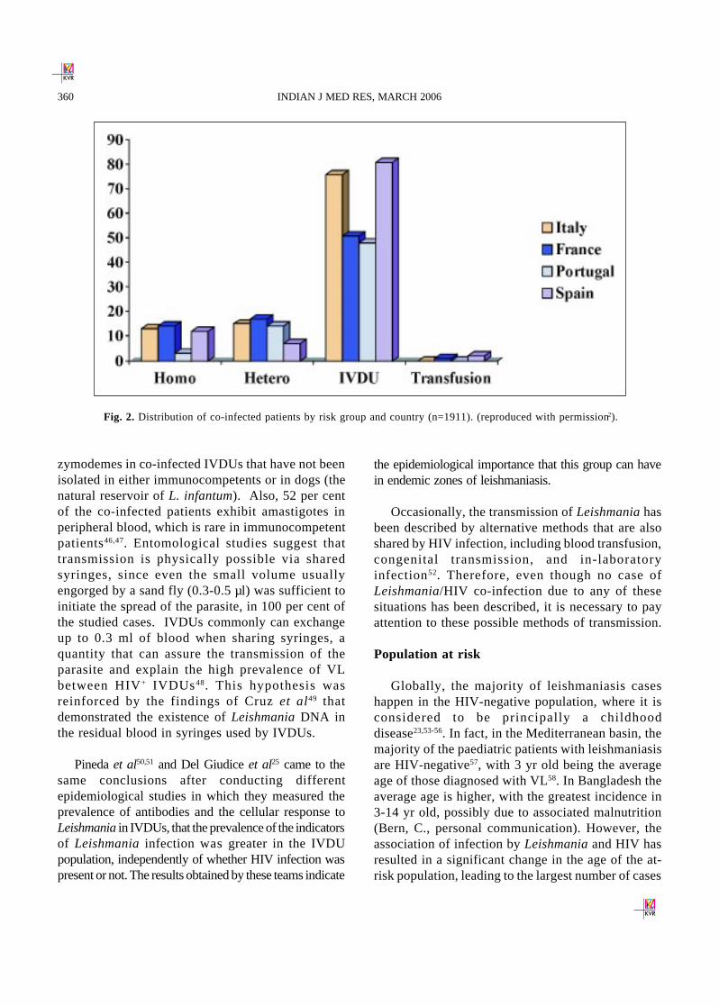
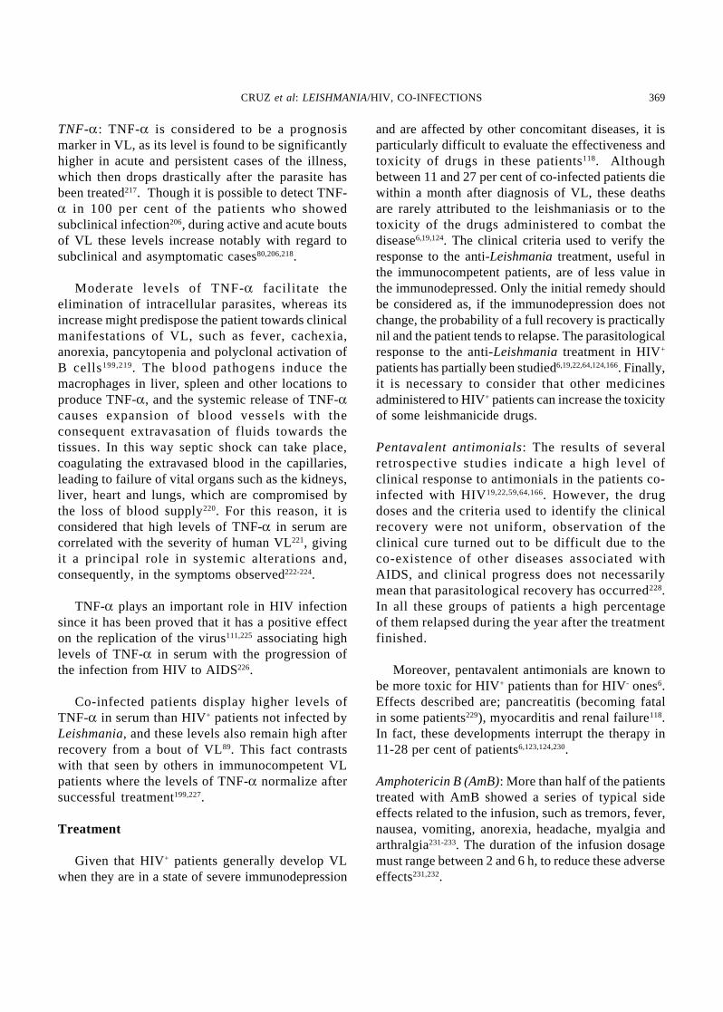
In the countries of the Mediterranean basin, up to70 per cent or more of the cases of VL in adults areassociated with infection by HIV. Of these patientsbetween 50 and 90 per cent, according to the country(Fig. 2), are intravenous drug users (IVDUs)21,45. Acommon factor between these individuals is thesharing of syringes and doses, such that Alvar et al8
proposed that this habit could be considered analternative cycle of Leishmania transmission,characterized by being artificial, epidemic andanthroponotic. A series of data support thishypothesis, such as the presence of L. infantum
CRUZ et al: LEISHMANIA/HIV, CO-INFECTIONS 359
zymodemes in co-infected IVDUs that have not beenisolated in either immunocompetents or in dogs (thenatural reservoir of L. infantum). Also, 52 per centof the co-infected patients exhibit amastigotes inperipheral blood, which is rare in immunocompetentpatients46,47. Entomological studies suggest thattransmission is physically possible via sharedsyringes, since even the small volume usuallyengorged by a sand fly (0.3-0.5 µl) was sufficient toinitiate the spread of the parasite, in 100 per cent ofthe studied cases. IVDUs commonly can exchangeup to 0.3 ml of blood when sharing syringes, aquantity that can assure the transmission of theparasite and explain the high prevalence of VLbetween HIV+ IVDUs48. This hypothesis wasreinforced by the findings of Cruz et al49 thatdemonstrated the existence of Leishmania DNA inthe residual blood in syringes used by IVDUs.
Pineda et al50,51 and Del Giudice et al25 came to thesame conclusions after conducting differentepidemiological studies in which they measured theprevalence of antibodies and the cellular response toLeishmania in IVDUs, that the prevalence of the indicatorsof Leishmania infection was greater in the IVDUpopulation, independently of whether HIV infection waspresent or not. The results obtained by these teams indicate
the epidemiological importance that this group can havein endemic zones of leishmaniasis.
Occasionally, the transmission of Leishmania hasbeen described by alternative methods that are alsoshared by HIV infection, including blood transfusion,congenital transmission, and in-laboratoryinfection52. Therefore, even though no case ofLeishmania/HIV co-infection due to any of thesesituations has been described, it is necessary to payattention to these possible methods of transmission.
Population at risk
Globally, the majority of leishmaniasis caseshappen in the HIV-negative population, where it isconsidered to be principally a childhooddisease23,53-56. In fact, in the Mediterranean basin, themajority of the paediatric patients with leishmaniasisare HIV-negative57, with 3 yr old being the averageage of those diagnosed with VL58. In Bangladesh theaverage age is higher, with the greatest incidence in3-14 yr old, possibly due to associated malnutrition(Bern, C., personal communication). However, theassociation of infection by Leishmania and HIV hasresulted in a significant change in the age of the at-risk population, leading to the largest number of cases
Fig. 2. Distribution of co-infected patients by risk group and country (n=1911). (reproduced with permission2).
360 INDIAN J MED RES, MARCH 2006
in people aged between 31-50 yr currently, coincidingwith the predominant age of the principal at-riskgroup, the IVDUs2.
In the southwest of Europe, 65 per cent of the HIV-
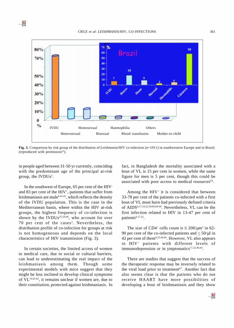
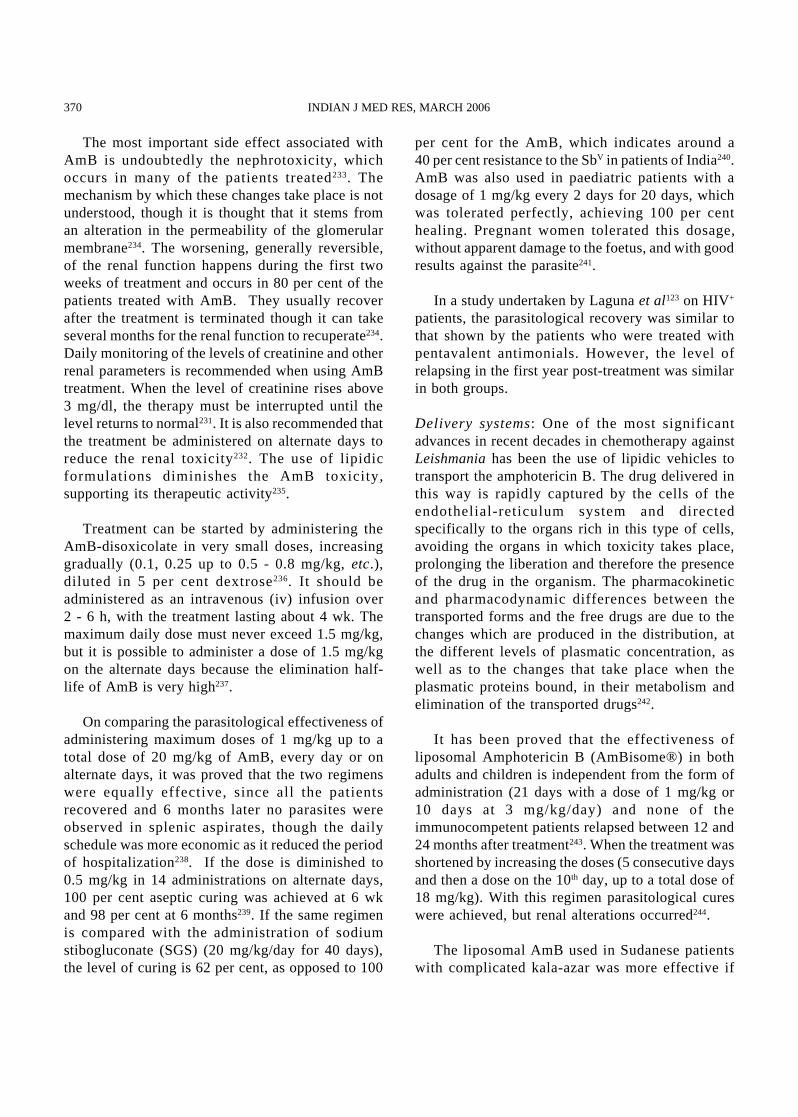
and 83 per cent of the HIV+, patients that suffer fromleishmaniasis are male6,42,43, which reflects the densityof the IVDU population. This is the case in theMediterranean basin, where within the HIV at-riskgroups, the highest frequency of co-infection isshown by the IVDUs25,59,60, who account for over70 per cent of the cases2. Nevertheless, thedistribution profile of co-infection for groups at riskis not homogeneous and depends on the localcharacteristics of HIV transmission (Fig. 3).
In certain societies, the limited access of womento medical care, due to social or cultural barriers,can lead to underestimating the real impact of theleishmaniasis among them. Though someexperimental models with mice suggest that theymight be less inclined to develop clinical symptomsof VL55,61,62, it remains unclear if women are, due totheir constitution, protected against leishmaniasis. In
fact, in Bangladesh the mortality associated with about of VL is 15 per cent in women, while the samefigure for men is 5 per cent, though this could beassociated with poor access to medical resources63.
Among the HIV+ it is considered that between33-78 per cent of the patients co-infected with a firstbout of VL must have had previously defined criteriaof AIDS6,17,19,22,50,60,64-66. Nevertheless, VL can be thefirst infection related to HIV in 13-47 per cent ofpatients6,17,22.
The size of CD4+ cells count is £ 200/µm3 in 62-90 per cent of the co-infected patients and < 50/µl in42 per cent of them6,25,46,66. However, VL also appearsin HIV + patients with different levels ofimmunodepression or in ymptomatics17,22,46,64.
There are studies that suggest that the success ofthe therapeutic response may be inversely related tothe viral load prior to treatment67. Another fact thatalso seems clear is that the patients who do notreceive HAART have more possibil it ies ofdeveloping a bout of leishmaniasis and they show
Fig. 3. Comparison by risk group of the distribution of Leishmania/HIV co-infection (n=1911) in southwestern Europe and in Brazil.(reproduced with permission10).
CRUZ et al: LEISHMANIA/HIV, CO-INFECTIONS 361
IVDU Homosexual Haemophilia Others
Heterosexual Bisexual Blood transfusion Mother-to-child
great risk of therapeutic failure, as well as of clinicaland parasitological relapse6,46,68.
Given that a good cellular immune response iscritical for the control of the infection of Leishmania,two mechanisms have been proposed by which theimmunodepresion might allow the appearance of aclinical VL bout16,46: (i) allowing the revival of alatent infection, as happens with a large proportionof the opportunistic infections associated with AIDS;and (ii ) facilitating the appearance of active VL afterthe initial infection.
Microbiological aspects
The majority of the cases of Leishmania/HIV co-infection has been described in adults infected byHIV-1; however, they have also been reported inpatients infected by HIV-246. In these patients, theVL is caused principally by L. infantum orL. donovani46. Other species of Leishmania, such asL. braziliensis69,73, L. aethiopica74, L.tropica75,76, andL. major77, have been described as responsible for casesof co-infection, according to the geographical areaconcerned.
Three important considerations can be derivedfrom the microbiological aspects of Leishmania/HIVco-infection: (i) a high variability of L. infantumzymodemes affecting co-infected people exists78;(ii ) several new zymodemes have been describedexclusively in HIV+ patients, which have not beendiscovered in immunocompetents or dogs of the samegeographical area46,47; and (iii ) the anergy state ofHIV + patients allows cutaneous Leishmaniazymodemes to visceralize and viceversa79. Variousdermotropic zymodemes of L. infantum, as well asof L. braziliensis, L. mexicana and L. amazonensis,have been described as causes of VL in HIV+
patients80-83. In addition, some visceral tropic variantsof L. infantum84 and L. chagasi85,86 have been foundin cutaneous lesions, and even in healthy skin of HIV+
patients87.
Pathogenesis of the Leishmania/HIV co-infection
Both infectious agents, in addition to producingsimilar effects in the host, share target cells. Leishmania
infects and multiplies inside the macrophages, and theHIV can also invade and replicate in these cells, inaddition to T CD4+ cells88,89. The presence of bothmicroorganisms in the same cellular type can haveimportant implications in their expression and spread90.Both infections change the predominant cellularimmune response of Th1 or Th0 to Th2 throughcomplex mechanisms mediated by cytokines,conferring susceptibility to both infections, accordingto the murine model91. Among the cytokines changesit has been recorded is an inhibition in the productionof inferon gamma (IFN-r), which causes a deficiencyin the leishmanicidal capacity of the macrophages92,disabling their ability to eliminate the intracellularamastigotes by means of nitric oxide’s toxic action93.The effects of the viral infection tend to predominateover those caused by the parasite, and theimmunodepresion induced by the HIV prevails over thecellular response caused by Leishmania46. The depletionof T CD4+ cells related to the HIV infection implies aloss of T cells capable of recognizing Leishmaniaantigens and of stimulating the B-lymphocytes. Thiswould explain the high valuation of false negatives inLeishmania serology in co-infected patients, in contrastto those found in immunocompetent patients, whopresent a strong specific humoral immuneresponse94-96. HIV also provokes an inhibition of theproliferative response against Leishmania, favouring thespread of the parasite, so that parasites can beencountered in atypical locations, and high parasitaemiais seen in these patients.
It has been observed that infection by Leishmaniaincreases the replication of the HIV, as much in invitro cultures as in co-infected individuals89,principally due to chronic activation of the immunesystem97,98, which is one of the principal determinantsof the progression of the illness caused by the HIV.The immune activation increases the expression ofviral co-receptors (CCR5 and CXR4), diminishes thesecretion of B cytokines99,100, and favours the entry,integration, formation and release of viral particles101.This brings with it an increase in the secretion oftumour necrosis factor-alpha (TNF-a), interleukinsIL-2, IL-4, IL-6, and IL-10 and affects the cellularcycle102,103. All this, therefore, results in a progressionof the immunodeficiency and a decline in thepatient’s survival rate104-109.
362 INDIAN J MED RES, MARCH 2006
Compared to HIV+ patients without leishmaniasis,co-infected patients show a cytokine profile with highlevels of IL-4, IL-10 and IL-2 receptor (IL-2R), anda decrease in the production (post-stimulation) ofIFN-γ97,98,110.
It has been proved that the lipophosphoglicon(LPG) of L. infantum can induce the expression ofHIV in mononuclear cells of peripheral bloodinfected in a latent manner111, probably due to thesecretion of TNF-a112,113. The initiation of theexpression of HIV has been suggested fromobservations that mark a progressive increase of theload of viral ribonucleic acid (RNA) in co-infectedpatients, associated with an increase in the levels ofIL-4, IL-6 and IL-1097. In fact, the success in thetreatment of cutaneous, mucocutaneous and visceralleishmaniasis has been correlated with the decreaseof the levels of TNF-a114. The response to theleishmaniasis treatment in co-infected patientsdepends on the initial viral load. Patients with goodresponse to the antileishmanial treatment showed amarked reduction67 in post treatment viral load, incontrast, post-treatment HIV viral concentrationswere markedly increased among patients with poorresponse to anti-leishmanial therapy.
Therefore, it is seen that VL promotes thedevelopment of the defining conditions of AIDS43
and its clinical progression. Equally it reduces thelife expectancy of HIV+ subjects. On the other hand,infection by HIV raises the possibility of developingVL by between 100 and 1,000 times (and sometimeseven more) in endemic areas41,43, it reduces thepossibil ity of post-treatment recovery, and itincreases the probability of relapsing6,21,41,43,46,68.
In this way, Leishmania/HIV co-infectionemerges as a very serious process, in which bothpathogens act in synergy.
Clinical presentation
Though the majority of infections by Leishmaniain HIV+ individuals show the classic signs of visceral,cutaneous and mucocutaneous leishmaniasis inimmunocompetents115,116, the leishmaniasis associatedwith HIV infection possesses several principalcharacteristics: (i) parasitic dissemination via thereticulo endothelial system without visceralinvolvement, creating diffuse cutaneous leishmaniasis;(ii ) it has been suggested that almost any organ thatcontains phagocytic cells could become infected117;
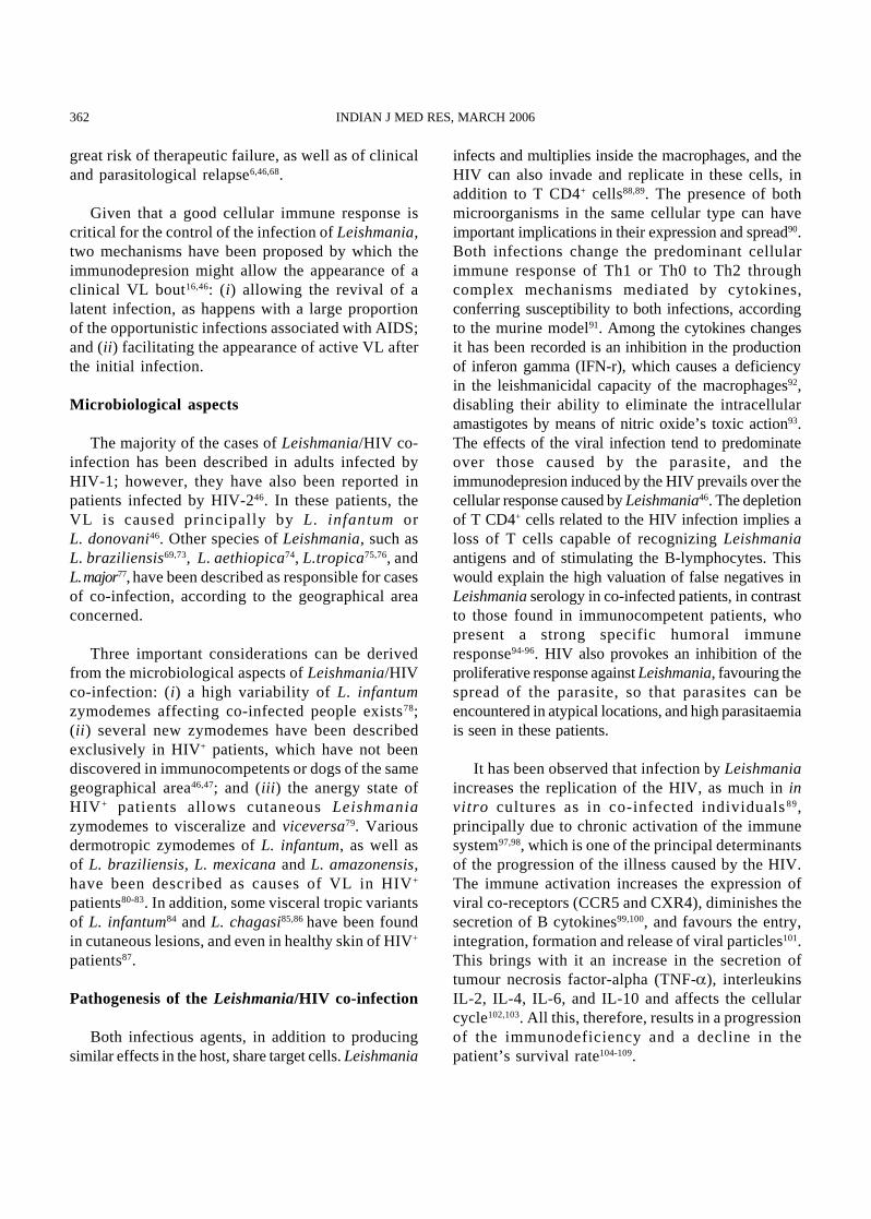
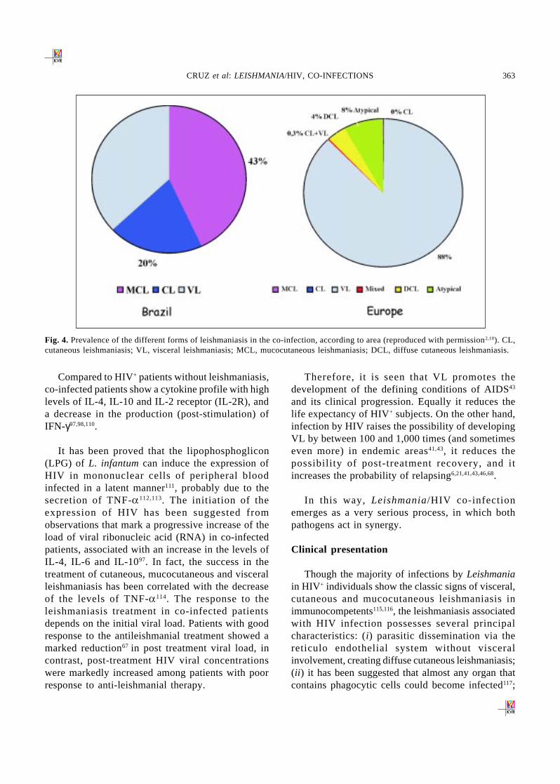
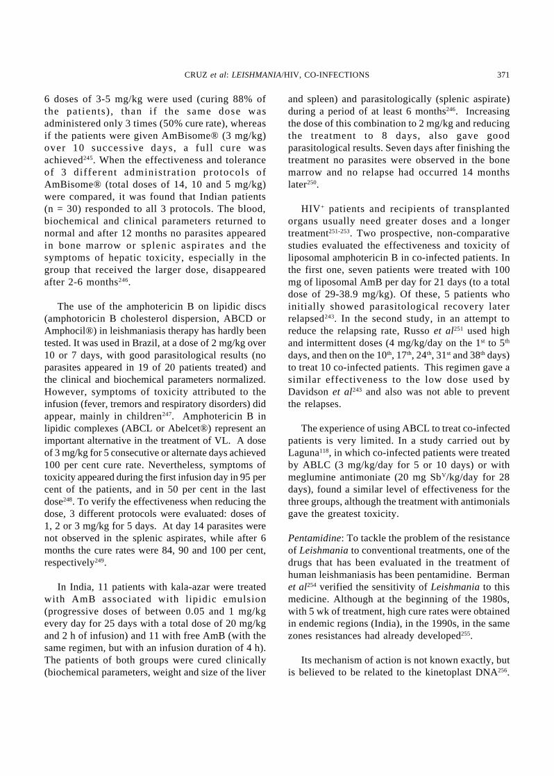
Fig. 4. Prevalence of the different forms of leishmaniasis in the co-infection, according to area (reproduced with permission2,10). CL,cutaneous leishmaniasis; VL, visceral leishmaniasis; MCL, mucocutaneous leishmaniasis; DCL, diffuse cutaneous leishmaniasis.
CRUZ et al: LEISHMANIA/HIV, CO-INFECTIONS 363
(iii ) atypical locations are affected as a consequenceof the parasite’s spread and deficiency in the cellularimmunity46; (iv) chronic progress and relapse115, witheach patient experiencing between two and threerelapses independently of the treatment received; (v)poor response to classic therapy118; (vii) low presenceof anti-Leishmania antibodies, a factor that can beobserved in many endemic areas119; and (viii ) theprevalence of the different forms of leishmaniasis isparticular to each zone, in relation to the causativespecies of Leishmania (Fig. 4).
The clinical aspects are comparable to those ofthe classic disease19,65,115,119-121. The period ofincubation is variable and can be related to age46,122.During a bout of VL, other concomitant opportunisticdiseases can be diagnosed in 42-68 per cent of HIV+
patients17,64. The visceral form is widespread andgenerally not only affects the organs of the reticuloendothelial system but also unusually in many otherorgans66.
A very important aspect in Leishmania/HIV co-infection is the appearance of post-treatment relapses,as happens to 27 per cent of patients in the first sixmonths after treatment and up to 60 per cent in firsttwelve months19,123. These bouts are caused by thereactivation of latent parasites because the immunesystem is incapable of eradicating the infection,although initially the patient may seem to haverecovered clinically22.
The prognosis of VL in HIV+ patients is verydifferent from that for immunocompetents. Thelevels of relapsing and of mortality are greater in thecases of co-infection. Before the introduction of thenew antiretroviral therapies, the mortality of co-infected patients reached 60 per cent in the first year,of which 27 per cent died during the first month afterthe diagnosis of VL124. Though the majority of thepatients die due to other diseases related to AIDS, itis considered that leishmaniasis contributes to this,causing immunosuppresion independently of theHIV, and stimulating the virus replication.
Systemic signs and symptoms: The majority of co-infected patients display fever, hepatomegaly and/or splenomegaly, hypergammaglobulinaemia andpancytopenia22,122.
Typically the splenomegaly is less frequent in theHIV +125. The majority of co-infected patients presentsome type of haematological cytopenia, thefrequency and the grade of anaemia, leucopoenia,lymphopenia and thrombocytopenia have beenobserved to be greater in this type of patientthan in the immunocompetent6,121. Thehypergammaglobulinaemia has limited diagnosticvalue because not only is it frequently found in VL,but also in HIV infection per se and in other chronicinfections. The constitutional syndromes (weakness,anorexia and loss of weight) are frequent in 50-70per cent of patients. The lymphadenopathy affects15-60 per cent of patients. The disease tends to spreadto the skin and other organs, so that the presentationoutside of the reticulo endothelial system can leadto clinical errors8,126,127. Between 7-17 per cent of thefevers of unknown origin in HIV+ patients are due toLeishmania infection, and 45 per cent of the HIV+
patients diagnosed with VL present fever of unknownorigin6.
Gastrointestinal implications: Gastrointestinalimplications are among the most frequentcomplications in HIV+ patients128,129. Leishmania hasbeen identified in the gastrointestinal tract of 50 percent of HIV- patients with VL130, and also in thosewho are HIV+. The parasite can appear in any sectionof the gastrointestinal tract130-139. In HIV- patients theprincipal digestive signs are diarrhoea,malabsorption, hypoalbuminaemia and loss ofweight. However, these symptoms are not specificin the AIDS patient, and therefore could have beencaused by another source140. Besides, Leishmaniacan co-exist with other pathogens in the same stretchof the digestive tract141. Therefore, the endoscopy androutine biopsy are the best diagnostic tools in HIV+
patients who display gastrointestinal symptoms ofunknown origin137-139,141. Endoscopic examinationgives very varied views, such as normal mucous,oesophagus mucous with diffuse erythemas,oesophagus mucous with extensive ulceration,erosive gastroduodenitis, gastric ulcers and multiplepetechial lesions in the colon137,141. The co-existenceof Leishmania in Kaposi’s sarcoma lesions, ulcerscaused by cytomegalovirus, or in the oesophaguscaused by Candida or Herpes simplex have beendescribed137,141.
364 INDIAN J MED RES, MARCH 2006
Cutaneous and mucocutaneous implications: Thecutaneous involvement in VL is rare142; neverthelessit is more frequent in VL associated with AIDS, beingobserved in 4.8 per cent of co-infected patients2.These lesions can occur simultaneously with a boutof VL and can be papular, macopapular or nodular.On occasions, the discovery of Leishmania isassociated with changes attributable to otherdermatological processes, such as dermatofibromas,psoriasis, Reiter’s syndrome, bacillary angiomatosis,cryptococcosis and oral aphtha, though its presencedoes not necessarily imply a causal role84. Cases ofcutaneous, mucocutaneous, and diffuse cutaneousleishmaniasis and post kala-azar dermalleishmaniasis (PKDL) have also been described73,143-
145. On the other hand, primary cutaneous lesions mayspread to the viscera in patients with severeimmunodepression80,81,83.
The mucocutaneous forms appear in 0.3 per centof European co-infection cases2. Practically all thespecies of Leishmania could be responsible formucocutaneous lesions in HIV+ patients, and they arealso not unknown in HIV- subjects due to otherspecies, including L. infantum146, distinct from thesubgenus Viannia. Although nasal septum and thesoft palate could appear to be involved due tometastasis, they might also be affected asconsequence of a primary lesion147. In the latter case,taking a nasal biopsy is usually necessary to establishthe definitive diagnosis of mucocutaneousleishmaniasis, a greater number of amastigotes beingobserved than in the case when the lesion is producedby species of the subgenus Viannia inimmunocompetent patients.
If the opposite is not found, for any cutaneousspecimen from HIV+ patients in whom there areLeishmania amastigotes, the first option to beconsidered should be a disseminated form of VLrather than a primary cutaneous lesion.
Respiratory tract forms: In anatomopathologicalstudies Leishmania amastigotes have been found inthe alveolar and pulmonary septums of 75 per centof the co-infected patients148. Nevertheless, theclinical meaning of this finding is hard to discerndue to the fact that the frequent pulmonary form is
not normally accompanied by clinical symptoms orcomplications and, when this happens, it is difficultto differentiate the role of Leishmania from that ofother, more frequent, pulmonary infections.
Renal form and acute renal failure: Glomerulopathy isassociated with VL and not with cutaneous ormucocutaneous leishmaniasis. In 60 per cent of HIV-
patients with VL proteinuria, haematuria andleucocituria have been described149. The frequency ofthese symptoms is not known in case of HIV+ patients.The pathological findings include glomerulonephritis,which changes from mesangioproliferative, sometimesassociated with the focal and segmental collapse of thecapillary handles, to tubulointerstitial damage150. Renalcondition is not common in human leishmaniasis.Nevertheless, a case of renal failure has been describedat the start of VL in an HIV+ patient151.
Other locations: A frequent aspect of bouts of VL inimmunodeficient patients is the appearance of atypicalsigns. The condition of severe immunodepresion thatcharacterizes these patients facilitates thehaematogenous dispersion of Leishmania to tissuesthat are not normally affected in immunocompetentconditions152,153. These types of signs are more frequentwhen the level of CD4+ cells is lower than 50 cells/µlof blood2. The list of infrequent signs includes, inaddition to those described previously (cutaneous,mucocutaneous, gastrointestinal, renal andpulmonary), others such as pleural, laryngeal,pancreatic, adrenal, peritoneal, synovial and cerebral,as well as of the pericardium, myocardium and tongueinvolvement46,59,84,117,139,148,154-160. A retrospective studyin France found amastigotes in atypical locations in34 per cent of the HIV+ patients with VL, while findingdiagnostic evidence in only 15 per cent of the cases161.It is important to bear in mind that the atypicallocations can be the first clinical manifestation of VLin immunodeficient patients. However, the majorityof these manifestations have been describedpreviously, though with a low frequency, inimmunocompetent patients130,148,162,163.
Diagnosis
In HIV+ patients the clinical diagnosis of VL hasan even more limited value since in the AIDS patient
CRUZ et al: LEISHMANIA/HIV, CO-INFECTIONS 365
leishmaniasis can first appear in an unusual way andwith not very specific clinical signs159,164,165. To thisshould be added that the splenomegaly is lessfrequent in these patients6. An aspect to bear in mindis that in 68 per cent of the cases of co-infection otheropportunistic infections associated with HIV alsoappear which display clinical and biological signssimilar to those of VL21,64,66,165.
Microscopy: In this case the experience of themicroscope user is fundamental, since they mightneed to recognise Histoplasma or other organismsthat could infect these patients and lead to falsepositive results.
For the microscopic observation of bone marrowa sensitivity has been described of between 67 and94 per cent6,22,166,167. On occasion, false negativescould be present for several reasons, as for example,the presence of a low number of leishmania infectedcells (as a consequence of pancytopenia) or thesample is haemodiluted due to incorrect collection.Another parameter that can affect the parasitic loadis if the patients have been treated with pentamidineor amphotericin B against mycosis orpneumocystosis168.
Amastigotes can be found in the peripheral bloodof approximately 50 per cent of HIV+ patients119,169,although Delgado et al170 established that thesensitivity of this method varies according to whetherthe patient presents symptomatic or asymptomaticexamination leishmaniasis. The study of other samplesdifferent than biopsies from blood or bone marrow isnot very common in HIV+ patients, though Montalbánet al22 found a sensitivity of 87.7 per cent for themicroscopic examination of hepatic biopsies. A typicalfeature in this type of patient is that, occasionally,parasitation can also be observed in unusual locationssuch as the lungs, larynx, gastrointestinal tract, rectum,spinal fluid and others46,153.
Culture: In patients co-infected by Leishmania/HIV,the culture of mononuclear peripheral blood cells hasa sensitivity of 67 per cent according to López-Vélezet al152. Mathis and Deplazes171 observed a similarpercentage (64%). This is interesting since, unlikein immunocompetents, the choice of non-invasive
sampling turns out to be useful in the diagnosis ofleishmaniasis. The culture of splenic aspirate canreach a greater sensitivity (63-100%)6,19, though dueto the danger involved in the collection of this typeof sample, myelocultivation is recommended forthese patients as it has a similar sensitivity accordingto some other authors46,172. However, when usingculture for diagnosis, one should remember that somespecial precautions are required in the routinehandling of leucocytic samples from AIDS patients.
Serology: In Leishmania/HIV co-infection, thehumoral specific response to Leishmania turns outto be partial, weak or absent, due to the fact that thecellular immunity is affected after infection by HIV64,causing a decrease of total lymphocytes in theproduction of antibodies95. Leishmania infectionaffects the appearance of antigens to the B cells asmuch as does HIV. Thus, the process of antibodiesproduction remains blocked173,174. This fact has beenconfirmed by Mary et al175, who estimated that thelevel of specific anti-Leishmania antibodies inpatients with AIDS was 50 times lower than inpatients with an intact immune system. Other authorsshown that the serology is positive for Leishmaniaonly in 40-50 per cent of co-infected patients22,176,this percentage being inversely related to the levelof depletion of T CD4+ cells. Therefore, it isrecommended that at least two different serologicaltechniques are used in the diagnosis of every patient2.
Lymphocyte proliferation test: Leishmania is capableof modulating the lymphocyte function177 causing asevere immunosupression in patients with VL178,179.During the course of the diseases there is apronounced depression of the cellular response toLeishmania antigens179,180 and a polyclonal activationof B cells with a high level of both specific andunspecific antibodies181. The immune response of Tcells is crucial for the cure and protection from laterinfections by Leishmania182. Therefore, after asuccessful treatment, the proliferation of T cells anddelayed hypersensitivity against the Leishmaniaantigens are developed179,183.
Though this test is currently not practical fordiagnosing a bout of VL, it is in epidemiologicalstudies in endemic zones to determine to determine
366 INDIAN J MED RES, MARCH 2006
the exposure of the population to the Leishmaniainfection184.
In co-infected individuals this technique hasallowed the verification of the finding that theabsence of a cellular response to the Leishmaniaantigens, on having finished the treatment, isresponsible for the high number of relapses sufferedby these patients185,186.
Indirect immunofluorescence test: Due to theimmunodepresion, this test applied on the sera ofLeishmania/HIV co-infected patients gives a lowsensitivity, between 11 and 58 per cent2,96 .
Enzyme immunoassay: Most of the assays performedon HIV+ patients samples where by using the rk39antigen, which is supposed to detect only active VLcases187. Medrano et al96 established a sensitivity of22 per cent for the rk39 ELISA in patients co-infectedby Leishmania/HIV. Other authors reporteda sensitivity of 20 per cent for theimmunochromatographic test and 62 per cent for theELISA form2,188.
Agglutination tests: Hailu and Berhe189 obtained asensitivity of 90 per cent for the direct agglutinationtest (DAT) in a work carried out in Ethiopia, similarto the result the same authors found forimmunocompetent patients. Vilaplana et al190, in astudy undertaken in Spain, found a sensitivity of 100per cent and a specificity of 96 per cent for thedetection of the Leishmania antigen in urine by meansof KAtex.
Immunoblot: Studying the serum of 37 patients withVL and 11 with HIV/VL using immunoblot, Mary etal175 showed that the antigens of 14 and 16 kDa hadthe highest specificity for VL. Those of 14 kDa weredetected in the serum of 92 per cent of the patientswith VL and 95 per cent for those of 16 kDa. Thesame standard was found in HIV/VL co-infectedpatients, although the 14 kDa band was not presentin any of the cases, whereas 16 kDa appeared in aconstant way for these patients. In another studyundertaken with co-infected patients, Medrano et al96
reported a sensitivity of 78 per cent for theimmunoblot technique, confirming that this type of
test provides an increase in sensitivity when detectinganti-Leishmania antibodies. Similar results have beenpresented by Moreno et al186, attributing a sensitivityof 85 per cent to the immunoblot in the diagnosis ofthese patients.
Molecular diagnosis - PCR: PCR has been shown tobe of great utility in the diagnosis of co-infectedpatients. Analyzing samples of bone marrow,Piarroux et al168 obtained a sensitivity of 82 per centfor the PCR, whereas other studies showed 100 percent165. In 165 blood sensitivity samples rangedbetween 72 and 100 per cent191,192.
Due to the relapsing character of leishmaniasis inHIV + patients, the PCR can be a useful technique inmonitoring patients post-treatment, as it can beapplied to non-invasive biopsies so avoidingunnecessary pain to the patient when evaluating theparasitic evolution during the follow up.
Real time quantitative PCR (RTQ-PCR): Althoughthe classic PCR technique is very useful as adiagnostic tool it has some disadvantages, such asthe need to analyze the amplified products usingelectrophoresis, and it has limited utility when theparasitic load needs to be monitored precisely as itis not able to quantify the obtained DNA.
The real time quantitative PCR allows continuousmonitoring of the PCR products generated during theamplification process. Thus it is possible to identifythe cycle in which the PCR product is generated in alogarithmic way and, by inference, the quantity ofthe target DNA present at the beginning of thereaction. Provided that the amplification product ismonitored in real time as is formed cycle by cycle,no post-amplification manipulation is needed.
The absolute quantif ication is real ized inaccordance with an internal standard coamplifiedwith the sample DNA, or in relation to a standardexternal curve obtained using the paral lelamplification of a series of known concentrationsof a sequence of reference DNA. An estimation ofthe parasitic load in a given sample can be obtainedfrom the quantification of the target DNA. Theadvantages, compared to the classic PCR, are a
CRUZ et al: LEISHMANIA/HIV, CO-INFECTIONS 367

reduction in the time needed for the assay and thepossibility of determining the parasitic load of thestudied sample.
Different approaches exist to monitor theamplification of the products, generally based on theuse of fluorescent markers. An example is thetechnology based on SYBR Green I, a detection systembased on a fluorescent agent that can be inserted in thedouble stranded DNA. Another example is the TaqMantechnology, which uses a specific fluorescent probe fora region of the product amplified during the PCR. Theintensity of the fluorescent signal accumulates at theend of every amplification cycle and is related to thequantity of PCR-generated product.
In the recent years, different methods ofquantitative PCR based on either the SYBR Green Imethod or on the TaqMan probes have been appliedin the study of leishmaniasis. Bretagne et al193 usedthe TaqMan system to determine the parasitic loadin the liver of mice infected with L. infantum; whereasNicolas et al194 applied the SYBR Green I system ina study of L. major infection, also in mice. In humanleishmaniasis Schulz et al195 managed to detect andto quantify parasites in different biopsies (peripheralblood, bone marrow aspirate, skin and liver) with amodel similar to the TaqMan probes, although theythought that its applicability to monitoring treatmentcould be affected by high variability in samples withlow parasitic loads. On the other hand, Bossolascoet al196 applied the real time PCR and also afluorescent probes model to monitor the parasiticload in the peripheral blood of patients co-infectedby HIV and Leishmania who had been undertreatment. Although the sensitivity obtained with thismethod in the initial attack did not turn out to bevery high (40%), it allowed a relationship betweenthe parasitic load in the blood and the clinical courseof the VL to be established. Mary et al197 obtainedsimilar results and improved the sensitivity, alsousing TaqMan probes, even though the variation inthe number of copies of the target that they used couldlead to a quantification calculation error betweenpatients infected by different strains.
Though the application of real time quantitativePCR in the diagnosis and monitoring of Leishmania
infection is in an early phase of development, thestandardization of a method capable of determiningthe parasitic load in a sensitive and reliable way willbe of great utility. It will find applications both inthe study of the effectiveness of treatments andvaccines and as an aid to diagnosis.
Plasmatic markers with prognostic value: IFN-g andTNF-a: As Leishmania is an intracellular parasite ofthe macrophages, specific immunity against VL has tocome from a Th1 type cellular immune response thatcan produce cytokine stimulators for macrophages(including IFN-g). On the other hand, susceptibility tothis illness is associated with an impairment to generatea Th1 response, generating Th2 type cytokines, as wellas high levels of TNF-a198-207. It has been observed thatoligosymptomatic individuals or those with subclinicalinfection produce cytokines of types Th1 and Th2, withlow or absent levels of detected IFN-g. Theseindividuals are in an intermediate position betweenillness and protective immunity206,208.
IFN-g : During a bout of VL the natural killer (NK)cells and the T cells produce IFN-r, influencingthe development of a protective Th1 response. Theproduct ion of IFN-g , wi th the consequentactivation of the macrophages, is crucial for theini t ia l containment of the parasi te’smultiplication205,209-211.
In the asymptomatic or subclinical cases, theproduction of IFN-g influences the self-limitingevolution of the clinical symptoms and signs,reaching in the spontaneous resolution of the bout ofVL 208,212-215. Gama et al206 found that 48 per cent ofthe children with asymptomatic infection showeddetectable levels of IFN-g, whereas this cytokine wasnot detected in children who suffered the acute form.Supporting this data, Carvalho et al201 also observedhigh levels of IFN-g in oligosymptomatic individuals,confirming the fact that resistance to the illness isassociated with an effective cellular immuneresponse.
As for the treatment of leishmaniasis, it has beenproved that IFN-g augments the therapeuticeffectiveness of antimonials, both in VL and incutaneous leishmaniasis216.
368 INDIAN J MED RES, MARCH 2006
TNF-a: TNF-a is considered to be a prognosismarker in VL, as its level is found to be significantlyhigher in acute and persistent cases of the illness,which then drops drastically after the parasite hasbeen treated217. Though it is possible to detect TNF-a in 100 per cent of the patients who showedsubclinical infection206, during active and acute boutsof VL these levels increase notably with regard tosubclinical and asymptomatic cases80,206,218.
Moderate levels of TNF-a facil itate theelimination of intracellular parasites, whereas itsincrease might predispose the patient towards clinicalmanifestations of VL, such as fever, cachexia,anorexia, pancytopenia and polyclonal activation ofB cells199,219. The blood pathogens induce themacrophages in liver, spleen and other locations toproduce TNF-a, and the systemic release of TNF-acauses expansion of blood vessels with theconsequent extravasation of fluids towards thetissues. In this way septic shock can take place,coagulating the extravased blood in the capillaries,leading to failure of vital organs such as the kidneys,liver, heart and lungs, which are compromised bythe loss of blood supply220. For this reason, it isconsidered that high levels of TNF-a in serum arecorrelated with the severity of human VL221, givingit a principal role in systemic alterations and,consequently, in the symptoms observed222-224.
TNF-a plays an important role in HIV infectionsince it has been proved that it has a positive effecton the replication of the virus111,225 associating highlevels of TNF-a in serum with the progression ofthe infection from HIV to AIDS226.
Co-infected patients display higher levels ofTNF-a in serum than HIV+ patients not infected byLeishmania, and these levels also remain high afterrecovery from a bout of VL89. This fact contrastswith that seen by others in immunocompetent VLpatients where the levels of TNF-a normalize aftersuccessful treatment199,227.
Treatment
Given that HIV+ patients generally develop VLwhen they are in a state of severe immunodepression
and are affected by other concomitant diseases, it isparticularly difficult to evaluate the effectiveness andtoxicity of drugs in these patients118. Althoughbetween 11 and 27 per cent of co-infected patients diewithin a month after diagnosis of VL, these deathsare rarely attributed to the leishmaniasis or to thetoxicity of the drugs administered to combat thedisease6,19,124. The clinical criteria used to verify theresponse to the anti-Leishmania treatment, useful inthe immunocompetent patients, are of less value inthe immunodepressed. Only the initial remedy shouldbe considered as, if the immunodepression does notchange, the probability of a full recovery is practicallynil and the patient tends to relapse. The parasitologicalresponse to the anti-Leishmania treatment in HIV+
patients has partially been studied6,19,22,64,124,166. Finally,it is necessary to consider that other medicinesadministered to HIV+ patients can increase the toxicityof some leishmanicide drugs.
Pentavalent antimonials: The results of severalretrospective studies indicate a high level ofclinical response to antimonials in the patients co-infected with HIV19,22,59,64,166. However, the drugdoses and the criteria used to identify the clinicalrecovery were not uniform, observation of theclinical cure turned out to be difficult due to theco-existence of other diseases associated withAIDS, and clinical progress does not necessarilymean that parasitological recovery has occurred228.In all these groups of patients a high percentageof them relapsed during the year after the treatmentfinished.
Moreover, pentavalent antimonials are known tobe more toxic for HIV+ patients than for HIV- ones6.Effects described are; pancreatitis (becoming fatalin some patients229), myocarditis and renal failure118.In fact, these developments interrupt the therapy in11-28 per cent of patients6,123,124,230.
Amphotericin B (AmB): More than half of the patientstreated with AmB showed a series of typical sideeffects related to the infusion, such as tremors, fever,nausea, vomiting, anorexia, headache, myalgia andarthralgia231-233. The duration of the infusion dosagemust range between 2 and 6 h, to reduce these adverseeffects231,232.
CRUZ et al: LEISHMANIA/HIV, CO-INFECTIONS 369
The most important side effect associated withAmB is undoubtedly the nephrotoxicity, whichoccurs in many of the patients treated233. Themechanism by which these changes take place is notunderstood, though it is thought that it stems froman alteration in the permeability of the glomerularmembrane234. The worsening, generally reversible,of the renal function happens during the first twoweeks of treatment and occurs in 80 per cent of thepatients treated with AmB. They usually recoverafter the treatment is terminated though it can takeseveral months for the renal function to recuperate234.Daily monitoring of the levels of creatinine and otherrenal parameters is recommended when using AmBtreatment. When the level of creatinine rises above3 mg/dl, the therapy must be interrupted until thelevel returns to normal231. It is also recommended thatthe treatment be administered on alternate days toreduce the renal toxicity232. The use of lipidicformulations diminishes the AmB toxicity,supporting its therapeutic activity235.
Treatment can be started by administering theAmB-disoxicolate in very small doses, increasinggradually (0.1, 0.25 up to 0.5 - 0.8 mg/kg, etc.),diluted in 5 per cent dextrose236. It should beadministered as an intravenous (iv) infusion over2 - 6 h, with the treatment lasting about 4 wk. Themaximum daily dose must never exceed 1.5 mg/kg,but it is possible to administer a dose of 1.5 mg/kgon the alternate days because the elimination half-life of AmB is very high237.
On comparing the parasitological effectiveness ofadministering maximum doses of 1 mg/kg up to atotal dose of 20 mg/kg of AmB, every day or onalternate days, it was proved that the two regimenswere equally effective, since all the patientsrecovered and 6 months later no parasites wereobserved in splenic aspirates, though the dailyschedule was more economic as it reduced the periodof hospitalization238. If the dose is diminished to0.5 mg/kg in 14 administrations on alternate days,100 per cent aseptic curing was achieved at 6 wkand 98 per cent at 6 months239. If the same regimenis compared with the administration of sodiumstibogluconate (SGS) (20 mg/kg/day for 40 days),the level of curing is 62 per cent, as opposed to 100
per cent for the AmB, which indicates around a40 per cent resistance to the SbV in patients of India240.AmB was also used in paediatric patients with adosage of 1 mg/kg every 2 days for 20 days, whichwas tolerated perfectly, achieving 100 per centhealing. Pregnant women tolerated this dosage,without apparent damage to the foetus, and with goodresults against the parasite241.
In a study undertaken by Laguna et al123 on HIV+
patients, the parasitological recovery was similar tothat shown by the patients who were treated withpentavalent antimonials. However, the level ofrelapsing in the first year post-treatment was similarin both groups.
Delivery systems: One of the most significantadvances in recent decades in chemotherapy againstLeishmania has been the use of lipidic vehicles totransport the amphotericin B. The drug delivered inthis way is rapidly captured by the cells of theendothelial-reticulum system and directedspecifically to the organs rich in this type of cells,avoiding the organs in which toxicity takes place,prolonging the liberation and therefore the presenceof the drug in the organism. The pharmacokineticand pharmacodynamic differences between thetransported forms and the free drugs are due to thechanges which are produced in the distribution, atthe different levels of plasmatic concentration, aswell as to the changes that take place when theplasmatic proteins bound, in their metabolism andelimination of the transported drugs242.
It has been proved that the effectiveness ofliposomal Amphotericin B (AmBisome®) in bothadults and children is independent from the form ofadministration (21 days with a dose of 1 mg/kg or10 days at 3 mg/kg/day) and none of theimmunocompetent patients relapsed between 12 and24 months after treatment243. When the treatment wasshortened by increasing the doses (5 consecutive daysand then a dose on the 10th day, up to a total dose of18 mg/kg). With this regimen parasitological cureswere achieved, but renal alterations occurred244.
The liposomal AmB used in Sudanese patientswith complicated kala-azar was more effective if
370 INDIAN J MED RES, MARCH 2006
6 doses of 3-5 mg/kg were used (curing 88% ofthe pat ients), than i f the same dose wasadministered only 3 times (50% cure rate), whereasif the patients were given AmBisome® (3 mg/kg)over 10 successive days, a ful l cure wasachieved245. When the effectiveness and toleranceof 3 di f ferent administrat ion protocols ofAmBisome® (total doses of 14, 10 and 5 mg/kg)were compared, it was found that Indian patients(n = 30) responded to all 3 protocols. The blood,biochemical and clinical parameters returned tonormal and after 12 months no parasites appearedin bone marrow or splenic aspirates and thesymptoms of hepatic toxicity, especially in thegroup that received the larger dose, disappearedafter 2-6 months246.
The use of the amphotericin B on lipidic discs(amphotoricin B cholesterol dispersion, ABCD orAmphocil®) in leishmaniasis therapy has hardly beentested. It was used in Brazil, at a dose of 2 mg/kg over10 or 7 days, with good parasitological results (noparasites appeared in 19 of 20 patients treated) andthe clinical and biochemical parameters normalized.However, symptoms of toxicity attributed to theinfusion (fever, tremors and respiratory disorders) didappear, mainly in children247. Amphotericin B inlipidic complexes (ABCL or Abelcet®) represent animportant alternative in the treatment of VL. A doseof 3 mg/kg for 5 consecutive or alternate days achieved100 per cent cure rate. Nevertheless, symptoms oftoxicity appeared during the first infusion day in 95 percent of the patients, and in 50 per cent in the lastdose248. To verify the effectiveness when reducing thedose, 3 different protocols were evaluated: doses of1, 2 or 3 mg/kg for 5 days. At day 14 parasites werenot observed in the splenic aspirates, while after 6months the cure rates were 84, 90 and 100 per cent,respectively249.
In India, 11 patients with kala-azar were treatedwith AmB associated with l ipidic emulsion(progressive doses of between 0.05 and 1 mg/kgevery day for 25 days with a total dose of 20 mg/kgand 2 h of infusion) and 11 with free AmB (with thesame regimen, but with an infusion duration of 4 h).The patients of both groups were cured clinically(biochemical parameters, weight and size of the liver
and spleen) and parasitologically (splenic aspirate)during a period of at least 6 months246. Increasingthe dose of this combination to 2 mg/kg and reducingthe treatment to 8 days, also gave goodparasitological results. Seven days after finishing thetreatment no parasites were observed in the bonemarrow and no relapse had occurred 14 monthslater250.
HIV + patients and recipients of transplantedorgans usually need greater doses and a longertreatment251-253. Two prospective, non-comparativestudies evaluated the effectiveness and toxicity ofliposomal amphotericin B in co-infected patients. Inthe first one, seven patients were treated with 100mg of liposomal AmB per day for 21 days (to a totaldose of 29-38.9 mg/kg). Of these, 5 patients whoinitially showed parasitological recovery laterrelapsed243. In the second study, in an attempt toreduce the relapsing rate, Russo et al251 used highand intermittent doses (4 mg/kg/day on the 1st to 5th
days, and then on the 10th, 17th, 24th, 31st and 38th days)to treat 10 co-infected patients. This regimen gave asimilar effectiveness to the low dose used byDavidson et al243 and also was not able to preventthe relapses.
The experience of using ABCL to treat co-infectedpatients is very limited. In a study carried out byLaguna118, in which co-infected patients were treatedby ABLC (3 mg/kg/day for 5 or 10 days) or withmeglumine antimoniate (20 mg SbV/kg/day for 28days), found a similar level of effectiveness for thethree groups, although the treatment with antimonialsgave the greatest toxicity.
Pentamidine: To tackle the problem of the resistanceof Leishmania to conventional treatments, one of thedrugs that has been evaluated in the treatment ofhuman leishmaniasis has been pentamidine. Bermanet al254 verified the sensitivity of Leishmania to thismedicine. Although at the beginning of the 1980s,with 5 wk of treatment, high cure rates were obtainedin endemic regions (India), in the 1990s, in the samezones resistances had already developed255.
Its mechanism of action is not known exactly, butis believed to be related to the kinetoplast DNA256.
CRUZ et al: LEISHMANIA/HIV, CO-INFECTIONS 371
In 1993, Murray et al257 demonstrated that in athymicmice (deficient in T cells) pentamidine was noteffective against Leishmania whereas it was effectivein euthymic mice.
The recommended dose depends on the salt used.If it is in the isotianate form the dose is 4 mg/kg(up to a maximum of 300 mg daily), if themetilsulphonate is used, the dose is 2.4 mg/kg (up toa maximum of 180 mg daily). Short treatmentregimes with low doses of pentamidine, which areused in cutaneous leishmaniasis treatments oftenresult in myalgia, pain in the injection site, nausea,headache and, rarely, a metallic taste. The incidenceand severity of these side effects are much greater invisceral leishmaniasis treatments, with their highdoses over long periods, sometimes also inducinghypotension, tachycardia and hyperglycaemias, andeven diabetes255,258.
In the treatment of visceral leishmaniasis, using4 mg/kg, three times per week up to theparasitological cure of the patient, 77 per cent of thepatients were cured after 15 injections (5 wk oftreatment) and 94 per cent after 27 injections (9 wkof treatment), however, 21 per cent relapsed withina year. In Colombia, a regimen of 2 mg/kg every twodays for seven days was used, with 96 per centeffectiveness259. The treatment of VL needs highdoses of pentamidine for several weeks, and is evenmore toxic than the treatments with antimonials.Thus, this drug should be used exclusively when thereare reasons to think that it is going to be moreeffective than any other, in geographical zones whereantimonial treatments habitually fail, or in areaswhere this product has not been in use for some timeand therefore resistance has not been created and, ofcourse, in individual cases where other therapies havenot succeeded.
Paramomycin: This is an aminoglycosid antibioticwith a short half-life, much used against intestinalparasites. The mechanism of action againstLeishmania is not known, though it is possible that itacts on the ribosome blocking the synthesis ofproteins260. The resistance to paramomicine that certainstrains of Leishmania develop is due to changes in themembrane that reduce absorption. The promastigotes
maintain this resistance in the absence of the antibioticin the culture system and it is transmitted to theamastigotes, which can infect macrophages261.
Paramomycin can be used alone or in combinationwith other drugs to increase their effectivenessagainst the parasite, with antimonials being thepreferred combination. According to Thakur et al262
the use of half doses of paramomycin partnered withantimonials turns out to be the most effectivecombination. In the treatment of VL in Kenya,Chunge et al263 used the sulphate of paramomycin(15 mg/kg/day, over an average of 19 days), healing79 per cent of the patients. In India, with thecombination of paramomycin (17 mg/kg) withPentostan® (20 mgSbV/kg) for 20 days, 82 per centof the patients were cured, a percentage similar tothat obtained with paramomycin alone for 40 days264.Similar results were obtained in Sudan265.
Though monotherapy with paramomycin iseffective, it never reaches the levels of healingobtained with antimonials or with amphotericin B266.However, as already indicated, the combination ofantimonials and paramomycin is more effective andallows shorter treatment periods. A recent Phase IIItrial of paromomycin (PM) efficacy resulted in a curerate of 94.6 per cent at 6 months after EOT267.
Miltefosine: Miltefosine is a synthetic phospholipid(alquil phospholipid), normally used as anantineoplastic, with a very long half-life, which meansthat resistance to it develops rapidly. It cannot be givento women during pregnancy or lactation. A particularadvantage is that it is administered orally, however adisadvantage is that this can lead to gastrointestinal sideeffects and, teratogenicity in the experimental model.Apparently it blocks the multiplication of Leishmaniaand alters its phospholipid and sterol composition268.Similarly, it seems that its anti-Leishmania activity isnot only related to a direct cytotoxic effect on theparasite, but also to an activation of the cellularimmunity269-271.
Kuhlencord et al272 verified that this drug iseffective against different Leishmania species as muchin vitro as in vivo. This effectiveness has beendemonstrated in both acute and chronic murine
372 INDIAN J MED RES, MARCH 2006

infections, showing a very notable reduction in thenumber of amastigotes after the treatment273. It hasalso been demonstrated that using topical miltefosineon experimental cutaneous leishmaniasis in miceproduced good results, giving a considerable reductionof the parasitic load in the lesions, and it was foundthat they did not usually reappear in the lesions afterthe treatment274. The recommended dose in the variousrecent clinical studies undertaken on humans in Indiawas 100 mg/day as higher doses can provoke toxicity,basically gastrointestinal disorders, and it isrecommended that it be taken with food275,276. It isbelieved that total doses of between 2,100 and 3,850mg were used in a clinical assay with 120 Indianpatients with VL, which achieved parasitological cure(absence of parasites in splenic aspirates two weeksafter the treatment ended) and clinic healing in 97 percent of the patients, with no relapsing in the followingsix months277. In another clinical assay the conclusionwas reached that the effective treatments must lastbetween 21 and 28 days with a recommended dose of100mg/day278. It has only been in use in a limitednumber of co-infected patients in a compassionateway, though the most suitable protocol has still notyet been established279,280.
Allopurinol: In general, the reports published withregard to allopurinol are quite contradictory. Kageret al281 treated 10 Kenyan patients only withallopurinol and cured three. However, Jha in 1983282
treated 16 patients in India and cured 13. Thecombination of allopurinol with ketoconzol wasshown to have been successful in a kidneytransplanted patient with VL283 and in cases ofcutaneous leishmaniasis in the New World284.
The combination of allopurinol and antimonials hasbeen widely used, though the results obtained withthis combination are contradictory, in the treatmentof relapsing patients285 and against MediterraneanVL 286. In a study of 124 patients, the combination ofthe two medicines did not demonstrate anyimprovement in effectiveness when compared with theuse of only pentavalent antimonials287.
The role of allopurinol in leishmaniasis is difficultto evaluate. It seems improbable that it hasleishmanicide effects by itself, though in combination
with other typical drugs its effects against Leishmaniaseem to improve. Ribera et al288 in a study onLeishmania HIV co-infected patients, in which allthe subjects were previously treated with antimonials,later administered a secondary prophylaxis. Therelapse rate was 65 per cent in the control grouppatients, who did not receive any secondarytreatment, 56 per cent in the group that receivedallopurinol, and 18 per cent in those who werereceiving antimony as a prophylaxis.
Immunotherapy
The progression of VL ends in immunosuppression,and follows the Th2 route with decrease in the levelsof IL-2 and IFN-r typical of the Th1 route. Therefore,in the treatment of resistant cases, where it is moreprobable that they will suffer immunologicaldepletion, it has been suggested to use a combinationof chemotherapy with microbial or syntheticimmunostimulants. Badaró et al289 were the first touse IFN-g in combination with pentavalent antimonialsin the treatment of VL, finding that seven of ninepatients with antimonial resistant kala-azar, were curedwith the combination of 100 µg/m2/day of IFN-g and20 mg/kg/day of pentavalent antimony over 28 days.In 1993, Squires et al290, demonstrated that the sametreatment produced a more rapid elimination of theparasites in Kenyan patients that had not received aprevious treatment. With the same treatment, in India,Sundar et al291, also observed a faster reduction in theparasitic load in the patients treated with thecombination of two products, than in those treated onlywith antimonials. A later assay demonstrated that IFN-ghas only a partial effectiveness by itself. Four out of9 patients in India, who previously had not receivedany treatment, showed no response and the others onlya partial elimination of the parasites292.
IFN-g has pronounced side effects such as fever,tremors and granulocytopenia, in addition to a highprice. Very recently, the immunomodulatortucaresol has been tested experimentally in infectedmice293. The effectiveness of recombinantgranulocyte-macrophage colony - stimulating factor(GM-SCF) has also been evaluated at a dose of 5µg/kg/day in combination with pentavalentantimony in 24 patients. This led to parasitological
CRUZ et al: LEISHMANIA/HIV, CO-INFECTIONS 373

healing after three months, although three of thepatients had relapses294. Information on thetherapeutic use of IL-12 and IL-2 is still scarce andthese have only been tested in experimental murineleishmaniasis in which an increase in the productionof INF-g and an important cellular response wereobserved295,296.
Factors that influence the choice of therapy
Several factors have to bear in mind whenchoosing the most appropriate reagent for thetherapeutic management of VL. Among them are notonly medical factors, but also socio-economic andcultural considerations. It is necessary to haveinformation about the endemicity of the strains ofLeishmania, the immune and nutritional state of thepopulation, the level and pattern of the parasite’sresistance to drugs, and obviously the cost. In HIV+
patients it is necessary to bear in mind the importantproblem posed by the frequent relapsing and the needof secondary chemoprophylaxis, which will dependon the level of CD4+ cells297,298.
Recommendations for the treatment of VL inimmunodepressed patients
The treatment of VL in HIV+ patients iscontroversial. The use of pentavalent antimonials oramphotericin B desoxycholate, though useful, resultsin high toxicity and the therapy takes a long time123.The new lipidic formulations of amphotericin B offergreater tolerability and can be used in short treatments.
The Leishmania infection reappears in HIV+
patients, including those that receive HAART, after asuccessful treatment. Apparently this is related to lowlevels of CD4+ cells297,298. Agreement does not existwith regard to the use of secondary prophylaxis, but itseems that the use of these can be suspended once thecount of CD4+ cells is greater than 300/ml298,299.
Secondary prophylaxis
In HIV + patients, including those that receiveHAART, relapsing of VL has been observed in spiteof having received an effective therapy300. However,
this is not very frequent, and is more probable due tolower level of the patient’s CD4+ cells297,298. In fact, ifa stable level of CD4+ is obtained after HAART, thereare those who recommend interrupting the secondaryprophylaxis, although this is controversial298.
In the absence of a controlled study, no consensusexists on what type of secondary prophylaxis toadminister to co-infected patients. Nevertheless, theperiod free of relapsing in patients treated withamphotericin B in lipidic complex (Abelcet® 3 mg/kg every 3 wk) compared to patients who do notreceive any prophylaxis has been evaluated byLópez-Vélez et al301, and after a year of monitoringit was observed that 50 per cent of the patients thatreceived the prophylaxis did not show any newepisode, whereas this percentage was 22 per cent inthe group that did not receive it. In a similar studywith five patients, Montana et al302 also indicated thebenefit of liposomal amphotericin B as the secondaryprophylaxis.
The improvement of treatment of leishmaniasisin HIV-positive patients should be based on both thedevelopment of new regimens based on already testeddrugs and the experimentation with oral drugs suchas miltefosine303.
References
1. De la Loma A, Alvar J, Martínez-Galiano E, Blázquez J,Alcalá-Muñoz A, Nájera R. Leishmaniasis or AIDS?Trans R Soc Trop Med Hyg 1985; 79 : 421-2.
2. Desjeux P, Alvar J. Leishmania/HIV co-infections:epidemiology in Europe. Ann Trop Med Parasitol 2003;97 : S3-15.
3. WHO. Report on the consultative meeting on Leishmania/HIV co-infection. WHO/LEISH/95.35. Geneva: WorldHealth Organization; 1995 p. 1-4.
4. Leishmania/HIV co-infection. Epidemiological analysisof 692 retrospective cases. Wkly Epidemiol Rec 1997;72 : 49-54.
5. Leishmania/HIV co-infection, south western Europe,1990-1998. Wkly Epidemiol Rec 1999; 74 : 365-75.
6. Pintado V, Martín-Rabadán P, Rivera ML, Moreno S,Bouza E. Visceral le ishmaniasis in human-immunodeficiency virus (HIV)-infected and non HIV-infected patients. Medicine 2001; 80 : 54-73.
374 INDIAN J MED RES, MARCH 2006
7. Kubar J, Marty P, Lelievre A, Quaranta JF, Staccini P,Caroli-Bosc C, et al. Visceral leishmaniosis in HIV-positive patients: primary infection, reactivation and latentinfection. Impact of the CD4+ T-lymphocyte counts.AIDS 1998; 12 : 2147-53.
8. Alvar J, Gutiérrez-Solar B, Molina R, López-Vélez R,García-Camacho A, Martínez P, et al. Prevalence ofLeishmania infection among AIDS patients. Lancet 1992;339 : 264-5.
9. Sampaio RN, Salaro CP, Resende P, De Paula CD.American cutaneous leishmaniasis associated with HIV/AIDS: report of four clinical cases. Rev Soc Bras Med Trop2002; 35 : 651-4.
10. Rabello A, Orsini M, Disch J. Leishmania/HIV co-infectionin Brazil: an appraisal. Ann Trop Med Parasitol 2003; 97 :S17-28.
11. Guiguemde RT, Sawadogo OS, Bories C, Traore KL,Nezien D, Nikiema L, et al. Leishmania major and HIVco-infection in Burkina Faso. Trans R Soc Trop Med Hyg2003; 97 : 168-9.
12. Mukhtar MM, Khal i l AM, Elhassan AM, Musa F,Modabber F. Leishmania f irst generation vaccines:vaccine trials in Sudan. In: Mas-Coma S, Bargues MD,Esteban JG, Valero MA, edi tors. IX EuropeanMulticolloquium of Parasitology, Valencia, Spain 2004;KS : 128.
13. Sundar S. Drug resistance in Indian visceral leishmaniasis.Trop Med Int Health 2001; 6 : 849-54.
14. Sundar S, Jha TK, Thakur CP, Engel J, Sindermann H,Fischer C, et al. Oral miltefosine for Indian visceralleishmaniasis. N Engl J Med 2002; 347 : 1739-46.
15. Sinha PK, Rabidas VN, Pandey K, Verma N, Gupta AK,Ranjan A, et a l. V isceral le ishmaniasis and HIVcoinfection in Bihar, India. J Acquir Immune Defic Syndr2003; 32 : 115-6.
16. Albrecht HI, Sobottka I, Emminger C, Jablonowski H,Just G, Stoehr A, et al. Visceral leishmaniasis emergingas an important opportunistic infection in HIV-infectedpersons l iving in areas nonendemic for Leishmaniadonovani. Arch Pathol Lab Med 1996; 120 : 189-98.
17. Rosenthal E, Marty P, Poizot-Martin I, Reynes J, Pratlong F,Lafeuillade A, et al. Visceral leishmaniasis and HIV-1 co-infection in southern France. Trans R Soc Trop Med Hyg 1995;89 : 159-62.
18. Gradoni L, Scalone A, Gramiccia M, Troiani M.Epidemiological surveillance of leishmaniasis in HIV-1-infected individuals in Italy. AIDS 1996; 10 : 785-91.
19. López-Vélez R, Pérez-Molina JA, Guerrero A, Baquero F,Villarrubia J, Escribano L, et al. Clinicoepidemiologiccharacteristics, prognostic factors, and survival analysis ofpatients coinfected with human immunodeficiency virus andLeishmania in an area of Madrid, Spain. Am J Trop MedHyg 1998; 58 : 436-43.
20. Botet J, Portús M. La leishmaniosis en la España peninsular.Revision histórico-bibliográfica (1912-1985). Rev San HigPub 1993; 67 : 255-66.
21. Alvar J. Leishmaniasis and AIDS co-infection: the Spanishexample. Parasitol Today 1994; 10 : 160-3.
22. Montalbán C, Calleja JL, Erice A, Laguna F, Clotet B,Podzamczer D, et al. Visceral leishmaniasis in patientsinfected with human immunodeficiency virus. Co-operativeGroup for the Study of Leishmaniasis in AIDS. J Infect 1990;21 : 161-70.
23. Palella FJ Jr, Delaney KM, Moorman AC, Loveless MO,Fuhrer J, Satten GA, et al. Declining morbidity and mortalityamong patients with advanced human immunodeficiencyvirus infection. HIV Outpatient Study Investigators. N EnglJ Med 1998; 338 : 853-60.
24. Mocroft A, Vella S, Benfield TL, Chiesi A, Miller V,Gargalianos P, et al. Changing patterns of mortality acrossEurope in patients infected with HIV-1. EuroSIDA StudyGroup. Lancet 1998; 352 : 1725-30.
25. Del Giudice P, Mary-Krause M, Pradier C, Grabar S,Dellamonica P, Marty P, et al. French Hospital Database onHIV Clinical Epidemiologic Group. Impact of highly activeantiretroviral therapy on the incidence of visceralleishmaniasis in a French cohort of patients infected withhuman immunodeficiency virus. J Infect Dis 2002; 186 :1366-70.
26.Autran B, Carcelain G, Li TS, Blanc C, Mathez D,Tub iana R , e t a l. Pos i t i ve e f fec ts o f comb inedantiretroviral therapy on CD4+ T cel l homeostasisand function in advanced HIV disease. Science 1997;277 : 112-6.
27. Tural C, Romeu J, Sirera G, Andreu D, Conejero M, RuizS, et al. Long-lasting remission of cytomegalovirus retinitiswithout maintenance therapy in human immunodeficiencyvirus-infected patients. J Infect Dis 1998; 177 : 1080-3.
CRUZ et al: LEISHMANIA/HIV, CO-INFECTIONS 375
28. Weverl ing GJ, Mocroft A, Ledergerber B, Kirk O,González-Lahoz J, D’Arminio-Monforte A, et al.Discontinuation of Pneumocystis carinii pneumoniaprophylaxis after start of highly active antiretroviraltherapy in HIV-1 infection. EuroSIDA Study Group.Lancet 1999; 353 : 1293-8.
29. Jouan M, Saves M, Tubiana R, Carcelain G, Cassoux N,Aubron-Olivier C, et al. RESTIMOP study team.Discontinuation of maintenance therapy for cytomegalovirusretinitis in HIV-infected patients receiving highly activeantiretroviral therapy. AIDS 2001; 15 : 23-31.
30. López Bernaldo de Quirós JC, Miró JM, Pena JM,Podzamczer D, Alberdi JC, Martínez E, et al. A randomizedtrial of the discontinuation of primary and secondaryprophylaxis against Pneumocystis carinii pneumonia afterhighly active antiretroviral therapy in patients with HIVinfection. N Engl J Med 2001; 344 : 159-67.
31. Carr A, Marrito D, Field A, Vasak E, Cooper DA. Treatmentof HIV-1-associated microsporidiosis and cryptosporidiosiswith combination antiretroviral therapy. Lancet 1998; 351 :256-61.
32. Foudraine NA, Weverling GJ, Van Gool T, Roos MT,De Wolf F, Koopmans PP, et al. Improvement of chronicdiarrhoea in patients with advanced HIV-1 infection duringpotent antiretroviral therapy. AIDS 1998; 12 : 35-41.
33. Miró JM, López JC, Podzamczer D, Peña JM, Alberdi C,
Claramonte X, et al. GESIDA 04/98-B Study.
Discontinuation of toxoplasmic encephalitis prophylaxis is
safe in HIV-1 and T. gondii coinfected patients after
immunological recovery with HAART: Preliminary results
of the GESIDA 04/98-B study. San Francisco: Proceedings
of the 7th Conference on Retroviruses and Opportunistic
Infections (Abstract Book); 2000.
34. López-Vélez R. The impact of highly active antiretroviraltherapy (HAART) on visceral leishmaniasis in Spanishpatients who are co-infected with HIV. Ann Trop MedParasitol 2003; 97 : S143-7.
35. Russo R, Nigro L, Panarello G, Montineri A. Clinical surveyof Leishmania/HIV co-infection in Catania, Italy: the impactof highly active antiretroviral therapy (HAART). Ann TropMed Parasitol 2003; 97 : S149-55.
36. De la Rosa R, Pineda JA, Delgado J, Macías J,Morillas F, Mira JA, et al. Incidence of and risk factors forsymptomatic visceral leishmaniasis among human
immunodeficiency virus type 1-infected patients from Spainin the era of highly active antiretroviral therapy. J ClinMicrobiol 2002; 40 : 762-7.
37. López-Vélez R, Casado JL, Pintado V. Decline of a visceralleishmaniasis epidemic in HIV-infected patients after theintroduction of highly active antiretroviral therapy(HAART). Clin Microbiol Infect 2001; 7 : 394-5.
38. Mira JA, Corzo JE, Rivero A, Macías J, De León FL,Torre-Cisneros J, et a l. Frequency of v isceralleishmaniasis relapses in human immunodeficiency virus-infected patients receiving highly active antiretroviraltherapy. Am J Trop Med Hyg 2004; 70 : 298-301.
39. UNAIDS. AIDS Epidemic Update 2002. www.unaids.org,accessed on April 8, 2005.
40. Fisk DT, Meshnick S, Kazanjian PH. Pneumocystis cariniipneumonia in patients in the developing world who haveacquired immunodeficiency syndrome. Clin Infect Dis 2003;36 : 70-8.
41. Desjeux P. Leishmania and HIV co-infection in south-western Europe, 1990-1998, retrospective analysis of 965cases. Geneva: World Health Organization, WHO/Leish/2000.42 2000.
42. WHO. The world health report. Geneva: World HealthOrganization 2001.
43. Guerin PJ, Olliaro P, Sundar S, Boelaert M, Croft SL,Desjeux P, et al. Visceral leishmaniasis: current statusof control, diagnosis, and treatment, and a proposedresearch and development agenda. Lancet Infect Dis2002; 2 : 494-501.
44. Molina R, Gradoni L, Alvar J. HIV and the transmission ofLeishmania. Ann Trop Med Parasitol 2003; 97 : S29-S45.
45. Desjeux P, UNAIDS. Leishmania and HIV in Gridlock.WHO/CTD/LEISH/98.9 Add.I, UNAIDS/98.23.1998.Geneva: World Health Organization; 1998.
46. Alvar J, Cañavate C, Gutiérrez-Solar B, Jiménez MI,Laguna F, López-Vélez R, et al. Leishmania and humanimmunodeficiency virus coinfection: the first 10 years.Clin Microbiol Rev 1997; 10 : 298-319.
47. Prat long F, Dereure J, Deniau M, Marty P,Faraut-Gambarelli F, Dedet JP. Enzymatic polymorphismduring Leishmania/HIV co-infection: a study of 381Leishmania strains received between 1986 and 2000 at the
376 INDIAN J MED RES, MARCH 2006
international cryobank in Montpellier, France. Ann TropMed Parasitol 2003; 97 : S47-56.
48. Molina R, Cañavate C, Cercenado E, Laguna F, López-VélezR, Alvar J. Indirect xenodiagnosis of visceral leishmaniasisin 10 HIV-infected patients using colonized Phlebotomusperniciosus. AIDS 1994; 8 : 277-9.
49. Cruz I, Morales MA, Noguer I, Rodríguez A, Alvar J.Leishmania in discarded syringes from intravenous drugusers. Lancet 2002; 359 : 1124-5.
50. Pineda JA, Macías J, Morillas F, Fernández-Ochoa J, Cara J,De La Rosa R, et al. Evidence of increased risk for Leishmaniainfantum infection among HIV-seronegative intravenous drugusers from southern Spain. Eur J Clin Microbiol Infect Dis2001; 20 : 354-7.
51. Pineda JA, Martín-Sánchez J, Macías J, Morillas F.Leishmania spp infection in injecting drug users. Lancet2002; 360 : 950-1.
52. Rosenthal PJ, Chaisson RE, Hadley WK, Leech JH. Rectalleishmaniasis in a patient with acquired immunodeficiencysyndrome. Am J Med 1988; 84 : 307-9.
53. Heisch R. Studies in leishmaniasis in East Africa. I.The epidemiology of an outbreak of kala-azar in Kenya.Trans R Soc Trop Med Hyg 1954; 48 : 449-69.
54. Aggarwal P, Wali JP. Profile of kala-azar in north India.Asia-Pacific J Public Health 1991; 5 : 90-3.
55. Evans TG, Teixeira MJ, McAuliffe IT, Vasconcelos I,Vasconcelos AW, Sousa A de A, et al. Epidemiology ofvisceral leishmaniasis in northeast Brazil. J Infect Dis 1992;166 : 1124-32.
56. Belazzoug S. Leishmaniasis in Mediterranean countries.Vet Pathol 1992; 44 : 15-9.
57. Cascio A, Colomba C, Antinori S, Orobello M, Paterson D,Titone L. Pediatric visceral leishmaniasis in Western Sicily,Italy: a retrospective analysis of 111 cases. Eur J ClinMicrobiol Infect Dis 2002; 21 : 277-82.
58. Kafetzis DA. An overview of paediatric leishmaniasis.J Postgrad Med 2003; 49 : 31-8.
59. Berenguer J, Moreno S, Cercenado E, Bernaldo de QuirósJC, García de la Fuente A, Bouza E. Visceral leishmaniasisin patients infected with human immunodeficiency virus(HIV). Ann Intern Med 1989; 111 : 129-2.
60. Pineda JA, Gallardo JA, Macías J, Delgado J, Regordan C,Morillas F, et al. Prevalence of and factors associated withvisceral leishmaniasis in human immunodeficiency virustype 1-infected patients in southern Spain. J Clin Microbiol1998; 36 : 2419-22.
61. Alexander J. Sex differences and cross-immunity in DBA/2mice infected with L. mexicana and L. major. Parasitology1988; 96 : 297-302.
62. Brabin L, Brabin BJ. Parasitic infections in women and theirconsequences. Adv Parasitol 1992; 31 : 1-81.
63. Ah luwal ia IB, Bern C, Wagatsuma Y, Costa C,Chowdhury R, Ali M, et al. Visceral leishmaniasis:consequences to women in a Bangladeshi community.J Womens Health (Larchmt) 2004; 13 : 360-4.
64. Medrano FJ, Hernández-Quero J, Jiménez E, Pineda JA,Rivero A, Sánchez-Quijano A, et al. Visceral leishmaniasisin HIV-1-infected individuals: a common opportunisticinfection in Spain? AIDS 1992; 6 : 1499-503.
65. Ribera E, Cucurull E, Ocaña I, Vallespi T, Gasser I, JusteC. Visceral leishmaniasis in patients with HIV infection.Enferm Infecc Microbiol Clin 1995; 13 : 73-9.
66. Russo R, Laguna F, López-Vélez R, Medrano FJ,Rosenthal E, Cacopardo B, et al. Visceral leishmaniasisin those infected with HIV: clinical aspects and otheropportunistic infections. Ann Trop Med Parasitol 2003;97 : S99-105.
67. Berhe N, Wolday D, Hailu A, Abraham Y, Ali A,Gebre-Michael T, et al. HIV viral load and response toantileishmanial chemotherapy in co-infected patients. AIDS1999; 13 : 1921-5.
68. Pintado V, López-Vélez R. Visceral leishmaniasis associated
with human immunodeficiency virus infection. EnfermInfecc Microbiol Clin 2001; 19 : 353-7.
69. Da-Cruz AM, Machado ES, Menezes JA, Rutowitsch MS,Coutinho SG. Cellular and humoral immune responses of apatient with American cutaneous leishmaniasis and AIDS.Trans R Soc Trop Med Hyg 1992; 86 : 511-2.
70. Machado ES, Braga MP, Da-Cruz AM, Coutinho SG,Vieira ARM, Rutowitsch MS, et al. Disseminated mericanmuco-cutaneous leishmaniasis caused by Leishmaniabraziliensis braziliensis in a patient with AIDS: a case report.Mem Inst Oswaldo Cruz 1992; 87 : 487-92.
CRUZ et al: LEISHMANIA/HIV, CO-INFECTIONS 377
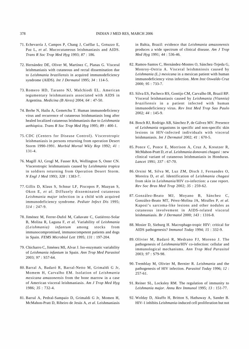
71. Echevarría J, Campos P, Chang J, Cuéllar L, Gotuzzo E,Paz L, et al. Mucocutaneous leishmaniasis and AIDS.Trans R Soc Trop Med Hyg 1993; 87 : 186.
72. Hernández DE, Oliver M, Martínez C, Planas G. Visceralleishmaniasis with cutaneous and rectal dissemination dueto Leishmania braziliensis in acquired immunodeficiencysyndrome (AIDS). Int J Dermatol 1995; 34 : 114-5.
73. Romero HD, Taranto NJ, Malchiodi EL. Americantegumentary leishmaniasis associated with AIDS inArgentina. Medicina (B Aires) 2004; 64 : 47-50.
74. Berhe N, Hailu A, Gemetchu T. Human immunodeficiencyvirus and recurrence of cutaneous leishmaniasis long afterhealed localized cutaneous leishmaniasis due to Leishmaniaaethiopica. Trans R Soc Trop Med Hyg 1995; 89 : 400-1.
75. CDC (Centers for Disease Control) . Viscerotropicleishmaniasis in persons returning from operation DesertStorm 1990-1991. Morbid Mortal Wkly Rep 1992; 41 :131-4.
76. Magill AJ, Grogl M, Fasser RA, Wellington S, Oster CN.Viscerotropic leishmaniasis caused by Leishmania tropicain soldiers returning from Operation Desert Storm.N Engl J Med 1993; 328 : 1383-7.
77. Gillis D, Klaus S, Schnur LF, Piscopos P, Maayan S,Okon E, et a l. D i f fusely d isseminated cutaneousLeishmania major infection in a child with acquiredimmunodeficiency syndrome. Pediatr Infect Dis 1995;514 : 247-9.
78. Jiménez M, Ferrer-Dufol M, Cañavate C, Gutiérrez-SolarB, Molina R, Laguna F, et al. Variability of Leishmania(Leishmania) infantum among stocks fromimmunocompromised, immunocompetent patients and dogsin Spain. FEMS Microbiol Lett 1995; 131 : 197-204.
79. Chicharro C, Jiménez MI, Alvar J. Iso-enzymatic variabilityof Leishmania infantum in Spain. Ann Trop Med Parasitol2003; 97 : S57-64.
80. Barral A, Badaró R, Barral-Netto M, Grimaldi G Jr,Momem H, Carvalho EM. Isolat ion of Leishmaniamexicana amazonensis from the bone marrow in a caseof American visceral leishmaniasis. Am J Trop Med Hyg1986; 35 : 732-4.
81. Barral A, Pedral-Sampaio D, Grimaldi G Jr, Momen H,McMahon-Pratt D, Ribeiro de Jesús A, et al. Leishmaniasis
in Bahia, Brazil: evidence that Leishmania amazonensisproduces a wide spectrum of clinical disease. Am J TropMed Hyg 1991; 44 : 536-46.
82. Ramos-Santos C, Hernández-Montes O, Sánchez-Tejeda G,Monroy-Ostria A. Visceral leishmniosis caused byLeishmania (L.) mexicana in a mexican patient with humanimmunodeficiency virus infection. Mem Inst Oswaldo Cruz2000; 95 : 733-7.
83. Silva ES, Pacheco RS, Gontijo CM, Carvalho IR, Brazil RP.Visceral leishmaniasis caused by Leishmania (Viannia)brazil iensis in a patient infected with humanimmunodeficiency virus. Rev Inst Med Trop Sao Paulo2002; 44 : 145-9.
84. Bosch RJ, Rodrigo AB, Sánchez P, de Gálvez MV. Presenceof Leishmania organisms in specific and non-specific skinlesions in HIV-infected individuals with visceralleishmaniasis. Int J Dermatol 2002; 41 : 670-5.
85. Ponce C, Ponce E, Morrison A, Cruz A, Kreutzer R,McMahon-Pratt D, et al. Leishmania donovani chagasi : newclinical variant of cutaneous leishmaniasis in Honduras.Lancet 1991; 337 : 67-70.
86. Orsini M, Silva M, Luz ZM, Disch J, Fernandes O,Moreira D, et al. Identification of Leishmania chagasifrom skin in Leishmania/HIV co-infection: a case report.Rev Soc Bras Med Trop 2002; 35 : 259-62.
87. González-Beato MJ, Moyano B, Sánchez C,González-Beato MT, Pérez-Molina JA, Miralles P, et al.Kaposi’s sarcoma-like lesions and other nodules ascutaneous involvement in AIDS-related visceralleishmaniasis. Br J Dermatol 2000; 143 : 1316-8.
88. Mosier D, Sieburg H. Macrophage-tropic HIV: critical forAIDS pathogenesis? Immunol Today 1994; 15 : 332-9.
89. Olivier M, Badaró R, Medrano FJ, Moreno J. Thepathogenesis of Leishmania/HIV co-infection: cellular andimmunological mechanisms. Ann Trop Med Parasitol2003; 97 : S79-98.
90. Tremblay M, Olivier M, Bernier R. Leishmania and thepathogenesis of HIV infection. Parasitol Today 1996; 12 :257-61.
91. Reiner SL, Locksley RM. The regulation of immunity toLeishmania major. Annu Rev Immunol 1995; 13 : 151-77.
92. Wolday D, Akuffo H, Britton S, Hathaway A, Sander B.HIV-1 inhibits Leishmania-induced cell proliferation but not
378 INDIAN J MED RES, MARCH 2006
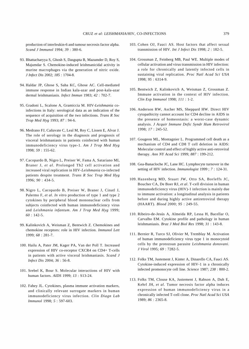
production of interleukin-6 and tumour necrosis factor alpha.Scand J Immunol 1994; 39 : 380-6.
93. Bhattacharyya S, Ghosh S, Dasgupta B, Mazumder D, Roy S,Majumdar S. Chemokine-induced leishmanicidal activity inmurine macrophages via the generation of nitric oxide.J Infect Dis 2002; 185 : 1704-8.
94. Haldar JP, Ghose S, Saha KC, Ghose AC. Cell-mediatedimmune response in Indian kala-azar and post-kala-azardermal leishmaniasis. Infect Immun 1983; 42 : 702-7.
95. Gradoni L, Scalone A, Gramiccia M. HIV-Leishmania co-infections in Italy: serological data as an indication of thesequence of acquisition of the two infections. Trans R SocTrop Med Hyg 1993; 87 : 94-6.
96. Medrano FJ, Cañavate C, Leal M, Rey C, Lissen E, Alvar J.The role of serology in the diagnosis and prognosis ofvisceral leishmaniasis in patients coinfected with humanimmunodeficiency virus type-1. Am J Trop Med Hyg1998; 59 : 155-62.
97. Cacopardo B, Nigro L, Preiser W, Fama A, Satariano MI,Braner J, et al. Prolonged Th2 cel l act ivat ion andincreased viral replication in HIV-Leishmania co-infectedpatients despite treatment. Trans R Soc Trop Med Hyg1996; 90 : 434-5.
98. Nigro L, Cacopardo B, Preiser W, Braner J, Cinatl J,Palermo F, et al. In vitro production of type 1 and type 2cytokines by peripheral blood mononuclear cells fromsubjects coinfected with human immunodeficiency virusand Leishmania infantum. Am J Trop Med Hyg 1999;60 : 142-5.
99. Kalinkovich A, Weisman Z, Bentwich Z. Chemokines andchemokine receptors: role in HIV infection. Immunol Lett1999; 68 : 281-7.
100. Hailu A, Pater JM, Kager PA, Van der Poll T. Increasedexpression of HIV co-receptor CXCR4 on CD4+ T-cellsin patients with active visceral leishmaniasis. Scand JInfect Dis 2004; 36 : 56-8.
101. Srebel K, Bour S. Molecular interactions of HIV withhuman factors. AIDS 1999; 13 : S13-24.
102. Fahey JL. Cytokines, plasma immune activation markers,and clinically relevant surrogate markers in humanimmunodeficiency virus infection. Clin Diagn LabImmunol 1998; 5 : 597-603.
103. Cohen OJ, Fauci AS. Host factors that affect sexualtransmission of HIV. Int J Infect Dis 1998; 2 : 182-5.
104. Grossman Z, Feinberg MB, Paul WE. Multiple modes ofcellular activation and virus transmission in HIV infection:a role for chronically and latently infected cells insustaining viral replication. Proc Natl Acad Sci USA1998; 95 : 6314-9.
105. Bentwich Z, Kalinkovich A, Weisman Z, Grossman Z.Immune activation in the context of HIV infection.Clin Exp Immunol 1998; 111 : 1-2.
106.Anderson RW, Ascher MS, Sheppard HW. Direct HIVcytopathicity cannot account for CD4 decline in AIDS inthe presence of homeostasis: a worst-case dynamicanalysis. J Acquir Immune Defic Syndr Hum Retrovirol1998; 17 : 245-52.
107. Gougeon ML, Montagnier L. Programmed cell death as amechanism of CD4 and CD8 T cell deletion in AIDS:Molecular control and effect of highly active anti-retroviraltherapy. Ann NY Acad Sci 1999; 887 : 199-212.
108. Gea-Banacloche JC, Lane HC. Lymphocyte turnover in thesetting of HIV infection. Immunologist 1999 ; 7 : 124-31.
109.Hazenberg MD, Stuart JW, Otto SA, Borleffs JC,Boucher CA, De Boer RJ, et al. T-cell division in humanimmunodeficiency virus (HIV)-1 infection is mainly dueto immune activation: a longitudinal analysis in patientsbefore and during highly active antiretroviral therapy(HAART). Blood 2000; 95 : 249-55.
110. Ribeiro-de-Jesús A, Almeida RP, Lessa H, Bacellar O,Carvalho EM. Cytokine profile and pathology in humanleishmaniasis. Braz J Med Biol Res 1998; 31 : 143-8.
111. Bernier R, Turco SJ, Olivier M, Tremblay M. Activationof human immunodeficiency virus type 1 in monocytoidcells by the protozoan parasite Leishmania donovani.J Virol 1995; 69 : 7282-5.
112. Folks TM, Justement J, Kinter A, Dinarello CA, Fauci AS.Cytokine-induced expression of HIV-1 in a chronicallyinfected promonocyte cell line. Science 1987; 238 : 800-2.
113. Folks TM, Clouse KA, Justement J, Rabson A, Duh E,Kehrl JH, et al. Tumor necrosis factor alpha inducesexpression of human immunodeficiency virus in achronically infected T-cell clone. Proc Natl Acad Sci USA1989; 86 : 2365-8.
CRUZ et al: LEISHMANIA/HIV, CO-INFECTIONS 379
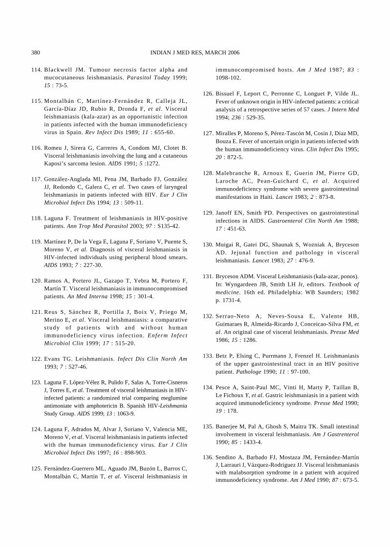
114. Blackwell JM. Tumour necrosis factor alpha andmucocutaneous leishmaniasis. Parasitol Today 1999;15 : 73-5.
115.Montalbán C, Martínez-Fernández R, Cal leja JL,García-Díaz JD, Rubio R, Dronda F, et al. Visceralleishmaniasis (kala-azar) as an opportunistic infectionin patients infected with the human immunodeficiencyvirus in Spain. Rev Infect Dis 1989; 11 : 655-60.
116. Romeu J, Sirera G, Carreres A, Condom MJ, Clotet B.Visceral leishmaniasis involving the lung and a cutaneousKaposi’s sarcoma lesion. AIDS 1991; 5 :1272.
117. González-Anglada MI, Pena JM, Barbado FJ, GonzálezJJ, Redondo C, Galera C, et al. Two cases of laryngealleishmaniasis in patients infected with HIV. Eur J ClinMicrobiol Infect Dis 1994; 13 : 509-11.
118. Laguna F. Treatment of leishmaniasis in HIV-positivepatients. Ann Trop Med Parasitol 2003; 97 : S135-42.
119. Martínez P, De la Vega E, Laguna F, Soriano V, Puente S,Moreno V, et al. Diagnosis of visceral leishmaniasis inHIV-infected individuals using peripheral blood smears.AIDS 1993; 7 : 227-30.
120. Ramos A, Portero JL, Gazapo T, Yebra M, Portero F,Martín T. Visceral leishmaniasis in immunocompromisedpatients. An Med Interna 1998; 15 : 301-4.
121.Reus S, Sánchez R, Porti l la J, Boix V, Priego M,Merino E, et al. Visceral leishmaniasis: a comparativestudy o f pat ients w i th and wi thout humanimmunodefic iency virus infect ion. Enferm InfectMicrobiol Clin 1999; 17 : 515-20.
122. Evans TG. Leishmaniasis. Infect Dis Clin North Am1993; 7 : 527-46.
123. Laguna F, López-Vélez R, Pulido F, Salas A, Torre-CisnerosJ, Torres E, et al. Treatment of visceral leishmaniasis in HIV-infected patients: a randomized trial comparing meglumineantimoniate with amphotericin B. Spanish HIV-LeishmaniaStudy Group. AIDS 1999; 13 : 1063-9.
124. Laguna F, Adrados M, Alvar J, Soriano V, Valencia ME,Moreno V, et al. Visceral leishmaniasis in patients infectedwith the human immunodeficiency virus. Eur J ClinMicrobiol Infect Dis 1997; 16 : 898-903.
125. Fernández-Guerrero ML, Aguado JM, Buzón L, Barros C,Montalbán C, Martín T, et al. Visceral leishmaniasis in
immunocompromised hosts. Am J Med 1987; 83 :1098-102.
126. Bissuel F, Leport C, Perronne C, Longuet P, Vilde JL.Fever of unknown origin in HIV-infected patients: a criticalanalysis of a retrospective series of 57 cases. J Intern Med1994; 236 : 529-35.
127. Miralles P, Moreno S, Pérez-Tascón M, Cosin J, Díaz MD,Bouza E. Fever of uncertain origin in patients infected withthe human immunodeficiency virus. Clin Infect Dis 1995;20 : 872-5.
128. Malebranche R, Arnoux E, Guerin JM, Pierre GD,Laroche AC, Pean-Guichard C, et al. Acquiredimmunodeficiency syndrome with severe gastrointestinalmanifestations in Haiti. Lancet 1983; 2 : 873-8.
129. Janoff EN, Smith PD. Perspectives on gastrointestinalinfections in AIDS. Gastroenterol Clin North Am 1988;17 : 451-63.
130. Muigai R, Gatei DG, Shaunak S, Wozniak A, BrycesonAD. Jejunal function and pathology in visceralleishmaniasis. Lancet 1983; 27 : 476-9.
131. Bryceson ADM. Visceral Leishmaniasis (kala-azar, ponos).In: Wyngardeen JB, Smith LH Jr, editors. Textbook ofmedicine. 16th ed. Philadelphia: WB Saunders; 1982p. 1731-4.
132. Serrao-Neto A, Neves-Sousa E, Valente HB,Guimaraes R, Almeida-Ricardo J, Conceicao-Silva FM, etal. An original case of visceral leishmaniasis. Presse Med1986; 15 : 1286.
133. Betz P, Elsing C, Purrmann J, Frenzel H. Leishmaniasisof the upper gastrointestinal tract in an HIV positivepatient. Pathologe 1990; 11 : 97-100.
134. Pesce A, Saint-Paul MC, Vinti H, Marty P, Taillan B,Le Fichoux Y, et al. Gastric leishmaniasis in a patient withacquired immunodeficiency syndrome. Presse Med 1990;19 : 178.
135. Banerjee M, Pal A, Ghosh S, Maitra TK. Small intestinalinvolvement in visceral leishmaniasis. Am J Gastrenterol1990; 85 : 1433-4.
136. Sendino A, Barbado FJ, Mostaza JM, Fernández-MartínJ, Larrauri J, Vázquez-Rodríguez JJ. Visceral leishmaniasiswith malabsorption syndrome in a patient with acquiredimmunodeficiency syndrome. Am J Med 1990; 87 : 673-5.
380 INDIAN J MED RES, MARCH 2006
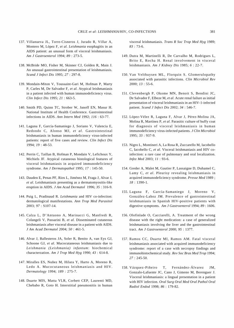
137. Villanueva JL, Torre-Cisneros J, Jurado R, Villar A,Montero M, López F, et al. Leishmania esophagitis in anAIDS patient: an unusual form of visceral leishmaniasis.Am J Gastroenterol 1994; 89 : 273-5.
138. McBride MO, Fisher M, Skinner CJ, Golden R, Main J.An unusual gastrointestinal presentation of leishmaniasis.Scand J Infect Dis 1995; 27 : 297-8.
139. Mondain-Miton V, Toussaint-Gari M, Hofman P, MartyP, Carles M, De Salvador F, et al. Atypical leishmaniasisin a patient infected with human immunodeficiency virus.Clin Infect Dis 1995; 21 : 663-5.
140. Smith PD, Quinn TC, Strober W, Janoff EN, Masur H.National Institute of Health Conference. Gastrointestinalinfections in AIDS. Ann Intern Med 1992; 116 : 63-77.
141. Laguna F, García-Samaniego J, Soriano V, Valencia E,Redondo C, Alonso MJ, et al. Gastrointestinalleishmaniasis in human immunodeficiency virus-infectedpatients: report of five cases and review. Clin Infect Dis1994; 19 : 48-53.
142. Perrin C, Taillan B, Hofman P, Mondain V, Lefichoux Y,Michiels JF. Atypical cutaneous histological features ofvisceral leishmaniasis in acquired immunodeficiencysyndrome. Am J Dermatopathol 1995; 17 : 145-50.
143. Dauden E, Penas PF, Rios L, Jiménez M, Fraga J, Alvar J,et al. Leishmaniasis presenting as a dermatomyositis-likeeruption in AIDS. J Am Acad Dermatol 1996; 35 : 316-9.
144. Puig L, Pradinaud R. Leishmania and HIV co-infection:dermatological manifestations. Ann Trop Med Parasitol2003; 97 : S107-14.
145. Calza L, D’Antuono A, Marinacci G, Manfredi R,Colangeli V, Passarini B, et al. Disseminated cutaneousleishmaniasis after visceral disease in a patient with AIDS.J Am Acad Dermatol 2004; 50 : 461-5.
146. Alvar J, Ballesteros JA, Soler R, Benito A, van Eys GJ,Schoone GJ, et al. Mucocutaneous leishmaniasis due toLeishmania (Leishmania) infantum: biochemicalcharacterzation. Am J Trop Med Hyg 1990; 43 : 614-8.
147.Miralles ES, Nuñez M, Hilara Y, Harto A, Moreno R,Ledo A. Mucocutaneous le ishmaniasis and HIV.Dermatology 1994; 189 : 275-7.
148. Duarte MIS, Matta VLR, Corbett CEP, Laurenti MD,Chebabo R, Goto H. Interstitial pneumonitis in human
visceral leishmaniasis. Trans R Soc Trop Med Hyg 1989;83 : 73-6.
149.Dutra M, Martinelli R, De Carvalho M, Rodrigues L,Bri to E, Rocha H. Renal involvement in visceralleishmaniasis. Am J Kidney Dis 1985; 6 : 22-7.
150. Van Velthuysen ML, Florquin S. Glomerulopathyassociated with parasitic infections. Clin Microbiol Rev2000; 13 : 55-6.
151. Clevenbergh P, Okome MN, Benoit S, Bendini JC,De Salvador F, Elbeze M, et al. Acute renal failure as initialpresentation of visceral leishmaniasis in an HIV-1-infectedpatient. Scand J Infect Dis 2002; 34 : 546-7.
152. López-Vélez R, Laguna F, Alvar J, Pérez-Molina JA,Molina R, Martínez P, et al. Parasitic culture of buffy coatfor diagnosis of visceral leishmaniasis in humanimmunodeficiency virus-infected patients. J Clin Microbiol1995; 33 : 937-9.
153. Nigro L, Montineri A, La Rosa R, Zuccarello M, IacobelloC, Iacobello C, et al. Visceral leishmaniasis and HIV co-infection: a rare case of pulmonary and oral localization.Infez Med 2003; 11 : 93-6.
154.Greder A, Malet M, Gautier P, Lassaigne D, Duhamel C,Lamy C, et al. Pleurisy revealing leishmaniasis inacquired immunodeficiency syndrome. Presse Med 1989 ;18 : 1390-1.
155. Laguna F, García-Samaniego J, Moreno V,González-Lahoz JM. Prevalence of gastrointestinalleishmaniasis in Spanish HIV-positive patients withdigestive symptoms. Am J Gastroenterol 1994; 89 : 1606.
156. Olofinlade O, Cacciarelli, A. Treatment of the wrongdisease with the right medication: a case of generalizedleishmaniasis involving the liver and the gastrointestinaltract. Am J Gastroenterol 2000; 95 : 1377.
157. Ramos CC, Duarte MI, Ramos AM. Fatal visceralleishmaniasis associated with acquired immunodeficiencysyndrome: report of a case with necropsy findings andimmunohistochemical study. Rev Soc Bras Med Trop 1994;27 : 245-50.
158. Vázquez-Piñeiro T, Fernández-Álvarez JM,Gonzalo-Lafuente JC, Cano J, Gimeno M, Berenguer J.Visceral leishmaniasis: a lingual presentation in a patientwith HIV infection. Oral Surg Oral Med Oral Pathol OralRadiol Endod 1998; 86 : 179-82.
CRUZ et al: LEISHMANIA/HIV, CO-INFECTIONS 381
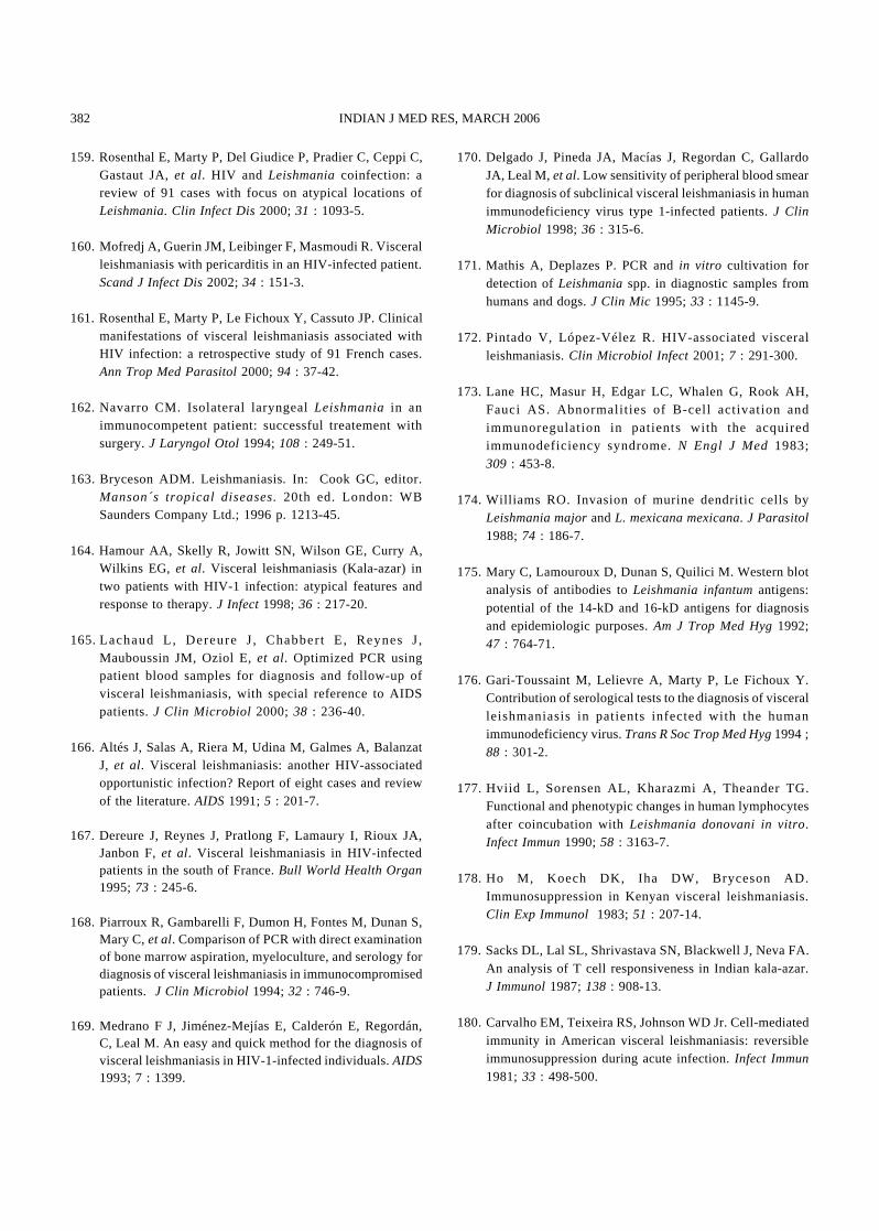
159. Rosenthal E, Marty P, Del Giudice P, Pradier C, Ceppi C,Gastaut JA, et al. HIV and Leishmania coinfection: areview of 91 cases with focus on atypical locations ofLeishmania. Clin Infect Dis 2000; 31 : 1093-5.
160. Mofredj A, Guerin JM, Leibinger F, Masmoudi R. Visceralleishmaniasis with pericarditis in an HIV-infected patient.Scand J Infect Dis 2002; 34 : 151-3.
161. Rosenthal E, Marty P, Le Fichoux Y, Cassuto JP. Clinicalmanifestations of visceral leishmaniasis associated withHIV infection: a retrospective study of 91 French cases.Ann Trop Med Parasitol 2000; 94 : 37-42.
162. Navarro CM. Isolateral laryngeal Leishmania in animmunocompetent patient: successful treatement withsurgery. J Laryngol Otol 1994; 108 : 249-51.
163. Bryceson ADM. Leishmaniasis. In: Cook GC, editor.Manson´s tropical diseases. 20th ed. London: WBSaunders Company Ltd.; 1996 p. 1213-45.
164. Hamour AA, Skelly R, Jowitt SN, Wilson GE, Curry A,Wilkins EG, et al. Visceral leishmaniasis (Kala-azar) intwo patients with HIV-1 infection: atypical features andresponse to therapy. J Infect 1998; 36 : 217-20.
165.Lachaud L, Dereure J, Chabbert E, Reynes J,Mauboussin JM, Oziol E, et al. Optimized PCR usingpatient blood samples for diagnosis and follow-up ofvisceral leishmaniasis, with special reference to AIDSpatients. J Clin Microbiol 2000; 38 : 236-40.
166. Altés J, Salas A, Riera M, Udina M, Galmes A, BalanzatJ, et al. Visceral leishmaniasis: another HIV-associatedopportunistic infection? Report of eight cases and reviewof the literature. AIDS 1991; 5 : 201-7.
167. Dereure J, Reynes J, Pratlong F, Lamaury I, Rioux JA,Janbon F, et al. Visceral leishmaniasis in HIV-infectedpatients in the south of France. Bull World Health Organ1995; 73 : 245-6.
168. Piarroux R, Gambarelli F, Dumon H, Fontes M, Dunan S,Mary C, et al. Comparison of PCR with direct examinationof bone marrow aspiration, myeloculture, and serology fordiagnosis of visceral leishmaniasis in immunocompromisedpatients. J Clin Microbiol 1994; 32 : 746-9.
169. Medrano F J, Jiménez-Mejías E, Calderón E, Regordán,C, Leal M. An easy and quick method for the diagnosis ofvisceral leishmaniasis in HIV-1-infected individuals. AIDS1993; 7 : 1399.
170. Delgado J, Pineda JA, Macías J, Regordan C, GallardoJA, Leal M, et al. Low sensitivity of peripheral blood smearfor diagnosis of subclinical visceral leishmaniasis in humanimmunodeficiency virus type 1-infected patients. J ClinMicrobiol 1998; 36 : 315-6.
171. Mathis A, Deplazes P. PCR and in vitro cultivation fordetection of Leishmania spp. in diagnostic samples fromhumans and dogs. J Clin Mic 1995; 33 : 1145-9.
172. Pintado V, López-Vélez R. HIV-associated visceralleishmaniasis. Clin Microbiol Infect 2001; 7 : 291-300.
173. Lane HC, Masur H, Edgar LC, Whalen G, Rook AH,Fauci AS. Abnormalit ies of B-cell activation andimmunoregulation in patients with the acquiredimmunodeficiency syndrome. N Engl J Med 1983;309 : 453-8.
174. Williams RO. Invasion of murine dendritic cells byLeishmania major and L. mexicana mexicana. J Parasitol1988; 74 : 186-7.
175. Mary C, Lamouroux D, Dunan S, Quilici M. Western blotanalysis of antibodies to Leishmania infantum antigens:potential of the 14-kD and 16-kD antigens for diagnosisand epidemiologic purposes. Am J Trop Med Hyg 1992;47 : 764-71.
176. Gari-Toussaint M, Lelievre A, Marty P, Le Fichoux Y.Contribution of serological tests to the diagnosis of visceralleishmaniasis in patients infected with the humanimmunodeficiency virus. Trans R Soc Trop Med Hyg 1994 ;88 : 301-2.
177. Hviid L, Sorensen AL, Kharazmi A, Theander TG.Functional and phenotypic changes in human lymphocytesafter coincubation with Leishmania donovani in vitro.Infect Immun 1990; 58 : 3163-7.
178. Ho M, Koech DK, Iha DW, Bryceson AD.Immunosuppression in Kenyan visceral leishmaniasis.Clin Exp Immunol 1983; 51 : 207-14.
179. Sacks DL, Lal SL, Shrivastava SN, Blackwell J, Neva FA.An analysis of T cell responsiveness in Indian kala-azar.J Immunol 1987; 138 : 908-13.
180. Carvalho EM, Teixeira RS, Johnson WD Jr. Cell-mediatedimmunity in American visceral leishmaniasis: reversibleimmunosuppression during acute infection. Infect Immun1981; 33 : 498-500.
382 INDIAN J MED RES, MARCH 2006
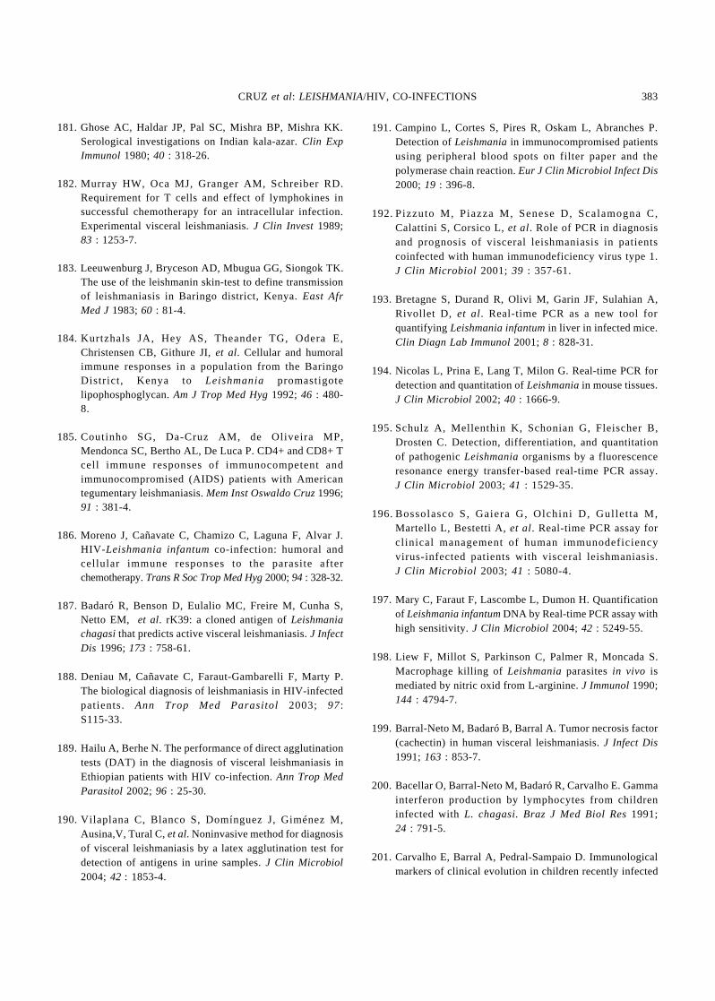
181. Ghose AC, Haldar JP, Pal SC, Mishra BP, Mishra KK.Serological investigations on Indian kala-azar. Clin ExpImmunol 1980; 40 : 318-26.
182. Murray HW, Oca MJ, Granger AM, Schreiber RD.Requirement for T cells and effect of lymphokines insuccessful chemotherapy for an intracellular infection.Experimental visceral leishmaniasis. J Clin Invest 1989;83 : 1253-7.
183. Leeuwenburg J, Bryceson AD, Mbugua GG, Siongok TK.The use of the leishmanin skin-test to define transmissionof leishmaniasis in Baringo district, Kenya. East AfrMed J 1983; 60 : 81-4.
184. Kurtzhals JA, Hey AS, Theander TG, Odera E,Christensen CB, Githure JI, et al. Cellular and humoralimmune responses in a population from the BaringoDistrict, Kenya to Leishmania promastigotelipophosphoglycan. Am J Trop Med Hyg 1992; 46 : 480-8.
185. Coutinho SG, Da-Cruz AM, de Oliveira MP,Mendonca SC, Bertho AL, De Luca P. CD4+ and CD8+ Tcell immune responses of immunocompetent andimmunocompromised (AIDS) patients with Americantegumentary leishmaniasis. Mem Inst Oswaldo Cruz 1996;91 : 381-4.
186. Moreno J, Cañavate C, Chamizo C, Laguna F, Alvar J.HIV- Leishmania infantum co-infection: humoral andcellular immune responses to the parasite afterchemotherapy. Trans R Soc Trop Med Hyg 2000; 94 : 328-32.
187. Badaró R, Benson D, Eulalio MC, Freire M, Cunha S,Netto EM, et al. rK39: a cloned antigen of Leishmaniachagasi that predicts active visceral leishmaniasis. J InfectDis 1996; 173 : 758-61.
188. Deniau M, Cañavate C, Faraut-Gambarelli F, Marty P.The biological diagnosis of leishmaniasis in HIV-infectedpatients. Ann Trop Med Parasitol 2003; 97:S115-33.
189. Hailu A, Berhe N. The performance of direct agglutinationtests (DAT) in the diagnosis of visceral leishmaniasis inEthiopian patients with HIV co-infection. Ann Trop MedParasitol 2002; 96 : 25-30.
190. Vilaplana C, Blanco S, Domínguez J, Giménez M,Ausina,V, Tural C, et al. Noninvasive method for diagnosisof visceral leishmaniasis by a latex agglutination test fordetection of antigens in urine samples. J Clin Microbiol2004; 42 : 1853-4.
191. Campino L, Cortes S, Pires R, Oskam L, Abranches P.Detection of Leishmania in immunocompromised patientsusing peripheral blood spots on filter paper and thepolymerase chain reaction. Eur J Clin Microbiol Infect Dis2000; 19 : 396-8.
192.Pizzuto M, Piazza M, Senese D, Scalamogna C,Calattini S, Corsico L, et al. Role of PCR in diagnosisand prognosis of visceral leishmaniasis in patientscoinfected with human immunodeficiency virus type 1.J Clin Microbiol 2001; 39 : 357-61.
193. Bretagne S, Durand R, Olivi M, Garin JF, Sulahian A,Rivollet D, et al. Real-time PCR as a new tool forquantifying Leishmania infantum in liver in infected mice.Clin Diagn Lab Immunol 2001; 8 : 828-31.
194. Nicolas L, Prina E, Lang T, Milon G. Real-time PCR fordetection and quantitation of Leishmania in mouse tissues.J Clin Microbiol 2002; 40 : 1666-9.
195.Schulz A, Mellenthin K, Schonian G, Fleischer B,Drosten C. Detection, differentiation, and quantitationof pathogenic Leishmania organisms by a fluorescenceresonance energy transfer-based real-time PCR assay.J Clin Microbiol 2003; 41 : 1529-35.
196.Bossolasco S, Gaiera G, Olchini D, Gul let ta M,Martello L, Bestetti A, et al. Real-time PCR assay forcl inical management of human immunodeficiencyvirus-infected patients with visceral leishmaniasis.J Clin Microbiol 2003; 41 : 5080-4.
197. Mary C, Faraut F, Lascombe L, Dumon H. Quantificationof Leishmania infantum DNA by Real-time PCR assay withhigh sensitivity. J Clin Microbiol 2004; 42 : 5249-55.
198. Liew F, Millot S, Parkinson C, Palmer R, Moncada S.Macrophage killing of Leishmania parasites in vivo ismediated by nitric oxid from L-arginine. J Immunol 1990;144 : 4794-7.
199. Barral-Neto M, Badaró B, Barral A. Tumor necrosis factor(cachectin) in human visceral leishmaniasis. J Infect Dis1991; 163 : 853-7.
200. Bacellar O, Barral-Neto M, Badaró R, Carvalho E. Gammainterferon production by lymphocytes from childreninfected with L. chagasi. Braz J Med Biol Res 1991;24 : 791-5.
201. Carvalho E, Barral A, Pedral-Sampaio D. Immunologicalmarkers of clinical evolution in children recently infected
CRUZ et al: LEISHMANIA/HIV, CO-INFECTIONS 383
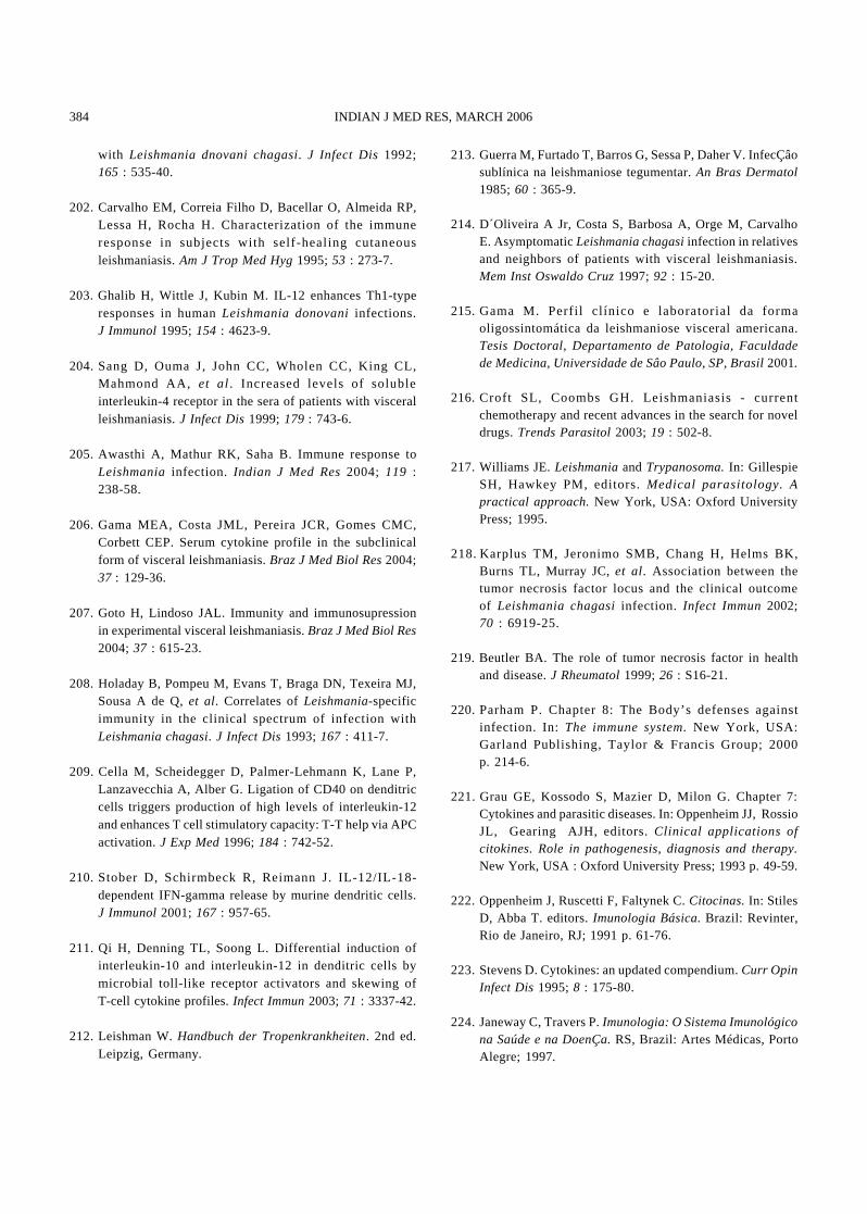
with Leishmania dnovani chagasi. J Infect Dis 1992;165 : 535-40.
202. Carvalho EM, Correia Filho D, Bacellar O, Almeida RP,Lessa H, Rocha H. Characterization of the immuneresponse in subjects with self-healing cutaneousleishmaniasis. Am J Trop Med Hyg 1995; 53 : 273-7.
203. Ghalib H, Wittle J, Kubin M. IL-12 enhances Th1-typeresponses in human Leishmania donovani infections.J Immunol 1995; 154 : 4623-9.
204. Sang D, Ouma J, John CC, Wholen CC, King CL,Mahmond AA, et al. Increased levels of solubleinterleukin-4 receptor in the sera of patients with visceralleishmaniasis. J Infect Dis 1999; 179 : 743-6.
205. Awasthi A, Mathur RK, Saha B. Immune response toLeishmania infection. Indian J Med Res 2004; 119 :238-58.
206. Gama MEA, Costa JML, Pereira JCR, Gomes CMC,Corbett CEP. Serum cytokine profile in the subclinicalform of visceral leishmaniasis. Braz J Med Biol Res 2004;37 : 129-36.
207. Goto H, Lindoso JAL. Immunity and immunosupressionin experimental visceral leishmaniasis. Braz J Med Biol Res2004; 37 : 615-23.
208. Holaday B, Pompeu M, Evans T, Braga DN, Texeira MJ,Sousa A de Q, et al. Correlates of Leishmania-specificimmunity in the clinical spectrum of infection withLeishmania chagasi. J Infect Dis 1993; 167 : 411-7.
209. Cella M, Scheidegger D, Palmer-Lehmann K, Lane P,Lanzavecchia A, Alber G. Ligation of CD40 on denditriccells triggers production of high levels of interleukin-12and enhances T cell stimulatory capacity: T-T help via APCactivation. J Exp Med 1996; 184 : 742-52.
210. Stober D, Schirmbeck R, Reimann J. IL-12/IL-18-dependent IFN-gamma release by murine dendritic cells.J Immunol 2001; 167 : 957-65.
211. Qi H, Denning TL, Soong L. Differential induction ofinterleukin-10 and interleukin-12 in denditric cells bymicrobial toll-like receptor activators and skewing ofT-cell cytokine profiles. Infect Immun 2003; 71 : 3337-42.
212. Leishman W. Handbuch der Tropenkrankheiten. 2nd ed.Leipzig, Germany.
213. Guerra M, Furtado T, Barros G, Sessa P, Daher V. InfecÇâosublínica na leishmaniose tegumentar. An Bras Dermatol1985; 60 : 365-9.
214. D´Oliveira A Jr, Costa S, Barbosa A, Orge M, CarvalhoE. Asymptomatic Leishmania chagasi infection in relativesand neighbors of patients with visceral leishmaniasis.Mem Inst Oswaldo Cruz 1997; 92 : 15-20.
215. Gama M. Perfi l clínico e laboratorial da formaoligossintomática da leishmaniose visceral americana.Tesis Doctoral, Departamento de Patologia, Faculdadede Medicina, Universidade de Sâo Paulo, SP, Brasil 2001.
216. Croft SL, Coombs GH. Leishmaniasis - currentchemotherapy and recent advances in the search for noveldrugs. Trends Parasitol 2003; 19 : 502-8.
217. Williams JE. Leishmania and Trypanosoma. In: GillespieSH, Hawkey PM, editors. Medical parasitology. Apractical approach. New York, USA: Oxford UniversityPress; 1995.
218. Karplus TM, Jeronimo SMB, Chang H, Helms BK,Burns TL, Murray JC, et al. Association between thetumor necrosis factor locus and the clinical outcomeof Leishmania chagasi infection. Infect Immun 2002;70 : 6919-25.
219. Beutler BA. The role of tumor necrosis factor in healthand disease. J Rheumatol 1999; 26 : S16-21.
220. Parham P. Chapter 8: The Body’s defenses againstinfection. In: The immune system. New York, USA:Garland Publishing, Taylor & Francis Group; 2000p. 214-6.
221. Grau GE, Kossodo S, Mazier D, Milon G. Chapter 7:Cytokines and parasitic diseases. In: Oppenheim JJ, RossioJL, Gearing AJH, editors. Clinical applications ofcitokines. Role in pathogenesis, diagnosis and therapy.New York, USA : Oxford University Press; 1993 p. 49-59.
222. Oppenheim J, Ruscetti F, Faltynek C. Citocinas. In: StilesD, Abba T. editors. Imunologia Básica. Brazil: Revinter,Rio de Janeiro, RJ; 1991 p. 61-76.
223. Stevens D. Cytokines: an updated compendium. Curr OpinInfect Dis 1995; 8 : 175-80.
224. Janeway C, Travers P. Imunologia: O Sistema Imunológicona Saúde e na DoenÇa. RS, Brazil: Artes Médicas, PortoAlegre; 1997.
384 INDIAN J MED RES, MARCH 2006
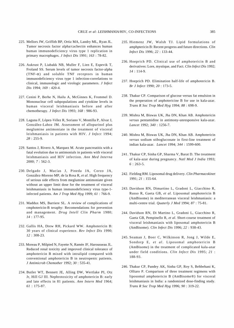
225. Mellors JW, Griffith BP, Ortiz MA, Landry ML, Ryan JL.Tumor necrosis factor alpha/cachectin enhances humanhuman immunodeficiency virus type 1 replication inprimary macrophages. J Infect Dis 1991; 163 : 78-82.
226. Aukrust P, Liabakk NB, Muller F, Lien E, Espevik T,Froland SS. Serum levels of tumor necrosis factor-alpha(TNF-α) and soluble TNF receptors in humanimmunodeficiency virus type 1 infection-correlations toclinical, immunologic and virologic parameters. J InfectDis 1994; 169 : 420-4.
227. Cenini P, Berhe N, Hailu A, McGinnes K, Frommel D.Mononuclear cell subpopulations and cytokine levels inhuman visceral leishmaniasis before and afterchemotherapy. J Infect Dis 1993; 168 : 986-93.
228. Laguna F, López-Vélez R, Soriano V, Montilla P, Alvar J,González-Lahoz JM. Assessment of allopurinol plusmeglumine antimoniate in the treatment of visceralleishmaniasis in patients with HIV. J Infect 1994;28 : 255-9.
229. Santos J, Rivero A, Marquez M. Acute pancreatitis with afatal evolution due to antimonials in patients with visceralleishmaniasis and HIV infection. Ann Med Interna2000; 7 : 562-3.
230. Delgado J, Macías J, Pineda JA, Corzo JA,González-Moreno MP, de la Rosa R, et al. High frequencyof serious side effects from meglumine antimoniate givenwithout an upper limit dose for the treatment of visceralleishmaniasis in human immunodeficiency virus type-1-infected patients. Am J Trop Med Hyg 1999; 61 : 766-9.
231. Maddux MS, Barriere SL. A review of complications ofcmphotericin-B teraphy: Recomendations for preventionand management. Drug Intel l Clin Pharm 1980;14 : 177-95.
232. Gallis HA, Drew RH, Pickard WW. Amphotericin B:30 years of clinical experience. Rev Infect Dis 1990;12 : 308-23.
233. Moreau P, Milpied N, Fayette N, Ramée JF, Harousseau JL.Reduced renal toxicity and improved clinical tolerance ofamphotericin B mixed with intralipid compared withconventional amphotericin B in neutropenic patients.J Antimicrob Chemother 1992; 30 : 535-41.
234.Butler WT, Bennett JE, Alling DW, Wertlake PI, OtzJr, Hill GJ III. Nephrotoxicity of amphotericin B: earlyand late effects in 81 patients. Ann Intern Med 1964;61 : 175-87.
235. Hiemenz JW, Walsh TJ. Lipid formulations ofamphptericin B: Recent progress and future directions. Clin
Infect Dis 1996; 22 : 133-44.
236. Hoeprich PD. Clinical use of amphotericin B andderivatives: Lore, mystique, and Fact. Clin Infect Dis 1992;
14 : 114-9.
237. Hoeprich PD. Elimination half-life of amphotericin B.Br J Infect 1990; 20 : 173-5.
238. Thakur CP. Comparison of glucose versus fat emulsion in
the preperation of anphotericine B for use in kala-azar.Trans R Soc Trop Med Hyg 1994; 88 : 698-9.
239. Mishra M, Biswas UK, Jha DN, Khan AB. Amphotericinversus pentamidine in antimony-unresponsive kala-azar.Lancet 1992; 340 : 1256-7.
240. Mishra M, Biswas UK, Jha DN, Khan AB. Amphotericinversus sodium stibogluconate in first-line treatment ofindian kala-azar. Lancet 1994; 344 : 1599-600.
241.Thakur CP, Sinha GP, Sharma V, Barat D. The treatment
of kala-azar during pregnancy. Natl Med J India 1993;6 : 263-5.
242. Fielding RM. Liposomal drug delivery. Clin Pharmacokinet
1991; 21 : 155-64.
243. Davidson RN, Dimartino L, Gradoni L, Giacchino R,Russo R, Gaeta GB, et al. Liposomal amphotericin B(AmBisome) in mediterranean visceral leishmaniasis: amulti-centre trial. Quaterly J Med 1994; 87 : 75-81.
244. Davidson RN, Di Martino L, Gradoni L, Giacchino R,
Gaeta GB, Pempinello R, et al. Short-course treatment ofvisceral leishmaniasis with liposomal amphotericin B(AmBisome). Clin Infect Dis 1996; 22 : 938-43.
245.Seaman J, Boer C, Wilkinson R, Jong J, Wilde E,Sondorp E, et a l. L iposomal amphoter ic in B
(AmBisome) in the treatment of complicated kala-azarunder field conditions. Clin Infect Dis 1995; 21 :188-93.
246. Thakur CP, Pandey AK, Sinha GP, Roy S, Behbehani K,Olliaro P. Comparison of three treatment regimens with
liposomal amphotericin B (AmBisome®) for visceralleishmaniasis in India: a randomized dose-finding study.Trans R Soc Trop Med Hyg 1996; 90 : 319-22.
CRUZ et al: LEISHMANIA/HIV, CO-INFECTIONS 385
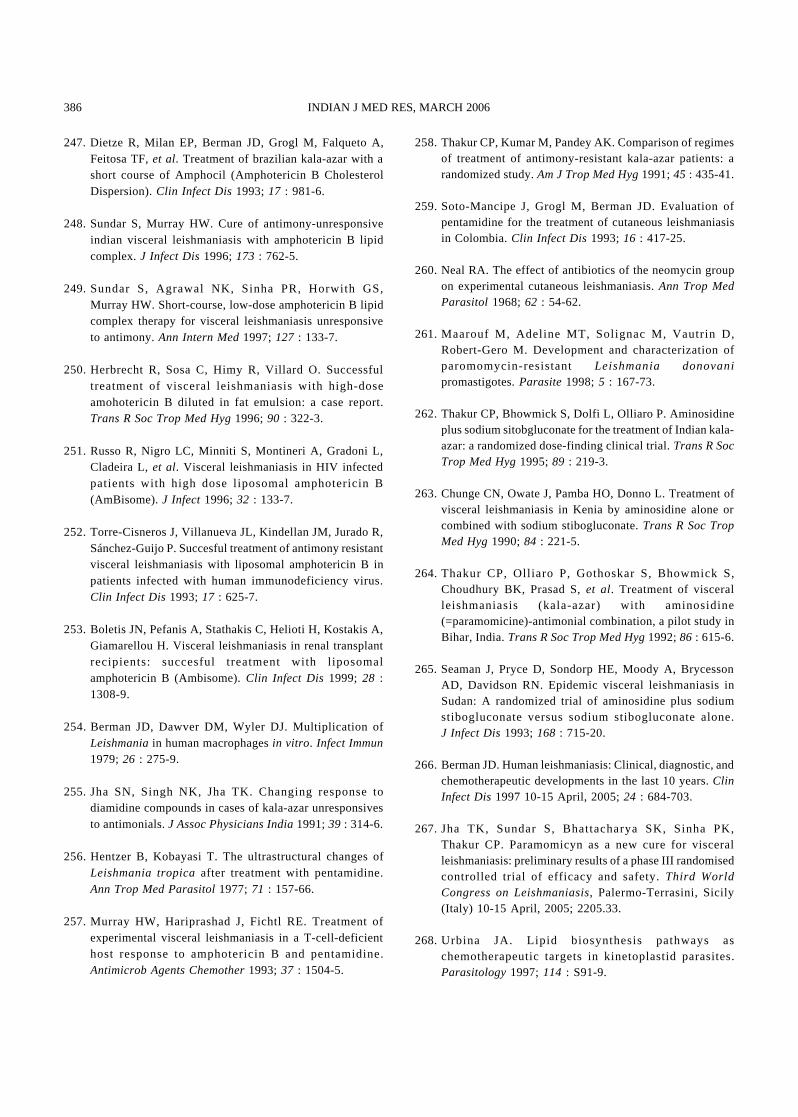
247. Dietze R, Milan EP, Berman JD, Grogl M, Falqueto A,Feitosa TF, et al. Treatment of brazilian kala-azar with ashort course of Amphocil (Amphotericin B CholesterolDispersion). Clin Infect Dis 1993; 17 : 981-6.
248. Sundar S, Murray HW. Cure of antimony-unresponsiveindian visceral leishmaniasis with amphotericin B lipidcomplex. J Infect Dis 1996; 173 : 762-5.
249. Sundar S, Agrawal NK, Sinha PR, Horwith GS,Murray HW. Short-course, low-dose amphotericin B lipidcomplex therapy for visceral leishmaniasis unresponsiveto antimony. Ann Intern Med 1997; 127 : 133-7.
250. Herbrecht R, Sosa C, Himy R, Villard O. Successfultreatment of visceral leishmaniasis with high-doseamohotericin B diluted in fat emulsion: a case report.Trans R Soc Trop Med Hyg 1996; 90 : 322-3.
251. Russo R, Nigro LC, Minniti S, Montineri A, Gradoni L,Cladeira L, et al. Visceral leishmaniasis in HIV infectedpatients with high dose l iposomal amphotericin B(AmBisome). J Infect 1996; 32 : 133-7.
252. Torre-Cisneros J, Villanueva JL, Kindellan JM, Jurado R,Sánchez-Guijo P. Succesful treatment of antimony resistantvisceral leishmaniasis with liposomal amphotericin B inpatients infected with human immunodeficiency virus.Clin Infect Dis 1993; 17 : 625-7.
253. Boletis JN, Pefanis A, Stathakis C, Helioti H, Kostakis A,Giamarellou H. Visceral leishmaniasis in renal transplantrecipients: succesful treatment with l iposomalamphotericin B (Ambisome). Clin Infect Dis 1999; 28 :1308-9.
254. Berman JD, Dawver DM, Wyler DJ. Multiplication ofLeishmania in human macrophages in vitro. Infect Immun1979; 26 : 275-9.
255. Jha SN, Singh NK, Jha TK. Changing response todiamidine compounds in cases of kala-azar unresponsivesto antimonials. J Assoc Physicians India 1991; 39 : 314-6.
256. Hentzer B, Kobayasi T. The ultrastructural changes ofLeishmania tropica after treatment with pentamidine.Ann Trop Med Parasitol 1977; 71 : 157-66.
257. Murray HW, Hariprashad J, Fichtl RE. Treatment ofexperimental visceral leishmaniasis in a T-cell-deficienthost response to amphotericin B and pentamidine.Antimicrob Agents Chemother 1993; 37 : 1504-5.
258. Thakur CP, Kumar M, Pandey AK. Comparison of regimesof treatment of antimony-resistant kala-azar patients: arandomized study. Am J Trop Med Hyg 1991; 45 : 435-41.
259. Soto-Mancipe J, Grogl M, Berman JD. Evaluation ofpentamidine for the treatment of cutaneous leishmaniasisin Colombia. Clin Infect Dis 1993; 16 : 417-25.
260. Neal RA. The effect of antibiotics of the neomycin groupon experimental cutaneous leishmaniasis. Ann Trop MedParasitol 1968; 62 : 54-62.
261. Maarouf M, Adeline MT, Solignac M, Vautrin D,Robert-Gero M. Development and characterization ofparomomycin-resistant Leishmania donovanipromastigotes. Parasite 1998; 5 : 167-73.
262. Thakur CP, Bhowmick S, Dolfi L, Olliaro P. Aminosidineplus sodium sitobgluconate for the treatment of Indian kala-azar: a randomized dose-finding clinical trial. Trans R SocTrop Med Hyg 1995; 89 : 219-3.
263. Chunge CN, Owate J, Pamba HO, Donno L. Treatment ofvisceral leishmaniasis in Kenia by aminosidine alone orcombined with sodium stibogluconate. Trans R Soc TropMed Hyg 1990; 84 : 221-5.
264. Thakur CP, Oll iaro P, Gothoskar S, Bhowmick S,Choudhury BK, Prasad S, et al. Treatment of visceralleishmaniasis (kala-azar) with aminosidine(=paramomicine)-antimonial combination, a pilot study inBihar, India. Trans R Soc Trop Med Hyg 1992; 86 : 615-6.
265. Seaman J, Pryce D, Sondorp HE, Moody A, BrycessonAD, Davidson RN. Epidemic visceral leishmaniasis inSudan: A randomized trial of aminosidine plus sodiumstibogluconate versus sodium stibogluconate alone.J Infect Dis 1993; 168 : 715-20.
266. Berman JD. Human leishmaniasis: Clinical, diagnostic, andchemotherapeutic developments in the last 10 years. ClinInfect Dis 1997 10-15 April, 2005; 24 : 684-703.
267. Jha TK, Sundar S, Bhattacharya SK, Sinha PK,Thakur CP. Paramomicyn as a new cure for visceralleishmaniasis: preliminary results of a phase III randomisedcontrolled trial of efficacy and safety. Third WorldCongress on Leishmaniasis, Palermo-Terrasini, Sicily(Italy) 10-15 April, 2005; 2205.33.
268. Urbina JA. Lipid biosynthesis pathways aschemotherapeutic targets in kinetoplastid parasites.Parasitology 1997; 114 : S91-9.
386 INDIAN J MED RES, MARCH 2006
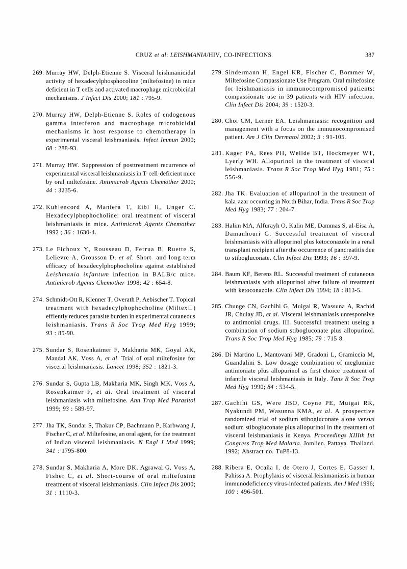
269. Murray HW, Delph-Etienne S. Visceral leishmanicidalactivity of hexadecylphosphocoline (miltefosine) in micedeficient in T cells and activated macrophage microbicidalmechanisms. J Infect Dis 2000; 181 : 795-9.
270. Murray HW, Delph-Etienne S. Roles of endogenousgamma interferon and macrophage microbicidalmechanisms in host response to chemotherapy inexperimental visceral leishmaniasis. Infect Immun 2000;68 : 288-93.
271. Murray HW. Suppression of posttreatment recurrence ofexperimental visceral leishmaniasis in T-cell-deficient miceby oral miltefosine. Antimicrob Agents Chemother 2000;44 : 3235-6.
272. Kuhlencord A, Maniera T, Eibl H, Unger C.Hexadecylphophocholine: oral treatment of visceralleishmaniasis in mice. Antimicrob Agents Chemother1992 ; 36 : 1630-4.
273. Le Fichoux Y, Rousseau D, Ferrua B, Ruette S,Lelievre A, Grousson D, et al. Short- and long-termefficacy of hexadecylphophocholine against establishedLeishmania infantum infection in BALB/c mice.Antimicrob Agents Chemother 1998; 42 : 654-8.
274. Schmidt-Ott R, Klenner T, Overath P, Aebischer T. Topicaltreatment with hexadecylphophocholine (Miltex)effiently reduces parasite burden in experimental cutaneousleishmaniasis. Trans R Soc Trop Med Hyg 1999;93 : 85-90.
275. Sundar S, Rosenkaimer F, Makharia MK, Goyal AK,Mandal AK, Voss A, et al. Trial of oral miltefosine forvisceral leishmaniasis. Lancet 1998; 352 : 1821-3.
276. Sundar S, Gupta LB, Makharia MK, Singh MK, Voss A,Rosenkaimer F, et al. Oral treatment of visceralleishmaniasis with miltefosine. Ann Trop Med Parasitol1999; 93 : 589-97.
277. Jha TK, Sundar S, Thakur CP, Bachmann P, Karbwang J,Fischer C, et al. Miltefosine, an oral agent, for the treatmentof Indian visceral leishmaniasis. N Engl J Med 1999;341 : 1795-800.
278.Sundar S, Makharia A, More DK, Agrawal G, Voss A,Fisher C, et al. Short-course of oral mi l tefosinetreatment of visceral leishmaniasis. Clin Infect Dis 2000;31 : 1110-3.
279. Sindermann H, Engel KR, Fischer C, Bommer W,Miltefosine Compassionate Use Program. Oral miltefosinefor leishmaniasis in immunocompromised patients:compassionate use in 39 patients with HIV infection.Clin Infect Dis 2004; 39 : 1520-3.
280. Choi CM, Lerner EA. Leishmaniasis: recognition andmanagement with a focus on the immunocompromisedpatient. Am J Clin Dermatol 2002; 3 : 91-105.
281. Kager PA, Rees PH, Wellde BT, Hockmeyer WT,Lyerly WH. Allopurinol in the treatment of visceralleishmaniasis. Trans R Soc Trop Med Hyg 1981; 75 :556-9.
282. Jha TK. Evaluation of allopurinol in the treatment ofkala-azar occurring in North Bihar, India. Trans R Soc TropMed Hyg 1983; 77 : 204-7.
283. Halim MA, Alfurayh O, Kalin ME, Dammas S, al-Eisa A,Damanhouri G. Successful treatment of visceralleishmaniasis with allopurinol plus ketoconazole in a renaltransplant recipient after the occurrence of pancreatitis dueto stibogluconate. Clin Infect Dis 1993; 16 : 397-9.
284. Baum KF, Berens RL. Successful treatment of cutaneousleishmaniasis with allopurinol after failure of treatmentwith ketoconazole. Clin Infect Dis 1994; 18 : 813-5.
285. Chunge CN, Gachihi G, Muigai R, Wassuna A, RachidJR, Chulay JD, et al. Visceral leishmaniasis unresponsiveto antimonial drugs. III. Successful treatment useing acombination of sodium stibogluconate plus allopurinol.Trans R Soc Trop Med Hyg 1985; 79 : 715-8.
286. Di Martino L, Mantovani MP, Gradoni L, Gramiccia M,Guandalini S. Low dosage combination of meglumineantimoniate plus allopurinol as first choice treatment ofinfantile visceral leishmaniasis in Italy. Tans R Soc TropMed Hyg 1990; 84 : 534-5.
287. Gachihi GS, Were JBO, Coyne PE, Muigai RK,Nyakundi PM, Wasunna KMA, et al. A prospectiverandomized trial of sodium stibogluconate alone versussodium stibogluconate plus allopurinol in the treatment ofvisceral leishmaniasis in Kenya. Proceedings XIIIth IntCongress Trop Med Malaria. Jomlien. Pattaya. Thailand.1992; Abstract no. TuP8-13.
288. Ribera E, Ocaña I, de Otero J, Cortes E, Gasser I,Pahissa A. Prophylaxis of visceral leishmaniasis in humanimmunodeficiency virus-infected patients. Am J Med 1996;100 : 496-501.
CRUZ et al: LEISHMANIA/HIV, CO-INFECTIONS 387
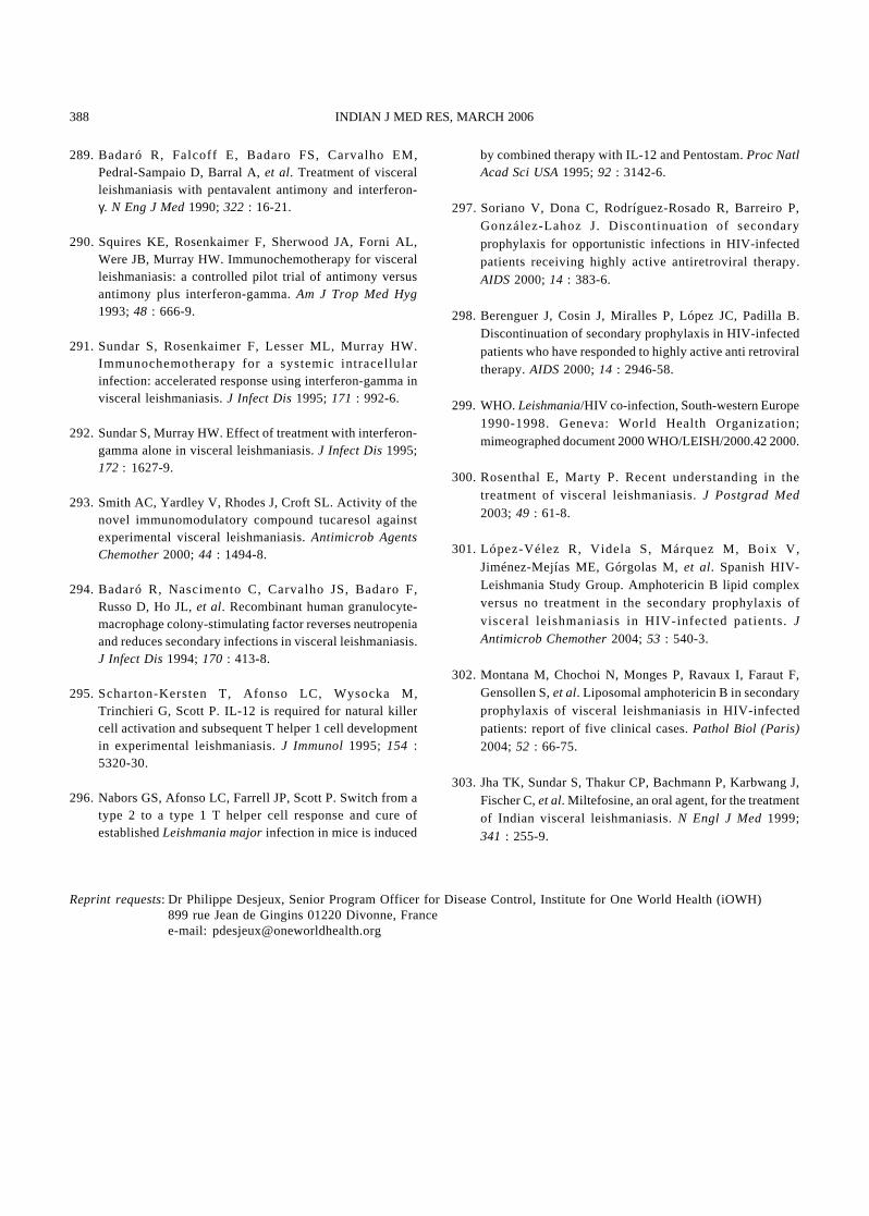
289. Badaró R, Falcoff E, Badaro FS, Carvalho EM,Pedral-Sampaio D, Barral A, et al. Treatment of visceralleishmaniasis with pentavalent antimony and interferon-γ. N Eng J Med 1990; 322 : 16-21.
290. Squires KE, Rosenkaimer F, Sherwood JA, Forni AL,Were JB, Murray HW. Immunochemotherapy for visceralleishmaniasis: a controlled pilot trial of antimony versusantimony plus interferon-gamma. Am J Trop Med Hyg1993; 48 : 666-9.
291. Sundar S, Rosenkaimer F, Lesser ML, Murray HW.Immunochemotherapy for a systemic intracellularinfection: accelerated response using interferon-gamma invisceral leishmaniasis. J Infect Dis 1995; 171 : 992-6.
292. Sundar S, Murray HW. Effect of treatment with interferon-gamma alone in visceral leishmaniasis. J Infect Dis 1995;172 : 1627-9.
293. Smith AC, Yardley V, Rhodes J, Croft SL. Activity of thenovel immunomodulatory compound tucaresol againstexperimental visceral leishmaniasis. Antimicrob AgentsChemother 2000; 44 : 1494-8.
294. Badaró R, Nascimento C, Carvalho JS, Badaro F,Russo D, Ho JL, et al. Recombinant human granulocyte-macrophage colony-stimulating factor reverses neutropeniaand reduces secondary infections in visceral leishmaniasis.J Infect Dis 1994; 170 : 413-8.
295. Scharton-Kersten T, Afonso LC, Wysocka M,Trinchieri G, Scott P. IL-12 is required for natural killercell activation and subsequent T helper 1 cell developmentin experimental leishmaniasis. J Immunol 1995; 154 :5320-30.
296. Nabors GS, Afonso LC, Farrell JP, Scott P. Switch from atype 2 to a type 1 T helper cell response and cure ofestablished Leishmania major infection in mice is induced
by combined therapy with IL-12 and Pentostam. Proc NatlAcad Sci USA 1995; 92 : 3142-6.
297. Soriano V, Dona C, Rodríguez-Rosado R, Barreiro P,González-Lahoz J. Discontinuation of secondaryprophylaxis for opportunistic infections in HIV-infectedpatients receiving highly active antiretroviral therapy.AIDS 2000; 14 : 383-6.
298. Berenguer J, Cosin J, Miralles P, López JC, Padilla B.Discontinuation of secondary prophylaxis in HIV-infectedpatients who have responded to highly active anti retroviraltherapy. AIDS 2000; 14 : 2946-58.
299. WHO. Leishmania/HIV co-infection, South-western Europe1990-1998. Geneva: World Health Organization;mimeographed document 2000 WHO/LEISH/2000.42 2000.
300. Rosenthal E, Marty P. Recent understanding in thetreatment of visceral leishmaniasis. J Postgrad Med
2003; 49 : 61-8.
301. López-Vélez R, Videla S, Márquez M, Boix V,Jiménez-Mejías ME, Górgolas M, et al. Spanish HIV-Leishmania Study Group. Amphotericin B lipid complexversus no treatment in the secondary prophylaxis ofvisceral leishmaniasis in HIV-infected patients. JAntimicrob Chemother 2004; 53 : 540-3.
302. Montana M, Chochoi N, Monges P, Ravaux I, Faraut F,Gensollen S, et al. Liposomal amphotericin B in secondaryprophylaxis of visceral leishmaniasis in HIV-infectedpatients: report of five clinical cases. Pathol Biol (Paris)2004; 52 : 66-75.
303. Jha TK, Sundar S, Thakur CP, Bachmann P, Karbwang J,Fischer C, et al. Miltefosine, an oral agent, for the treatmentof Indian visceral leishmaniasis. N Engl J Med 1999;341 : 255-9.
388 INDIAN J MED RES, MARCH 2006
Reprint requests: Dr Philippe Desjeux, Senior Program Officer for Disease Control, Institute for One World Health (iOWH)899 rue Jean de Gingins 01220 Divonne, Francee-mail: [email protected]