IMMUNODIAGNOSIS OF PARASITIC INFECTIONS USING ...
108
IAEA-TECDOC-339 IMMUNODIAGNOSIS OF PARASITIC INFECTIONS USING NUCLEAR TECHNIQUES REPORT OF AN ADVISORY GROUP MEETING ON IMMUNODIAGNOSIS OF PARASITIC INFECTIONS USING NUCLEAR TECHNIQUES ORGANIZED BY THE INTERNATIONAL ATOMIC ENERGY AGENCY AND HELD IN VIENNA, 7-10 MAY 1984 A TECHNICAL DOCUMENT ISSUED BY THE (.. INTERNATIONAL ATOMIC ENERGY AGENCY, VIENNA, 1985
-
Upload
khangminh22 -
Category
Documents
-
view
1 -
download
0
Transcript of IMMUNODIAGNOSIS OF PARASITIC INFECTIONS USING ...
IAEA-TECDOC-339
IMMUNODIAGNOSISOF PARASITIC INFECTIONS
USING NUCLEAR TECHNIQUESREPORT OF AN ADVISORY GROUP MEETING
ON IMMUNODIAGNOSIS OF PARASITIC INFECTIONSUSING NUCLEAR TECHNIQUES
ORGANIZED BY THEINTERNATIONAL ATOMIC ENERGY AGENCY
AND HELD IN VIENNA, 7-10 MAY 1984
A TECHNICAL DOCUMENT ISSUED BY THE(.. INTERNATIONAL ATOMIC ENERGY AGENCY, VIENNA, 1985

IMMUNODIAGNOSIS OF PARASITIC INFECTIONS USING NUCLEAR TECHNIQUESIAEA, VIENNA, 1985IAEA-TECDOC-339
Printed by the IAEA in AustriaJuly 1985

PLEASE BE AWARE THATALL OF THE MISSING PAGES IN THIS DOCUMENT
WERE ORIGINALLY BLANK

The IAEA does not maintain stocks of reports in this series. However,microfiche copies of these reports can be obtained from
INIS ClearinghouseInternational Atomic Energy AgencyWagramerstrasse 5P.O. Box 100A-1400 Vienna, Austria
Orders should be accompanied by prepayment of Austrian Schillings 80.00in the form of a cheque or in the form of IAEA microfiche service couponswhich may be ordered separately from the INIS Clearinghouse.
FOREWORD
Parasitic infections are an important cause of morbidity and mortalityparticularly in the tropics and sub-tropics and in recent years increasing atten-tion has been focussed on their control. This focus has highlighted the needfor effective diagnostic methods as the distinction between the presence of theinfection and the clinical manifestation is frequently difficult to interpret.Direct identification of parasites and their ova in faeces, urine, blood and biopsymaterial can be difficult and immunodiagnostic methods have been lookedupon as the more attractive option.
The initial advances in immunodiagnostic technology were largely in thedevelopment of antibody-detection systems such as the radioimmunoassay.However, the limitations of such systems, in terms of specificity, ability todiagnose pre-clinical infections and to distinguish between current and pastinfections; and the availability of new technologies such as hybridoma andgenetic engineering, have led to attention being directed to antigen-detectionsystems. Nuclear techniques employed in conjunction with others, areimportant tools in the development and, to a lesser extent, the routine applica-tion of both antibody and antigen detection systems in immunodiagnosis.
In May 1984 the International Atomic Energy Agency (IAEA) convenedan Advisory Group Meeting of experts to review and appraise the recentadvances in the development and application of immunodiagnostic techniques,including biotechnical methods for the production of diagnostic antigensand antibodies. This publication originates from that meeting and consists of asummary report prepared collectively by the Advisory Group, followed by aseries of working papers prepared by individual contributors.
CONTENTS
I. SUMMARY REPORT ........................................ .......................... 7Immunodiagnosis of parasitic infections using nuclear techniques
1. INTRODUCTION .............................................................. 7
2. CURRENT STATUS ........................................ ...................... 7
3. GOALS FOR THE DEVELOPMENT OF IMMUNODIAGNOSTICMETHODOLOGIES ............................................................................................ 10
4. PROBLEMS IN IMMUNODIAGNOSIS ................................................................ 11
5. AREAS FOR DEVELOPMENT ............................................................................ 12
6. CONCLUSIONS .................................................................................................... 12
II. WORKING PAPERS
Isotopic immunoassay systems for measuring antibodies, antigens and immune complexesin parasitic/infectious diseases ........................................................................................ 15D.K. Hazra
Developments in immunoassays for diagnosis of parasitic diseases ........................................ 27E. Linder
Immunoassays of antibodies and circulating antigens in schistosomiasis ................................ 35J.P. Dessaint and A. Capron
Production of schistosome antigens for immunodiagnosis and vaccines:The role of recombinant DNA technology ........................................................................ 51D. W. Taylor, J.S. Cordingley, K.S. Johnson, A.E. Butterworth
Studies on epitopic and idiotypic regulation in schistosomiasis:The use of monoclonal antibodies in radio-immuno- and avidin-biotin-enzyme assays ........ 63S.M. Phillips, E.G. Fox, M.N. Fathelbab, D.J. Walker, and D.M. Zodda
Radiotracer techniques in the immunodiagnosis of filariasis in man .................................... 75R. G. Hamilton
Im m unodiagnosis of m alaria ................................................................................................ 91L.H. Perrin and M. Gabra
Use of radionuclide labels in the identification and isolation of merozoite antigens fromthe human malarial parasite, Plasmodium falciparum for the development ofim m unodiagnostic m ethods ......................................................................................... 99H. G. Heidrich
Immunodiagnosis of filariasis, schistosomiasis and malaria .................................................... 103V. Houba, K.E. Mott and L.J. Martinez
III. LIST OF PARTICIPANTS ............................................................................................ 11
I. SUMMARY REPORT
IMMUNODIAGNOSIS OF PARASITIC INFECTIONS USINGNUCLEAR TECHNIQUES
Advisory Group of theInternational Atomic Energy Agency,Vienna
1. INTRODUCTION
This report documents the recommendations of the "Advisory Group onImmunodiagnosis of Parasitic Infections Using Nuclear Techniques" with afocus on malaria, schistosomiasis and filariasis. Radionuclide tracersare considered an important component of present and future immunologicalmethods for the assessment of the host's humoral and cellular immunity tothe parasite and the detection of parasite antigen(s) in human bodyfluids. The Advisory Group has concluded that there is a continuing needfor the development and application of immunodiagnostic methods inparasitic diseases.
This report concerns methods which are currently or potentiallyapplicable to immunodiagnostic investigations in parasitic diseases.Other analytical methods are being used in research for thecharacterization of parasite antigens and antibodies, but thesetechniques are not at present directly applicable to immunodiagnosis.Therefore, they will not be addressed in this report. Reference is made,where appropriate, to recent developments in research which may lead toimprovement and standardization of methods now available and thedevelopment of new methodology.
2. CURRENT STATUS
2.1. Immunodiagnosis of Malaria:
Plasmodium falciparum is the most pathogenic species of plasmodiainfecting man. Therefore, most of the test systems noted in this reporthave been developed in relation to P.falciparum infection. Some are usedfor other human plasmodial species and others should be adapted forP.vivax and P.malariae.
2.1.1. Tests related to in vitro functional assays
a. Antibody mediated immunity
A number of assays have been used to evaluate the ability ofantibody to interfere with specific functions of the malaria parasites.These include: inhibition of growth and maturation of asexual bloodforms; inhibition of invasion of RBC by merozoites; aggregation ofmerozoites; agglutination of schizont infected cells (P.knowlesi);inhibition of binding of infected RBC to endothelial cells; inhibition ofsporozoite invasion; and inhibition of fertilization and development ofsporogonic stages.
7
b. Cell mediated immunity
A limited number of assays reflecting cellular immune responsesare being used in the course of research. These may lead to developmentof test systems for application in clinical and epidemiologicalinvestigations. These assays include: delayed-type hypersensitivity(DTH); lymphocyte proliferation; enhanced phagocytosis; released solublemediators including lymphokines.
2.1.2. Detection of host antibody
Several methods are available for the detection of human circulatingantibody in clinical and seroepidemiological studies. The presence ofspecific antibody proves a host-parasite contact but does notdiscriminate between present and past infection, nor does it indicate thelevel of protective immunity. Antibody measurement is a valuableepidemiological tool for the assessment of transmission and formonitoring of malaria control measures. Most of the work in this areaconcerns antibody to asexual blood stages. A limited number of studiesconcern antibodies directed against sporozoites and gametocytes.
Tests involving antibodies directed against asexual blood formsinclude: indirect immunofluorescence (IFA); indirect haemagglutination(IHA); radioimmunoassay (RIA); enzyme linked immunosorbent assay (ELISA);
Tests involving antibodies directed against sporozoites include IFAand circumsporozoite precipitation (CSP);.
2.1.3. Detection of parasite antigens
a. Sporozoites
Monitoring of vector control in endemic areas up to now requiredmicroscopic examination of individual mosquitoes for the detection ofsporozoites. These procedures did not identify specific plasmodiaspecies. The recent development of a solid-phase, sandwich assay, whichis specific for one of several species of plasmodia infecting man, shouldprovide a valuable method for the evaluation of malaria transmission.The assay in its present form uses radiolabelled monoclonal antibody tosporozoite surface protein.
b. Asexual blood stages
The purpose of tests for the detection of blood stage antigens is toindicate the presence of a specific current infection and to quantitatethe intensity of that infection. Such tests will be of value inepidemiological studies and in a future evaluation of malaria vaccines.Methods, available for the detection of soluble circulating antigens,include: Ouchterlony precipitation and countercurrentimmunoelectrophoresis (CIE). Methods to detect parasitized RBC include:solid-phase RIA and solid-phase ELISA.
The presently available tests for soluble antigen detection are notsensitive enough for large scale epidemiological use. These tests arepositive only if the parasitemia exceeds 1%. In recently developedsolid-phase assays, parasitized RBC can be detected at a level ofapproximately 5 per 106 RBC. However, these test systems are not yet
8
suitable for general epidemiological application. Defined and/orpurified reagents will be required for their improvement andstandardization.
2.2. Immunodiagnosis of Schistosomiasis:
At the present time, the detection and quantitation ofschistosomiasis relies primarily on microscopic examination of stool andurine samples and there remains a need for sensitive, inexpensive, field-reliable serological tests. Ideally, future tests should accuratelyreflect a number of epidemiological aspects including current intensityof infection; prevalance; morbidity; response to therapy (chemical/vaccine); and resistance to re-infection. At the current time no suchtests exist.
2.2.1. Tests related to in vitro functional assays
Tests in this category should be considered to have potential as invitro correlates to in vivo characteristics such as resistance orpropensity to morbidity. A number of such tests have been developed andstandardized. These include antibody-dependent cell-mediated reactionsinvolving the ability of a number of classes of immunoglobulins tomediate schistosomule killing by a number of effector cells includingeosinophils and macrophages. These tests would reflect both specificreactivity against the schistosomes as well as general immunologicalcompetence. Other tests in this category include in vitro measurement ofdirect humoral and cellular cytotoxicity, and in vivo skin tests whichmeasure the immediate or delayed hypersensitivity skin test reactivityagainst injected antigen.
2.2.2. Detection of host antibody
Tests in this category should be considered to be indicators of theprevalence of infection. A number of tests in this category have beendeveloped and standardized. These include the use of radionuclide andother tracers in tests involving antibody binding to live organisms andto a variety of antigenic preparations; measurement of antigen - specificblast transformation; and the release of lymphokines. During the pastfew years the sensitivity and specificity of these tests have improveddramatically but there is still a need for improving the reagents used inthese tests.
2.2.3. Detection of parasite antigens
Tests in this category should be considered to be indicative of theintensity of current infection. A number of tests in this category havebeen developed and standardized. These include the use of radionuclideand other tracers in tests to detect antigen directly and a variety ofinhibition and immune complex formation techniques. At the present timethis category of tests is most in need of development.
2.3. Immunodiagnosis of Filariasis:
Despite advances in serological methods for the diagnosis of humanfilariasis, a definitive diagnosis of a filarial infection often can beestablished only after a careful clinical history, physical examinationand a microscopic confirmation of microfilariae in the blood (Wuchereriabancrofti, Brugia malayi) or skin (Onchocerca volvulus) of the infectedhost. The major problem with the blood smear or skin biopsy is the
9
limited sensitivity of the test. This characteristic limits its abilityto differentiate, in regions where filariasis is endemic, between anon-infected individual and one who is infected but does not have micro-filariae in the peripheral circulation. Patients with microfilaremiaand/or chronic pathology (skin pathology and lymphatic blockage) are morereadily diagnosed.
2.3.1. Detection of host antibody
Antibody assays are presently at a refined state from the technicalpoint of view. Solid-phase immunoassays using radionuclide(immunoradiometric assays) and enzyme tracers (ELISA) are availableworld-wide in reference laboratories. These techniques permit theaccurate and precise quantitation of antifilarial, antigen-specific IgEand IgG antibodies in serum. The major constraint in these assaysresides in the "quality of antigen" employed and not on the tracer orsolid phase technology per se. The reasons for the apparent poorcorrelation between the levels of antifilarial, antigen-specific IgE andIgG antibodies and the degree of infection relates to many factors.Clearly, reagent (specificity of antigen) and parasitological (absorptionby circulating microfilariae) constraints are important in this regard.
2.3.2. Detection of parasite antigen
Multiple immunological methods including competitive andnon-competitive forms of immunoassays have been investigated for theirability to detect filarial antigen in the human body fluids. Of theavailable immunoassays, the non-competitive immunoradiometric assayappears to provide the most sensitive design using the existentreagents. The current major issue regarding immunoassays is reagentquality and not assay design.
3. GOALS FOR THE DEVELOPMENT OF IMMUNODIAGNOSTIC METHODOLOGIES
3.1. Preparation and Standardization of Reagents:
These reagents must be related to appropriate in vivo events. Theirproduction will require the use of existing methodology and the furtherdevelopment of sophisticated in vitro technology. This technologyincludes: the improvement of in vitro culture of parasites; methods forantigen fractionation; monoclonal antibody production; and techniques forantigen production (DNA technology and producton of syntheticpolypeptides).
3.2. Development of Suitable Methods for the Detection of Stage-specificParasite Antigens in Human Body Fluids:
These methods should be specific, sensitive, standardized, simpleand cheap, and permit the accurate assessment of a prepatent state, thelevel of current infection, and of responses to treatment, vaccinationand re-exposure.
3.3. Improvement, Particularly in Standardization, of tihe Methods for theDetection of Antibodies:
It is hoped that parasite antigens, now being produced or purifiedfor vaccine research, will become available in the near future for use inthese tests.
10
3.4. Development of Tests for Assessment of Protective Immunity andPropensity to Morbidity:
Such tests will be necessary for the evaluation of future parasitevaccines. Some of the existing functional assays may be relevant tothese questions but further investigation is required to establish thiswith certainty.
4. PROBLEMS IN IMMUNODIAGNOSIS
4.1. Availability of Defined Reagents:
At the present time, monoclonal antibodies are increasinglyavailable; however, the definition of their specificity and theirdistribution must be improved. Antigens remain the major problem at thepresent time. Few biochemically defined antigens exist in adequatequantities.
Good and reliable tests for antigen detection depend on the qualityof the antisera. At the present time no standard antisera or monoclonalantibodies are generally available nor has standardized methodology beendeveloped. Standard antibodies (either mono- or polyclonal) are requiredand test systems should be simplified for use in endemic areas.
4.2. Establishment of Biological Relevance of Reagents and Assays:
Specific antigens or antibodies may reflect unique specificbiological phenomena. For example, a given antigen or antibody mayreflect disease prevalence, intensity, stage of infection, hostresistance, and/or morbidity. The given reagent must be correlated tothe biological phenomena using detailed co-ordinated clinical studies.
4.3. Optimization of Technical Aspects of Immunoassays:
There is reason to believe that different reagents and tests will berequired to measure different parameters. Assays must be specificallydesigned for their appropriate purposes. Clearly the requirements forfield and central laboratories will differ. One would expect continualre-evaluation and technical improvement as new reagents and techniquesbecome available.
4.4. Availability of Well-Characterized Epidemiologic Material:
Defined patient populations from endemic areas must be established.Specific disease, clinical morbidity, parasitological criteria, andcomprehensive clinical histories are required. Long term sequentialspecimens, including pre and post-treatment material (blood, urine, stooland where applicable, tissue samples), must be collected. They should bestored appropriately in adequate quantities, and made rapidly availableto investigators to permit standardization of tests. Attention should bepaid to the specific clinical course of infection and the requirements ofinvestigators and assay systems vis-a vis each specific parasitosis.
4.5. Improvement and Implementation of Comprehensive EpidemiologicalStudies:
Comprehensive clinical histories, parasite exposure patterns,morbidity, effects of chemotherapy and/or vaccination and attention toappropriate immunological parameters must be included. Field studies and
11
detailed biochemical and immunological evaluations must be carefullyco-ordinated. These studies would provide the reference standards forthe evaluation of immunological tests.
4.6. Establishment of Field Feasibility Testing Programmes:
Several aspects are important. First, the tests per se must havethe required characteristics vis-a-vis simplicity, reproducibility andcost. Second, appropriately trained professional and technical personnelshould be available. Therefore, exchanges of investigators and technicalpersonnel between field and central facilities are critical. Finally,equipment and equipment maintenance must be adequate.
4.7. Establishment of Adequate Support for these Projects:
Of considerable concern is the rapidly increasing costs of reagents,materials, personnel, equipment, and other logistical considerations.These latter considerations include transport, housing, shipment, and anappreciation of local customs requirements.
5. AREAS FOR DEVELOPMENT
The problems as defined in Section III merit individual attentionand each deserves emphasis. These projects must not be viewed inisolation. The techniques and problems associated ;with the immuno-diagnosis of parasitoses are equally applicable to other serodiagnosticproblems involving other infectious diseases. Furthermore, such studiesmay contribute to a basic understanding of the mechanisms involved inpathology and resistance; and thus, the results of these studies mayfacilitate vaccine development.
6. CONCLUSIONS
It is appropriate for the IAEA to undertake support of those of theactivities defined above which involve the use of nuclear techniques andwhich will lead to the improvement of existing radioimmunoassays and tothe development of new immunodiagnostic techniques. In particular thefollowing areas merit attention:
1. Encourage the production of purified and defined antigens andantibodies: The evaluation of assays can not be logicallyapproached until reagents are standardized. The priority of theIAEA should be to encourage the development of defined antigens andantibodies for immunoassays.
2. Encourage the development of new techniques for immunodiagnosisbased on radionuclide tracers and radiation. Examples of suchtechniques include: immobilization of antibodies and antigens andfield applicable radiotracer counting techniques such as thecombined use of photographic film and a densitometer.
3. Encourage the evaluation of existing radioimmunoassays andtheir comparison to developing technologies which utilize immunecomplex analysis, idiotypic reactivity, and measurements of poorlyimmunogenic parasite-derived products.
4. Identify assays which accurately reflect protective immunity:With the development of vaccination programs, it will becomecritical to identify populations at risk and to assess the efficacy
12
of these programs under field conditions. Radioimmunoassays usingdefined antigens which detect protective antibodies should bedeveloped and evaluated.
5. Disseminate technical information and relevant field studyfindings. The Agency should circulate to investigators engaged inthe development of immunoassays, all information on new detectiontechniques which could improve field applicability of RIAs.
6. Encourage the use of radioisotopes in in vitro culture toassess parasite growth and metabolism, antigen production andimplication of cytotoxic reactions.
7. Encourage the investigation and use of other nuclear techniquesto monitor clinical morbidity and effects of therapy or vaccination.
8. Encourage the evaluation and distribution of monoclonalantibodies: A number of MABs already exist but their clinicalutility is not yet established by comparative center and fieldtrials.
9. Establish collaboration between laboratories receiving supportin endemic areas and appropriate laboratories in developedcountries. Such efforts ought to include the encouragement ofappropriate training of personnel and the supply of basic reagents.This support ought to be aimed at the field implementation ofexisting and newly developed immunoassays.
13

II. WORKING PAPERS
ISOTOPIC IMMUNOASSAY SYSTEMS FOR MEASURINGANTIBODIES, ANTIGENS AND IMMUNE COMPLEXES INPARASITIC/INFECTIOUS DISEASES*
D.K. HAZRANuclear Medicine & Radioimmunoassay Unit,P.G. Dept. of Medicine, S.N. Medical College,Agra, India
Abstract
The diagnostic aims of immunoassay are enumerated and analytes ofinterest in immunodiagnosis are listed - antigens, antibodies, immunecomplexes and anti-idiotype antibodies. The various problems ofcompetitive assay systems, and of immunoradiometric systems are listed,as well as the limitations of immunoprecipitation techniques. The needto measure antigenemia rather than antibody is emphasised. The choice ofantigen for measurement is discussed. The special problems withbiological samples as regards assay performance and safety incommunicable diseases are discussed. The problems of measuring immunecomplexes are listed and possible solutions suggested. The need forensuring the world-wide availability of highly purified monospecificantibodies (monoclonal or affinity chromatographed polyclonal antibodiesor a mixture of these) and of purified antigens (by Genetic Engineering)as well as clinically validated assay protocols is emphasised.
1. INTRODUCTION
The creation of sensitive and specific immunoassay systems of whichradioimmunoassay was the prototype in the late '60's ushered in aquantitative era in endocrinology in the '70's, and great expectationshave been raised in the '80's for their application to communicablediseases. This review will seek to define realistic targets for suchattempts and also seek to examine some developments in generalimmunoassay methodology and immunology [1, 2], to assess their impact oncommunicable diseases and parasitic infections.
Present day immunological interest in communicable diseases focuseson four apparently distinct areas - immunopathology, immunodiagnosis,immunoprophylaxis and immunotherapy, but these are interlinked.
2. GOALS FOR IMMUNOASSAY
Our expectations from immunoassay are primarily in immunodiagnosisbut immunoassay may unravel aetiopathology and help assess the results oftherapy. Further the search for the protective antigens and protectiveantibodies for immunoprophylaxis is furnishing information which promisesto be helpful in immunodiagnosis.
*This paper was not presented at the Advisory Group Meeting but isincluded in this document as it provides a general background to theother working papers.
15

2.1. It may be helpful to list the following stages in the interaction ofman or other host with the microorganism or parasite.
a. Unexposed and uninfected e.g. the North American in relation toschistosomiasis.
b. Exposed but uninfected e.g. a Western traveller in the tropicswho ingests helminths or protozoa and destroys them withoutinfection being set up.
c. Exposed infected individual before clinical illness i.e. thestage of incubation.
d. Exposed and infected with symptoms of acute clinical illness,e.g. a patient with Plasmodium falciparum malarial fever.
e. Exposed infected without clinical illness but neverthelesscapable of transferring illness e.g. the typhoid carrier.
f. Exposed and chronically infected e.g. the filarial patientharbouring adult filaria, with or without intermittentmicrofilaraemia, or the malarial patient with hepatosplenomegaly andexo-erythrocytic schizogony.
g. The exposed, infected and cured subject e.g. subject who hashad Plasmodium falciparum malaria two months ago.
h. The exposed, infected and cured subject now exposed to renewedinfection e.g. post primary tuberculosis in the adult or a secondattack of typhoid.
2.2. The immunoassayist must not only determine whether infection isoccurring but also define to which of the stages (B-G) the patientbelongs. In addition it is desirable to differentiate between localizedand metastatic infection e.g. between intestinal and hepatic amoebiasis.
It is thus clear that the diagnostic aims in a communicable diseaseimmunoassay are far more ambitious than in an endocrine disease where itsuffices to measure a ligand in a body fluid to assess the functionalstatus of a particular endocrine organ.
2.3. The biological fluid in a communicable disease is sometimesdifferent from those chosen in endocrinology. For hormones, the serum,urine or saliva is usually chosen but for communicable diseases it may benecessary to sample also haemolysate from RBC, stool or cerebro spinalfluid (CSF) and sometimes other tissue fluids e.g. hydrocele fluids infilariasis. Interfering factors in such materials can be different.
2.4. The analytes to be measured in communicable diseases include (i)antibodies, (ii) antigen, and (iii) immune complexes. Apart from theseone can try to measure the cellular immune response e.g. sensitizedT-lymphocytes. Anti idiotype antibodies (antibodies directed against
antibodies elicited by the foreign organism) may exist and like immunecomplexes may complicate the measurement of antigens or antibodies. TheNetwork Theory [3] suggests that antibodies, anti-idiotype, and anti-anti-idiotypes create a fine regulatory cascade which may be significantin modifying the clinical picture of the disease. The anti-idiotypepossesses a conformation similar to that of the original antigenicepitope and may, therefore, masquerade as the antigen in immunoassay
16
systems or deliberately be used instead of scarce antigen in assay design[4]. Conversely the antigen can be detected by its' ability to inhibitthe idiotype - anti-idiotype interaction and such assays have now beenreported for schistosomiasis [5].
2.5. The formation of immune complexes is dependent upon (a) thequantities of antigen and antibodies present, (b) antigen-antibodyaffinity which determines stability of the immune complexes and (c) uponthe rate of clearance through the reticulo-endothelial system or thekidneys and other target organs where immune complexes commonly deposit.
The quantum of circulating antigen will depend upon the parasiteload, it's developmental stage and the clinical stage of the infection(para 2.1.). The quantum, type, affinity and specificity of the antibodywill depend upon the degree of exposure to antigen (which will varyaccording to the aforesaid factors) and the response of the immunesystem, which is controlled not only by genetic factors but also byenvironmental influences e.g. steroids.
3. TYPES OF ASSAY SYSTEMS
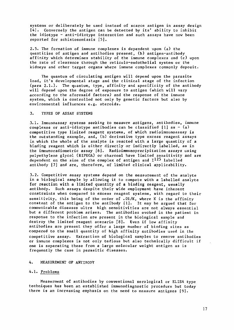
3.1. Immunoassay systems seeking to measure antigens, antibodies, immunecomplexes or anti-idiotype antibodies can be classified [1] as - (a)competitive type limited reagent systems, of which radioimmunoassay isthe outstanding example, and, (b) derivative type excess reagent assaysin which the whole of the analyte is reacted with a large quantity of abinding reagent which is either directly or indirectly labelled, as inthe immunoradiometric assay [6]. Radioimmunoprecipitation assays usingpolyethylene glycol (RIPEGA) or charcoal have limited sensitivity and aredependent on the size of the complex of antigen and I125 labelledantibody [7] and are, therefore, of limited clinical applicability.
3.2. Competitive assay systems depend on the measurement of the analytein a biological sample by allowing it to compete with a labelled analytefor reaction with a limited quantity of a binding reagent, usuallyantibody. Such assays despite their wide employment have inherentconstraints when compared to excess reagent systems, with regard to their
sensitivity, this being of the order of .01/K, where K is the affinityconstant of the antigen to the antibody [1]. It may be argued that forcommunicable diseases ultra high sensitivities are not always essentialbut a different problem arises. The antibodies evoked in the patient inresponse to the infection are present in the biological sample anddestroy the limited reagent scenario [8]. Even if low affinityantibodies are present they offer a large number of binding sites ascompared to the small quantity of high affinity antibodies used in the
competitive assay. Extraction of biological samples to remove antibodiesor immune complexes is not only tedious but also technically difficult ifone is separating these from a large molecular weight antigen as isfrequently the case in parasitic diseases.
4. MEASUREMENT OF ANTIBODY
4.1. Problems
Measurment of antibodies by conventional serological or ELISA typetechniques has been an established immunodiagnostic procedure but todaythere is an increasing emphasis on the need to measure antigens [9].
17

Firstly, the presence of antibodies does not differentiate between
present and past infection nor categorise the stage of infection (para2.1.). Secondly, the humoral immune response takes a finite time to
develop and very early infection may therefore not be diagnosed.Thirdly, the antibody response may never appear e.g. in an immunologicallycompromised host, or with very small amounts of secluded antigens such asan infection confined to the anterior chamber of the eye. Certain
overwhelming infections may depress the immune response e.g. measles,miliary tuberculosis and the acquired immunodeficiency syndrome (AIDS).Certain infections are known to elicit a cellular rather than humoralimmune response e.g. tuberculosis [9A].
Fourthly, anamnestic reactions and the prozone phenomenon have longbeen recognized as bedevilling the immunodiagnosis of typhoid in theWidal test. A patient suffering an unrelated fever may show a raised
titre of antibodies to salmonella and demonstrating a rising titre toclinch the diagnosis may involve waiting for two weeks.
Fifthly, it is possible in some diseases such as rubella todifferentiate acute from past infection by analysing whether the antibody
response is IgG or IgM, the latter being common in recent infection.
However, this approach may fail e.g. in African trypanosomiasis, and
"big spleen disease" where there is a persistent IgM response [10].
Sixthly, methods for measurements of antibodies are criticallydependent on the availability of purified antigenic material as
standard. Use of intact parasite or a crude mixture of antigens may lead
to the measurement of an irrelevant or even cross reacting antibody. The
parasites have a complex life cycle involving different developmental
stages each of which has a bewildering surfeit of antigens. Radiation
immobilisation of whole microbial cell has been described for
constituting solid phase [11, 12].
ANTIBODY DETECTION -
SOLID PHASE COUPLED 'Ag
INCUBATED WITHUNKNOWN SAMPLE
(Ab?) 4
SPECIFIC ANTIBODIES I -BOUND
INCUBATED WITHLABELLEDANTI-IG
i1']G(;. ]. Use o labelled anti-lgG antibody Jor detection of circularing antibodies.
An antibody response to be diagnostically useful should be speciesspecific. Responses to widely shared antigens e.g. Nematode classantigens, or highly restricted antigens e.g. variant specific responsesto trypanosomes, are not useful. The antigen source used for antibodymeasurement has usually been a whole organism or soluble extracts.
18
Homologous parasites would be the obvious source. This is possible forEntamoeba histolytica but in some diseases it is difficult to obtain e.g.
for Plasmodium vivax, a close relative, such as Plasmodium cynomolgi, hasto be used. This aggravates diagnostic confusion due to cross reactingantibody. It is sometimes possible to draw meaningful conclusions fromantibody responses by considering the context of the geographical andclinical situation but it would intrinsically be desirable if an assaycould provide a species specific diagnosis without additional data.
4.2. Why measure antibodies at all?
Nevertheless the measurement of antibodies remains important fromseveral points of view. The production of antibodies is evidence of (a)exposure to parasites and (b) of the immune status of the host. It isalso valuable epidemiologically e.g. to determine geographicaldistribution and prevalence of malaria. Antibody measurements may beused during surveillance to determine the effectiveness of public healthmeasures in the community or of individual chemoprophylaxis or therapy.The presence or absence of protective antibody may identify individualsat risk of developing infection among contacts or residents in an endemicarea. Studying antibody level also helps to understand the complexity ofa host response to a parasite, e.g. in filariasis, one subject achievescomplete cure, another has recurrent microfilaraemia and the thirddevelops the syndrome of tropical pulmonary eosinophilia withoutdemonstrable filaraemia.
5. WHICH ANTIGEN TO MEASURE?
It is therefore, clear that one must try to measure antigenemia butthe question arises as to which antigen is to be measured. Antigens maybe secreted in the body fluids or be associated with cell walls ormembranes or found in the cytosol. For organisms which are ininaccessible sites a secreted antigen would be preferred so that there isan appreciable quantity in the body fluid being sampled. Ekins hassuggested that in the future intra corporeal immunoprobes [13] may beemployed rather than the in vitro measurments in body fluids. Theantigen measured should correlate with the quantum of infection but itneed not be identical with the protective antigen eliciting theprotective immune response. It may be preferable in fact to measure aparasite product or antigen which is not immunogenic so that it is notmasked by antibodies evoked in the body fluids.
Parasite antigens can be proteins, polysaccharides or lipoidal innature. It is easy to obtain antisera against protein antigens.Competitive assays require that the antigen should be chemically stableduring iodination and also not damaged in body fluids during samplestorage/transport or during the assay incubation. The antigen should bespecific for the diagnostic need (para 2.1), non-infective andnon-toxic. For competitive assay systems the antigen should be easilylabelled. For such systems the labelled antigen need not be identicalwith immunogen or even with the antigen being measured. All that isrequired is that the standards and the unknowns compete in the samemanner for the antibody with the labelled antigen. However, the specificactivity of the labelled antigen must be high and the yield of thelabelling procedure must be such that the desired labelled antigenconstitutes a sufficient proportion of the total radiolabelled material.In a competitive assay system the lowest amount that can be measured issometimes apparently dependent on the amount of labelled antigen beingemployed and a high specific activity is therefore desirable.
19
ANTIGEN DETECTION -
WjLABELELET --- ,/--- | - INCUBATED WITHLAELE ^ SPECIFIC
ANTISERUM
INCUBATED WITH ....LABELLED ANTI- IG
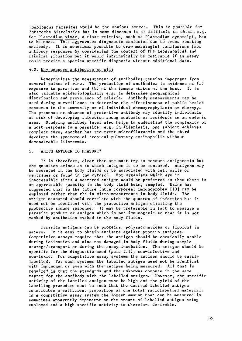
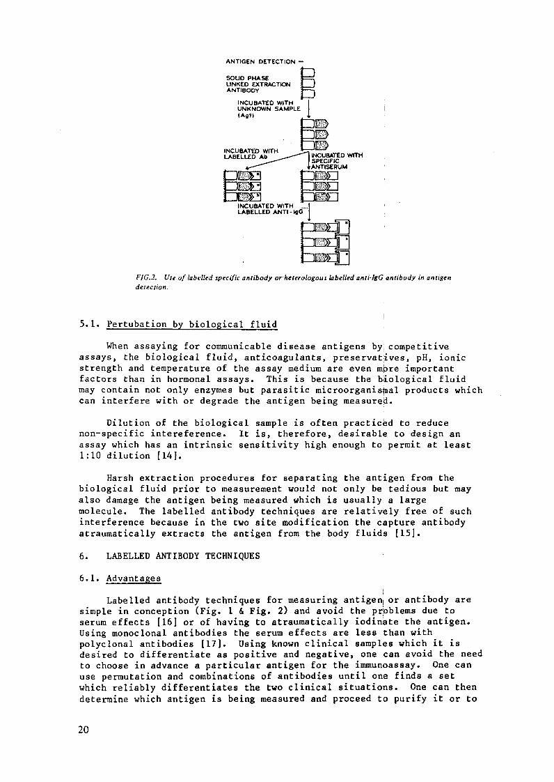
FIG.2. Use of labelled specific antibody or heterologous labelled anti-IgG antibody in antigendetection.
5.1. Pertubation by biological fluid
When assaying for communicable disease antigens by competitiveassays, the biological fluid, anticoagulants, preservatives, pH, ionicstrength and temperature of the assay medium are even mpre importantfactors than in hormonal assays. This is because the biological fluidmay contain not only enzymes but parasitic microorganistal products whichcan interfere with or degrade the antigen being measured.
Dilution of the biological sample is often practiced to reducenon-specific intereference. It is, therefore, desirable to design anassay which has an intrinsic sensitivity high enough to permit at least1:10 dilution [14].
Harsh extraction procedures for separating the antigen from thebiological fluid prior to measurement would not only be tedious but mayalso damage the antigen being measured which is usually a largemolecule. The labelled antibody techniques are relatively free of suchinterference because in the two site modification the capture antibodyatraumatically extracts the antigen from the body fluids [15].
6. LABELLED ANTIBODY TECHNIQUES
6.1. Advantages
Labelled antibody techniques for measuring antigen or antibody aresimple in conception (Fig. 1 & Fig. 2) and avoid the problems due toserum effects [16] or of having to atraumatically iodinate the antigen.Using monoclonal antibodies the serum effects are less than withpolyclonal antibodies [17]. Using known clinical samples which it isdesired to differentiate as positive and negative, one can avoid the needto choose in advance a particular antigen for the immunoassay. One canuse permutation and combinations of antibodies until one finds a setwhich reliably differentiates the two clinical situations. One can thendetermine which antigen is being measured and proceed to purify it or to
20

clone it. However, with competitive assays the antigen must first bechosen and purified before the assay is even set up.
6.2. Problems
Despite these advantages of labelled antibody systems we haveearlier pointed out the problems in their application to communicable andparasitic diseases [9] e.g. Interglobulin reactions due to rheumatoidfactor like substances [18-20], non specific stickiness of IgM to solidsurfaces, cross-reaction between various species globulins and aheterologous IgG raised against a particular species, anti-idiotypemasquerading as antigen and hook effects during the assay. Many of theseproblems will hopefully be reduced, by the use of monoclonal antibodiesboth as antigen specific or antigen capture antibodies or as heterologousanti-IgG.
7. MEASUREMENT OF IMMUNE COMPLEXES
The different ratios of circulating antibodies to antigen and theoccurrence of antigen-antibody complexes interfere in the detection ofantigen as discussed earlier. There are various methods [21-25] ofdemonstrating the presence of immune complexes in general but theisolation of a particular immune complex involving a particularcommunicable disease antigen is not straight forward. Immune complexesmay activate and bind complement, and link to immunoglutins(anticomplement antibodies), or anti-immunoglobulins, which are naturallyoccurring (Rheumatoid factor) or induced (anti-idiotypic antibodies), andcan, therefore, vary in size. The isolation of immune complexes may bebased either on their biological properties (binding to Fc or complementreceptors) or on their physico-chemical properties - size, charge anddifferential solubility. Ultracentrifugation, ultrafiltration,cryoprecipitation and PEG precipitation or Gel chromatography depend uponphysicochemical properties. But all these are tedious. Fc receptors maybe solid phase Clq or other solid phases e.g. monoclonal rheumatoidfactor while complement receptors may be solid phase conglutin, solidphase Anti - Clq antibodies, or Raji cells. One can thus immobilize theimmune complex by using solid phases, looking at complement or the Fcreceptor of the antibody. After this one can either dissociate theantigen-antibody bond by changing the pH or ionic strength and thenmeasure the antigen, or one may attempt to measure the communicabledisease particle linked to the antibody using another labelled antibody,looking at a different epitope of the communicable disease particle.Separating immune complexes from large molecular weight antigens may notbe possible by polyethylene glycol [9].
Similar separation of antigen-antibody complexes from antibodies orantibody aggregates is also troublesome. However, several groups arewresting with these problems and it is hoped that simpler methods willsoon become available.
8. ASSAY HAZARDS IN COMMUNICABLE DISEASES
Certain special problems in applying isotopic assays to biologicalspecimens must now be mentioned. The biological sample carries not onlythe risks of unsuspected Hepatitis B, but also the risk to laboratoryworkers from the infection being diagnosed. This has been a realconstraint in the development of certain assays which are obviouslyneeded e.g. an assay for rabies antigen in the saliva of dogs or the bodyfluids of dog bite cases where the offending dog is not traceable.
21
Straus and Van Der Horst [14] have approached this problem fortuberculosis samples by developing an RIA using heat alteredtuberculo-protein as immunogen, the antibody recognizing the heat alteredmaterial. This allows all samples to be autoclaved before assay. It isclaimed that apart from safety such heat treatment dissociates immunecomplexes and denatures endogenous antibody, removing the interferencedue to these in a competitive assay for antigen. This approach dependsupon the existence of an antiserum for a heat stable or Specifically heataltered protein coupled with heat labile immune complexes andantibodies. The principle is elegant and needs to be explored for otherdiseases.
The safety considerations regarding biological samples also apply tothe assay standards. Measurement of antibodies in body fluids requiresthe use of antigens which may themselves be infective. The competitiveassay methods for antigen measurement require the presence of antigensboth in the standards as well as in the labelled tracer material. Theimmunoradiometric methods for antigen measurement also require standardantigens to set up a standard curve although one sometimes avoids thisproblem by using a negative and a positive control serum (which is ineffect a two point standard curve) e.g. assay for Australia antigen.
9. NEED FOR STANDARDIZED PURE MONOSPECIFIC ANTIBODIES, ANTIGENS,AND FOR CLINICALLY VALIDATED ASSAY PROTOCOL
As discussed earlier measurements of antigens by labelled antibodytechniques requires monospecific antisera (preferably two looking atdifferent epitopes) as assay reagents and either purified antigens asassay standards or known positive and negative samples. Competitiveassay methods for measuring antigens, require one monospecific antiserum,purified antigen as standard and as a source of labelled antigen and alsoperhaps as immunogen to raise antisera.
Detection of antibodies by labelled antibody techniques requirespurified antigen for coating solid surfaces which will capture thedesired antibody. Thus for both these aims monospecific antisera andpurified antigens are prime requisites. Over the last decade theintroduction of affinity chromatography for the purification ofpolyclonal antisera and the hybridoma technology for producing monoclonalantibodies have shown the way to obtain monospecific antibodies which maybe used either as such or as an "engineered polyclonal clocktail" forassays. Simultaneously genetic engineering promises [26, 27] to obtainunlimited quantities of an antigen without having to extract and purifyit from patients, in vitro cultures, vectors or animal hosts. DNA hybridprobes have been used to identify malaria infected blood [28]. Similarlyfilarial antigens have been detected using a PAGE - Western Blot -labelled antibody overlay autoradiography [29] but these are researchmethods not suitable for widespread clinical application. Unfortunatelyin the common communicable diseases/parasitic infections, monospecificantibodies and genetic engineering produced pure antigens have either notbeen made or are not available for distribution. An assay which is to beuseful for individual diagnosis or epidemiology must be available aroundthe world and utilize easily available material and it should thereforebe an urgent priority of international organisations tolfund thedevelopment of monospecific antisera or pure antigens for worldwidedistribution.
22

Once such purified materials are available protocols must be testedso as to standardize which combination of antibodies and/or antigensprovides the most meaningful results answering clinical questions e.g. ina two site immunoradiometric assay one must choose the best combinationof solid phase capture antibody and of second antibody to measure aparticular antigen. It is hoped that over the next decade monospecificantisera, pure antigens and validated assay protocols are introduced inall the major communicable diseases and this may require setting up taskforces for each of them.
10. ISOTOPIC VERSUS NON ISOTOPIC LABELS
For antibody detection isotopic and non isotopic labels have thesame order of sensitivity and specificity [30, 31]. For the detection ofantigens or of immune complexes, the non specific signal in radioactivityis much less than that due to autofluorescence/enzymes using conventionalfluorochrome or enzyme labels. However, using the time resolvedfluorescence of Europium chelates linked to antibodies combined withpulsed excitation of the sample, the sensitivity theoretically attainableis much greater than with isotopic labels [32]. The cost ofsophisticated equipment for measuring non isotopic label is not lowerthan that of radioactivity counters and therefore because of availabilityof isotopic counting equipment, isotopic assays will continue to be usedfor a long time. The real limiting factor in isotopic label is not thehealth hazard but the limited shelf life and this may eventually causenon-isotopic methods to replace them.
ACKNOWLEDGEMENT
Some of this work was supported by grants from International AtomicEnergy Agency, Vienna (IAEA); Indian Council of Medical Research (ICMR),Delhi; Medical Research Council, Bombay Hospital and Department of AtomicEnergy (DAE), Government of India.
REFERENCES
1. Ekins, R.P.: The future development of immunoassay. In:Radioimmunoassay and Related Procedures in Medicine, (Proc. Symp.Berlin (West) 1977) Vol. 1 IAEA Vienna (1978) 247.
2. Hunter, W.M.: Recent advances in radioimmunoassay and relatedProcedures. In: Radioimmunoassay and Related Procedures inMedicine. IAEA-SM-259/101, IAEA, Vienna, (1982), 3.
3. Jerne, N.K,: Toward a Network Theory of the immune system. Ann.Immunol. Paris 125C (1974), 373.
4. Mitchell, G.F., Garcia, E.G., Cruise, K.M.: Competitiveradioimmunoassays using hybridoma and anti-idiotype antibodies inidentification of antibody responses to, and antigens of,Schistosoma japonicum. Aust. J. Exp. Biol. Med. Sci. 61 (1983), 27.
5. Potojnak, P., Zavala, F., Nussenzweig R., Nussenzweig, V.:Inhibition of idiotype-anti-idiotype interaction for detection ofparasitic antigen: A new immunoassay. Science, 215 (1982), 1637.
6. Miles, L.E., Hales, C.N.: Immunoradiometric assay procedures: Newdevelopments. In: In Vitro Procedures with Radioisotopes inMedicine, IAEA, Vienna, (1970), 483.
23
7. Hamilton, R.G. et al: Limitations of the RIPEGA for detection offilarial antigens in serum. J. Immunol. Meth. 68 (1984), 389.
8. Hamilton, R.G., Hussain, R., Ottesen, E.A., Adkinsdn, N.E. Jr.:Serological diagnosis of human filarial infections. Progress inPerinatal Medicine. Excerpta Medic (1983).
9. Hazra, D.K., Ekins, R.P., Williams, E.S., Pathak, A.K., Bharati, A.and Bansal, R.P.: Problems on the application of labelled antibodytechniques to communicable and parasitic diseases. In: nuclearTechniques in the Study of Parasitic Infections, IAEA-SM256/37,IAEA, Vienna, (1982).
9A. Ivanyi J., Kramboritz E. and Keen, M.: Evaluation of a MonoclonalAntibody (TB72) based serological test for tuberculosis. Clin. andExperimental Immunology, 50 (1983), 337-345.
10. Greenwood, B.M., Whittle, H.C.: Immunodiagnosis of Infection. InImmunology of Medicine in the Tropics. Immunology 14, Edwin Arnold,London (1981), 136-177.
11. Kumakura, M., Yoshida, M. and Kaistu, I.: European, J. App.Microbial, Biotechnol 6 (1978), 13.
12. Kumakura, M., Yashida, M. and Kaistu, I.: Biotechnol. Bioeng. 21(1979), 679.
13. Ekins, R.P.: in "Alternative Immunoassays" Ed. Collins W.P. JohnWiley, New York (In Publication, 1984).
14. Straus, E., Harstdervan, C.: Radioimmunoassay methods for the studyof communicable diseases. In: Radio Immunoassay and RelatedProcedures in Medicine. IAEA-SM-259/107 673-688. IAEA, Vienna,(1982).
15. Hazra, D.K.: Immunoradiometric methods in glycoprotein estimation.M.Sc. Thesis (Nuclear Medicine) London University, (1975).
16. Hazra, D.K., Ekins, R.P., Williams, E.S.: Labelled antibodytechniques in glycoprotein estimation. In: RIA and RelatedProcedures in Medicine, IAEA, (1977).
17. Perrin, L.H. and Gabra, M.: Immunodiagnosis in Malaria - IAEA TECDOC(1984).
18. Decker, R.H.: In: Radioimmunoassay and Related Procedures inMedicine (Proc. Symp. Berlin (West) 1977), Vol.I. IAEA, Vienna(1978), 275.
19. Ling, C.M. et al: Comparison of confirmation methods for hepatitis Bantigen and the nature of false positives detected by 1251Immunoglobulins. J. Lab. Clin. Med. 86 (1975).
20. Sabnen, E.M., Vahen, A., Suni, J., and Wager, 0.: Rheumatoid factorin acute viral Infections; Interference with determination of IgM,lgG and lgA antibodies in an enzyme immunoassay. J. Inf. Dis. 142(1980), 250 - 255.
24

21. Lunde, M.N.: A Clq Enzyme Linked Immunosorbent Assay (ELISA) usingPolyethylene Glycol for immune complexes Am. J. Trop. Med. Hyg. 32(1983), 392.
22. D'Amelio, R., Potesilli, R., Palmisano, L., Pezzella, M., Vullo, V.,Della, S., De Rose, F., Sorice, F., and Aiuti, F.: Detection andpartial characterization of circulating immune complexes in hydatiddiseases. J. Clin. Microbiol. 18 (1983), 1026.
23. Degraves, F., and Cox, H.W.: Interrelationship of immunoconglutinin,immune complexes and complement in anemia thromocytopenia and
parasitemia of acute and chronic malaria in rats. J. Parasitol. 69(1983), 262.
24. Smith, M.D., Verroust, P.J., Morel - Maroger, L.M., Pastiver, A.,and Couland, J.P.: Circulating immune complexes in schistosomiasis.Br. Med. J. 2 (1975), 274.
25. Pini, C., Pastore, R., and Valesini, G.: Circulating immunecomplexes in sera of patients infected with Echinococcus granulosus,Clin. Exp. Immunol. 51 (1983), 572.
26. Kemp, D.T., Cappel, R.L., Cowman, R.F., Saint, R.B., Brown G.V. andAnders, R.F.: Expression of Plasmodium falciparum blood stageantigens in Escherichia coli: Detection with antibodies from immunehumans. Proc. Natn. Acad. Sci. USA 80 (1983), 3787-3791.
27. Ozaki, L.S. et al: Structure of the Plasmodium knowlesi gene codingfor the circumsporozoite protein. Cell 30 (1983), 815-822.
28. Franzen, L., Shabo, R., Perlmann, H., Wigzell, H., Wostin, G.,Aslund, L., Persson, T., and Petterson, U.: Analysis of clinicalspecimens by hybridisation with probe containing respective DNA fromPlasmodium falciparum. Lancet, 1 (1984), 526.
29. Hamilton, R.G.: Radiotracer techniques in the immunodiagnosis offilariasis in man. IAEA-TECDOC (1984).
30. Voller, A., Edwards, R. et al: Elisa and isotopically labelledantibody studies trans. R. Soc. Trop. Med. Hyg. 755 (1977), 431.
31. Hill, R.H. and Matsen, M.J: Enzyme-linked immunosorbent assay andradioimmunoassay in the serologic diagnosis of infectious diseases.J. Infect. Dis. 147 No. 2, (1983), 258 - 263.
32. Soini, S., and Kojola, H.: Time resolved fluorometer for lanthanidechelates - A new generation of non-isotopic immunoassays. Clin.Chem. 29 (1983), 65.
25
DEVELOPMENTS IN IMMUNOASSAYS FORDIAGNOSIS OF PARASITIC DISEASES
E. LINDERNational Bacteriological Laboratory,Stockholm, Sweden
Abstract
The purpose of this paper is to define the major problems involved inimmunodiagnosis of parasitic diseases and to point at their solutionsbased on recent literature. The topics to be examined are advances inassay technology, development, purification and standardization of thereagents employed in the assays, and reasons for false positive and falsenegative reactions.
INTRODUCTION
Serological diagnosis of parasitic diseases is usually confirmatorybut may be essential under certain conditions such as infection with onlya few parasites, early infection before typical disease manifestationsare seen and disease caused by parasites located at secluded or atypicalsites. Immunodiagnosis of parasitic diseases may also be the only usefuldiagnostic tool in epidemiological studies. Such tests may also be ofimportance as screening tests for parasitic infections in patients withnon-specific symptoms after travel to endemic areas. Furthermore,immunoparasitology is closely related to both immunopathology andprevention of parasitic infections by vaccination.
Several problems are associated with immunodiagnosis of infectionscaused by various agents. The demonstration of specific antibodies inthe circulation is a secondary event dependent on the immune response ofthe host. Several circumstances, such as route, stage and location ofthe infectious agent, as well as genetic properties of the host effectthe timing, duration, quantity, fine specificity and class of theantibodies formed. The simultaneous presence of foreign antigen andspecific antibodies may lead to neutralization of the antibody andformation of circulating immune complexes.
In addition to these common problems there are those typicallyassociated with immunological diagnosis of parasitic diseases. A majorproblem is a consequence of a complex immune response. The parasites arein general well differentiated organisms with a complicated life cycleeven within the host. They may therefore generate a spectrum ofantibodies of different specificities and directed not only againstdifferent parts of the parasite but also against different developmentalstages. The parasite may on the other hand have developed means ofevading the host immune response. Protective measures include antigenicvariation, shedding of surface components and covering of the surfacewith host derived material.
Immunodiagnosis in parasitic infections is currently mainly based onidentification of circulating anti-parasite antibodies but there is aclear tendency showing that demonstration of parasite antigen is ofdiagnostic significance (1). The specificity and sensitivity of the
27
diagnostic methods used are dependent on one hand on the technology, andon the other, on the reagents used. It is concluded that the availabletechnology provides the necessary tools for immunodiagnosis of parasiticdiseases. The problems are caused by the lack of well defined reagentsand the false positive and false negative reactions which are seen as aconsequence of this.
Parasite Antigens
The use of crude mixtures of parasite antigen for demonstration ofmixtures of antibodies present in patient sera leads tol problems ofspecificity, reproducibility and standardization. A major problem hastherefore been to prepare antigenic material useful as target antigen inthe assay (2). The starting situation is the need to demonstrateantibodies to unknown target antigens using poorly characterized crudeantigen material.
The immunofluorescence (IFL) technique is a basic immunodiagnosticmethod in parasitology as it makes use of whole parasites as antigen andis still capable of differentiating distinct antibody responses. Othertechniques, not only the classical ones based on precipitation oragglutination but also enzyme- and radioimmunoassays are dependent onantigen preparations which still largely consist of crude parasiteextracts. This causes problems in standardization and comparison betweenindividual samples, and risk false positive reactions. However, thetendency to prepare defined fractions containing immunodominant parasiteantigens is evident from numerous published reports.
New strategies for analysis of mixtures of antigenic material andfor demonstration and isolation of immunodominant antigens have evolvedwith development of biochemical separation techniques such as affinitychromatography using antibodies and lectins as binders and high powerliquid chromatography and isoelectric focusing. Combination ofbiochemical separation on polyacrylamide gels followed by transfer ofisolated polypeptides to nitrocellulose membranes have made analysis ofmixtures of antibodies and antigens possible. Radionuclide labelling ofparasite antigen followed by immunoprecipitation and polyacrylamide gelelectrophoresis is another powerful analytical procedure for definingtarget antigens.
Revolutionary possibilities are provided by DNA technology forpurification of parasite antigens by development of cDNA clones ofantigen genes of both protozoa and helminths. Such reagents willprobably have a fundamental impact on immunodiagnostic techniques in thenear future.
Furthermore, the facinating possibility of solving the problem oflack of available antigen by substituting parasite antigen withanti-idiotypic antibodies has been pointed out (2, 3).
Monoclonal antibodies
Monoclonal antibodies are directed against a single antigenicepitope. They can be induced by immunization with crude antigen mixturesor during natural or experimental infection and the antibodies can beproduced in unlimited amounts.
Monoclonal antibodies have been used for the demonstration of bothparasite antigens and anti-parasite antibodies, and they have a central
28
role in solving the major problem on immunodiagnosis of parasiticdiseases, that of specificity of the reagents (4, 5).
A possible solution to the problem may lie in the design ofinhibition assays. Antibody assays employing solid phase antigen andmonoclonal antibodies have now been developed. The use of a monoclonalantibody eliminates the problem involved in assays employing mixtures ofantigens for demonstration of mixtures of specific antibodies. In suchan assay specific antibody activity can be measured by preincubatingpatient serum with test antigen followed by monoclonal antibody, thebinding of which is measured. Assays employing solid phase monoclonalantibodies have also been designed for demonstration of antigen. Thiseliminates the need for purified labelled antigen in competitive bindingassays.
Immunoassays
The basic immunoassay principles were established almost 30 yearsago in studies on binding of radiolabelled insulin to anti-insulinantibodies by Berson, Yalow and co-workers. The basis for theradioimmunoassay, the RIA, was laid by the observation that labelledinsulin can be displaced by unlabelled insulin in the assay. Thecompetitive binding assays which subsequently evolved, have been used ina variety of situations for quantitation of ligands present in tracequantities in body fluids. The recently developed immunoradiometric(IRMA) assays employ "labelled antibody" in excess which binds a limitedamount of analyte present in the test sample (6). Removal of unreactedreagent involved problems which were solved by using solid phasemodifications; unlabelled solid phase antibody "catching antibody" iscapable of binding the ligand which can be detected with a labelledsecond antibody (two-site assay) or the second antibody can be detectedwith a labelled anti-antibody.
The basic principles of immunoassays have been applied for differentdiagnostic purposes and the radionuclide label has been substituted bystable labels, most frequently an enzyme. The future of radioactiveversus stable labels in immunoassay technology has been the subject ofseveral studies (7, 8). The choice of suitable labels has to be decidedin casu and depends on a number of considerations. Immunoassays forparasitological diagnosis are dependent above all on defined reagents andinsights in the possible interference mechanisms causing false positiveand false negative reactions.
The solid phase assay employing parasite antigen bound to a plasticsupport is used in parasitological diagnosis for demonstration ofspecific anti-parasite antibody. The assay can also be modified fordemonstration of antigen (1). The immunoassays employed are commonlynon-isotopic solid phase assays using polystyrene microtiter platespassively coated with antigen. Bound antibodies are detected withlabelled anti-immunoglobulin conjugates. Recent advances include newlabels, non-covalent coupling techniques, novel separation techniques andhomogenous assays.
New labels have involved, in addition to the commonly usedenzyme-chromogen combinations (peroxidase with orthophenylene diamine(OPD) and alkaline phosphatase with 4 nitrophenyl phosphate), fluorescentsubstrates derived from methylumbeliferin. The use of fluorescentsubstrates involves problems with light scattering and autofluorescenceoriginating both from the samples and the equipment.
29
The fluorescent probes commonly used, FITC, TRITC and dansylderivatives have excitation and emission wavelength maxima separated bynot more than about 30 nm. These properties of the fluorochromes causebackground problems and weak signals. The introduction of fluorescentchelates of rare earth metals, especially europium (Eu) has overcome themajor problems involved in fluorometry. Their advantage is a very largeStokes' shift - for Eu over 270 nm, narrow emission spectra and longdecay times of the fluorescence. The latter property makes possible theuse of short excitation light pulses of about 1 ns which are followed bya rather long counting time after a delay during which autofluorescenceof the background has decayed. As a consequence background fluorescencecan be efficiently eliminated (9).
Further developments in immunoassays are the homogeneousimmunoassays in which the complex formed after antibody and antigen havecombined does not need to be separated from unbound ligand (10). Suchtypes of assay employing fluorescent probes make use of changes in thedegree of polarization taking place in the fluorescent probe afterbinding. Decrease in fluorescence (fluorescence quenching) and increasedfluorescence (fluorescence enhancement) may also occur in somefluorescent probes depending on the change in the microenvironment (11).
Homogeneous assays have also been developed in which the enzyme ofan antibody-enzyme conjugate is prevented through steric hindrance fromreacting with the substrate after antibody has bound to a large antigen(10, 12).
False positive and negative reactions
Class specific antibodies are determined especially in neonatalinfections as IgG but not IgM may be transferred from the mother to thefetus. The presence of IgM rheumatoid factor(RF) in serum samples maycause false positive reactions in IFL, ELISA and RIA designed toquantitate specific IgM class antibodies especially in diagnosis ofneonatal infections (13 - 15). Demonstration of IgM class antibodies isindicative of recent or active infection and is of special importance indiagnosis of neonatal infections (15). Diagnosis of active diseaseduring pregnancy may have important therapeutic consequences and falsepositive results have to be avoided by all means. The problem isespecially important as a significant increase in RF occurs duringpregnancy (16). IgM rheumatoid factor is routinely demonstrated usinglatex particles coated with denatured IgG which are commerciallyavailable. Also the ELISA can be used for determination of IgM RF (17).
Positive test results indicating presence in patient sera of IgMclass anti-parasite antibodies frequently have to be further examined inorder to exclude the possibility of interference by IgM rheumatoidfactor. False positive reactions may result from binding of RF to boundanti-parasite IgG class antibodies. If positive for RF, absorption withdenatured IgG can be performed e.g. using IgG bound to latex particle(18). Fractionation of IgG, IgM and IgA can be achieved by a combinationof ion exchange chromatography, lectin affinity chromatography andabsorption with staphylococcal protein A.
An alternative to elimination of IgM RF from the test sample is touse specific antibody against IgM in the solid phase for binding of serumIgM including specific anti-parasite antibodies. Labelled specificparasite antigen or a labelled second anti-parasite antibody is used fordetection of bound antigen.
30
False positive reactions, or more exactly unwanted positivereactions can be seen which are basically due to the use of impureantigens in the assay. Potential sources of error are constituents ofthe parasite contractile apparatus e.g. actin and myosin, andcytoskeletal proteins like tubulin, as well as various nuclear antigens.Little is known about the frequency of such interferring reactions. Thatthey deserve attention is suggested by the observation that anti-nuclearanti-bodies may cause false positive reactions in tests for Toxoplasmaantibodies (19). The possibility of false positive reactions due toanti-myosin has also been considered in the case of antibodies reactingwith Toxoplasma gondii.
False negative reactions may occur not only from deficient antigenor antibody preparations but also from limitations of the technique e.g.blocking antibodies against Toxoplasma which may block binding of IgMclass antibodies. Blocking antibodies may be removed by fractionation.Absorption with staphylococcal protein A is the most commonly usedprocedure for removal of IgG. The absorption is considered useful evenif protein A binds only IgG subclasses 1, 2 and 4 and binding of IgM by afew per cent also takes place.
Several parasitic infections are associated with release ofantigenic material into the circulation (2, 20 - 25). False negative,and in some cases false positive reactions can be caused by circulatingimmune complexes depending bn their composition and on the assay: thecomplexes can be precipitated by polyethylene glycol and bind to solidphase antibodies against antigen, antibody or complement (26). Thecomplexes may have the capacity to activate and bind complement, bindanti-complement antibodies (immunoconglutinins), anti-immunoglobulins (RFand anti-idiotypic antibodies) (27 - 32).
Conclusions
Development of rapid, sensitive assays based on non-radionuclidelabels in immunoassay technology continue to challenge establishedradioimmunological assays in the field of immunoparasitology. Comparisonbetween different assay techniques have to depend on well definedreagents. Immunodiagnosis of parasitic diseases is complicated by thecomplexity of both the parasite and the host immune response. Seriousproblems involving false positive and negative reactions are due to lackof standardized reagents, but recent advances in in vitro culturetechniques, biochemical isolation procedures, development of monclonalantibodies and DNA technology provide the necessary tools for rapiddevelopment in the field.
REFERENCES
1. VOLLER, A., BARTLETT, A. and BIDWELL, D. - Immunoassays for the80s. New York University Park Press (1981).
2. MITCHELL, G. and ANDERS R.F. - Parasite antigens and theirimmunogenicity in infected hosts. In: The Antigens Vol VI. AcademicPress (1982) 69.
31
3. MITCHELL, G.F., GARCIA, E.G. and CRUISE, K.M. - Competitiveradioimmunoassays using hybridoma and anti-idiotype antibodies inidentification of antibody responses to, and antigens of,Schistosoma japonicum. Aust. J. Exp. Biol. Med. Sci. 61 (1983) 27.
4. HOUBA, V. and CHAN SH, eds. - Properties of the monoclonalantibodies produced by hybridoma technology and their application tothe study of diseases UNDP/WORLD BANK/WHO, (1982).
5. MITCHELL, G.F. - Hybridoma antibodies in immunodiagnosis ofparasitic infection. Immunology Today 13 (1981) 140.
6. EKINS, R. - Merits and disadvantages of different labels and methodsof immunoassay. In: Immunoassays for the 80s. (eds) - A. Voller, A.Bartlett and D. Bidwell, University Park Press (1983) 5.
7. THOMPSON, M. - Are monoclonal antibodies the end ofradioimmunoassay? TIBS (1982) 419.
8. WITHERSPOON, L.R. - Immunoassay - is there a future role for nuclearmedicine? J. Nucl. Med. 24 (1983) 952.
9. SOINI, S. and KOJOLA, H. - Time-resolved fluorometer for lanthanidechelates - a new generation of non-isotopic immunoassay. Clin.Chem. 29 (1983) 65.
10. RUBENSTEIN, K.E. - New homogeneous assay methods for thedetermination of proteins. In: Immunoassays for the 8 0s. (eds) A.Voller, Al Bartlett and D. Bidwell New York University Park Press(1981).
11. SOINI, S. and HEMMILA, I. - Fluoroimmunoassay: Present status andkey problems. Clin. Chem. 25 (1979) 353.
12. YOLKEN, R.H. - Enzyme immunoassay for the detection of infectiousantigens in body fluids: Current limitations and future prospects.Rev. Infect. Dis. 4 (1982) 35.
13. BONOLO, A., DOVIS, M., MALVANO, R. and ZANNINO, M. - ELISA forspecific anti-toxoplasma IgM antibodies: Aspects related to seruminterference, J. Imm. Meth. 59 (1983) 113.
14. FILICE, G.A., YEAGER, A.S. and REMINGTON, J.S. - Diagnosticsignificance of immunoglobulin M antibodies to ToXoplasma gondiidetected after separation of immunoglobulin M from immunoglobulin Gantibodies, J. Clin. Microbiol. 12 (1980) 3336.
15. REMINGTON, J.S., MILLER, M.J. and BROWNLEE, I. - IgM antibodies inacute toxoplasmosis, II Prevalence and significance in acquiredcases. J. Lab. Clin. Med. 71 (1968) 855.
16. MEURMAN, O.H., TERHO, P. and SALMI, A. - Activation of rheumatoidfactor during pregnancy, Lancet (1978) 686.
17. VEJTORP, M. - The Interference of IgM rheumatoid factor inenzyme-linked immunosorbent assays of Rubella IgM and IgGantibodies, J. Vir. Meth. 1 (1980) 1.
32
18. WINCHESTER, R. - the nature of rheumatoid factors and theirpotential for interfering with assays. In: Workshop on new anduseful techniques in rapid viral diagnosis. J. Infect. Dis. 142(1980) 793.
19. ARAUJO, K., BARNETT, E.V., GENTRY, L.O. and REMINGTON, J.S. -False-positive anti-Toxoplasma fluorescent-antibody tests inpatients with antinuclear antibodies. Appl. microbiol. 22 (1971)270.
20. DEELDER, A.M., KLAPPE, H.T.M., van den AARDWEG, G.H.M.J. and vanMEERBEKE, E.H.E.M. - Schistosoma mansoni: Demonstration of twocirculating antigens in infected hamsters. Exp. Parasitol. 40 (1976)189.
21. BOUT, D., SANTORO, F., CARLIER, Y., BINA, J.C. and CAPRON, A. -Circulating immune complexes in schistosomiasis. Immunology 33(1977) 17.
22. LAWLEY, T.J., OTTESEN, E.A., HIATT, R.A. and GAZZE, L.A. -Circulating immune complexes in actue schistosomiasis. Clin. Exp.Immunol. 37 (1979) 221.
23. MADWAR, M.A. and VOLLER, A. - Circulating soluble antigens andantibody in schistosomiasis, Br. Med. J. 1 (1975) 435.
24. PHILLIPS, T.M. and DRAPER, C.C. - Circulating immune complexes inschistosomiasis due to Schistosoma mansoni, Br. Med. J. 2 (1975) 476.
25. PINI, C., PASTORE, R. and VALESINI, G. - Circulating immunecomplexes in sera of patients infected with Echinococcus granulosus.Clin. Exp. Immunol. 51 (1983) 572.
26. LUNDE, M.N. - A Clq enzyme-linked immunosorbent assay (ELISA) usingPolyethylene Glycol for immune complexes. Am. Trop. Med. Hyg. 32(1983) 392.
27. D'AMELIO, R., PONTESILLI, O., PALMISANO, L., PEZZELLA, M., VULLO,V., DELIA, S., DE ROSE, F., SORICE, F. and AIUTI, F. - Detection andpartial characterization of circulating immune complexes in hydatiddisease, Journal of Clinical Microbiology, 18 (1983) 1026.
28. CARVALHO, E.M., ANDREWS, B.S., MARTINELLI, R., DUTRA, M. and ROCHA,H. - Circulating immune complexes and rheumatoid factor inschistosomiasis and visceral leishmaniasis, Am. J. Trop. Med. Hyg.82 (1983) 61.
29. DeGRAVES, F. and COX, H.W. - Interrelationship of immunoconglutinin,immune complexes, and complement in anemia, trombocytopenia, andparasitemia of acute and chronic malaria in rats, J. Parasitol. 69(1983) 262.
30. ROBINSON, A. and LEWERT, R.M. - Immunoglobulin M-immunoglohulin G.mixed cryoglobulinemia in Schistosoma japonicum-infected rabbits.Infection and Immunity 39 (1983) 1477.
33

31. SMITH, M.D., VERROUST, P.F., MOREL-MAROGER, L.M., PASTICIER, A. andCOULAUD, J.P. - Circulating immune complexes in schistosomiasis. Br.Med. J. 2 (1975) 274.
32. STEVENS, W.J., FELDMEIR, H., BRIDTS, C.H. and DAFFALLA, A.A. - IgGand IgE circulating immune complexes, total serum IgE and parasiterelated IgE in patients with mono- or mixed infection withSchistosoma mansoni and/or S.haematobium: Influence of therapy.,Clin. Exp. Immunol 52 (1983) 142.
34
IMMUNOASSAYS OF ANTIBODIES AND CIRCULATINGANTIGENS IN SCHISTOSOMIASIS
J.P. DESSAINT, A. CAPRONCentre d'immunologie et de biologie parasitaire,Institut Pasteur,Lille, France
Abstract
Recent progress in immunodiagnostic tests for schistomiasis haveallowed evaluation of several purified antigens from S. mansoni egg oradult worms to detect anti-schistosome antibodies. Egg antigens allowedmore sensitive and specific assays of anti-schistosome antibody althoughno antigenic preparation proved to be decisively superior as animmunodiagnostic tool. Promising results were obtained by measurement ofIgM antibodies which correlate with acute infection and IgE antibodies orimmune complexes which decrease rapidly after parasitological cure.Assay of antibodies in schistosomiasis japonicum by sandwich immunoassaysor competitive radioimmunoassays with monoclonal antibody yieldedsensitive results in epidemiological studies. Measurement of circulatingantigens is made difficult by interference of immune complexes and thecorrelation of antigenemia with parasite load is still controversial.Yet decrease in circulating levels after treatment appears as valuableinformation to be evaluated in large scale studies. Detection of non-immunogenic parasite-derived products might avoid the difficultiesintroduced in immunoassays by complexed antigens. Purification ofschistosome antigens or absorption of cross-reactive antigens from crudepreparations and preparation of specific monoclonal antibodies should beencouraged.
1. INTRODUCTION
Immunological tests for the diagnosis of schistosomiasis have beenin use since the beginning of this century. Yet, these still represent achallenge to immunoparasitologists, mainly because of the intense cross-reactivity among parasites, the limited availability of pure schistosomeantigens and the marked variability in seroreactivity of the patients.Extensive efforts are presently directed to the improvement of old andnew immunodiagnostic tests in order to increase their specificity whichis still the limiting factor in most assays.
It is generally accepted that the specificity of immunodiagnostictests will rely mainly on the use of genus- or species--specific reagents,but that particular requirements for individual tests will depend ontheir intended use. A primary goal of research in immunoassays is todevelop a rapid, cheap and technically easy test which can beepidemiologically used in endemic areas, particularly to evaluate theeffects of various national or international schemes of control, as analternative to the reliable but tedious and time-consuming quantitativeegg count techniques. On the other hand, assays for clinical studies orresearch may take advantage of the sophisticated methodology available toachieve maximum specificity or sensitivity, which are of particular valuein certain stages of the infection when egg excretion may be scant. Afurther goal of immunoassays is the distinction between infection anddisease, which is one of the most important features from a clinicalviewpoint, as it is recognized that whilst the majority of a population
35
in an endemic country may be actively infected by schistosomes, only aportion will present clinical morbidity. Another purpose of researchoriented assays is to provide means to assess effectiveness ofchemotherapy or other curative measures. Besides their enormous appealin epidemiology, such assays might be used for monitoring of treatment orpharmacological research. Finally, the ability of immunodiagnostic teststo differentiate those individuals that develop a protective immuneresponse against schistosomes deserves consideration.
Almost every serologic technique has been applied to immunodiagnosisof schistosomiasis. For many years, the tests depended on theidentification of an antibody response against the three most importantspecies, Schistosome mansoni, S. japonicum and S. haematobium. Thealternative of immunoassays to detect circulating schistosome antigenshas been more recently explored, since current serologic assays forantiparasite antibody lack the ability to differentiate individuals withactive infection from those with past infections and from those who haverecently been cured. It has been recognized however that detection ofprecise antibody isotypes might bring useful information to our appraisalof the host-parasite relationship.
2. ASSAYS OF ANTI-SCHISTOSOME ANTIBODIES
One of the most important factors in these assays is represented bythe antigen preparation employed in the test. Crude schiistosome-derivedantigen preparations commonly used in assays for the detection of anti-schistosome antibodies are in fact heterogeneous mixture of specificitiesobtained by extraction or homogenization of the different stages of thelife cycle of schistosomes. Thus the relevant antigen(s) may be eitherhindered by large amounts of irrelevant or cross-reactive molecules, thatdepreciate the results of serological tests, or present in too smallquantities to be bound by the antibodies with sufficient sensitivity.The specificity of the test, which is determined by the antigens used,might be improved when the crude antigenic preparations are replaced byfractions of them, or by purified genus- or species- specific antigens.
The necessity for the isolation, purification and standardization ofschistosome antigens has been emphasized since 1974 [1] and becomes moreand more evident for the new generation of highly sensitive methodologiesfor immunoassays. Gel filtration, ion exchange chromatography andaffinity chromatography with lectins or with polyclonal or monoclonalantibodies, are being used in several laboratories. Several purifiedantigenic preparations are now available although in limited quantities,which have been evaluated in immunodiagnostic tests [2].
2.1. Assays using purified antigens
Antigen preparations for use as reagents in the detection ofinfection-specific antibodies should meet the following requirements: (a)
sufficiently large quantities of the antigen are needed to allowevaluation of the test; (b) the antigen should be stable under storage
conditions, which might be a major problem in field studies; (c) theantigen should be recognized by the antibodies in a ma ority of specific
infection sera; conversely (d) it should contain minimal cross-reactive
epitopes; and (e) the antigen should be compatible witi the assay systemi.e. it can be either labelled for Farr-type assays orlbound to a solidphase for sandwich-type assays. This might be difficult to achieve,particularly in the case of polysaccharide antigens that do not absorb
readily to polystyrene. However, modifications of thelinert matrix can
36
be used, like for instance precoating the wells of microtitration plateswith poly-L-Lysin [3]. Cross-linking of antigens, particularly those oflow molecular weight can also improve the assay system [4].
Two stages of the life cycle of schistosomes, the egg and the adultworm, present lasting antigenic stimuli to the infected host, andantigens prepared from these two stages therefore are the most obviouschoice for use in immunodiagnostic tests. Three basically differentapproaches are possible for the purification of schistosomal antigens.(a) A single defined antigen, previously recognized as presentingdiagnostic interest, can be purified using a specific isolation procedureby appropriate chemical or immunochemical methods; (b) alternatively,immunochemical techniques can be used to deplete cross-reactive antigensin parasite extracts [5]; (c) general purification methods can be appliedto crude schistosome homogenates, this step being followed by theselection of the fraction(s) with the desired properties; (d) as it maybe difficult to obtain large amounts of purified antigens, a furtherpossibility is to use anti-idiotypic antibodies instead of the antigen ina sandwich assay, but no success with this approach has been reportedyet, although anti-idiotypic antibodies proved to be useful in monitoringthe isolation of target antigens [5, 6].
2.1.1. Fractionated schistosome egg antigens
Several purified antigenic preparations have been evaluated inimmunoassays. The major serological antigen (MSA1) of S. mansoni eggswas fractionated by a combination of lectin affinity chromatography, ionexchange chromatography and gel filtration and its preparation monitoredwith monoclonal antibodies. Fractions of 68-200 kDa have been labelled
with 125 I and used in a Farr-type assay with ammonium sulphate toprecipitate the antigen-antibody complexes. Although thecross-reactivity of this egg fraction was very low (< 1%), the assayshowed only quantitative differences among sera from patients infectedwith one of the three schistosome species, significantly higher resultsbeing obtained with sera from patients with schistosomiasis mansoni [7].S. mansoni soluble egg antigen (SEA) was purified by immunoaffinity [8]while CEF-6 S. mansoni egg antigen was isolated by elution fromCM-Sepharose columns [9, 10].
Egg antigen from S. japonicum was fractionated using gel filtration,ion exchange and lectin affinity chromatography [111, a combination ofConA and immunoaffinity chromatography [12] or Con A-Sepharose and gelfiltration chromatography (GP-2) [Tracy & Mahmoud, in 2], while 8M-ureasoluble egg-antigen fraction was reported to retain higher antigenicactivity than aqueous soluble fractions, the former containing highmolecular weight glycoproteins [13]. Hydrophobic glycoproteins fromS. japonicum soluble egg antigen were shown to have higher specificityand sensitivity than hydrophylic glycoproteins or SEA proteins [14].Preliminary purification of S. haematobium egg glycoprotein of 70 kDaallowed to demonstrate its reactivity with antibodies from patients withS. haematobium infection and its partial cross-reactivity with antigensin S. mansoni eggs [15].
2.1.2. Purification of adult worm antigens
Excretory-secretory antigens collected from the incubation ofS. mansoni adult worms were semi-purified by affinity chromatography onConA-Sepharose [Capron A., in 2] or fractionated by gel filtration on
37
Ultrogel ACA-44. Antigens of low molecular weight (< 60 kDa) gave about30 % false negative results in schistosomiasis mansoni but littlecross-reactivity; whereas, high molecular weight fractions (> 60 kDa)gave no false negative but showed cross-reactions which surpassed that ofthe total worm antigen preparations [16, 17]. Circulating antigens havethemselves been used to detect antibodies to S. mansoni [3]. Recently,S. mansoni adult microsomal antigen (MAMA) has been isolated bydifferential centrifugation of adult worm homogenates followed by gelfiltration and ion-exchange chromatography [18] and this antigen showedmuch higher reactivity with sera from patients infected by S. mansonithan by other schistosomes and no cross-reactivity with other helminthparasites [19]. Two carbohydrate antigens eliciting specific antibodyresponses in humans have been characterized in S. mansoni. Theschistosome gut-associated proteoglycan (GASP) and the phenol sulfurictest active peak (PSAP) were labelled with 12 5I-Bolton-Hunter reagentand their respective antibodies were precipitated using eitherpolyethylene-glycol, antihuman immunoglobulins or Staphylococcal proteinA [20, 21].
2.1.3. Evaluation of the antigens for immunodiagnosis ofschistosomiasis
The purified antigen preparations reviewed above were evaluated inimmunodiagnostic tests. Each proved to be recognized by antibodies inpatient sera but cross-reactivity or high percentage of false negativewere the main problems encountered. A recent collaborative study of mostof these antigens has been coordinated by the World Health Organizationusing 395 well-documented sera from four endemic areas in Brazil, Kenya,and the Philippines.
Of the antigens and immunoassay combinations tested inschistosomiasis mansoni, none proved to be definitively superior withrespect to specificity and sensitivity. S. mansoni egg antigens, bothcrude and purified, yielded a higher combined sensitivity and specificitythan the adult worm antigens, the latter being particularly inaccuratewith post-treatment sera from individuals with repeated negative stoolexaminations. The sensitivity of immunoassays using labelled reagents insera from untreated persons ranked from 63.5 to 91.7 % and thespecificity from 73.1 to 93.8 %. The egg antigens detected over 90 % ofindividuals with more than 400 eggs/g stool while adult worm antigensdetected at least 95 % individuals with > 800 eggs/g. No correlationbetween clinical morbidity related to S. mansoni and seroreactivity wasobserved in any assays. One of the most disappointing conclusion of thiscollaborative study was that the circum ovale precipitin test (COPT)developed 30 years ago appeared to be the most reliable! [2].
On the other hand, S. japonicum fractionated egg antigens showedhigh sensitivity and specificity in relation to egg output in thehomologous infection [2]. However, it is generally agreed that theclinical sensitivity and the specificity of immunodiagnostic tests usingcrude S. japonicum egg antigen are sufficiently high, so that there is noconsensus that purification of the crude antigen may have any advantageon epidemiologic sensitivity [11, 14]. A striking conclusion of a recentseroepidemiological study in the Philippines was that unrepeated stoolestimations did underestimate serologic positives [22]. Nevertheless, astudy using a radioimmunoassay with extracted S. japonicum egg antigensin the same country showed that high titers of anti-egg antibodies didnot correlate with faecal egg output [23].
38
In agreement with epidemiological findings on maximum increase ofprevalence during childhood and maximum egg outputin this age group, thehighest antibody titers were found in young people both in S. mansoni andS. japonicum infections, and the serological data with the egg antigensusually showed a negative correlation with age [2, 23]. Althoughsignificantly higher levels of antibodies were found in homologousinfection, high levels of cross-reactivity were observed between one andthe other schistosome species, particularly with S. mansoni egg antigensCEF-6 and fractionated SEA or the purified adult glycoprotein antigens.Yet these antigens appeared highly specific for the schistosome genus asvery low percentages of false positives were obtained with egg-negativepeople [2]. Similar findings on shared reactivity were reported for theS. mansoni microsomal antigen [20] and for the S. haematobium eggantigens [15].
2.2. Discrimination of schistosomiasis patients in relation to clinicalor immunological status
In an attempt to discriminate patients in relationship to theduration or severity of infection, or to provide means to assess theefficiency of the treatment or immunity against schistosomiasis, severalpromising approaches have been developed recently.
2.2.1. Comparative measurement of IgG and IgS antibodies
Initial studies with S. mansoni carbohydrate antigens indicated thatIgG antibody responses to GASP were highest in acutely infected patientsbut depressed in chronic schistosomiasis; whereas, antibody responses toPSAP were highest in heavily infected patients [20]. The ratio ofstaphylococcal Protein A precipitation of IgG containing complexes, topolyethylene-glycol precipitation, which detects in addition IgM-specificantibodies, could discriminate between patients with acuteschistosomiasis, showing depressed ratios (i.e. high IgM antibodies), andchronically infected individuals, who had mostly IgG antibodies to PSAP.In some individuals, when antibodies to PSAP were undetectable, levels ofantibodies to GASP allowed the correct classification of almost allpatients [21]. Accordingly, IgH antibodies were preferentially detectedby the ELISA in acute or early infection [24]. Chronic infection couldalso be discriminated from recent infection by the ratio of anti-cercarial to anti-adult worm antibodies (24, 25].
2.2.2. Measurements of IKE antibody or IgE circulating immunecomplexes
Although levels of total or parasite-specific IgE antibody (measuredby a RAST-elution technique) do not discriminate patients with mild orsevere forms of chronic schistosomiasis [26] S. mansoni specific IgEantibodies (measured by a RAST-type assay) and the fraction of the totalIgE response that was parasite-specific were significantly lower in earlyinfection (< 1 year) than in patients with chronic schistosomiasis (3,5 %versus 12 % of total IgE) [27]. Furthermore, IgE circulating immunecomplexes (assayed by polyethylene-glycol precipitation followed byincubation of the precipitate with 1251 labelled anti IgE), althoughcorrelated to levels of both total and parasite specific IgE, weresignificantly related to the intensity of infection [28]. Interestingly,a reinfection did not lead to a further increase in IgE complexes [29]while parasitogical cure was followed by a significant and rapid (1-4months) decrease in IgE complexes [28, 29] contrary to schistosome-
39

specific IgE antibody levels which showed heterogenous results, as insome patients IgE antibody levels rose in the early months followingtreatment whereas they declined in others [26, 28, 29]. These promisingresults lend therefore a particular value to the follow up study ofantischistosome IgE antibodies and immune complexes, as is the case inhydatidosis [301.
Such assays presently using crude schistosome antigens could beimproved using stage-specific allergens, which have been characterizedrecently. Differences in immunoreactivity were found inhuman andexperimental infections. Sera from patients infected by S. mansonicontained IgE antibodies that bound in RAST to a larger range of adultworm antigens of 20-150 kDa with pI values of 4.4 - 6.0 than to specificegg allergens in the range 35 - 120 kDa, lower molecular weight eggantigens being, contrary to adult worm allergens, not recognized by humanIgE antibodies [31, 32]. Most of these allergens were glycoproteins, asfor S. japonicum egg allergens [33]. Allergens are also found inproducts released by schistosomula, which induce a persistent specificIgE response. Two major allergens of 22 - 26 kDa bound to rat IgEantibodies, and preliminary studies using RAST indicated that patientsinfected with S. mansoni also possessed IgE antibodies to schistosomulumallergen [34]. Schistosomulum allergens induced a protective responseagainst a challenge schistosome infection as shown by IgE antibody-dependent cytotoxicity [34]. On the other hand, a significant positivecorrelation was found between quantitative antischistosome IgE levels andage [2], the latter being epidemiologically linked to resistance. Thus,measurement of IgE antibodies to stage-specific allergens might, in thefuture provide the means to assess an in vitro correlate of immunity.
2.3. Hybridoma antibody immunoassays
The major problem encountered in antibody immunoassays being theparasite specificity of the antigenic preparation used, which is far frombeing overcome with the purified antigens presently available as shownabove, an alternative has been systematically explored by G. Mitchell andhis co-workers since 1979. By using a competitive solid-phaseradioimmunoassay with 1 2 5I-labelled monoclonal antibody, theydemonstrated that anti-parasite antibodies in patient sera wereresponsible for the inhibitory activity in the test rather thancirculating antigens, immune complexes or anti-idiotypic antibodies. Thebinding of the labelled (1-134) monoclonal to a crude S. japonicum adultantigen was inhibited by > 90 % of the sera from infected individuals andno false positive reactions have been reported yet with patients infectedby parasites other than schistosomes. Most patients with high S.japonicum egg output or prominant disease had high serum inhibitoryactivity. Although the 1-134 monoclonal antibody did not bind to S.mansoni adult worm extract, the species-specificity of this competitiveradioimmunoassay has to be confirmed by screening of seria fromindividuals with S. mansoni or S. haematobium infections [35, 37].
By using another monoclonal antibody (P.41) which produces apositive COPT and which is capable of detecting a small subset ofpatients, these authors could observe antithetic reactions, positive
results being obtained with one but not the other of their labelledmonoclonals [6]. This very promising approach therefore replaces the asyet poorly successful attempts to isolate specific parasite antigens bythe use of specific monoclonal antibodies prepared by inmmunization ofanimals with crude schistosome antigens. The selection for monoclonals
40
with immunodiagnostic potential has to be carried out by screening notonly for parasite specificity i.e. non cross-reactive epitopes, but alsofor their inhibitory action, by a large panel of sera from infectedpatients so as to select for a major immunodominant reactivity. Yet thedetection of patient antibodies is based upon competition for a singleepitope and it is therefore not surprising that a combination of severaldistinct monoclonals might yield higher sensitivity.
A similar approach can also be used as a means to assess a putativeprotective immune response against schistosomes. Surface antigens ofS. mansoni schistosomula are recognized by polyclonal antibodies in serafrom infected animals or schistosomiasis patients. Among these surfaceantigens, several components, ranging from 30-40 kDa, showed nocross-reactivity with other trematodes while the 32 kDa molecule appearedspecific for S. mansoni [37]. A 38 kDa surface antigen was defined byusing a rat monoclonal antibody (IPL sm) [38] and the epitope recognizedby this monoclonal was also found on S. mansoni cercariae and on a 115kDa molecule from adult worms [39]. When the antibody response to these30-40 kDa schistosomulum surface antigens was analysed in humans withschistosomiasis mansoni or haematobium, antibodies binding to thisparticular antigen were detected in 97 % of patients' sera, with amaximal response in the second decade of life and a decline thereafter.A correlation was also found between levels of antibodies to the 38 kDasurface antigen and antibodies to the 30-40 kDa antigenic group [37].This indicated that these antigens are potent immunogens in humans andthat the corresponding antibody response is linked with the prevalenceand intensity of schistosomiasis. That these antibodies mightparticipate in the protective immune response against schistosomes wasfurther suggested by several observations: in experimentalschistosomiasis, antibodies specific for the 30-40 kDa antigens weredetected in sera when the animals began to develop resistance to achallenge infection [40]. More importantly, the IgG2a monoclonalantibody (IPL sM) that defines the 38 kDa schistosomulum surface antigenwas able to confer resistance to S. mansoni when injected into naive ratsand to mediate high levels of cytotoxicity to schistosomula by rateosinophils [41). Thus, in addition to being markers of schistosomeinfection, antibodies to 30-40 kDa schistosomulum surface antigens mightalso behave as protective antibodies in immunity to schistosomes. Thelimited availability of the schistosomulum surface antigens in purifiedform precludes their use in a Farr-type or sandwich immunoassay. But theuse of the IPL-Sml antibody in competitive binding assays could provide,not only an immunodiagnostic test with a very low percentage of falsenegatives, but also, a means to assess immunity in schistosomiasispatients, both approaches being currently evaluated in our laboratory.
2.4. Choice of the label in immunoassays for antibodies
Results of immunoassays for antischistosome antibodies have beenanalysed above irrespective of the label used. Indeed, in sandwich-typeassays, radioisotopes, enzymes or fluorochromes all have been used aslabels and comparisons made between 1 25I-labelled antiglobulin andenzyme-linked immunosorbent assay (ELISA) have generally found similarresults with respect to sensitivity, particularly with the recentimprovements of the ELIS', allowing the preparation of labelledantiglobulins free of unlabelled antibodies, and the availability ofenzyme labels with higher detectability, like immunogalactosidase with afluorogenic substrate, which increases the sensitivity of the assay by30-50 times [42]. Likewise, fluoroimmunoassay compared favourably with
41
ELISA [43]. It is thus only in Farr-type assays that radioactive labelsplays a pre-eminent role, all the more since the competitiveradioimmunoassay using a monoclonal antibody was recently adapted to acompetitive ELISA [44]. When IgE antibodies or complexes are measured,radioimmunoassay still appear as the choice method [271. In fact, it isclear that the immunological reagent used, whether purified antigen ormonoclonal antibody, is the critical factor in immunodiagnosis ofschistosomiasis and that the accuracy of the immunodiagnostic test willdepend more on the specificity of the immunological reagent than on thenature of the label itself. It should also be stressed that improvementsin the sensitivity of non-isotopic immunoassays required the use ofsophisticated read-out instruments like microfluorometers and thus thedisadvantage for developing countries is almost as great as withradioimmunoassays!
3. ASSAYS OF CIRCULATING ANTIGENS
As a possible means of improving the immunodiagnosis ofschistosomiasis, recent interest has focused on assays of circulatingschistosome antigens. Being released by infective worms, such antigenstheoretically would indicate active infection and their levels wouldaccurately reflect worm burden. Most of the circulating antigens howeverinduce antibody responses and the level of free schistosome antigen, i.e.in excess of molecules bound by circulating antibodies,! is low in man orexperimental animals. Furthermore, circulating antigen-antibodycomplexes introduce an additional complication in the assay systems, asthere is a competition between the antibody reagent of the test and theantibodies in patient sera together with variable in viltro association-dissociation of complexed antigens depending on the antigen: antibodyratio of the immune complexes and the respective avididities of thereagent and patient antibodies.
3.1. Heterogeneity of circulating schistosome antigens
Schistosomes regurgitate many antigenic specificities in thebloodstream of their host. Among these, some are unsuitable forimmunodiagnostic tests because of their cross-reactivity with a varietyof parasites. Of less practical importance is the cross-reactivitybetween antigens restricted to the schistosomes (see below).
Schistosome-specific antigens may also be of little use fordiagnosis owing to their rapid removal and degradation by the host.Factors which modulate clearance and the ultimate fate of two specificschistosome antigens have been investigated by Nash. When 1 2 5I-Bolton-Hunter- labelled GASP was injected in the circulation of normal mice, theantigen was shown to be sequestred in the liver and eliminated in theurine as non-antigenic, low molecular weight materials with a half lifeof 60 minutes. In infected mice, enhanced antigen clearance was observed(half-life 12 min.) in parallel with the increase in antibody to GASP.The level of serum antibody correlated with enhanced clearance, and mostif not all, circulating GASP was found in the form of immune complexes[45]. After intravenous injection, labelled PSAP was Cleared from thecirculation with a very short half life (2.5 min.) in Control mice andexcreted as non-antigenic materials in the urine. In nfected mice onthe other hand, antibodies to PSAP induced a delay in clearance of 1-1.5min., this prolongation of clearance being correlated with the amount ofantibody. About 89 % of the labelled PSAP had been eliminatedtwenty-four hours after injection. As early as 1 min. after injection in
42
infected mice, 56.2 to 69.5 % of the label was precipitated bypolyethyleneglycol (without the addition of specific antibody),indicating that most of the antigen circulated in the infected mice inthe form of immune complexes whilst the antigen was unbound in controlanimals [46]. Rapid clearance and elimination as non antigenic materialsis fortunately not the case of all circulating antigens, and antigen M ofS. mansoni for instance can be detected in urine of infected patients[47]. The rate of clearance of immune complexes is related to thecomposition of the antigens, type and amount of antibody bound, the sizeof the immune complexes formed and the quantity of other circulatingantigen- antibody complexes, and these critical factors will govern notonly the deposit of the complexes in host tissues, but also theapplicability of the antigen assays as an immunodiagnostic tool Afurther complexity is introduced in pregnant women by transplacentalpassage. In a study of the foeto-maternal relationship in S. mansoniinfection, 24/26 infected mothers and their new born children both haddetectable circulating M antigen [48]. Schistosome M antigen was alsodemonstrated in milk [49].
Another factor that has to be considered is the heterogeneity of theimmune response of the hosts [50] which introduces a source ofvariability in the specificity, the affinity and the amount of antibodiesproduced during the course of the infection. This has been recentlydocumented in schistosomiasis japonicum, where rabbits and hamsters donot respond with antibodies against one or the other circulating antigens[51]. In capuchin monkeys infected with S. laponicum or S. mansoni,striking differences were also found. Although mounting a vigorousantibody response against both parasite antigens, monkeys infected withS. japonicum failed to develop detectable levels of circulating immunecomplexes while animals infected with S. mansoni exhibited high levels ofantigen-antibody complexes with negative correlation with intensity ofworm burden. Sera from monkeys with S. japonicum also contained morecross-reactive antibodies [52].
Finally, another set of factors which governs the level ofcirculating antigens in the host is the mechanisms that control theproduction and subsequent release of antigens by schistosomes into thebloodstream. Very little information on these factors is available yet,but a comparison of polysaccharide antigens from S. mansoni andS. japonicum showed that, while two major circulating antigens arereleased by the former, i.e. the so-called circulating anodic antigen(CAA) and the antigen "M" or circulating cathodic antigen (CCA), bothderived from the cells lining the schistosome gut, eight TCA-solubleantigens of S. japonicum, of which seven are circulating, could becharacterized [51, 53, 54]. There are also quantitative variations inthe level of ecdysteroid molecules detected in sera from infectedpatients or animals along with the stage of the parasite [55].
Owing to these numerous sources of variation in the release and thefate of antigens, it is therefore not surprising that evidence inschistosomiasis that the amount of circulating parasite antigencorrelates with the worm burden is still controversial. Yet severalassay systems have been described which allow quantitative measurement ofcirculating schistosome antigens in infections by S. mansoni orS. japonicum with promising results.
43

3.2. Measurement of circulating schistosome antigens by radioimmunoassaysor related procedures
3.2.1. Application to S. mansoni immunodiagnosis
A two site inmmunoradiometric assay was used with rabbit antiserum orpooled patient sera to detect circulating schistosome M antigen [48,56]. Similar sandwich immunoassays using either fluorescent conjugates[57] or peroxydase conjugates [54] were used to measure CAA levels but nocorrelation was found between antigen levels and worm burden in infectedmice or hamsters. An inhibition enzyme-linked immunosorbent assay(IELISA) was devised by coating wells of polyvinylchloride microtiterplates with cercarial or soluble worm antigenic preparations or withsoluble egg antigen. Binding of the mouse antiserum or monoclonalantibody to these (l:l/v:v ratio) was revealed by peroxydase-labelledantiglobulin and circulating antigen was assayed by its inhibition of thebinding of the specific antibody with a detection limit of less than10 mg/ml. Significant inhibition was detected in infected mouse serastarting 4 weeks after infection and reaching 60 % inhibition after 10weeks of infection. The degree of inhibition correlated with theintensity of infection and the antigenemia fell after praziquanteltreatment with a half-life of approximately 72-96 hour, contrasting withan absence of change in the antibody titre. Infected human sera gave36 ± 17% mean inhibition compared to 4 i 3% with normal serum.Monoclonal antibody predictably proved to be more specific (32.3 - foldincrease in specificity) with respect to S. japonicum cross-reactiveantigens [58]. Immunogalactosidase labelling of the antibody in ELISAallowed the detection of levels as low as 0,1 mg/ml S E in a model system[42]. The discrepancies in correlation between circulating antigenlevels and worm burdens were generally attributed to the formation andclearance of immune complexes and assay systems using polyclonalantibodies showed significant cross-reactivity between variousschistosome species or cercarial, adult worm or egg antigens, which couldhowever be reduced by the use of more specific antibody reagents, i.e.monoclonals [58].
As an alternative to sandwich-type immunoassays which are deeplyaffected by the occurence of circulating antigens in the form of immunecomplexes, a radioimmunoassay using polyethylene glycol precipitation hasbeen described (RIPEGA) [59]. A defined concentration of polyethyleneglycol is used to optimize precipitation of bound antibodies, whether inimmune complexes or as a 125I-labelled reagent - while minimizingnon-specific precipitation of the labelled antibodies. In a study of 121patients with schistosomiasis mansoni, a significant relationship wasfound between egg-count and the percentage of 1 25I-antibodyprecipitate, together with a correlation between levels of circulatingantigen and of immune complexes. By using a monospecific antiserumagainst S. mansoni antigen 4, the RIPEGA was positive In all the patientsthat excreted 800 eggs/g or more, although no correlation was foundbetween the levels of total circulating antigen as measured with apolyspecific antiserum and of antigen 4 [59, 60].
The RIPEGA has recently been used with a monoclon l antibody againstS. mansoni (40, B1) prepared by fusion of lymphocytes from miceimmunized with excretory/secretory antigens. This antibody gave one lineof precipitation by immunoelectrophoresis or radioimmunoelectrophoresisagainst S. mansoni adult worm antigens or serum or urine from infectedanimals but did not precipitate with egg antigens. It cross-reacted with
44

S. haematobium or S. bovis but not with other trematode or cestodeantigens. The assay using this monoclonal is presently being evaluatedin a collaborative study of the International Atomic Energy Agency. Frompreliminary results, infected patients showed a significantly higherpercentage of 1 25I-monoclonal antibody precipitate although there was awide dispersion of the results with little relationship to the eggoutput. Yet a correlation was found between the decrease in antigenemiaand the reduction in egg counts.
3.2.2. Application to S. japonicum immunodiagnosis
In This schistosome species, the circulating anodic polysaccharideantigen (CAA) appears as the major specificity in the serum, but fourother molecules with anodic mobility were found as circulating antigensin infected animals. The major anodic antigen was measured using theELISA with a detection limit of 100 ng CAA/ml and a direct relationshipwas reported between CAA level and worm burden in S. japonicum infectedrabbits [51]. This antigen was detectable in sera from rabbits with alight infection from 5-6 weeks post-infection [61]. This anodic antigen,which can be considered as the major S. japonicum antigen with regard toits frequency of occurence and circulating amount, was shown to beimmunologically identical with the S. mansoni CAA while unfortunately theonly species- specific TCA-soluble antigen (antigen 2) was found not tobe circulating [51]. A favourable situation however exists inS. japonicum infection, when compared to schistosomiasis mansoni, atleast in experimental animals, since during the entire infection very lowconcentration of CAA-specific immune complexes are detected [51, 57].
3.2.3. Measurement of ecdysteroids
The non-immunogenic hormones, ecdysone and 20 hydrozyecdysone, aresynthetized by schistosomes and released in the blood stream of theirhost. In the serum, the concentration of ecdysteroids in various animalspecies was correlated with the permissiveness of the host to schistosomeinfection as early as day 6 post-infection. Interestingly, theconcentration of ecdysteroids in urine from infected patients decreasedsharply in the few days following parasitological cure, thereforeconferring to radioimmunoassay of ecdysteroids in serum or urine adiagnostic potential which is strengthened by the detectability ofecdysteroids at the early stages of a primary infection or in infectedanimals with chronic schistosomiasis but negative stool examination[55]. These preliminary observations have however to be confirmed onlarge scale studies.
4. CONCLUSION
In an effort to discriminate schistosomiasis patients in relation toduration and itensity of infection or as an alternative means ofimmunodiagnosis, many immunoassays have been devised to detectcirculating parasite antigens or antibodies. Numerous studies have showna high degree of false negative results and of cross-reactivity withother parasites, when humoral response is measured, particularly in thecase of S. mansoni infection. There has been for long a marked contrastbetween the high technicity of the tests with their low detection limitof the label, whether a radioisotope, an enzyme or a fluorochrome, andthe use of basically undefined schistosome antigens. In an internationaleffort, recent emphasis has been placed on antigenic purification andcharacterization as well as on preparation of more specific antibodies,whether by absorption of cross-reactive antigens or by the use of
45
hybridoma technology. At present, several purified antigenicpreparations from S. mansoni are available, yet the results ofcollaborative evaluation do not indicate the decisive superiority of anyfor detecting anti-schistosome antibodies, although measurement of anti-S. mansoni egg antibodies yielded a higher sensitivity than adult worm,and the World Health Organization recommended that "work should continueon characterization and purification of antigens, and their comparisonwith standard crude antigens". A serious consideration should be givento eliminate the shared antigens from crude preparations instead of usinga single defined antigen since this restriction in the epitopes for usein immunoassays might possibly increase the rate of false negative.While immunodiagnostic tests appeared to perform better in the case ofS. japonicum, one of the most promising results have been obtainedthrough the characterization of the antibody classes produced by infectedpatients. Results of comparative measurement of IgM and IgG antibodiesreadily allowed the discrimination between acute and chronic infectionand assays of circulating IgE antibodies or immune complexes coulddistinguish early from chronic schistosomiasis. Provided theseobservations are confirmed in field studies, this would suggest thatimmunodiagnostic tests for anti-schistosome antibodies could performbetter when precise antibody isotypes are investigated. Theirsignificance with regard to the level of the infection, proximity toexposure, correlation with expression of morbidity and resistance shouldbe evaluated. In that respect, since anaphylactic antibody isotypesappear to play a prominent role in immunity to reinfection byschistosomes 1621, measurement of these (sub) classes of antibodies couldprovide the means to follow the development of the protective responses,as is suggested by the parallel increase in IgE antibody levels with theage of the patients, which is correlated with appearance of resistance[2]. From general purpose immunoassays therefore, we may well have tomove to devising tests that fulfil specific requirements, as demands ofassays for use in epidemiology or in field conditions, i.e. specificityand reliability but preferential use of unsophisticated read-outinstruments, obviously are not the same as those for assessment of apotent immune response which is protecting the host against reinfection.Once evaluated, however, such host protection-oriented assays should leadto the improvement of large scale assays for application in controlprogrammes.
More extensive use should be made of antibodies of more restrictedspecificity whether polyclonal or monoclonal, as radioiommunoassays orrelated procedures based upon the competition between labelledmonoclonals and patient antibodies yield promising results although withalmost 10 % false negative. From presently available data, it appearsthat combination of several monoclonals in competitive assays should leadto a further improvement in the specificity. Finally, an alternative tocompetition between antibodies for parasite antigens may be in the use oflabelled antiidiotypes antibodies which recognize parasite specificimmunoglobulins.
While an indication of previous exposure and a gross correlationwith the level of infection is expected from the demonstration ofspecific antibodies, this relationship is often inaccurate, especially atlower antibody levels, and the antibody titres do not decrease rapidlyenough to test the effects of chemotherapy (with the possible exceptionof IgE antibody or immune complexes). This prompted numerous studies onmeasurement of circulating schistosome antigens. Current immunologicaltechniques to detect circulating antigens have not been fully evaluated
46

in large scale or field studies. While the assay systems appear ,sensitive enough, in the case of free antigens, the detection of antigensin the form of circulating immune complexes or as a mixture of free, andbound antigens in the serum of infected humans appears to be much more
difficult. Although a decrease in circulating antigen levels parallel tothe reduction in worm burden is found after treatment of schistosomeinfection in man, or experimental animals, a correlation with parasiteload in chronic infection is not always observed. A strategy to improveassays for circulating antigens has thus to be developed. Fundamentalstudies are required to identify schistosome-specific antigens, and toselect those which are not cleared too rapidly from the circulation. Thepurification of such antigens would improve the accuracy of the standardcurves of immunodiagnostic tests, open the possibility to use competitivetechniques with labelled antigens and the preparation of antibodyreagents with better specificity. Again, the use of carefully selectedmonoclonals or combinations of monoclonals should be recommended. Inorder to avoid interference of immune complexes in the assays, deliberatechoice of non-immunogenic parasite-derived molecules might also be made,like for instance schistosome ecdysteroids.
Immunodiagnostic tests for schistosomiasis have receivedconsiderable attention and have benefitted from numerous improvements inthe past few years. Although no entirely satisfactory test is presentlyavailable that meets all the criteria of specificity, sensitivity,prognostic value and practicability, important progress has been madewhich will allow in the near future, through antigen purification and useof schistosome-specific antibodies, the development of analytical toolsfor immunodiagnosis of schistosome infection, although it might prove abetter strategy to devise specialized tests, according to their intendeduse. These assays will in turn not only improve the diagnosis of theinfection but also provide means to clarify both parasite and hostfactors intermixed in the complex network of the host parasiterelationship.
REFERENCES
1. Immunology of schistosomiasis: a WHO Memorandum. BULL.WHO 51 (1974)553.
2. Mott, K.E., Dixon, H. Bull. WHO 60 (1982) 829.
3. Kelsoe, G.H., Weller, T.H. Proc. Natl. Sci. USA 75 (1978) 5715.
4. Rotmans, J.P., Delwel, H.R. J. Immunol. Methods 57 (1983) 87.
5. Mitchell, G.F. et al., Proc. Natl. Sci. USA 78 (1981) 3165.
6. Mitchell, G.F., Garcia, E.G., Cruise, K.M. Aust. J. Exp. Biol. Med.Sci. 61 (1983) 27.
7. Pelley, R.P., Warren, K.S., Jordan, P. Lancet ii (1977) 781.
8. Harrison, D.J., Carter, C.E., Colley, D.G. J. Immunol. 122 (1979)2210.
9. Dunne, D.W. Et al., Trans. Roy. Soc. Trop. Med. Hyg. 75 (1981) 59.
10. McLaren, M.L. et al. Trans. Roy. Soc. Trop. Med. Hyg. 75 (1981) 72.
47
11. Ishii, A., Owhasi, M.Z. Parasitenkd 67 (1982) 279.
12. Carter, C.E., Colley, D.G. J. Immnol. 18 (1981) 219.
13. Tsang, V.C., et al., J. Parasitol. 68 (1982) 1034.
14. Long, G.W. et al., Am. J. Trop. Ned. Hyg. 31 (1982) 1006.
15. Hamburger, J. et al., J. Immunol. 129 (1982) 1711.
16. Rotmans, J.P. Trans. Roy. Soc. Trop. Ned. Hyg. 74 (1980) 463.
17. Rotmans, J.P. Exp. Parasitol. 52 (1981) 171.
18. Tsang, V.C. et al., J. Immunol. 130 (1983) 1359.
19. Tsang, V.C.W. et al., J. Immunol. 130 (1983) 1366.
20. Nash, T.E., Lunde, N.N., Cheever, A.W. J. Immunol. 126 (1981) 805.
21. Nash, T.E. et al., Am. J. Trop. Med. Hyg. 32 (1983) 776.
22. Yogore, N.G., Jr., Lewert, R.N., Bias, B.L. Am. J. Trop. Ned. Hyg.32 (1983) 1322.
23. Tapales, F. et al, Southeast Asian J. Trop. Med. 1 (1981) 19.
24. Feldmeyer, H., Buttner, D.W. Zentralbl. Bakteriol. Mikobiol. Hyg.255 (1983) 413.
25. Lunde, N.N., Ottesen, E.A. Am. J. Trop. Med. Hyg. 29 (1980) 82.
26. Dessaint, J.P. et al., Clin. Exp. Immunol. 20 (1975) 427.
27. Hussain, R. et al., Am. J. Trop. Med. Hyg. 32 (1983) 1347.
28. Stevens, W.J. et al., Clin. Exp. Immunol. 52 (1983) 142.
29. Feldmeyer, H. et al., Int. Archs. Allergy 72 (1983) 211.
30. Dessaint, J.P., Capron, A. "Nuclear techniques in seroepidemiologyand diagnosis of parasitic diseases", Nuclear techniques in thestudy of Parasite Infections, IAEA, Vienna (1982) 317.
31. Pierce, R.J. et al., Int. Archs. Allergy 72 (1983) 219.
32. Damonneville, N. et al., Int. Archs. Allergy 73 (1983) 248.
33. Owhashi, N., Ishii, A., Int. Archs. Allergy 64 (1981) 146.
34. Auriault, C. et al., Eur. J. Immunol. 14 (1984) 132.
35. Nitchell, G.F. et al. Proc. Natl. Acad. Sci. USA 78 (1981) 3165.
36. Cruise, K.N. et al., Acta. Tropica 38 (1981) 437.
37. Dissous, C., Prata, A., Capron, A. J. Infect. Dis 149 (1984) 227.
38. Dissous, C., Grzych, J.N., Capron, A. J. Immunol. 129 (1982) 2232.
48
39. Dissous, C., Capron, A., FEBS Letters 162 (1983) 355.
40. Dissous, C., Capron, A. "Isolation of surface antigens fromSchistosoma mansoni schistosomula", Protides of the BiologicalFluids (PEETERS, H., Ed.), Pergamon Press, Oxford (1982) 179.
41. Grzych, J.M. et al., J. Immunol. 129 (1902) 2739.
42. Deelder, A.M. et al., J. Immunol. Methods 36 (1980) 269.
43. Slemenda, S.B., Hitchings, M., Maddison, S.E. J. Parasitol. 66(1980) 893.
44. Mitchell, G.F. et al., Am. J. Trop. Med. Hyg. 32 (1983) 114.
45. Nash, T.E. J. Immunol. 128 (1982) 1608.
46. Nash, T.E. J. Immunol. 131 (1983) 2520.
47. Carlier, Y. et al., Am. J. Trop. Med. Hyg. 24 (1975) 949.
48. Carlier, Y. et al., Am. J. Trop. Med. Hyg. 79 (1980) 74.
49. Santoro, F. et al., Ann. Trop. Med. Par. 71 (1977) 121.
50. Sher, A., Scott, P.A. Clinics Immunol. Allergy 2 (1982) 489.
51. Qian, Z.L., Deelder, A.M. Exp. Parasitol. 55 (1983) 168.
52. Barral-Netto, M. et al., Am. J. Trop. Med. Hyg. 32 (1983) 1335.
53. Carlier, Y. et al., J. Immunol. 124 (1980) 2442.
54. Deelder, A.M. et al., Exp. Parasitol. 50 (1980) 16.
55. Nirde, P. et al., FEBS letters 168 (1984) 235.
56. Capron, M. et al., Parasite Immunol. 2 (1980) 223.
57. Deelder, A.M., Van Dalen, D.P., Van Egmond, J.G. Exp. Parasitol. 44(1978) 216.
58. Abdel-Hafez, S.K., Philipps, S.M., Zodda, D.M. Exp. Parasitol. 55(1983) 219.
59. Santoro, F., Vandemeleubrouke, B., Capron, A. J. Immunol. Methods24 (1978) 229.
60. Santoro, F. et al., Clin. Exp. Immunol. 42 (1980) 219.
61. Qian, Z.L., Deelder, A.M. Exp. Parasitol. 55 (1983) 394.
62. Capron, A. et al., Progr. Immunology V (T. TADA, Ed.), AcademicPress Japan, Inc., Kyoto (1983) 1305.
49
PRODUCTION OF SCHISTOSOME ANTIGENS FORIMMUNODIAGNOSIS AND VACCINES: THE ROLE OFRECOMBINANT DNA TECHNOLOGY
D.W. TAYLOR, J.S. CORDINGLEY, K.S. JOHNSON,A.E. BUTTERWORTHDepartment of Pathology,University of Cambridge,Cambridge, United Kingdom
Abstract
A major problem to confront biochemists studying the immunologyof parasitic infection is a paucity of the organisms themselves.Conventional biochemical techniques for the isolation andpurification of individual antigens are inappropriate. Thisproblem has been eleviated by the application of recombinant DNAtechnology. It is now possible to produce large quantities ofindividual antigens by cloning the corresponding genes intoplasmids (or other vectors) and subsequent expression inbacteria. Antigens produced in this way may provide the basisof a specific diagnostic test and vaccines. This paperdescribes the identification of cDNA clones of Schistosomamansoni which encode a major egg antigen and schistosomulasurface antigens. These antigens are thought to be speciesspecific and may form the basis of a diagnostic test. Theschistosomula antigens are also possible candidates forinclusion in an experimental vaccine against infection with S.mansoni.
Probably the most frustrating problem encountered bybiochemists and immunologists studying eukaryote parasites is apaucity of the organisms themselves. This problem is mostacute when it comes to asking questions about the antigenicityof individual molecules and their suitability as diagnosticreagents or their ability to elicit a protective immuneresponse. While bacteriologists and virologists are frequentlyable to grow up large vats of their favourite "bug",parasitologists often have to make do with a few thousandorganisms. In this situation, conventional biochemicaltechniques for purification of individual molecules areinappropriate. However, with the advent of recombinant DNAtechnology [1] this material problem has to a large extent beeneleviated. It is now possible to produce large quantities ofindividual antigens by cloning parasite genes into plasmids orother vectors and subsequently obtaining expression of the genein E. coli. These techniques coupled with the use ofmonoclonal antibodies [2] offer the prospect of development ofmore sensitive and specific diagnostic tests and hopefullyvaccines against parasitic diseases. Indeed, in the case ofmalaria considerable progress has already been made in thisdirection [3,4]. This paper will discuss recent experimentsconcerned with the molecular cloning of antigens ofSchistosoma mansoni and their possible use as diagnosticreagents and production of experimental vaccines. Within the
51
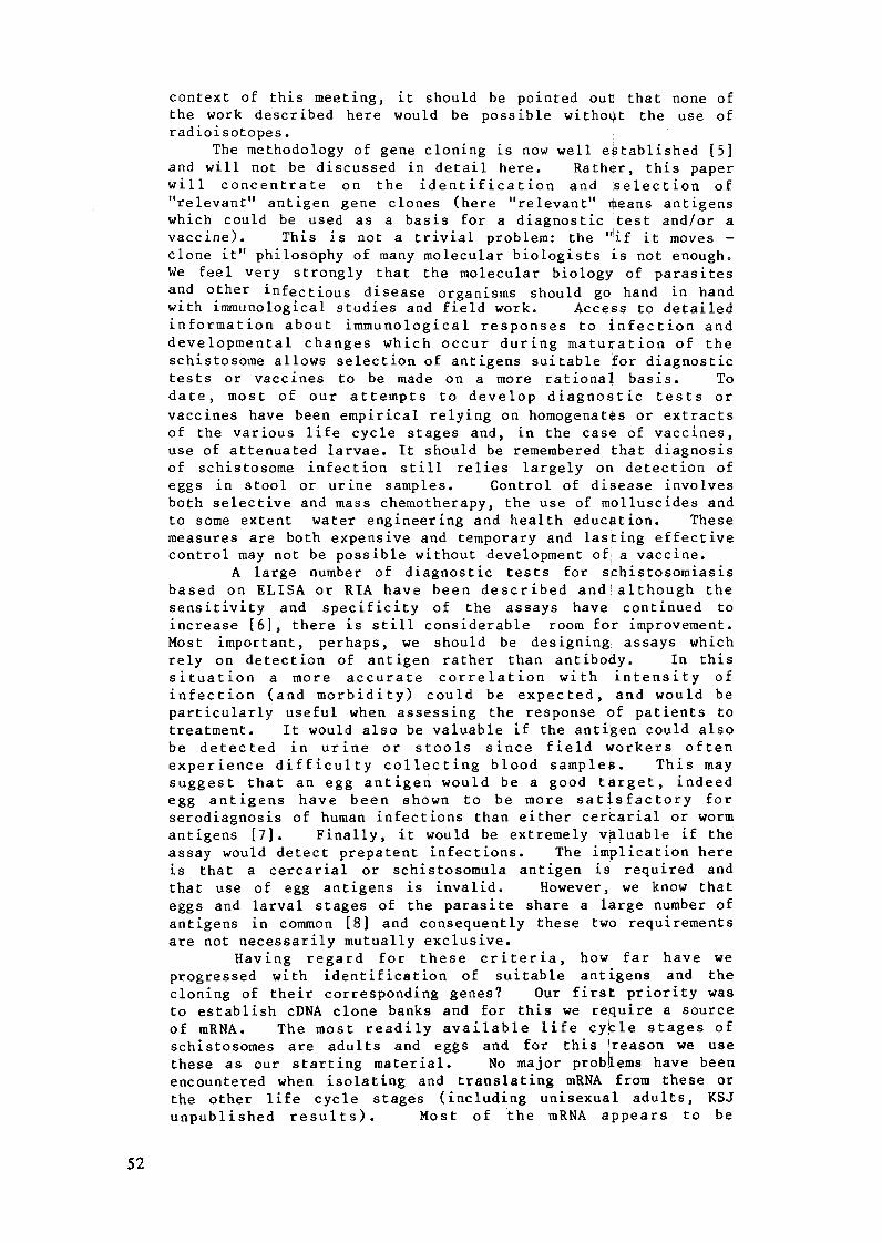
context of this meeting, it should be pointed oud that none ofthe work described here would be possible without the use ofradioisotopes.
The methodology of gene cloning is now well established [5]and will not be discussed in detail here. Rather, this paperwill concentrate on the identification and selection of"relevant" antigen gene clones (here "relevant" means antigenswhich could be used as a basis for a diagnostic test and/or avaccine). This is not a trivial problem: the "if it moves -clone it" philosophy of many molecular biologists is not enough.We feel very strongly that the molecular biology of parasitesand other infectious disease organisms should go hand in handwith immunological studies and field work. Access to detailedinformation about immunological responses to infection anddevelopmental changes which occur during maturation of theschistosome allows selection of antigens suitable for diagnostictests or vaccines to be made on a more rational basis. Todate, most of our attempts to develop diagnostic tests orvaccines have been empirical relying on homogenates or extractsof the various life cycle stages and, in the case of vaccines,use of attenuated larvae. It should be remembered that diagnosisof schistosome infection still relies largely on detection ofeggs in stool or urine samples. Control of disease involvesboth selective and mass chemotherapy, the use of mplluscides andto some extent water engineering and health education. Thesemeasures are both expensive and temporary and lasting effectivecontrol may not be possible without development of a vaccine.
A large number of diagnostic tests for schistosomiasisbased on ELISA or RIA have been described and'although thesensitivity and specificity of the assays have continued toincrease [6], there is still considerable room for improvement.Most important, perhaps, we should be designing assays whichrely on detection of antigen rather than antibody. In thissituation a more accurate correlation with intensity ofinfection (and morbidity) could be expected, and would beparticularly useful when assessing the response of patients totreatment. It would also be valuable if the antigen could alsobe detected in urine or stools since field workers oftenexperience difficulty collecting blood samples. This maysuggest that an egg antigen would be a good target, indeedegg antigens have been shown to be more satisfactory forserodiagnosis of human infections than either cercarial or wormantigens [7]. Finally, it would be extremely valuable if theassay would detect prepatent infections. The implication hereis that a cercarial or schistosomula antigen is required andthat use of egg antigens is invalid. However, we know thateggs and larval stages of the parasite share a large number ofantigens in common [8] and consequently these two requirementsare not necessarily mutually exclusive.
Having regard for these criteria, how far have weprogressed with identification of suitable antigens and thecloning of their corresponding genes? Our first priority wasto establish cDNA clone banks and for this we require a sourceof mRNA. The most readily available life cycle stages ofschistosomes are adults and eggs and for this reason we usethese as our starting material. No major problems have beenencountered when isolating and translating mRNA from these orthe other life cycle stages (including unisexual adults, KSJunpublished results). Most of the mRNA appears to be
52
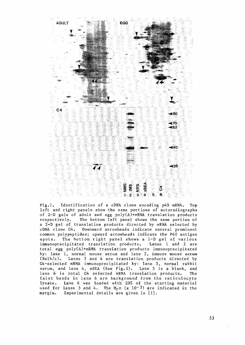
ADULT EGG:
C4
4~4Z- 00002a~ l
left and right panels show the same portions of autoradiographsof 2-D gels of adult and egg poly(A)+mRNA translation productsrespectively. The bottom left panel shows the same portion ofa 2-D gel of translation products directed by mRNA selected bycDNA clone C4. Downward arrowheads indicate several prominentcommon polypeptides; upward arrowheads indicate the P40 antigenspots. The bottom right panel shows a 1-D gel of variousimmunoprecipitated translation products. Lanes 1 and 2 aretotal egg poly(A)+mRNA translation products immunoprecipitatedby: lane 1, normal mouse serum and lane 2, immune mouse serum(Balb/c). Lanes 3 and 4 are translation products directed byC4-selected mRNA immunoprecipitated by: lane 3, normal rabbitserum, and lane 4, aSEA (See Fig.3). Lane 5 is a blank, andlane 6 is total C4 selected mRNA translation products. Thefaint bands in lane 6 are background from the reticulocytelysate. Lane 6 was loaded with 20% of the starting materialused for lanes 3 and 4. The Mrs (x 10-3) are indicated in themargin. Experimental details are given in [1].
:!: ̂ :; : ^ ̂ !:^ j 11^ : ̂ ; ^ ! :j[^ ;, ;^ 1i ^ | | :| | :::, , ̂ M K ::; ::\: !1^ ̂ ^ ̂̂ : ^.: ;.:| ,: :1\: ^ | :, : i' ̂ :: :,
* ' ' * * : * * : * ** .^ '*' ' *' " ; ̂ ^ ' * " ^ ; ̂ :
? .; ; - ' ; ; ; *: ' i ' ; / * * | f j : * * : : : " .' ' < :'* * ' ' * ' ' * ' ' ' ' ' ' ' .* * ' * ̂ y ''* . : * *
,: : : ;:- :1: ' 1: ::' . , : ; < ::^ : i;. ,;!; ::: ; ~u : ; ^ "" B * ̂ :;:- ::1::: :: 1 ^ . 1; : 11 '< *?x ' !, ̂" . : ' : ::: : ::! : ::. : : '!: .:.\ : i :;::::;. ::^:^: ^ 1^ : .: : : :.* i i~ f ̂ : :' : :**1 : : " :: : : ** ̂s2 i ::^ ' .* :
, ~ ~ ~ ~ ~ ~ ~ ~ · ~ -1:;: ;.
:^
1, :
1:^ :
l ::N
;:
: : lls? s S N:':: ^ ^
: 11:
1 1:
: l^
l l l™
<'::''
1
: 11 : ::11:1 ^ " : ' :^ :: i :r ; :^ : 1: ::; :: : ;:: :: :: :"1-: : : 'fl l i i " - : l : " s ;: l ' M't © ^ l :' 1
1: I\^ ^ '' [ :Iir I' ! :"1^ ? 1^^ : '^ :: '11!^ ' 1̂ :̂ 'I:^^ ^I^^ -^^:^^ ^ ^ 1 ::'-: :
margin. Experimental details are given in [I].
53
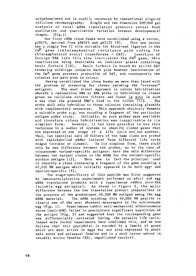
polyadenylated and is readily recovered by conventional oligo-dtcellulose chromatography. Single and two dimension IEF/SDS gelanalysis of total mRNA translation products reveal bothqualitative and quantitative variation between developmentalstages. (Fig.l)
Our first cDNA clone banks were established using a vector,pJSC73, derived from pBR325 and pAT153 [9]. This new vectorhas a single Pvu II site suitable for blunt-end ligation in theCmR gene (chloramphenicol resistance gene coding forchloramphenicol acetyl transferase - CAT). Insertion offoreign DNA into this site inactivates the CmR gene, thisinactivation being detectable on indicator plates containingbasic fuchsin [10]. Basic fuchsin is bound by active CATrendering wild-type colonies dark pink however inactivation ofthe CmR gene prevents production of CAT, and consequently thecolonies are pale pink in colour.
Having established the clone banks we were then faced withthe problem of screening for clones encoding the "relevant"antigens. The most direct approach is colony hybridisationwhereby a radioactive DNA or RNA probe is hybridised to clonesgrown on cellulose nitrate filters and lysed in $itu in sucha way that the plasmid DNA's bind to the filter [11]. Theprobe will only hybridise to those colonies containing plasmidswith complementary sequences. This approach obviously demandsa suitable probe e.g. one encoding a fragment of the particularantigen under study. Initially, no such probes were availableand therefore colony hybridisation was inapplicable in itssimplest form. However, it has been possible to use thistechnique in a number of systems to identify cloned genes thatare expressed at one stage of a life cycle and not another.Thus, two identical sets of filters of the same clone are probedwith radiolabelled mRNAs isolated from different life cyclestages (strains or clones). In its simplest form, there couldonly be one difference between the probes, as in the case oftrypanosome variant-specific antigens: here the only differencebetween two variant clones is the mRNA for the variant-specificsurface antigen [12]. This was in fact the principal usedto identify a clone containing a fragment of the gene encoding a40,000 MW antigen which initially appeared to be both egg- andspecies-specific [9].
The stage-specificity of this peptide was first suggestedby immunoprecipitation experiments performed on adult and eggmRNA translation products with a hyperimmune rabbit anti-SEA(soluble egg antigens). As shown in Figure 2, the majordifference between the two translation product preparations is
the presence of the predominant 40,000 MW antigen among the eggmRNA material. The mRNA encoding this 40,000 MW peptide isclearly one of the most abundant messengers in the schistosomeegg (Fig. 1). Hyperimmune rabbit anti-mechanical schistosomulaserum (anti-SOM) failed to precipitate significant quantities ofthe antigen (Fig. 2) and suggested that the corresponding genewas differentially activated during the parasite life cycle.Indeed more recent experiments have confirmed this, and we nowbelieve that this peptide(s) is encoded by a family of geneswhich are most active in eggs but are also expressed by adultmale worms and unisexual females and to a much lesser extent insexually active females (KSJ, unpublished results).
54
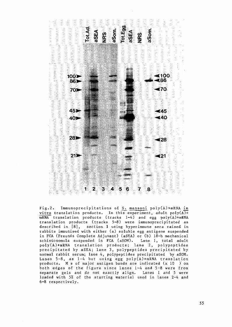
Z:41 Ozf cc J-t
i 10017
~!C-?OQ
-,45
1 [28
l218'I: :; :
i5 i6: :i;0 700 ;80
Fig.2. Immunoprecipitations of S. mansoni poly(A)+mRNA invitro translation products. In this experiment, adult poly(A)+mRNA translation products (tracks 1-4) and egg poly(A)+mRNAtranslation products (tracks 5-8) were immunoprecipitated asdescribed in [8], section 1 using hyperimmune sera raised inrabbits immunised with either (a) soluble egg antigens suspendedin FCA (Freunds Complete Adjuvant) (aSEA) or (b) 18-h mechanicalschistosomula suspended in FCA (aSOM). Lane 1, total adultpoly(A)+mRNA translation products; lane 2, polypeptidesprecipitated by aSEA; lane 3, polypeptides precipitated bynormal rabbit serum; lane 4, polypeptides precipitated by aSOM.Lanes 5-8, as 1-4 but using egg poly(A)+mRNA translationproducts. M s of major antigen bands are indicated (x 10 ) onboth edges of the figure since lanes 1-4 and 5-8 were fromseparate gels and do not exactly align. Lanes 1 and 5 wereloaded with 5% of the starting material used in lanes 2-4 and6-8 respectively.
55
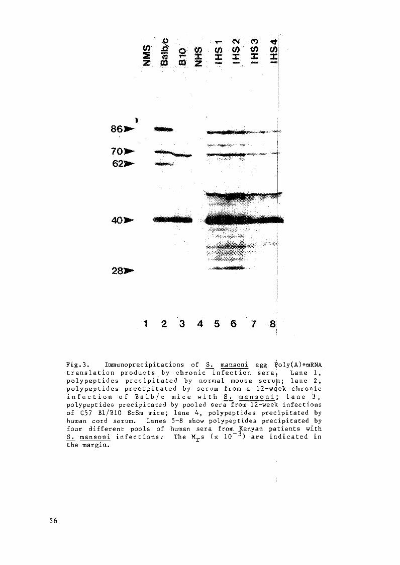
1 2 3 4 5 6 7 8 x
anain rc crnic inf in ra, an
infection of Balb/c mice with S. mansoni; lane 3,
~ cS. mansoni infections. The Mr ( 10- 3 ) are indicated in
the margin.
1 2 3 4 5 6 7 8
Fig.3. Immunoprecipitations of S. mansoni egg Poly(A)+mRNAtranslation products by chronic infection sera, Lane 1,polypeptides precipitated by normal mouse serum; lane 2,polypeptides precipitated by serum from a 12-week chronicinfection of Balb/c mice with S. mansoni; lane 3,polypeptides precipitated by pooled sera from 12-week infectionsof C57 B1/B10 ScSm mice; lane 4, polypeptides precipitated byhuman cord serum. Lanes 5-8 show polypeptides precipitated byfour different pools of human sera from Kenyan patients withS. mansoni infections. The Mrs (x 10 ) are indicated inthe margin.
56
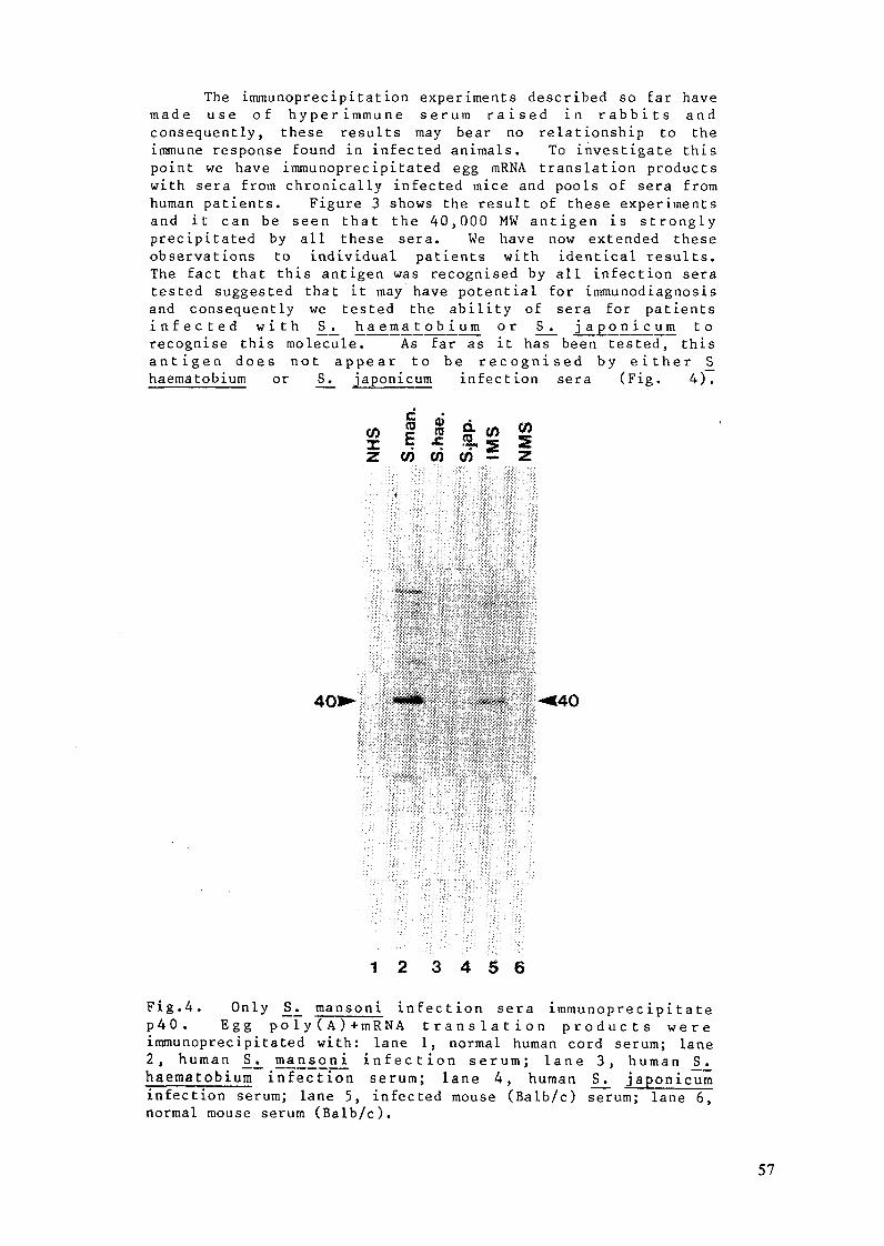
The immunoprecipitation experiments described so far havemade use of hyperimmune serum raised in rabbits and
consequently, these results may bear no relationship to theimmune response found in infected animals. To investigate thispoint we have immunoprecipitated egg mRNA translation productswith sera from chronically infected mice and pools of sera fromhuman patients. Figure 3 shows the result of these experimentsand it can be seen that the 40,000 MW antigen is stronglyprecipitated by all these sera. We have now extended theseobservations to individual patients with identical results.The fact that this antigen was recognised by all infection seratested suggested that it may have potential for immunodiagnosisand consequently we tested the ability of sera for patientsinfected with S. haematobium or S. japonicum torecognise this molecule. As far as it has been tested, thisantigen does not appear to be recognised by either Shaematobium or S. japonicum infection sera (Fig. 4).
z I: X z
1 2 34 5 6
Fig.4. Only S. mansoni infection sera immunoprecipitatep4 0 . Egg poly(A)+mRNA translation products wereimmunoprecipitated with: lane 1, normal human cord serum; lane2, human S. mansoni infection serum; lane 3, human S.haematobium infection serum; lane 4, human S. japonicuminfection serum; lane 5, infected mouse (Balb/c) serum; lane 6,normal mouse serum (Balb/c).
57
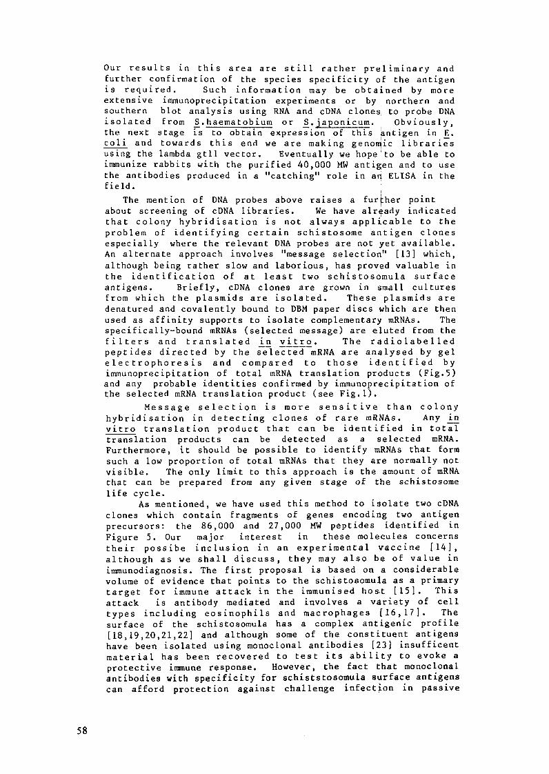
Our results in this area are still rather preliminary andfurther confirmation of the species specificity of the antigenis required. Such information may be obtained by moreextensive immunoprecipitation experiments or by northern andsouthern blot analysis using RNA and cDNA clones to probe DNAisolated from S.haematobium or S.japonicum. Obviously,the next stage is to obtain expression of this antigen in E.coli and towards this end we are making genomic librariesusing the lambda gtll vector. Eventually we hope to be able toimmunize rabbits with the purified 40,000 MW antigen and to usethe antibodies produced in a "catching" role in an ELISA in thefield.
The mention of DNA probes above raises a further point
about screening of cDNA libraries. We have already indicated
that colony hybridisation is not always applicable to the
problem of identifying certain schistosome antigen clones
especially where the relevant DNA probes are not yet available.
An alternate approach involves "message selection" [13] which,
although being rather slow and laborious, has proved valuable inthe identification of at least two schistosomula surface
antigens. Briefly, cDNA clones are grown in small culturesfrom which the plasmids are isolated. These plasmids are
denatured and covalently bound to DBM paper discs which are then
used as affinity supports to isolate complementary mRNAs. The
specifically-bound mRNAs (selected message) are eluted from thefilters and translated in vitro. The radiolabelled
peptides directed by the selected mRNA are analysed by gel
electrophoresis and compared to those identified byimmunoprecipitation of total mRNA translation products (Fig.5)and any probable identities confirmed by immunoprecipitation ofthe selected mRNA translation product (see Fig.l).
Message selection is more sensitive than colony
hybridisation in detecting clones of rare mRNAs. Any in
vitro translation product that can be identified in total
translation products can be detected as a selected mRNA.
Furthermore, it should be possible to identify mRNAs that form
such a low proportion of total mRNAs that they are normally not
visible. The only limit to this approach is the amount of mRNA
that can be prepared from any given stage of the schistosome
life cycle.
As mentioned, we have used this method to isolate two cDNA
clones which contain fragments of genes encoding two antigen
precursors: the 86,000 and 27,000 MW peptides identified in
Figure 5. Our major interest in these molecules concerns
their possibe inclusion in an experimental vaccine [14],
although as we shall discuss, they may also be of value in
immunodiagnosis. The first proposal is based on a considerable
volume of evidence that points to the schistosomula as a primary
target for immune attack in the immunised host [15]. This
attack is antibody mediated and involves a variety of cell
types including eosinophils and macrophages [16,17]. The
surface of the schistosomula has a complex antigenic profile
[18,19,20,21,22] and although some of the constituent antigens
have been isolated using monoclonal antibodies [ 3] insufficent
material has been recovered to test its ability to evoke a
protective immune response. However, the fact that monoclonal
antibodies with specificity for schiststosomula surface antigens
can afford protection against challenge infection in passive
58
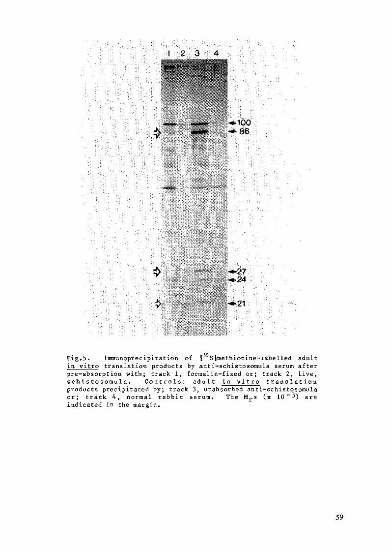
AK0 : E :00--. ,86
Fig.5. Immunoprecipitation of [C S]methionine-labelled adultin vitro translation products by anti-schistosomula serum afterpre-absorption with; track 1, formalin-fixed or; track 2, live,schistosomula. Controls: adult in vitro translationproducts precipitated by; track 3, unabsorbed anti-schistosomulaor; track 4, normal rabbit serum. The Mrs (x 10 - 3) areindicated in the margin.
59
transfer experiments argues strongly in favour of a protectiverole for these antigens [24,25].
Recently our own experiments [8] and others carried out inDr. Smithers' laboratory at NIMR, Mill Hill [26] have identifiedschistosomula surface antigen precursors among in vitrotranslation products directed by mRNA isolated for adults andeggs. The synthesis of schistosomula antigen by adults and eggwas demonstrated by immunoprecipitation of the correspondingtranslation product by an anti-schistosomula serum (Fig.2),while the surface location was suggested by failure ofanti-schistosomula serum to precipitate certain antigensfollowing absorption with line or formalin-fixed larvae. Theresult of one such experiment is illustrated in Figure 5. Heretwo translation products (86,000 and 27,000 MW) are shown to beprecursors of surface components while similar experiments, inwhich formalin-fixed schistosomula were used asimmunoabsorbants, have identified three other surface antigenprecursors with apparent molecular weights 100,000; 28,000; and24,000 [8]. These and similar experiments provide a confirmationand indeed are a prerequisite for the identification of relevantcDNA clones by message selection [13]. The next step is toobtain expression of these antigens in bacteria.
Immunoprecipitation experiments carried out using invitro translation products show the antibody response ofindividual patients towards these surface antigens isvariable. This is not unexpected, and such observations arepertinent to development of both vaccines and diagnostic tests.A poor response to a particular antigen by a proportion ofinfected patients should not necessarily preclude its use in avaccine. One of the most interesting and fascinating aspects ofthe immune response concerns the question of antigenpresentation. Simply producing large quantities of purifiedantigens will not provide us with a vaccine overnight, therecertainly will be many questions to ask about apparent trivalpoints such as the route of inoculation, amount of antigen andwhether a carrier or adjuvant is required to evoke anapropriated response. As for diagnosis, it is clear that apoor response to a purified antigen used as a target in an'ELISAor RIA designed to measure antibody levels could lead tospurious results. For example, a patient may have a high wormburden and egg output yet have only a poor response to, say, the86000 surface antigen: this would produce a false negativeresult. There is need for caution here and probably thesolution would be to use a cocktail of antigens. Better still,and as mentioned at the beginning of this paper, we shouldconsider designing the assay to detect antigen rather thanantibody [27]. Polyclonal or monoclonal antibodies raisedagainst surface antigens (expressed by bacteria) could be usedas both catching and detecting antibody in an ELISA or RIA.Use of surface antigens in such a test may also allow thedetection of prepatent infections. Finally there is thequestion of specificity: immunoprecipitation experimentsperformed on S. haematobium in vitro translationproducts using anti S. mansoni serum have demonstrated thespecies specificity of at least three of the putative surfaceantigen precursors namely the 100,000; 86,000 and 27,000 MWpeptides (DWT and JSC unpublished results).
60
A logical extension of the work described above is thesynthesis of peptides based on the nucleotide sequence of thecloned antigen genes. It is now well established that evenquite small amino acid sequences can evoke a strong immuneresponse against the native parent protein [28,29] andconsiderable progress has been reported in development ofvaccines against a number of infectious agents [30], forexample, Foot and Mouth Disease virus [31] and Hepatitis B [32].Use of synthetic vaccines is particularly appealing for anumber of reasons. First cost, it is important that any vaccineused in developing countries is cheap to produce. Second,quality control is easy to maintain. Third, it should bepossible to avoid undesirable side effects which sometimesaccompany complex antigenic mixtures (eg, immunosupression andhypersensitivity). In our opinion it is only a matter of timebefore such vaccines will become available for control ofeukaryote parasitic diseases.
ACKNOWLEDGEMENTS
This work is supported by grants from the Medical ResearchCouncil (UK), the Edna McConnell Clark Foundation, and the RoyalSociety. J.S. Cordingley is an MRC Training Fellow and K.S.Johnson is in receipt of an MRC studentship. The Figures arereproduced with permission.
REFERENCES
[1] WILLIAMSON , R. "Genetic Engineering" Vols 1-4 AcademicPress, London, (1981-82)
[2] MELCHERS, F., POTTER, M. and WARNER, N. "LymphocyteHybridomas", Springer-Verlag, Berlin, 1979.
[3] KEMP, D.J. et al. Proc. Natn. Acad. Sci., USA 80 (1983)3787-3791.
[4] OZAKI, L.S. et al. Cell 34 (1983) 815-822.[5] MANIATIS, T., FRITSCH, E.F., SAMBROOK, J. "Molecular
Cloning", Cold Spring Harbour Laboratory, 1982.[6] MCLAREN, M.L. et al. Trans. R. Soc. Trop. Med. Hyg. 75
(1981) 72-79.[7] MCLAREN, M.L. et al. Annls. Trop. Med. Parasit. 72
(1978).[8] TAYLOR, D.W., CORDINGLEY, J.S. and BUTTERWORTH, A.E. Mol.
Biochem. Parasit. 10 (1984) 305-318.[9] CORDINGLEY, J.S., TAYLOR, D.W., DUNNE, D.W. and BUTTERWORTH
A.E. Gene 26 (1983) 25-39.[10] PROCTOR, G.N. and ROUWND, R.H. J. Bacteriol. 150 (1982)
1375-1382.[11] GRUNDSTEIN, M. and HOGNESS, D.S. Proc. Natn. Acad. Sci.
USA 72 (1975) 3961-3965.[12] HOEIJMAKERS, J.H.J. Gene 8 (1980) 391-417.[13] SMITH, D.F., SEARLE, P.F. and WILLIAMS, J.G. Nuclei Acids
Res. 6 (1979) 487-506.[14] TAYLOR, D.W., CORDINGLEY, J.S. and BUTTERWORTH, A.E. Vet.
Parasitol. (1984) in press.[15] SMITHERS, S.R., MCLAREN, D.J. and RAMALHO-PINTO, F.J.
Amer. J. Trop. Med. Hyg. 26 suppl. (1977) 11-19.
61

[16] BUTTERWORTH, A.E. et al. Immunological Revs. 61 (1982)5-39.
[17] CAPRON, A. et al. Immunological Revs. 61 (1982) 41-66.[18] SNARY, D., SMITH, M.A. and CLEGG, J.A. Eur. J. Immunol.
10 (1980) 573-575.[19] DISSOUS, C., DISSOUS, C. and CAPRON, A. Mol. Biochem.
Parasitol. 3 (1981) 215-225.[20] TAYLOR, D.W., HAYUNGA, E.G. and VANNIER, W.E. Mol.
Biochem. Parasit. 3 (1981) 157-168.[21] SHAH, J. and RAMASAMY, R. Int. J. Parasit. 12 (1982)
451-461.
[22] SIMPSON, A.J.G., JAMES, S.L. and SIER, A. Infect. Immun.41 (1983) 591-597.
[23] TAYLOR, D.W. and BUTTERWORTH, A.E. Parasitology 84(1982) 65-82.
[24] SMITH, M.A., CLEGG, J.A., SNARY, D. and TREJDOSIEWECZ,A.J. Parasitology 84 (1982) 83-91.
[25] CRZYCH, J-M., CAPRON, M., BAZIN, H. and CAPRON, A.J. Immunol. 129 (1982) 2739-2743.
[26] KNIGHT, M. et al. EMBO J. 3 (1984) 213-219.[27] ROTMANS, J.P. Exp. Parasit. 52 (1981) 319-330.[28] LERNER, R.A. Nature 299 (1982) 592-597.[29] YOUNG, C.R., SCHMITZ, H.E. and ATASSI, M.Z. Mol.
Immunol. 20 (1983) 567-570.[30] YOUNG, C.R. J. Roy. Soc. Med. (1984) in press.[31] BITTLE, J.L. et al. Nature 298 (1982) 30-33.[32] PRINCE, A.M. Proc. Natn. Acad. Sci. 79 (1982) 579-
582.
62
STUDIES ON EPITOPIC AND IDIOTYPIC REGULATIONIN SCHISTOSOMIASIS: THE USE OF MONOCLONALANTIBODIES IN RADIO-IMMUNO- AND AVIDIN-BIOTIN-ENZYME ASSAYS
S.M. PHILLIPS, E.G. FOX, M.N. FATHELBAB,D.J. WALKER, D.M. ZODDAAllergy and Immunology Section,Department of Medicine,University of Pennsylvania,Philadelphia, PA, USA
Abstract
A series of monoclonal antibodies (MAbs) of both protectiveand non-protective nature against schistosomiasis mansoni havebeen produced in our laboratory. We attempted to identify andpurify the specific antigen which was recognized by theprotective MAb in hopes of obtaining a putative protectiveimmunogen. Initial attempts at identification using crude 125I-soluble cercarial immunogen in radioimmunoprecipitation protocolsproduced similar molecular weight banding patterns for all MAbstested. Discrimination on the basis of antibody affinity alsoproved unrewarding as did isotype analysis; affinity of theprotective MAb was not distinctive and all MAbs used in theseassays were IgGl. Purifications of the crude antigen by affinitychromatography, using either protective or non-protective MAbs,produced bands that remained immunologically reactive as assayedby overlay analysis with 1 2 5 I-1Ab, but again of identicalmolecular weight. We thus developed an avidin-biotin-likPimmunoassay to test for epitopic competition. Competition assaysusing biotinylated antisera or MAb demonstrated that theprotective MAb was recognizing a distinct epitope on the antigen.It appears that the non-protective MAbs are cross-reactive for asecond epitope. Other experiments demonstrated that binding ofMAb could be blocked by addition of soluble antigen, and thatanti-idiotypic antisera raised against the protective NAb celllines would specifically inhibit binding of the inducingidiotypic MAb. Thus, we have demonstrated the utility of thismethod for assessing the basic components - epitope, idiotype andparatope directed anti-idiotype - of the idiotypic network as itpertains to schistosomiasis.
INTRODUCTION
Recognition of the interactive nature of the humoral andcellular components of the immune system and the manner in whichthe total immune response is regulated has proven to be of greatimportance. Understanding these processes is critical in theelucidation of mechanisms of resistance and morbidity in humandisease. For example, the immune response to a metazoaninfectious organism such as Schistosoma mansoni is polyclonalconsisting of many different events. A variety of cellular andhumoral events occur which are in response and directed against
63
many different immunogenic species (antigens or epitopes). Aevents a cascade of modulating events occur to regulate thesereactions. These events include production of helper andsuppressor events in a complex network of interactions. Toregulate this network the existance of idiotypic-anti-idiotypicinteractions have been postulated [11. According to thishypothesis, the regulatory cells or antibodys recognize andrespond against the primary receptors or idiotypes which aredirected against the parasite associated immunogens. In turn theidiotypes are recognized by other anti-idiotypic reactions. Thisprocess proceeds sequentially, producing a cascade ofregulation.
This network provides a number of theoretical implications.For example, anti-idiotypic moieties may compete with antigensand contribute to immune complex formation. This occurence willcomplicate analysis of antigenic characteristics and antibodyactivity. Anti-idiotypes may also provide alternatives toantigen for immunization 12,3,4].
There are reasons to suggest that such a network isoperative in schistosomiasis. Radioimmune assays havedemonstrated such responses in the blood of patients infectedwith S. japonicum 151. Anti-idiotypic reactions have beendemonstrated at the polyclonal and oligoclonal level 1.6] (Fox andPhillips, unpublished observation respectively). 1
Additional regulatory mechanisms involve competition ofreactant moeities for the same critical determinant or epitope.For example, in schistosomiasis, several antibody isotypes havebeen demonstrated to promote antibody-dependent cell mediatedcytotoxicity, ie. (ADCC), directed against schistosomlules. IgEand IgG2a in rats, while another isotype, i.e. IgG2c, with across-reactive epitopic specificity, will provide animmunoregulatory function. This latter antibody will not promoteADCC and will actually block the protective monoclonal's abilityto promote ADCC and adoptively transfer protection 171. A similarevent has been described in vitro using mouse derivedimonoclonalantibodies (Butterworth, A., Personal Communication). Thus,multiple antibodies will react with the same antigen and/orsterically effect the binding of antibodies directed againstdifferent antigens. To analyze these complex interactions ofantigens, idiotypes, and anti-idiotypes, we have developedsensitive assays for each component. These techniques appearpromising for antigen fractionation and in immunodiagnosis.
In the past, the majority of assays providing information asregards differentiation of epitopic recognition by varyingmonoclonal antibodies have relied heavily upon the use ofradiolabeled materials, both antigens and antibodie s . Problemsassociated with these procedures are several including the shorthalf-life of the radioisotopes; inherent potential1dangers ofworking with and disposing of such materials; the'expense ofequipment needed to analyze radioreactants; field applicability.Several of these procedures are slowly being supplimented orsupplanted by the use of ELISA or enzyme-linked immunologicassays. A recent advancement in this latter technoldgy has beenthe introduction of the avidin-biotin binding system. The low
64
molecular weight biotin molecule (M.W.274) may be covalentlybound to protein using free amino groups. The gentle complexingprocedure does not alter the biological properties of themolecule [81.
In the present work we have demonstrated the utility ofusing combined radiobiologic and biotinylation techniques asimmunoanalytic probes for analysis. The results indicate that:1. Biotinylation or radiologic labelling do not destroybiological integrity and are sensitive at least to the longlevel; 2. Competition assays with biotinylated MAbs (B-MAbs)will provide quick and complete characterization of epitopicdistinctiveness. For example, the protective MAb combines withan epitope which is distinct from that of the non-protective MAbalthough both react with a similar membrane component; 3.Blocking assays may estimate specific antigen levels or provideevidence of anti-idiotypic responses. The technology provides aprecise quantitative procedure for analyzing polyclonal andmonoclonal anti-idiotypic reactants.
MATERIALS AND METHODS
Production of protective hybridoma lines: Splenocytes frominfected S. mansoni C57B1/6 mice were fused with SP2/0 myelomacells as previously described [9]. Lines producing antibody withparticular affinity for the tegumental surface of S. mansonicercariae as assayed by ELISA and immunofluorescence testing,were selectively sub-cloned and expanded in C57B1/6 x Balb/c F1mice to produce ascites fluid. Ascitic fluids wereintraperitoneally administered to naive C57B1/6 recipients whichwere then infected with the parasite. Several antibodies haveproduced protection and the antibody product of one line, S80-D6(IgG1) has been particularly carefully studied. For completedetails see Zodda and Phillips [9J and Phillips and Zodda [10].
Preparation and purification of monoclonal antibodies: Allantibody producing hybridoma clones were maintained in definedserum-free medium, (HB-101, Hana Biologics, Berkeley, CA).Initial purification of supernatant material was accomplished byprecipitation in a 50% saturated ammonium sulfate solution anddialysis against PbS. Affinity purification was accomplishedusing a Protein-A column (Pharmacia Fine Chemicals, Piscataway,NJ). Assays for purity, activity and protein concentration wereby standard PAGE [11], ELISA [9] and Lowery techniques [12]respectively. Normal and immune mouse serum were similarlyprepared except for an additional 45% pre-precipitation withsaturated ammonium sulfate.
Radioiodination Procedures: Soluble cercarial membraneantigen (SCI) was obtained by 4 M KC1 digestions of S. mansonicercariae and radiolabeling was accomplished using thechloramine-T method and 125I 113]. This method successfullylabeled SCI but inactivated our purified MAb; thereforeradiolabelling of antibody was accomplished using the Bolton-hunter technique [14].
Kadioimmunoprecipitations: 1 x 10 CPMI of 1 25I-SCI was addedto 1 ml of hybridoma supernatant and mixed overnight at 4° .
65
Twenty-five ul of Protein-A was added and the reactants mixed for1 hour at 4°. The material was then centrifuged in an "air-fuge"and washed 4 times, terminally in starting PAGE buffer. Theprecipitate-complex was uncoupled and reduced by 2-mercaptoethanol and SDS and then run in 7.5-15% polyacrylamidegels. Visualization was accomplished by means of the Bio-radsilver staining procedure.
Immunoaffinity purification: CNBr-Sepharose 4t (Pharniacia,Piscataway, NJ) immunoaffinity columns were prepared by couplingpurified MAb, 2-3 mg/ml. The SCI antigen in PBS, pH 8.2, waspassed over the column and specifically bound material was elutedusing glycine-HCI, pH 2.5.
Electrophoresis technicology: Polyacrylimide gel electro-phoresis (PAGE) was conducted at 7.5-15% [11]. "Western"electroblotting and overlays were made onto nitrocellulosemembranes [15] (Bio-rad, Richmond, CA).
Biotinylation of monoclonal antibodies: 100 ul of dimethyl-sulfoxide containing 1.1 mg/ml of N-hydroxy-succinimide ester ofbiotin was added to each ml. of purified mouse serum or MAb, (1mg protein/ml in PBS pH8.4). The reactants were incubated fortwo hours at room temperature with occasional shaking. Unreactedester was blocked by the addition of 100 ul of 1 M NtH4Cl andincubated for 10 minutes. One ml. ot PBS 7.2 containing 1% BSA(Fraction V - fatty acid free; Cal-Biochem, LaJolla, CA) was thenadded and the samples were dialyzed in PbS, 7.2. i
Avidin-biotin Procedures: Varying concentrations ofschistosomal egg antigen (SEA)[91 in 100 ul of PBS 7.2 or lOOulof 0.1% BSA were added to experimental or control wells,respectively. The contents of the flexible PVC microtiter plates(Dynatech; Alexandria, VA) were flicked out. The wells werewashed and blocked with 4X BSA in carbonate buffer (pH 9.6) andincubated for 1 hour at 20°C on a rocker platform. The plateswere washed witt an 0.1% BSA-gelatin buffer (ptH7.4). In thedirect assay, 50ul of the biotinylated MAb was added to bothexperimental and control wells and incubated for 2 hours at 20°Con a rocker platform. The plates were then washed with 0.1% BSA-gelatin buffer. The Avidin-biotin-HRP-complex (ABC) (V.P.Scientific; San Diego, CA) working solution, 50 ul, was added tothe plates and incubated an additional 30 min. at 20°C on arocker platform. The plate was then washed six times with thegelatin buffer without BSA and 200 ul of the chromagen substrate(0-phenylenediamine) in citrate buffer (pH 4.5) and H202 wasadded. Incubation was continued for appropriate times and colordevelopment was read using a Titertek multiscan. Specificreadings were established by substracting control from
experimental OD readings.
Production of anti-idiotypic antisera: This procedure wasbased upon the methodology of Noseworthy et al. [ 161. Briefly,hybridoma cells in log growth phase were counted and irradiated
with 1500 mrads. The cells were emulsified in CFA (Img PPD/ml)
and injected into the rear foot pads and dorsum of mice. A total
of 5 x 10⁶ cells/0.5ml per animal was injected. Thereafterinjections were in the presence of incomplete Freind's adjuant.
66

This protocal was repeated every two weeks for a total of atleast 3 injections.
RESULTS
Rationale and standardization of assays: We have produced aseries of murine monoclonal antibodies directed against thesurface antigen of the trematode Schistosoma mansoni. Some ofthese can provide protection in passive transfer againstinfection when administered to naive recipient mice.Differentiation of the specific antigens recognized by theprotective and non-protective M1Ab's by radioimmunoprecipitationdemonstrated that both MAb types could precipitate proteinmoieties of the same molecular weight (Figure 1). Subsequentaffinity purified material, obtained from protective S80-D6 ornon-protective N122-9 (IgG1) affinity columns, also demonstratedidentical molecular weight banding patterns. These antigensremain immunologically intact as determined by electroblot 1251-MAb overlay procedures.
Sample STOS N3-4 N-158 M-122 N-34 S-80/f06 CfAppleoation (C) S*200(
200,000- --~ -.
-" SM 160
94,000-' - ...
68,000 -a
43,000 -'*
'IMMUWOPRElCITAT: ' : OtI :PiUTIUJZN< MONOCLONAL.iATOl : AIE o gs0
Figure 1: Immunoprecipitation/PAGE autoradiograph utilizingmonoclonal antibodies and 12 5 I-SCI.
SCI (1 x 10⁷ CPM) was immunoprecipitated by five differentMAbs. Four MAbs recognizing SCI precipitated indenticalmolecular weight components, while the control (N34-4) did not.
67
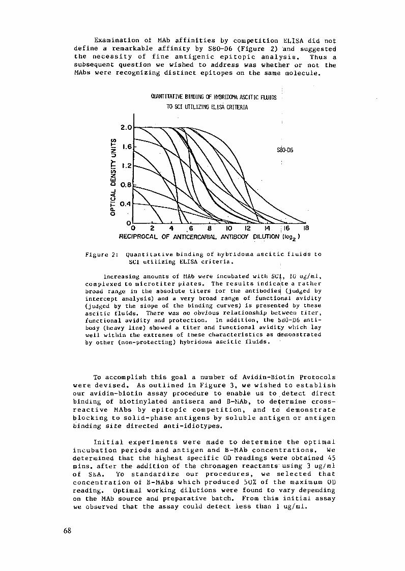
Examination of MAb affinities by competition ELISA did notdefine a remarkable affinity by S80-D6 (Figure 2) and suggestedthe necessity of fine antigenic epitopic analysis. Thus asubsequent question we wished to address was whether or not theMAbs were recognizing distinct epitopes on the same molecule.
Q(NTITATIVE BIDlING OF HYBRIDO1M ASCITIC FUIDS
TO SCI UTILIZING ELISA CRITERIA
2.0
1.2 0
o 0.8
O 2 4 6 8 10 12 14 16 18RECIPROCAL OF ANTICERCARIAL ANTIBODY DILUTION (log 2 )
Figure 2: Quantitative binding of hybridoma ascitic fluids toSCL utilizing ELISA criteria.
Increasing amounts of MAb were incubated with SCI, 10 ug/ml,complexed to uicrotiter plates. The results indicate a ratherbroad range in the absolute titers tor the antibodies (judged byintercept analysis) and a very broad range of functional avidity(judged by the slope of the binding curves) is presented by theseascitic fluids. There was no obvious relationship between titer,functional avidity and protection. In addition, the SdO-D6 anti-body (heavy line) showed a titer and functional avidity which laywell within the extremes of these characteristics as demonstratedby other (non-protecting) hybridoma ascitic fluids.
To accomplish this goal a number of Avidin-Biotin Protocols
were devised. As outlined in Figure 3, we wished to establish
our avidin-biotin assay procedure to enable us to detect direct
binding of biotinylated antisera and B-bAb, to determine cross-
reactive MAbs by epitopic competition, and to demonstrate
blocking to solid-phase antigens by soluble antigen or antigen
binding site directed anti-idiotypes.
Initial experiments were made to determine the optimal
incubation periods and antigen and B-MAb concentrations. We
determined that the highest specific OD readings were obtained 45
mins. after the addition of the chromagen reactants using 3 ug/ml
of SEA. To standardize our procedures, we selected that
concentration of B-MAbs which produced 50% of the maximum OD
reading. Optimal working dilutions were found to vary depending
on the MAb source and preparative batch. From this initial assay
we observed that the assay could detect less than 1 ug/mnl.
68
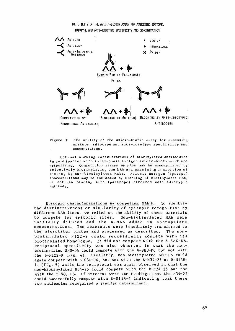
THE UTILITY OF THE AVIDIN-BIOTIN ASSAY FOR ASSESSING EPITOPE,
IDIOTYPE AND ANTI-IDIOTYPE SPECIFICITY AD CCNCENTPATION
AA ANTIGEN I BIOTIN
'~ ANTIBODY * PEROXIDASE
NATI-IDIOTYPIC X AVIDIN
AAN+ AVIDIN-BIOTIN-PEROXIDASE
ELISA
AA+* AA+ I AA +COMPETITION BY BLOCKING BY ANTIrEN I BLOCKING BY ANTI-IDIOTYPIC
MONOCLONAL ANTIBODIES; ANTIBODIES
Figure 3: The utility of the avidin-biotin assay for assessingepitope, idiotype and anti-idiotype specificity andconcett ration.
Optimal working concentrations of biotinylated antiboidiesin combination with solid-phase antigen avidin-biotin-iiKP areestaDlished. Comipetition assays by hiAbs may be accomplished byselectively biotinylating one MAb and examining inhibition otbinding by non-biotinylated MAbs. Soluble antigen (epitope)concentrations may be estimated by blocking of biotinylated NAb,or antigen binding site (paratope) directed anti-idiotypicantibody.
Epitopic characterizations by competing MAb's: To identifythe distinctiveness or similarity of epitopic recognition bydifferent MAb lines, we relied on the ability of these materials
to compete for epitopic sites. Non-biotinylated MAb were
initially diluted and the B-MAb added in appropriateconcentrations. The reactants were immediately transferred tothe microtiter plates and processed as described. The non-biotinylated N122-9 could successfully compete with itsbiotinylated homologue. It did not compete with the B-S80-D6.Reciprocal specificity was also observed in that the non-biotinylated S80-D6 could compete with the B-S80-D6 but not withthe B-N122-9 (Fig. 4). Similarly, non-biotinylated S80-D6 couldagain compete with B-S80-D6, but not with the B-N34-25 or B-N158-
1, (Fig. 5) while the reciprocal was again observed in that thenon-biotinylated N34-25 could compete with the B-N34-25 but notwith the B-S80-D6. Of interest were the findings that the N34-25could successfully compete with B-N158-1 indicating that thesetwo antibodies recognized a similar determinant.
69
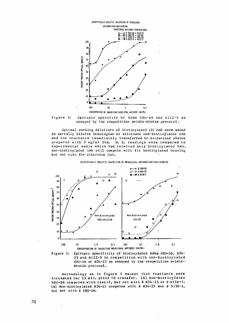
IDIOTYPICALLY SPICIFIC INHIBITION CF KLOCALOL
FNriBaY/ANIIGEN BIwINGXOCLOUAL AN1TIBCY COMIINATIONS
= B S80-D6 x N122-9U- B S80-D6 x S80-D6*--- B N122-9 x S80-DGU--- B N122-9 x N122-9
g
*/ / /
11/ m/
-"-------- --.---- "--- '-
100 10 1 0.1
CCEMWR4 TIlc OF IIIBITING I.lD1CLCII . ANTIBSoY IULG.)
Figure 4: Epitopic specifity of MAbs S8U-D6 and N122-9 asassayed by the competition avidin-biotin protocol.
Optimal working dilutions of biotinylated (B) MAb were addedto serially diluted homologous or alternate non-biotinylated ,Aband the reactants immediately transferred to microtiter platesprepared with 3 ug/ml SEA. O. D. readings were compared toexperimental wells which had received only biotinylated fMAb.hon-biotinylated ifAb will compete with its biotinylated homologbut not with the alternate I-Ab.
IDIOTYPICALLY SPECIFIC INHIBITION OF MONOCLONAL ANTIBODY/ANTIGENI BINDING
-- B S0-D60 o--- B N34-25
100 ° ° .8 .1 o1900 A n'y'' ^ O 0 B0 -- B N{58-1
9 0 A
3 0 */ NON-BIOTINYLATED NON-BIOTINYLATED 0
230 // S80-3AF12-D6 134-25
2010
~~~1O~~~L~~~i~~~ f -^ ^...a.............
100 10 1.0 0,1 100 10 1.0 0.1
CONCENTRATION OF INHIBITING MONOCLONAL ANTIBODY (UR/ML)
Figure 5: Epitopic specificity of biotinylated HAbs S80-D6, N34-25 and N122-9 in competition with non-biotinylatedS80-D6 or N34-25 as assayed by the competition avidin-biotin protocol.
Methodology as in Figure 4 except that reactants wereincubated tor 15 min. prior to transfer. (A) Non-biotinylatedS80-D6 competes with itself, but not with B N34-25 or B N158-1.(B) Non-biotinylated N34-25 competes with B N34-25 and B N158-1,but not with b S80-D6.
:70
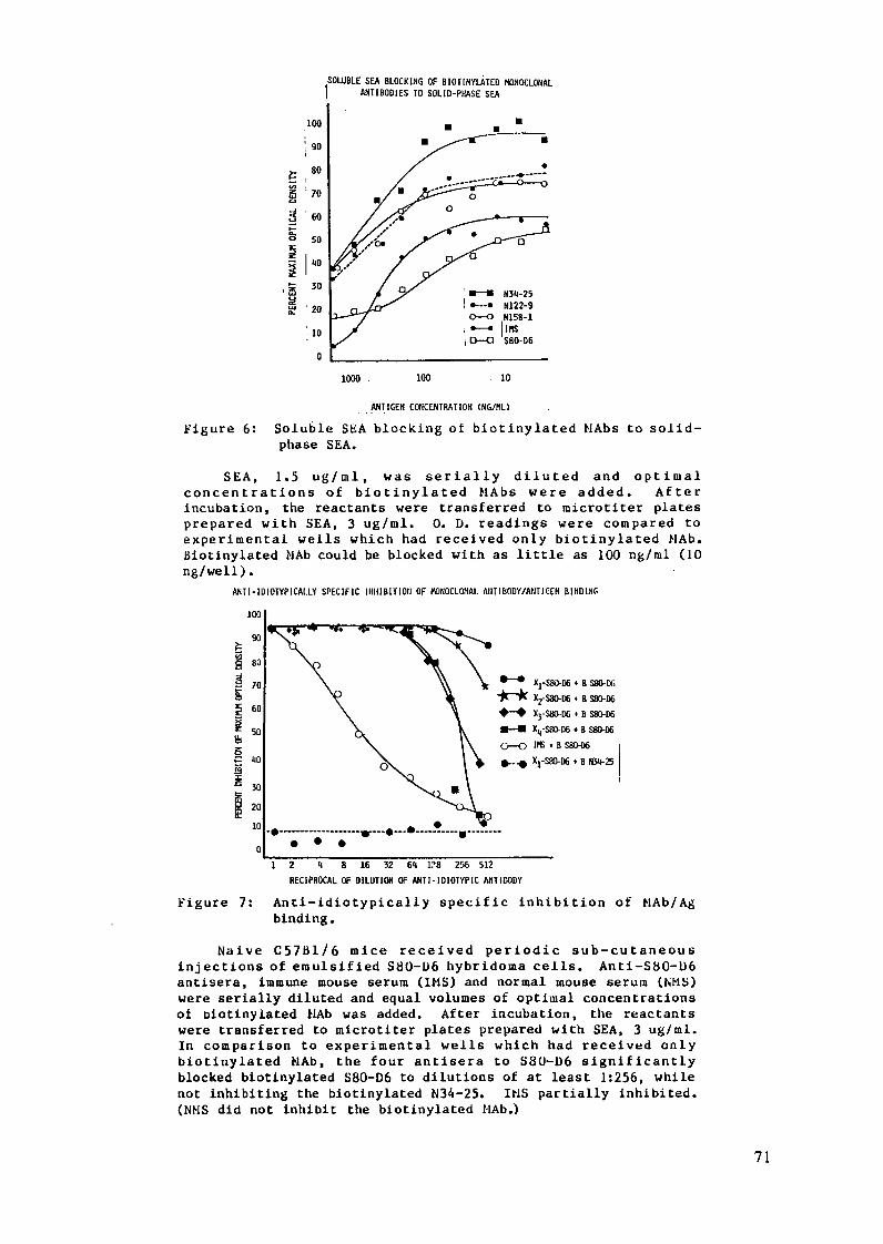
SOLUBLE SEA BLOCKING OF BIOTINYLATED MONOCLONALANTIBODIES TO SOLID-PHASE SEA
e:
9
2
100
90
80
70
60
140
30
·20
10
0
1000 100 10
ANTIGEN CONCENTRATION (NG/ML)
Figure 6: Soluble SEA blocking of biotinylated MAbs to solid-phase SEA.
SEA, 1.5 ug/ml, was serially diluted and optimalconcentrations of biotinylated MAbs were added. Afterincubation, the reactants were transferred to microtiter platesprepared with SEA, 3 ug/ml. 0. D. readings were compared toexperimental wells which had received only biotinylated PlAb.Biotinylated MAb could be blocked with as little as 100 ng/ml (10ng/well).
ANTI-IDIOTYPICALLY SPECIFIC IH1IBIT1011 OF MONOCLONAL AIITIBODY/ANITICEH BINDING
100
90
i 80
S 70 \ \< * ^< X1-S8"-D6 + B Sm-M
S \ ' -kM X,-S0-D6 t B S80-D660 - X3-SBO-DG * B S80-
50 'X K W-M X-S-D6 80* B S80-D6
\-O IS * B S80-D6
4 X40 X1-S 0-D6, B 3B4-25
1 2 4 8 16 32 64 1?8 256 512
RECIPROCAL OF DILUTION OF ANTI-IDIOTYPIC ANTIDODY
Anti-idiotypically specific inhibition ofbinding.
Figure 7: MAb/Ag
Naive C57B1/6 mice received periodic sub-cutaneousinjections of emulsified S80-D6 hybridoma cells. Anti-S80-D6antisera, immune mouse serum (IMS) and normal mouse serum (NMS)were serially diluted and equal volumes of optimal concentrationsof biotinylated IfAb was added. After incubation, the reactantswere transferred to microtiter plates prepared with SEA, 3 ug/ml.In comparison to experimental wells which had received onlybiotinylated MAb, the four antisera to S80-D6 significantlyblocked biotinylated S80-D6 to dilutions of at least 1:256, whilenot inhibiting the biotinylated N34-25. IMS partially inhibited.(NMS did not inhibit the biotinylated l1Ab.)
71
Antigen-specific blocking of B-MAb: We next investigatedthe ability of soluble antigen to specifically block adherence ofthe B-MAb to the solid-phase antigen. SEA was diluted 2-fold ingelatin buffer in glass test tubes. The highest concentrationwas 1.5 ug/tube. B-MAb, at previously determined optimaldilution, was added in equal volume. The tubes were incubatedfor 15 min at 20°C to allow Ag-Ab complexing and then thereactants were transfered to Ag complexed plates. The plateswere then incubated and assayed as usual. For all B-MAb tested,sensitivity was observed to levels of less than 100 ng/ml (Fig.6).
Anti-idiotypic blocking of B-MAb: Sera from S80-D6hybridoma cell stimulated mice were obtained by retroorbitalbleeding. The serum samples were serially diluted in glass tubesas above and the optimal concentrations of B-MAb were added. Thereactants were incubated for 15 minutes and then transferred tomicrotiter plates and processed as usual. Sera obtained from allfour of the individual mice, specifically sensitized with theS80-D6 hybridoma cells, were observed to specifically bind B-S80-D6 to dilutions of at least 1:250, while when tested againstanother MAb, B-N34-25, no blocking of the reaction Was observed.Immune but not normal mouse serum also blocked B-S80-D6, howeverthe titer and inhibition slopes were less impressive than thoseproduced by the anti-idiotypic sera.
DISCUSSION
kesearch into the immunological aspects of infectiousdiseases is directed to three major areas: 1. The immunologicalresponses concerned with resistance and immunopathology; 2.Immunodiagnosis; and 3. Immunotherapeutics. To promote progressin these areas, parameters important to immune requlation mustbe ellucidated. In this regard we and others have identifiedMAbs that provide significant protection to homologousinfections. Therefore these antibodies identified antigens whichare relevant to protection. Radioinimunoprecipitation studiesprovided preliminary evidence that the S80-D6 protective MAb andother non-protective MAbs combined with antigens of the samemolecular weight. Additionally, immunologically reactiveantigens obtained by affinity chromatographic methods usingeither the protective S80-D6 or the non-protective N122-9 againdemonstrated antigens of equivalent molecular weight. Evidencefrom the affinity and subclass studies of the different MAbsproduced no significant differences and thus again no obviousexplanation for the protection afforded by the S80-D6 wasevident. One remaining explanation, that would explain theunique character of the S80-D6, was that it was recognizing anepitope on the same molecule which was distinct from thatrecognized by the non-protecting MAbs. This event has beenpreviously suggested by Stahli et al. 1171. In our competitionassays, utilizing the high avidity avidin-biotin system, we havedemonstrated that our protective S80-D6 is indeed recognizing adistinct epitope from that recognized by the non-protectiveblAb's. Also of interest is the indication that these non-protective MAb's can compete with each other, e.g. N34-25 and
N158-1, and thus are cross-reacting with a second epitope.
72

Avidin-biotin procedures for measuring antibodyconcentration have become quite common. The utility of thisprocedure for measuring antigen concentration using solid-phaseantibody-bound reagents or measuring antibody using secondarybiotinylated antibody has been demonstrated to be most useful[18j. The ability to define hybridoma epitopic specificities forschistosomiasis agents with similar competition assays has beendescribed with radiolabeled 151 and penicillinasa-bound MAb [19Jas well. In addition this technology has proven feasible tomeasure circulating antigen [20] and to assess antigenic salience121].
In the future we anticipate the assays will be of value in anumber of other modes. These include: Assessment of otherrecognized S. mansoni protective MAb's, derived from mice andother species or produced by recombinant technology (N. Agabian,E. Joy, E. Fox, S. M. Phillips, unpublished observations). 2.Determination of the role of anti-idiotypic components. 3. Usein precise sera diagnostic assessment of antigenemia and antibodyactivity.
REFERENCES
[1] JERNE, N. K., Toward a network theory of the immune system,Ann. Immunol. Paris 125C 1-2 (1974) 373.
[2] SACKS, D. L., ESSER, K. M., SHER, A., Immunization of miceagainst African trypanosomiasis using anti-idiotypicantibodies J. Exp. Med. 155 4 (1982) 1108.
[31 HAHN, B. H., EBL1NG, F. i., Suppression of murine lupusnephritis by administration of an anti-idiotypic antibody toanti-DNA, J. Immunol. 132 1 (1984) 187.
14] AUGUSTIN, A. A., SIM, G. K., BONA, C. A., Internal images ofantigens within the immune network, Sur. Immunol. Res. 2(1983) 78.
151 MITCHELL, G. F., GARCIA, E. G., CRUISE, K. M., Competitiveradioimmunoassays using hybridoma and anti-idiotypeantibodies in identification of antibody response toantigens of S. japonicum, Aust. J. Exp. Biol. Med. Sci. 61 1(1983) 27.
[61 POWELL, M. R., COLLEY, D. G.: The production of anti-idiotypic antibody in murine schistosomiasis Fed. Amer. Soc.Exp. Biol.
[7] CAPRON, A., DESSAINT, J. P., CAPRON, M., JOSEPH, M.,Effector Mechanisms against schistosomes (Proc. Int. Cong.Immunol. V, Kyoto, Geneva, 1983) In Press.
[8] GUESDON, J. L., TERNYNCK, T., AVRAMEAS, S., The use ofavidin-biotin interaction in immunoenzymatic techniques, J.Hist. Cytochem. 27 8 (1979) 1131.
[9] ZODDA, D. H., PHILLIPS, S. M., Monoclonal antibody-mediatedresistance to Schistosoma mansoni infection in mice, J.Immunol. 129 6 (1983) 2326.
1101 PHILLIPS, S. M., ZODDA, D. M., "Monoclonal antibodies andimmunoparasitology", Monoclonal Antibodies, 2nd Ed.(KLNNETT, R. IH., McKEARN, T. J., BECtTOL, K. B., Eds),Plenum Press, New York (In Press).
[I11 LAEMMLI, U. K., Cleavage of structural proteins during theassembly of the head of bacterioplague T 4, Nature London227 (1970) 680.
73
[12] LOWRY, O. Hi., ROSEBROUGH, N. J., FARR, L., RANPALL, R. J.,Protein measurement with the folin phenol reagent, J. Biol.Chem. 193 2 (1951) 265.
1131 KLINMAN, U. M., HOWARD, J. C., "Protein iodination suitablefor labeling hybridoma antibodies", Monoclonal Antibodies(Kennett, R. H., McKearn, T. J., Bechtol, K. B., Eds),Plenum Press, New York (1980) 401.
1141 BOLTON, A. E., HUNTER, W.M., The labelling of proteins tohigh specific radioactivities by conjugation to a 1251-containing acylating agent, Biochem. J. 133 3 (1973) 529.
[15] TOWBIN, H., STAEHELIN, T., GORDON, J., Electrophoretictransfer of proteins from polyacrylamide gels to nitro-cellulose sheets: Procedure and some applications, Proc.Nat. Acd. Sci., USA 76 3 (1979) 4350.
[16] NOSEWORTHY, J. H., FIELDS, B. N., DICHTER, M. A., SOBOTKA,C., PIZER, E., PERRY, L. L., NEPOM, J. T., GREENE, M. I.,Cell receptors for the mammulian reovirus. I. Syngeneicmonoclonal anti-idiotypic antibody identifies a cell surfacereceptor for reovirus, J. Immunol. 131 5 (1983) 2533.
[171 STAhLl, C., MIGGIANO, V., STOCKER, J., STAEHELIN, TH.,HARING, P., TAKALS, B., Distinction of epitopes bymonoclonal antibodies, Methods Enzm. 92 (1983) 242.
(181 SPATZ, L., WHITMAN, L. MESSITO, M. J., NILAVER, G.,GINSbERG, S., LATOV, N., Measurement of myelin basicprotein and anti-basic protein antibodies by ELISA utilizingbiotinylated antibodies, Immunol. Comm. 12 1 (1983) 31.
[19] MITCHELL, G. F., PREMIER, R. R., GARCIA, E. G., HURRELL, J.G. R., CHANDLER, H. M., CRUISE, K. M., TAPALES, F., TIU, W.U., Hybridoma antibody-based competitive ELISA inSchistosoma japonicum infection, 32 1 (1983) 114.
[20] ABDEL-HAFEZ, S. K., PHILLIPS, S. M., ZODDA, D. M.,Schistosoma mansoni: Detection and characterization ofantigens and antigenemia by inhibition enzyme-linkedimmunosorbent assay (IELISA), Exp. Parasitol. 55 2 (1983)219.
[21 ABDEL-HAFEZ, S. K., PHILLIPS, S. M., ZODDA, D. M.,Schistosoma mansoni: Detection and characterization ofschistosome derived antigens by inhibition enzyme-linkedimmunosorbent assay (IELISA) utilizing monoclonalantibodies Zeit. Parasiten. 70 1 (1984) 105.
74
RADIOTRACER TECHNIQUES IN THE IMMUNODIAGNOSISOF FILARIASIS IN MAN
R.G. HAMILTONDepartment of Medicine,Johns Hopkins University School of Medicine,Baltimore, Maryland, United States of America
Abstract
Multiple laboratory methods have been examined for theirability to differentiate between the two important clinicalstates of human filariasis: the asymptomatic microfilaremic(Mf+) and exposed but not-infected patient. The peripheralblood smear and skin biopsy, immediate-type hypersensitivityskin test and specific IgG antibody immunoassay have beenunable to effectively discriminate between these two patientpopulations. While the presence of specific antibody is asensitive indicator for a past or present filarial infection,it does not differentiate between the two. The detection ofantigen in human body fluids therefore appears to provide themost specific indicator of a filarial infection in man. A con-current antigen-specific human IgG antibody measurement cansupplement the diagnostic utility of the antigen determinationby providing an assessment of the immunologic status of theindividual.
This report reviews the state of antibody assays and exploresthe design and performance of selected filarial antigen immuno-assays. The immunoradiometric assay (IRMA) and its non-isotopiccounterpart appear to be most sensitive and specific assay forfilarial antigen detection in human serum and urine. The IRMAemploys solid phase antibody to extract antigen from the testspecimen and labeled antibody to detect bound antigen by bindingto a second antigenic determinant. Immunoassay methods whichemploy labeled antigen, non-specific precipitation reactions(PEG) and solid phase anti-C3 or Clq to extract immune-complexesare less sensitive and precise.
The future of filarasis immunodiagnostics (antigen assays)rests in our ability to define selected antigens which arepoorly immunogenic or present in large quantities in serum andurine. Once defined, they may be isolated from crude wormhomogenates or culture supernatants and used to produce highaffinity polyclonal and monoclonal antibody reagents. Improvedefficacy of the filarial antigen immunoassay at present residesin the preparation of highly avid, specific antibody reagentsand not on the refinement of immunoassay methodology.
1. INTRODUCTION
Filariasis is a serious world health problem in which 3 of 7infective nematodes are responsible for most of the significanthuman pathology [1]. Among 4 billion people in the world today,there are approximately 44 million cases of cutaneous filariasis[Onchocerca volvulus (Ov)] [2], and 400 million cases of lymph-atic filarasis [Wuchereria bancrofti (Wb), Brugia malayi (Bm)][3].
75

The most widespread filariasis is caused by the tro lymphaticdwelling nematodes, Wb and Bm, which are commonly found in trop-ical zones of Africa, the Indian sub-continent, South-East Asia,the Pacific Islands, and South and Central America [4]. Thesenematodes produce a broad spectrum of clinical manifestations inman which are only partially differentiated by currently employ-ed diagnostic procedures [5].
The wide spectrum of clinically defined states of Filariasisis commonly divided into four groups [5]. Individuals in theexposed but not infected group live in filarasis endemic areaswhere they are exposed to infected larvae but they exhibit noindication of clinical disease. The asymptomatic microfilarem-ics live in endemic areas and are clinically asymptomatic withmicrofilariae circulating in their peripheral blood. The fil-arial fever group includes infected individuals with microfil-aremia who exhibit recurrent inflammation of their lymphaticsand episodes of fever 2 to 6 times per year. Tropical pulmonaryeosinophilia is at the other extreme of the clinical spectrumwith a bronchial-asthmatic condition and extremely elevatedserum IgE levels.
Multiple assays have been used in an attempt to different-iate first two groups: asymptomatic microfilaremics from theexposed but not-infected [2]. These tests have included peri-pheral blood smears and skin biopsies for visual detection ofmicrofilariae, immediate-type hypersensitivity skin tests todetect filarial antigen-specific IgE antibody and immunoassayanalysis of serum for specific IgG antibody. Despitle the excel-lent sensitivity of immunoassays for human antibodies, specificIgG levels have been unable to differentiate between the clin-ically important infected-amicrofilaremic and exposed but notinfected groups. The major thesis of this report is that thedetection of filarial antigen in human body fluids is the mostspecific indicator of an infection in man. Concurrent quanti-tative analysis of specific human IgG antibody may aid in thediagnosis and subsequent therapeutic intervention by providinginformation about the immunologic status of the individual.
Based on this premise, the objectives of this report are twofold: First, variables will be examined which can influencethe quantity and specificity of detectable circulating parasiteantigen and host antibody in serum and urine of infected indiv-iduals. Second, selected immunoassay methods will be discussedwhich permit quantitation of human anti-filarial antibody anddetection of filarial antigen(s) in human body fluids. B. mal-ayi, the lymphatic dwelling nematode, will be used throughoutthis report as an exemplary model antigen system.
II. Variables influencing Antigen and Antibody Detection
Prior to discussion of immunoassays for filarial antigen andhost antibody detection, a moment should be spent reflectingupon several uncontrollable variables with which the diagnostic-ian must contend that can alter the level of antigen and anti-body in circulation at the time of the specimen collection [1,6].
76
The number of worms, their stage of development (microfilariaversus adult) and periodicity, and the state of the infection(occult, pre-patent, microfilaremic) will determine to a largeextent the amount and specificity of antigen that is secretedinto the blood and eventually excreted into the urine.
The degree and type of the humoral immune response to filar-ial antigen will define the quantity, specificity, avidity andclass of antibody in circulation. The antibody level will inturn effect the level of detectable circulating (serum) andexcreted (urine) antigen. The immune response is govern byenvironmental (exposure) and genetic (immuno-competency) factorswhich are beyond the control of the clinical investigator.
The levels of antibody and antigen present at any point intime will influence immune-complex formation and clearance ofboth antigen and antibody via the body's reticuloendothelialsystem. The immune-complex's size and stability are govern byLaw of Mass Action considerations which depend upon concentrat-ion and affinity.
Other factors which may influence the amount of antigendetected include the circulatory system dynamics (degree ofhydration as both antigen and antibody are measured per unitvolume of blood or urine), concomitant infections which mayalter the host's immune-responsiveness and individual's life-style sex and general health (nutritional state).
Ideally, the antigen(s) which are the least immunogenic,highest in concentration and most filarial-specific are the bestcandidates for a diagnostic indicator of human filariasis. Toachieve the detection of such a population of antigens, thefollowing areas of future research are needed. First, filarialantigens in the blood and urine of infected individuals must bedefined by size, charge and composition and isolated. Second,specific antisera (both polyclonal and monoclonal) must be pre-pared to these antigens for use as specific immunoassay probes.Third, the immunoassay design must be continually improved usingnew techniques to maximize its sensitivity and specificity asnew antibody reagents become available. As these three fociprogress in parallel, the diagnostician's chance will improvefor detecting the low pg/ml to ng/ml levels antigen which cir-culate in the presence of ug/ml levels of specific host anti-body.
III. Quantitation of Host Antibody
While antigen-specific IgG antibody does not discrimatebetween the asymptomatic microfilaremic (Mf+) and exposed butnot-infected patient, it continues to be an important analytefor several reasons. The level of human antibody specific forfilarial antigens provides an indicator of a past or presentfilarial infection. The level of criculating antibody may beused as an indicator of the infected individual's immune status(degree of resistance against the parasite). Third, specificantibody is the major interferring factor in antigen immuno-assays.
77
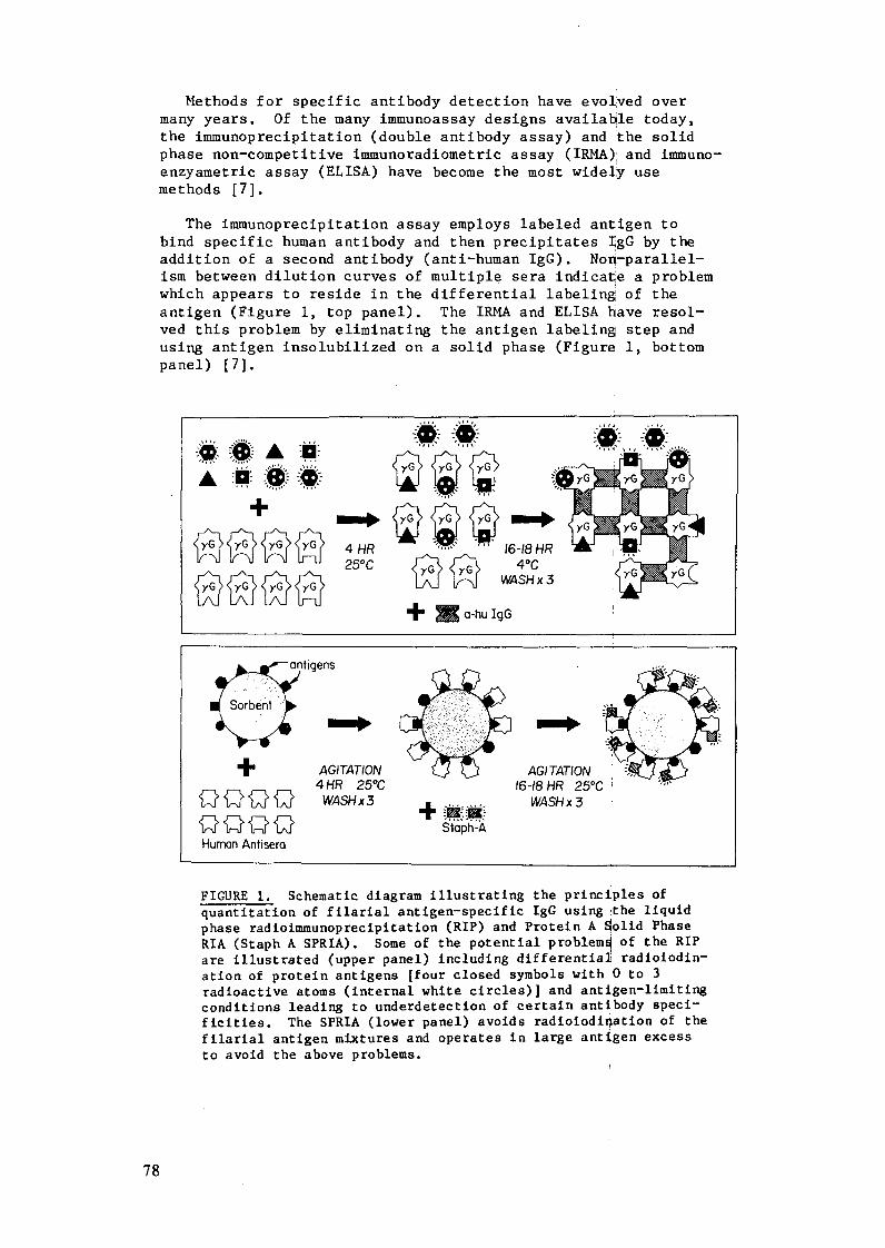
Methods for specific antibody detection have evolved overmany years. Of the many immunoassay designs available today,the immunoprecipitation (double antibody assay) and :the solidphase non-competitive immunoradiometric assay (IRMA); and immuno-enzyametric assay (ELISA) have become the most widely usemethods [7].
The immunoprecipitation assay employs labeled antigen tobind specific human antibody and then precipitates IgG by theaddition of a second antibody (anti-human IgG). Non-parallel-ism between dilution curves of multiple sera indicate a problemwhich appears to reside in the differential labeling of theantigen (Figure 1, top panel). The IRMA and ELISA have resol-ved this problem by eliminating the antigen labeling step andusing antigen insolubilized on a solid phase (Figure 1, bottompanel) [7].
*~AU:' ', a 'A t1
+ld y: --- G / , yG ^ 1yG
'O' 'c'<%L^ 1. 1-, [7\J b-
*0.
250C 40
"`` ` "'yG YC G12 1 r
3
4 HR25°C
16-18 HF4°C
WASH x .
+ o a-hu IgG
+ AGITATION 4HR 25°C
Q'Q Q 4 {. WASH x 3
<Q Q> ' Stoph-AHuman Antisera
AGITATION16-18 HR 25°C
WASH x 3
FIGURE 1. Schematic diagram illustrating the principles of
quantitation of filarial antigen-specific IgG using the liquidphase radioimmunoprecipitation (RIP) and Protein A Solid PhaseRIA (Staph A SPRIA). Some of the potential problems of the RIPare illustrated (upper panel) including differential radioiodin-ation of protein antigens [four closed symbols with 0 to 3radioactive atoms (internal white circles)] and antigen-limitingconditions leading to underdetection of certain antibody speci-ficities. The SPRIA (lower panel) avoids radiolodi ation of thefilarial antigen mixtures and operates in large antigen excessto avoid the above problems.
78
In the IRMA and ELISA, antibody is extracted from a dilutionof serum with solid phase (agarose particle, microtiter platebased) filarial antigen. Following removal of unbound serumproteins with a buffer wash, bound antibody is detected with aimmunoglobulin class specific labeled detection antibody orprobe (anti-human IgE, anti-human IgG, Protein A). The specificdetails of these assays are discussed elsewhere [8].
Recent precision profile analyses has demonstrated using thesame antigen preparation that the IRMA and ELISA provide equiv-alent antibody results [9]. Both display good precision andreproducibility with coefficients of variation (CV) generallyless than 10% throughout the typical 0.5 to 30 ug/ml workingrange. The quality (accuracy) of antibody measurements appearsto be reasonably independent of the type of solid phase (part-icle, plate) and label (1-125, enzyme, fluorochrome) employedin the assay and more related to the specificity and quantity ofantigen attached to the solid phase. Cross-reactivity (specif-icity) continues to be the single most difficult problem in theaccurate measurement of antibody in filariasis patients [8].More comprehensive characterization of filarial antigens willimprove the performance of host IgG antibody assays.
IV. Detection of Filarial Antigen
The concept of filarial antigen detection in human bodyfluids is not new. Investigators since 1863 have visualizedmicrofilaria in blood smears and skin biopsies which is detect-ing antigen albeit the source itself. The present day use ofthe immunoassay methods to detect filarial antigen is simplyour attempt to improve upon the blood smear's limited sensit-ivity while hoping to maintain its excellent specificity.
A. Competitive Binding Immunoassay
Our experience with the competitive binding immunoassay (CB-IA) for the detection of filarial antigen in human serum hasbeen disappointing. There are several reasons for our lack ofsuccess with this assay design. One form of the CB-IA (Figure2, top panel) involves the competitive inhibition of labeledantigen from limited binding sites by unlabeled antigen. Thebinding reagent (antibody) concentration must by definition belimiting with regard to the amount of labeled antigen. Anyhuman anti-filarial antibody in the serum will increase theconcentration of total binding sites and shift the equilibriumestablished by the Law of Mass Action. In doing so, host anti-body eliminates the CB-IA as a serious contender for a diag-nostic filarial antigen assay [6].
In addition to the problem of host antibody interference,there is a requirement for labeled filaral antigen. The manyproteins commonly present in a filarial antigen extract aredifferentially radioiodinated due to the varying number oftyrosines on different proteins. Once radioiodinated, alimited number of labeled proteins must be isolated by columnchromatography from the unlabeled radioiodine. Antigensemployed in the CB-IA must not only be labeled but also immuno-reactive.
79
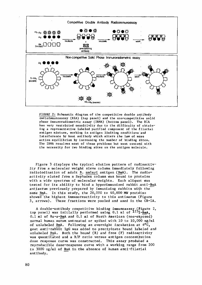
Competitive Double Antibody Radioimmunoossay
-Ag * *@* *@@:
+ Incubate ncubote wash
(limded)ooO &precipitaling
antibody
Non-competitive Solid Phase Immunorodiometric assay
Ag * # Q Q p e
-1 ' ,
S-Ab , Incubate Inhcubate ash(excess)
'125-Ab2 (excess)
FIGURE 2: Schematic diagram of the competitive double antibodyradioimmunoassay (RIA) (top panel) and the non-competitive solidphase immunoradiometric assay (IRMA) (bottom panel). The RIAhas very restricted sensitivity due to the difficulty of obtain-ing a representative labeled purified component of the filarialantigen mixture, working in antigen limiting conditions andinterference by host antibody which alters the law of massaction equilibrium by increasing the number of binding Sites.The IRMA resolves most of these problems but must contend withthe necessity for two binding sites on the antigen molecule.
Figure 3 displays the typical elution pattern of radioactiv-ity from a molecular weight sieve column immediately followingradioiodination of adult B. malayi antigen (BmA). The radio-
activity eluted from a Sephadex column was bound to proteinswith a wide spectrum of molecular weights. Each aliquot was
tested for its ability to bind a hyperimmunized rabbit anti-BmA
antiserum previously prepared by immunizing rabbits with the
same BmA. In this study, the 20,000 to 40,000 MW proteinsshowed the highest immunoreactivity to this antiserum (Figure3, arrows). These fractions were pooled and used in the CB-IA.
A double-antibody competitive binding immunoassay (Figure 2,top panel) was initially performed using 0.1 ml of l2 -BmA,0.1 ml of Ra-a-BmA and 0.1 ml of North American (non-eXposed)normal human serum untreated or spiked with 10 to 10,000 ng/mlof unlabeled BmA. Following an overnight incubation at 4°C,goat anti-rabbit IgG was added to precipitate bound labeled andunlabeled BmA. Both the bound (B) and free (F) radioactivitywas quantitated and a B/F ratio versus antigen concentrationdose response curve was constructed. This assay produced areproducible dose-response curve with a working range from 200to 3000 ng/ml of BmA in the absence of human anti-filatialantibody.
80
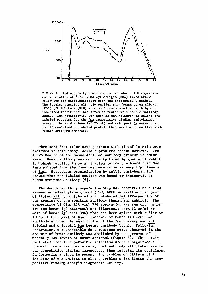
cpm
1000-
1520 50 45 50 55 60 65 70 75 15 20 25 30 35 40 45 50 55 60 65 70 75 80
Eluole Volume(ml)
FIGURE 3: Radioactivity profile of a Sephadex G-100 superfinecolumn elution of 1 2 5I-B. malayl antigen (BmA) immediatelyfollowing its radioiodination with the chloramine T method.The labeled proteins slightly smaller than human serum albumin(HSA) (20,000 to 40,000) were most immunoreactive with hyper-immunized rabbit anti-BmA serum as tested in a double antibodyassay. Immunoreactivity was used as the criteria to select thelabeled proteins for the BmA competitive binding radioimmuno-assay. The void volume (20-25 ml) and salt peak (greater than55 ml) contained no labeled protein that was immunoreactive withrabbit anti-BmA antibody.
When sera from filariasis patients with microfilaremia wereanalyzed in this assay, serious problems became obvious. TheI-125-BmA bound the human anti-BmA antibody present in thesesera. Human antibody was not precipitated by goat anti-rabbitIgG which resulted in an artifactually low cpm bound that wasinterpolated from the dose-response curve as very high levelsof BmA. Subsequent precipitation by rabbit anti-human IgGshowed that the labeled antigen was bound predominantly tohuman anti-BmA antibody [6].
The double-antibody separation step was converted to a lessexpensive polyethylene glycol (PEG) 6000 separation that pre-cipitates all bound labeled and unlabeled BmA irrespective ofthe species of the specific antibody (human and rabbit). Thecompetitive binding RIA with PEG separation was run with negat-ive (no human IgG anti-BmA) and filariasis sera (1 ug/ml ormore of human IgG anti-BmA) that had been spiked with buffer or10 to 10,000 ng/ml of BmA. Presence of human IgG anti-BmAantibody shifted the equilibrium of the immunoassay and alllabeled and unlabeled BmA became antibody bound. Followingseparation, the acceptable dose response curve observed in theabsence of human antibody was abolished by the present ofmodestly low levels of human anti-BmA (Figure 4). This studyindicated that in a parasitic infection where a significanthumoral immune-response occurs, host antibody will interfere inthe competitive binding immunoassay thus reducing its usefulnessin detecting antigen in serum. The problem of differentiallabeling of the antigen is also a problem which limits the com-petitive binding assay's diagnostic utility.
81
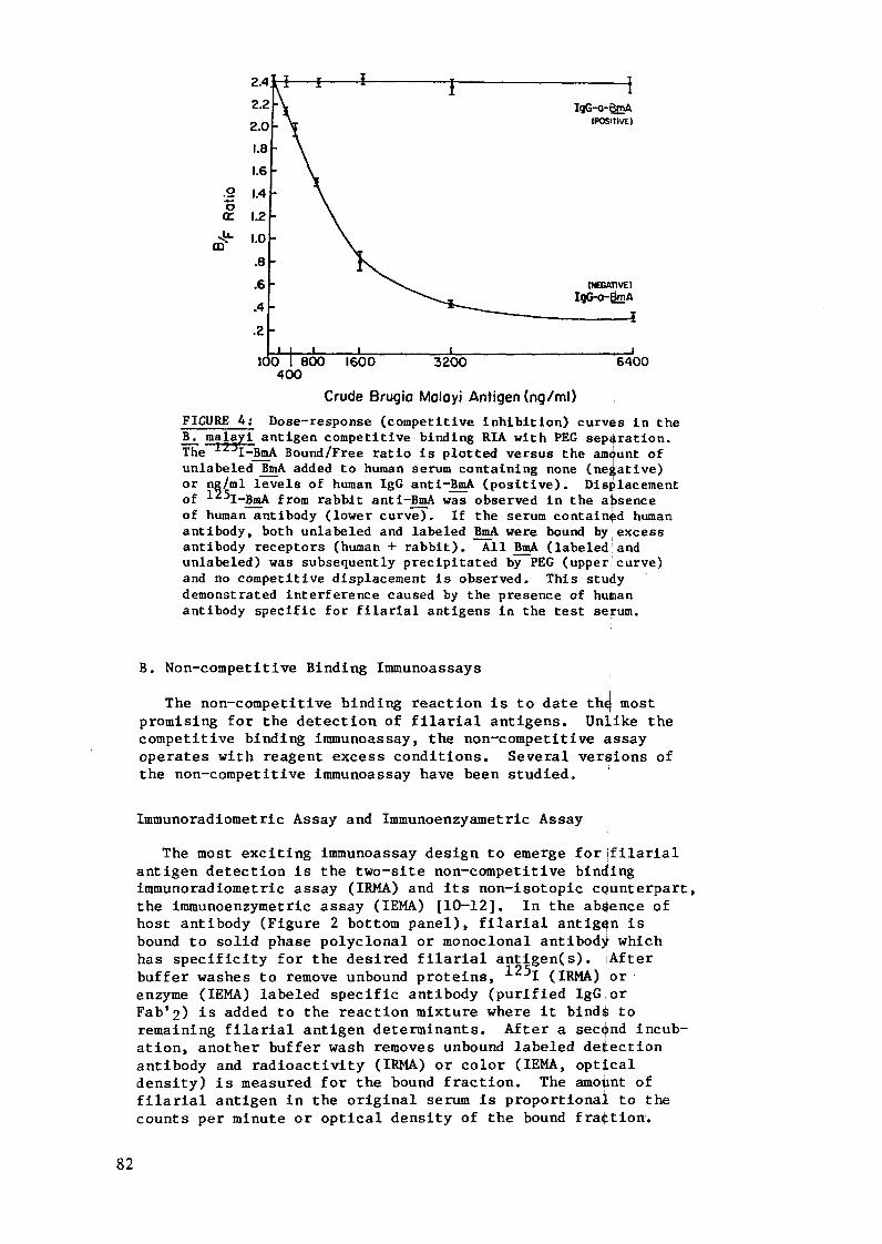
I POG-o-- A(POSITIVE)
.° 1.4
(r 1.2
-m 1.0
.8
.6 \ INUEAnl^"^^..4 ~IgG-C-.A
.4
.2
10 800 1600 3200 6400400
Crude Brugio Moloyi Antigen (ng/ml)FIGURE 4: Dose-response (competitive inhibition) curves in theB. malayi antigen competitive binding RIA with PEG separation.The -LJI-BmA Bound/Free ratio is plotted versus the amount ofunlabeled BmA added to human serum containing none (negative)or ng/ml levels of human IgG anti-BmA (positive). Displacementof 1 5I-BmA from rabbit anti-BmA was observed in the absenceof human antibody (lower curve). If the serum contained humanantibody, both unlabeled and labeled BmA were bound by excessantibody receptors (human + rabbit). All BmA (labeled andunlabeled) was subsequently precipitated by PEG (uppercurve)and no competitive displacement is observed. This studydemonstrated interference caused by the presence of humanantibody specific for filarial antigens in the test serum.
B. Non-competitive Binding Immunoassays
The non-competitive binding reaction is to date the most
promising for the detection of filarial antigens. Unlike the
competitive binding immunoassay, the non-competitive assayoperates with reagent excess conditions. Several versions ofthe non-competitive immunoassay have been studied.
Immunoradiometric Assay and Immunoenzyametric Assay
The most exciting immunoassay design to emerge for filarial
antigen detection is the two-site non-competitive bindingimmunoradiometric assay (IRMA) and its non-isotopic counterpart,
the immunoenzymetric assay (IEMA) [10-12]. In the absence ofhost antibody (Figure 2 bottom panel), filarial antigen is
bound to solid phase polyclonal or monoclonal antibody which
has specificity for the desired filarial antigen(s). After
buffer washes to remove unbound proteins, 125I (IRMA) or
enzyme (IEMA) labeled specific antibody (purified IgGiorFab'2) is added to the reaction mixture where it binds to
remaining filarial antigen determinants. After a second incub-
ation, another buffer wash removes unbound labeled detection
antibody and radioactivity (IRMA) or color (IEMA, optical
density) is measured for the bound fraction. The amount of
filarial antigen in the original serum is proportional to the
counts per minute or optical density of the bound fraction.
82
The diagnostic utility of the IRMA is dependent on therestricted specificity of the antibody which is insolubilizedor labeled. The IRMA's sensitivity is dependent upon severalfactors: the amount of human antibody present which can maskantigenic determinants, the specific-activity (amount of labelper mass) of the labeled detection antibody and the level ofnon-specific binding (NSB) which is proportional to the concent-ration of solid phase antibody and human serum added to thereaction mixture [6].
Interferring Factors: Filarial antigen-specific human antibodyis the major substance in serum which can interfere in theIRMA. An inverse relationship has been indicated between theamount of antigen detected in the IRMA and the level of hostantibody [10,11]. The use of both solid-phase and labeledliquid-phase IgG antibodies in the IRMA allows the possibilitythat rheumatoid factor (RF) which is infrequently found inserum may cause falsely elevated antigen levels. To test thispossibility, the IRMA protocol permits an analysis of antigenpositive sera using normal IgG coupled to the solid phase. Theuse of a solid phase Fab'2 fragment of the IgG detectionantibody reduces this potential artifact to insignificantlevels. In general, the number of false positive RF artifactsis minor as compared to the apparent number of false negativeresults which can be attributed to host antibody interference.
Performance: Several reports have demonstrated acceptableperformance characteristics for the IRMA. A non-specificbinding level of 2-4% Bmax (Bmax = % of added immunoreactivecounts) with non-exposed North American human serum (NEHS) iscommonly observed with the agarose based IRMA [12]. Theresponse range of 4% to 35% Bmax provides a working range of15-1000 ng/ml of added BmA (protein concentration). The IRMA'ssensitivity (minimal detectable dose) as defined statistically(mean cpm bound + 3 SD produced by the NEHS) is 15 ng/ml equiv-alents of BmA. Intra- and inter- assay precision (reproducibi-lity between replicates) over the IRMA's working range aregenerally good: intra-assay <8% CV; inter-assay <14% CV for the15-1000 ng/ml working range. No hook effect (i.e. decrease indetectable antigen at high antigen levels due to steric hind-erance or negative cooperativity) was observed by analyzingsamples with 60 ug/ml of BmA.
Quality Control: Consistency of the IRMA's performance overmonths has been monitored with multiple dilutions of controlsera analyzed at high (800-1000 ng/ml), medium (80-100 ng/ml)and low (15-20 ng/ml) levels on the BmA reference curve. Agree-ment between antigen levels obtained by analyzing multipledilutions of each specimen in an assay (parallelism) was good(inter-dilutional CV less than 20%) for sera without humananti-BmA antibody. Approximately 50% of sera from naturally-infected individuals which contained varying amounts of anti-BmA antibody diluted in a manner non-parallel to the referenceserum containing BmA and no antibody ( >20% inter-dilutionalCVs). Non-parallelism in most cases results in final BmAlevels which can vary 20-50% as a function of the serum dilut-ion tested. The interpolated value was therefore used as anestimate of filarial antigen and given the units: ng/ml"equivalents". Absolute quantitation of the results does not
83
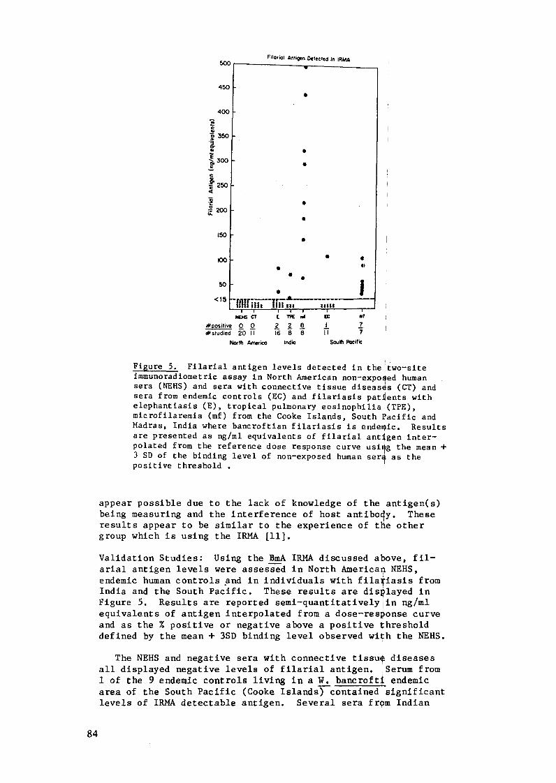
~500 ~ Filaricl Antigen Detected in IRMA500
450
400
C
t 250
200 *
0*
250
<?i- , |i- 5 , ,,,-.-wH5 CT , T w C i f
#positive 0 0 2 2 8 1 72studied 20 II 16 8 8 II
North A erico Indl Soult Pcific
Ia.
Figure 5. Filarial antigen levels detected in the two-siteimmunoradiometric assay in North American non-exposed humansera (NEHS) and sera with connective tissue diseases (CT) andsera from endemic controls (EC) and filariasis patients withelephantiasis (E), tropical pulmonary eosinophilia (TPE),microfilaremia (mf) from the Cooke Islands, South Pacific andMadras, India where bancroftian filariasis is endemic. Resultsare presented as ng/ml equivalents of filarial antigen inter-polated from the reference dose response curve usitg the mean +3 SD of the binding level of non-exposed human sera as thepositive threshold .
appear possible due to the lack of knowledge of the antigen(s)being measuring and the interference of host antibody. Theseresults appear to be similar to the experience of the othergroup which is using the IRMA [11].
Validation Studies: Using the BmA IRMA discussed above, fil-arial antigen levels were assessed in North American NEHS,
endemic human controls and in individuals with filariasis fromIndia and the South Pacific. These results are displayed inFigure 5. Results are reported semi-quantitatively in ng/mlequivalents of antigen interpolated from a dose-response curveand as the % positive or negative above a positive thresholddefined by the mean + 3SD binding level observed with the NEHS.
The NEHS and negative sera with connective tissue diseases
all displayed negative levels of filarial antigen. Serum from1 of the 9 endemic controls living in a W. bancrofti endemicarea of the South Pacific (Cooke Islands) contained significantlevels of IRMA detectable antigen. Several sera from Indian
84

patients (Madras) with elephantiasis and tropical pulmonaryeosinophilia had IRMA detectable filarial antigen. All serafrom microfilaremic patients from India and the South Pacificcontained IRMA detectable filarial antigen [12].
Another report [11] displays the exciting promise of the IRMAusing an IgM monoclonal antibody against 0. gibsoni egg anti-gen (Gib-13). Recent results at the WHO workshop in Lille(1983) were encouraging with regard to its sensitivity indetecting filarial antigen in documented cases of filariasis.Further clinical trials will document the specificity of theGib-13 IgM antibody. Another group has been using human serumimmunoglobulin from Wb microfilaremic patients (FSI or filarialserum immunoglobulin) as the solid phase and labeled detectionantibody in an IEMA [14]. The difficulty of obtaining largequantities of this human antibody and the wide spectrum of anti-body specificities (possible cross-reactivity with other para-sites) suggests a limited utility for FSI as a diagnostic testreagent.
Inhibition ELISA for Antigen
An inhibition assay has been reportedly used to detect micro-filarial excretory-secretory antigen (ES-Ag) in the blood ofpatients who received diethylcarbamazine therapy [13]. In thisassay, FSI (human IgG antibody isolated from Wb microfilaremicpatients) was insolubilized on wells of a microtiter plate.Following blocking of unbound plate sites with bovine serumalbumin, the test serum (1:150 to 1:300 dilution) was added andantigen if present bound to the insolubilized antibody. Aftera wash, penicillinase conjugated ES-Ag was added which bound toremaining unsatuated binding sites. After a final wash, sub-strate was added and the color was shown to be inversely pro-portional to the amount of antigen in the test serum.
As discussed with regard to the competitive immunoassay, therecan be problems associated with the uniform labeling of antigenmixtures. As a semi-quantitative assay this design appears tobe sensitive. Further documentation may prove this assay to bea useful future diagnostic test.
Non-Competitive Dot ELISA
An interesting variation of the two-site IEMA uses nitro-cellulose paper instead of a bead or plastic surface to insol-ubilize the specific antibody. Antibody is localized in a doton the nitrocellulose paper and other remaining binding sitesare saturated with bovine serum albumin. As in the IEMA, filar-ial antigen is bound by the solid phase antibody and followinga washing step, enzyme-conjugated detection antibody binds asecond antigen determinant, if available.
This assay is constrained by the same limitations discussedabove for the IRMA and IEMA. The use of a paper solid phaseand small reaction volume makes it more suitable to field usethan its test tube counterpart. Before this assay finds accept-ance, the problem of host antibody interference must beaddressed especially due to the small quantity of antibody iscoupled to the paper. The tube IRMA and IEMA employ more solid
85
phase antibody than the Dot ELISA which facilitates the bindingof complexed antigen. Preliminary results indicate that the DotELISA has a lower sensitivity than its tube counterpart.
Antigen-Specific Immune Complex Assays
Most individuals living in filariasis endemic areas have cir-culating antibodies specific for filarial antigens. This isthe major reason for the limited use of specific antibody as adiagnostic analyte. The presence of antibody as discussed aboveposes serious consequences for the immunoassay detection offilarial antigen in circulation.
Once antigen is released into the blood, it may complex withantibody which enhances its rate of clearance from the blood.The level of circulating immune-complexed filarial antigen is adynamic process related to the rates of complex formation andclearance. The rate of immune-complex clearance in turn isinfluenced by the antigen valency, energy of binding betweenthe antigen and antibody, complex size, concentration ofcomplexes in blood, route of antigen administration and theimmune-competency of the individual. The efficiency of thehuman phagocytic system for complexes is also dependent on theclass (predominantly IgG) and subclass of human IgG antibody.IgGl and IgG3 can most readily bind to the Fc receptors ofliver macrophages. Complexes greater in size than Ag2Ab2are quickly removed from circulation.
Lattice formation (cross-linking) of antigens and antibodyis an important phenomenon which requires special attentionwhen designing an immunoassay for filarial antigen detection.The relative affinities (tightness of binding) and concentrat-ions of filarial antigen and host antibody will define theextent (quantity), the physical nature (size, charge) of immunecomplex(es) and eventually how the complex is extracted fromserum.
Assays with the potential for detecting filarial antigens incirculating immune complexes are non-competitive solid-phaseassays that isolate or extract immune-complexes from serum in afirst reaction and probe for antigen with a labeled-specificantibody probe in a second reaction.
Immune-complex isolation methods may take advantage of thephysical and chemical properties (size, charge and solubility)of the immune complex(s). Ultracentrifugation, gel chromato-graphy, ultrafiltration, cryoprecipitation and PEG precipit-ation have all been used as isolation methods but most of thesecontinue to be research tools and not useful in large scaleclinical tests. Other investigators have exploited the biolog-ical properties of immune-complex to bind to immunoglobulin Fcor complement receptors. Receptors reported for extraction ICsfrom blood are humoral receptors for immunoglobulin Fc (solidphase Clq, solid phase monoclonal rheumatoid factor) and com-plement (solid phase Conglutinin, solid phase anti-Ci3 anti-bodies) and cellular receptors of which the complement receptorson the surface of the Raji Cells for C3, C3b, C3d is the mostcommonly employed extraction reagent.
86

While there have been indication of that filarial antigencirculating in immune complexes can be detected in patientswith Wb filariasis [15-16], the general experience has beenpoor. We have found solid phase anti-C3 to be the most effic-ient and reproducible for extracting complement-fixed immune-complexes from serum. Using solid phase anti-C3 in the firststage and I-125-labeled anti-BmA antibody in the second step,we have been unable to demonstrate immune-complexed filarialantigen in sera of patients known to have both antigen andantibody using the IRMA and Protein A SPRIA, respectively.
Radioimmunoprecipitation Polyethylene Glycol Assay (RIPEGA)
The RIPEGA has been applied to the detection of 0. volvulusantigens in the serum of infected patients [17]. Initially,antigen in a body fluid is bound by 1 25I-specific antibody.Polyethylene glycol is then added to precipitate antigen-labeledantibody complexes. Commonly a PEG washing step is then per-formed to remove unbound labeled antibody that may be non-speci-fically trapped and the pelleted (bound) radioactivity shouldbe proportional to the amount of parasite antigen (free andimmune-complexed) in the original serum.
Criticism of the RIPEGA has largely focused on its apparentlimited sensitivity and the dependence of precipitation step onthe size of the complex [18]. When compared with the IRMA inthe recent WHO meeting, the RIPEGA which used an IgM monoclonalantibody and 2.5% PEG displayed a lower sensitivity than theIRMA. The RIPEGA while on an experimental basis can be used totest for antigen should not be seriously considered as a diag-nostically useful filarial antigen assay.
V. Strategy for Future Research
It is the opinion of this author and I believe other investi-gators in this field that the non-competitive two-site immuno-radiometric assay is the best assay design to date for detect-ing filarial antigen in the presence of host antibody in humanbody fluids. Several reports show much promise for this assaydesign [10-13]. Research must now turn to the most importantproblem, namely that of preparing highly avid, specific antibodyprobes which will be someday be employed in the IRMA or theIEMA. The rate limiting step at present is not the immunoassaytechnology but rather the reagents which are to be employed inthe assay.
There is reason to be optimistic about the future of filar-ial assay reagents. Several investigators have begun thearduous task of defining and isolating circulating and excretedantigens in serum and urine. Serum and urine from Wb microfil-aremic patients have been precipitated and chromatographed inorder to isolate components called circulating filarial antigen(CFA) and urinary filarial antigen (UFA). These preparationshave more recently been shown to contain antigen by an BmAantigen IRMA. Excretory-secretory antigen from microfilaria ina maintenance culture has also been examined as a possiblesource of diagnostically relevant antigen. Both polyclonal andmonoclonal antibodies are being prepared against the CFA, UFAand ES protein fractions.
87
Recently, antigens have been identified in the blood of Wbmicrofilaremic patients using a multi-step polyacrylamide gelelectrophoresis-Western blot-labeled antibody overlay-autorad-iography technique. These high molecular weight antigens maybe the same antigens which are detected in the IRMA using thesame polyclonal antibody against B. malayi antigen. The workof the monoclonal IgM antibody to O. gibsoni egg antigen whichrecognizes cross-reactive antigen in human serum is anotherexample of movement toward more targeted, diagnostically usefulspecific antibody reagents.
Specific Recommendations: These present successes will hope-fully evolve into a battery of reagents which can be appliedand tested in the IRMA and other immunoassays. The followingissues may be considered important when planning a strategy forthe development of improved diagnostic reagents and testing ofimmunoassays for detection of filarial antigen.
Identification of filarial antigen(s) [somatic, egg, excret-ory, secretory] in the serum and urine of microfilaremicpatients should be at the top of the wish list. This willinvolve determination of the antigen(s) molecular weight(s)distribution, isolectric point(s), SDS-PAG-blotting-labeledantibody overlay identification and definition of its char-acteristics interms of stability (TCA, heat, proteinase) andcross-reactivity with antibodies specific for other helminthantigens.
Once identified, these antigens then need to be isolatedfrom crude worm homogenates, egg isolates, and excretory-secretory antigen. When isolated, they can be used (in part-ially or highly purified form) for immunization and preparationof polyclonal and monoclonal antibodies.
As new promising antibodies become available, they can beinserted into the immunoradiometric assay or its non-isotopicequivalent for testing. While the IRMA is the assay of choiceat present, this is not to say that it remain so in light ofthe rapid evolution of immunoassay technology. Immunoassaysinvolving non-specific precipitation (PEG, charcoal) should beavoided due to their dependence on the size of complexes andtheir limited sensitivity. Immunoprecipitation assays orinhibition assays employing labeled antigens should be used forexperimental purposes but not as diagnostic tests due toproblems with differential labeling of the antigens which leadsto uncontrollable non-parallelism and underestimation of someantigen specificities.
As promising antibody reagents become available, the issuesof immunoassay and clinical sensitivity and specificity need tobe addressed. Standardization and quality control of the assayusing a reference serum needs to be a part of the future strat-egy. The geographic location where blood and urine specimensare collected for assay testing should be considered in lightof the specificity of the antibody used in the assay and thespecies of worm in the region. Specimens from potentially-exposed amicrofilaremic endemic controls or non-exposed non-endemic controls from Europe or North America should both beincluded in a field evaluation of the assay because the issue
88
of the "appropriate" negative control specimen still remainsunanswered.
We have reason to be very optimistic about the future ofFilariasis immunodiagnostic methods.
Acknowledgements: This work was supported by the Filariasiscomponent of the UNDP/World Bank/WHO Special Programme forResearch and Training in Tropical Diseases (# 820109) and theNational Institutes, of Health, Bethesda, Maryland (# AI-19417).
REFERENCES
[1] SASA, M. "Human Filariasis", Univ. of Tokyo Press, 1976.[2] KAGAN, I.G., "The Immunodiagnosis of Filariasis", WHO,
Geneva, 1981 (TDR/FIL/SWG (6)/81[3] NELSON, G.S., "Current Concepts in Parasitology, Filaria-
sis", N. Engl. J. Med. 300 (1979) 1136.[4]- DUKE, B.O.L., "Filiariasis. Handbook for Scientific Work-
ing Groups", WHO, Geneva, 1979 (TDR/AR (1)/77.10 (1979).[5] OTTESEN, E.A. "Immunopathology of Lymphatic Filariasis",
Springer Semin Immunopathol 2 (1980) 373.[6] HAMILTON, R.G., HUSSAIN R., OTTESEN E.A., ADKINSON, N.F.
Jr., "Serological Diagnosis of Human Filarial Infections",Progress in Perinatal Medicine, Excerpta Medica, 1983.
[7] HAMILTON, R.G., ADKINSON, N.F. Jr., "Quantitation of Anti-gen-Specific IgG in Human Serum", J. Allergy Clin. Immunol.67, 1981, 14.
[8] HAMILTON, R.G. et al., "Quantitation of Parasite-SpecificImmunoglobulins: Comparison of RIA and ELISA Methods", J.Immunol. Meth. 44, 1981, 101.
[9] HAMILTON, R.G. et al., "Dirofilaria immitis: Performanceand Standardization of Specific Immunoassays in Filaria-sis.", Exp. Parasitol. 56, 1983, 298.
[10] HAMILTON R.G., SCOTT A., "Immunoradiometric assay forQuantitation of Filarial Antigen using Dirofilaria immitisas a Model System", Am. J. Vet Res. (in press).
[11] DISANAYAKE S. et al., "Detection of Circulating Antigen inBancroftian Filariasis using a Monoclonal Antibody" Am. J.Trop. Med. Hyg. (in press).
[12] HAMILTON, R.G. et al., "Immunoradiometric Assay for theDetection of Filarial Antigens in Human Serum", J. Immunol.(in press).
[13] MALHOTRA A., HARINATH B.C., "Detection and Monitoring ofMicrofilarial ES Ag by inhibition ELISA during DEC Ther-apy", Ind.J. Med. Res 79 (1984) 194.
[14] KALIRAJ P. et al., "The Utility of Human Filarial Serum inthe Detection of Circulating Antigen", J. Helminth 55,1981, 133.
[15] PRASAD G.B.K.S. et al., "Immune complexes and immunoglob-ulins in human filariasis", Ind. J. Med. Res 77 (1983) 813.
[16] PRASAD G.B.K.S. et al., "Detection of antimicrofilarial ESAg-Ab in immune complexes in filariasis by EIA", TransRoyal Soc. Trop Med Hyg. 77 (1983) 771.
89

[17] DES MOUTIS I., et al. 'Onchocerca volvulus Detection ofCirculating Antigen by Monoclonal ANtibodies in HumanOnchocerciasis", Am. J. Trop. Med. Hyg. 32 (1983), 533.
[18] HAMILTON R.G. et al., "Limitations of the RIPEGA forDetection of Filarial Antigens in Serum. J. Immunol.Meth. 68 (1984) 349.
[19] KALIRAJ P. et al., "Fractionation and Evaluation of W.bancrofti microfilaral antigens in immunodiagnosis ofBancroftian Filariasis", Ind. J. Exp. Biol. 20, 1982, 440.
90
IMMUNODIAGNOSIS OF MALARIA
L.H. PERRIN, M. GABRAWHO Unit, Geneva Blood Center,Department of Medicine, Geneva University Hospital,Geneva, Switzerland
Abstract
The need for improved diagnostic tests for malaria over conventionalmethods based on indirect immunofluorescence for the measure ofantimalarial antibodies, and for identification of malaria parasites onstained blood films for antigen detection (diagnostic of ongoinginfection) has led to the development of several solid phase assays.These assays have been used in limited trials for both antibody andantigen detection. Solid phase assays for antimalarial antibodies arerelatively easy to perform but the currently available assays for antigendetection which are based on solid phase antibody binding inhibition arestill complicated, poorly standardised and time consuming. They can notbe used on a large scale in endemic areas. Several new developmentsincluding the availability of monoclonal antimalarial antibodies of knownspecifications, the cloning of several malarial antigens and thesynthesis of malaria specific nucleotides and polypeptides may allow inthe near future the development of simple and reliable assays formalarial antigens detection or the identification of genomic malaria DNAby hybridisation on infected blood samples. Moreover the measure ofantimalarial antibodies of known specificities would be easily achievable.
INTRODUCTION
Immunodiagnosis tests have two main fields of application: first inepidemiological studies in communities where malaria is endemic andsecond, in various applications in research laboratories. The testsystems required for these two applications are different in many waysbut some requirements apply for both applications, e.g. antigenproduction and purification, standardization of reagents andreproducibility. The immunodiagnostic tests aim at the detection ofspecific antimalarial antibodies or at the detection of malaria antigenswhich are released in the sera of infected patients or associated withparasites growing within red blood cells. We will therefore considerseparately tests for antibody and antigen detection.
1. Antimalarial antibody detection
The detection of antimalarial antibodies for immunodiagnosticpurposes has certain limitations since the presence of antibodies inserum does not necessarily indicate an active disease. Antimalarialantibody detection is therefore of little help in the diagnosis of activedisease in individual cases but may be used for follow-up patients andhave applications, for example, in blood transfusion centers innon-endemic areas to determine whether individuals coming back fromendemic areas may be accepted as regular blood donors. At the presenttime, the main interest of these tests is that they may serve as toolsfor the epidemiological surveillance of malaria incidence. They havebeen used to determine the geographical distribution and prevalence ofmalaria and for the evaluation of the transmission of the disease (1).
91
This latter aspect is particularly important for the evaluation of theimpact of malaria eradication programmes. The advantages of thepresently available tests for antimalarial antibody detection is theirrelatively low cost and their sensitivity and simplicity, but theproblems of standardization are still not completely solved. The mostwidely used assays for antimalarial antibody detection are the indirectimmunofluorescence technique (IFA) (1, 2), the indirect hemagglutination(IHA) (3) and a variety of solid phase immunoassays (4, 5, 6).
The indirect immunofluorescence technique (IFA) was introduced inmalaria in the early sixties and has been widely used for epidemiologicalstudies which have clearly established the age-related variations inantibody titers in endemic areas and have helped in the eradication ofremaining malaria foci following eradication programmes (1). Morerecently, IFA has been used for the selection of monoclonal antibodiesreacting with malarial antigens present in defined anatomical sites (7,8, 9). The main advantages of IFA is its rapidity, relative simplicityand the possibility to identify the antibody binding site on definedstructures of the parasites.
For the indirect hemagglutination test, erythrocytes coated withmalarial antigens are incubated with dilutions of sera to be tested. IHAhas been used in epidemiological studies but its use in tropical areas ishampered by false positive results due to frequent non-specificerythrocyte agglutination (3).
Solid-phase immunoassays are based on the property that mostantigens become firmly attached to plastic surface and preserve theirreactivity after attachment. Following antigen absorption, dilutions ofthe sera to be tested are incubated in the wells of plastic microplatesand the amount of antibody is determined in a second step by measuringthe binding of radiolabelled anti-immunoglobulin or 1251 protein A(radioimmunoassay = RIA) (4) and the binding of enzyme labelledanti-immunoglobulin antibody (enzyme-linked immunoabsorbent assay ELISA) (5).
Another application of radiolabelled anti-immunoglobulin is theidentification of the specificities of monoclonal antibodies usingwestern blotting; for these experiments, parasite extracts are applied onSDS PAGE and transferred to nitrocellulose sheets. These sheets arelatter incubated with monoclonal antibodies and finally with 1251antimouse IgG (10). The specificity of the monoclonal antibody is latterdetermined by autoradiography. This technology allows the selection ofmonoclonal antibodies of interest.
One of the critical points for the development of immunodiagnostictests and particularly for antibody detection is the production andstandardization of antigens. The main sources of antigens available are:blood from infected individuals or experimental animals, sera frominfected patients, and parasites obtained from in vitro cultures. Othersources of antigens such as sporozoites and gametocyteS are not availablein sufficiently large amounts. These sources represent a wide range ofantigens and are usually used as total extracts. Therefore, measurementof antibody levels using whole parasitic extracts as a source of malarialantigen represents only a mean assessment and does not differentiatebetween the various antibody specificities involved.
Recent studies have shown that individuals with a high degree ofresistance to malaria preferentially react with some defined antigenic
92
determinants (11, 12) and vaccination trials against malaria may bedeveloped in the future. In these two instances, the availability ofpurified defined antigens for antibody measurement would represent anessential tool. Progress in the characterization and identification ofsingle malarial antigens has been achieved in recent years and severalmalarial antigens have been expressed in E. coli plasmids using DNArecombinant technology (13, 14). One may therefore expect that, in thenear future, antibody measurement against a single antigen would beeasily achievable and allow for a more precise and quantitativeevaluation of the degree of resistance of populations living in endemicareas and of vaccinated individuals.
II. Malarial antigen detection
The detection of antigens in biological fluids is widely used inmicrobiology for the diagnosis of recent or ongoing infection; such testsare usually extremely specific and sensitive. A variety of techniqueshave been similarly developed for the detection of malarial antigens,mainly for the detection of P. falciparum antigens, in the serum and/orin the blood of infected patients (15, 16, 17).
The antigens associated with asexual blood stages of malaria includesoluble antigens present in the serum and intraerythrocytic antigens.Immunoprecipitation in gel (15, 16, 17) and counterimmuno-electrophoresis(18, 19) have been used for the detection of soluble malaria antigensduring or after P. falciparum infection. These antigens have beencharacterized according to their resistance to heating at differenttemperatures. There is a sequential appearance of different antigenseven in the serum of a single individual. The analysis of heat stable(S) antigens in Gambian children has shown a heterogeneicity of thisgroup of antigens, suggesting an antigenic diversity within theP. falciparum species (15, 17). Indeed, this technique allows thediagnosis of recent infections but is limited by its low sensitivity.
The simplest way to detect intraerythrocytic antigens in the studyof individual cases is obviously the examination of blood films.However, in endemic areas, infected individuals frequently have very lowparasitemia and in large scale epidemiological studies the method istime-consuming and requires highly skilled personnel. To bypass thisproblem, solid-phase inhibition RIA and ELISA assays have been developed(4, 5, 6, 20). Presently available assays use extracts of patients' RBCwhich are incubated with enzyme or radiolabelled antimalarial antibodies,and after centrifugation, the supernatants are incubated in plastic tubescoated with malarial antigens. The fixation of the antibody to thecoated tube is specifically inhibited by the prior reaction of theantibody with lysed infected cells. The measurement of the amount ofradioactivity bound to the tube or the measurement of the absorbence ofthe enzyme activity associated with the antimalarial antibodies allowsfor the determination of the specific binding inhibition. These testsare very sensitive since parasites were detected at a level of 8parasites/10 6 normal RBC. There are however some limitations relatedto the standardization of these tests, the relatively complicatedmanipulations including washing of red cells to eliminate antimalarialantibodies contained in the patient's blood since they compete with thelabelled antibody, and the specificity, since correlation betweenmicroscopic parasite detection and significant binding inhibition hasstill to be improved.
93
There are suggestions that some of these problems may be solved bythe use of anti P. falciparum monoclonal antibodies which have beendeveloped by several laboratories. Pools of monoclonal antibodiesdirected against selected determinants would help in the standardizationof the test and may be less sensitive to competition with naturalantibodies present in the blood of infected patients. Another possiblemodification would be the selection of a sandwich assay using polyclonalantibodies or a pool of monoclonal antibodies for the coating of theplastic well on which lysate of parasitized blood wouldidirectly beapplied. The final step would be the application of ra4iolabelledmonoclonal antibodies of known specificities. The technology has beenrecently developed in our laboratory by using a pool of five monoclonalantibodies reacting with ring forms as shown by indirect immuno-fluorescence and western blotting both as first coating and as detectorafter labelling with 125I. Using this solid phase sandwich assay, itwas possible to detect parasitemia at a level of 0.002%, For theseexperiments blood from 5 infected patients lysed by NP40 and Tween 20 wasdiluted with normal RBC in serial dilutions and applied to 96 wellsplastic dishes. Standardization of this methodology may allow a largescale use.
As malarial antigens are released in sera of infected individuals,similar technology may be developed for malarial antigen detection insera, providing the relevant monoclonal antibodies are selected. Thisimplicates the identification of the malarial antigens released at themolecular level. The main antigen released in sera of infectedindividuals is the so-called "S" antigen but this antigen by itself isprobably not suitable since it shows antigenic variation among variousisolates of P. falciparum (9, 15). Other antigens have been identifiedin the supernatant of P. falciparum in vitro culture and are probablyalso present in sera of infected patients (figure 1) (21).
The selection of adequate monoclonal antibodies for immunodiagnostictests rely on basic investigations concerning the identification ofmalaria antigens associated with the various developmental stages of theasexual cycle since mainly ring forms are found in the blood of infectedpatients and since antigens released in the sera of infected individualsare of schizont and merozoite origin. Several group of investigatorshave pursued such studies using synchronized P. falciparum culturelabelled with radiolabelled amino acids (mainly [35S] methionine) (22,23, 24, 25). The samples of the various labelled developmental stagesare analysed by SDS PAGE and autoradiogrpahy. An example of such anexperiment is shown in figure 2. Such studies clearly indicate which arethe major antigens associated with the various developmental stages.
Obviously, solid phase assays for antigen detection can be developedfor P. vivax, P. malariae and P. ovale antigens which may cross-reactwith P. falciparum antigens; for the detection of soluble malarialantigens present in the serum and for the determination ofisolate-specific antigens since monoclonal antibodies with isolatespecificity have been recently described (9); for the detection of singleantigens by using polyclonal sera or monoclonal antibodies directedagainst several epitopes expressed on the same antigen.
An interesting recent development concerns a test for the detectionof a single epitope of the anti-sporozoite protein of P. berghei (Pb44)(26). The principle of this solid-phase assay (inhibition of idiotypeanti-idiotype interaction) is that the antigen (Pb44) inhibits the
94
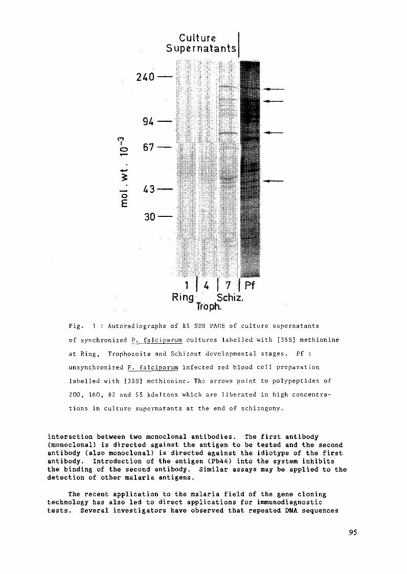
CultureSupernatants
240-
94 -
1
I
0
E
**
67 -
43-
30-
1 14 17 PfRing Schiz.
Troph.
Fig. 1 : Autoradiographs of 8% SDS PAGE of culture supernatants
of synchronized P. falciparum cultures labelled with [35S] methionine
at Ring, Trophozoite and Schizont developmental stages. Pf :
unsynchronized P. falciparum infected red blood cell preparation
labelled with [35S] methionine. The arrows point to polypeptides of
200, 160, 82 and 55 kdaltons which are liberated in high concentra-
tions in culture supernatants at the end of schizogony.
interaction between two monoclonal antibodies. The first antibody(monoclonal) is directed against the antigen to be tested and the secondantibody (also monoclonal) is directed against the idiotype of the firstantibody. Introduction of the antigen (Pb44) into the system inhibitsthe binding of the second antibody. Similar assays may be applied to thedetection of other malaria antigens.
The recent application to the malaria field of the gene cloningtechnology has also led to direct applications for immunodiagnostictests. Several investigators have observed that repeated DNA sequences
95
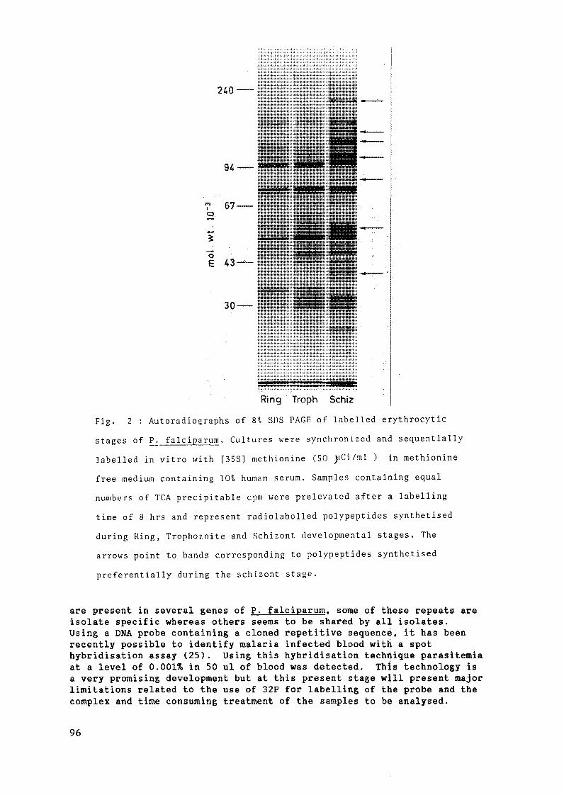
240 -
67-
43
30-
Ring Troph Schiz
Fig. 2 : Autoradiographs of 8 SDS PAGE of labelled erythrocytic
stages of P. falciparum. Cultures were synchronized and sequentially
labelled in vitro with [35S] methionine (50 pCi/ml ) in methionine
free medium containing 10% human serum. Samples containing equal
numbers of TCA precipitable cpm were prelevated after a labelling
time of 8 hrs and represent radiolabelled polypeptides synthetised
during Ring, Trophozoite and Schizont developmental stages. The
arrows point to bands corresponding to polypeptides synthetised
preferentially during the schizont stage.
are present in several genes of P. falciparum, some of these repeats areisolate specific whereas others seems to be shared by all isolates.Using a DNA probe containing a cloned repetitive sequence, it has beenrecently possible to identify malaria infected blood with a spothybridisation assay (25). Using this hybridisation technique parasitemiaat a level of 0.001% in 50 ul of blood was detected. This technology isa very promising development but at this present stage will present majorlimitations related to the use of 32P for labelling of the probe and thecomplex and time consuming treatment of the samples to be analysed.
96

REFERENCES
1. Sulzer, A.J. and Wilson, M.: The fluorescent antibody test formalaria. CRC Critical Reviews in Laboratory Sciences 2 (1971), 601.
2. Voller, A. and Bray, R.S.: Fluorescent antibody staining as ameasure of antimalarial antibody. Proc. Soc. Exp. Biol. Med. 110(1962), 907.
3. Kagan, I.G.: Evaluation of an indirect haemaglutinuation test as anepidemiological technique for malaria. Am. J. Trop. Med. Hyg. 21(1972), 683.
4. Avraham, H., Golenser, J.G., Gazitt, Y., Spira, D.T. and Sutitzeanu,D.: A highly sensitive solid-phase radioimmunoassay for Plasmodiumfalciparum antigens and antibodies. J. Immunol. Methods 53 (1982),61.
5. Avraham, H., Golenser, J., Bunnag, D., Sunthasamai, P., Traravanij,S., Harinasuta, K.T., Spira, D.T. and Sultizeanu, D.: Preliminaryfield trial of a radioimmunoassay for the diagnosis of malaria. Am.J. Trop. Med. Hyg. 82 (1983), 11.
6. Mackey, L., McGregor, I.A. and Lambert, P.H.: Diagnosis ofPlasmodium falciparum infections using a solid-phaseradioimmunoassay for the detection of malaria antigens. Bull WHO 58(1980), 439.
7. Yoshida, N., Nussenzweig, R.S., Potocnjak, P., Nussenzweig, V. andAikawa, M.: Hybridoma produces protective antibodies directedagainst the sporozoite stage of malaria parasite. Science 207(1980), 71.
8. Perrin, L.H., Ramirez, E., Lambert, P.H. and Miescher, P.A.:Inhibition of P. falciparum growth in human erythrocytes bymonoclonal antibodies. Nature 289 (1981), 301.
9. McBride, J., Walliker, D. and Morgan, G.: Antigenic diversity in thehuman malaria parasite, Plasmodium falciparum. Science 217 (1982),254.
10. Freeman, R. and Holder, A.: Surface antigens of malaria merozoites.J. Exp. Med. 158 (1983), 1647,
11. Perrin, L.H. and Dayal, R.: Immunity to asexual erythrocytic stagesof Plasmodium falciparum: role of defined antigens in the humoralimmune response. Immunological Rev. 61 (1982), 245.
12. Brown, G.V., Anders, R.F., Mitchell, G.F. and Heywood, P.F.: Targetantigens of purified human immunoglobulins which inhibit growth ofPlasmodium falciparum in vitro. Nature 297 (1982), 591.
13. Godson, G.N., Ellis, J., Svec, P., Schlessinger, D.H. andNussenzweig, V.: Identification and chemical synthesis of an epitopeof the Plasmodium knowlesi circumsporozoite protein. Evidence forits tandemly repeated nature. Nature 305 (1983), 29.
97
14. Coppel, R., Cowman, A.F., Lingelbach, K.R., Brown, G.V., Saint,R.B., Kemp, D.J. and Anders, R.F.: Isolate specific S-antigen ofPlasmodium falciparum contains a repeated sequence of eleven aminoacids, Nature 306 (1983), 751.
15. Wilson, R.J.M.: Serotyping Plasmodium falciparum malaria withS-antigens. Nature 284 (1980), 451.
16. Wilson, R.J.M., McGregor, I.A., Hall, P., Williams, K. andBartholomew, R.: Antigens associated with Plasmodium falciparuminfections in man. Lancet ii (1969), 201.
17. Wilson, R.J.M., McGregor, I.A. and Williams, K.: Occurrence ofS-antigens in serum in Plasmodium falciparum infection in man.Nature 284 (1980), 451.
18. Seitz, H.M.: The Plasmodium falciparum infection in isogenic Fl(C57BL x DBA) mice. II. Antibodies and antigens in the serum.Z. Tropenmed. Parasitol. 27 (1976), 33.
19. Perrin, L.H., Mackey, L., Ramirez, E., Celada, T., Lambert, P.H. andMiescher, P.A.: Demonstration d'antigenes parasitaires solubles,d'anticorps correspondants et de complexes immuns lors du traitementde la malaria. Schweiz. med. Wochenschr. 109 (1979), 1983.
20. Mackey, L., McGregor, I.A., Paounova, N. and Lambert, P.H.:Diagnosis of Plasmodium falciparum infection in man: detection ofparasite antigens by ELISA. Bull. WHO 60 (182), 69.
21. Rodriguez da Silva, L., Loche, M., Dayal, R. and Ferrin, L.:Plasmodium falciparum polypeptides released during in vitrocultivation. Bull. WHO 61 (1983), 105.
22. Kilejan, A.: Stage specific proteins and glycoproteins of Plasmodiumfalciparum: identification of antigens unique to schizonts andmerozoites. Proc. Natl. Acad. Sci. USA 77 (1980), 3695.
23. Perrin, L.H., Dayal, R. and Rieder, H.: Characterization of antigenfrom erythrocytic stages of Plasmodium falciparum reacting withimmune sera. Trans. R. Soc. Trop. Med. Hyg. 75 (1981), 163.
24. Deans, J.A., Thomas, A.W., Inge, P.M.G. and Cohen, S.: Stagespecific protein synthesis by asexual blood stage parasites ofPlasmodium falciparum. Mol. Biochem. Parasitol. 8 (1983), 45.
25. Heidrich, H.G., Strych, W. and Mrema, J.E.K.: Identification ofsurface and internal antigens from spontaneously released Plasmodiumfalciparum merozoites: radio-iodination and metabolically labellingof merozoites. z. Parasiteked 69 (1983), 715.
26. Potojnak, P., Zavala, F., Nussenzweig, R. and Nus~enzweig, V.:Inhibition of idiotype-anti-idiotype interaction for detection ofparasite antigen: a new immunoassay. Science 215 (1982), 1637.
27. Franzen, L., Shabo, R., Perlmann, H., Wigzell, H., Wostin, G.,Aslund, L., Persson, T. and Petterson, U.: Analys.s of clinicalspecimens by hybridisation with probe containing repetitive DNA fromPlasmodium falciparum. Lancet i (1984), 526.
98

USE OF RADIONUCLIDE LABELS IN THE IDENTIFICATIONAND ISOLATION OF MEROZOITE ANTIGENS FROM THEHUMAN MALARIA PARASITE Plasmodium falciparumr
FOR THE DEVELOPMENT OF IMMUNODIAGNOSTIC METHODS
H.G. HEIDRICHMax-Planck-Institut fur Biochemie,Martinsried, Federal Republic of Germany
Abstract
Radionuclide tracers play an important role in the study of antigens ofthe human malaria parasite, Plasmodium falciparum. The use of such tracersto study the synthesis and the biochemical and immunological characteristicsof various parasite products, is discussed.
INTRODUCTION
Radionuclides play an important role in identification, characterizationand isolation studies of putative protective antigens from the human malariaparasite Plasmodium falciparum. They are particularly important in studiesinvolving antigens from isolated merozoites - the infectious and host cellinvading stage of the parasite. These antigens are only available in smallquantities because merozoite production in culture is a costly and difficulttask. Thus sensitive markers such as radiotracers are a prerequisite formeaningful research in this field.
Radiolabelling of protein in culture
Various types of radiolabelling techniques and nuclides must be appliedwhen studying merozoite antigens in detail. Metabolic labelling of parasiteproteins in culture with radioactive amino acids during the developmentalcycle of the parasite provides information about the total set of proteinspresent in merozoites. In order to obtain such information the radiolabelmust be present in culture from the early trophozoite stage - when proteinsynthesis starts - up to the merozoite harvest. The compartmentisation ofproteins, that is, whether they are membrane-bound or soluble in the parasite,can be studied using cell fractionation techniques after merozoite isolation(1, 2). Similarly the possible immunological activity of the proteins can beinvestigated using immune sera from patients or animals or polyclonal ormonoclonal antibodies, in polyacrylamide gel electrophoresis and immuneblotting analyses, both in combination with Protein-A techniques.
Biosynthesis or processing of antigens can be studied by pulse-chaseexperiments. In these the initial step of protein synthesis is labelled by ashort, hot pulse with a suitable radiolabelled amino acid, and the radiolabelis then chased by the addition of the corresponding cold amino acid. In thisway only those proteins synthesized during the pulse-period are labelled andtheir processing and fate in the cell during the developmental cycle can befollowed. This method also offers the possiblity of identifying proteinprecursors.
During the developmental cycle of the parasite several important proteinsare released from the parasite into the culture medium. These can beidentified if they are radiolabelled since they are present in very smallquantities (3).
99

3 5S-methionine or various 3H- or 14C-labelled amino acids -preferentially leucine, iso-leucine, proline or histidine - are used in allthe studies described.
Merozoite glycoproteins
Merozoite glycoproteins are identified by metabolically labellingmerozoites in culture with 3H- or 1 4C- glucosamine or with various 3H-or 1 4C-sugars such as glucose, mannose, galactose or fucose (4). Inparticular, with glucose care must be taken to avoid commercial productscontaminated with chemically unidentified impurities. These impurities can beincorporated into proteins via non-enzymatic mechanisms, and be interpretedfalsely as an enzymatic incorporation of sugars. Such "glycoproteins",however, are glycoprotein artifacts (5).
Merozoite lipoproteins
Lipoproteins are identified by metabolically labelling merozoites inculture with 1 4C-palmitic acid.
Merozoite cell surface antigens
Merozoite surface antigens can be identified by applying cell surfaceradiolabelling techniques, preferentially with 125Iodine. Variousprocedures are available. The most reliable is the lactopelroxidase-catalyzediodination of tyrosine residues in surface proteins (2, 6). A prerequisitefor applying this technique is that the isolated merozoites are fully intact,i.e., that their membrane is tight and not leaky so that the radiolabel cannotdiffuse into the cell and label internal proteins. In various modificationsof the procedure the 1 2 5Iodine label is bound to glass carriers (IODO-GEN)or matrix beads (lodo-beads) in order to prevent such diffusion. Amino acidresidues from merozoite surface antigens, other than tyrosine, can be labelledusing other procedures (7). For example lysine can be labelled withNaB3H4. These labelling techniques, however, are not commonly used.
Identification of unlabelled merozoite antigens
Merozoite antigens can also be identified without priQr labelling ofmerozoites in culture. In this case the reagents used for antigenidentification must carry the radiolabel. For example polyclonal ormonoclonal antibodies used in immunoprecipitation can be iodinated usingsodium 1 2 5Iodine. This label can be incorporated into tyrosine residues ofthe antibody using the Chloramine-T procedure (8) or the Bolton-Hunter reagent(9). Both techniques can also be used to label Protein-A, which can identifyimmune complexes formed between an unlabelled merozoite antigen with anunlabelled antibody. 1 2 5Iodine-labelled lectins can often be used toidentify antigens which are glycoproteins. As yet, however, no lectin hasbeen found which can be used to identify merozoite glycoproteins.
The most reliable technique for identifying unlabelled merozoite antigenswith radiolabelled reagents is the transfer of electrophoretically separatedmerozoite antigens from polycrylamide gels to nitrocellulose sheets (10) or tomembrane filters for zeta-blotting (11), followed by detection with theradiolabelled reagents. In this way the quantities of theivaluableradiolabelled reagents needed can be reduced to a minimum.
100
Isolation of merozoite antigens
Radiolabelled merozoite antigens can be used as radiotracers mixed withthe corresponding cold material, when isolating merozoite antigens usingvarious chromatographic isolation techniques such as conventionalchromatography, high pressure chromatography or affinity chromatography.Isolated antigens are often used in animal experiments, i.e. for raisingpolyclonal antibodies in small animals or for vaccination trials in monkeys.In such experiments rather large quantities of antigens are needed and itwould be harmful for the animals as well as being costly if all the antigenicmaterial was radiolabelled. Large quantities of radioactive label in animalcadavers also pose a problem in waste disposal.
CONCLUSIONS
Both our work and the many publications by others show that some form ofradioactive label is a prerequisite in virtually all research into thedetailed structure of the malaria parasite. Without such labels the progresswhich has been made towards the development of a vaccine against the humanmalaria parasite P. falciparum would not have been possible. Radiolabelcompounds are expensive research tools and the handling of radioactive wastebecomes more and more difficult. These problems cannot be compared, however,with the problem of the 200 million malaria cases world-wide and the 1 milliondead children in Africa per annum. This problem must be fought with everymeans available.
REFERENCES
(1) Mrema, J.E.K., Langreth, S.G., Jost, R.C., Rieckmann, K.H., Heidrich,H.G., Plasmodium falciparum: Isolation and purification of spontaneouslyreleased merozoites by nylon membrane sieves. Exp. Parasitol. 54 (1982)285.
(2) Heidrich, H.G., Strych, W., Mrema, J.E.K., Identification of surface andinternal antigens from spontaneously released Plasmodium falciparummerozoites by radioiodination and metabolic labelling. Parasitenkd. 69(1983) 715.
(3) Perrin, L.H., Dayal, R., Immunity to asexual erythrocytic stages ofPlasmodium falciparum: Role of defined antigens in the humoralresponse. Immunol. Rev. 61 (1982) 245.
(4) Heidrich, H.G., Strych, W., Prehm, P., Spontaneously released Plasmodiumfalciparum merozoites from culture possess glycoproteins. Parasitenkd.submitted.
(5) Trueb, B., Holenstein, C.G., Fischer, R.W, Winterhalter, K.H.,Nonenzymatic glycosylation of proteins. J. Biol. Chem. 255 (1980) 6717.
(6) Hubbard, A.L., Cohn, Z.A., Externally disposed plasma membrane proteins.J. Cell. Biol. 64 (1975) 438.
(7) Hynes, R.O., Surface labelling techniques of eukaryotic cells. In: NewTechniques in Biophysics and Cell Biology vol. 3 (1976) 147.
(8) Hunter, W.M., Greenwood, F.C., Preparation of 125-Iodine labelled humangrowth hormone of high specific activity. Nature 194 (1962) 495.
101

(9) Langone, J.J., Boyle, M.D.P., Borsos, T., 125-I-Protein-A applications tothe quantitative determination of fluid phase and cell-bound IgG. J.Immunol. Meth. 18 (1977) 281.
(10) Gibson, W., Protease-facilitated transfer of high-molecular-weightproteins during electro-transfer to nitrocellulose. Analy. Biochem. 118(1981) 1.
(11) Gershoni, J.M., Palade, G.E, Electrophoretic transfer of proteins fromsodium dodecyl sulfate -polyacrylamide gels to a positively chargedmembrane filter. Analy. Biochem. 124 (1982) 396.
102
IMMUNODIAGNOSIS OF FILARIASIS,SCHISTOSOMIASIS AND MALARIASummary of research carried out underthe auspices of theUNDP/World Bank/WHO Special Programmefor Research and Trainingin Tropical Diseases
V. HOUBA, K.E. MOTT, L.J. MARTINEZWorld Health Organization, Geneva
Abstract
Results are reported from studies on the immunodiagnosis offilariasis, schistosomiasis and malaria which have been carried out underthe auspices of the UNDP/World Bank/WHO Special Programme for Researchand Training in Tropical Diseases. These studies include an evaluationof the RIPEGA and IRMA for the detection of filarial antigens; theselection and standardization of schistosome antigens for diagnosticpurposes; and the development of tests for the detection of malariaantigens and antibodies.
INTRODUCTION
Research on the immunodiagnosis of tropical diseases carried outunder the auspices of the UNDP/World Bank/WHO Special Programme forResearch and Training in Tropical Diseases has as its main objective thedevelopment of simple techniques which can be applied in endemic areas atlow cost. Ideally, the methods should be sensitive, specific, simple tocarry out and not require sophisticated equipment or expensive reagents.Research projects supported by the Special Programme are concerned withthe improvement of some existing tests and with the development of newtests where these are needed. The precision of serodiagnostic methodsshould improve with the application of defined and purified reagents,including monoclonal antibodies and cloned gene products, which arebeginning to become available for some of the tropical diseases. TheSpecial Programme also supports workshops at which reagents are exchangedand compared. When new methods are developed to a point where fieldapplication is practicable, it is envisaged that training courses will beheld to expedite widespread use of the methods.
1. Filariasis
Of the eight species of filaria that infect man, three producesignificant disease with a wide spectrum of clinical manifestations.These are Wucheresia bancrofti, Brugia malayi and Onchocerca volvulus.By definition, the diagnosis of filarial infection requires thedemonstration of the parasite in the host. However, the development ofinfective larvae in the host into adult worms with production ofmicrofilariae takes time and delays the diagnosis of new infections. Inaddition, the parasites may be present in small numbers, in certainperiods of time (nocturnal microfilariaemia) and/or sequestered ininaccessible sites.
103
For these reasons, immunological techniques e.g. antibody assays andskin tests, have been employed for diagnosis of filariasis for a longtime. The older serological tests with moderate sensitivity, e.g.complement fixation, indirect haemagglutination, gel diffusion, etc. werereplaced later on by more sensitive assays using reagents labelled withradio-isotopes, fluorochromes and/or enzymes (Kempen 1979,Ambroise-Thomas 1980, Ottesen 1984). These tests improved the clinicalsensitivity and facilitated early diagnosis. However, none of thesetests either discriminate between past exposure and active infection orreflect the parasite burden. The demonstration of parasitic antigenscirculating in the blood or excreted in urine in malaria andschistosomiasis has stimulated research to detect filarial antigens inblood serum and/or in urine. To avoid an undesirable cross-reactivity,experienced in the assays of filarial antibodies, monoclonal antibodieshave been employed. Because antigens are released into circulation inminute amounts, assays of high sensitivity had to be developed.
Under the auspices of the UNDP/World Bank/WHO Special Programme forResearch and Training in Tropical Diseases efforts are being made toimprove antibody detection tests for filariasis. Attempts are being madeto define better antigens for these assays, by purification, to givebetter specificity. To date, no species-specific filarial antigen hasbeen identified, though work on this is still in progress. In general,it has been found that in vitro released and membrane antigens are morefilaria- specific than cruder extracts.
Tests are also being developed for the detection of filarialantigens for diagnostic purposes. Two techniques using radio-isotopelabelled reagents have recently been evaluated. These are:
RIPEGA - Radioimmunoassay using polyethylene-glycol (Montis et al1983) and IRMA-Immunoradiometric assay (Forsyth et al 1984, Hamilton andScott, 1984).
In RIPEGA, a radioiodinated monoclonal IgM antibody, specific forfilarial antigens, is incubated with test specimens (serum, urine).Polyethylene glycol (PEG) is added as a separating reagent and themixture is incubated for 14 - 16 hours at 40° and centrifuged. Theprecipitate is washed with PEG, and the radioactivity measured. Theamount of precipitated radioactivity is proportional to the amount offilarial antigen in the test-specimen, reacting with IgM antibody (IgMantibody has been used to avoid "false" positive reactions withRheumatoid factors which may be present in the serum).
In IRMA, the test specimen (serum, urine) is mixed with isotopelabelled monoclonal antibody, incubated overnight and the mixture istransferred to a microtitre plate containing wells precoated with thesame non-labelled monoclonal antibody. The immune complex producedduring the first incubation binds to the non-labelled antibody in thesolid phase during a second overnight incubation. Unbound material isremoved by washing and the bound radioactivity is quantitated in acounter. A modification of this assay has been described usingpolyclonal antifilarial antibody bound to Sepharose beads, reacting withantigens from test samples (serum, urine). Radio-labelled monoclonalantibody is then used to detect the antigen bound on the particles.
The clinical sensitivity of RIPEGA and IRMA was compared during theWorkshop on "The Application of monoclonal antibodies to Immunodiagnosisand studies of antigens in Filariasis", organized by the Scientific
104

Working Group on Filariasis, and held at the Centre d'Immunologie et deBiologie Parasitaire of the Institut Pasteur, Lille, France in November1983 (Bulletin World Health Organization, 62, 695-6, 1984). Filarialantigens were detected in sera from patients with parasitologicallyconfirmed filariasis by both techniques. However, both assays showedsome false positive results with sera from patients with connectivetissue diseases and a reduced sensitivity with sera from filarialpatients containing host antibody to filarial antigens. Both theseartifacts were eliminated by pretreatment of sera with acid (pH 2.0)followed by boiling (100°C for 10 minutes), demonstrating thethermostable and acid-resistant nature of the antigens. Studies are inprogress to refine and standardize these assays, which have betterpotential than antibody detection tests for early diagnosis, fordiscrimination between past exposure and active infection as well as forevaluation of chemotherapy.
This collaborative study demonstrated the need for more and bettermonoclonal antibodies for immunodiagnosis. Most of those tested to dateshowed wide cross-reactivity within filarial species and very few showedrestricted species and stage specificity.
As a result of this study, a serum bank has recently beenestablished with support from the Special Programme. Well characterizedsera are being collected from patients with all types of filarialdisease. The need for an antigen bank was also recognized.
2. Schistosomiasis
Under the auspices of the UNDP/World Bank/WHO Special Programme forResearch and Training in Tropical Diseases, the Scientific Working Groupon Schistosomiasis has organized collabortive studies for the selectionand standardization of antigens for the immunodiagnosis ofschistosomiasis.
In the first collaborative study, (Mott & Dixon, 1982) eightresearch laboratories in Europe and the United States of America wereselected on the basis of having published data on Schistosoma mansoni andS. japonicum antigens to participate in a study of various antigen/testcombinations for immunodiagnosis of schistosomiasis. The serum bankconsisted of 395 well documented sera from four endemic areas in Brazil(2 areas), Kenya, and the Philippines. Altogether, 21 S. mansoni andfour S. japonicum antigen and immunoassay combinations were evaluated.
S. mansoni egg antigens yielded a higher combined sensitivity thanadult worm antigens, irrespective of their purity, in active S. mansoniinfections before and after specific treatment. Quantitativeseroreactivity of characterized S. mansoni egg antigens (McLaren, 1981)showed good correlation whith faecal egg counts in the 5-14 year agegroup. No correlation between morbidity related to S. mansoni andseroreactivity was observed in any test system.
Three S. japonicum egg antigens showed high sensitivity andspecificity in relation to the presence or absence of eggs in the stool.The quantitative seroreactivity of the characterized S. japonicum egg
antigens correlated directly with the intensity of S. japonicum infectionin all age groups.
105
The enzyme-linked immunosorbent assay (ELISA), using severaldifferent procedures, performed well with the antigens used in thestudy. The indium slide immunoassay, a simple qualitative visual testsystem using an S. mansoni egg antigen, demonstrated a high degree ofsensitivity and specificity.
The results did not indicate the superiority of any particularimmunodiagnostic method for detecting antischistosome antibodies.
Original observations during this study were that antibody to eggantigen diminishes six months after treatment, and that the indium slidetest was as sensitive as ELISA using the same egg antigen. It was morespecific than ELISA for S. mansoni infection. Tests were S. japonicumshowed high sensitivity and specificity, but many S. mansoni antigenscross-react with S. japonicum (Tsang et al.). It is therefore possiblethat S. mansoni antigens could be used for the immunodiagnosis ofS. japonicum. It was notable that crude antigen preparations gave justas good specificity as "purified" antigens, and greater sensitivity.
The main recommendations made on the basis of the firstcollaborative study were that work should continue on characterizationand purification of antigens, and their comparison with standard crudeantigens. Use should be made of all available technology, includingmonoclonal antibodies, to facilitate antigen purification,standardization, and quality control. Epidemiologically defined humansera are prerequisites for antigen quality control and standardizationprocedures. International collaborative efforts are needed betweenlaboratories in the endemic areas and developed countries. S. mansoniegg and adult antigens should be evaluated in the field, in order toassess the correlation between serological and parasitological data.Current immunological techniques to detect circulating Schistosomaantigens have not been fully developed (Deelder and Dozy, 1980). As newtechnology becomes available, research in this area should beencouraged. Limited field testing of antigens and test systems thatperform well in collaborative trials should be carried out.
In January 1983, another serum bank was set up for the evaluation ofS. japonicum antigens (egg and adult). Definitive results are not yetavailable but the preliminary immunodiagnostic data look extremelypromising.
The collaborative studies supported by the Special Programme willattempt to identify antigens which can be used in existing test systemsto reflect the status of infection. In addition, efforts are being madeto identify antigens using newer techniques, which could be used fordiagnosis or to monitor therapy (Norden and Strand, 1984).
Cost is a major factor to be considered in the development ofimmunodiagnostic tests for schistosomiasis. Most such tests areconsidered to be too expensive for most of the 74 countries whereschistosomiasis is endemic. However, tests could be valuable in PuertoRico, St. Lucia and in China, where the level of infection is low and thesensitivity of detection could be improved by the use of immunodiagnosticmethods.
Based on the experience gained during the collaborative studies, itis strongly recommended that when such studies are set up, the testreading should be truly blind. Samples should be coded and the coderetained in the country of origin. The testing laboratory should receive
106
only numbered samples. This applies even when the test systems used areapparently instrinsically objective in nature. It is also stronglyrecommended that such studies be carried out in close collaboration withepidemiological and control programmes, to ensure full documentation ofthe sera collected.
3. Malaria
New and improved immunodiagnostic methods are required for malaria,for use in epidemiological studies, in monitoring control programmes andin future, for the evaluation of malaria vaccines. Research supported bythe UNDP/World Bank/WHO Special Programme for Research and Training inTropical Diseases is concerned with the development and improvement oftests to detect antigens of an antibodies to sporozoites and asexualblood stage parasites, as well as the development of methods for theassessment of protective immunity in malaria.
(a) Sporozoites
A small pilot study carried out in a hyperendemic malaria areaemploying intact sporozoites of P. falciparum as antigen, revealedthat antibodies to sporozoites are frequently demonstrable byindirect immunofluorescence in the sera taken from members ofresident populations (Nardin et al., 1979). Age-specific antibodyprofiles indicated that these antibodies were commonly present insera from newborn infants, implying transplacental acquisition, andtended to disappear in the months following birth. They remainedinfrequent in the early years of life but reappeared in laterchildhood and increased in prevalence and concentration into adultlife. The relevance of these antibodies to protection remains to beinvestigated.
An immunoradiometric (IRMA) test which detects and estimates thenumbers of sporozoites carried by infected mosquitoes has been devisedand assessed in field conditions. The assay has three importantcharacteristics. First, it employs monoclonal antibodies to detectspecies-specific circumsporozoite (CS) protein present in mosquitoes.Secondly, it remains capable of detecting CS protein in dried mosquitoesfor up to 4 months after insect-death. Thirdly, as CS protein contains arepeated epitope, only one monoclonal is necessary for a two-site assay.In a hyperendemic area of the Gambia, West Africa, the sporozoite rate intrapped mosquitoes was estimated using first the IRMA and secondly,classical mosquito dissection methodology (Zavala et al., 1982). Therewas a good correlation between the results obtained by the two methods.However, the rates determined by dissection were consistently around 70%of those found by IRMA. The difference is probably due to the fact thatthe IRMA detects CS protein in sporozoites present in late-stage oocystsin the mosquito midgut as well as in mature forms in the salivary gland.Assessment of sporozoite numbers showed that while loads in excess of150,000 sporozoites were sometimes encountered, the great majority ofinfections did not exceed 8,000. These encouraging preliminary findingsindicate that this type of test will in future provide an extremelyvaluable tool for the epidemiology assessment of malaria in endemicareas. The method has recently been adapted to an ELISA system, which isexpected to be more readily applicable under field conditions than theIRMA.
107
(b) Asexual blood stages
Emphasis is currently placed on the development of methods forthe detection of parasite antigens in infected blood. The detectionof parasites in blood by microscopic examination ofithick bloodfilms is rapid and accurate when parasitaemia is relatively high,but becomes slow and less reliable when parasitaemia is low andlarge numbers of slides have to be examined. Various types of fieldstudies, including vaccine evaluation, would be facilitated by asimple serological test that could detect parasites in peripheralblood with a high degree of sensitivity. Both RIA and ELISA methodshave been devised for the detection of P. falciparu" parasites inblood and useful levels of sensitivity were achieved (Avraham etal., 1981; Bidwell & Voller, 1981; Mackey et al., 1982). However,greater test precision as well as standardization will be requiredbefore such tests are ready for widespread application in fieldstudies.
It can be expected that considerable improvements will be made inserodiagnostic methods for malaria, as a result of the present intensiveresearch on plasmodial antigens. As more monoclonal antibodies andpurified antigens are produced in the course of vaccine research, theyshould also become available for serodiagnostic purposes. A recentdevelopment in this connection has been the establishment of a Registryfor monoclonal malaria antibodies in the WHO Immunology and TrainingCentre, Geneva. Monoclonal antibodies being produced in variouslaboratories, mainly for purposes of antigen analysis, are beingcollected and tested for their suitability as immunodiagnostic reagents.Monoclonal antibodies, by virtue of their extreme specificity,homogeneity and potentially unlimited availability could bring majorimprovements to the serology of malaria. A number of scientists havecontributed samples to the Registry and the antibodies are being testedfor species, strain and stage specificity. They are also being assessedin tests for the detection of whole P. falciparum parasites in infectedblood and for the detection of soluble antigens in the serum of infectedpatients.
(c) Protective Immunity
Several methods are available for the measurement of serumantibodies to blood parasites (IFA, IHA, RIA, ELISA), and their use inseroepidemiological studies can provide much valuable information.Unfortunately, the presence of antibody does not indicate the level ofprotective immunity. It is possible that the antibody response tocertain individual parasite antigens may give some measure of protection,since sera from "immune" adults in endemic areas preferentially recognizea number of antigens that are seldom recognized by sera from patientsrecovering from a first infection. Several functional assays have beendevised, in which antibody reacts with antigens that appear to beimportant for the growth and development of the parasites. Merozoiteinvasion inhibition is one such assay, in which merozoite re-invasion isquantitated in in vitro cultures of P. falciparum (WilsOn & Phillips,1976; Green et al., 1981; Myler et al., 1982). However, although immunehuman and Aotus sera have been shown to be inhibitory, a proportion ofimmune sera have an enhancing effect on parasite growth, Some serainhibit some strains of P. falciparum and not others. Another functionalassay uses an in vitro model of the sequestration of mature P. falciparumparasites that occurs in natural infections. The binding of trophozoite-containing erythrocytes to endothelial cells or amelanotic melanoma cells
108
in vitro was shown to be inhibited or reversed by immune Aotus serum(Udeinya et al. 1983). This phenomenon is now being studied using humansera from people with different degrees of malaria experiences.
Tests for cell-mediated immunity in malaria are being developed, butthis work is still at an early stage. The response of peripheral bloodlymphocytes to plasmodial antigens in vitro is also being studied, but itis not yet clear whether this will lead to the development of an assay ofprotective immunity. Further studies are planned in this area.
REFERENCES
1. Ambroise-Thomas, P., Filariasis In: Immunological Investigations inTropical Parasitic Diseases, ed. V. Houba, Publ.Churchill-Livingston, Edinburgh, (1980) pp. 84-103
2. Avraham, H., Golenser, J., Spria, D.T., and Sulitzeanu, D.,Plasmodium falciparum assay of antigens and antibodies by means of asolid phase radioimmunoassay with radioiodinated staphylococcalprotein A, Trans. Roy. Soc. Trop. Med. and Hyg., 75 (1981) 421.
3. Bidwell, D.E., and Voller, A., Malaria diagnosis by enzyme-linkedimmunosorbent assays, Br. Med. J., 282 (1981) 1757.
4. Deelder, A.M. and Dozy, M.H., Applicability of sol particleimmunoassay (SPIA) for detection of Schistosoma mansoni circulatingantigens Acta Leidenesis, 48 (1980) 17.
5. Forsyth, K.P., Spark, R., Karura, J., Brown, G.V., Peters, P.,Heywood, P., Dissanayake, S., Mitchell, G.F., Detection ofcirculating antigens in Baneroftian filariasis using a monoclonalantibody (submitted for publication, (1984)).
6. Green, T.J., Morhardt, M., Brackett, R.G., Jacobs, R.L., Seruminhibition of merozoite dispersal from Plasmodium falciparumschizonts: indicator of immune status, Infect. Immun., 31 (1981)1203.
7. Hamilton, R.G. and Scott, A., Immunoradiometric assays forquantitation of Dirofilaria immitis antigen in dogs with heartworminfections. Am. J. Vet. Res. 45 (1984) 1.
8. Kagan, I., Current status of serological testing for parasiticdiseases. Hospital Practice, 9 (1974) 157.
9. Mackey, L., McGregor, I.A., Paounova, N., and Lambert, P.H., Thediagnosis of Plasmodium falciparum infection in man by enzyme-linedimmunosorbent assay, Bull. WHO, 60 (1982) 69.
10. McLaren, M.L., Lillywhite, J.E., Dunne, D.W. and Doenhoff, M.,Serodiagnosis of human Schistosoma mansoni infections: enhancedsensitivity and specificity in ELISA using a fraction containing S.mansoni egg antigens W, and, Trans. Royal. Soc. Trop. Med. and Hyg.,75 (1981)72.
109
11. Montis des, I., Quaissi, A., Graych, J.M. Yarrabal, L. Haque, A.,and Capron, A., Onchocerca volvulus: Detection of circulatingantigen by monoclonal antibodies. Am. J. Trop. Med. Hyg. 32 (1983)533.
12. Mott, K.E. & Dixon, H., Collaborative study on antigens for theimmunodiagnosis of schistosomiasis. Bull. WHO, 6q (1982) 729 .
13. Myler, P., Saul, A., Mangan, T., Kidson, C., An automated assay ofmerozoite invasion of erythrocytes using highly synchronizedPlasmodium falciparum cultures, Aust. J. Exp. Biol. Med. Sci., 60(1982) 83.
14. Nardin, E.H., Nussenzweig, R.S., McGregor, I.A., and Bryan, J.,Antisporozoite antibodies: Their frequent occurrertce in individualsliving in an area of hyperendemic malaria, Science, 206 (1979) 597.
15. Norden, A.P. and Strand, M., Schistosoma mansoni, S. haematobium andS. japonicum: identification of genus-, species- and gender-specificantigenic worm glycoproteins, Exp. Parasitol., 57 (1984) 110.
16. Ottesen, E.A., Immunolgical aspects of lymphatic filariasis andonchocerciasis in man. Trans. Roy. Soc. Trop. Med. Hyg. 78(Supplement): (1984) 9.
17. Tsang, V.C., Hancock, K., Maddison, S.E., Beatty, A.L., Moss, D.M.,Demonstration of species-specific and cross-reactive components ofthe adult microsomal antigens from Schistosoma mansoni and S.japonicum (MAMA and JAMA), J. Immunol., 132 (1984) 2607.
18. Udeinya, I.J., Miller, L.H., McGregory, I.A. and Jensen, J.B., P.falciparum strain-specific antibody blocks binding of infectederythrocytes to amelanotic melanoma cells, Nature 303 (1983) 429.
19. Wilson, R.J.M., and Phillips, R.S., Method to test inhibitoryantibodies in human sera to wild populations of P. falciparum,Nature, 263 (1976) 132,.
20. Zavala, F., Gwadz, R.W., Collins, F.H., Nussenzweig, R.S.,Nussenzweig, V., Monoclonal antibodies to circumsporozoite proteinsidentify the species of malaria parasite in infected mosquitoes,Nature, 299 (1982) 737.
110

III. LIST OF PARTICIPANTS
1. Dessaint, J.P.
2. Hamilton, R.G.
3. Heidrich, H.G.
4. Linder, E.
5. Perrin, L.H.
6. Phillips, S.M.
7. Taylor, D.W.
8. Martinez, L.J.
Centre d'immunologie et de biologieparasitaire, Institut Pasteur,15 Rue Camille, Guerrin, Lille, France
The Good Samaritan Hospital,5601 Loch Raven Boulevard,Baltimore, Maryland 21239, USA
Max Planck Institute for Biochemistry,D-8033 Martinrieb, Munich, FRG
National Bacteriology Laboratory (S.B.L.),10521 Stockholm, Sweden
Centre de Transfusion, H8pital Cantonal,CH-1211, Geneva 4, Switzerland
Allergy & Immunology Section,Dept. of Medicine,University of Pennsylvania,5th Floor, Johnson Pavilion,36th & Hamilton Walk G2, PhiladelphiaPA 19104, USA
Dept. of Pathology, University of Cambridge,Tennis Court Road, Cambridge CB2 1QP, UK
World Health Organization,CH-1211 Geneva 27, Switzerland
OBSERVERS
1. Aspbck, H.
2. Picher, O.
3. Wiedermann, J.
Department of Medical Parasitology,Institute of Hygiene,Kinderspitalg. 15, A-1095 Vienna, Austria
Department of Medical Parasitology,Institute of Hygiene,Kinderspitalg. 15, A-1095 Vienna, Austria
Institute of Tropical MedicineKinderspitalg. 15, A-1095 Vienna, Austria
International Atomic Energy AgencySECRETARIAT
Castelino, J.B. Division of Life Sciences, InternationalAtomic Energy Agency, A-1400 Vienna, Austria
111