intestinal parasitic infections in patients attending moi
94
INTESTINAL PARASITIC INFECTIONS IN PATIENTS ATTENDING MOI TEACHING AND REFERRAL HOSPITAL ELDORET, KENYA BY KIMOSOP JEPKOSGEI ROSE A THESIS SUBMITTED IN PARTIAL FULFILMENT OF THE REQUIREMENTS FOR THE DEGREE OF MASTER OF SCIENCE IN ZOOLOGY (PARASITOLOGY) OF THE UNIVERSITY OF ELDORET, KENYA OCTOBER, 2017
-
Upload
khangminh22 -
Category
Documents
-
view
2 -
download
0
Transcript of intestinal parasitic infections in patients attending moi

INTESTINAL PARASITIC INFECTIONS IN PATIENTS ATTENDING MOI
TEACHING AND REFERRAL HOSPITAL ELDORET, KENYA
BY
KIMOSOP JEPKOSGEI ROSE
A THESIS SUBMITTED IN PARTIAL FULFILMENT OF THE
REQUIREMENTS FOR THE DEGREE OF MASTER OF SCIENCE IN
ZOOLOGY (PARASITOLOGY) OF THE UNIVERSITY OF ELDORET,
KENYA
OCTOBER, 2017

ii
DECLARATION
This thesis is my original work and has not been submitted for any Degree or any other
award in any other University or Institution. No part of this thesis may be reproduced
without the prior written permission of the author and/or the University of Eldoret.
Candidate: Kimosop Jepkosgei Rose Sign…………………
Date…………………
Regn. Number: SC/PGB/028/08
This Thesis has been submitted for examination with our approval as the University
supervisors.
Dr. Moses M. Ngeiywa Sign………………………
Date………………………
Department of Biological Sciences, School of Science, University of Eldoret.
P.O. Box 1125, ELDORET.
Prof. Chrispinus S. Mulambalah. Sign………………………
Date………………………
Department of Medical Microbiology and Parasitology, School of Medicine, Moi
University.
P.O. Box 4606, ELDORET.

iii
DEDICATION
This work is dedicated to my loving husband Joseph, children Nordia , Bilha, Immaculate
and all the members of my entire family for their support , patience and co-operation that
they have tirelessly accorded me.

iv
ABSTRACT
Intestinal parasitic infections are major public health problems in developing countries.
The distribution of these infections is mainly associated with poor personal hygiene,
environmental sanitations and limited access to clean water. Intestinal parasitic infections
(IPIS), in patients attending Moi Teaching and Referral Hospital Eldoret (MTRH), Kenya
has not been studied although they present serious public health problem nationally and
worldwide. The aim of the study was to determine the prevalence and distribution of
intestinal parasites in patients referred to the laboratory from the outpatient clinics and the
wards at MTRH. The demographics and social – economic variables of 185 patients
investigated were done between April-December 2015. Direct saline and formal - ether
sedimentation techniques were used for detection and identification of the protozoan and
helminth parasites in stool samples while air dried fresh stool smears were stained with
modified acid fast stain for identification of the coccidian parasites. Preliminary
macroscopic assessment of fresh stool specimens was performed for identification of
helminthic segments, larvae and/or adult stages. The results revealed an overall prevalence
of 86 (46.5%) of intestinal parasites while 99 (53.5%) were negative. The specific parasites
prevalence and distributions were Entamoeba histolytica 43 (23.9%), Cryptosporidium
parvum 24(13%), Entamoeba coli 12(6.5 %), Giardia lamblia 12 (6.5%), Iadomoeba
butschlii 12 (6.5%), Ascaris lumbricoides 3 (1.6 %), Hymenolepis nana 1 (0.5%), Trichuris
trichiura 1 (0.5 %), and Hookworm species 1 (0.5%). The most prevalent parasitic
infections encountered were amoebiasis and cryptosporidiosis. Study participants of all
ages were susceptible to parasitic infections with varied magnitudes in the study
population and both genders were found to be susceptible to both protozoal and helminth
infections. The high prevalence of IPIS among the patients attending MTRH indicates that
parasitic infections should be considered a public health problem.

v
TABLE OF CONTENTS
DECLARATION ........................................................................................................................... ii
DEDICATION.............................................................................................................................. iii
ABSTRACT .................................................................................................................................. iv
TABLE OF CONTENTS ............................................................................................................... v
LIST OF TABLES...................................................................................................................... viii
LIST OF FIGURES ....................................................................................................................... ix
LIST OF APPENDICES ................................................................................................................ x
LIST OF ABBREVIATIONS AND ACRONYMS ...................................................................... xi
ACKNOWLEDGEMENT ........................................................................................................... xii
CHAPTER ONE ............................................................................................................................. 1
1.0 Introduction .............................................................................................................................. 1
1.1 Background Information ........................................................................................................... 1
1.2 Statement of the Problem .......................................................................................................... 3
1.3 Justification of the Study ........................................................................................................... 4
1.4 Objectives of the Study ............................................................................................................. 5
1.4.1 General Objective of the study ............................................................................................... 5
1.4.2 The Specific Objectives of the study: ..................................................................................... 5
1.5 Hypothesis ................................................................................................................................. 5
1.5.1 Null-Hypothesis ...................................................................................................................... 5
CHAPTER TWO ............................................................................................................................ 6
2.0 Literature review........................................................................................................................ 6
2.1 Intestinal helminthiases ............................................................................................................. 7
2.1.1 Ascariasis ................................................................................................................................ 8
2.1.2 Trichuriasis ............................................................................................................................. 8

vi
2.1.3 Hookworm disease ................................................................................................................. 9
2. 2 Intestinal protozoal infections ................................................................................................ 10
2.2.1 Cryptosporidiosis .................................................................................................................. 11
2.2.2 Giardiasis .............................................................................................................................. 11
2.2.3 Intestinal Amoebiasis ........................................................................................................... 12
2.3 Social economic factors associated with intestinal parasites .................................................. 13
2.4 Gender- related issues versus intestinal parasitic infections ................................................... 14
CHAPTER THREE ...................................................................................................................... 16
3.0 Materials and methods ............................................................................................................. 16
3.1 Study Site and Setting ............................................................................................................. 16
3.2 Study Design, Sample Size and Population............................................................................. 16
3.3 Inclusion and Exclusion criteria .............................................................................................. 18
3.4 Collection of Stool Specimens ................................................................................................ 18
3.5 Preservation of specimens ....................................................................................................... 19
3.6 Microscopic examination-staining methods ............................................................................ 20
3.6.1 Saline and iodine wet mount ................................................................................................ 20
3.6.2 Formal-ether concentration................................................................................................... 20
3.6.3 Modified Ziehl-Neelsen stain (acid fast staining technique) ................................................ 21
3.7 Data management .................................................................................................................... 21
3.7.1 Data Entry and Analysis ....................................................................................................... 22
3.8 Ethical Considerations ............................................................................................................. 23
3.9 Limitations of the study ........................................................................................................... 23
CHAPTER FOUR ........................................................................................................................ 25
4.0 Results ..................................................................................................................................... 25
4.1 Identified intestinal parasite species ........................................................................................ 25
4.2 Intestinal parasitic infections distribution according to specific age groups ........................... 26
vii
4.3 Intestinal parasitic infections distribution by gender ............................................................... 27
4.4 Socio-economic risk factors associated with intestinal parasitic infections ............................ 28
4.4.1 The effects of domestic water used, faecal disposal facility and presence of pets .............. 28
4.4.2 The effects of Education level , Occupation and Residence ................................................ 28
CHAPTER FIVE .......................................................................................................................... 30
5.0 Discussion ................................................................................................................................ 30
CHAPTER SIX............................................................................................................................. 36
6.0 Conclusions and Recommendations ........................................................................................ 36
6.1 Recommendations ................................................................................................................... 36
REFERENCES ............................................................................................................................. 38
APPENDICES .............................................................................................................................. 57

viii
LIST OF TABLES
Table 4.1 Parasitic infections Distribution, Prevalence and Absolute values by age group ......... 26
Table 4.2 Intestinal infections in relation to Gender and Absolute values .................................... 27
Table 4.3 The water source, faecal disposal and presence of pets ................................................ 28
Table 4.4 Test of Hypothesis on Education, Occupation and Residence ..................................... 29

ix
LIST OF FIGURES
Figure 4.1 Identified parasite species and relative prevalence ...................................................... 25

x
LIST OF APPENDICES
APPENDIX I(A-I): Parasite Genera / species associated with intestinal infection(s) among
age groups ...................................................................................................................................... 57
APPENDIX II: Identified parasite species and the relative prevalence (%) ................................. 62
APPENDIX III (A-I): Associations of the identified intestinal parasitic infections on age,
level of education and gender ........................................................................................................ 63
APPENDIX IV(A-B): Effects of water source, toilet type and presence of pets .......................... 72
APPENDIX V: Sample Questionnaire Form ................................................................................ 74
APPENDIX VI: Informed Consent Form ..................................................................................... 75
APPENDIX VII: Identification Criteria ........................................................................................ 77
APPENDIX VIII : IREC Approval Letter ..................................................................................... 80

xi
LIST OF ABBREVIATIONS AND ACRONYMS
CDC Center for Disease Control and Prevention
DALYS Disability Adjusted Life Years
GNNTD Global network for neglected tropical diseases
IPIs Intestinal Parasitic Infections
IREC Institutional Research and Ethics Committee
MTRH Moi Teaching and Referral Hospital
PI Principal Investigator
SES Socio-Economic Status
Spp Species
SPSS Statistical Package for Social Sciences
STHs Soil Transmitted Helminthes
UK United Kingdom
USA United States of America
WHO World Health Organization
Z-N Ziehl- Nielsen

xii
ACKNOWLEDGEMENT
I sincerely thank the Almighty God for guidance and good health to perform my work with
sound health. I am humbled in expressing my sincere gratitude to my thesis supervisors: Dr
M. Ngeiywa and Prof. C. S. Mulambalah for their guidance, encouragement and tireless
support in the entire writing up of the proposal and the thesis.
My Sincere gratitude also goes to the department of Biological sciences, University of
Eldoret for offering me all the support I needed in order to accomplish the course work.
Appreciation also goes to Dr. A. Obala, Department of Medical Microbiology and
parasitology, Moi University for assisting me with all the required laboratory equipment
used for the study and my former HOD, Prof. H. Nyamogoba for allowing me the
opportunity and time to collect and analyze the data for the study.
Iam indebted to VLIR OUS Programme which funded the study and clearing the fee
balance at the University. My gratitude also goes to Moi Teaching and Referal hospital
(MTRH), Uasin gishu County hospital for allowing me to use their staff and facilities
during this study. Finally, I wish to appreciate my entire family, friends and colleagues for
their constant love, prayers, cooperation and support they gave me during the course and
all those who assisted me in one way or another.
1
CHAPTER ONE
INTRODUCTION
1.1 Background Information
Parasitic infections are endemic worldwide and have been described as constituting the
greatest single worldwide cause of illness and disease (Lwambo et al., 1999). These
infections are associated with poor sanitary habits, lack of access to safe water and
improper hygiene, thereby occurring wherever there is poverty (Mbuh et al., 2010). The
prevalence of infections varies from one region to another (Mazigo et al., 2010).
Contaminated environment and the socio-cultural habits of communities could be the
contributing factors for the high prevalence of intestinal parasitic infections in the
developing countries (Abbosie et al., 2014 and Mbanugo et al., 2002). Therefore, intestinal
parasitic infections in humans are important threats to healthy living in many developing
countries (Kia et al., 2008).
Intestinal parasitic infections are amongst the most common worldwide. It is estimated that
some 3.5 billion people are affected, that 450 million are ill as a result of these infections,
the majority being children (WHO, 2003). One fourth of the known human infectious
diseases are caused by the helminthes and protozoan species known to infect humans since
prehistoric times and have evolved with man throughout history (Cleaveland et al., 2001).
The World Health Organization estimates the global prevalence of intestinal geohelminth
infections to be over 1 billion cases of Ascaris lumbricoides, 740 million cases of Necator
americanus and Ancylostoma duodenale, and 795 million cases of Trichuris trichiura
(WHO, 2010a).The four intestinal geohelminth nematodes sometimes occur concurrently
in the same community, resulting in multiple infections in an individual at a time (Ruto and
Mulambalah 2016). Current estimates show that at least one-quarter of the world’s
2
population is infected with intestinal geohelminth nematodes with about 90 million school-
aged children and the poor being infected in Africa (WHO, 2010a).
This relationship has been influenced by global changes in the human socio-cultural
spectrum. The highest rates of ascariasis infection have been reported in China, Southeast
Asia, coastal regions of West Africa, and Central Africa (Ortega et al., 1997). However,
trichuriasis infection is at its highest rate in Central Africa, Southern India, and Southeast
Asia while hookworm infections are most common in Sub-Saharan Africa, South China,
and Southeast Asia (Meyer,1985).
In industrialized countries, the prevalence of intestinal protozoan parasites such as Giardia
infection ranges from 2% to 5%, whereas in developing countries it ranges from 20% to
40% ( Ali and Hill, 2003). Giardia lamblia, causing giardiasis, is the most prevalent
protozoan parasite worldwide with about 200 million people being currently infected
(Minenoa and Avery, 2003). Blastocystis hominis whose parasitic status is under debate
(Pillai and Kain, 2003) is also another common protozoan affecting the human. Parasitic
infections caused by helminths and protozoa are the major causes of human diseases in
most countries of the tropical region. It is estimated that about 3.5 billion people in the
world are infected with intestinal parasites, of which 450 million are ill (Keiser and
Utzinger, 2010; WHO, 2003). Majority of these cases are children (Brooker et al., 2009).
About 1.45 billion people in the world were infected with Soil-Transmitted Helminthes
(STHs) and 5.19 million showed associated morbidity in 2010 (Pullan et al., 2014). Out of
1.45 billion infections due to STHs, 438.9 million people were infected with hookworm,
819.0 million with A. lumbricoides and 464.6 million with T. trichiura (Pullan et al.,
2014). STHs are the second leading cause of mortality in children aged less than 6 years

3
who live in Africa (WHO, 2010b) while the estimated disease burden due to
schistosomiasis was 3.31 million during 2010 (Hotez et al., 2014).
Intestinal parasites are among the major contributors to the global infectious disease load.
A wide variety of intestinal parasites are prevalent in different parts of the world. Parasite
species in the genera: Ascaris, Entamoeba, Toxoplasma, Cyclospora, Giardia, and
Cryptosporidium are among the major contributors to the global intestinal parasitic disease
burden. Intestinal parasitic infections have serious consequences on human health, such as
hepatomegaly, splenomegaly, esophageal varices and bleeding (WHO, 2010b).
Prevention of intestinal parasitic infections usually involves treatment of cases with
appropriate drugs. However, infection with these parasites remains a major public health
problem in most of the endemic areas due to continued exposure (Nacher, 2011).
Therefore, there is need to undertake integrated control strategies involving improved
sanitation, health education and chemotherapy to effectively control intestinal parasitic
infections in endemic African countries (WHO, 2013b).Intervention against intestinal
helminthic infections is based on regular anti-helminthic treatment, improved water supply
and sanitation and health education (Belayhun et al., 2010). In developing countries,
however, control measures are difficult to implement due to lack of clean water, sanitation
and education problems. As a result, intestinal helminths infection remains a significant
health problem in these regions.
1.2 Statement of the Problem
Growth and development disabilities due to frequently undetected health problems are
prevalent in developing countries such as Kenya. Intestinal parasitic infections are
prevalent in all age-groups who visit the MTRH and most of these patients present with

4
nonspecific clinical manifestations have their diagnosis based on clinical observations and
it is often misleading and may lead to wrong treatment. However, there is growing need for
targeted approach for treatment based on evidence-based diagnostic tests results. This will
go a long way in improving patient treatment outcome, rational use of drugs and
preventive measures.
1.3 Justification of the Study
Intestinal parasitic infections are endemic world-wide and are responsible for the greatest
cause of illnesses and disease. The prevalence of intestinal protozoal and helminthic
infections varies with location even within a region in the country (Gunawardena et al.,
2004, Thiongo et al., 2001). Prevalence studies provides basis for design of appropriate
and specific intervention programs in the community. If not diagnosed and appropriately
treated, the outcome of intestinal protozoal and helminthic infections lead to adverse
consequences in specific population segments for instance school children and pregnant
women. In women of child- bearing age, infections are associated with adverse pregnancy
outcomes (Wekesa et al., 2014). It has been observed that children living in less developed
countries are likely to be infected with one or more STH which may affect their physical
and cognitive development (Bethony et al., 2006).
The findings of the study therefore forms a valuable basis for further epidemiological
studies on intestinal parasitic infections and formulation of better informed policy on
disease(s) prevention and control by public health sector. For instance, age group and
gender-related prevalence findings will enable targeted investigations for risk factors and
prevention/control.

5
1.4 Objectives of the Study
1.4.1 General Objective of the study
To investigate the prevalence, distribution and risk factors for intestinal parasitic infections
among patients referred to the laboratory for stool analysis at the MTRH, Eldoret, Kenya.
1.4.2 The Specific Objectives of the study:
1. To identify parasite species associated with intestinal infections in referred
patients.
2. To determine the prevalence of intestinal parasitic infections amongst different
age groups.
3. To determine the prevalence of gender-related intestinal parasitic infections.
4. To determine the possible socio-economic risk factors associated with the
intestinal parasitic infections distribution amongst referred patients.
1.5 Hypothesis
1.5.1 Null-Hypothesis
Ho: There is no significant difference in the species distribution, prevalence and
socio- economic factors of intestinal parasitic infections among the children,
teenagers, adults and between genders in patients referred to the laboratory at
MTRH, Eldoret, Kenya.

6
CHAPTER TWO
LITERATURE REVIEW
Intestinal parasitic infections (IPIs) are globally endemic and have been described as
constituting the greatest single worldwide cause of illness and disease (Sketetee, 2003).
IPIs are linked to lack of sanitation, access to safe water and improper hygiene; therefore
they occur wherever there is poverty (Bethony et al., 2006). Intestinal parasitic infections
deprive the poorest of the poor of health, contributing to economic instability and social
marginalization (Garson, 2003). The poor people of under developed nations experience a
cycle where under nutrition and repeated infections lead to excess morbidity that can
continue from generation to generation.
Intestinal parasitic infections are particularly rampant in areas of the world where climate
and poor sanitary conditions promote their survival, reproduction and transmission (Alum
et al., 2010). People of all ages are affected by this cycle of prevalent parasitic infections;
however, children are the worst affected (Curtale et al., 1998). About one third of the
worlds, more than two billion people, are infected with intestinal parasites WHO, (2006).
IPIs rarely cause death but because of the size of the problem, the global number of related
deaths is substantial (WHO, 2006). About 39 million Disability Adjusted Life Years
(DALYs) are attributed to IPIs and these infections thus represent a substantial economic
burden (Stephenson et al., 2000). These infections have a worldwide distribution, being
present in almost all geographic and climatic regions, except for those with extremes of
cold or heat where survival of infectious stages in the environment is impossible.
Prevalence tends to be highest in warm, moist climates and is closely correlated with poor
environmental hygiene such as lack of adequate excreta disposal facilities and access to
health services. Whipworm (Trichuris trichiura) and hookworm account for 604–795 and

7
576–740 million infections respectively and the highest incidence was discovered in the
Sub-Saharan Africa and East Asia (Bethony et al., 2006).
In a recent study done by (WHO, 2002), DALYs lost due to Ascaris, whipworm, and
hookworm infections were estimated to be 1.2, 1.6, and 1.8 million, respectively. These
DALY estimates were significantly lower than those of previous reports: 10.5, 6.4, and
22.1 million, respectively studied by (Chan et al., 1994). Soil-transmitted helminthiasis is a
major public health problem in low and middle-income countries affecting about 2 billion
people across the globe, WHO, (2010b).
2.1 Intestinal helminthiases
The common geohelminth infections are caused by Ascaris lumbricoides, Trichuris
trichiura and hookworm (Ancylostoma duodenale and Necator americanus). The estimated
global prevalence of A. lumbricoides, T. trichiura and hookworm are 1.5 billion, 1.3 billion
and 900 million, respectively, and have more than 2 billion humans are infected with at
least one of these parasites (Hotez et al., 2006).
The chief form of morbidity caused by intestinal geohelminth nematodes is the negative
effect on nutritional status that includes malabsorption of nutrients, loss of appetite and
reduction of food intake (WHO, 2010a). Soil-transmitted helminthiasis (STH) and
schistosomiasis are the most important helminthiases, and are among the neglected tropical
diseases (CDC, 2011). This group of helminthiases have been targeted under the joint
action of the world's leading pharmaceutical companies and non-governmental
organizations through an ambitious project launched in 2012 called the London
Declaration on Neglected Tropical Diseases which aims to control or eradicate certain
neglected tropical diseases by 2020 (London Declaration, 2012). Transmission of intestinal

8
geohelminth nematode infections is predominantly related to human behavioral habits with
regard to cleanliness, personal hygiene.
2.1.1 Ascariasis
Ascariasis is the most common helminth infection, with an estimated worldwide
prevalence of 25% (0.8-1.22 billion people) (Bethony et al., 2006). This infection is caused
by the parasitic roundworm Ascaris lumbricoides which is usually asymptomatic usually in
more than 85% of cases, especially if the number of worms is small. The Center for
Disease Control and Prevention (CDC), estimated the worldwide ascariasis rates in 2005
and found the following: 86 million cases in China, 204 million elsewhere in East Asia
and the Pacific, 173 million in sub-Saharan Africa, 140 million in India, 97 million
elsewhere in South Asia, 84 million in Latin America and the Caribbean, 23 million in the
Middle East and North Africa. About 0.8 to 1.2 billion people globally have ascariasis with
the most heavily affected populations being in sub-Saharan Africa, Latin America, and
Asia (Keiser and Utzinger, 2010). Ascariasis still remains the most common intestinal
parasite with 807–1221 million infections globally (Senior, 2008).
2.1.2 Trichuriasis
Trichuriasis, also known as whipworm infection, is caused by the parasitic worm Trichuris
trichiura (whipworm) (CDC, 2014). The global prevalence of trichurisias is estimated as
795 million cases representing about 31.36% of the total worldwide infection by intestinal
geohelminth nematodes and Africa takes the second largest burden of infection with 162
million cases reported WHO, (2010a). It is most common in tropical countries and the
developing world, thus those infected with whipworm often have hookworms and
ascariasis infections (Bethony, 2006). In spite of its classification as one of the neglected

9
tropical disease (CDC , 2013) its effects on the economy of many countries is enormous
(Jamison, 2006).
When infection is associated with only a few worms, there are often no symptoms and in
those who are infected with many worms, there may be abdominal pain, tiredness, diarrhea
which may be blood stained (WHO,2014). Infections in children may cause poor
intellectual and physical development due to low red blood cell counts according studies
done by (CDC and WHO, 2014). Young children playing in the soil and putting their
fingers in the mouth also become infected easily (WHO, 2014). Whipworm infection
affects about 600 to 800 million people worldwide (Fenwick, 2012).Trichuriasis is
associated with poverty, inadequate sanitation and hygiene, and certain sanitary behaviors
such as defecating in the open fields (Ezeagwuna et al., 2010).
The prevalence of trichuriasis increases during childhood and maintains a relatively
constant value in adulthood (Bundy et al., 1987). Children below 10 years of age tend to
have higher intensity of trichuriasis than older age groups (Thiongo et al., 2001). In Kenya,
variable prevalence have been reported in the arid, semi-arid, highlands and Lake Region
(Otieno et al., 1985). The prevalence of 21.85% and 24% in the Bondo then Kisumu
districts was recorded respectively (Olsen et al., 1998; Thiongo et al., 2001). In other
studies, prevalence rates of 3.6%, 30.6% and 55.2% were reported among schoolchildren
in southwestern Kenya (Peterson et al., 2011), Nairobi city (Mwanthi and Kinoti, 2008)
and Busia (Brooker et al., 2000) respectively.
2.1.3 Hookworm disease
Hookworm infection in humans is caused by two species, namely Ancylostoma duadenale
which is more prevalent in the Middle East, North China, Europe and South East Asia; and

10
Necator americanus which is prevalent in Central and South America and Tropical Africa
(WHO, 2010b). Globally, the prevalence of hookworm disease is estimated as 740 million
cases of N.americanus and A.duodenale, with Africa harboring the largest disease burden
of 198 million cases (WHO, 2010a). It was previously estimated that these hookworm
infections annually account for 65,500 human deaths and 22.1 million disabilities adjusted
life span years (WHO, 2002; De Silva et al., 2003.
In Kenya hookworm infection was found to be more prevalent in the coastal region and
western Kenya (Olsen et al., 1998; Thiongo et al., 2001). Other studies reported different
percentage prevalence of 36% (Thiongo et al., 2001) and 63% (Oslen et al., 1998) in
Bondo and Kisumu districts respectively. Booker et al., (2000) estimated the percentage
prevalence and intensity of infection of hookworm infection among schoolchildren in
Busia District at 77.5% and 8.6% respectively. In southwestern Kenya and Nairobi city,
prevalence of 9% and 1.6% was registered respectively (Mwanthi et al., 2008; Peterson et
al., 2011). Hookworms contribute significantly to iron deficiency (Olsen et al., 1998) and
impair the intellectual, cognitive and physical development of infected children
(Stephenson et al., 2000).
2. 2 Intestinal protozoal infections
The most important intestinal protozoan pathogens are Entamoeba histolytica, Giardia
lamblia, Balantidium coli, Cryptosporidium parvum, Cyclospora cayetanensis, Isospora
belli and members of the phylum Microsporidia (Hague et al., 2007).Infections with
pathogenic intestinal protozoa (e.g. Entamoeba histolytica and Giardia intestinalis) result in
considerable gastrointestinal morbidity, malnutrition and mortality worldwide, particularly
among young children in developing countries (Feng and XiaoL, 2011). It has been
estimated that E. histolytica, the causative agent of amoebiasis, kills between 40,000 and

11
100,000 people each year; hence, is one of the deadliest parasitic infections worldwide
(Stanley, 2003). In the People’s Republic of China alone, G. intestinalis affects an
estimated 28.5 million people every year (Lozano et al., 2010).The prevalence of G.
intestinalis has been estimated at 2–3% in the industrialized world and 20–30% in
developing countries (Jex et al., 2011). Blastocystis hominis is a common additional
anaerobic intestinal protozoan and its pathogenicity is still under debate (Stensvold et al.,
2009). Lack of access to clean water, sanitation and hygiene are strong drivers for infection
with intestinal protozoa (Yonder et al., 2010).
2.2.1 Cryptosporidiosis
Cryptosporidiosis has a worldwide distribution and in most surveys is among the four
major pathogens causing diarrheal diseases in children. It has major public health
implications because infections can result from exposure to low doses of Cryptosporidium
Oocysts (Xiao et al., 2000).In developing countries, Cryptosporidium is responsible for 8–
19% of cases of diarrheal disease, with a significant effect on mortality (Molbak et al.,
1993). A recent study on cryptosporidiosis conducted in Egypt examined 1,275 children
attending two hospitals and found a prevalence of 17% (Abdel-Messih et al., 2005).
2.2.2 Giardiasis
Giardiasis is popularly known as beaver fever and it is a Zoonotic parasitic disease caused
by the flagellate protozoan Giardia lamblia (formerly called Giardia intestinalis / Giardia
duodenalis) (Esch et al., 2013).There are three identified species of Giardia: Giardia
lamblia, Giardia muris, and Giardia agilis. Giardia lamblia is the only species of three
known to infect humans. It is the most common pathogenic parasitic infection in humans
worldwide. In 2013, there were about 280 million people worldwide with symptomatic
giardiasis (Barry et al., 2013).

12
Giardiasis usually represents a zoonosis with cross-infectivity between animals and
humans and is believed to play a role in keeping infections present in an environment
(Auerbach et al., 2012). It is a cosmopolitan parasite with an overall prevalence rate of 20–
30% in developing countries, higher numbers of infections are seen in the late summer
months. Travelers to regions of Africa, Asia, and Latin America where clean water
supplies are low are at increased risk of contracting the infection (Savioli et al., 2006).
Children, seniors, and people with long-term illnesses may be more prone to contracting
the infection as the risk of transmission is higher in day care centers and seniors' residences
and may become opportunistic infection (WHO, 2000a).
2.2.3 Intestinal Amoebiasis
Amoebiasis is the third most important reason for death from parasitic diseases worldwide,
with its further most impact on the people of developing countries (Wang et al., 2009). The
(WHO, 2012) estimates that approximately 50 million people worldwide endure insidious
amoebic infection each year, resulting in 40–100 thousand deaths yearly. Out of the 50
million symptomatic cases occurring each year, up to 100,000 are fatal (Stanley,
2001).There is three species of intestinal amebae with identical morphologic
characteristics: E. histolytica, E. dispar, and E. moshkovskii (Peterson et al., 2011). Most
symptomatic disease is caused by E. histolytica while E. dispar is considered
nonpathogenic. Approximately 10 percent of the world's population is infected, yet 90
percent of infected persons are asymptomatic (Reed et al., 2001). Reported infections with
E. moshkovskii are becoming more frequent but its pathogenic potential remains unclear
(Heredia et al., 2012). Globally, approximately 50 million people develop colitis or extra
intestinal disease, with over 100,000 deaths annually (Haque et al., 2007).

13
2.3 Social economic factors associated with intestinal parasites
Age is an important risk factor for IPIs and the pre-school and school going children have
been reported to be at highest risk for IPIs (Bethony et al., 2002). Lower socioeconomic
status (SES) is a risk factor for IPIs (Nematian et al., 2004).The effect of SES on risk of
infectious diseases in general, and parasitic infections in particular, is complex in nature
and could be attributed to several other factors such as lack of access to clean water, poor
hygienic environment, lack of access to education due to financial constraints and
overcrowded conditions (Houweling et al., 2003). Local conditions such as quality of
domestic and village infrastructure; economic factors such as monthly income,
employment and occupation and social factors such as education influence the risk of
infection, disease transmission and associated morbidity and mortality (Wang et al., 2009).
STHs is enhanced by favourable natural factors such as temperature, humidity and
socioecological factors, structure of dwelling, life style, and habits of food consumption.
However, the construction of latrine and improvement of education might contribute to the
decrease of infection rate of STHs (Toma et al, 1999).
According to (WHO, 2012) estimates, developing countries are the most affected, majority
being school children because of their typical hand-mouth activity, uncontrolled fecal
activity and their immature immune systems. The climatic conditions in this part of the
world favor the development and survival of these parasites. The high prevalence is as a
result of infection and diseases that may lead to malnutrition and death in young children.
Intestinal parasitic diseases constitute a global health burden in numerous developing
countries mainly due to fecal contamination of water and food (Odu et al., 2011).
sympathetic climatic, and environmental and sociocultural factors enhancing parasitic
transmissions (Mordi and Ngwodo 2007, Alli et al.,2011). These parasites dwell in the
gastrointestinal tract in humans and other animals (Loukopoulos et al.,2007). In urbanized

14
countries, protozoan parasites commonly cause gastrointestinal infections in contrast to
helminthes as reported by (Haque, 2007).
Intestinal parasite prevention methods are not isolated to specific geographical areas
however, many of the research-based interventions have primarily taken place in
underdeveloped countries and regions, where sanitation is a large concern for the spreading
of diseases (Birn and Armando ,1999). Current best practice behaviors that prevent
intestinal parasites include: using proper hand washing practices, using correctly-built
latrines with ample ventilation, having a piped water source, and wearing shoes (Abossie
and Seid, 2014).Currently, in some parts of Ethiopia where IPIs prevalence is high, up to
80% of people in a population lack access to washing facilities and 93% did have access
to a latrine, but only 29.2% of those latrines had proper construction to decrease parasitic
infections (Abossie and Seid, 2014). Behavioral interventions have been focused on
promoting washing, sometimes with soap, in context of education at schools and child care
facilities and the best interventions following multidisciplinary health approaches (Ejemot
et al., 2015). The factors influencing.
2.4 Gender- related issues versus intestinal parasitic infections
Parasitic infections caused by protozoa and helminths are major global health problems.
The prevalence of parasitic infections varies with the level of sanitation and is generally
higher in the tropics and sub-tropics than in more temperate climates Singh et al., (2010).
In addition, poverty, malnutrition, high population density, the unavailability of potable
water, low health status and a lack of personal hygiene provide optimal conditions for the
growth and transmission of intestinal parasites. Other barriers which are likely to
increasing the rates of parasitic infections include insufficient parasitic disease research,

15
neglect of the problem in developing countries and lack of follow-up treatment Sayyari et
al., ( 2005).
According to a study of the Tribhuvan University Teaching Hospital recorded from 1985-
1992 revealed that the positive rates of the intestinal parasites were 29.1% - 43% and
higher prevalence was found in children and some of the study also revealed that the
prevalence of parasitic infection is more common in girls in comparison to boys Rai et al.,
(1993). Another study done in Nepal also revealed that the prevalence of parasitic infection
is more common in girls in comparison to boys (Sherchand et al, 1997).
16
CHAPTER THREE
MATERIALS AND METHODS
3.1 Study Site and Setting
This study was conducted among patients seeking treatment at MTRH who were referred
to the Medical Laboratory for stool analysis. Moi Teaching and Referral Hospital is the
second largest National Referral Hospital after Kenyatta National Hospital in Kenya. It is
located along Nandi Road in Eldoret town, which is 310 kilometers Northwest of Nairobi,
the capital city of Kenya. It is in Uasin Gishu County in the North Rift region of Western
Kenya.
The Hospital has an 800 bed capacity and receives patients from Western Kenya, parts of
Eastern Uganda, and the Southern Sudan. It offers a wide range of services both out-patient
and in-patient. The services are supported by modern state of the art clinical and diagnostic
equipment manned by trained and qualified medical, para-medical and support staff of
different cadres both from the hospital and the college of Health Sciences administered
through various clinical departments in the hospital.
Eldoret’s altitude is about 2100 meters above sea level (7,000-9,000 feet) and Uasin Gishu
is a cosmopolitan County.
3.2 Study Design, Sample Size and Population
This was an analytical cross-sectional, health facility-based study. The study was
conducted between April and December 2015. Informed consent was obtained from each
individual before the study (Appendix 6).
Demographic details were obtained using a questionnaire which was administered to the
participants (Appendix 5). Fresh stool specimen was collected from each individual who
were also interviewed on their social - economic status such as their toilet and water
17
facilities, hand washing habits, level of education, the presence of pets in the homes and
previous parasitic infections. The study population consisted of all consenting age groups
(in case of children, consent was obtained from their parents/guardians) and both sexes
who were referred to the laboratory for stool analysis at MTRH. The sample size was
calculated basing on a previous study done by Wekesa et al., (2014) on intestinal helminth
infections, which showed an overall prevalence of 13.7% and was used as the standard.
The calculation was done basing on 95% confidence level and 5% marginal error. The
sample size (n) was estimated using modified Fischer’s formula as described by Mugenda
and Mugenda, (1999).
n=z2pq/d2
n=desired sample size
z= standard normal deviate (1.96)
p= Prevalence of intestinal parasites from previous study of 13.7%.
q=1.0-p
d= degree of accuracy
n = (1.96)2 (0.14) (1.0-0.14)/ (0.05)2
n=185 patients. Therefore, the minimum sample size aimed at was 185 patients.
The participants were separated and recorded according to the following age groups; below
9, 10-19, 20-29, 30-39, 40-49, 50 and above years (age was rounded to the nearest year).
Through the hospital management team, the clinicians working in the outpatient clinics and
the wards were informed about the ongoing study so that willing patients could be
recruited and thus benefit from the study.

18
A questionnaire (Appendix 5) was used to obtain demographic data and the variables of the
suspected risk factors. The two Senior Laboratory Technologists who were trained by the
Principal Investigator for this purpose to collect the data took charge from the reception to
the processing of the specimens. To ensure the reliability of the information, the patients
were interviewed in the language they understood best. The interview included information
on several factors such as age, hand washing habits, housing status, level of education and
the source of drinking water. Those patients who were willing to participate and were
unable to get stool specimen during request, were provided with the polypots and advised
accordingly so that they would deliver it the following day or at any convenient time of the
day. All the questionnaires were checked regularly for accuracy and completeness.
3.3 Inclusion and Exclusion criteria
All patients who were sent to the laboratory for stool analysis and consented by signing the
provided form were included in the study.
Patients of unsound mind and those whose parents/ guardians did not consent were
excluded from the study.
3.4 Collection of Stool Specimens
At the time of interview, the patients were given a dry, clean, leak proof container labeled
with the serial code for identification in the collection of stool specimen. They were guided
on how to collect the specimen appropriately. In the case of small children, either the stool
was to be collected directly as the child defecates or a small piece of the feces was to be
put into the sample bottle after the child defecates, through the help of a sterile wooden
scoop which was provided with the collecting container.

19
A small screw capped leak-prove plastic container, wooden scoop and a tissue paper was
provided to each participant. They were advised to fill half the container and safely discard
the scoop after use. The stool specimens were collected and brought to the laboratory for
processing.
All the containers along with specimen were properly labeled with the respective
individual code, date, age, time and gender. The stool specimens was examined
macroscopically for the adult and the larval stages of helminth parasites and
microscopically for trophozoites, ova and cysts of parasites using both direct saline and
iodine mounts on clean grease-free slides in the laboratory. Slides were then prepared
directly for wet mount in saline as well as in iodine and then were microscopically
examined initially under low power (10× = 100 times magnification) bright field then under
high power (40× = 400 times magnification) bright field for helminth cysts or eggs and at
oil immersion power (100x = 1000 times magnification) for protozoan parasites.
Laboratory examinations was conducted at the Department of Parasitology MTRH, by the
Principal Investigator and two other experienced laboratory technologists .Therefore, every
patient who was recruited signed the consent form. In the case of children and minors the
parents/guardians were informed verbally before appending their signatures on the medical
consent form (Appendix 6) for the purpose of authorization. The two technical staff at the
laboratory’s specimen collection room were responsible for delivering this information.
Patients, who were referred from other hospitals or clinics to the laboratory for stool
analysis outside MTRH, also had a chance to be recruited into the study.
3.5 Preservation of specimens
Once the specimen was received in the laboratory, saline and iodine wet mount techniques
were immediately performed. The remaining specimen was preserved in 10% formalin

20
until formal ether concentration technique was performed. Preservation of the specimens
was essential for maintenance of protozoal morphology and also to prevent further
development of helminthic eggs and larvae and thus render the specimens safe.
3.6 Microscopic examination-staining methods
The recognition of intestinal parasites was achieved by using a binocular microscope under
10x and confirmed by observing under 40x objectives.
3.6.1 Saline and iodine wet mount
Approximately 2 mg of stool specimen was picked up using a wooden stick and mixed
with a drop of normal saline (0.9%) on a glass slide with applicator stick. For the formed
stool, materials were taken from well inside the specimen to look for parasite eggs. The
preparation was covered with a cover slip and observed under the microscope. For iodine
wet mount preparation, approximately 2 mg of stool specimen was picked up using a
wooden stick and mixed with a drop of dilute Lugol’s iodine. It was covered with a cover
slip and observed under the microscope.
3.6.2 Formal-ether concentration
One (1) g of stool specimen was fixed by emulsifying in 7 ml of 10% formal saline and left
to stand for 10 min. It was then strained through a wire gauge and the filtrate was collected
in a centrifuge tube. Three (3) ml of ether was added to it and the mixture was shaken
vigorously for 1 min. It was then centrifuged at 3,000 rpm for 2 min and then allowed to
settle. The debris was loosened with a stick; the upper part of the test tube was cleared of
fatty debris and the supernatant fluid was decanted, leaving 1 or 2 drops. The deposit, after
shaking by use of vortex, it was poured on to a glass slide, and a cover slip placed over it
21
and examined. This process was suitable for both protozoal cysts and helminthes’ eggs
which are examined microscopically (Cheesbrough, 2000).
3.6.3 Modified Ziehl-Neelsen stain (acid fast staining technique)
Stool smears were prepared from the concentrated stool specimen; air dried and stained by
the Modified Ziehl–Neelsen (Z-N) staining technique for identification of oocysts of
Cryptosporidium species, Isospora belli, Cyclospora cayetanensis as described by
Cheesbrough (1985). The smears were fixed with methanol for 10 min and 7 drops of
carbol fuchsin were flooded for 3 minutes. Decolourization was done with 5% sulphuric
acid for 30 seconds. Then, it was counter-stained with methylene blue for a minute. The
smear was rinsed, drained, air-dried, and examined under oil immersion power. This
diagnostic technique is the most suitable for demonstration of oocysts of the protozoans.
Microscopy was done first with medium power (x40=400 times magnification) to
determine the distribution then high power (x100=1000 times magnification) bright field
for identification. For each batch of smears which was processed through the Modified Z-
N stain, positive control was included for quality assurance. Each sample was observed
microscopically by two other technologists for confirmation and verification before
declaring the final result. The colored charts published by Cheesbrough, (1987) were used
in differentiations of various trophozoites, cysts and ova in the faecal specimens. The
identification of the various intestinal parasites species was achieved through the
identification criteria summarized in a table (Appendix 7).
3.7 Data management
Demographic data and personal information on residence, housing, sanitation and socio-
economic characteristics which were obtained by use of structured questionnaires were

22
entered in the study register identified by specific codes to ensure confidentiality. The
results were stored in a soft copy and the password was retained only by the PI.
The data collection was performed by the PI and the trained investigators who recorded
first the coded information in a book/file as the raw data and then these were transferred
into the soft copy. During the data collection, completed questionnaires were checked
regularly in order to rectify any discrepancy which could have caused possible logical
errors or missing values. The responsibilities of each person in the study research team was
to make sure that at every step, good clinical practice was maintained in supervision,
quality control, and protocol procedures for the effective production of valid and
interpretable results.
3.7.1 Data Entry and Analysis
The data entry was carried out using Excel spread sheets ® 2007 template, cleaned and
coded before exporting to statistical package for social sciences (SPSS) Version 16.0 for
analysis.
The data analysis was performed using SPSS version 16.0 SE. Descriptive and inferential
statistics such as mean median, standard deviations and ranges were carried out for
continuous data while frequency listing and percentages were used to explore categorical
data. The prevalence was calculated directly for the identified parasites and their respective
percentages were obtained. Association between categorical variables like the gender
status was assessed using Pearson’s Chi Square test. In all analyses, a p-value of less than
0.05 was considered statistically significant. Data collected was presented in tables and
graphs.

23
3.8 Ethical Considerations
The proposal for this study was submitted to the Institutional Research and Ethics
Committee (IREC) of the School of Medicine – Moi University and Moi Teaching and
Referral Hospital (MTRH), Eldoret, for scientific merit and ethical review (Approval
Number: 0001601- Appendix 8). The purpose and benefit of the study was explained to the
patients through informed verbal consent before signing the consent form followed by a
structured questionnaire. For those below the age of 18 years, consent was obtained from
their parents / guardians before recruitment. The participants were identified by specific
codes and none of them was identified by name. Therefore all the results were coded and
recorded in a soft copy and the principal investigator kept the secret code for
confidentiality. There was no monetary or any form of inducements benefits for taking part
in the study. However, those patients found with intestinal parasites were treated and were
advised on prevention and control of those specific intestinal parasites for future
protection. All individuals in the population were recruited regardless of age, ethnic origin,
education, marital status, or social status so long as consent was obtained.
3.9 Limitations of the study
This study did not assess opportunistic intestinal parasitic infections since it required
specialized serological techniques to be performed. Although important risk factors such as
age, sex and gender were considered, some other risk factors were not evaluated like the
handling and cooking of food stuffs in the current study.
This was a health facility based study which might have missed out those in the population
who did not seek for medical treatment. Apart from these limitations the study had the
following strengths; it is the first of its kind in the area, that is, the pattern of intestinal
parasitism had not been studied earlier than this current study in the region. Moreover, all

24
the participants at MTRH were sampled at one specific time during the sampling period to
avoid seasonal biases.
In addition, the sample represented the entire population both urban and rural dwellers
hence giving equal probability for each individual in which is a reflection of the real
prevalence of IPIs in the region.

25
CHAPTER FOUR
RESULTS
4.1 Identified intestinal parasite species
The total number of patients who were found to have parasitic infections was 86 (46.5%)
and the negative were 99 (53.5%). This is shown in Figure 4.1 which depicts most
prevalent being E. histolytica 43 (23.2 %), followed by C. parvum 24 (13.0%) , E. coli 12
(6.5%), G. lamblia 12(6.5%), I. butschlii 12(6.5%), A. lumbricoides 3(1.6%), A. duodenale
2(1.1%), H. nana 1(0.5%), and T. trichura 1(0.5%) being the lowest.
Figure 4.1 Identified parasite species and relative prevalence
26
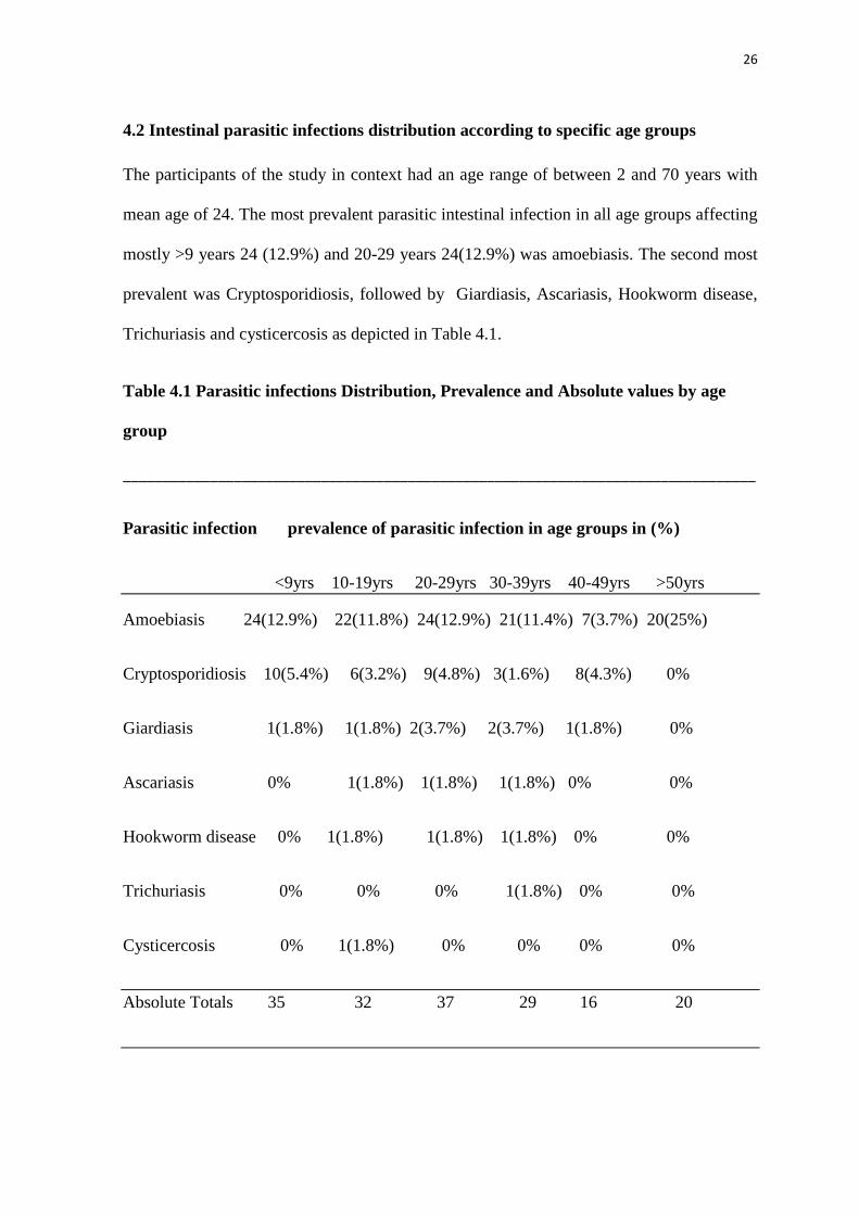
4.2 Intestinal parasitic infections distribution according to specific age groups
The participants of the study in context had an age range of between 2 and 70 years with
mean age of 24. The most prevalent parasitic intestinal infection in all age groups affecting
mostly >9 years 24 (12.9%) and 20-29 years 24(12.9%) was amoebiasis. The second most
prevalent was Cryptosporidiosis, followed by Giardiasis, Ascariasis, Hookworm disease,
Trichuriasis and cysticercosis as depicted in Table 4.1.
Table 4.1 Parasitic infections Distribution, Prevalence and Absolute values by age
group
________________________________________________________________________________
Parasitic infection prevalence of parasitic infection in age groups in (%)
<9yrs 10-19yrs 20-29yrs 30-39yrs 40-49yrs >50yrs
Amoebiasis 24(12.9%) 22(11.8%) 24(12.9%) 21(11.4%) 7(3.7%) 20(25%)
Cryptosporidiosis 10(5.4%) 6(3.2%) 9(4.8%) 3(1.6%) 8(4.3%) 0%
Giardiasis 1(1.8%) 1(1.8%) 2(3.7%) 2(3.7%) 1(1.8%) 0%
Ascariasis 0% 1(1.8%) 1(1.8%) 1(1.8%) 0% 0%
Hookworm disease 0% 1(1.8%) 1(1.8%) 1(1.8%) 0% 0%
Trichuriasis 0% 0% 0% 1(1.8%) 0% 0%
Cysticercosis 0% 1(1.8%) 0% 0% 0% 0%
Absolute Totals 35 32 37 29 16 20

27
4.3 Intestinal parasitic infections distribution by gender
The total number of females who participated in the study was 104 while the males were
81 totaling to 185 participants. Amoebiasis was the most identified parasitic infection in
the both genders; 67(36.2%) in males and 51(27.6%) in females. Cryptosporidiosis had a
higher prevalence in females 27(14.6%) than males 10(5.4%). The helminthic infections
were generally very low in both genders 1(0.5%). The least prevalent in the males was
trichuriasis (0%) and in the females cysticercosis (0%) as depicted in Table 4.2.
Table 4.2 Intestinal infections in relation to Gender and Absolute values
Parasitic infection Percentage prevalence in gender status
Males Females
Amoebiasis 67(36.2%) 51(27.6%)
Giardiasis 3(1.6%) 4(2.2%)
Ascariasis 1(0.5%) 2(1.1%)
Trichuriasis 0% 1(0.5%)
Hookworm disease 1(0.5%) 1(0.5%)
Cysticercosis 1(0.5%) 0%
Cryptosporidiosis 10(5.4%) 27(14.6%)
Absolute Totals 83 86

28
4.4 Socio-economic risk factors associated with intestinal parasitic infections
4.4.1 The effects of domestic water used, faecal disposal facility and presence of pets
The majority of the participants used tap and borehole water 88 (47.8), and the least used
borehole 6 (3.3%). Pit latrine was mostly used 140 (76.1%) with only 14 (7.6%) using
flash toilets. Possesion of pets was found in 119 (64.7%) while those without pets were 66
(35.7%) as depicted in Table 4.3
Table 4.3 The water source, faecal disposal and presence of pets
Utility Factor Percentage found
• Using tap water only 48(22.8%)
• Using tap and borehole 88(47.8%)
• Using borehole and river 42(22.8%)
• Using borehole only 6(3.3%)
• Using pit latrine and flash toilets 30(16.3%)
• Using pit latrine only 140(76.1%)
• Using flash toilets only 14(7.6%)
• Sharing toilet more than five persons 133(72.3%)
• Using toilet less than five persons 5(27.7%)
• Possessing pets 119(64.7%)
• Do not have pets 65 (35.3%)
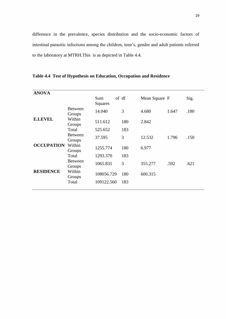
4.4.2 The effects of Education level , Occupation and Residence
The study results showed a p-value of 0.180 (Education level), 0.15 (Occupation) and 0.61
(Residence) respectively which is greater than the threshold value of (0.05). This means
therefore that the study findings accepted the null hypothesis that there is no significant
29
difference in the prevalence, species distribution and the socio-economic factors of
intestinal parasitic infections among the children, teen’s, gender and adult patients referred
to the laboratory at MTRH.This is as depicted in Table 4.4.
Table 4.4 Test of Hypothesis on Education, Occupation and Residence
ANOVA
Sum of
Squares
df Mean Square F Sig.
E.LEVEL
Between
Groups 14.040 3 4.680 1.647 .180
Within
Groups 511.612 180 2.842
Total 525.652 183
OCCUPATION
Between
Groups 37.595 3 12.532 1.796 .150
Within
Groups 1255.774 180 6.977
Total 1293.370 183
RESIDENCE
Between
Groups 1065.831 3 355.277 .592 .621
Within
Groups 108056.729 180 600.315
Total 109122.560 183

30
CHAPTER FIVE
DISCUSSION
The present study was undertaken to investigate the prevalence of intestinal parasitic
infections, its relationship with age, gender and the association with specific socio-
economic factors. A total of 185 stool samples were analyzed for intestinal parasites. The
residential areas varied from urban estates to the rural villages in all over the Western
region of Kenya. In this study the total number of protozoans identified were 103 (55.1%)
while the helminths were 7 (3.8%). It was in agreement with Ashtiani et al., (2011) who
found parasitic infection in Iran at (33%) and (4.8%) by protozoa and helminths
respectively. On the other hand, it was in disagreement with Gelaw et al., (2013) who
reported infection of (13.2%, 26.9%) with protozoa and helminths, respectively in
Ethiopia. Inadequate sanitary measures and problems of drainage may have contributed to
this high prevalence of protozoan parasitic infection in the study area. However, out of the
total number of the identified parasites, 110 /185 some of the patients were found to have
more than one parasite.
The general prevalence of parasitic infections in this study of 86 (46.5%) was lower than
previous report from Karachi in India that had observed 52.8% (Mehraj et al., 2003), and
higher than similar studies done in Nigeria that observed a prevalence of 34.6% (Nduka et
al., 2006) and 30.6% (Mbanugo et al., 2002) respectively. The difference could have been
attributed to the type of patients used. For instance, in this study, both the in and out-
patients, and those from the tertiary hospitals with signs and symptoms of diarrhea and
abdominal pains were recruited. The studies mentioned previously used subjects from rural
community (Nduka et al., 2006) or those from the general population (Mbanugo et al.,
2012). It is possible that the patients in the current study may have not been treated at the

31
available primary healthcare facilities where they lack diagnostic facilities This may
explain the higher prevalence in this study.
The prevalence of E. histolytica among males and females below 9 years was 30.0% and
26.3%, respectively indicating that both genders are equally susceptible to infection
(p>0.05). These results are not in agreement with many studies done in Thailand, where
the prevalence was 18.5% (males), 16.1% (females) (Wongstitwilairoong et al., 2007), in
Italy 17.1% (males), 12.7% (females) (Manganelli et al.,2012). The higher standard of
living in both Thailand and Italy could be the possible reason for the lower prevalence’s in
comparison to developing country like Kenya. Previous similar studies showed even higher
rates in Morocco 63.5% (males), 60.4% (females), (El Fatni et al., 2014) and in Nairobi,
Kenya 51.6% (males), 48.4% (females) (Mbae et al., 2013). However, it was not in
agreement with the study that was done in Nepal where the prevalence was (16.9%, 22%),
(Mukhiya et al., 2012), in Brazil (26.1%, 30.3%), (Nobre et al., 2013), in northwest and
southern Ethiopia (32.1%, 35.9%) (Gelaw et al., 2013), (80.6%, 81.4%) (Abossie and Sied,
2014) representing the males and females respectively. This varied difference could have
been attributed by the improper disposal of the sewage refuse and lack of treated water for
domestic uses.
In this study 43(23.9) were infected with E. histolytica which was in agreement with
related study done in Iraq which showed that E. histolytica was the most common
protozoan infection with a prevalence rate of 24.0% (Abbas, 2012). The frequency of the
parasitic infestations was slightly higher among males 67 (36.2%) than females 51
(27.6%), but this difference was not statistically significant (p>0.05)
The high prevalence rate of Entamoeba spp. 43 (23.9%) in the current study showed that
the infection transfer between persons probably through food or water is high and this may

32
indicate that there is likelihood of contamination by human faeces (Fernandez et al., 2002).
The infections are likely to be linked to the everyday activities of the individuals rather
than gender. The present study findings showed an equal exposure of both genders to
parasitic infections due to sharing almost the same environmental conditions therefore
gender did not influence the prevalence of the intestinal parasitic infections. The high
prevalence of E. histolytica could be due to the existence of resistant cysts of the parasite
as reported by Mbuh et al., (2010) in Cameroon. Nevertheless, children most often have a
tendency of eating food without hand washing unless reminded or may lick their
contaminated fingers. This age group fall within the period when children are increasingly
involved with outdoor activities, including an increased chance of handling fecal
contaminated materials, which predispose them to parasitic infections. Among the non-
pathogenic protozoa found in all age groups, was Entamoeba coli 12 (6.5%) and
iodamoeba butschlii 12 (6.5 %).
Infection with Ascaris lumbricoides and Hymenolepis nana which had the lowest
prevalence-one case of each 2 (1.6 %), was comparable with similar studies of 1(0.5%) by
(Patel and Khandekar, 2006; Al-Braiken, 2008; Al-Megrin, 2010; Sharif et al., 2010). The
ova of the two mentioned helminths have a tough outer coat that enables them to resist
adverse external environmental conditions which enhance their survival and higher
probability of transmission (Bhutta et al., 2014). Indeed, earlier studies in the Magu district
in Tanzania, reported a prevalence of <1% of A. lumbricoides, Trichuris trichiura and E.
vermicularis (Lwambo et al., 1999) while another previous study done in Sengerema
District in Tanzania did not detect any A. lumbricoides, T. trichiura or E. vermicularis
(Mazigo et al., 2010). However, it was not in agreement with other studies done in
Nicaragua (12%) (Munoz-Antoli et al., 2014) and in Malaysia (Sinniah et al., 2014). The
low prevalence of the helminths in this study is an indication of effective regular

33
intermittent treatment with anthelminthic drugs in the primary health facilities. However,
the drug mass administration with albendazole could explain the low rate of helminthes
infection as previously reported by (Supali et al., 2013).
The prevalence of Cryptosporidium parvum was 20 (13%), and it was comparable with
several other studies (12%) (Yilmaz et al., 2008; Dogan et al., 2012; Vahedi et al., 2012).
The high prevalence rate of cryptosporidiosis recorded in the females could been attributed
by other underlying medical conditions that may have weakened the immune system hence
giving a chance to the opportunistic infections like cryptosporidiosis (Herwaldt et al.,
2000). In some areas of the studies, the water pipes are passed through drainage pathways
and, in some instances, the pipes are broken and left unattended, which eventually may
contaminate the drinking water (Lopez et al., 2003). Nevertheless, with the suitable
temperature range, humidity, and other environmental factors sporulation and endurance of
oocysts in the water may be enhanced.
However, since modified Z-N test is not employed as a routine stool test in most of the
hospital laboratories, some cases of cryptosporidiosis are missed out therefore it is possible
that many healthy carriers exist in the communities. Such cases can only be detected when
patients are referred to facilities with modern diagnostic techniques like MTRH.
Out of the infected patients 46.5 % (86/185) who were infected by the intestinal parasites,
6.4% (12/185) had multiple infections. In the multiple infection groups, the most common
combinations were Entamoeba histolytica and E. coli, Entamoeba histolytica and I.
butschlii followed by hookworm spp and I. butschlii. There was no statistically significant
difference (P value of 0.562). The multiple infections were not specific to a particular age
nor gender. For the case of small children the outdoor activities including handling of
feacal contaminated materials could have predisposed them to parasitic infections.The
34
sharing of the toilet facility by many people as shown in the current study may have lead to
high chances of contamination 133 (72.3%). Polyparasitism was common within the
protozoa than in the helminthic infections, and this could probably be due to the more
hostile weather to the mode of transmission of the helminths. Ascaris lumbricoides and
Trichuris trichiura is feco-oral requiring suitable environment for egg maturation, survival
and transmission. The accessibility of improved hygiene, sanitation and awareness of the
infection associated with lack of access to potable water could be one plausible explanation
for low STH prevalence within these patients.
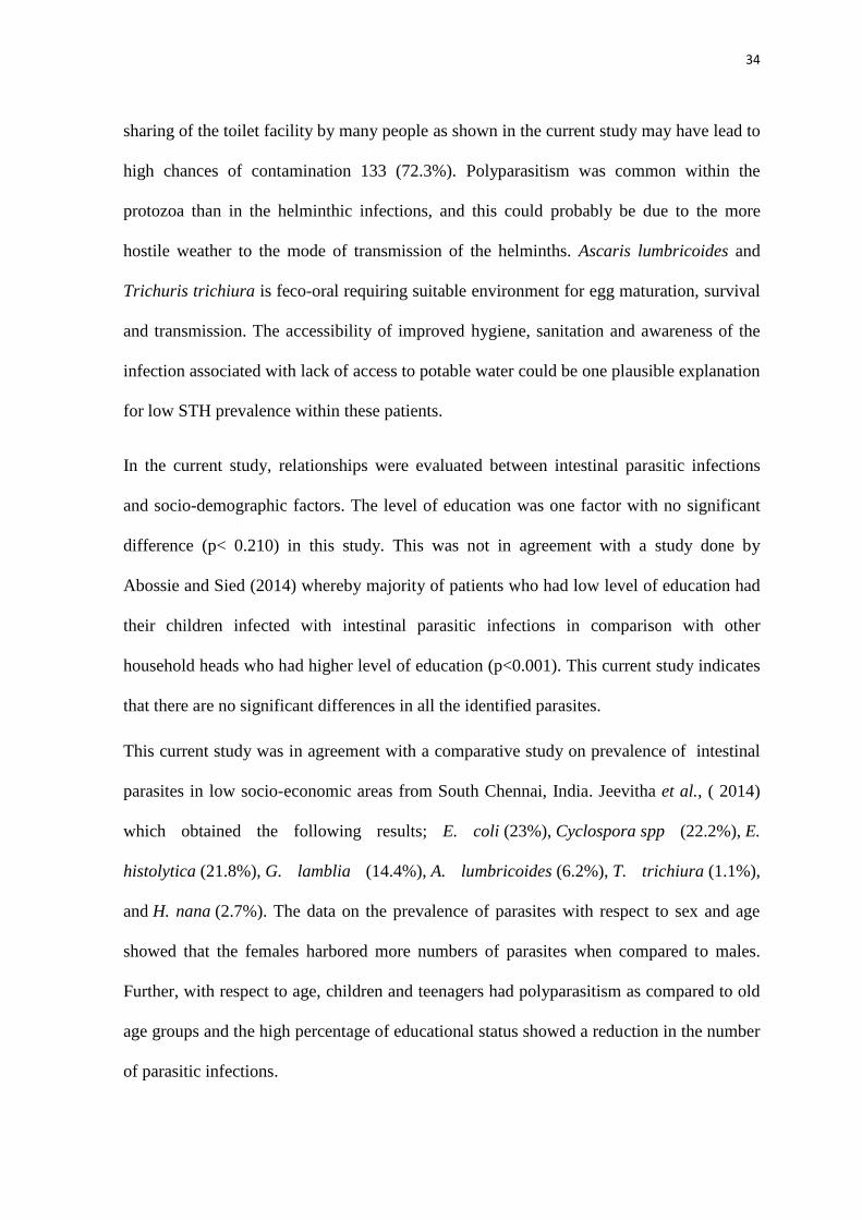
In the current study, relationships were evaluated between intestinal parasitic infections
and socio-demographic factors. The level of education was one factor with no significant
difference (p< 0.210) in this study. This was not in agreement with a study done by
Abossie and Sied (2014) whereby majority of patients who had low level of education had
their children infected with intestinal parasitic infections in comparison with other
household heads who had higher level of education (p<0.001). This current study indicates
that there are no significant differences in all the identified parasites.
This current study was in agreement with a comparative study on prevalence of intestinal
parasites in low socio-economic areas from South Chennai, India. Jeevitha et al., ( 2014)
which obtained the following results; E. coli (23%), Cyclospora spp (22.2%), E.
histolytica (21.8%), G. lamblia (14.4%), A. lumbricoides (6.2%), T. trichiura (1.1%),
and H. nana (2.7%). The data on the prevalence of parasites with respect to sex and age
showed that the females harbored more numbers of parasites when compared to males.
Further, with respect to age, children and teenagers had polyparasitism as compared to old
age groups and the high percentage of educational status showed a reduction in the number
of parasitic infections.

35
Conclusively, these IPIs could be prevented by possible grouping of better ecological
designs, examination of personal hygiene as well as routine medical examination and
treatment should be strongly recommended in the low socio-economic areas in the current
study region.
36
CHAPTER SIX
CONCLUSIONS AND RECOMMENDATIONS
Based on the present study findings, Entamoeba histolytica and Giardia lamblia were the
most prevalent pathogenic intestinal protozoa while A. lumberciodes, Encylostoma
duodenale and T. trichiura were less common in the patients observed.
Study participants of all ages were susceptible to parasitic infections with varied
magnitudes in the study population. The most prevalent parasitic infections encountered
were amoebiasis and cryptosporidiosis.
Both genders were found susceptible to both protozoal and helminth infections though
cryptosporidiosis was more prevalent in females than males.
Effects of education level, source of domestic water, residence and fecal disposal facility
did not show a significant influence on intestinal infections among the patients referred to
the laboratory at MTRH. The high prevalence rate of intestinal parasitic infections among
the patients attending MTRH indicates that parasitic infections should be considered as a
public health problem.
6.1 Recommendations
As this is the first study in the region to provide comprehensive information related to
prevalence of intestinal parasites in patients attending MTRH, the given recommendations
from the study will assist Government health officials in policy development. Interventions
such as deworming and health education programs will provid proper mechanism to be put
in place through the public health department in liaison with the County health authorities.
For this reason, preventive measures should be implemented by adhering to the following;
1. Community-based health promotions including regular checkups and treatment.

37
2. Adequate treatment of domestic water for the community to reduce the incidences
of IPIs.
3. Modified Z-N technique to should be included as a routine test for stool analysis in
the health facility Laboratories.
4. Research is needed to elucidate why amoebiasis is higher in the males and
cryptosporidiosis in the females.
38
REFERENCES
Abbas, Obaid, Farhan. (2012). Prevalence of Intestinal Parasitic Infestations in
Al-Anbar Province, West of Iraq. J. of university of anbar for
pure science: Vol.6: NO.1.
Abdel-M, I., A., Wierzba, T., F., Abu-Elyazeed, R., Ibrahim, A., F., Ahmed, S.,
F., Kamal, K., Sanders, J., Frenck, R. (2005). Diarrhea Associated
with Cryptosporidium parvum among young Children of the Nile
River Delta in Egypt. J Trop Pediatr 51:154–159.
Abossie, A. and M. Seid.(2014). Assessment of the prevalence of intestinal
parasitosis and associated risk factors among primary School
Children in Chencha town, Southern Ethiopia. BMC Public Health,
Vol. 14. 10.1186/1471-2458-14-166.
Al-Braiken, F., A. (2008). Is intestinal parasitic infection still a public health
concern among Saudi children? Saudi Med. J., 29: 1630-1635.
Al-Braiken, F., A., A., Amin., N., J., Beeching., M., Hommel., and C., A., Hart.
(2003). Detection of Cryptosporidium amongst diarrhoeic and
asymptomatic children in Jeddah, Saudi Arabia. Ann. Trop. Med.
Parasitol., 97: 505-510.
Ali, S., A. and Hill, D., R. (2003).Giardia intestinalis. Curr Opin Infect Dis.;
16: 453–460.
Alli, J., A., A., F., Kolade. I., O., Okonko. et al., (2011). “Prevalence of intestinal
nematode infection among pregnant women attending antenatal clinic
at the University College Hospital, Ibadan, Nigeria,” Advances in
Applied Science Research, vol. 2, pp. 1–13. View at Google Scholar.

39
Al-Megrin, W., A., I. (2010). Intestinal parasites infection among
immunocompromised patients in Riyadh, Saudi Arabia. Pak.
J. Biol. Sci., 13: 390-394.
Alum, A., J., R., I., jaz, M., K. (2010). The global war against intestinal
parasites— should we use a holistic approach? Int J.
Infect Dis.14:e732–e738.
Ashtiani, M., T., H., M., Monajemzadeh., B., Saghi., S., Shams. and S, H.,
Mortazavi. (2013). Prevalence of intestinal parasites among
children referred to Children's Medical Center during 18
years (1991-2008), Tehran, Iran. Ann. Trop. Med.
Parasitol., 105: 507-513.
Auerbach, Karp., S. (2012).Intestinal parasites in Cambodia .Chronic
Strongyloides stercoralis in Loatian immigrants .CDC 27/7:
Saving Lives. Protecting people.
Barry, M.,A., Weatherhead J., E., Hotez, P., J., Woc-Colburn , L. (2013).
"Childhood parasitic infections endemic to the United States".
Pediatrics Clin. North Am 60 (2): 471–85. doi : 10.1016
/j. pcl. 2012.12.011 PMID 23481112.
Belayhun Y., Medhin G., Amberbir, A. (2010). Prevalence and risk factor for
geohelminthic Infection in infants in Butajera Ethiopia, a population
based study. BMC Public Health 10: 21. doi: 10.1186/1471-
2458-10-21.
Bethony, J., Brooker, S., Albonico, M., Geiger., S.M., Loukas., A., Diemert., D.,

40
and Hotez, P.J. (2006). Soil-transmitted helminth infections:
ascariasis, trichuriasis, and hookworm. Lancet; 367: 1521–1532.
Birn, Anne, Emanuelle, Armando, Solórzano. (1999). "Public health policy
paradoxes: science and politics in the Rockefeller Foundation's
hookworm campaign in Mexico in the 1920s". Social Science
& Medicine. 49 (9): 1197–1213. PMID 10501641. doi:10.
/S0277-9536(99)00160-4.
Bhutta, Z., A., Sommerfeld, J., Lassi , Z., S., Salam ,R., A., Das, J.,
K.(2014).Global burden, distribution, and Interventions for
infectious diseases of poverty. Infect Dis poverty; 3:21.
Brooker , S., Kabatereine, N., B., Smith, J., L., Mupfasoni, D., Mwanje, M., T.
(2009). An updated Atlas of human helminth infections: the example
of East Africa. In. J. Health Geogr 8: 42. doi: 10.1186/1476-
072x-8-42.
Brooker, S., Michael., E. (2000).The Potential of Geographical Information
Systems and Remote Sensing in Epidermiology and control
of Human Helminth Infections. Anvances in Parasitology ;
47:254-87. (PubMed)
Bundy, D., C., T, Cooper, R., S., Thomson, D., Didler, M, and Simmons., S.
(1987).Epidemiology and population dynamics of A. lumbricoides
and Trichuris trichiura infections in the same community.
Trans roy. Soc. Trop. Med. Hgy. 81:987-994
CDC. (2014). Centre for disease control and prevention.
41
http://www.cdc.gov/parasites/children.html.
CDC. (2013). "Parasites - Trichuriasis (also known as Whipworm Infection)"
Retrieved 5 March2014.
CDC. (2013). "Giardiasis". Cdc.gov. Retrieved 26 February 2015.
Chan, M., S. (1997). The global burden of intestinal nematode infections—fifty
years on. Parasitol Today. ; 13: 438–443.
Chan, M., S., Medley, G., F., Jamison, D. and Bundy, D., A. (1994). The
evaluation of potential Global morbidity attributable to
intestinal nematode infections. Parasitology. 1373–387
Cheesbrough, M. (1987).Medical Laboratory Manual for tropical Countries. VOL
1. ELSB Ed. 1989. Pitkanen Y., T., Petonen., M. Published 1995.
Cheesborough, M. (1987). Medical Laboratory Manual for Tropical Countries.
Vol. 1. 2nd Edition Pgs 181-186, 204-205, 217.
Cheesbrough, M. (1985). Parasitological Tests. In: District Laboratory Practice in
Tropical Countries, Chessbrough, M. (Ed.).Tropical Health
Technology, World Health Organization, Geneva, Switzerland,
pp: 184-235
Cleaveland, S., Laurenson, M., K. and Taylor, L., H. (2001). Diseases of humans
and their domestic mammals: pathogen characteristics, host range
and the risk of emergence. Phil Trans R Soc Lond B.; 356:
991– 999.
Curtale, F., Pezzotti. P., Sharbini. A., L., AlMaadat. H., Ingrosso. P. (1998).
Knowledge, Perceptions and behavior of mothers towards
intestinal helminths in Upper Egypt: Implication for control.
Health Policy Plan.

42
De Silva. N., R., Brooker, S., Hotez., P., J., Montresor, a., Engles , D., Savioli., L.
(2003). Soil-Transmitted Helminth Infections:Updating
the Global Picture. Trends in Parasitology.
19:547- 51(PubMed).
Dogan, N., Y., O., Z., N., U., Kocman. and A , F., Nursal. (2012). [Comparison of
individual differences in the direct microscopic examination in
the diagnosis of intestinal parasites]. Turkiye Parazitolojii
Dergisi, 36: 211-214, (In Turkish).
Ejemot, Nwadiaro, Regina, I., Ehiri, John, E., Arikpo, Dachi, Meremikwu,
Martin, M., Critchley, Julia, A. (2015). “Hand washing
promotion for preventing diarrhoea” . The Cochrane
Database of Systematic Reviews (9): 1–95. ISSN 1469-493X.
El Fatni, C., F., Olmo., H., El Fatni., D., Romero. and M , J., Rosales.(2014). First
genotyping of Giardia duodenalis and prevalence of
enteroparasites in children from Tetouan (Morocco).
Parasite, Vol. 21. 10.1051/parasite/2014049.
Esch, Thimothy, M., Goater., Cameron., P., Goater., Gearal., W.,
Esch.(2013).Parasitism In human. 2nd edition. The diversity and
ecology of animal parasites.
Ezeagwuna, Okwelogu, Ekejindu, Ogbuagu. C. (2010). The Prevalence and
Socio-Economic Factors Of Intestinal Helminth Infections
Among Primary School Pupils In Ozubulu, Anambra State,
Nigeria. The internet Journal of Epidemiology. Vol. 9 No 1.

43
Feng, Y., Xiao, L. (2011). Zoonotic potential and molecular epidemiology of
Giardia species and giardiasis. Clin Microbiol Rev.24:110–140.
doi: 10.1128/CMR.00033-10.
Fenwick, A. (March 2012). “The global burden of neglected tropical diseases”
Public health 126(3):233–6.doi:10.1016/ j.puhe.2011.11.015.
PMID 22325616.
Fernandez, M., C., S., Verghese. , R ., Bhuvaneswari (2002). “A comparative
study of the intestinal parasites prevalent among children living
in rural and urban settings in and around Chennai,” Journal of
Communicable Disease. vol. 34, no. 1, pp. 35–39.
Garson, M. (2003). Capital University of Intergrated Medicine. Parasites-
A Holistic Approach. In: Associates NIH, editor.
Gelaw, A., B., Anagaw., B., Nigussie., B., Silesh. and A, Yirga.( 2013)
. Prevalence of intestinal parasitic infections and risk factors
among schoolchildren at the University of Gondar
Community School, Northwest Ethiopia: A cross-sectional study.
BMC Public Health, Vol. 13. 10.1186/1471-2458-13-304.
GNNTD, (2009). "Global network for neglected tropical diseases receives $34
million from Gates Foundation: IDB leads campaign to greatly
reduce the burden of most neglected Diseases by 2020 in Latin
America and the Caribbean. “Press Release.Global Network for
Neglected Tropical Diseases.
Gunarwardena, C., Jennings. , B., Ortegano-Layne. L., Freehette. C., Carbajal

44
K., Linderrmann. K.(2004).Building an on line wisdom community:
A transformation Design model. Journal of computing in
Higher Education 15(2), 40-62.
Haque, R., (2007).“Human intestinal parasites,” Journal of Health, Population
and Nutrition, vol. 25, no. 4, pp. 387–391. View at Google Scholar ·
View at Scopus.
Hellard, M., E., Sinclair, M., I., Hogg, G., G., Fairley, C., K. (2000). Prevalence
of enteric pathogens among Community based asymptomatic
individuals. J Gastroenterol Hepatol. ; 15: 290–293.
doi: 10.1046/j.1440-1746.2000. 02089.
Heredia, R., D., Fonseca, J., A., López , M., C. (2012). Entamoeba moshkovskii
perspectives of a new Agent to consider in the Diagnosis of
amebiasis. Acta Trop 123:139.
Herwaldt, B., L. (2000).“Cyclospora cayetanensis: a review, focusing on
the outbreaks of cyclosporiasis in the 1990s,”
Clinical Infectious Diseases, vol. 31, no. 4, pp. 1040–1057.
Hotez, P., J., Alvarado, M., Basa'ñez, M., G., Bolliger, I., Bourne, R. (2014).The
Global Burden of Disease Study 2010: Interpretation and
Implications for the Neglected Tropical Diseases.
PLoS Negl Trop Dis 8(7): e 2865. doi:
10.1371/ journal.pntd.0002865.
Hotez, P., J. (2009). One world health: neglected tropical diseases in a flat world.
PLoS Negl Trop Dis.; 3: e405.
Hotez, P., J. (2008). Neglected infections of poverty in the United States of
America. PLoS Negl Trop Dis.; 2: e256.
45
Hotez, P., J., Bottazzi, M., E., Franco-Paredes, C., Ault, S., K. and Periago, M., R.
(2008). The neglected tropical diseases of Latin America and the
Caribbean: a review of Disease burden and distribution and a
roadmap for control and elimination.
PLoS Negl Trop Dis.; 2 e300.
Hotez, P., J., Bottazzi. M., E.,Bethony. J., Buss., P. (2006). New technologies for
the control of human hookworm infection Trend Parasitol.
22:327-331.
Hotez, P, J., Bethony, J., Bottazzi, M., E., Brooker, S., Buss, P. (2005). The Great
Infection of Mankind.PLoS Med. 2 (3): e67. doi:
10.1371/journal.pmed.0020067PMC 1069663. PMID 15783256.
Houweling, T., A., Kunst, A., E., Mackenbach, J., P. (2003). Measuring health
inequality among children in Developing countries: does the choice
of the indicator of economic status matt . Int J. Equity Health. 2:8.
Jamison, Dean. (2006). "Helminth Infections: Soil-transmitted Helminth
Infections and Schistosomiasis. “Disease control priorities in
developing countries.” New York: Oxford University Press.
p. Chapter 24. ISBN 9780821361801.
Jeevitha, D., Pradeep. Pushpara, j., Selvadoss. and Kanchana, Muthuswamy.
(2014). Comparative Study of the Prevalence of Intestinal
Parasites in Low Socioeconomic Areas from South Chennai,
India. Research Article. Journal of Parasitology Research.
Volume 2014 , Article ID 630968,
7 pages. http://dx.doi.org/10.1155/2014/630968
Jex, A., R., Smith, H., V., Nolan, M., J., Campbell, B., E., Young, N., D.,

46
Cantacessi , C., Gasser, R., B. (2009). Cryptic parasite
revealed improved prospects for treatment and
control of human Cryptosporidiosis through advanced technologies.
Adv Parasitol. 77:141–173.
Keiser, J., Utzinger, J. (2010). The drugs we have and the drugs we need against
major helminth infections. Adv Parasitol73: 197–230.
doi:10.1016/s0065-308x (10)73008.
Kia, E., B., Hossein,M., Nilforoushan, M.,R,, Meamar, A.,R., Rezaeian, M.
(2008). Study of intestinal protozoan Parasites in rural inhabitants
of Mazandaran Province, Northern Iran.Iranian J Parasitol; 3: 22–25.
Loukopoulos, P., A., Komnenou. E., Papadopoulos. and V., Psychas. (2007).
“Lethal ozolaimus megatyphlon infection in a green iguana
(Iguana iguana rhinolopa),” Journal of Zoo and Wildlife Medicine,
vol. 38, no. 1, pp. 131–134. View at Publisher
View at Google Scholar · View at Scopus.
Lozano, R., Naghavi, M., Foreman, K., Lim, S., Shibuya, K., Aboyans, V.,
Abraham, J., Adair, T., Aggarwal, R., Ahn, S.,Y. (2012).
Global and regional mortality from 235 causes of death
for 20 age groups in 1990 and 2010: a systematic analysis for
the Global Burd of Disease Study 2010
.Lancet.380:2095–2128. doi: 1016/S0140- 6736(12)61728-0.
Lwambo, N., J., S., Siza, J., E., Brooker, S., Bundy, D., A., P. & Guyatt, H.
(1999).Patterns of concurrent hookworm infection and
schistosomiasis in schoolchildren in Tanzania. Transactions
of the Royal Society of Tropical Medicine and Hygiene
93, 497‐502.

47
Manganelli, L., F., Berrilli., D., D., I., Cave., L., Ercoli., G., Capelli., D., Otranto.
and A, Giangaspero. (2012). Intestinal parasite infections in
immigrant children in the city of Rome, related risk factors and
possible impact on nutritional status. Parasites Vectors,
Vol. 5. 10.1186/1756-3305-5-265.
Mazigo, H., D., Waihenya, R., Lwambo, N., J., S., Mnyone, L., L., Mahande, A.,
M., Seni, J., Zinga, M., Kapesa , A., Kweka, E., J., Mshana
S., E., Heukelbach, J., & Mkoji, G., M. (2010). Co‐infections
with Plasmodium falciparum, Schistosoma mansoni
and in test helminth among school children in endemic
areas of north western Tanzania. BMC Parasites and Vectors 3:4.
Mbae, C., K., D., J., Nokes., E., Mulinge., J., Nyambura. , A., Waruru, and S.
Kariuki.(2013). Intestinal parasitic infections in children presenting
with diarrhoea in outpatient and inpatient settings in an informal
settlement of Nairobi , Kenya. BMC Infect. Dis.,
Vol. 13. 10.1186/1471-2334-13-243.
Mbanugo, J., I., Onyebuchi , C., J. (2012). Prevalence of intestinal parasites in
Ezinifite Community, Aguata Local Government Area of
Anambra State. Nigerian J. Parasitol.23:27–34.

48
Mbanugo, J., I., Abazie, O., C. (2002). A comparative study of intestinal parasite
infections of pregnant and non-pregnant women in Nkpor ,
Anambra State. Nigerian J Parasitol. 23:19–26.
Mbuh, J., V., Ntonifor, H., N., & Ojong, J., T. (2010).The incidence, intensity and
host morbidity of parasitic protozoan infections in gastrointestinal
disorder outpatients in Buena Sub Division, Cameroon. Journal
of Infection in Developing Countries 4, 38‐43.
Mehraj, V., Hatcher, J., Akhtar, S., Rafique, G., Beg, M., A. (2008). Prevalence
and factors associated with Intestinal parasitic infection
among children in an urban slum of Karachi.
PLoS One. 3(11):e3680.
Meyer, E., A. (1985). The epidemiology of giardiasis.Parasitol Today. (1985)1:
101–105.
Minenoa, M., and Avery, A. (2003). “Giardiasis: recent progress in chemotherapy
and drug development,” Current Pharmaceutical Design,
vol. 9, pp. 841–855.
Molbak, K., Hojlyng , N., Gottschan. A., Sa., J., C., Ingholt, L., da Silva., A., P.
and Aaby. , P. (1993).Cryptosporidiosis in infancy and
childhood Mortality in Guinea Bissau, West Africa Br.
Medd. J. 307, 417-420.
Mordi, R., M., and P., O., A., Ngwodo. (2007). “A study of blood and gastro-
intestinal parasites in Edo state,” African Journal of Biotechnology,
vol. 6, no. 19, pp. 2201–2207. View at Google Scholar ·

49
View at Scopus.
Mugenda, O., M. and Mugenda, A., G. (1999). Research Methods: Quantitative
and Qualitative approaches, African Center of Technology
Studies, Nairobi, Kenya.
Mukhiya, R., K., S., K., Rai., A., B., Karki, and A. Prajapati.(2012). Intestinal
protozoan parasitic infection among school children.
J. Nepal Health Res. Council, 10: 204-207.
Munoz-Antoli, C., A., Pavon., A., Marcilla., R., Toledo. and J ,G., Esteban.
(2014). Prevalence and risk factors related to intestinal parasites
among children in Department of Rio San Juan, Nicaragua.
Trans. R. Soc. Trop. Med. Hyg., 108: 774-782.
Mwanthi, M., A., Kinoti. (2008). Prevalence of intestinal worm infection among
primary school children in Nairobi city. East African Journal
of Public Health, Vol. 5. No. 2
Nacher, M. (2011). Interactions between worms and malaria: good worms or bad
worms? Malar J.10:259.
Nduka, F., O., Nwaugo, V., O., Nwachukwu, N., C. (2006). Human intestinal
parasite infections in Ishiagu, a leading mining Area of
Abia State. Animal Research International; 3 (3):505–507.
Nematian, J., Nematian, E., Gholamrezanezhad, A., Asgari, A., A., (2004).
Prevalence of intestinal Parasitic infections and their
relation with socio-economic factors and hygienic
habits in Tehran primary school students.

50
Acta Trop.92:179–186.
Nobre, L., N., R., V., Silva., M., S., Macedo., R., A., Teixeira., J., A., Lamounier.
and S, C. , Franceschini.(2013). Risk factors for intestinal
parasitic infections in pre-schoolers in a low socio-economic
area, Diamantina, Brazil. Pathog. Global Health, 107: 103-10.
NYSDH. (2011). “Giardiasis (beaver fever)’’ Retrieved 21 June 2015.
Odu, N., N., C., O., Akujobi. S., N., Maxwell, and A., R., Nte. (2011). “Impact of
mass deworming of school children in rural communities in
Rivers State, Nigeria: option for programme sustainability,”
Acta Parasitologica, vol. 2, pp. 20–24.
View at Google Scholar.
Ogbe , M., G., Odudu, L., A. (1990). Gastro-intestinal helminthiasis in Epe
L.G.A., Lagos State, Nigeria. Nigerian Journal Parasitol. ;
9-11:95–106.
Olsen, A., Magnusen, P., Ouma, J., H., Anderso, J., Friis, H.(1998). The
contribution of hookworm and other parasitic infections to
haemoglobin and iron stsus among children and adults in
Western Kenya.Trans Roy Soc Trop Med Hyg. Nov-Dec;
92(6):643-9.
Ortega, Y., R. and Adam, R., D. (1997). Giardia: overview and update. Clin.
Infect Dis.; 25: 545–550.
Otieno, L., S., kamunui: F., j., Nyaga, D. and Nderitu , J., W. (1985). The
intestinal parasitic characteristics in the rural health centres
51
in Kisii District. In Recent advances in the management and
control of infections in East Africa. Tukei P.M (ed)
East African Lit.Bureau. Nairobi pp248-253.
Patel, P., K., and R., Khandekar. (2006). Intestinal parasitic infections among
school children of the Dhahira Region of Oman. Saudi Med. J.,
27: 627-632.
Peterson, K., M., Singh, U., Petri, W., A., Jr. (2011). Enteric Amebiasis. In:
Tropical Infectious Diseases: Principles, Pathogens and
Practice, 3rd ed, Guerrant R, Walker DH, Weller PF (Eds),
Saunders Elsevier, Philadelphia p.614.
Pillai D., R., Kain ., K.,C. (2003). Common intestinal parasites. Curr Treat
Infect. Dis 2003 ; 5: 207-17.
Pullan, R., L., Smith, J., L., I., Jasrasaria, R., Brooker, S., J. (2014). Global
numbers of infection and disease burden of soil transmitted
helminth infections in 2010. Parasites & Vectors 7: 37.
doi: 10.1186/1756-3305-7-37.
Rai, S., K., Bajracharya. K., Shrestha. M., K. (1993). Status of intestinal parasites
in Nepal. J Nep Med Assoc 31: 382-9.
Reed, S., L., (2012). Amobiasis and infections with free-living amoebas. In:
Harrison T., R., Fauci A., S., Braunwald., E., et al ., Principles
of internal medicine. 15 th ed. New York: McGraw-Hill;
pg. 1199-2002.
Ruto, J., Chrispinus. S., Mulambalah. (2016). Epidermiology of parasitism and
poly-parasitism involving intestinal helminthes among school
children from different residential settings in Nandi County,Kenya.

52
CHRISMED journal of Health and Research.
Sanad, M., M., and J., S., Al-Malki. (2007). Cryptosporidiosis among
immunocompromised patients in Saudi Arabia. J. Egypt. Soc.
Parasitol., 37: 765-774.
Sayyari, A., A., F. Imanzadeh, S., A., Bagheri, Yazdi, H., Karami, M., Yaghoobi.
(2005).Prevalence of intestinal parasitic infections in the Islamic
Republic of Iran Eastern Mediterranean Health Journal,
11 (3) (2005), pp. 377-383
Senior, K. (2008). Neglected tropical diseases affect thousands in the USA.
Lancet. ; 8: 427.
Sharif, M., A., Daryani., F., Asgarian., and M., Nasrolahei. (2010). Intestinal
parasitic infections among intellectual disability children in
rehabilitation centers of northern Iran. Res. Dev. Disabil.,
31: 924-928.
Sherchhand, J., B., Ohara, H., Cross. J., S., Shrestha, M., P., Sherchand. S. (1997).
Intestinal parasitic infections in rural areas of southern Nepal.
J Inst Med 1997; 19: 115-21.
Singh, C., S., A., Zargar, I., Masoodi, A., Shoukat, B., Ahmad. (2010). Predictors
of intestinal parasitosis in school children of Kashmir:
a prospective study .Tropical Gastroenterology,
31 (2) pp. 105-107
Sinniah, B., A., K., R., Hassan., I., Sabaridah., M., M., Soe., Z., Ibrahim. and O.,
53
Ali.( 2014). Prevalence of intestinal parasitic infections
among communities living in different habitats and its
comparison with one hundred and one studies conducted
over the past 42 years (1970 to 2013) in Malaysia.
Trop. Biomed., 31: 190-206.
Stanley, S., L., Jr. (2001). Pathophysiology of amoebiasis. Trends in Parasitology
17280–285.285.
Steketee, R., W. (2003). Pregnancy, nutrition and parasitic diseases. J. Nutr.
;133:1661S– 1667S.
Stensvold , C., R., Lewis. H., C., Hammerum. A., M., Porsbo. L., J., Nielsen. S.,
S., Olsen. K., E., Arendrup. M., C., Nielsen. H., V.,
Mølbak. K. (2009). Blastocystis: unravelling potential risk
factors and clinical significance of a common but neglected
parasite. Epidemiol Infect.137:1655–1663.
doi: 10.1017/S0950268809002672.
Stephenson, M. C., Latham. E., A., Ottesen. (2000). Malnutrition and parasitic
helminth Infections .Parasitology, 121 (Suppl.) (2000), pp. S23-28
Supali, T., Djuardi, Y., Brabley, M., Noordin, R., Ruckert, P., Fischer., P. (2013).
Impact of six rounds of mass drug administrationon Brugian
filariasis and soil-tranmitted helminth infections in eastern
Indonesia. PLoS Negl Trop Dis.7 (12):e2586.
Thiongo, F., W., Luoba, A. and Ouma., J., H. (2001). Intestinal helminths and
schistosomiasis among school children in a rural district

54
in Kenya. East Africa Medical Journal 78(6): 279-282.
Toma, T., Miyagi, I., Kamimura. K. (1999). Questionnaire survey and prevalence
of intestinal helminthes infections in Barru, Sulawesi, Indonisia.
Southeast Asian J. Trop Med Public Health 1999; 30 : 68-77.
Vahedi, M., S., Gohardehi., M., Sharif. and A., Daryani. (2012). Prevalence of
parasites in patients with gastroenteritis at East of Mazandaran
Province, Northern Iran. Trop. Biomed., 29: 568-574.
Wang, L., D, Guo., J., G., Wu., X., H., Chen., H., G., Wang., T., P., Zhu., S., P.,
Zhang., Z., H., Steinmann., P., Yang., G., J.,Wang., S., P. (2009).
China’s new strategy to block Schistosoma japonicum
transmission:experiences and impact beyond schistosomiasis.
Trop. Med., Int. Health. 14:1475-1483. 10.1111/j.1365-3156.02403.x.
Wekesa, A. W., Mulambalah, C., S., Muleke, C., I., and Odhiambo, R. (2014).
Intestinal Helminth Infections in Pregnant Women Attending
Antenatal Clinic at Kitale District Hospital, Kenya
Journal of Parasitology Research.
WHO (2014). Soil- transmitted helminthiasis: number of children treated in 2010.
Week Epidemio Rec.(3014), 87: 225-232).
WHO (2013)."Soil-transmitted helminth infections Fact sheet N°366".Retrieved 5
March 2014.Wolfe MS (1975)."Giardiasis". JAMA 233 (13): 1362–5.
doi:10.1001/jama.233.13.1362.
WHO (2013). Second report on neglected tropical diseases. Geneva: Sustaining
the drive to Overcome the global impact of neglected

55
tropical diseases. 9789241564540/en.
WHO (2012). The Use and Interpretation of Anthropometry. Report of a
WHO Expert Committee. Geneva, Switzerland: 2012. pp. 2–5.
WHO (2010a). First report on neglected tropical diseases? Geneva: Working to
overcome the Global impact of neglected
Tropical disease who.int/publications/9789241564090.
WHO (2010b). Soil-transmitted helminthiasis. Number of children treated 2007–
2008: update on the 2010 global target. Wkly Epidemiol
Rec 85: 141–8.
WHO (2006). Preventive Chemotherapy in Human Helminthiasis:
CoordinatedUse of Anthelminthic Drugs in Control Interventions:
A Manual for Health Professionals and Programme Managers.
WHO (2003). The Global Burden of Intestinal Parasitic infections in man.
www.E-mjm org/2003/v58n/ intestinal parasites.
WHO (2002). World health report. Annex Table 2: deaths by cause, sex,
and mortality stratum In WHO regions and annex Table 3:
Burden of disease in DALYs by cause, Sex and mortality
stratum in WHO regions. Geneva World Health.
WHO (2000). Informal consultation on intestinal helminthes infection. Geneva,
Switzerland: WHO; pp. 16–18. Organization p. 186–93.
Wongstitwilairoong, B., A., Srijan, O., Serichantalergs., C., D., Fukuda, P.,

56
McDaniel, L., Bodhidatta. and C., J., Mason.(2007). Intestinal
parasitic infections among pre-school children in Sangkhlaburi,
Thailand. Am. J. Trop. Med. Hyg., 76: 345-350.
Xiao, L., Morgan, U., M., Fayer, R., Thompson, R., C., Lal, A., A. (2000).
Cryptosporidium systematic and implications for public Health.
Parasitol Today16: 287–292.
Yilmaz, H., Z., Tas, Cengiz, and M., Cicek. (2008). Investigation of
cryptosporidiosis by enzyme-linked immunosorbent assay and
microscopy in children with diarrhea. Saudi Med. J., 29: 526-529.
Yoder, J., S., Harral, C., Beach, M., J. (2010).Giardiasis surveillance-United
States, 2006– 2008. MMWR Surveillance Summit; 59:15–25.

57
APPENDICES
APPENDIX I (A-I): Parasite Genera / species associated with intestinal infection(s)
among age groups
A. Prevalence (%) of Entamoeba coli amongst age groups and gender

58
B. Prevalence (%) of Giardia lamblia amongst age groups and gender
C. Prevalence (%) of Iodamoeba butschlii amongst age groups and gender

59
D. Prevalence (%) of Ascariasis amongst age groups and gender
E. Prevalence (%) of Hymenolepis nana amongst age groups and gender

60
G. Prevalence (%) of Hookworm disease amongst age groups and gender
H. Prevalence (%) of Trichuriasis amongst age groups and gender

61
I. Prevalence (%) of Cryptosporidiosis amongst age groups and gender

62
APPENDIX II: Identified parasite species and the relative prevalence (%)
Name Developmental stage Classification Associated infection Technique used ( % )
E. histolytica Cyst sarcodina Amoebiasis Concentration 43(23.9%)
C. parvum Oocyst coccidia Cryptosporidiosis Mod.Z-N 24 (13%)
E. coli Cyst sarcodina Amoebiasis Concentration 12 (6.5)
G. lamblia Cyst mastigophora Giardiasis Concentration 12 (6.5%)
I. butschlii Cyst sarcodina Amoebiasis Concentration 12 (6.5%)
A. lumbricoides Ova Nematoda Ascariasis Concentration 3 (1.6%)
H. nana Ova Cestoda Cysticercosis Concentration 1 (0.5%)
T. trichiura Ova Nematoda Trichuriasis Concentration 1 (0.5%)
A. duodenale Ova Nematoda Hookworm disease Concentration 2 (1.1%)

63
APPENDIX III (A-I): Effects of the identified intestinal parasitic infections on age,
level of education and gender
A. Effects of Amoebiasis on age, level of education and gender
The results showed that there was no significant difference of E.histolytica in age groups
of the respondents (χ2= 3.525 and P-value of 0.620). The study findings showed that there
was no significant relation between E.histolytica and Education (χ2= 7.143 and p-value of
0.210). There was no significant relationship between E.histolytica and sex (χ2= 1.820 and
p-value of 0.177).
Variable
E. histolytica
AGE Positive Negative Total
Below 9 years 8 21 29
18.20% 15.00% 15.80%
10-19 years 12 33 45
27.30% 23.60% 24.50%
20-29 years 12 35 47
27.30% 25.00% 25.50%
30-39 years 9 28 37
20.50% 20.00% 20.10%
40-49 years 1 16 17
2.30% 11.40% 9.20%
above 50 years 2 7 9
4.50% 5.00% 4.90%
χ2= 3.525 Sig=0.620
EDUCATION LEVEL Positive Negative
Primary 18 40 58
40.90% 28.60% 31.50%
Secondary 4 13 17
9.10% 9.30% 9.20%
O-level 6 44 50
13.60% 31.40% 27.20%
A-level 0 3 3
0.00% 2.10% 1.60%
College 13 33 46
29.50% 23.60% 25.00%
University 3 7 10
6.80% 5.00% 5.40%
χ2= 7.143 sig=0.210
SEX Positive Negative
F 21 83 104

64
47.70% 59.30% 56.50%
M 23 57 80
52.30% 40.70% 43.50%
χ2= 1.820 sig=0.177
B. Effects of E. coli on age, level of education and gender
The results showed that there was no significant difference of E.coli in age groups of the
respondents (χ = 1.521 and P-value of 0.911). The study findings showed that there was no
significant relation between E.coli and Education (χ = 0.445 and p-value 0.994) and sex (χ
= 0.017 and p-value of 0.896)
Variable E. coli
AGE Positive Negative TOTAL
BELOW 9 YEARS 2 27 29
16.70% 15.70% 15.80%
10-19 YEARS 3 42 45
25.00% 24.40% 24.50%
20-29 YEARS 4 43 47
33.30% 25.00% 25.50%
30-39 YEARS 1 36 37
8.30% 20.90% 20.10%
40-49 YEARS 1 16 17
8.30% 9.30% 9.20%
ABOVE 50 YEARS 1 8 9
8.30% 4.70% 4.90%
χ2= 1.521 Sig=0.911
EDUCATION LEVEL Positive Negative
PRIMARY 4 54 58
33.30% 31.40% 31.50%
SECONDARY 1 16 17
8.30% 9.30% 9.20%
O-LEVEL 3 47 50
25.00% 27.30% 27.20%
A-LEVEL 0 3 3
0.00% 1.70% 1.60%
COLLEGE 3 43 46
25.00% 25.00% 25.00%
UNIVERSITY 1 9 10
8.30% 5.20% 5.40%
χ2= 0.455 Sig=0.994
GENDER Positive Negative
F 7 97 104
58.30% 56.40% 56.50%
M 5 75 80
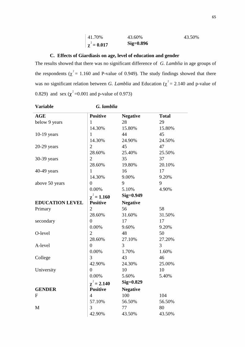
65
41.70% 43.60% 43.50%
χ2= 0.017 Sig=0.896
C. Effects of Giardiasis on age, level of education and gender
The results showed that there was no significant difference of G. Lamblia in age groups of
the respondents (χ2= 1.160 and P-value of 0.949). The study findings showed that there
was no significant relation between G. Lamblia and Education (χ2= 2.140 and p-value of
0.829) and sex (χ2=0.001 and p-value of 0.973)
Variable G. lamblia
AGE Positive Negative Total
below 9 years 1 28 29
14.30% 15.80% 15.80%
10-19 years 1 44 45
14.30% 24.90% 24.50%
20-29 years 2 45 47
28.60% 25.40% 25.50%
30-39 years 2 35 37
28.60% 19.80% 20.10%
40-49 years 1 16 17
14.30% 9.00% 9.20%
above 50 years 0 9 9
0.00% 5.10% 4.90%
χ2= 1.160 Sig=0.949
EDUCATION LEVEL Positive Negative
Primary 2 56 58
28.60% 31.60% 31.50%
secondary 0 17 17
0.00% 9.60% 9.20%
O-level 2 48 50
28.60% 27.10% 27.20%
A-level 0 3 3
0.00% 1.70% 1.60%
College 3 43 46
42.90% 24.30% 25.00%
University 0 10 10
0.00% 5.60% 5.40%
χ2= 2.140 Sig=0.829
GENDER Positive Negative
F 4 100 104
57.10% 56.50% 56.50%
M 3 77 80
42.90% 43.50% 43.50%

66
χ2=0.001 Sig=0.973
D. Effects of I. butschlii on age, level of education and gender
The results showed that there was no significant difference of I. Butschlii in age groups of
the respondents (χ2= 5.213 and P-value of 0.390). The study findings showed that there
was no significant relation between I. Butschlii and Education (χ2= 5.138 and p-value
0.399) and sex (χ2=0.017 and p-value of 0.896)
Variable I. butschlii
AGE Positive Negative Total
below 9 years 1 28 29
8.30% 16.30% 15.80%
10-19 years 5 40 45
41.70% 23.30% 24.50%
20-29 years 4 43 47
33.30% 25.00% 25.50%
30-39 years 0 37 37
0.00% 21.50% 20.10%
40-49 years 1 16 17
8.30% 9.30% 9.20%
above 50 years 1 8 9
8.30% 4.70% 4.90%
χ2=5.213 Sig=0.390
EDUCATION LEVEL Positive Negative
Primary 4 54 58
33.30% 31.40% 31.50%
secondary 1 16 17
8.30% 9.30% 9.20%
O-level 2 48 50
16.70% 27.90% 27.20%
A-level 1 2 3
8.30% 1.20% 1.60%
College 4 42 46
33.30% 24.40% 25.00%
University 0 10 10
0.00% 5.80% 5.40%
χ2= 5.138 Sig=0.399
GENDER Positive Negative
F 7 97 104
58.30% 56.40% 56.50%
M 5 75 80
41.70% 43.60% 43.50%
χ2= 0.017 Sig=0.896

67
E. Effects of Ascariasis on age, level of education and gender
The results revealed that there was no significant difference of A. Lumbricoides in age
groups of the respondents (χ2=1.348 and P-value of 0.930). The study findings showed
that there was no significant relation between A. Lumbricoides and Education (χ2= 3.447
and p-value 0.631) and sex (χ2=0.667 and p-value of 0.414).
Variable A. lumbricoides
AGE Positive Negative Total
below 9 years 0 29 29
0.00% 16.00% 15.80%
10-19 years 1 44 45
33.30% 24.30% 24.50%
20-29 years 1 46 47
33.30% 25.40% 25.50%
30-39 years 1 36 37
33.30% 19.90% 20.10%
40-49 years 0 17 17
0.00% 9.40% 9.20%
above 50 years 0 9 9
0.00% 5.00% 4.90%
χ2=1.348 Sig=0.930
EDUCATION LEVEL Positive Negative
Primary 1 57 58
33.30% 31.50% 31.50%
Secondary 0 17 17
0.00% 9.40% 9.20%
O-level 0 50 50
0.00% 27.60% 27.20%
A-level 0 3 3
0.00% 1.70% 1.60%
College 2 44 46
66.70% 24.30% 25.00%
University 0 10 10
0.00% 5.50% 5.40%
χ2=3.447 Sig=0.631
SEX Positive Negative
F 1 103 104
33.30% 56.90% 56.50%
M 2 78 80
66.70% 43.10% 43.50%
χ2=0.667 sig=0.414

68
F. Effects of H. nana on age, level of education and gender
The results revealed that there was no significant difference of H. nana in age groups of
the respondents (χ2=3.106 and P-value of 0.684). The study findings showed that there
was no significant relation between H. nana and Education (χ2= 9.877 and p-value of
0.079). There was no significant relationship between H. nana and sex (χ2=1.307 and p-
value of 0.253).
Variable H. nana
AGE Positive Negative Total
Below 9 years 0 29 29
0.00% 15.80% 15.80%
10-19 years 1 44 45
100.00% 24.00% 24.50%
20-29 years 0 47 47
0.00% 25.70% 25.50%
30-39 years 0 37 37
0.00% 20.20% 20.10%
40-49 years 0 17 17
0.00% 9.30% 9.20%
above 50 years 0 9 9
0.00% 4.90% 4.90%
χ2=3.106 sig=0.684
EDUCATION LEVEL Positive Negative
Primary 0 58 58
0.00% 31.70% 31.50%
Secondary 1 16 17
100.00% 8.70% 9.20%
O-level 0 50 50
0.00% 27.30% 27.20%
A-level 0 3 3
0.00% 1.60% 1.60%
College 0 46 46
0.00% 25.10% 25.00%
University 0 10 10
0.00% 5.50% 5.40%
χ2=9.877 Sig=0.079
GENDER Positive Negative
F 0 104 104
0.00% 56.80% 56.50%
M 1 79 80
100.00% 43.20% 43.50%
χ2=1.307 Sig=0.253

69
G. Effects of Hookworm disease on age, level of education and gender
The results revealed that there was no significant difference of A. duodenale in age
groups of the respondents (χ2=3.252 and P-value of 0.661). The study findings showed
that there was no significant relation between A. duodenale and Education (χ2= 4.393 and
p-value 0.494) and sex (χ2=0.035 and p-value of 0.852).
Variable A. duodenale
AGE Positive Negative Total
below 9 years 1 28 29
50.00% 15.40% 15.80%
10-19 years 1 44 45
50.00% 24.20% 24.50%
20-29 years 0 47 47
0.00% 25.80% 25.50%
30-39 years 0 37 37
0.00% 20.30% 20.10%
40-49 years 0 17 17
0.00% 9.30% 9.20%
Above 50 years 0 9 9
0.00% 4.90% 4.90%
χ2=3.252 sig=0.661
EDUCATION LEVEL Positive Negative
Primary 2 56 58
100.00% 30.80% 31.50%
Secondary 0 17 17
0.00% 9.30% 9.20%
O-level 0 50 50
0.00% 27.50% 27.20%
A-level 0 3 3
0.00% 1.60% 1.60%
College 0 46 46
0.00% 25.30% 25.00%
University 0 10 10
0.00% 5.50% 5.40%
χ2=4.393 Sig=0.494
GENDER Positive Negative
F 1 103 104
50.00% 56.60% 56.50%
M 1 79 80
50.00% 43.40% 43.50%
χ2=0.035 Sig=0.852

70
H. Effects of trichuriasis on age, level of education and gender
The results revealed that there was no significant difference of T. trichiura in age groups of
the respondents (χ2=3.995 and P-value of 0.550). The study findings showed that there
was a significant relation between T. trichiura and Education (χ2= 17.995 and p-value of
0.004) and sex (χ2=0.773 and p-value of 0.379).
Variable T. trichiura
AGE Positive Negative Total
Below 9 years 0 29 29
0.00% 15.80% 15.80%
10-19 years 0 45 45
0.00% 24.60% 24.50%
20-29 years 0 47 47
0.00% 25.70% 25.50%
30-39 years 1 36 37
100.00% 19.70% 20.10%
40-49 years 0 17 17
0.00% 9.30% 9.20%
Above 50 years 0 9 9
0.00% 4.90% 4.90%
χ2=3.995 Sig=0.550
EDUCATION LEVEL Positive Negative
Primary 0 58 58
0.00% 31.70% 31.50%
Secondary 0 17 17
0.00% 9.30% 9.20%
O-level 0 50 50
0.00% 27.30% 27.20%
A-level 0 3 3
0.00% 1.60% 1.60%
College 0 46 46
0.00% 25.10% 25.00%
University 1 9 10
100.00% 4.90% 5.40%
χ2=17.995 Sig=0.004
SEX Positive Negative
F 1 103 104
100.00% 56.30% 56.50%
M 0 80 80
0.00% 43.70% 43.50%
χ2=0.773 sig=0.379

71
I. Effects of Cryptosporidiosis on Age, Level of Education and Gender
The results revealed that there was no significant difference of Cryptosporidiosis in age
groups of the respondents (χ2=3.809 and P-value of 0.557). The study findings showed
that there was no significant relation between the C. Parvum and Education (χ2= 4.764
and p-value 0.445), and sex (χ2=1.283 and p-value of 0.257).
Variable C. Parvum
AGE Positive Negative Total
Below 9 years 6 23 29
25.00% 14.40% 15.80%
10-19 years 6 39 45
25.00% 24.40% 24.50%
20-29 years 7 40 47
29.20% 25.00% 25.50%
30-39 years 3 34 37
12.50% 21.20% 20.10%
40-49 years 2 15 17
8.30% 9.40% 9.20%
Above 50 years 0 9 9
0.00% 5.60% 4.90%
χ2=3.809 Sig=0.577
EDUCATION LEVEL Positive Negative
Primary 10 48 58
41.70% 30.00% 31.50%
secondary 2 15 17
8.30% 9.40% 9.20%
O-level 4 46 50
16.70% 28.80% 27.20%
A-level 0 3 3
0.00% 1.90% 1.60%
College 8 38 46
33.30% 23.80% 25.00%
University 0 10 10
0.00% 6.20% 5.40%
χ2=4.764 Sig=0.445
SEX Positive Negative
F 11 93 104
45.80% 58.10% 56.50%
M 13 67 80
χ2=1.283 Sig=0.257

72
APPENDIX IV(A-B): Effects of water source, toilet type and presence of pets
A. Effects of water source, toilet type, housing and presence of pets
Variable Category Frequency Percentages
Water Source Tap/Borehole 88 47.8%
Tap 48 26.1%
Borehole/River 42 22.8%
Borehole 6 3.3%
Toilet Type Pit latrine/Flash 30 16.3%
Pit Latrine 140 76.1%
Flash 14 7.6%
No. of Users >5 133 72.3%
<5 51 27.7%
House Type Permanent 109 59.2%
Semi-Permanent 75 40.8
Any Pets Yes 119 64.7%
No 65 35.3%

73
B. Effects of Education level, Age and Sex in correlation with their X- and P-
Values
Infection Education level Age Sex Comment
Amoebiasis x-7.143 & p-0.210 x-3.525 & p-0.620 x-1.820 &p-0.177 No association
Giardiasis x-2.140 & p-0.829 x-1.160 & p-0.949 x-0.001 & p-0.973 No association
Ascariasis x-3.447 & p-0.631 x-1.348 & p-0.930 x-0.667 & p-0.414 No association
Trichuriasis x-17.995 & p-0.004 x-3.995 & p-0.550 x-0.773 & p-0.379 No association
Hookworm disease x-4.393 & p-0.494 x-3.252 & p-0.661 x-0.035 & p-0.852 No association
Cysticercosis x-9.877 & p-0.079 x-3.106 & p-0.684 x-1.307 & p-0.253 No association
Cryptosporidiosis x-4.764 & p-0.445 x-3.809 & p-0.557 x-1.283 & p-0.257 No association

74
APPENDIX V: Sample Questionnaire Form
Personal details
Age ………………………………………………………………………….
Sex …………………………………………………………………………..
Location/Residence …………………………………………………………
Occupation …………………………………………………………………..
Level of Education…………………………………………………………...
Housing Permanent Semi-permanent Temporary
General Information
(a) Have you suffered from any stomach upsets in the last one year?
Yes No
(b) How frequent do you wash your hands, fruits/ vegetables before eating?
Always Frequently Rarely Never
(c) What is the source of your drinking water?
River Borehole Tap water Rain water
(d) Do you treat your water before drinking? Yes No
(e) If yes in (d) above, how?
Boiling
Chlorinating
Other specify ………………………………………………………………………
(f) Which type of latrine do you have for use? Pit latrine Flush toilet
Portable toilet None
(g) How many people use the same toilet? One only less than 5
More than 5
(h) How often do you clean the toilet? Once a day once a week once in a
while
(i) Who cleans your toilet? Cleaner volunteer landlord/ Landlady
(j) Do you have any pet i.e. cat or dog or any domestic animal?
Yes No
T
y
p
e
a
q
u
o
t
e
f
r
o
m
t
h
e
d

75
APPENDIX VI: Informed Consent Form
Title of the project proposal: Intestinal parasitic infestation in patients attending Moi
Teaching and Referral Hospital, Kenya.
Patients Name………………………………………………………………………
Date of birth……………………………..Age……………Sex…………………
Tel. /Mobile No……………………………Fax……………E-mail……………….
Purpose of the study
To determine the prevalence of intestinal parasitic infections and distribution among
patients referred to the laboratory with request for stool analysis.
You are invited to participate in a research study conducted by investigators from Moi
Teaching and Referral Hospital, to find out the prevalence, species distribution and the
socio- economic factors of intestinal parasitic infections among the children, teens and
adult patients referred to the laboratory with request for stool analysis.
Procedure to be followed
In this study the stool sample will be required from you for routine stool analysis for the
diagnosis of various intestinal parasites. Therefore, if you accept to participate in this
study, you will be requested to provide a small amount of faeces in a clean, dry, wide-
necked container such as a polypot.
Benefits of Taking Part in the Study
There are no monetary benefits to you for taking part in this study. While there is no
monetary, if any intestinal parasite will be diagnosed you will be treated.
Risks
This study will not expose you to unusual risks as trained hospital staff using approved
methods will be handling the specimens for analysis.
Alternatives to Taking Part in the Study
It is import for you to know that you have the freedom to decline to participate in the study
and your refusal will not affect relationship between you and those treating you and your
caretakers. When not taking part in the study, you will continue with your usual medical
care.
Confidentiality
Codes will be used to identify the samples in order to observe confidentiality. Your
identity will be held in confidence in report in which the study may be published.
Organizations that may inspect and/or copy your research records for quality assurance and

76
data analysis include groups such as my academic supervisors, MTRH/Moi University
(IREC). No identity of any specific patient in this study will be disclosed in any public
reports or publication.
Costs/Compensation
There will be no cost to you to participate in this study.
Questions about the Study
You can ask the study staff any question that you may have about the study. They will be
happy to answer any question at any time during the study. If you have any question
regarding this study, your participation in it, or you develop any problem because of your
participation in this study, you may contact the laboratory in-charge or the principal
investigator using the following number: 0721-456-927.
Signature
I have read the above information and have had an opportunity to ask questions and all of
my questions have been answered. I consent to participate in the study. I fully understand
that there are no risks associated with the provision of the stool sample. I have been given a
copy of this consent form.
Patient Signature………………………Date………………………………....Parent or
guardian Signature…………………….Date………………………..I, the undersigned,
have fully explained the relevant details of this study to the patient named above and /or
the person authorized to consent for the patient. I am qualified to perform this role.
Name: Rose Jepkosgei Kimosop
Signature……………Date……………...…
Investigator
Signature……………………Name:……………………………………Date……………
Witness
Address of Witness…………………………………………………………………

77
APPENDIX VII: Identification Criteria
Parasitic Species Stage found Size Characteristics
Entamoeba histolytica -Cyst
-Amoeba
10-25 um
25*20um
-Have 1-4 nuclei
-Have 1 nucleus, red blood cells and pseudopodia.
Entamoeba coli Cyst 15-30 um Have 1-8 nuclei
Entamoeba hartmanni Cyst 7-9 um Very small in size when compared with cyst of
Entamoeba histolytica
Iodamoebabutschlii Cyst 9-15 um Have one nucleus with a compact mass of glycogen
inclusion.
Endolimax nana Cyst 7-9 um Have 4 Hole-like nuclei
Giardia lamblia -Cyst
Trophozoite
10*6 um
10*12 um
Have 4 nuclei with the remains of
Axoneme and flagella.
-Have 8 flagella , 2 nuclei and
Concavity at the anterior end.
Chilomastixmeslinii Cyst 5-7 um
-Have one nuclei with remains of flagella and
cystosome.(lemon shaped)
Balantidium coli Cyst
Ciliate
50-60 um
100*70 um
-Have a thick wall with a visible macronucleus.
-Have beating cilia with a cytostome.Macronucleus
and micronucleus can be seen.
Isosporabelii Oocyst 32* 16 um -Oval in shape and contains a central undivided mass
of protoplasm (zygote).

78
Cryptosporidium parvum Oocyst 7 um in
diameter
-They are small, round to oval pink-red stained bodies
with a single deeply stained dot.
Toxoplasma gondii Oocyst 3*7 um in
diameter
-Have small, crescent shapes with one end rounded
and the other end pointed. The cytoplasm stains blue
while the nucleus stains dark red and its position is
towards the rounded end.
Sarcocystishominis Sporocyst 20 um in
diameter
Large in size and stain red.
Ascarislumbricoides Ova
Adult
60* 40 um
90*45 um
35cm long*3
mm wide
-Fertilized-is yellow-brown with uneven albuminous
coat.
-Unfertilized-is darker in color with more granular
albuminous covering and a central mass of large
retractile granules.
-Is pink in color with a mouth which is surrounded by
three lips.
Enterobiusvermicularis Ova
Adult
55*30 um
8-13mmlong
-Is oval in shape, with one side flattened.
-Have cervical alae on each side of the head.
Strongyloidesstercoralis Larvae
Adult
300*15 um
2mm long
-Have bulbous esophagus (typical rhabditiform)
shallow buccal cavity.
-Have small buccal cavity surrounded by four lips
Trichuristrichiura Ova
Adult
50*25 um
50 mm long
-Has a barrel shape with colorless protruding mucoid
plugs at each end and a central granular mass.
-Is whip- like in shape, coiled and narrow at the
anterior end and wide at the tail end.
Hookworm species Ova
Adult
65*40 um
15 mm long
-It is colorless, oval in shape with a thin shell and a
segmented ovum.
-Have a large mouth with teeth on its ventral surface
and a smaller pair of teeth on its dorsal surface.
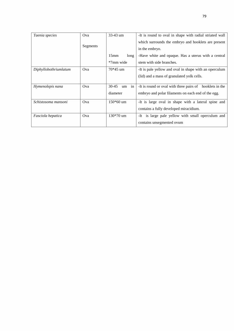
79
Taenia species Ova
Segments
33-43 um
15mm long
*7mm wide
-It is round to oval in shape with radial striated wall
which surrounds the embryo and hooklets are present
in the embryo.
-Have white and opaque. Has a uterus with a central
stem with side branches.
Diphyllobothriumlatum Ova 70*45 um -It is pale yellow and oval in shape with an operculum
(lid) and a mass of granulated yolk cells.
Hymenolepis nana Ova 30-45 um in
diameter
-It is round or oval with three pairs of hooklets in the
embryo and polar filaments on each end of the egg.
Schistosoma mansoni Ova 150*60 um -It is large oval in shape with a lateral spine and
contains a fully developed miracidium.
Fasciola hepatica Ova 130*70 um -It is large pale yellow with small operculum and
contains unsegmented ovum

80
APPENDIX VIII : IREC Approval Letter

81

82