Genomics of the Parasitic Nematode Ascaris and Its Relatives
15
genes G C A T T A C G G C A T Review Genomics of the Parasitic Nematode Ascaris and Its Relatives Jianbin Wang 1,2 Citation: Wang, J. Genomics of the Parasitic Nematode Ascaris and Its Relatives. Genes 2021, 12, 493. https://doi.org/10.3390/ genes12040493 Academic Editor: Jean-Lou Justine Received: 24 February 2021 Accepted: 26 March 2021 Published: 28 March 2021 Publisher’s Note: MDPI stays neutral with regard to jurisdictional claims in published maps and institutional affil- iations. Copyright: © 2021 by the author. Licensee MDPI, Basel, Switzerland. This article is an open access article distributed under the terms and conditions of the Creative Commons Attribution (CC BY) license (https:// creativecommons.org/licenses/by/ 4.0/). 1 Department of Biochemistry and Cellular and Molecular Biology, University of Tennessee, Knoxville, TN 37996, USA; [email protected] 2 UT-Oak Ridge National Laboratory Graduate School of Genome Science and Technology, University of Tennessee, Knoxville, TN 37996, USA Abstract: Nematodes of the genus Ascaris are important parasites of humans and swine, and the phylogenetically related genera (Parascaris, Toxocara, and Baylisascaris) infect mammals of veterinary interest. Over the last decade, considerable genomic resources have been established for Ascaris, including complete germline and somatic genomes, comprehensive mRNA and small RNA transcrip- tomes, as well as genome-wide histone and chromatin data. These datasets provide a major resource for studies on the basic biology of these parasites and the host–parasite relationship. Ascaris and its relatives undergo programmed DNA elimination, a highly regulated process where chromosomes are fragmented and portions of the genome are lost in embryonic cells destined to adopt a somatic fate, whereas the genome remains intact in germ cells. Unlike many model organisms, Ascaris transcription drives early development beginning prior to pronuclear fusion. Studies on Ascaris demonstrated a complex small RNA network even in the absence of a piRNA pathway. Comparative genomics of these ascarids has provided perspectives on nematode sex chromosome evolution, programmed DNA elimination, and host–parasite coevolution. The genomic resources enable comparison of proteins across diverse species, revealing many new potential drug targets that could be used to control these parasitic nematodes. Keywords: Ascaris; parasitic nematode; genome; transcriptome; small RNA; histone modification; chromatin; comparative genomics; chromosome; evolution 1. Introduction Ascaris is a large nematode (roundworm) that lives in the small intestine of its hosts, pigs and humans [1]. It infects ~800 million people, mostly children [2–4]. Ascaris infection, known as ascariasis, contributes significantly to global disability-adjusted life years, per- petuating the cycle of poverty in areas of endemic infection, particularly in parts of Africa, Southeast Asia, and South America [5–7]. Historically, two species of Ascaris that infect hu- mans (Ascaris lumbricoides) or pigs (Ascaris suum) have been described, but cross-infections are observed [8–10] and these two species have little difference in anatomy, physiology, and genome sequences [11]. Thus, for simplicity, Ascaris refers to both species in this review. Ascaris and its relatives have been the subject of many basic scientific studies, leading to the Boveri–Sutton chromosome theory of inheritance [12,13] and insights into programmed DNA elimination (also known as chromatin diminution) [14–16], trans splicing [17–20], neurobiology [21,22], metabolism [23–26], and amoeboid sperm in nematodes [27–30]. Ascaris is well-suited for biochemical and genomic studies due to the size of adults (on average 30 cm for females and 15 cm for males) and the large numbers of eggs produced [1]. This allows easy isolation of large amounts of material from distinct developmental stages, including various germline and somatic tissues. Ascaris is sexually dimorphic, and the male and female reproductive systems are about 1 to 2 m in length when fully extended, enabling dissecting and collecting of the mitotic germline, transition zone, and all stages of meiosis, including mature oocytes and spermatids [1]. For example, one can isolate 20–40 μL of packed spermatids from a single seminal vesicle. In addition, a single female is Genes 2021, 12, 493. https://doi.org/10.3390/genes12040493 https://www.mdpi.com/journal/genes
-
Upload
khangminh22 -
Category
Documents
-
view
2 -
download
0
Transcript of Genomics of the Parasitic Nematode Ascaris and Its Relatives

genesG C A T
T A C G
G C A T
Review
Genomics of the Parasitic Nematode Ascaris and Its Relatives
Jianbin Wang 1,2
�����������������
Citation: Wang, J. Genomics of the
Parasitic Nematode Ascaris and Its
Relatives. Genes 2021, 12, 493.
https://doi.org/10.3390/
genes12040493
Academic Editor: Jean-Lou Justine
Received: 24 February 2021
Accepted: 26 March 2021
Published: 28 March 2021
Publisher’s Note: MDPI stays neutral
with regard to jurisdictional claims in
published maps and institutional affil-
iations.
Copyright: © 2021 by the author.
Licensee MDPI, Basel, Switzerland.
This article is an open access article
distributed under the terms and
conditions of the Creative Commons
Attribution (CC BY) license (https://
creativecommons.org/licenses/by/
4.0/).
1 Department of Biochemistry and Cellular and Molecular Biology, University of Tennessee,Knoxville, TN 37996, USA; [email protected]
2 UT-Oak Ridge National Laboratory Graduate School of Genome Science and Technology,University of Tennessee, Knoxville, TN 37996, USA
Abstract: Nematodes of the genus Ascaris are important parasites of humans and swine, and thephylogenetically related genera (Parascaris, Toxocara, and Baylisascaris) infect mammals of veterinaryinterest. Over the last decade, considerable genomic resources have been established for Ascaris,including complete germline and somatic genomes, comprehensive mRNA and small RNA transcrip-tomes, as well as genome-wide histone and chromatin data. These datasets provide a major resourcefor studies on the basic biology of these parasites and the host–parasite relationship. Ascaris and itsrelatives undergo programmed DNA elimination, a highly regulated process where chromosomes arefragmented and portions of the genome are lost in embryonic cells destined to adopt a somatic fate,whereas the genome remains intact in germ cells. Unlike many model organisms, Ascaris transcriptiondrives early development beginning prior to pronuclear fusion. Studies on Ascaris demonstrateda complex small RNA network even in the absence of a piRNA pathway. Comparative genomicsof these ascarids has provided perspectives on nematode sex chromosome evolution, programmedDNA elimination, and host–parasite coevolution. The genomic resources enable comparison ofproteins across diverse species, revealing many new potential drug targets that could be used tocontrol these parasitic nematodes.
Keywords: Ascaris; parasitic nematode; genome; transcriptome; small RNA; histone modification;chromatin; comparative genomics; chromosome; evolution
1. Introduction
Ascaris is a large nematode (roundworm) that lives in the small intestine of its hosts,pigs and humans [1]. It infects ~800 million people, mostly children [2–4]. Ascaris infection,known as ascariasis, contributes significantly to global disability-adjusted life years, per-petuating the cycle of poverty in areas of endemic infection, particularly in parts of Africa,Southeast Asia, and South America [5–7]. Historically, two species of Ascaris that infect hu-mans (Ascaris lumbricoides) or pigs (Ascaris suum) have been described, but cross-infectionsare observed [8–10] and these two species have little difference in anatomy, physiology, andgenome sequences [11]. Thus, for simplicity, Ascaris refers to both species in this review.Ascaris and its relatives have been the subject of many basic scientific studies, leading to theBoveri–Sutton chromosome theory of inheritance [12,13] and insights into programmedDNA elimination (also known as chromatin diminution) [14–16], trans splicing [17–20],neurobiology [21,22], metabolism [23–26], and amoeboid sperm in nematodes [27–30].
Ascaris is well-suited for biochemical and genomic studies due to the size of adults (onaverage 30 cm for females and 15 cm for males) and the large numbers of eggs produced [1].This allows easy isolation of large amounts of material from distinct developmental stages,including various germline and somatic tissues. Ascaris is sexually dimorphic, and themale and female reproductive systems are about 1 to 2 m in length when fully extended,enabling dissecting and collecting of the mitotic germline, transition zone, and all stagesof meiosis, including mature oocytes and spermatids [1]. For example, one can isolate20–40 µL of packed spermatids from a single seminal vesicle. In addition, a single female is
Genes 2021, 12, 493. https://doi.org/10.3390/genes12040493 https://www.mdpi.com/journal/genes
Genes 2021, 12, 493 2 of 15
estimated to release 0.2 to 1 million eggs per day and the uterus (~20–25 cm in length) canhold up to 27 million eggs [31,32]. The fertilized eggs can be easily isolated. They undergosynchronized development under controlled lab conditions, allowing the collection ofhundreds of millions of embryos at discrete stages of development, as well as larvae, forin vitro biochemical [17,20,33], cellular, molecular, and genomics studies [34–39].
The development of genomics technologies and the decrease of sequencing costs havemade genome sequencing, transcriptome profiling, ChIP-seq, and comparative genomicsfeasible in individual labs. In the last decade, many large-scale genomics studies have beencarried out on Ascaris and its relatives (Table 1). This led to a rich trove of genomic data thathas provided key insights into many biological questions, such as the function and mecha-nism of programmed DNA elimination [35,37–39], the contribution of transcription to earlyanimal development [36,40], and the diversity of small RNAs in nematodes [34,41–45].The studies have also provided genomics resources for identifying potential drug tar-gets [46–51] and worm control [9,11,52–55]. In this review, I summarize the development ofcurrent Ascaris genomic resources, including the complete germline genome and a compar-ison to the somatic genome, transcriptomes and small RNAs, epigenomes, and chromatinaccessibility datasets. I also discuss population genomics, as well as comparative genomicsand chromosome evolution in Ascaris and its related nematodes.
Table 1. Current genome assemblies for Ascaris and its relatives.
Features Ascaris suum Ascarislumbricoides
Parascarisunivalens
Toxocaracanis
Baylisascarisschroederi
Baylisascarisailuri
Toxascarisleonina
Major host pig human horse dog giant panda red panda lionAssembled bases
(Mb) 279 296 253 317 282 267 285
N50 (kb) 12,191 4633 1826 375 889 51 36Scaffold number 109 * 415 1274 22,857 2834 30,943 49,543Largest scaffold
(Mb) 23.1 13.2 5.6 1.9 5.8 0.5 0.4
Protein-coding genes 15,714 17,902 15,027 18,596 13,284 12,252 16,087Accession number JACCHR01 SMSY01 NJFU01 JPKZ01 NA NA NA
Major technologiesused
PacBio,Hi-C
Illumina,PacBio
Illumina,PacBio,
BioNanoIllumina Illumina,
PacBio Illumina Illumina
Reference
Wang et al.2020
Curr. Biol.[39]
Easton et al.2020
eLife [11]
Wang et al.2017
Genome Res.[38]
Zhu et al.2015Nat.
Commun.[49]
Hu et al. 2020 Mol. Biol. Evol. [50]
* With 24 chromosomes, a mitochondrial genome, and 84 unplaced small contigs.
2. History of Ascaris Genome Assemblies
The Ascaris genome was studied extensively before the development of moderngenomic approaches. One particular interest was to determine the amount of sequence lostand identify the eliminated DNA and the sites of DNA breaks during programmed DNAelimination [14,15]. Using biochemistry and chromosome staining, the germline genomewas estimated to be 300 to 800 Mb by different research groups [56–59] and an estimated~20–40% of DNA was suggested to be lost during DNA elimination. Through molecularcloning and sequencing, the nature of eliminated DNA began to be unveiled, with thefirst identified element as an abundant 121-bp tandem repeat [60,61]. Later, other minorrepeats [62] as well as three protein-coding genes [63–65] were found to be eliminated. Itwas also established that the DNA break ends are healed by telomere addition during DNAelimination [66–68].
At the turn of the 21st century, first-generation high-throughput Sanger sequencingwas used to generate many high-quality reference genomes, including those of important

Genes 2021, 12, 493 3 of 15
human parasites [69–77]. However, the cost to generate a de novo genome assemblywas still very expensive. Although being a parasite infecting upwards of 800 millionpeople, Ascaris was not promoted as a priority to be sequenced at large genome sequencingcenters mainly due to the small number of researchers working on Ascaris and its relativelylarge genome size. Indeed, before next-generation sequencing technologies, there wereonly ~1× coverage of Sanger and ~3× coverage of 454 Ascaris genomic reads available.The first significant Ascaris draft genome was generated using the Sanger and 454 readstogether with ~14× Illumina reads [34] (Table S1). The draft genome and the extensivetranscriptomes were mainly used to facilitate small RNA data analysis [34]. Soon after, twoadditional draft assemblies using high coverage of Illumina reads were published (Table S1),one focused on genomics resources for drug discovery [46] and the other comparing thegermline and somatic genomes for programmed DNA elimination [35].
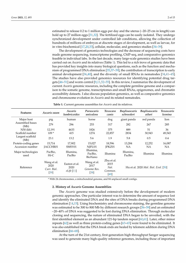
The germline and somatic assemblies were further improved with new technologies,including long reads, optical mapping, and Hi-C, to produce two major recent updates(Table S1) [38,39]. The latest Ascaris germline and somatic genomes represent fully as-sembled chromosomes [39], with 24 germline chromosomes and 36 in somatic cells; allrepetitive sequences were anchored in these assembled chromosomes (Figure 1).
Autosomes
Sex Chromosomes
5 Mb 10 Mb 15 Mb 20 Mb
Eliminated DNADNA Break RegionUnknown RepeatsTelomere
* 121bp Repeats● CENP−A sites (top 2000)
CorrespondingC. elegans Chromosome
IIIIIIIVVX
1 18.8
2 14.2
3 14.1
4 13.9
5 13.4
6 13.2
7 13.1
8 12.2
9 11.5
10 11.0
11 10.3
12 9.8
13 8.7
14 8.5
15 8.3
16 8.1
17 7.3
18 7.1
19 7.1
X1 23.1
X2 12.4
X3 9.9
X4 9.0
X5 7.1
* **
* ** ** *
* ** * *
*
** *
* **** *
**
*
* **
** *
*
●●● ●● ●●●●● ● ● ● ● ● ● ●●●● ●●●● ●●●● ● ●● ●● ● ●● ● ● ● ●● ● ● ●●● ● ●● ●● ● ●● ●● ● ● ● ●● ● ● ●● ● ● ●●●● ● ● ● ●● ● ● ● ● ● ● ●●● ● ● ●● ● ● ● ● ●● ●●● ●●● ● ● ●●● ●●● ● ● ●●● ●● ● ● ● ● ● ● ● ● ●● ● ●●● ●● ●● ●●●
● ●● ●●● ● ● ● ● ●●● ● ● ● ● ● ● ● ● ●● ● ● ● ●● ●● ● ● ●●● ● ● ●● ●●● ●●● ● ●● ● ● ● ●● ● ●● ● ● ●● ● ● ● ●● ●●●● ●● ● ●● ● ●● ● ● ●
●● ● ● ●● ● ● ● ●● ● ● ●●● ● ● ● ● ● ●● ●● ● ● ● ● ● ● ●●● ● ● ● ●● ● ●●●● ● ● ●● ● ●●●●●● ● ● ● ●● ●● ●● ● ●● ● ● ●●● ●● ●
● ● ●● ● ● ● ● ●●● ● ●●● ●●●● ●●●●●● ● ●● ●●● ● ●● ● ● ● ● ● ● ●● ● ●● ● ●● ●●● ●● ●● ●● ● ● ● ● ● ●●● ● ● ●● ● ●
● ● ●●● ● ● ● ●● ● ● ●● ● ● ● ●●● ● ●●● ● ● ● ● ●●●● ● ● ● ●● ●●● ●● ● ●● ● ●● ● ● ● ● ● ●● ●● ●
● ● ●●●●●● ● ●● ● ● ● ●●● ●●● ● ● ● ● ●● ●● ● ●●●●●● ●● ● ●●● ●●●● ●● ● ●● ● ● ● ● ● ● ● ● ● ●●● ●●● ● ● ● ● ●●
● ● ●● ●● ● ● ●● ● ●● ●● ●● ●●● ●●●● ● ● ● ● ● ● ● ●●●● ●● ● ● ● ● ● ●● ● ●●●● ●●● ● ●● ● ● ● ● ●● ●
●●● ● ●● ● ● ● ●● ● ● ● ● ● ●● ●●●●● ●●●● ● ● ● ● ● ● ● ●●● ● ●● ● ●● ●●● ● ● ● ● ● ● ● ●● ●
●● ● ● ●● ●● ● ● ● ●● ●● ●●● ● ● ● ●● ●● ● ● ●● ● ● ● ● ● ●● ● ●●●● ● ● ● ●● ●● ● ●● ● ●
● ● ● ● ●● ● ●● ● ● ● ● ● ● ● ●● ● ● ● ● ● ● ● ● ●●● ● ● ● ● ●●●● ●● ● ● ● ● ● ●
● ● ●●● ● ●●●● ●●● ●● ●● ● ● ● ● ● ● ●●●●● ● ● ● ●●● ●● ● ●● ●● ●● ● ● ● ●● ●●●● ●●
● ● ●● ● ● ● ●● ● ● ● ● ● ● ● ● ● ●●● ● ● ● ●● ●● ● ● ●● ● ● ●● ●● ● ● ● ● ●● ● ● ●● ●● ● ● ● ● ● ● ● ●● ●●● ● ●●●● ● ● ● ● ● ● ● ●● ●● ●● ● ● ● ● ● ●●●● ●
●●●● ● ● ●●● ● ● ●● ●● ● ● ●● ● ● ● ●●●●● ●● ● ●●●●●●● ● ●●● ● ● ●●● ● ● ● ● ● ●●● ● ● ●●● ● ● ● ● ● ● ● ● ●●●● ●● ●●●● ● ● ● ●● ● ●●●● ● ●● ● ●● ●● ● ● ● ●●●●●●● ●●● ●●●●●● ●●●
● ● ●●● ●● ● ●● ● ● ●●●●● ● ●● ● ● ● ● ● ● ● ● ● ● ●●● ● ● ● ●●●● ●●● ● ● ●● ●●● ● ●●●●●●● ● ● ● ● ● ●● ● ●●●● ●● ● ● ● ●●● ●● ●● ● ● ●●● ●●●●● ●●● ● ● ●● ●● ● ● ● ●●
●● ●● ●● ●●● ● ● ● ● ● ● ● ● ●● ● ●● ● ●●● ● ●●● ● ●●●● ● ●● ● ● ● ● ● ● ● ● ●●●●●● ●●● ● ● ● ●●●● ● ● ● ● ●●● ● ● ● ● ● ●●●● ● ●●● ● ● ● ● ●●● ● ●● ●●● ●●● ● ● ● ●● ●●●●
● ●● ● ● ● ● ● ● ● ● ●● ● ● ● ●●● ●● ● ● ● ●● ●● ● ●●● ●●● ● ● ● ●● ●●●●● ● ● ●● ● ● ●●● ● ● ● ●●● ●●●● ● ●● ●●● ●●●● ● ● ● ● ● ● ● ● ● ● ● ● ●● ● ● ● ● ● ●
●● ●● ● ● ● ● ● ● ●● ●● ●● ● ● ● ● ● ● ● ● ●●●● ● ●● ● ● ● ● ●● ● ● ● ●● ●●● ● ● ●●● ● ● ● ● ● ● ● ●● ● ●● ● ● ● ● ● ● ● ● ● ● ● ● ● ● ● ● ● ●●●●● ● ●●● ● ●●
● ● ● ● ● ●● ● ● ●●●● ●● ● ● ● ● ● ● ● ●● ● ● ● ● ● ●● ●● ●●●●●● ● ● ● ● ●●● ●● ●● ● ● ●● ●● ●●●●●● ●● ● ● ● ● ● ● ● ● ●●● ● ● ●● ● ● ●● ●●
●● ●●● ●●●● ● ●●● ● ● ● ● ● ● ● ●● ● ●●● ●● ● ● ●●●● ● ● ● ● ●●●● ● ● ● ● ● ● ● ● ●● ● ● ● ● ●●● ● ● ● ● ● ● ● ● ● ● ● ● ● ● ●● ●●●●● ●
● ●● ● ● ●● ● ● ●● ● ● ●● ● ● ● ● ●● ● ●●● ● ●●●● ●●●● ●● ● ● ●● ● ● ● ● ● ●● ● ● ● ●● ● ● ●●● ●● ●● ●●● ●● ● ● ●● ● ●● ● ●● ● ● ● ●● ● ●●● ● ●● ●● ● ●● ● ●● ●● ● ● ●● ● ● ●● ● ● ● ● ●● ● ● ● ●● ● ● ● ● ● ● ● ● ● ● ●● ● ●● ● ●● ●●●● ● ● ●● ● ● ●●●●●● ● ● ●●●● ● ● ●●●●
● ● ● ● ● ● ● ●● ●●● ● ● ● ● ● ●●● ● ● ● ● ● ● ●●●●● ●● ● ● ● ● ● ● ●● ● ● ●● ●● ●● ● ● ● ● ● ● ● ● ● ● ● ● ●● ●● ●● ● ● ● ●● ●●● ● ●●
●● ●● ● ● ● ● ● ● ● ● ● ● ●●●● ● ● ● ●● ●● ● ●●● ●●● ● ●● ● ●●●● ● ● ● ●● ●● ● ● ●● ● ●●● ● ● ●●●●●●● ●●● ● ● ●● ●● ●● ●
● ● ●●●● ●●●●●●●● ● ● ● ● ● ●●● ● ●●● ● ●●●●●● ● ●●● ● ● ● ● ●●● ● ● ● ● ● ● ● ● ● ●● ●●● ●● ●●●●●●
●● ● ● ● ● ● ● ●●● ●●●● ● ● ● ● ●● ● ● ● ●● ● ●●●●●● ●●●● ● ● ● ● ● ● ● ● ● ● ●
Figure 1. Ascaris genome, programmed DNA elimination, and chromosome evolution. Germlinechromosomes are illustrated with the length of the chromosome in Mb on the right. Chromosomeregions that are eliminated are colored in red. The retained regions, which will become somaticchromosomes after DNA elimination, are colored based on their corresponding C. elegans chromo-somes (see legends). CENP-A deposition (data from 12 developmental stages [37]) are illustrated bygreen dots underneath the chromosomes, illustrating possible centromeric regions for the holocentricchromosomes; shown are the top 2000 (out of 3342) CENP-A enriched sites.
Along with these genomes, extensive transcriptomes, small RNA datasets, andepigenomes have been produced that provide comprehensive genomic data for Ascaris. In

Genes 2021, 12, 493 4 of 15
addition, a comparison of Ascaris genomes from humans and pigs revealed that the twogenomes are very similar, with extensive heterozygosity, existing as genetic mosaics, andthus reflecting the highly interbred nature of Ascaris species [11].
3. Ascaris Genomes
One of the most striking features of Ascaris is that the adult worms have two genomes:an intact germline genome and a reduced somatic genome. This reduced somatic genomeis the result of a process called programmed DNA elimination, where parts of the germlinegenome are lost during the differentiation of germ cells into somatic cells in early embryo-genesis [14–16]. This process occurs in five independent cell lineages between the 4–16 cellstages of development and is achieved through double-stranded DNA breaks in chro-mosomes followed by selective loss of portions of the chromosome fragments [35,37–39].Comparison of the germline and somatic genomes suggest that there are 72 chromosomalbreak regions (CBRs), with 48 at the chromosome ends that removes all subtelomericand telomeric sequences to form the somatic genome (Figure 1). The other 24 CBRs arein the middle of the chromosomes that contribute to an increased number of somaticchromosomes (Figure 1).
About 55 Mb (~18%) of the Ascaris germline DNA is eliminated in somatic cells duringprogrammed DNA elimination. This includes ~30 Mb of a 121-bp tandem repeat and~5 Mb of subtelomeric and telomere repeats. Strikingly, the other 20 Mb of eliminated DNAis unique and encodes ~1000 germline-expressed genes [35,38,39]. The majority of theseeliminated genes are specifically expressed in the testes and therefore are likely involved inAscaris spermatogenesis. The data suggests that DNA elimination is an alternative way ofsilencing these germline genes in the somatic cells; rather than repressing their expression,they simply remove them from the genome as a permanent way of gene silencing. Thesequences lost and the break regions are the same in the five independent eliminationevents, in individual males and females, and in worms from pigs and humans [35,38,39].This programmed and high-fidelity removal of DNA during early development suggestsspecific and regulated mechanisms are involved in Ascaris DNA elimination.
Another interesting feature of the Ascaris genome is that it has multiple sex chromo-somes, with five in the germline and nine in the somatic cells. This is in contrast withmost nematodes (including the model C. elegans) that have only one sex chromosome.Comparison of nematode chromosomes using orthologous proteins (reciprocal best blasthits) indicates that many of the Ascaris germline sex chromosomes are likely derived fromrecent chromosome fusion events (Figure 1) [39]. This is further supported by chromosome“painting” analyses using conserved Nigon elements [78]. These comparative analyses ofnematode chromosomes revealed that while extensive intra-chromosomal rearrangementsoccurred, the karyotypic unit for nematodes remains largely intact except for occasionalscission and fusion events [78]. Interestingly, the fused Ascaris germline sex chromosomesare broken at the sites of these fusions during DNA elimination, resulting in the pre-fusionkaryotypes in somatic cells (Figure 1) [39]. In addition, Hi-C data suggests that the sexchromosomes in the testis have greater interactions with each other than with the auto-somes [39]. This high level of interaction among the sex chromosomes suggests that theyare likely physically close to each other during meiosis, reminiscent of the chromosomebehavior during meiosis in other systems with multiple sex chromosomes [79–82]. Morechromosomal assemblies from ascarids and other nematodes are needed to further studythe evolution and interactions of nematode chromosomes.
4. Ascaris Genes and Transcriptomes
The predicted number of genes for Ascaris genome varies from 15,000 to 19,000 de-pending on the genome assembly and transcriptome data (see Table S1). The differences area consequence of completeness of the genome assemblies and the transcriptome datasetsused for gene identification. Early draft genomes tend to predict higher number of genesdue to split gene fragments and the presence of multiple uncollapsed contigs. More com-
Genes 2021, 12, 493 5 of 15
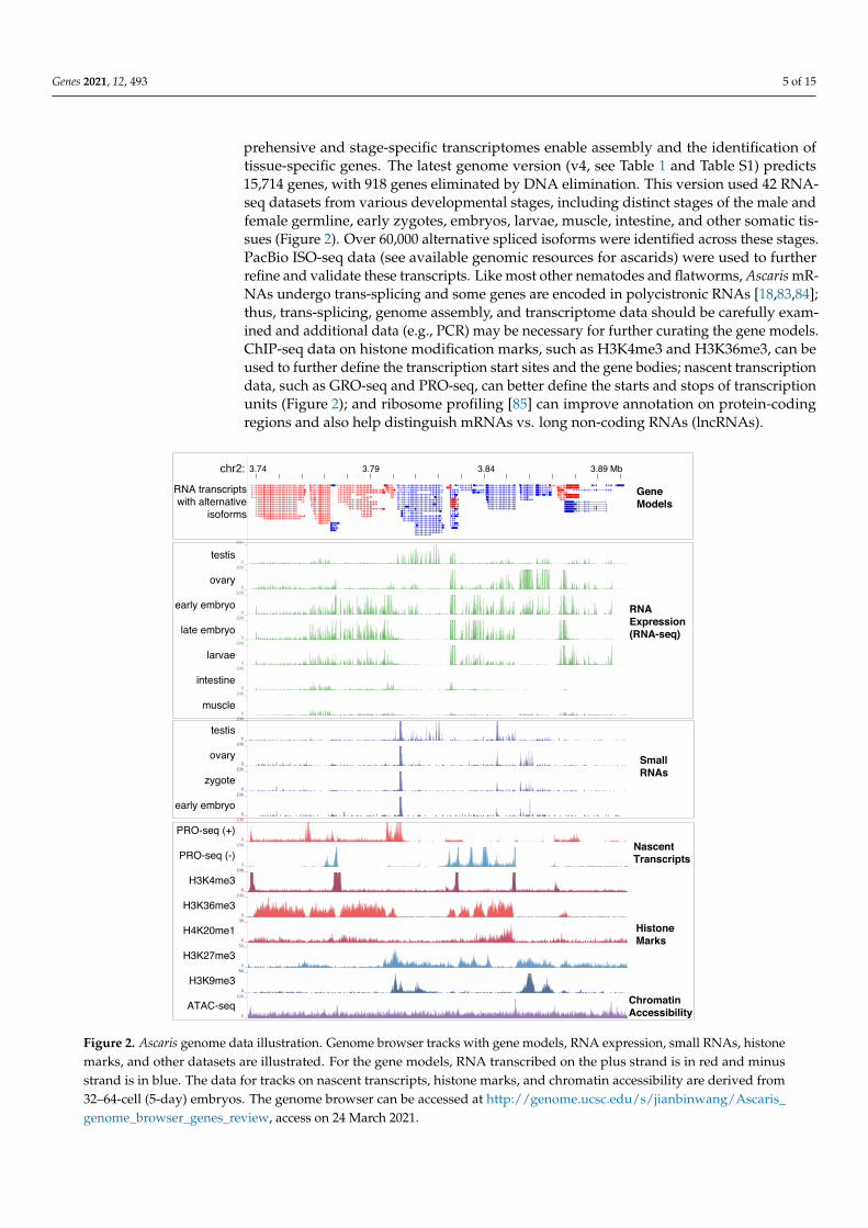
prehensive and stage-specific transcriptomes enable assembly and the identification oftissue-specific genes. The latest genome version (v4, see Table 1 and Table S1) predicts15,714 genes, with 918 genes eliminated by DNA elimination. This version used 42 RNA-seq datasets from various developmental stages, including distinct stages of the male andfemale germline, early zygotes, embryos, larvae, muscle, intestine, and other somatic tis-sues (Figure 2). Over 60,000 alternative spliced isoforms were identified across these stages.PacBio ISO-seq data (see available genomic resources for ascarids) were used to furtherrefine and validate these transcripts. Like most other nematodes and flatworms, Ascaris mR-NAs undergo trans-splicing and some genes are encoded in polycistronic RNAs [18,83,84];thus, trans-splicing, genome assembly, and transcriptome data should be carefully exam-ined and additional data (e.g., PCR) may be necessary for further curating the gene models.ChIP-seq data on histone modification marks, such as H3K4me3 and H3K36me3, can beused to further define the transcription start sites and the gene bodies; nascent transcriptiondata, such as GRO-seq and PRO-seq, can better define the starts and stops of transcriptionunits (Figure 2); and ribosome profiling [85] can improve annotation on protein-codingregions and also help distinguish mRNAs vs. long non-coding RNAs (lncRNAs).
chr2: 3.74 3.79 3.84 3.89 Mb
200 _
0 _200 _
0 _200 _
0 _200 _
0 _200 _
0 _200 _
0 _200 _
0 _100 _
0 _100 _
0 _100 _
0 _100 _
0 _100 _
0 _100 _
0 _100 _
0 _100 _
0 _30 _
0 _50 _
0 _50 _
0 _100 _
0 _
testis
ovary
early embryo
late embryo
larvae
intestine
muscle
testis
ovary
zygote
early embryo
PRO-seq (+)
PRO-seq (-)
H3K4me3
H3K36me3
H4K20me1
H3K27me3
H3K9me3
ATAC-seq
RNA transcriptswith alternative
isoforms
GeneModels
RNAExpression(RNA-seq)
SmallRNAs
Nascent Transcripts
HistoneMarks
ChromatinAccessibility
Figure 2. Ascaris genome data illustration. Genome browser tracks with gene models, RNA expression, small RNAs, histonemarks, and other datasets are illustrated. For the gene models, RNA transcribed on the plus strand is in red and minusstrand is in blue. The data for tracks on nascent transcripts, histone marks, and chromatin accessibility are derived from32–64-cell (5-day) embryos. The genome browser can be accessed at http://genome.ucsc.edu/s/jianbinwang/Ascaris_genome_browser_genes_review, access on 24 March 2021.
Genes 2021, 12, 493 6 of 15
RNA-seq through Ascaris early development has provided some novel insights intoearly gene expression and gene regulation during embryogenesis [36,40]. In most modelorganisms, the maternal to zygotic transition—the turnover of maternally contributedmRNAs and the activation of zygotic transcription—does not start until ≥2 cell numberduring embryogenesis [86–88]. However, RNA-seq data from Ascaris oocytes, sperms,zygote maturation, and early embryogenesis revealed ~4000 genes are transcribed priorto pronuclear fusion and in the 1–4 cell embryos [36]. Transcription prior to pronuclearfusion is in drastic contrast to most model organisms. The free-living nematode C. eleganshas almost identical cell lineages and morphological patterns during early development.While major transcription is not active until late gastrulation and organogenesis (~100 cells)in C. elegans, many orthologous C. elegans maternally contributed RNAs are transcribedin Ascaris zygotes and early embryos [36]. Due to its rapid embryonic development,C. elegans largely relies on maternal regulators to drive early zygotic gene expressionand embryogenesis. In contrast, Ascaris transcribes new RNAs as needed during earlydevelopment. Thus, gene regulation in early development is largely transcriptional inAscaris, whereas it is mainly regulated post-transcriptionally in C. elegans. The difference inthe gene regulation programs is likely due to the differences in cell cycle length during earlydevelopment. The extended maturation of zygotes prior to pronuclear fusion and longearly cell cycles of Ascaris (15–20 h) provides ample time for the transcriptional machineryto make new RNAs [36,40]. Additional transcriptome analyses of early development indifferent nematodes are likely to yield insights into gene regulation in early development.
RNA-seq analyses from Ascaris intestines have facilitated the understanding of thenematode intestine and efforts to develop the intestine as a potential target for new drugtherapies [45,89–91]. Multiomics across diverse nematode species, including mRNAs andmiRNAs from Ascaris, were used to define and investigate the biology of the intestine. Thelarge size of adult Ascaris allows easy dissection of anterior (close to head), middle, andposterior regions of the intestine, enabling the identification of differentially expressedtranscripts in these regions. Analysis of the intestinal transcriptome shows many uniqueand highly expressed transcripts are present in the anterior region [45]. Together withproteomics [92] and the development of Ascaris intestinal cannulation and a perfusionmodel [93], an experimental system was established for testing and validation of drugcandidates. The Ascaris intestine model represents an important system for developingnovel anthelmintic drugs against nematode intestinal cells [91,94].
5. Ascaris Small RNAs
In addition to the protein coding mRNAs, transcriptomes also contain structural andregulatory RNAs, such as rRNAs, tRNAs, lncRNAs, as well as various types of small RNAs.The nematode C. elegans has three major classes of known small RNAs: miRNAs, siRNAs(22G-RNAs and 26G-RNAs) and piRNAs (21U-RNAs) [95,96]. These small RNAs interactwith the genome, transcripts, or foreign sequences to silence or regulate their targets. Inan early study, ~100 high-confidence Ascaris miRNAs as well as numerous siRNAs wereidentified [34]. Most of the siRNAs are 22G-RNAs with 5’-triphosphates that target repeatsand/or mRNAs that are dynamically expressed throughout germline and embryonicdevelopment. Ascaris also has 26G-RNAs with 5’-monophosphates that are specific tothe testis and only target testis-specific genes [34]. Surprisingly, piRNAs, Piwi-cladeArgonautes, and other proteins associated with the piRNA pathway are lost in Ascaris [34].This loss of the piRNA pathway was later found to be common in other nematodes [44].Initial studies found no evidence to suggest that small RNAs may contribute to DNAelimination [34]. However, this could be due to the large number and complexity of totalAscaris small RNAs present and thus the inability to identify unique small RNAs associatedwith DNA elimination.
Small RNAs are associated with Argonaute proteins. While C. elegans has an expandedArgonaute family of 25 proteins [97], Ascaris has a reduced number of 10 Argonautes [34].Most studies on Ascaris small RNAs have focused on miRNAs [41,42,45,52]. However, the
Genes 2021, 12, 493 7 of 15
predominant Ascaris small RNAs are endogenous siRNAs, estimated to be over 90% ofall small RNAs in Ascaris and other parasitic nematodes. The role of these endogenoussmall RNAs in nematodes needs to be explored. In the parasitic nematode Heligmosomoides,an Argonaute and siRNAs were found in secreted extracellular vesicles that may play arole in host–parasite interactions [98,99]. Outstanding questions related to small RNAs inAscaris and other nematodes include: (1) What specific small RNAs are associated witheach Argonaute? (2) What are the biological roles of each Argonaute and its associatedsmall RNAs? (3) Do any particular Argonautes and their associated small RNAs play a rolein DNA elimination? (4) How do small RNA contribute to host–parasite interactions andtransgenerational epigenetic inheritance? And (5) how do Ascaris and other nematodescompensate for the loss of piRNAs?
6. Ascaris Chromatin and Epigenome
In eukaryotes, a common mechanism of gene regulation is through the differentialorganization of nucleosomes and chromatin, forming heterochromatin or euchromatin.Chromatin status can be associated with histone modification marks that activate or repressspecific genomic regions [100]. In Ascaris, antibodies against many common histonemodifications have been used in immunohistochemistry, electron microscopy, and ChIP-seq [37–39]. ChIP-seq data are available for common modifications, such as the activemarks H3K4me3, H3K36me3, and H4K20me1, as well as the repressive marks H3K27me3and H3K9me3 in a few developmental stages (Figure 2). These data provide initial insightsinto the Ascaris epigenome. They can also be used to further curate gene annotationand facilitate gene expression and other genomic analysis. Future data on the changesof histone marks through Ascaris development may also help elucidate the nature ofepigenetic inheritance during gametogenesis, fertilization, and early embryogenesis in thisparasitic nematode.
The accessibility of Ascaris chromatin during early development was also evaluatedusing ATAC-seq [38]. ATAC-seq uses a Tn5 transposon to insert into regions of the genomethat are more open, thus providing a measurement of DNA accessibility [101]. The sitesof Ascaris chromatin accessibility are enriched at the promoter regions of active genes,where H3K4me3 is also enriched (Figure 2). Interestingly, Ascaris chromosomal breakregions (CBRs) for DNA elimination become more accessible just prior to DNA elimination(4-cell stage at 60 hour) compared with earlier embryo stages (0 hour) and germline tissues,with the open regions matching where new telomeres are added to the broken DNAends [38,39]. This more accessible chromatin in the CBRs might be due to a reduced numberor compactness of nucleosomes or other epigenetic changes. It is plausible that factors,such as DNA replication stress, RNA transcription, R-loops, and/or the 3-D organizationof the chromosomes, might lead to the more open chromatin at these CBRs. These factorsand processes might also recruit the telomere addition machinery to the regions, furtheropening the CBRs in the subsequent cell cycles after elimination [38,39].
An epigenetic mark for centromere deposition is the variant of histone H3 calledCENP-A [102–104]. Unlike its hosts (pig and human) that have monocentric chromosomeswith one centromere per chromosome, Ascaris has holocentric chromosomes with manycentromeres distributed along the length of the chromosomes. This is consistent withobservations in the model nematode C. elegans [105,106]. ChIP-seq using specific anti-bodies against Ascaris CENP-A (and CENP-C) identified many enriched loci with thesemarks across the length of the chromosomes (Figure 1). Deposition of CENP-A is notassociated with repetitive or any specific sequences, is inversely related with transcription,and the pattern of deposition changes through gametogenesis and embryogenesis [37].Interestingly, Ascaris CENP-A is enriched in all regions in both male and female germline,but its level is reduced prior to and during a DNA elimination mitosis in the eliminatedregions. The loss of CENP-A and thus functional centromeres in the eliminated regionsprovides a mechanism for the selective loss of sequences after the DNA breaks during DNAelimination [37]. However, questions on what identifies the CENP-A deposition sites and

Genes 2021, 12, 493 8 of 15
how much CENP-A is required in nematodes to form a functional microtubule attachmentsite remain to be determined.
7. Population Genomics of Ascaris from Pigs and Humans
Ascaris has historically been considered to have two main species, distinguished bytheir host: A. suum from pigs and A. lumbricoides from humans. They are, however, knownto cross-infect both hosts [8–10] and are capable of interbreeding [107,108]. Populationgenetic studies generally agree that the genetic differences in Ascaris collected from aroundthe world are caused by geographic reproductive isolation [9,109]. Data from mitochondrialsequence analyses suggest there are human-associated and pig-associated clades in As-caris [110,111] or multiple haplotype clusters [112]. A recent study using a reference-qualityA. lumbricoides genome and 68 worms from human hosts in Kenyan villages identified over11 million SNPs [11]. Comparative phylogenomic analyses of these SNPs indicates thatthese worm genomes had extensive heterozygosity with genetic mosaics, suggesting ahighly interbred Ascaris species genetic complex. Analysis of the complete mitochondrialgenomes from these 68 individual worms and other sequences available also supports theidea that worms from pigs and humans form a genetic complex that is capable of inter-breeding [11]. This large-scale phylogenomic study on both the nuclear and mitochondrialgenomes in Ascaris suggests recent and multiple cross-infection events occurred in Ascarispopulations, likely caused by the immigration of humans and livestock. As Ascaris caninfect both pigs and humans, a one-health approach is critical to control the spread ofhuman ascariasis.
8. Comparative Genomics and Ascarids Evolution
Large parasitic nematodes like Ascaris are known as ascarids. A variety of ascaridsare known to infect essentially all large mammals. Common genera include Ascaris (frompigs, sheep, monkeys, apes, and humans), Parascaris (from horses, donkeys, and zebras),Toxocara (from dogs, cats, tigers, and lions), Baylisascaris (from raccoons, pandas, bears,badgers, and marmots), and the more distantly related marine ascarids Anisakis (fish andmarine mammals). Most of these parasites have relatively large (~300 Mb) genomes andchromosome number (>20) (Table 1), except for some species of Parascaris, one of which hasa single chromosome and a huge 2.5-Gb genome (P. univalens) and another one with a muchsmaller genome (~130 Mb for Anisakis simplex). Adult ascarid infections in general causemild symptoms in their hosts. However, heavy infections can lead to intestinal obstructionand sometimes death. Fatality is more common in horses and pandas.
A number of other ascarids genomes have recently been sequenced (Table 1). TheToxocara canis genome from dogs was used to identify genes for potential new drug targetsand to provide a genomic resource for future molecular studies [49]. An improved versionof this genome was generated using additional reads from the 50 helminth genomesinitiative at the Sanger Institute that enabled characterization of DNA elimination inToxocara [38]. The germline and somatic genomes from the horse parasite Parascaris werealso sequenced (Table 1). Comparative analysis of Ascaris, Parascaris, and Toxocara genomesreveals that about 1000 to 2000 germline-expressed genes are eliminated, with 35% of theseeliminated genes conserved among the genera that are preferentially expressed duringspermatogenesis [38]. These data support the idea that DNA elimination is a way to silencethese germline genes in the somatic cells. Testes are known to be the birthplace for manynew genes [113–116]. In these ascarids, the removal and thus permanent silencing of thetestis genes in the somatic cells may enable large sampling of new genes for reproduction,providing a mechanism for more rapid evolutionary changes in the testis [66] while theirelimination in the soma prevents deleterious effects in other tissues. Additional ascaridgenomes and transcriptomes will likely reveal new insights into the evolution of testis-specific genes and programmed DNA elimination. Recently, an analysis of the initialdraft genome of the marine ascarids A. simplex infecting herring provided informationon carbohydrate metabolism genes in the parasite’s development [117]. Future improved

Genes 2021, 12, 493 9 of 15
genomes and omic studies promise to shed lights on the genomics and molecular processesof these important marine parasites (see review [118]).
More recently, genomic data from ascarids and their hosts were also used to elucidatethe coevolution between host and parasites [50]. Three ascarids from the giant panda,the red panda, and the lion were sequenced (Table 1) and used to construct a genome-wide phylogenetic tree. The topology of the tree for these ascarids based on the genome-wide data was not consistent with their host phylogeny, indicating that these parasiteshave not phylogenetically coevolved with their hosts [50]. However, analysis of thehost–parasite protein–protein interactions (PPIs) reveals that seven of the PPIs, includingthe host neutrophil matrix metalloproteinase-8 (MMP8) and parasite lysosomal cysteineprotease cathepsin Z (CTSZ), the host insulin-like growth factor-binding protein 7 (IGFBP7)and parasite prolyl 4-hydroxylase subunit beta (P4HB), and the host acute-phase protein(CRP) and parasite secreted member of the phospholipase A2 (PLA2G1B), had consistentphylogenetic topology [50]. These coevolutionary PPIs are involved in immune regulationand thus they may be relevant to the immune response during the antagonistic coevolutionbetween ascarids and their hosts.
9. Future Perspectives
Over the last decade, the research community has established high-quality genomes,comprehensive transcriptomes, small RNA data, as well as other epigenomic datasets forAscaris. Many related ascarids have also been sequenced and compared (Table 1). Thesegenomic resources provide a starting point for future research in ascarids genomics andbiology. I predict three major areas that will continue to grow and benefit from these andfuture genomic studies.
First, the sequencing of new species and continuing improvement of the genomeassemblies [119] will be enabled by technologies, such as long reads (PacBio and Nanopore)and chromosome conformation capture (Hi-C), as well as genome annotation using longreads (ISO-seq), nascent transcription (PRO-seq), ribosome profiling, and epigenomics(ChIP-seq) data. The high-quality data will enable in-depth and chromosomal viewsof many biological processes, as seen in the improved Ascaris genomes and transcrip-tomes [34–39,46]. Research areas that have not been extensively studied in ascarids includealternative splicing [26], lncRNAs, epigenomes, regulation of tissue-specific transcripts, therepertoire and role of repeats, and more. New emerging technologies, such as single-cellgenomics [120–122] and in situ sequencing for RNAs [123] and the genome [124], mayalso be used in the future to study the high-resolution, heterogeneity, and spatial orga-nizations of DNA and RNA in the cells. Continuing the development of computationaltools, genome browsers, and databases specifically designed for nematodes, such as Worm-Base ParaSite [125], will facilitate data mining and discovery for biologists working onthese parasites.
Second, the resources will help researchers interested in various areas of nematodebasic biology to carry out extensive genomic and molecular studies. For example, onecan now characterize the nature and timing of the DNA breaks for Ascaris DNA elimina-tion using these genomic resources by methods, such as END-seq [126]. The completedgenome also enables the study of DNA replication timing, nascent transcriptional land-scape, R-loops [127,128], and the 3-D genome organization that may be involved in theDNA break sites recognition and/or break generation as well as many other processes. Inaddition, histone modification profiles using ChIP-seq would enable the quantificationof epigenetic changes throughout spermatogenesis, oogenesis, fertilization, zygote devel-opment, embryogenesis, and larvae development. Finally, comparative genomics usingmore completed chromosomal assemblies will delineate the evolution of nematode sexchromosomes [78] as well as the evolutionary developmental biology of embryogenesisand other developmental processes across nematode species.
Third, Ascaris and other ascarids are parasites that cause important zoonotic diseases.The genomic resources will enable future genome-scale studies on the epidemiology,

Genes 2021, 12, 493 10 of 15
population genomics, host–parasite interactions, metagenomics, and immune responseof the diseases. High-quality genomes have now been used for extensive populationgenetics, linkage mapping, and genome-wide quantitative trait loci (QTLs) to examinedisease transmissions, drug resistance, and mining for new therapeutic targets in parasiticworms (see recent reviews [129–132]). There is increasing evidence for drug resistancein parasitic nematodes, including ascarids [133,134]. Ascaris and other intestinal wormcontrol programs are currently based on recurrent drug treatment [135–137] known to leadto drug resistance in nematodes, such as Parascaris. Thus, there is an urgency to identifyand characterize new drug targets in parasitic nematodes. Future large-scale efforts usingpopulation and comparative genomics of major parasitic worms, such as the ones led bythe International Helminth Genomes Consortium [47], are likely to identify new potentialdrug targets and screen compounds to combat parasitic worm infections.
Supplementary Materials: The following are available online at https://www.mdpi.com/article/10.3390/genes12040493/s1, Table S1: History of Ascaris genome assemblies.
Funding: Work in the Wang lab is supported by NIH grant AI155588 and the University of TennesseeKnoxville Startup Funds.
Institutional Review Board Statement: Not applicable.
Informed Consent Statement: Not applicable.
Data Availability Statement: Genomics and transcriptomes data for most ascarids are available inWormBase ParaSite (https://parasite.wormbase.org/species.html) (accessed on 27 March 2021) [125].The NCBI accession numbers for the genome assemblies are available in Table 1 and Table S1. TheAscaris genome is also available in UCSC Genome Browser track data hubs [138] that can be accesswith this link: http://genome.ucsc.edu/s/jianbinwang/Ascaris_genome_browser_genes_review(accessed on 27 March 2021). The genome, transcripts and proteomes datasets for various ascaridsare also available in https://dnaelimination.utk.edu/protocols-data/ (accessed on 27 March 2021).
Acknowledgments: I thank the research community for contributing and sharing genomic datasetson Ascaris and other parasitic nematodes. I also thank the members of the Davis and Wang labs forcritical reading and helpful discussions.
Conflicts of Interest: The authors declare no conflict of interest.
References1. Wang, J.; Davis, R.E. Ascaris. Curr. Biol. 2020, 30, R423–R425. [CrossRef] [PubMed]2. Bethony, J.; Brooker, S.; Albonico, M.; Geiger, S.M.; Loukas, A.; Diemert, D.; Hotez, P.J. Soil-transmitted helminth infections:
Ascariasis, trichuriasis, and hookworm. Lancet 2006, 367, 1521–1532. [CrossRef]3. Hotez, P.J. The Unholy Trinity: The soil-transmitted helminth infections ascariasis, trichuriasis, and hookworm infection. Forgot.
People Forgot. Dis. ASM Press 2013, 17–40. [CrossRef]4. Jourdan, P.M.; Lamberton, P.H.L.; Fenwick, A.; Addiss, D.G. Soil-transmitted helminth infections. Lancet 2018, 391, 252–265.
[CrossRef]5. Hotez, P.J.; Brindley, P.J.; Bethony, J.M.; King, C.H.; Pearce, E.J.; Jacobson, J. Helminth infections: The great neglected tropical
diseases. J. Clin. Investig. 2008, 118, 1311–1321. [CrossRef] [PubMed]6. Brooker, S. Estimating the global distribution and disease burden of intestinal nematode infections: Adding up the numbers-a
review. Int. J. Parasitol. 2010, 40, 1137–1144. [CrossRef]7. Pullan, R.L.; Smith, J.L.; Jasrasaria, R.; Brooker, S.J. Global numbers of infection and disease burden of soil transmitted helminth
infections in 2010. Parasit. Vectors 2014, 7, 37. [CrossRef]8. Sadaow, L.; Sanpool, O.; Phosuk, I.; Rodpai, R.; Thanchomnang, T.; Wijit, A.; Anamnart, W.; Laymanivong, S.; Aung, W.P.P.;
Janwan, P.; et al. Molecular identification of Ascaris lumbricoides and Ascaris suum recovered from humans and pigs in Thailand,Lao PDR, and Myanmar. Parasitol. Res. 2018, 117, 2427–2436. [CrossRef]
9. Betson, M.; Nejsum, P.; Bendall, R.P.; Deb, R.M.; Stothard, J.R. Molecular epidemiology of ascariasis: A global perspective on thetransmission dynamics of Ascaris in people and pigs. J. Infect. Dis. 2014, 210, 932–941. [CrossRef]
10. Miller, L.A.; Colby, K.; Manning, S.E.; Hoenig, D.; McEvoy, E.; Montgomery, S.; Mathison, B.; de Almeida, M.; Bishop, H.; Dasilva,A.; et al. Ascariasis in humans and pigs on small-scale farms, Maine, USA, 2010–2013. Emerg. Infect. Dis. 2015, 21, 332–334.[CrossRef]
Genes 2021, 12, 493 11 of 15
11. Easton, A.; Gao, S.; Lawton, S.P.; Bennuru, S.; Khan, A.; Dahlstrom, E.; Oliveira, R.G.; Kepha, S.; Porcella, S.F.; Webster, J.; et al.Molecular evidence of hybridization between pig and human Ascaris indicates an interbred species complex infecting humans.Elife 2020, 9. [CrossRef]
12. Satzinger, H. Theodor and Marcella Boveri: Chromosomes and cytoplasm in heredity and development. Nat. Rev. Genet. 2008, 9,231–238. [CrossRef] [PubMed]
13. Maderspacher, F. Theodor Boveri and the natural experiment. Curr. Biol. CB 2008, 18, R279–R286. [CrossRef]14. Muller, F.; Tobler, H. Chromatin diminution in the parasitic nematodes Ascaris suum and parascaris univalens. Int. J. Parasitol.
2000, 30, 391–399. [CrossRef]15. Wang, J.; Davis, R.E. Programmed DNA elimination in multicellular organisms. Curr. Opin. Genet. Dev. 2014, 27, 26–34. [CrossRef]16. Streit, A.; Wang, J.; Kang, Y.; Davis, R.E. Gene silencing and sex determination by programmed DNA elimination in parasitic
nematodes. Curr. Opin. Microbiol. 2016, 32, 120–127. [CrossRef]17. Hannon, G.J.; Maroney, P.A.; Denker, J.A.; Nilsen, T.W. Trans splicing of nematode pre-messenger RNA in vitro. Cell 1990, 61,
1247–1255. [CrossRef]18. Davis, R.E. Spliced leader RNA trans-splicing in metazoa. Parasitol. Today 1996, 12, 33–40. [CrossRef]19. Lall, S.; Friedman, C.C.; Jankowska-Anyszka, M.; Stepinski, J.; Darzynkiewicz, E.; Davis, R.E. Contribution of trans-splicing, 5′
-leader length, cap-poly(A) synergism, and initiation factors to nematode translation in an Ascaris suum embryo cell-free system. J.Biol. Chem. 2004, 279, 45573–45585. [CrossRef]
20. Cohen, L.S.; Mikhli, C.; Friedman, C.; Jankowska-Anyszka, M.; Stepinski, J.; Darzynkiewicz, E.; Davis, R.E. Nematode m7GpppGand m3(2,2,7)GpppG decapping: Activities in Ascaris embryos and characterization of C. elegans scavenger DcpS. RNA 2004, 10,1609–1624. [CrossRef]
21. Davis, R.E.; Stretton, A.O. The motornervous system of Ascaris: Electrophysiology and anatomy of the neurons and their controlby neuromodulators. Parasitology 1996, 113, S97–S117. [CrossRef] [PubMed]
22. Stretton, A.O.; Maule, A.G. The Neurobiology of Ascaris and Other Parasitic Nematodes. Ascaris Negl. Parasite 2013, 127–152.[CrossRef]
23. Rathbone, L. Oxidative metabolism in Ascaris lumbricoides from the pig. Biochem. J. 1955, 61, 574–579. [CrossRef] [PubMed]24. Beis, I.; Barrett, J. Energy metabolism in developing Ascaris lumbricoides eggs. II, The steady state content of intermediary
metabolites. Dev. Biol. 1975, 42, 188–195. [CrossRef]25. Barrett, J.; Beis, I. Energy metabolism in developing Ascaris lumbricoides eggs. I. The glycolytic enzymes. Dev. Biol. 1975, 42,
181–187. [CrossRef]26. Tan, J.H.; Lautens, M.; Romanelli-Cedrez, L.; Wang, J.; Schertzberg, M.R.; Reinl, S.R.; Davis, R.E.; Shepherd, J.N.; Fraser, A.G.;
Salinas, G. Alternative splicing of coq-2 controls the levels of rhodoquinone in animals. Elife 2020, 9. [CrossRef] [PubMed]27. Theriot, J.A. Worm sperm and advances in cell locomotion. Cell 1996, 84, 1–4. [CrossRef]28. Italiano, J.E., Jr.; Roberts, T.M.; Stewart, M.; Fontana, C.A. Reconstitution in vitro of the motile apparatus from the amoeboid
sperm of Ascaris shows that filament assembly and bundling move membranes. Cell 1996, 84, 105–114. [CrossRef]29. Bottino, D.; Mogilner, A.; Roberts, T.; Stewart, M.; Oster, G. How nematode sperm crawl. J. Cell Sci. 2002, 115, 367–384. [PubMed]30. Roberts, T.M.; Stewart, M. Role of major sperm protein (MSP) in the protrusion and retraction of Ascaris sperm. Int. Rev. Cell Mol.
Biol. 2012, 297, 265–293. [CrossRef]31. Cram, E.B. The egg-producing capacity of Ascaris lumbricoides. J. Agric. Res. 1925, 30, 977–983.32. Olsen, L.S.; Kelley, G.W.; Sen, H.G. Longevity and egg-production of Ascaris suum. Trans. Am. Microsc. Soc. 1958, 77, 380–383.
[CrossRef]33. Wallace, A.; Filbin, M.E.; Veo, B.; McFarland, C.; Stepinski, J.; Jankowska-Anyszka, M.; Darzynkiewicz, E.; Davis, R.E. The
nematode eukaryotic translation initiation factor 4E/G complex works with a trans-spliced leader stem-loop to enable efficienttranslation of trimethylguanosine-capped RNAs. Mol. Cell Biol. 2010, 30, 1958–1970. [CrossRef]
34. Wang, J.; Czech, B.; Crunk, A.; Wallace, A.; Mitreva, M.; Hannon, G.J.; Davis, R.E. Deep small RNA sequencing from the nematodeAscaris reveals conservation, functional diversification, and novel developmental profiles. Genome Res. 2011, 21, 1462–1477.[CrossRef] [PubMed]
35. Wang, J.; Mitreva, M.; Berriman, M.; Thorne, A.; Magrini, V.; Koutsovoulos, G.; Kumar, S.; Blaxter, M.L.; Davis, R.E. Silencing ofgermline-expressed genes by DNA elimination in somatic cells. Dev. Cell 2012, 23, 1072–1080. [CrossRef] [PubMed]
36. Wang, J.; Garrey, J.; Davis, R.E. Transcription in pronuclei and one- to four-cell embryos drives early development in a nematode.Curr. Biol. 2014, 24, 124–133. [CrossRef]
37. Kang, Y.; Wang, J.; Neff, A.; Kratzer, S.; Kimura, H.; Davis, R.E. Differential chromosomal localization of centromeric histoneCENP-A contributes to nematode programmed DNA elimination. Cell Rep. 2016, 16, 2308–2316. [CrossRef] [PubMed]
38. Wang, J.; Gao, S.; Mostovoy, Y.; Kang, Y.; Zagoskin, M.; Sun, Y.; Zhang, B.; White, L.K.; Easton, A.; Nutman, T.B.; et al. Comparativegenome analysis of programmed DNA elimination in nematodes. Genome Res. 2017, 27, 2001–2014. [CrossRef] [PubMed]
39. Wang, J.; Veronezi, G.M.B.; Kang, Y.; Zagoskin, M.; O’Toole, E.T.; Davis, R.E. Comprehensive Chromosome end remodelingduring programmed DNA elimination. Curr. Biol. 2020, 30, 3397–3413.e4. [CrossRef] [PubMed]
40. Wang, J.; Davis, R.E. Contribution of transcription to animal early development. Transcription 2014, 5, e967602. [CrossRef]41. Shao, C.C.; Xu, M.J.; Alasaad, S.; Song, H.Q.; Peng, L.; Tao, J.P.; Zhu, X.Q. Comparative analysis of microRNA profiles between
adult Ascaris lumbricoides and Ascaris suum. BMC Vet. Res. 2014, 10, 99. [CrossRef]
Genes 2021, 12, 493 12 of 15
42. Xu, M.J.; Fu, J.H.; Nisbet, A.J.; Huang, S.Y.; Zhou, D.H.; Lin, R.Q.; Song, H.Q.; Zhu, X.Q. Comparative profiling of microRNAs inmale and female adults of Ascaris suum. Parasitol. Res. 2013, 112, 1189–1195. [CrossRef]
43. Ma, G.; Luo, Y.; Zhu, H.; Luo, Y.; Korhonen, P.K.; Young, N.D.; Gasser, R.B.; Zhou, R. MicroRNAs of Toxocara canis and theirpredicted functional roles. Parasit. Vectors 2016, 9, 229. [CrossRef] [PubMed]
44. Sarkies, P.; Selkirk, M.E.; Jones, J.T.; Blok, V.; Boothby, T.; Goldstein, B.; Hanelt, B.; Ardila-Garcia, A.; Fast, N.M.; Schiffer, P.M.;et al. Ancient and novel small RNA pathways compensate for the loss of piRNAs in multiple independent nematode lineages.PLoS Biol. 2015, 13, e1002061. [CrossRef] [PubMed]
45. Gao, X.; Tyagi, R.; Magrini, V.; Ly, A.; Jasmer, D.P.; Mitreva, M. Compartmentalization of functions and predicted miRNAregulation among contiguous regions of the nematode intestine. RNA Biol. 2017, 14, 1335–1352. [CrossRef] [PubMed]
46. Jex, A.R.; Liu, S.; Li, B.; Young, N.D.; Hall, R.S.; Li, Y.; Yang, L.; Zeng, N.; Xu, X.; Xiong, Z.; et al. Ascaris suum draft genome.Nature 2011, 479, 529–533. [CrossRef] [PubMed]
47. IHG Consortium. Comparative genomics of the major parasitic worms. Nat. Genet. 2019, 51, 163–174. [CrossRef] [PubMed]48. Stroehlein, A.J.; Young, N.D.; Gasser, R.B. Advances in kinome research of parasitic worms-implications for fundamental research
and applied biotechnological outcomes. Biotechnol. Adv. 2018, 36, 915–934. [CrossRef]49. Zhu, X.Q.; Korhonen, P.K.; Cai, H.; Young, N.D.; Nejsum, P.; von Samson-Himmelstjerna, G.; Boag, P.R.; Tan, P.; Li, Q.; Min, J.;
et al. Genetic blueprint of the zoonotic pathogen Toxocara canis. Nat. Commun. 2015, 6, 6145. [CrossRef] [PubMed]50. Hu, Y.; Yu, L.; Fan, H.; Huang, G.; Wu, Q.; Nie, Y.; Liu, S.; Yan, L.; Wei, F. Genomic signatures of coevolution between non-model
mammals and parasitic roundworms. Mol. Biol. Evol. 2020. [CrossRef] [PubMed]51. Zheng, W.B.; Zou, Y.; Zhu, X.Q.; Liu, G.H. Toxocara “omics” and the promises it holds for medicine and veterinary medicine. Adv.
Parasitol. 2020, 109, 89–108. [CrossRef]52. Hansen, E.P.; Fromm, B.; Andersen, S.D.; Marcilla, A.; Andersen, K.L.; Borup, A.; Williams, A.R.; Jex, A.R.; Gasser, R.B.; Young,
N.D.; et al. Exploration of extracellular vesicles from Ascaris suum provides evidence of parasite-host cross talk. J. Extracell.Vesicles 2019, 8, 1578116. [CrossRef] [PubMed]
53. Gerhard, A.P.; Krucken, J.; Heitlinger, E.; Janssen, I.J.I.; Basiaga, M.; Kornas, S.; Beier, C.; Nielsen, M.K.; Davis, R.E.; Wang, J.;et al. The P-glycoprotein repertoire of the equine parasitic nematode Parascaris univalens. Sci. Rep. 2020, 10, 13586. [CrossRef][PubMed]
54. Nielsen, M.K.; Wang, J.; Davis, R.; Bellaw, J.L.; Lyons, E.T.; Lear, T.L.; Goday, C. Parascaris univalens—A victim of large-scalemisidentification? Parasitol. Res. 2014, 113, 4485–4490. [CrossRef]
55. Pilotte, N.; Maasch, J.; Easton, A.V.; Dahlstrom, E.; Nutman, T.B.; Williams, S.A. Targeting a highly repeated germline DNAsequence for improved real-time PCR-based detection of Ascaris infection in human stool. PLoS Negl. Trop. Dis. 2019, 13, e0007593.[CrossRef] [PubMed]
56. Tobler, H.; Smith, K.D.; Ursprung, H. Molecular aspects of chromatin elimination in Ascaris lumbricoides. Dev. Biol. 1972, 27,190–203. [CrossRef]
57. Moritz, K.B.; Roth, G.E. Complexity of germline and somatic DNA in Ascaris. Nature 1976, 259, 55–57. [CrossRef] [PubMed]58. Goldstein, P.; Straus, N.A. Molecular characterization of Ascaris suum DNA and of chromatin diminution. Exp. Cell Res. 1978, 116,
462–466. [CrossRef]59. Roth, G.E.; Moritz, K.B. Restriction enzyme analysis of the germ line limited DNA of Ascaris suum. Chromosoma 1981, 83, 169–190.
[CrossRef] [PubMed]60. Muller, F.; Walker, P.; Aeby, P.; Neuhaus, H.; Back, E.; Tobler, H. Molecular cloning and sequence analysis of highly repetitive
DNA sequences contained in the eliminated genome of Ascaris lumbricoides. Prog. Clin. Biol. Res. 1982, 85 Pt A, 127–138.61. Muller, F.; Walker, P.; Aeby, P.; Neuhaus, H.; Felder, H.; Back, E.; Tobler, H. Nucleotide sequence of satellite DNA contained in the
eliminated genome of Ascaris lumbricoides. Nucleic Acids Res. 1982, 10, 7493–7510. [CrossRef]62. Aeby, P.; Spicher, A.; de Chastonay, Y.; Muller, F.; Tobler, H. Structure and genomic organization of proretrovirus-like elements
partially eliminated from the somatic genome of Ascaris lumbricoides. EMBO J. 1986, 5, 3353–3360. [CrossRef] [PubMed]63. Etter, A.; Aboutanos, M.; Tobler, H.; Muller, F. Eliminated chromatin of Ascaris contains a gene that encodes a putative ribosomal
protein. Proc. Natl. Acad. Sci. USA 1991, 88, 1593–1596. [CrossRef]64. Spicher, A.; Etter, A.; Bernard, V.; Tobler, H.; Muller, F. Extremely stable transcripts may compensate for the elimination of the
gene fert-1 from all Ascaris lumbricoides somatic cells. Dev. Biol. 1994, 164, 72–86. [CrossRef] [PubMed]65. Huang, Y.J.; Stoffel, R.; Tobler, H.; Mueller, F. A newly formed telomere in Ascaris suum does not exert a telomere position effect
on a nearby gene. Mol. Cell. Biol. 1996, 16, 130–134. [CrossRef] [PubMed]66. Bachmann-Waldmann, C.; Jentsch, S.; Tobler, H.; Muller, F. Chromatin diminution leads to rapid evolutionary changes in the
organization of the germ line genomes of the parasitic nematodes A. suum and P. univalens. Mol. Biochem. Parasitol. 2004, 134,53–64. [CrossRef]
67. Jentsch, S.; Tobler, H.; Muller, F. New telomere formation during the process of chromatin diminution in Ascaris suum. Int. J. Dev.Biol. 2002, 46, 143–148. [PubMed]
68. Muller, F.; Wicky, C.; Spicher, A.; Tobler, H. New telomere formation after developmentally regulated chromosomal breakageduring the process of chromatin diminution in Ascaris lumbricoides. Cell 1991, 67, 815–822. [CrossRef]
69. Gardner, M.J.; Hall, N.; Fung, E.; White, O.; Berriman, M.; Hyman, R.W.; Carlton, J.M.; Pain, A.; Nelson, K.E.; Bowman, S.; et al.Genome sequence of the human malaria parasite Plasmodium falciparum. Nature 2002, 419, 498–511. [CrossRef] [PubMed]
Genes 2021, 12, 493 13 of 15
70. Jones, T.; Federspiel, N.A.; Chibana, H.; Dungan, J.; Kalman, S.; Magee, B.B.; Newport, G.; Thorstenson, Y.R.; Agabian, N.; Magee,P.T.; et al. The diploid genome sequence of Candida albicans. Proc. Natl. Acad. Sci. USA 2004, 101, 7329–7334. [CrossRef]
71. Nierman, W.C.; Pain, A.; Anderson, M.J.; Wortman, J.R.; Kim, H.S.; Arroyo, J.; Berriman, M.; Abe, K.; Archer, D.B.; Bermejo, C.;et al. Genomic sequence of the pathogenic and allergenic filamentous fungus Aspergillus fumigatus. Nature 2005, 438, 1151–1156.[CrossRef]
72. Loftus, B.; Anderson, I.; Davies, R.; Alsmark, U.C.; Samuelson, J.; Amedeo, P.; Roncaglia, P.; Berriman, M.; Hirt, R.P.; Mann, B.J.;et al. The genome of the protist parasite Entamoeba histolytica. Nature 2005, 433, 865–868. [CrossRef] [PubMed]
73. Ivens, A.C.; Peacock, C.S.; Worthey, E.A.; Murphy, L.; Aggarwal, G.; Berriman, M.; Sisk, E.; Rajandream, M.A.; Adlem, E.; Aert, R.;et al. The genome of the kinetoplastid parasite, Leishmania major. Science 2005, 309, 436–442. [CrossRef] [PubMed]
74. Berriman, M.; Ghedin, E.; Hertz-Fowler, C.; Blandin, G.; Renauld, H.; Bartholomeu, D.C.; Lennard, N.J.; Caler, E.; Hamlin, N.E.;Haas, B.; et al. The genome of the African trypanosome Trypanosoma brucei. Science 2005, 309, 416–422. [CrossRef]
75. Ghedin, E.; Wang, S.; Spiro, D.; Caler, E.; Zhao, Q.; Crabtree, J.; Allen, J.E.; Delcher, A.L.; Guiliano, D.B.; Miranda-Saavedra, D.;et al. Draft genome of the filarial nematode parasite Brugia malayi. Science 2007, 317, 1756–1760. [CrossRef]
76. Berriman, M.; Haas, B.J.; LoVerde, P.T.; Wilson, R.A.; Dillon, G.P.; Cerqueira, G.C.; Mashiyama, S.T.; Al-Lazikani, B.; Andrade,L.F.; Ashton, P.D.; et al. The genome of the blood fluke Schistosoma mansoni. Nature 2009, 460, 352–358. [CrossRef] [PubMed]
77. Mitreva, M.; Jasmer, D.P.; Zarlenga, D.S.; Wang, Z.; Abubucker, S.; Martin, J.; Taylor, C.M.; Yin, Y.; Fulton, L.; Minx, P.; et al. Thedraft genome of the parasitic nematode Trichinella spiralis. Nat. Genet. 2011, 43, 228–235. [CrossRef] [PubMed]
78. Gonzalez de la Rosa, P.M.; Thomson, M.; Trivedi, U.; Tracey, A.; Tandonnet, S.; Blaxter, M. A telomere-to-telomere assembly ofOscheius tipulae and the evolution of rhabditid nematode chromosomes. G3 2021, 11. [CrossRef]
79. Gruetzner, F.; Ashley, T.; Rowell, D.M.; Marshall Graves, J.A. How did the platypus get its sex chromosome chain? A comparisonof meiotic multiples and sex chromosomes in plants and animals. Chromosoma 2006, 115, 75–88. [CrossRef]
80. Galian, J.; Hogan, J.E.; Vogler, A.P. The origin of multiple sex chromosomes in tiger beetles. Mol. Biol. Evol. 2002, 19, 1792–1796.[CrossRef]
81. Zhou, Y.; Shearwin-Whyatt, L.; Li, J.; Song, Z.; Hayakawa, T.; Stevens, D.; Fenelon, J.C.; Peel, E.; Cheng, Y.; Pajpach, F.; et al.Platypus and echidna genomes reveal mammalian biology and evolution. Nature 2021. [CrossRef] [PubMed]
82. Grutzner, F.; Rens, W.; Tsend-Ayush, E.; El-Mogharbel, N.; O’Brien, P.C.; Jones, R.C.; Ferguson-Smith, M.A.; Marshall Graves, J.A.In the platypus a meiotic chain of ten sex chromosomes shares genes with the bird Z and mammal X chromosomes. Nature 2004,432, 913–917. [CrossRef] [PubMed]
83. Allen, M.A.; Hillier, L.W.; Waterston, R.H.; Blumenthal, T. A global analysis of C. elegans trans-splicing. Genome Res. 2011, 21,255–264. [CrossRef]
84. Blumenthal, T. Trans-splicing and operons in C. elegans. WormBook 2012, 1–11. [CrossRef]85. Ingolia, N.T. Ribosome profiling: New views of translation, from single codons to genome scale. Nat. Rev. Genet. 2014, 15,
205–213. [CrossRef] [PubMed]86. Vastenhouw, N.L.; Cao, W.X.; Lipshitz, H.D. The maternal-to-zygotic transition revisited. Development 2019, 146. [CrossRef]
[PubMed]87. Lee, M.T.; Bonneau, A.R.; Giraldez, A.J. Zygotic genome activation during the maternal-to-zygotic transition. Annu Rev. Cell Dev.
Biol. 2014, 30, 581–613. [CrossRef]88. Tadros, W.; Lipshitz, H.D. The maternal-to-zygotic transition: A play in two acts. Development 2009, 136, 3033–3042. [CrossRef]
[PubMed]89. Rosa, B.A.; Jasmer, D.P.; Mitreva, M. Genome-wide tissue-specific gene expression, co-expression and regulation of co-expressed
genes in adult nematode Ascaris suum. PLoS Negl. Trop. Dis. 2014, 8, e2678. [CrossRef] [PubMed]90. Wang, Z.; Gao, X.; Martin, J.; Yin, Y.; Abubucker, S.; Rash, A.C.; Li, B.W.; Nash, B.; Hallsworth-Pepin, K.; Jasmer, D.P.; et al. Gene
expression analysis distinguishes tissue-specific and gender-related functions among adult Ascaris suum tissues. Mol. Genet.Genom. 2013, 288, 243–260. [CrossRef] [PubMed]
91. Jasmer, D.P.; Rosa, B.A.; Tyagi, R.; Mitreva, M. Omics driven understanding of the intestines of parasitic nematodes. Front. Genet.2019, 10, 652. [CrossRef] [PubMed]
92. Rosa, B.A.; Townsend, R.; Jasmer, D.P.; Mitreva, M. Functional and phylogenetic characterization of proteins detected in variousnematode intestinal compartments. Mol. Cell Proteom. 2015, 14, 812–827. [CrossRef] [PubMed]
93. Rosa, B.A.; McNulty, S.N.; Mitreva, M.; Jasmer, D.P. Direct experimental manipulation of intestinal cells in Ascaris suum, withminor influences on the global transcriptome. Int. J. Parasitol. 2017, 47, 271–279. [CrossRef] [PubMed]
94. Jasmer, D.P.; Rosa, B.A.; Tyagi, R.; Bulman, C.A.; Beerntsen, B.; Urban, J.F., Jr.; Sakanari, J.; Mitreva, M. De novo identification oftoxicants that cause irreparable damage to parasitic nematode intestinal cells. PLoS Negl. Trop. Dis. 2020, 14, e0007942. [CrossRef][PubMed]
95. Almeida, M.V.; Andrade-Navarro, M.A.; Ketting, R.F. Function and evolution of nematode RNAi pathways. Noncoding RNA 2019,5, 8. [CrossRef]
96. Billi, A.C.; Fischer, S.E.; Kim, J.K. Endogenous RNAi pathways in C. elegans. WormBook 2014, 1–49. [CrossRef]97. Buck, A.H.; Blaxter, M. Functional diversification of Argonautes in nematodes: An expanding universe. Biochem. Soc. Trans. 2013,
41, 881–886. [CrossRef]
Genes 2021, 12, 493 14 of 15
98. Buck, A.H.; Coakley, G.; Simbari, F.; McSorley, H.J.; Quintana, J.F.; Le Bihan, T.; Kumar, S.; Abreu-Goodger, C.; Lear, M.; Harcus,Y.; et al. Exosomes secreted by nematode parasites transfer small RNAs to mammalian cells and modulate innate immunity. Nat.Commun. 2014, 5, 5488. [CrossRef]
99. Chow, F.W.; Koutsovoulos, G.; Ovando-Vazquez, C.; Neophytou, K.; Bermudez-Barrientos, J.R.; Laetsch, D.R.; Robertson, E.;Kumar, S.; Claycomb, J.M.; Blaxter, M.; et al. Secretion of an Argonaute protein by a parasitic nematode and the evolution of itssiRNA guides. Nucleic Acids Res. 2019, 47, 3594–3606. [CrossRef]
100. Jenuwein, T.; Allis, C.D. Translating the histone code. Science 2001, 293, 1074–1080. [CrossRef]101. Buenrostro, J.D.; Wu, B.; Chang, H.Y.; Greenleaf, W.J. ATAC-seq: A method for assaying chromatin accessibility genome-wide.
Curr. Protoc. Mol. Biol. 2015, 109, 21–29. [CrossRef]102. Fukagawa, T.; Earnshaw, W.C. The centromere: Chromatin foundation for the kinetochore machinery. Dev. Cell 2014, 30, 496–508.
[CrossRef]103. Black, B.E.; Cleveland, D.W. Epigenetic centromere propagation and the nature of CENP-a nucleosomes. Cell 2011, 144, 471–479.
[CrossRef]104. Allshire, R.C.; Karpen, G.H. Epigenetic regulation of centromeric chromatin: Old dogs, new tricks? Nat. Rev. Genet. 2008, 9,
923–937. [CrossRef] [PubMed]105. Buchwitz, B.J.; Ahmad, K.; Moore, L.L.; Roth, M.B.; Henikoff, S. A histone-H3-like protein in C. elegans. Nature 1999, 401, 547–548.
[CrossRef] [PubMed]106. Gassmann, R.; Rechtsteiner, A.; Yuen, K.W.; Muroyama, A.; Egelhofer, T.; Gaydos, L.; Barron, F.; Maddox, P.; Essex, A.; Monen, J.;
et al. An inverse relationship to germline transcription defines centromeric chromatin in C. elegans. Nature 2012, 484, 534–537.[CrossRef] [PubMed]
107. Criscione, C.D.; Anderson, J.D.; Sudimack, D.; Peng, W.; Jha, B.; Williams-Blangero, S.; Anderson, T.J. Disentangling hybridizationand host colonization in parasitic roundworms of humans and pigs. Proc. Biol. Sci. 2007, 274, 2669–2677. [CrossRef]
108. Peng, W.; Criscione, C.D. Ascariasis in people and pigs: New inferences from DNA analysis of worm populations. Infect. Genet.Evol. 2012, 12, 227–235. [CrossRef] [PubMed]
109. Peng, W.; Anderson, T.J.; Zhou, X.; Kennedy, M.W. Genetic variation in sympatric Ascaris populations from humans and pigs inChina. Parasitology 1998, 117 Pt 4, 355–361. [CrossRef]
110. Zhou, C.; Li, M.; Yuan, K.; Hu, N.; Peng, W. Phylogeography of Ascaris lumbricoides and A. suum from China. Parasitol. Res. 2011,109, 329–338. [CrossRef]
111. Cavallero, S.; Snabel, V.; Pacella, F.; Perrone, V.; D’Amelio, S. Phylogeographical studies of Ascaris spp. based on ribosomal andmitochondrial DNA sequences. PLoS Negl. Trop. Dis. 2013, 7, e2170. [CrossRef]
112. Nejsum, P.; Hawash, M.B.; Betson, M.; Stothard, J.R.; Gasser, R.B.; Andersen, L.O. Ascaris phylogeny based on multiple wholemtDNA genomes. Infect. Genet. Evol. 2017, 48, 4–9. [CrossRef] [PubMed]
113. Levine, M.T.; Jones, C.D.; Kern, A.D.; Lindfors, H.A.; Begun, D.J. Novel genes derived from noncoding DNA in Drosophilamelanogaster are frequently X-linked and exhibit testis-biased expression. Proc. Natl. Acad. Sci. USA 2006, 103, 9935–9939.[CrossRef] [PubMed]
114. Van Oss, S.B.; Carvunis, A.R. De novo gene birth. PLoS Genet. 2019, 15, e1008160. [CrossRef] [PubMed]115. Kaessmann, H. Origins, evolution, and phenotypic impact of new genes. Genome Res. 2010, 20, 1313–1326. [CrossRef]116. Rodelsperger, C.; Ebbing, A.; Sharma, D.R.; Okumura, M.; Sommer, R.J.; Korswagen, H.C. Spatial transcriptomics of nematodes
identifies sperm cells as a source of genomic novelty and rapid evolution. Mol. Biol. Evol. 2021, 38, 229–243. [CrossRef]117. Lopienska-Biernat, E.; Paukszto, L.; Jastrzebski, J.P.; Myszczynski, K.; Polak, I.; Stryinski, R. Genome-wide analysis of Anisakis
simplex sensu lato: The role of carbohydrate metabolism genes in the parasite’s development. Int. J. Parasitol. 2019, 49, 933–943.[CrossRef]
118. D’Amelio, S.; Lombardo, F.; Pizzarelli, A.; Bellini, I.; Cavallero, S. Advances in omic studies drive discoveries in the biology ofAnisakid nematodes. Genes 2020, 11, 801. [CrossRef]
119. Wang, J. Genome analysis of programmed DNA elimination in parasitic nematodes. Methods Mol. Biol. 2021, in press.120. Camp, J.G.; Platt, R.; Treutlein, B. Mapping human cell phenotypes to genotypes with single-cell genomics. Science 2019, 365,
1401–1405. [CrossRef]121. Tanay, A.; Regev, A. Scaling single-cell genomics from phenomenology to mechanism. Nature 2017, 541, 331–338. [CrossRef]122. Macaulay, I.C.; Voet, T. Single cell genomics: Advances and future perspectives. PLoS Genet. 2014, 10, e1004126. [CrossRef]
[PubMed]123. Lee, J.H.; Daugharthy, E.R.; Scheiman, J.; Kalhor, R.; Yang, J.L.; Ferrante, T.C.; Terry, R.; Jeanty, S.S.; Li, C.; Amamoto, R.; et al.
Highly multiplexed subcellular RNA sequencing in situ. Science 2014, 343, 1360–1363. [CrossRef] [PubMed]124. Payne, A.C.; Chiang, Z.D.; Reginato, P.L.; Mangiameli, S.M.; Murray, E.M.; Yao, C.C.; Markoulaki, S.; Earl, A.S.; Labade, A.S.;
Jaenisch, R.; et al. In situ genome sequencing resolves DNA sequence and structure in intact biological samples. Science 2020.[CrossRef]
125. Howe, K.L.; Bolt, B.J.; Shafie, M.; Kersey, P.; Berriman, M. WormBase ParaSite—A comprehensive resource for helminth genomics.Mol. Biochem. Parasitol. 2017, 215, 2–10. [CrossRef] [PubMed]
126. Wong, N.; John, S.; Nussenzweig, A.; Canela, A. END-seq: An unbiased, high-resolution, and genome-wide approach to mapDNA double-strand breaks and resection in human cells. Methods Mol. Biol. 2021, 2153, 9–31. [CrossRef]

Genes 2021, 12, 493 15 of 15
127. Santos-Pereira, J.M.; Aguilera, A. R loops: New modulators of genome dynamics and function. Nat. Rev. Genet. 2015, 16, 583–597.[CrossRef]
128. Aguilera, A.; Garcia-Muse, T. R loops: From transcription byproducts to threats to genome stability. Mol. Cell 2012, 46, 115–124.[CrossRef]
129. Anderson, T.J.C.; LoVerde, P.T.; Le Clec’h, W.; Chevalier, F.D. Genetic crosses and linkage mapping in schistosome parasites.Trends Parasitol. 2018, 34, 982–996. [CrossRef] [PubMed]
130. Bennuru, S.; O’Connell, E.M.; Drame, P.M.; Nutman, T.B. Mining filarial genomes for diagnostic and therapeutic targets. TrendsParasitol. 2018, 34, 80–90. [CrossRef]
131. Doyle, S.R.; Cotton, J.A. Genome-wide approaches to investigate anthelmintic resistance. Trends Parasitol. 2019, 35, 289–301.[CrossRef]
132. Wit, J.; Dilks, C.M.; Andersen, E.C. Complementary approaches with free-living and parasitic nematodes to understandinganthelmintic resistance. Trends Parasitol. 2021, 37, 240–250. [CrossRef] [PubMed]
133. Adugna, S.; Kebede, Y.; Moges, F.; Tiruneh, M. Efficacy of mebendazole and albendazole for Ascaris lumbricoides and hookworminfections in an area with long time exposure for antihelminthes, Northwest Ethiopia. Ethiop. Med. J. 2007, 45, 301–306. [PubMed]
134. Matthews, J.B. Anthelmintic resistance in equine nematodes. Int. J. Parasitol. Drugs Drug Resist. 2014, 4, 310–315. [CrossRef]135. Vercruysse, J.; Albonico, M.; Behnke, J.M.; Kotze, A.C.; Prichard, R.K.; McCarthy, J.S.; Montresor, A.; Levecke, B. Is anthelmintic
resistance a concern for the control of human soil-transmitted helminths? Int. J. Parasitol. Drugs Drug Resist. 2011, 1, 14–27.[CrossRef]
136. Jia, T.W.; Melville, S.; Utzinger, J.; King, C.H.; Zhou, X.N. Soil-transmitted helminth reinfection after drug treatment: A systematicreview and meta-analysis. PLoS Negl. Trop. Dis. 2012, 6, e1621. [CrossRef]
137. Hawdon, J.M. Controlling soil-transmitted helminths: Time to think inside the box? J. Parasitol. 2014, 100, 166–188. [CrossRef][PubMed]
138. Raney, B.J.; Dreszer, T.R.; Barber, G.P.; Clawson, H.; Fujita, P.A.; Wang, T.; Nguyen, N.; Paten, B.; Zweig, A.S.; Karolchik, D.; et al.Track data hubs enable visualization of user-defined genome-wide annotations on the UCSC Genome Browser. Bioinformatics2014, 30, 1003–1005. [CrossRef]