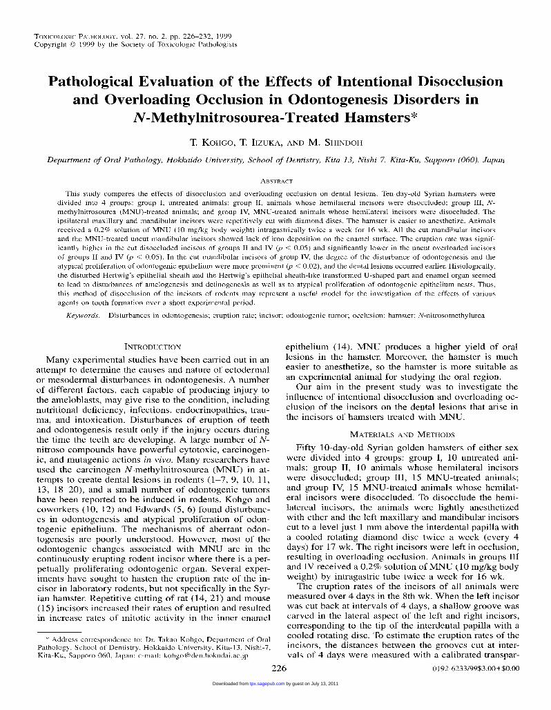
Cytokine expression in hamsters experimentally infected with Opisthorchis viverrini
http://tpx.sagepub.com/Toxicologic Pathology
http://tpx.sagepub.com/content/27/2/226The online version of this article can be found at:
DOI: 10.1177/019262339902700209
1999 27: 226Toxicol PatholT. Kohgo, T. Zuka and M. Shindoh
Odontogenesis Disorders in N-Methylnitrosourea-Treated HamstersPathological Evaluation of the Effects of Intentional Disocclusion and Overloading Occlusion in
Published by:
http://www.sagepublications.com
On behalf of:
Society of Toxicologic Pathology
can be found at:Toxicologic PathologyAdditional services and information for
http://tpx.sagepub.com/cgi/alertsEmail Alerts:
http://tpx.sagepub.com/subscriptionsSubscriptions:
http://www.sagepub.com/journalsReprints.navReprints:
http://www.sagepub.com/journalsPermissions.navPermissions:
http://tpx.sagepub.com/content/27/2/226.refs.htmlCitations:
by guest on July 13, 2011tpx.sagepub.comDownloaded from
226
Pathological Evaluation of the Effects of Intentional Disocclusionand Overloading Occlusion in Odontogenesis Disorders inN-Methylnitrosourea-Treated Hamsters*
T. KOHGO, T. IIZUKA, AND M. SHINDOH
Department of Oral Pathology, Hokkaido University, School of Dentistry, Kita 13, Nishi 7, Kita-Ku, Sapporo (060), Japan
* Address correspondence to: Dr. Takao Kohgo, Department of OralPathology, School of Dentistry, Hokkaido University, Kita-13, Nishi-7,Kita-Ku, Sapporo 060, Japan; e-mail: [email protected].
ABSTRACT
This study compares the effects of disocclusion and overloading occlusion on dental lesions. Ten-day-old Syrian hamsters weredivided into 4 groups: group I, untreated animals; group II, animals whose hemilateral incisors were disoccluded; group III, N-
methylnitrosourea (MNU)-treated animals; and group IV, MNU-treated animals whose hemilateral incisors were disoccluded. Theipsilateral maxillary and mandibular incisors were repetitively cut with diamond discs. The hamster is easier to anesthetize. Animalsreceived a 0.2% solution of MNU (10 mg/kg body weight) intragastrically twice a week for 16 wk. All the cut mandibular incisorsand the MNU-treated uncut mandibular incisors showed lack of iron deposition on the enamel surface. The eruption rate was signif-icantly higher in the cut disoccluded incisors of groups II and IV (p < 0.05) and significantly lower in the uncut overloaded incisorsof groups II and IV (p < 0.05). In the cut mandibular incisors of group IV, the degree of the disturbance of odontogenesis and theatypical proliferation of odontogenic epithelium were more prominent (p < 0.02), and the dental lesions occurred earlier. Histologically,the disturbed Hertwig’s epithelial sheath and the Hertwig’s epithelial sheath-like transformed U-shaped part and enamel organ seemedto lead to disturbances of amelogenesis and detinogenesis as well as to atypical proliferation of odontogenic epithelium nests. Thus,this method of disocclusion of the incisors of rodents may represent a useful model for the investigation of the effects of variousagents on tooth formation over a short experimental period.
Keywords. Disturbances in odontogenesis; eruption rate; incisor; odontogenic tumor; occlusion; hamster; N-nitrosomethylurea
INTRODUCTION
Many experimental studies have been carried out in anattempt to determine the causes and nature of ectodermalor mesodermal disturbances in odontogenesis. A numberof different factors, each capable of producing injury tothe ameloblasts, may give rise to the condition, includingnutritional deficiency, infections, endocrinopathies, trau-ma, and intoxication. Disturbances of eruption of teethand odontogenesis result only if the injury occurs duringthe time the teeth are developing. A large number of N-nitroso compounds have powerful cytotoxic, carcinogen-ic, and mutagenic actions in vivo. Many researchers haveused the carcinogen N-methylnitrosourea (MNU) in at-tempts to create dental lesions in rodents (1-7, 9, 10, 11,13, 18-20), and a small number of odontogenic tumorshave been reported to be induced in rodents. Kohgo andcoworkers (10, 12) and Edwards (5, 6) found disturbanc-es in odontogenesis and atypical proliferation of odon-togenic epithelium. The mechanisms of aberrant odon-togenesis are poorly understood. However, most of theodontogenic changes associated with MNU are in thecontinuously erupting rodent incisor where there is a per-petually proliferating odontogenic organ. Several exper-iments have sought to hasten the eruption rate of the in-cisor in laboratory rodents, but not specifically in the Syr-ian hamster. Repetitive cutting of rat (14, 21) and mouse(15) incisors increased their rates of eruption and resultedin increase rates of mitotic activity in the inner enamel
epithelium (14). MNU produces a higher yield of orallesions in the hamster. Moreover, the hamster is mucheasier to anesthetize, so the hamster is more suitable asan experimental animal for studying the oral region.Our aim in the present study was to investigate the
influence of intentional disocclusion and overloading oc-clusion of the incisors on the dental lesions that arise inthe incisors of hamsters treated with MNU.
MATERIALS AND METHODS
Fifty 10-day-old Syrian golden hamsters of either sexwere divided into 4 groups: group I, 10 untreated ani-
mals ; group II, 10 animals whose hemilateral incisorswere disoccluded; group III, 15 MNU-treated animals;and group IV, 15 MNU-treated animals whose hemilat-eral incisors were disoccluded. To disocclude the hemi-latereal incisors, the animals were lightly anesthetizedwith ether and the left maxillary and mandibular incisorscut to a level just 1 mm above the interdental papilla witha cooled rotating diamond disc twice a week (every 4days) for 17 wk. The right incisors were left in occlusion,resulting in overloading occlusion. Animals in groups IIIand IV received a 0.2% solution of MNU (10 mg/kg bodyweight) by intragastric tube twice a week for 16 wk.
The eruption rates of the incisors of all animals weremeasured over 4 days in the 8th wk. When the left incisorwas cut back at intervals of 4 days, a shallow groove wascarved in the lateral aspect of the left and right incisors,corresponding to the tip of the interdental papilla with acooled rotating disc. To estimate the eruption rates of theincisors, the distances between the grooves cut at inter-vals of 4 days were measured with a calibrated transpar-
by guest on July 13, 2011tpx.sagepub.comDownloaded from
227
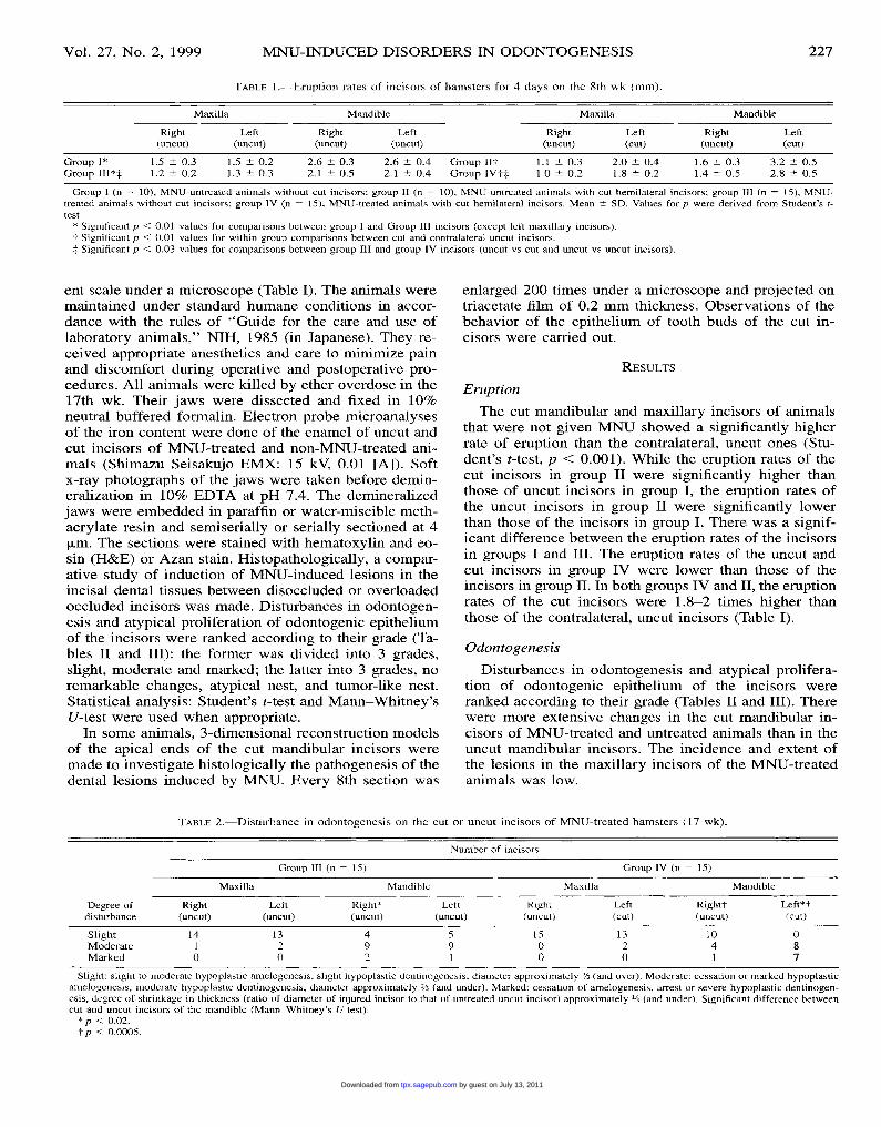
TABLE 1.-Eruption rates of incisors of hamsters for 4 days on the 8th wk (mm).
Group I (n = 10), MNU-untreated animals without cut incisors; group II (n = 10), MNU-untreated animals with cut hemilateral incisors; group III (n = 15), MNU-treated animals without cut incisors; group IV (n = 15), MNU-treated animals with cut hemilateral incisors. Mean ± SD. Values for p were derived from Student’s t-test.
* Significant p < 0.01 values for comparisons between group I and Group III incisors (except left maxillary incisors).t Significant p < 0.01 values for within group comparisons between cut and contralateral uncut incisors.i Significant p < 0.03 values for comparisons between group III and group IV incisors (uncut vs cut and uncut vs uncut incisors).
ent scale under a microscope (Table I). The animals weremaintained under standard humane conditions in accor-dance with the rules of &dquo;Guide for the care and use of
laboratory animals,&dquo; NIH, 1985 (in Japanese). They re-ceived appropriate anesthetics and care to minimize painand discomfort during operative and postoperative pro-cedures. All animals were killed by ether overdose in the17th wk. Their jaws were dissected and fixed in 10%neutral buffered formalin. Electron probe microanalysesof the iron content were done of the enamel of uncut andcut incisors of MNU-treated and non-MNU-treated ani-mals (Shimazu Seisakujo EMX: 15 kV, 0.01 [A]). Softx-ray photographs of the jaws were taken before demin-eralization in 10% EDTA at pH 7.4. The demineralizedjaws were embedded in paraffin or water-miscible meth-acrylate resin and semiserially or serially sectioned at 4[Lm. The sections were stained with hematoxylin and eo-sin (H&E) or Azan stain. Histopathologically, a compar-ative study of induction of MNU-induced lesions in theincisal dental tissues between disoccluded or overloadedoccluded incisors was made. Disturbances in odontogen-esis and atypical proliferation of odontogenic epitheliumof the incisors were ranked according to their grade (Ta-bles II and III): the former was divided into 3 grades,slight, moderate and marked; the latter into 3 grades, noremarkable changes, atypical nest, and tumor-like nest.Statistical analysis: Student’s t-test and Mann-Whitney’sU-test were used when appropriate.
In some animals, 3-dimensional reconstruction modelsof the apical ends of the cut mandibular incisors weremade to investigate histologically the pathogenesis of thedental lesions induced by MNU. Every 8th section was
enlarged 200 times under a microscope and projected ontriacetate film of 0.2 mm thickness. Observations of thebehavior of the epithelium of tooth buds of the cut in-cisors were carried out.
RESULTS
EruptionThe cut mandibular and maxillary incisors of animals
that were not given MNU showed a significantly higherrate of eruption than the contralateral, uncut ones (Stu-dent’s t-test, p < 0.001). While the eruption rates of thecut incisors in group II were significantly higher thanthose of uncut incisors in group I, the eruption rates ofthe uncut incisors in group II were significantly lowerthan those of the incisors in group I. There was a signif-icant difference between the eruption rates of the incisorsin groups I and III. The eruption rates of the uncut andcut incisors in group IV were lower than those of theincisors in group II. In both groups IV and II, the eruptionrates of the cut incisors were 1.8-2 times higher thanthose of the contralateral, uncut incisors (Table I).
OdontogenesisDisturbances in odontogenesis and atypical prolifera-
tion of odontogenic epithelium of the incisors wereranked according to their grade (Tables II and III). Therewere more extensive changes in the cut mandibular in-cisors of MNU-treated and untreated animals than in theuncut mandibular incisors. The incidence and extent ofthe lesions in the maxillary incisors of the MNU-treatedanimals was low.
TABLE 2.-Disturbance in odontogenesis on the cut or uncut incisors of MNU-treated hamsters (17 wk).
Slight: slight to moderate hypoplastic amelogenesis, slight hypoplastic dentinogenesis, diameter approximately 2/~ (and over). Moderate: cessation or marked hypoplasticamelogenesis, moderate hypoplastic dentinogenesis, diameter approximately 2% (and under). Marked: cessation of amelogenesis, arrest or severe hypoplastic dentinogen-esis, degree of shrinkage in thickness (ratio of diameter of injured incisor to that of untreated uncut incisor) approximately 1/3 (and under). Significant difference betweencut and uncut incisors of the mandible (Mann-Whitney’s U-test).
* p < 0.02.
t p < 0.0005.
by guest on July 13, 2011tpx.sagepub.comDownloaded from

228
TABLE 3.-Incidence and degree of manifestation of proliferating odontogenic epithelial nests in the periodontal ligament of the cut and uncutincisors of MNU-treated hamsters (17 wk).
Significant difference between cut and uncut incisors of the mandible. Mann-Whitney’s V-test.* p < 0.005.
t p < 0.0005.
Non-MNU GroupsThere were no remarkable changes in the incisors of
untreated animals. In the animals with cut hemilateralincisors (group II), at 10 days after the first cutting, thebrownish pigmentation on the enamel surface of the cutincisors had faded; this change was more prominent in
mandibular incisors (Fig. 1A). At 17 wk, the cut maxil-lary and mandibular incisors had slight dilatation of pulpspaces on soft x-ray film. In the MNU-treated animals
(group III) at 17 wk, some of the mandibular incisors hadbecome somewhat thin. Arrest of amelogenesis and dis-turbances in dentinogenesis were frequently seen. There
FIG. 1.-A) MNU-untreated animal with cut incisors at 10 days. Normal brownish color of the uncut right incisor and whitish color of the cutleft incisor. X 1.2. B) MNU-treated animal with hemilateral cut incisors at 17 wk. The cut mandibular incisor became thinner than the contralateraluncut incisor. X 1.2. C) Soft x-ray photograph of the animal shown in Fig. 1B, revealing severe hypoplasia of the left cut mandibular incisor(arrowhead) and slight hypoplasia of the right uncut incisor. X2.6. D, E) Radiogram of the same animal in Fig. 1B. D) The lateral view of theuncut mandibular incisor. E) The lateral view of the cut mandibular incisor (arrowhead). X2.3.
by guest on July 13, 2011tpx.sagepub.comDownloaded from

229
FIG. 2.-Photographs of x-ray microanalysis of iron deposition on the enamel surface of the mandibular incisors (arrowheads). x 850. A) GroupI: iron detected strongly in the enamel surface. B) Group III: iron detected weakly in the enamel surface. C) Group IV: iron not detected in theenamel surface of the cut incisor.
were atypical odontogenic epithelial cell nests of varioussizes in the periodontal ligaments of the mandibular in-cisors. The maxillary incisors showed slight dental hy-poplasia.
MNU GroupsIn MNU-treated animals (group IV) (Fig. 1B-E), from
9 to 12 wk, the uncut mandibular incisors had reducedpigmentation on the enamel surface and had becomeslightly hypoplastic. At 17 wk, the uncut mandibular in-cisors showed marked enamel hypoplasia with occasionalfocal areas of arrested amelogenesis and defects in theameloblastic layer. The disturbances in odontogenesis ofthe uncut incisors were not as prominent as in the con-tralateral cut teeth or in the teeth of group III. Hertwig’sepithelial root sheath was atrophied, occasionally dis-rupted, folded, and furcated. Sometimes the dentine wasirregular in width. The atypical odontogenic epithelialnests were chiefly in the lingual part of the periodontalligament. They took various forms and tended to squa-mous metaplasia with keratinization and, sometimes, liq-uefaction. In the uncut overloaded maxillary incisors den-tal hypoplasia was very slight. In the cut disoccludedmandibular incisors, at 10 days after the first cutting, thepigmentation on the enamel surface had faded away.From 9 to 12 wk, the cut mandibular incisors becamethin, with mesial and lateral constrictions. At 17 wk, thecut mandibular incisors had become very short and thin
(Fig. 1B-E). The cut maxillary incisors were brownishwhite and there was slight dilatation of the pulp spacefrom 9 wk. At 17 wk, the dentine showed slight irregu-larity in width.
X-ray MicroanalysisIn untreated animals (group I), x-ray microanalysis re-
vealed iron deposition on the incisal enamel surface withbrownish pigmentation (Fig. 2A). While the mandibularincisors in MNU-treated animals (group III) had reducediron deposition on the enamel surface (Fig. 2B), the cutmandibular incisors in groups II and IV (Fig. 2C) showedlack of iron deposition on the enamel surface.
Odontogenic Epithelium
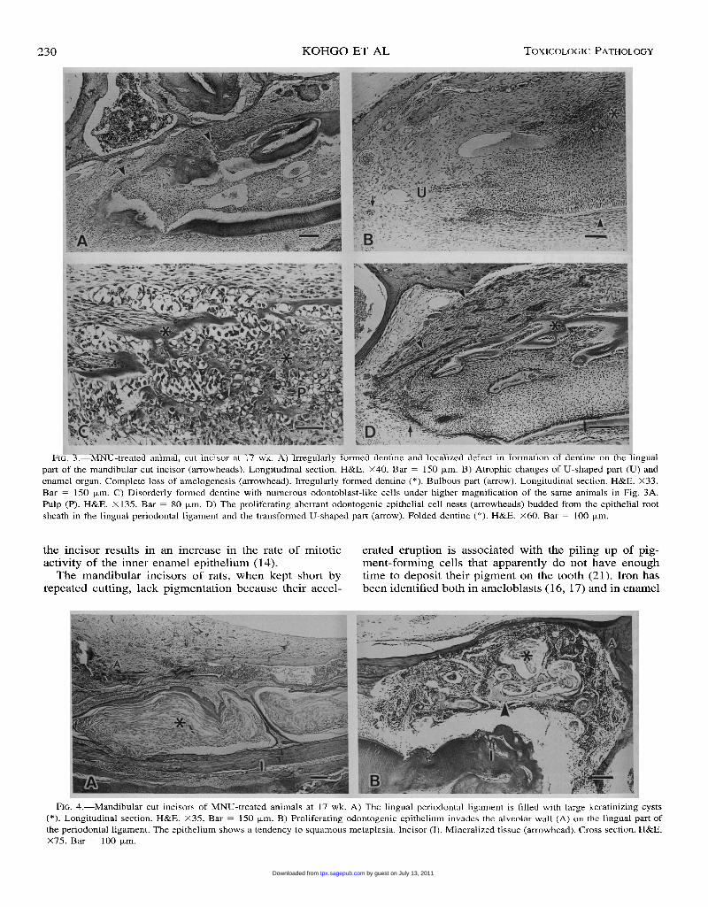
Histologically, disturbed odontogenesis and prolifera-tive changes in the odontogenic epithelium of the toothbuds were seen in MNU-treated animals, and these
changes were more prominent in the cut than in the con-tralateral uncut mandibular incisors (group IV) and themandibular incisors of group III (Tables II and III). Inmarked deficiency of odontogenesis, dentine became ir-regular in width on the lingual part of the root of theincisor (Fig. 3A and B). The bulbous part, U-shaped part,and enamel organs showed atrophic changes. Sometimesthere was complete loss of enamel formation (Fig. 3B),and dentine was irregularly formed (Fig. 3C). Sometimesthe U-shaped part and enamel organ were transformed toa Hertwig’s epithelial root sheath-like appearance bothfunctionally and behaviorally, and often irregular, foldeddentine was induced (Fig. 3D). Some of the aberrantodontogenic epithelial nests showed a range of histolog-ical features including foci of a keratinizing cyst-likestructure (Fig. 4A) and tumor-like growth with inductionof dentine-like calcified masses (Fig. 4B). Sometimes afew, small atypical epithelial nests were seen in the uncutoverloaded maxillary incisors.
DISCUSSION
The hamster is much easier to anesthetize lightly.Moreover, MNU produces a higher yield of oral lesionsin the hamster. Further, the hamster is a small but large-mouthed animal, so we can more conveniently maketreatments in the oral cavity. Therefore, the hamster ismore suitable as an experimental animal for studying theoral region.
Findings in this study suggested that while disocclu-sion (groups II and IV) increased the eruption rates ofthe incisors, MNU administration (groups III and IV) andocclusal overloading (groups II and IV) reduced the erup-tion rates of the incisors. The mandibular incisors ofwhite rats, when kept short by repeated cutting, reachedan eruption rate of more than 200% of the normal level(21). Repetitive cutting of the incisor or disocclusion of
by guest on July 13, 2011tpx.sagepub.comDownloaded from
230
FIG. 3.-MNU-treated animal, cut incisor at 17 wk. A) Irregularly formed dentine and localized defect in formation of dentine on the lingualpart of the mandibular cut incisor (arrowheads). Longitudinal section. H&E. X40. Bar = 150 p,m. B) Atrophic changes of U-shaped part (U) andenamel organ. Complete loss of amelogenesis (arrowhead). Irregularly formed dentine (*). Bulbous part (arrow). Longitudinal section. H&E. X33.
Bar = 150 wm. C) Disorderly formed dentine with numerous odontoblast-like cells under higher magnification of the same animals in Fig. 3A.Pulp (P). H&E. X 135. Bar = 80 pm. D) The proliferating aberrant odontogenic epithelial cell nests (arrowheads) budded from the epithelial rootsheath in the lingual periodontal ligament and the transformed U-shaped part (arrow). Folded dentine (*). H&E. X60. Bar = 100 f.Lm.
the incisor results in an increase in the rate of mitotic
activity of the inner enamel epithelium (14).The mandibular incisors of rats, when kept short by
repeated cutting, lack pigmentation because their accel-
erated eruption is associated with the piling up of pig-ment-forming cells that apparently do not have enoughtime to deposit their pigment on the tooth (21). Iron hasbeen identified both in ameloblasts (16, 17) and in enamel
FIG. 4.-Mandibular cut incisors of MNU-treated animals at 17 wk. A) The lingual periodontal ligament is filled with large keratinizing cysts(*). Longitudinal section. H&E. X35. Bar = 150 Vm. B) Proliferating odontogenic epithelium invades the alveolar wall (A) on the lingual part ofthe periodontal ligament. The epithelium shows a tendency to squamous metaplasia. Incisor (I). Mineralized tissue (arrowhead). Cross section. H&E.X75. Bar = 100 p.m.
by guest on July 13, 2011tpx.sagepub.comDownloaded from
231
(16). Electron microprobe analysis shows a very highiron content in the outer portion of enamel (8). We foundby x-ray microanalysis that the loss of pigmentation seenboth in the cut control incisors and in the MNU-treatedincisors was due to a loss of iron in the outer portion ofthe enamel. Moreover, giving MNU resulted in severehypoplasia or complete loss of the mandibular incisalenamel. The cessation of amelogenesis can be related tothe fact that cytotoxic injury to the cells in the U-shapedpart and the enamel organ prevented the differentiationor the maturation of the cells of the internal enamel ep-ithelium into ameloblasts. In the cut, disoccluded man-dibular incisors of MNU-treated hamsters, disturbancesin odontogenesis and atypical proliferation of odontogen-ic epithelial nests were more frequent and more extensivethan in the contralateral uncut incisors. Moreover, thesechanges occurred earlier than reported in previous studies(10) on hamsters with intact incisors, which suggestedthat the odontogenic epithelial cells of the tooth bud withaccelerated mitotic activity may be more susceptible, atan earlier time, to the effects of MNU than normal. How-ever, early visible changes may also occur in Hertwig’sepithelial root sheath, possibly because it is a thin epi-thelial layer that is apt to manifest injuries. In advancedstages, the U-shaped part and enamel organ were trans-formed to a Hertwig’s epithelial root sheath-like appear-ance and function, resulting in loss of amelogenesis. Thealtered odontogenic epithelium of the root sheath, U-shaped part, and enamel organ were often associated withthe induction of aberrant dentine formation, whichseemed to lead to disturbances in odontogenesis. Edwards(6) suggested that the herniation or separation of a por-tion of active odontogenic tissue from the basal odonto-genic organ might precede the formation of an odonto-genic tumor or malformation. In our study, the prolifer-ating odontogenic epithelium in the incisal periodontalligaments of MNU-treated hamsters seemed to originatefrom small epithelial cell nests irregularly migrating fromthe furcated Hertwig’s epithelial sheath, the Hertwig’s ep-ithelial root sheath-like transformed U-shaped part, andthe disturbed enamel organ. These atypically migratingepithelial nests were rarely associated with hard dentaltissues.
Overloading occlusion seemed to decrease the rate ofproliferative activity of the tooth bud, and then the erup-tion rate of the incisors seemed to be reduced. Moreover,the tooth bud with reduced mitotic activity may be lesssusceptible to the effects of MNU. However, further in-vestigation is required.The maxillary incisors did not suffer from the severe
morphological changes seen in the mandibular incisors inthe MNU-treated animals, and the eruption rates of cutdisoccluded and uncut overloaded occluded maxillary in-cisors were not remarkably changed by giving MNU.However, at the half-way point of the experiment, theeruption rates of cut maxillary incisors in MNU-treatedanimals were significantly higher than those of the uncutincisors of the mandible. Thus, manifestation of the ef-fects of MNU in the maxillary incisor region may beinfluenced by a lower sensitivity to the compound, lesserreaction to the stimulus of repetitive cutting, and other
anatomical factors; however, further investigation is re-quired to confirm this.Some of the MNU-induced atypical proliferating odon-
togenic epithelium in the disoccluded mandibular incisorshistologically resembled early neoplastic changes; the re-sulting range of histological appearances was reminiscentof some odontogenic lesions in man, including the odon-togenic keratocyst, dysplastic dentine, and even odonto-genic tumors. Thus, this method of disocclusion of theincisors of rodents may represent a useful model for theinvestigation of the effects of various agents on toothformation over a short experimental period.
REFERENCES
1. Azuma T and Komori A (1985). An experimental study of odon-togenic tumors in rats. Induction of tumors by gastric intubation ofMNU. Jpn. J. Oral Biol. 27: 631-639.
2. Berman JJ and Rice JM (1980). Odontogenic tumours produced inFisher rats by a single intraportal injection of methylnitrosourea.Arch. Oral Biol. 25: 213-220.
3. Druckrey H, Steinhoff D, Preussmann R, and Ivankovic S (1964).Erzeugung von Krebs durch eine einmalige Dosis von Methylni-troso-hamstoff und verschiedenen Dialkyl-nitrosaminen an Ratten.Z. Krebsforsch. 66: 1-10.
4. Eblin H, Barbachan JJD, Do Valle JGC, and De Oliveira LY (1973).N-methyl-N-nitrosourea-induced odontogenic neoplasms in rats. J.Dent. Res. 52: 177.
5. Edwards MB (1978). Some effects of N-methylnitrosourea on theoral and odontogenic tissues of the Syrian golden hamster. Arch.Oral Biol. 23: 515-524.
6. Edwards MB (1980). Effects of N-methylnitrosourea on oral anddental tissues in the inbred Lewis rat. Arch. Oral Biol. 25: 59-65.
7. Eisenberg E, Murthy AS, Vawter GF, and Krutchkoff DJ (1983).Odontogenic neoplasms in Wistar rats treated with N-methylnitro-sourea. Oral Surg. 55: 481-486.
8. Halse A (1972). An electron microprobe investigation of the dis-tribution of iron in rat incisor enamel. Scand. J. Dent. Res. 80:
26-39.
9. Herrold KM (1968). Odontogenic tumors and epidermoid carcino-mas of the oral cavity. Oral Surg. Oral Med. Oral Pathol. 25: 262-272.
10. Kohgo T (1972). An experimental study of odontogenic tumors inhamsters by N-nitrosomethylurea. J. Stomatol. Soc. Jpn. 39: 191-212.
11. Kohgo T, Acob TB, Iizuka T, Shindoh M, and Amemiya A (1996).Histopathologic evaluation of the effect of natural and synthetic β-carotene on tumorigenesis and disturbances in odontogenesis in N-nitrosomethylurea-treated hamsters. Hokkaido J. Dent. Sci. 17: 62-71.
12. Kohgo T, Uy GH, Iizuka T, Shindoh M, and Amemiya A (1990).Histopathologic study of squamous cell carcinoma of the gingivain edentulous and dentulous jaws of hamsters treated with N-meth-ylnitrosourea. J. Oral Pathol. Med. 19: 202-207.
13. Leaver DD, Swann PF, and Magee PN (1969). The induction oftumours in the rat by a single oral dose of N-nitrosomethylurea.Br. J. Cancer 223: 177-187.
14. Nakagawa K (1968). The response of the cell proliferation at thebase of the rat incisor tooth to experimental shortening. J. Stomatol.Soc. Jpn. 35: 143-154.
15. Ness AR (1965). Eruption rates of impeded and unimpeded man-dibular incisors of the adult laboratory mouse. Arch. Oral Biol. 10:439-451.
16. Pindborg JJ (1947). Studies on incisor pigmentation in relation toliver iron and blood picture of the white rat. V. Histo-chemicaldemonstration of the embedding of the pigment in the enamel.Odontol. Tidskr. 55: 443-446.
17. Reith EJ (1959). The enamel organ of the rat’s incisor, its histologyand pigment. Anat. Rec. 133: 75-87.
by guest on July 13, 2011tpx.sagepub.comDownloaded from
232
18. Smulow JB, Konstantinidis A, and Sonnenschein C (1983). Age-dependent odontogenic lesions in rats after a single i.p. injectionof N-nitroso-N-methylurea. Carcinogenesis 14: 1085-1088.
19. Tani Y (1986). Induction of odontogenic tumors by transplacental ad-ministration of N-methylnitrosourea. Jpn. J. Oral Biol. 28: 551-564.
20. Tashiro T, Matsushima T, and Kai S (1984). The induction of tu-mors in the rat by intra-arterial injection of N-nitroso-methylurea.Jpn. J. Oral Maxillofac. Surg. 30: 1-4.
21. Taylor AC and Butcher EO (1951). The regulation of eruption ratein the incisor teeth of the white rat. J. Exp. Zool. 117: 165-188.
17th ESVP Meeting
The European Society of Veterinary Pathology, jointly with the French Society ofVeterinary Pathology and the French Society of Toxicologic Pathology, has scheduled its17th meeting at the Veterinary School of Nantes (France), on September 14-17, 1999.
Parallel sessions for Veterinary Pathology and Toxicologic Pathology will be arrangedand themes of the sessions will be announced later. Invited lectures will take place at thebeginning of each session. Topics will include: aging, cytokines in inflammatory process,cell proliferation, Helicobacter and Helicobacteriosis in man and animals. Oral commu-nications and posters are invited from all of the fields of Veterinary Pathology of bothexperimental and spontaneous diseases.
Deadline for early registration and submission of abstracts is June 15, 1999.For further information, contact:
Congress Secretariat5/5 ESVPBP 2071644007 Nantes cedex 1
France
E-mail: [email protected] site: http://www.vet-nantes.fr/congress99.htm
by guest on July 13, 2011tpx.sagepub.comDownloaded from
Copyright © 2022 FDOKUMEN