Citrullinated vimentin as an important antigen in immune complexes from synovial fluid of rheumatoid...
10
Van Steendam et al. Arthritis Research & Therapy 2010, 12:R132 http://arthritis-research.com/content/12/4/R132 Open Access RESEARCH ARTICLE © 2010 Van Steendam et al.; licensee BioMed Central Ltd. This is an open access article distributed under the terms of the Creative Com- mons Attribution License (http://creativecommons.org/licenses/by/2.0), which permits unrestricted use, distribution, and reproduc- tion in any medium, provided the original work is properly cited. Research article Citrullinated vimentin as an important antigen in immune complexes from synovial fluid of rheumatoid arthritis patients with antibodies against citrullinated proteins Katleen Van Steendam 1 , Kelly Tilleman 1 , Marlies De Ceuleneer 1 , Filip De Keyser 2 , Dirk Elewaut 2 and Dieter Deforce* 1 Abstract Introduction: Rheumatoid arthritis (RA) is an inflammatory disease, which results in destruction of the joint. The presence of immune complexes (IC) in serum and synovial fluid of RA patients might contribute to this articular damage through different mechanisms, such as complement activation. Therefore, identification of the antigens from these IC is important to gain more insight into the pathogenesis of RA. Since RA patients have antibodies against citrullinated proteins (ACPA) in their serum and synovial fluid (SF) and since elevated levels of citrullinated proteins are detected in the joints of RA patients, citrullinated antigens are possibly present in IC from RA patients. Methods: IC from serum of healthy persons, serum of RA patients and IC from synovial fluid of RA patients and Spondyloarthropathy (SpA) patients were isolated by immunoprecipitation. Identification of the antigens was performed by SDS-PAGE, mass spectrometry and immunodetection. The presence of citrullinated proteins was evaluated by anti-modified citrulline (AMC) staining. Results: Circulating IC in the serum of RA patients and healthy controls contain fibrinogenβ and fibronectin, both in a non-citrullinated form. Additionally, in IC isolated from RA SF, fibrinogenγ and vimentin were identified as well. More importantly, vimentin and a minor portion of fibrinogenβ were found to be citrullinated in the isolated complexes. Moreover these citrullinated antigens were only found in ACPA+ patients. No citrullinated antigens were found in IC from SF of SpA patients. Conclusions: Citrullinated fibrinogenβ and citrullinated vimentin were found in IC from SF of ACPA+ RA patients, while no citrullinated antigens were found in IC from SF of ACPA- RA patients or SpA patients or in IC from serum of RA patients or healthy volunteers. The identification of citrullinated vimentin as a prominent citrullinated antigen in IC from SF of ACPA+ RA patients strengthens the hypothesis that citrullinated vimentin plays an important role in the pathogenesis of RA. Introduction Rheumatoid arthritis (RA) is a progressive autoimmune disease characterized by chronic inflammation of the peripheral joints. It is a complex multifactorial pathology, in which genetic and environmental factors, like smok- ing, can play an important role in the onset of disease and the progression of the joint damage [1,2]. The presence of immune complexes (IC) in serum and synovial fluid (SF) of RA patients is likely to contribute to the pathogenesis of the disease and to articular damage, since they are responsible for the activation of complement, the stimu- lation of phagocytes through their Fc receptor and the release of chemotactic factors, cytokines, metalloprotei- nases and reactive oxygen intermediates [3-6]. The for- mation of IC as such is not specifically related to autoimmune pathologies as it is a natural process, com- pleting an immune response in the body. The antigen- antibody complexes are usually effectively removed by phagocytosis. However, it is known that an impaired * Correspondence: [email protected] 1 Laboratory for Pharmaceutical Biotechnology, Ghent University, Harelbekestraat 72, B-9000 Ghent, Belgium Full list of author information is available at the end of the article
-
Upload
independent -
Category
Documents
-
view
0 -
download
0
Transcript of Citrullinated vimentin as an important antigen in immune complexes from synovial fluid of rheumatoid...
Van Steendam et al. Arthritis Research & Therapy 2010, 12:R132
http://arthritis-research.com/content/12/4/R132
Open AccessR E S E A R C H A R T I C L E
© 2010 Van Steendam et al.; licensee BioMed Central Ltd. This is an open access article distributed under the terms of the Creative Com-mons Attribution License (http://creativecommons.org/licenses/by/2.0), which permits unrestricted use, distribution, and reproduc-tion in any medium, provided the original work is properly cited.
Research articleCitrullinated vimentin as an important antigen in immune complexes from synovial fluid of rheumatoid arthritis patients with antibodies against citrullinated proteinsKatleen Van Steendam1, Kelly Tilleman1, Marlies De Ceuleneer1, Filip De Keyser2, Dirk Elewaut2 and Dieter Deforce*1
Abstract
Introduction: Rheumatoid arthritis (RA) is an inflammatory disease, which results in destruction of the joint. The
presence of immune complexes (IC) in serum and synovial fluid of RA patients might contribute to this articular
damage through different mechanisms, such as complement activation. Therefore, identification of the antigens from
these IC is important to gain more insight into the pathogenesis of RA. Since RA patients have antibodies against
citrullinated proteins (ACPA) in their serum and synovial fluid (SF) and since elevated levels of citrullinated proteins are
detected in the joints of RA patients, citrullinated antigens are possibly present in IC from RA patients.
Methods: IC from serum of healthy persons, serum of RA patients and IC from synovial fluid of RA patients and
Spondyloarthropathy (SpA) patients were isolated by immunoprecipitation. Identification of the antigens was
performed by SDS-PAGE, mass spectrometry and immunodetection. The presence of citrullinated proteins was
evaluated by anti-modified citrulline (AMC) staining.
Results: Circulating IC in the serum of RA patients and healthy controls contain fibrinogenβ and fibronectin, both in a
non-citrullinated form. Additionally, in IC isolated from RA SF, fibrinogenγ and vimentin were identified as well. More
importantly, vimentin and a minor portion of fibrinogenβ were found to be citrullinated in the isolated complexes.
Moreover these citrullinated antigens were only found in ACPA+ patients. No citrullinated antigens were found in IC
from SF of SpA patients.
Conclusions: Citrullinated fibrinogenβ and citrullinated vimentin were found in IC from SF of ACPA+ RA patients, while
no citrullinated antigens were found in IC from SF of ACPA- RA patients or SpA patients or in IC from serum of RA
patients or healthy volunteers. The identification of citrullinated vimentin as a prominent citrullinated antigen in IC
from SF of ACPA+ RA patients strengthens the hypothesis that citrullinated vimentin plays an important role in the
pathogenesis of RA.
IntroductionRheumatoid arthritis (RA) is a progressive autoimmune
disease characterized by chronic inflammation of the
peripheral joints. It is a complex multifactorial pathology,
in which genetic and environmental factors, like smok-
ing, can play an important role in the onset of disease and
the progression of the joint damage [1,2]. The presence of
immune complexes (IC) in serum and synovial fluid (SF)
of RA patients is likely to contribute to the pathogenesis
of the disease and to articular damage, since they are
responsible for the activation of complement, the stimu-
lation of phagocytes through their Fc receptor and the
release of chemotactic factors, cytokines, metalloprotei-
nases and reactive oxygen intermediates [3-6]. The for-
mation of IC as such is not specifically related to
autoimmune pathologies as it is a natural process, com-
pleting an immune response in the body. The antigen-
antibody complexes are usually effectively removed by
phagocytosis. However, it is known that an impaired
* Correspondence: [email protected]
1 Laboratory for Pharmaceutical Biotechnology, Ghent University,
Harelbekestraat 72, B-9000 Ghent, BelgiumFull list of author information is available at the end of the article

Van Steendam et al. Arthritis Research & Therapy 2010, 12:R132
http://arthritis-research.com/content/12/4/R132
Page 2 of 10
clearance of these complexes can elicit or sustain an
inflammatory response [7,8].
The pathological nature of IC has been suggested by
several groups based on in vitro studies. The effect of the
SF IC from juvenile RA patients on healthy PBMCs was
studied by Jarvis et al. They found that especially the high
molecular weight IC, separated by size exclusion chroma-
tography from the other immunoglobulins and low
molecular weight IC, were responsible for inducing a
spectrum of pro-inflammatory cytokines, such as TNFα,
IL-1β, IL6, IL8 and granulocyte-macrophage colony-
stimulating factor (GM-CSF) [9]. A comparison between
IC from SF of RA patients, serum of RA patients and
serum of healthy persons was made by Schuerwegh et al.
They demonstrated that IC isolated from RA serum and
RA SF, in contrast to IC from healthy persons, had an
effect on chondrocyte growth, NO production and apop-
tosis, thereby contributing directly to cartilage destruc-
tion in RA [10]. Mathsson et al. showed that polyethylene
glycol (PEG) precipitated IC from RA SF induced the
production of the pro-inflammatory cytokine TNFα in
peripheral blood mononuclear cell (PBMC) cultures from
healthy donors. When IC from RA serum or healthy
serum were used, no elevated levels in TNFα could be
seen [11]. These reports show the relevance of IC in the
joint destruction and the pathogenesis of RA.
The best known IC in RA is the rheumatoid factor (RF)
bound to its antigen, the Fc domain of IgG. The RF, which
is mainly IgM [12], is used in diagnostic tests for RA and
has a sensitivity of 78.6% and a specificity of 80.8% [13].
The RF factor is also found in other diseases such as sys-
temic sclerosis (20 to 30%) [14] and occasionally in
healthy persons (1.3 to 4%) [5]. Besides the RF, immuno-
globulins and complement factors, other components can
also be present in IC from serum of RA patients. Indeed,
recently, it has been shown that fibrinogen and citrulline-
containing fibrinogen are present in the IC of RA patients
[15]. Because of the pathogenic nature of IC in RA, it is
important to identify the antigens in these complexes.
After identification of these antigens, a better under-
standing of the immunological process in the affected
joints can be achieved.
Since anti-citrullinated protein/peptide antibodies
(ACPA) are very specific for RA (specificity of 98%, sensi-
tivity 68%) [16] and high amounts of citrullinated pro-
teins, like fibrinogen, have been detected in the joint of
RA patients, it is likely that some antigens in IC of RA
patients are citrullinated.
The isolation of IC and subsequent identification of the
antigens is therefore of great importance in the under-
standing of RA. The isolation of IC from biological matri-
ces has been tackled by many different techniques such as
PEG precipitation [10,11,17], C1q ELISA [15] and immu-
noprecipitation [18]. PEG precipitation is broadly used
for the isolation of IC but the IC-fraction also contains a
considerable amount of non immune complex (IC)-
related proteins, such as albumine, haptoglobin and α1-
antitrypsin [17]. C1q ELISA will isolate IC that are bound
to the C1q component of the complement and this
method is gaining interest because of the high through-
put possibilities. However, to capture IC by C1q ELISA,
C1q must be present and accessible in the IC. IC from
serum and SF can be isolated with a high purity by means
of immunoprecipitation with proteinG, but it has the dis-
advantage of isolating the free immunoglobulins as well.
For the identification of the antigens in IC, a sensitive
method like mass spectrometry and immunodetection is
necessary because of their low abundance.
In this report a broad range proteome approach, by
means of mass spectrometry, is used in order to find new
antigens in IC. Because of the low abundance of the anti-
gens and the excess of immunoglobulins, it is possible
that not all antigens will be detected by this approach,
especially antigens that have a molecular weight that cor-
respond to those of immunoglobulins. Therefore, a sec-
ond, very sensitive method such as immunodetection on
2D-PAGE, was chosen to confirm the results of the broad
range proteome approach and to investigate whether
known antigens in RA (e.g. fibrinogenβ (Fibβ), fibrino-
genγ (Fibγ), fibronectin and vimentin) are present in
these complexes. Besides the high sensitivity of immuno-
detection, Western blot makes it also possible to visualize
different isoforms of a certain protein.
Since not only the identification of the antigens, but
also their citrullination status was of interest, the choice
of antibodies for immunodetection was based on previ-
ous reports on citrullinated proteins either in serum, SF
or synovial tissue of RA patients. The comparison
between citrullinated proteins in serum and SF was
already reported by Takizawa et al. [19]. In their study,
soluble antigens were studied in RA serum, RA SF and
osteo-arthritis (OA) SF. They could only identify citrulli-
nated fibrinogen in RA SF. However, two years later, also
citrullinated fibronectin and citrullinated vimentin were
found as soluble antigens in RA SF and synovial tissue
[20-22]. Citrullinated fibronectin was also detected in RA
SF and synovial exosomes [23,24]. Additionally, the pres-
ence of citrullinated Fibβ and Fibγ in RA synovium has
been reported by Matsuo et al. [25]. Based on these find-
ings, immunodetection was performed with anti-Fibβ,
anti-Fibγ, anti-fibronectin and anti-vimentin antibodies
on 2D-PAGE with IC, followed by anti-modified citrul-
line (AMC) detection.
The citrullination of the antigens perfectly fits the
model for the development and chronic nature of RA
proposed by van Venrooij and Pruijn. They divided the
process of autoimmunity in RA into five steps: an inno-
cent inflammation in combination with massive apopto-

Van Steendam et al. Arthritis Research & Therapy 2010, 12:R132
http://arthritis-research.com/content/12/4/R132
Page 3 of 10
sis or impaired clearance can lead to the elevation of
cytosolic Ca2+ concentrations (1) followed by the activa-
tion of peptidylarginine deiminase (PAD) and the citrulli-
nation of proteins (2). When citrullinated antigens are
presented to T cells, the production of ACPAs is trig-
gered (3). Immune complexes can be formed if the anti-
gens react with the auto-antibodies (4). These IC
stimulate inflammatory processes (5) and cause a vicious
circle of inflammation resulting in joint destruction for
years [26].
This study describes the isolation and characterization
of antigens residing in IC of RA patients. We found that
circulating IC in the serum of RA patients and healthy
controls contain Fibβ and fibronectin, both in a non-cit-
rullinated form. In IC, isolated from RA SF, on the other
hand, Fibrinogenβ, Fibrinogenγ, fibronectin and vimen-
tin were identified. More importantly, vimentin and a
minor portion of Fibβ were found to be citrullinated in
the isolated complexes from RA SF. However, these cit-
rullinated antigens were only found in IC from SF of
ACPA+ RA patients, while no citrullinated antigens were
found in IC from SF of ACPA- RA patients or SpA
patients.
Materials and methodsPatients and controls
Serum and synovial fluid were collected from patients
fulfilling the American College of Rheumatology criteria
for RA [27] and European Spondyloarthropathy Study
Group criteria for SpA [28] (for patient information see
Table 1). Sera from healthy donors were used as controls.
Informed consent was obtained from patients and
healthy controls and the study was approved by the local
ethics committee. Detailed information on the identity of
the samples used in each experiment is provided in Table
1. RF titers were determined with the Waaler Rose test
and ACPA titers were measured with anti-CCP-EliA
(Phadia, Freiburg, Germany).
Immunoprecipitation with Immobilized Protein G
IC were further purified by affinity immunoprecipitation
with Immobilized Protein G (Pierce, Rockford, IL, USA).
400 μL beads were washed twice with 500 μL phosphate
buffered saline (PBS). A total of 50 μL serum or SF was
mixed with 450 μL PBS and added to the beads. Sample
and beads were placed on a rocker for four hours at 4°C.
The beads with the bound IC were washed five times in
500 μL PBS. The pellet of protein G beads was resus-
pended in reducing Laemmli sample buffer for five min-
utes at 95°C. After centrifugation (5 minutes, 460 g) the
supernatant was stored at -20°C.
Protein concentrations were determined by Coomassie
(Bradford) Protein Assay (Pierce, Rockford, IL, USA) and
2 D Quant (GE Healthcare, Uppsala, Sweden).
One-dimensional gel electrophoresis (1D-PAGE)
Protein samples were dissolved in Laemmli buffer (50
mM TrisHCl, pH 6.8, 2% SDS, 10% glycerol, bromophe-
nol blue) with 5% β-mercapto-ethanol and incubated at
95°C for five minutes. The samples were loaded on a 10%
TrisHCl polyacrylamidegel (Biorad, Hercules, CA, USA)
and electrophoresis was performed by applying 150 V for
30 minutes, followed by 200 V for one hour.
Two-dimensional gel electrophoresis (2D-PAGE)
For 2D-PAGE, protein samples were first precipitated
overnight in acetone at -20°C. After centrifugation at
18,000 g for 10 minutes the samples were air dried. A
total of 100 μg was resuspended in 200 μL rehydration
buffer (7 M Urea, 2 M Thiourea, 2% CHAPS, 0.2% carrier
ampholytes, 100 mM DTT, bromophenol blue). The sam-
ple was introduced passively in an IPG strip (11 cm, pH 4
to 7) (Biorad) as previously described [29]. Iso-electric
focussing was performed in a Protean IEF Chamber (Bio-
rad) according to the following program: 100 V, 30 min-
utes, linear voltage slope - 250 V, 30 minutes, linear - 500
V, one hour, linear - 1,000 V, one hour, linear - 8,000 V,
four hours, rapid - 8,000 V, 35,000 V hours, rapid - 500 V,
20 h, rapid. Subsequently the strips were equilibrated in
equilibration buffer (50 mM TrisHCl, pH 8.8, 6 M Urea,
20% glycerol, 2% SDS) containing 1.5% DTT for 15 min-
utes, followed by 4% IAA in equilibration buffer for 15
minutes.
Gel electrophoresis was carried out on a 10% TrisHCl
PAGE using 150 V for 30 minutes, followed by 200 V for
one hour.
Western blot
After a 15-minute equilibration of the gels and the nitro-
cellulose membranes (Biorad) in CAPS (pH 11), electro-
phoretic transfer of proteins was performed by tank
blotting in a Trans Blot Cell (Biorad), with CAPS (pH =
11), at 50 V for three hours. Successful transfer of pro-
teins was checked by means of Ponceau S staining.
Detection of citrullinated proteins
The presence of citrullinated proteins on the nitrocellu-
lose blots was detected using the anti-modified citrulline
(AMC) detection kit (Upstate, Charlottesville, VA, USA)
according to the manufacturer's protocol. Each AMC
detection was accompanied with a positive control, as
indicated in the manufacturer's protocol.
Protein identification
Visualization of proteins in the gels was performed using
Sypro Ruby Protein Gel staining (Invitrogen, Carlsbad,
CA, USA) for at least three hours after a 30-minute fixa-
tion in a 10% MeOH, 7% acetic acid solution. After stain-
ing, the gel was washed twice with a 10% MeOH, 7%
acetic acid solution. Proteins of interest were excised

Van Steendam et al. Arthritis Research & Therapy 2010, 12:R132
http://arthritis-research.com/content/12/4/R132
Page 4 of 10
Table 1: Rheumatoid factor and CCP values of RA patients
Used in experiment diagnosis RF (U/ml) CCP (U/ml)
RA1 results 1&2 RA 1,280 2,839
RA2 results 1&2 RA 320 265
RA3 results 1&2 RA 0 > 1,600
SF1 results 3 RA 6 0
SF2 results 1&2&3 RA 80 1
SF3 results 3 RA 0 1
SF4 results 3 RA 0 1,6
SF5 results 3 RA 0 2
SF6 results 3 RA 5 2
SF7 results 3 RA 0 3
SF8 results 3 RA 40 4
SF9 results 3 RA 0 5
SF10 results 3 RA 0 5
SF11 results 3 RA 80 7
SF12 results 3 RA 160 10
SF13 results 1&2&3 RA 351 107
SF14 results 3 RA 183 340
SF15 results 1&2&3 RA 1,280 533
SF16 results 3 RA 1,280 608
SF17 results 3 RA 1,280 710
SF18 results 3 RA 2,560 740
SF19 results 3 RA 10,240 767
SF20 results 1 RA 1,280 1,141
SF21 results 3 RA 640 1,294
SF22 results 3 RA 0 > 1,600
SF23 results 3 RA 160 > 1,600
SF24 results 3 RA 0 > 1,600
SF25 results 3 RA 640 1,775
SF26 results 2 RA 227 ND
SF27 results 3 SpA 0 0
SF28 results 3 SpA ND 1,6
SF29 results 3 SpA 0 2
SF30 results 3 SpA 0 2
SF31 results 3 SpA 0 3
SF32 results 3 SpA 0 4
SF33 results 3 SpA 0 11
SF34 results 3 SpA 0 ND
SF35 results 3 SpA ND ND
SF36 results 3 SpA 0 ND
SF37 results 3 SpA 0 ND
SF38 results 3 SpA 0 ND
The rheumatoid factor was determined with Waaler Rose (U/ml) and CCP (U/ml) values were determined with anti-CCP-EliA. Serum (RA1-RA3)
as well as SF was used. The section of the article in which the sera and SF are used is mentioned in the second column. ND, not determined
Van Steendam et al. Arthritis Research & Therapy 2010, 12:R132
http://arthritis-research.com/content/12/4/R132
Page 5 of 10
from the gel and digested with modified sequence grade
porcine trypsin (Promega, Madison, WI, USA) as
described earlier [30]. Proteins were analyzed and identi-
fied by LC-MSMS, using a Q-TOF Ultima Mass spec-
trometer (Waters, Milford, MA, USA) combined with ESI
source. The data were processed using Mascot Distiller
and searched against the Swissprot human database,
using the in-house mascot daemon searching algorithm.
Identification was considered positive with a P-value <
0.05.
Immunodetection
Before immunodetection, each blot was blocked for one
hour in 0.3% Tween-20 in PBS. Vimentin was detected
with the mouse anti-human vimentin antibody (clone V9,
Sigma, St. Louis, MI, USA) at a concentration of 1/400 in
0.3% Tween-20 in PBS. After overnight incubation,
vimentine was detected with HRP labelled goat anti-
mouse IgG followed by ECL detection. Detection of Fibβ,
Fibγ and fibronectin was performed using respectively
rabbit anti-human Fibβ, rabbit anti-human Fibγ and rab-
bit anti-human fibronectin. Anti-rabbit HRP labelled
antibody was used as a secondary antibody. ECL detec-
tion was carried out by means of Supersignal West Dura
Extended Duration Substrate (Pierce).
Following each immunodetection, the blot was stripped
for 30 minutes at 50°C with stripping buffer (2% SDS, 0.1
M β-mercapto-ethanol, 0.05 M Tris pH 6.8) and washed
three times with 0.3% Tween20 in PBS. To check the
stripping efficiency, the blot was re-incubated with sec-
ondary antibody and detected with ECL. Afterwards the
blot was stripped for another 15 minutes, before incuba-
tion with a new primary antibody. Additionally, the
sequence of antibodies used for immunodetection varied
throughout the different experiments in order to exclude
false positive results. Protein patterns were scanned and
digitized using the VersaDoc Imaging System (Biorad).
ResultsBroad range proteome approach to identify potential
antigens in RA serum and RA SF
Immunoprecipitation (IP) was used in order to isolate the
IC from serum and synovial fluid. Because of the high vis-
cosity of SF, a hyaluronidase treatment was necessary.
Both, the flow-through and the eluted IC fraction from a
pool of RA SF (SF2; SF13; SF15; SF26) were subjected to 1
D gel electrophoresis (20 μg/lane). In order to identify
potential autoantigens in the eluted IC fractions from SF,
each lane (20 μg) from the gel was divided in 30 different
plugs and analysed separately by mass spectrometry after
in gel digestion. Mass spectrometric analysis revealed
that the eluted IC fraction from RA SF contained mainly
immunoglobulins, while almost none were detected in
the flow-through fraction (data not shown). Additionally,
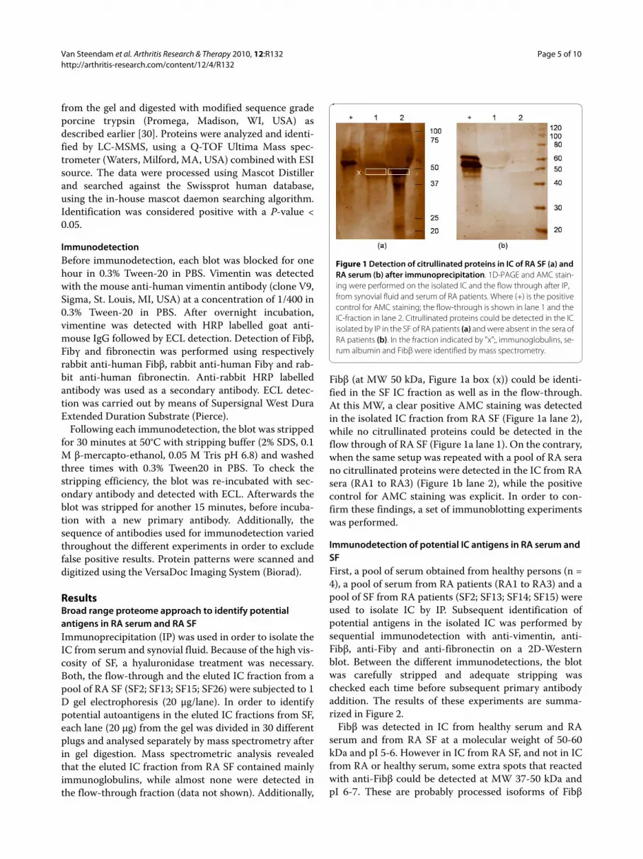
Fibβ (at MW 50 kDa, Figure 1a box (x)) could be identi-
fied in the SF IC fraction as well as in the flow-through.
At this MW, a clear positive AMC staining was detected
in the isolated IC fraction from RA SF (Figure 1a lane 2),
while no citrullinated proteins could be detected in the
flow through of RA SF (Figure 1a lane 1). On the contrary,
when the same setup was repeated with a pool of RA sera
no citrullinated proteins were detected in the IC from RA
sera (RA1 to RA3) (Figure 1b lane 2), while the positive
control for AMC staining was explicit. In order to con-
firm these findings, a set of immunoblotting experiments
was performed.
Immunodetection of potential IC antigens in RA serum and
SF
First, a pool of serum obtained from healthy persons (n =
4), a pool of serum from RA patients (RA1 to RA3) and a
pool of SF from RA patients (SF2; SF13; SF14; SF15) were
used to isolate IC by IP. Subsequent identification of
potential antigens in the isolated IC was performed by
sequential immunodetection with anti-vimentin, anti-
Fibβ, anti-Fibγ and anti-fibronectin on a 2D-Western
blot. Between the different immunodetections, the blot
was carefully stripped and adequate stripping was
checked each time before subsequent primary antibody
addition. The results of these experiments are summa-
rized in Figure 2.
Fibβ was detected in IC from healthy serum and RA
serum and from RA SF at a molecular weight of 50-60
kDa and pI 5-6. However in IC from RA SF, and not in IC
from RA or healthy serum, some extra spots that reacted
with anti-Fibβ could be detected at MW 37-50 kDa and
pI 6-7. These are probably processed isoforms of Fibβ
Figure 1 Detection of citrullinated proteins in IC of RA SF (a) and
RA serum (b) after immunoprecipitation. 1D-PAGE and AMC stain-
ing were performed on the isolated IC and the flow through after IP,
from synovial fluid and serum of RA patients. Where (+) is the positive
control for AMC staining; the flow-through is shown in lane 1 and the
IC-fraction in lane 2. Citrullinated proteins could be detected in the IC
isolated by IP in the SF of RA patients (a) and were absent in the sera of
RA patients (b). In the fraction indicated by "x";, immunoglobulins, se-
rum albumin and Fibβ were identified by mass spectrometry.

Van Steendam et al. Arthritis Research & Therapy 2010, 12:R132
http://arthritis-research.com/content/12/4/R132
Page 6 of 10
(Figure 2a, box x), which are specifically found in IC from
SF.
Fibγ was visualized as three spot trains at a molecular
weight around 100 kDa. As Fibγ has a molecular weight
of 56 kDa, the presence of spots at 100 kDa indicated the
dimeric form of Fibγ. These dimers of Fibγ were only
seen in IC from RA SF and not in IC from RA serum or
healthy serum (Figure 2b).
Fibronectin could be detected in IC from healthy
serum, RA serum and RA SF at a molecular weight of
150-250 kDa. However, fibronectin in IC from RA SF
covered a wider range of isoforms in comparison with IC
from RA serum and healthy serum. It is known that
fibronectin is present in biological samples in many iso-
forms [31]. This explains the large spreading of fibronec-
tin protein spots in molecular weight (150-250 kDa) and
pI (pH 5-7). Many more isoforms of fibronectin were
observed in IC from RA SF, in comparison to IC from RA
serum and healthy RA (Figure 2c). Since these extra iso-
forms in IC from RA SF are located at a lower molecular
weight range, we presume that these isoforms are cleav-
age products of fibronectin.
Vimentin was detected in IC from RA SF at MW 50-60
kDa and pI 4.6 (Figure 2d). On the contrary, in healthy
serum and RA serum, no vimentin was detected in the IC
pool (Figure 2d).
In order to reveal citrullinated proteins, AMC detec-
tion was performed after successful stripping. In IC from
Figure 2 Identification of antigens in IC of healthy serum, RA serum and RA SF by immunodetection. 100 μg IC purified from healthy serum,
RA serum and RA SF were analysed on Western blot (pH 4 to 7) and immunodetection was performed with anti-fibrinogenβ (a), anti-fibrinogenγ (b),
anti-fibronectin (c), anti-vimentin (d) and AMC staining (e). Note that different mass and pI scales were used for clarity. Box × indicates processed iso-
forms of Fibβ.
Van Steendam et al. Arthritis Research & Therapy 2010, 12:R132
http://arthritis-research.com/content/12/4/R132
Page 7 of 10
healthy serum and RA serum no citrullination was
observed, confirming our prior 1 D analysis. In IC from
RA SF on the contrary, spots corresponding to citrulli-
nated proteins could be detected around 50 kDa and pI
4.5-6 (Figure 2e).
To make sure that the detection of these spots was due
to binding of the primary antibody and not to aspecific
binding, 2D-Western blotting and immunodetection
were performed using only the secondary antibodies. The
few spots detected on these blots did not correspond to
the spots detected with anti-vimentin, anti-Fibβ, anti-
Fibγ, or anti-fibronectin (data not shown).
In order to compare the AMC results with the immu-
nodetection results, landmarks were positioned on the
blot. Using these landmarks, accurate comparison
between the different stainings was possible. This com-
parison revealed that vimentin and Fibβ were the citrulli-
nated proteins in IC from RA SF. Remarkably, only a
minor fraction of the detected Fibβ was also positive with
AMC staining. In IC from RA serum no citrullinated Fibβ
or citrullinated vimentin could be detected (Figure 2e).
The observed results were confirmed in a second pool
of IC from RA SF. A different sequence of antibody stain-
ing was used to minimize technical variation and dupli-
cate blots were run to make sure that the AMC detection
was not influenced by previous detections or multiple
stripping steps. Again, citrullinated vimentin and some
citrullinated Fibβ isoforms were present in IC from RA
SF (data not shown).
Identification of citrullinated antigens in SF of RA (CCP+
versus CCP-) and SpA
Immunoprecipitation and subsequent immunodetection
and AMC staining were performed on individual SF sam-
ples of 24 RA patients (12 CCP- patients: SF1 to SF12 and
12 CCP+ patients: SF13 to SF25) and 12 SpA patients
(SF27 to SF38). In all the SF samples (n = 36) from RA
CCP+ patients as well as RA CCP- patients and SpA
patients, Fibβ and/or the processed isoforms (Figure 2a
box x) could be detected. Fibγ was found in the isolated
IC from 9 out of 12 CCP+ patients, 10 out of 12 CCP-
patients and 9 out of 12 SpA patients (Table 2). Interest-
ingly, vimentin was detected in half of the CCP- patients
(6 out of 12) and SpA patients (7 out of 12), while 11 out
of 12 CCP+ patients were positive for vimentin. The 12th
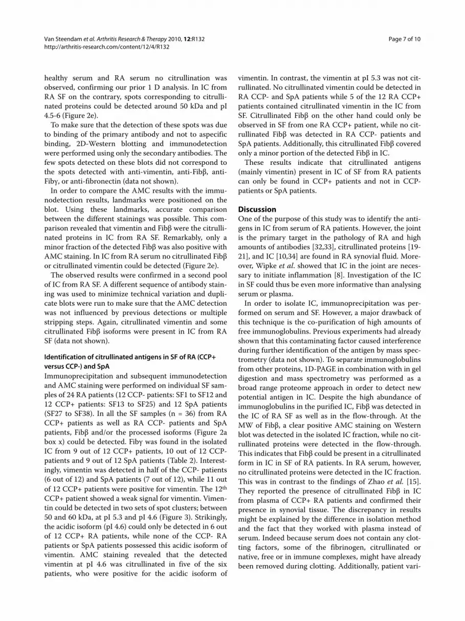
CCP+ patient showed a weak signal for vimentin. Vimen-
tin could be detected in two sets of spot clusters; between
50 and 60 kDa, at pI 5.3 and pI 4.6 (Figure 3). Strikingly,
the acidic isoform (pI 4.6) could only be detected in 6 out
of 12 CCP+ RA patients, while none of the CCP- RA
patients or SpA patients possessed this acidic isoform of
vimentin. AMC staining revealed that the detected
vimentin at pI 4.6 was citrullinated in five of the six
patients, who were positive for the acidic isoform of
vimentin. In contrast, the vimentin at pI 5.3 was not cit-
rullinated. No citrullinated vimentin could be detected in
RA CCP- and SpA patients while 5 of the 12 RA CCP+
patients contained citrullinated vimentin in the IC from
SF. Citrullinated Fibβ on the other hand could only be
observed in SF from one RA CCP+ patient, while no cit-
rullinated Fibβ was detected in RA CCP- patients and
SpA patients. Additionally, this citrullinated Fibβ covered
only a minor portion of the detected Fibβ in IC.
These results indicate that citrullinated antigens
(mainly vimentin) present in IC of SF from RA patients
can only be found in CCP+ patients and not in CCP-
patients or SpA patients.
DiscussionOne of the purpose of this study was to identify the anti-
gens in IC from serum of RA patients. However, the joint
is the primary target in the pathology of RA and high
amounts of antibodies [32,33], citrullinated proteins [19-
21], and IC [10,34] are found in RA synovial fluid. More-
over, Wipke et al. showed that IC in the joint are neces-
sary to initiate inflammation [8]. Investigation of the IC
in SF could thus be even more informative than analysing
serum or plasma.
In order to isolate IC, immunoprecipitation was per-
formed on serum and SF. However, a major drawback of
this technique is the co-purification of high amounts of
free immunoglobulins. Previous experiments had already
shown that this contaminating factor caused interference
during further identification of the antigen by mass spec-
trometry (data not shown). To separate immunoglobulins
from other proteins, 1D-PAGE in combination with in gel
digestion and mass spectrometry was performed as a
broad range proteome approach in order to detect new
potential antigen in IC. Despite the high abundance of
immunoglobulins in the purified IC, Fibβ was detected in
the IC of RA SF as well as in the flow-through. At the
MW of Fibβ, a clear positive AMC staining on Western
blot was detected in the isolated IC fraction, while no cit-
rullinated proteins were detected in the flow-through.
This indicates that Fibβ could be present in a citrullinated
form in IC in SF of RA patients. In RA serum, however,
no citrullinated proteins were detected in the IC fraction.
This was in contrast to the findings of Zhao et al. [15].
They reported the presence of citrullinated Fibβ in IC
from plasma of CCP+ RA patients and confirmed their
presence in synovial tissue. The discrepancy in results
might be explained by the difference in isolation method
and the fact that they worked with plasma instead of
serum. Indeed because serum does not contain any clot-
ting factors, some of the fibrinogen, citrullinated or
native, free or in immune complexes, might have already
been removed during clotting. Additionally, patient vari-

Van Steendam et al. Arthritis Research & Therapy 2010, 12:R132
http://arthritis-research.com/content/12/4/R132
Page 8 of 10
Table 2: Immunodetection of selected antigens in IC
obtained from SF of individual RA CCP+ and CCP- patients
Patient n° (RF;CCP) Fibβ Fibγ Vimentin
RA CCP- SF1 (6;0) + + +
SF2 (80;1) + + -
SF3 (0;1) + + -
SF4 (0;1,6) + + +
SF5 (0;2) + + +
SF6 (5;2) + + +
SF7 (0;3) + + +
SF8 (40;4) + - -
SF9 (0;5) + + -
SF10 (0;5) + - -
SF11 (80;7) + + +
SF12 (160;10) + + -
RA CCP+ SF13 (351;107) +* - +
SF14 (183;340) + - +
SF15 (1,280;533) + - +*
SF16 (1,280;608) + + +*
SF17 (1,280;710) + + +
SF18 (2,560;740) + + +
SF19 (10,240;767) + + weak
SF21 (640;1294) + + +
SF22 (0;1,600) + + +*
SF23 (160;1,600) + + +*
SF24 (0;1,600) + + +
SF25 (640;1,775) + + +*
SpA SF27 (0;0) + - -
SF28 (ND;1,6) + + +
SF29 (0;2) + + +
SF30 (0;2) + + +
SF31 (0;3) + + +
SF32 (0;4) + + -
SF33 (0;11) + + -
SF34 (0;ND) + + -
SF35 (ND;ND) + + -
SF36 (0;ND) + - +
SF37 (0;ND) + - +
SF38 (0;ND) + + +
(+) Indicates the positive immunodetection; (-) indicates absence of
the positive antibody staining and (*) indicates that the antigen was
citrullinated as confirmed by AMC staining. ND, not determined
ability or disease status variation can not be excluded as
contributing factors.
By means of this broad range approach, Fibβ, possibly
citrullinated, was found as an antigen in SF from RA
patients. However, it should be noted that due to the low
abundance of the antigens and the excess of immuno-
globulins, some antigens will not be detected in this
approach, especially the antigens that co-migrate with
immunoglobulins on 1 D SDS PAGE.
Therefore, in a second approach, 2 D PAGE was com-
bined with immunodetection to overcome the interfer-
ence of the immunoglobulins as well as the low
abundance of the antigens. By means of 2 D PAGE and
immunodetection we wanted to confirm the previous
results from the broad proteome approach and also anal-
yse the presence of well-known antigens in RA, such as
Fibβ, Fibγ, fibronectin and vimentin.
Fibβ and fibronectin were found in IC obtained from
healthy serum, RA serum and RA SF, indicating that the
presence of Fibβ and fibronectin in IC is not specific for
RA or RA SF. Fibγ and vimentin could only be detected in
the IC from RA SF (Figure 2). The extra isoforms of Fibβ
(Figure 2a, box x) were also exclusively found in SF.
Besides the presence/identification of the antigens in IC,
the citrullination status of these antigens was analyzed.
No citrullinated proteins were detected in IC of RA
serum, while RA SF contained different citrullinated pro-
teins, which confirmed previous 1D-PAGE analysis. The
citrullinated proteins in IC from RA SF were identified as
vimentin and Fibβ. The fact that vimentin was found as
antigen during 2 D analysis and not during the broad
range proteome approach with 1 D SDS PAGE, can be
explained by the fact that vimentin has the same molecu-
lar weight as the heavy chain of immunoglobulins. The
detection of citrullinated Fibβ on 2D-PAGE confirmed
our results from the broad range approach. During later
analysis we found that citrullinated Fibβ could only be
detected in patient 13 (SF13) (Table 2), which was present
in the pool for 1D-PAGE as well as the pool for 2D-PAGE.
However, it should be noted that fibrinogen, citrullinated
or not, could possibly be deposited in synovium tissue
and thereby not, or in a lesser extend, be detected in syn-
ovial fluid.
Next, we analysed individual SF samples from CCP+
RA patients and CCP- RA patients. SpA patients were
included as disease controls. Since fibronectin was pres-
ent in healthy serum as well as RA serum and RA SF, we
focussed on Fibβ, Fibγ and vimentin during further analy-
sis. When individual samples of CCP+, CCP- and SpA
synovial fluid were processed, we observed absence of
citrullinated proteins in IC in the CCP- patients and SpA
patients (n = 24) while half of the CCP+ patients con-
tained citrullinated vimentin or Fibβ.

Van Steendam et al. Arthritis Research & Therapy 2010, 12:R132
http://arthritis-research.com/content/12/4/R132
Page 9 of 10
Citrullinated fibrinogen is known to be present in SF of
RA patients [25]. Our data showed that in IC of SF only
Fibβ was citrullinated and that Fibγ was present exclu-
sively in IC of SF, but in a non-citrullinated form. The
Fibβ isoforms detected on the AMC blot of one patient,
however, were only a minor fraction of the Fibβ isoforms
detected with anti-Fibβ. The lower abundance of Fibβ
could be due to residing fibrinogen in the synovial tissue.
A remarkable difference between our data and previous
reports is the presence of citrullinated vimentin in IC of
CCP+ RA SF. This antigen could not be detected in IC of
healthy or RA serum. Citrullinated vimentin is known to
be an important antigen in RA [35] and is present in syn-
ovial fluid [21], but its presence in IC has not been
reported. Moreover, no citrullinated antigens were found
in IC of SF from our control groups, consisting of CCP-
RA patients and SpA patients.
Since CCP+ patients have a more destructive course of
disease and because CCP+ patients contain citrullinated
vimentin in their IC, we hypothesize that citrullinated
vimentin plays an important pathophysiological role in
the perpetuation of RA. Moreover, in contrast to Fibβ,
vimentin is an intracellular antigen and therefore not
expected in IC. Additionally, the presence of intracellular
citrullinated proteins in the synovium is specific for RA,
while extracellular citrullinated proteins lack this speci-
ficity [36]. These RA specific synovial intracellular citrul-
linated proteins are also associated with significantly
higher systemic and local ACPA levels and with local
ACPA production in the joint [36].
ConclusionsOur data reveal the presence of citrullinated vimentin
and a less pronounced presence of citrullinated Fibβ in
RA SF (of CCP+ patients), while no citrullinated proteins
could be detected in IC from RA serum and healthy
serum or in IC from SF of RA CCP- patients and SpA
patients. Combining these findings with the five-point
circle of van Venrooij [26] we conclude that CCP+ RA
patients are more susceptible to the perpetuation of
inflammation and possibly have a more severe disease
state because of the presence of citrullinated vimentin
and Fibβ in their SF IC. Taken together, our data indicate
that citrullinated vimentin is an important antigen in IC
of CCP+ RA patients and therefore implies its impor-
tance in the pathology of RA.
Abbreviations
2D-PAGE: two dimensional polyacrylamide gelelectrophoresis; ACPA: anti-cit-
rullinated protein/peptide antibody; AMC: anti-modified citrulline; CAPS: 3-
(cyclohexylamino)-1-propanesulfonic acid; CCP: anti-cyclic citrullinated pep-
tide; CHAPS: 3-((3-Cholamidopropyl)dimethylammonio)-1-propanesulfonate;
DTT: dithiothreitol; ECL: enhanced chemiluminescence; ELISA: enzyme linked
immunosorbent assay; ESI: electrospray ionisation; Fib: fibrinogen; GM-CSF:
granulocyte-macrophage colony-stimulating factor; HRP: horse radish peroxi-
dise; IAA: iodoacetamide; IC: immune complex; Ig: immunoglobulin; IL: inter-
leukin; IP: immunoprecipitation; IPG: immobilized pH gradient; LC: liquid
chromatography; M: molar; MS: mass spectrometry; MW: molecular weight;
NO: nitrogen oxide; OA: osteo-arthritis; PAD: peptidyl arginine deiminase;
PBMC: peripheral blood mononuclear cells; PBS: phosphate buffer saline; PEG:
polyethylene glycol; pI: iso-electric point; Q-TOF: quadrupole time of flight; RA:
rheumatoid arthritis; RF: rheumatoid factor; SDS: sodium dodecyl sulphate;
SDS-PAGE: sodium dodecyl sulphate-polyacrylamide gelelectrophoresis; SF:
synovial fluid; SpA: Spondyloarthropathy; TNFα: tumor necrosis factor.
Competing interests
The authors declare that they have no competing interests.
Authors' contributions
KVS performed most of the practical work and data-analysis and wrote the
manuscript. KT assisted in performing ultracentrifugation, immunoprecipita-
tion and Western blot analysis. She also helped to draft the manuscript. MDC
gave practical assistance during the experiments. DE, FDK and DD helped in
the design of the study and the critical analysis of the data. All authors read and
approved the manuscript.
Authors' information
The locations where the authors' completed their education follow: KVS,
PharmD, Ghent University, Belgium; KT, MSc, PhD, Ghent University Hospital,
Belgium; MDC, PharmD, Ghent University, Belgium; DE, MD, PhD, Ghent Univer-
sity Hospital, Belgium; FDK, MD, PhD, Ghent University Hospital, Belgium; DD,
PharmD, PhD, Ghent University, Belgium
Acknowledgements
This study has been supported by FWO Flanders (Belgium). The authors kindly
acknowledge Lars Hulpio and Jens Van Praet for assistance in sample collec-
tion.
Figure 3 Different isoforms of vimentin on Western blot. Immunodetection with anti-vimentin on Western blot. A: shows the combination of the
acidic (pI = 4.6) and the basic (pI = 5.3) isoform of vimentin; B: acidic isoform of vimentin; C: the basic isoform of vimentin. The acidic isoforms (A and
B) correspond with citrullinated vimentin as detected with AMC.

Van Steendam et al. Arthritis Research & Therapy 2010, 12:R132
http://arthritis-research.com/content/12/4/R132
Page 10 of 10
Author Details1Laboratory for Pharmaceutical Biotechnology, Ghent University,
Harelbekestraat 72, B-9000 Ghent, Belgium and 2Department of
Rheumatology, Ghent University Hospital, De Pintelaan 185, B-9000 Ghent,
Belgium
References
1. Alamanos Y, Drosos AA: Epidemiology of adult rheumatoid arthritis.
Autoimmun Rev 2005, 4:130-136.
2. Klareskog L, Ronnelid J, Lundberg K, Padyukov L, Alfredsson L: Immunity
to citrullinated proteins in rheumatoid arthritis. Annu Rev Immunol
2008, 26:651-675.
3. Weissmann G: The pathogenesis of rheumatoid arthritis. Bull NYU Hosp
Jt Dis 2006, 64:12-15.
4. Weissmann G: Pathogenesis of rheumatoid arthritis. J Clin Rheumatol
2004, 10:S26-31.
5. Dorner T, Egerer K, Feist E, Burmester G: Rheumatoid factor revisited.
Current Opinion In Rheumatology 2004, 16:246-253.
6. Scrivo R, Di Franco M, Spadaro A, Valesini G: The immunology of
rheumatoid arthritis. Ann N Y Acad Sci 2007, 1108:312-322.
7. Shmagel KV, Chereshnev VA: Molecular bases of immune complex
pathology. Biochemistry (Mosc) 2009, 74:469-479.
8. Wipke BT, Wang Z, Nagengast W, Reichert DE, Allen PM: Staging the
initiation of autoantibody-induced arthritis: a critical role for immune
complexes. J Immunol 2004, 172:7694-7702.
9. Jarvis JN, Wang W, Moore HT, Zhao L, Xu C: In vitro induction of
proinflammatory cytokine secretion by juvenile rheumatoid arthritis
synovial fluid immune complexes. Arthritis Rheum 1997, 40:2039-2046.
10. Schuerwegh AJ, Dombrecht EJ, Stevens WJ, Van Offel JF, Kockx MM, Bridts
CH, De Clerck LS: Synovial fluid and peripheral blood immune
complexes of patients with rheumatoid arthritis induce apoptosis in
cytokine-activated chondrocytes. Rheumatol Int 2007, 27:901-909.
11. Mathsson L, Lampa J, Mullazehi M, Ronnelid J: Immune complexes from
rheumatoid arthritis synovial fluid induce FcgammaRIIa dependent
and rheumatoid factor correlated production of tumour necrosis
factor-alpha by peripheral blood mononuclear cells. Arthritis Res & Ther
2006, 8:R64.
12. Gioud-Paquet M, Auvinet M, Raffin T, Girard P, Bouvier M, Lejeune E,
Monier JC: IgM rheumatoid factor (RF), IgA RF, IgE RF, and IgG RF
detected by ELISA in rheumatoid arthritis. Ann Rheum Dis 1987,
46:65-71.
13. De Rycke L, Peene I, Hoffman IE, Kruithof E, Union A, Meheus L, Lebeer K,
Wyns B, Vincent C, Mielants H, Boullart L, Serre G, Veys EM, De Keyser F:
Rheumatoid factor and anticitrullinated protein antibodies in
rheumatoid arthritis: diagnostic value, associations with radiological
progression rate, and extra-articular manifestations. Ann Rheum Dis
2004, 63:1587-1593.
14. Renaudineau Y, Jamin C, Saraux A, Youinou P: Rheumatoid factor on a
daily basis. Autoimmunity 2005, 38:11-16.
15. Zhao X, Okeke NL, Sharpe O, Batliwalla FM, Lee AT, Ho PP, Tomooka BH,
Gregersen PK, Robinson WH: Circulating immune complexes contain
citrullinated fibrinogen in rheumatoid arthritis. Arthritis Res Ther 2008,
10:R94.
16. Schellekens GA, Visser H, de Jong BA, van den Hoogen FH, Hazes JM,
Breedveld FC, van Venrooij WJ: The diagnostic properties of rheumatoid
arthritis antibodies recognizing a cyclic citrullinated peptide. Arthritis
Rheum 2000, 43:155-163.
17. Robinson MW, Scott DG, Bacon PA, Walton KW, Coppock JS, Scott DL:
What proteins are present in polyethylene glycol precipitates from
rheumatic sera? Ann Rheum Dis 1989, 48:496-501.
18. McDougal JS, Redecha PB, Inman RD, Christian CL: Binding of
immunoglobulin G aggregates and immune complexes in human sera
to Staphylococci containing protein A. J Clin Invest 1979, 63:627-636.
19. Takizawa Y, Suzuki A, Sawada T, Ohsaka M, Inoue T, Yamada R, Yamamoto
K: Citrullinated fibrinogen detected as a soluble citrullinated
autoantigen in rheumatoid arthritis synovial fluids. Ann Rheum Dis
2006, 65:1013-1020.
20. Tabushi Y, Nakanishi T, Takeuchi T, Nakajima M, Ueda K, Kotani T, Makino S,
Shimizu A, Hanafusa T, Takubo T: Detection of citrullinated proteins in
synovial fluids derived from patients with rheumatoid arthritis by
proteomics-based analysis. Ann Clin Biochem 2008, 45:413-417.
21. Tilleman K, Van Steendam K, Cantaert T, De Keyser F, Elewaut D, Deforce
D: Synovial detection and autoantibody reactivity of processed
citrullinated isoforms of vimentin in inflammatory arthritides.
Rheumatology (Oxford) 2008, 47:597-604.
22. Chang X, Yamada R, Suzuki A, Kochi Y, Sawada T, Yamamoto K:
Citrullination of fibronectin in rheumatoid arthritis synovial tissue.
Rheumatology (Oxford) 2005, 44:1374-1382.
23. Sawada T, Hashimoto S, Kanzaki T, Suzuki A, Yamada R, Shoji A, Hayashi H,
Tahara K, Yamamoto K: Identification of citrullinated antigen as
component of immune complex in synovial fluids from patients with
rheumatoid arthritis. Arthritis and Rheumatism 2008, 58:S520-S520.
24. Skriner K, Adolph K, Jungblut PR, Burmester GR: Association of
citrullinated proteins with synovial exosomes. Arthritis Rheum 2006,
54:3809-3814.
25. Matsuo K, Xiang Y, Nakamura H, Masuko K, Yudoh K, Noyori K, Nishioka K,
Saito T, Kato T: Identification of novel citrullinated autoantigens of
synovium in rheumatoid arthritis using a proteomic approach. Arthritis
Res Ther 2006, 8:R175.
26. van Venrooij WJ, Pruijn GJ: An important step towards completing the
rheumatoid arthritis cycle. Arthritis Res Ther 2008, 10:117.
27. Arnett F, Edworthy S, Bloch D, Mcshane D, Fries J, Cooper N, Healey L,
Kaplan S, Liang M, Luthra H, Medsger T, Mitchell D, Neustadt D, Pinals R,
Schaller J, Sharp J, Wilder R, Hunder G: The American-Rheumatism-
Association 1987 Revised Criteria For The Classification Of
Rheumatoid-Arthritis. Arthritis and Rheumatism 1988, 31:315-324.
28. Dougados M, van der Linden S, Juhlin R, Huitfeldt B, Amor B, Calin A, Cats
A, Dijkmans B, Olivieri I, Pasero G, et al.: The European
Spondylarthropathy Study Group preliminary criteria for the
classification of spondylarthropathy. Arthritis Rheum 1991,
34:1218-1227.
29. Rabilloud T, Valette C, Lawrence JJ: Sample application by in-gel
rehydration improves the resolution of two-dimensional
electrophoresis with immobilized pH gradients in the first dimension.
Electrophoresis 1994, 15:1552-1558.
30. Tilleman K, Van Beneden K, Dhondt A, Hoffman I, De Keyser F, Veys E,
Elewaut D, Deforce D: Chronically inflamed synovium from
spondyloarthropathy and rheumatoid arthritis investigated by protein
expression profiling followed by tandem mass spectrometry.
Proteomics 2005, 5:2247-2257.
31. Przybysz M, Borysewicz K, Szechinski J, Katnik-Prastowska I: Synovial
fibronectin fragmentation and domain expressions in relation to
rheumatoid arthritis progression. Rheumatology (Oxford) 2007,
46:1071-1075.
32. Rodriguez-Bayona B, Perez-Venegas JJ, Rodriguez C, Brieva JA: CD95-
Mediated control of anti-citrullinated protein/peptides antibodies
(ACPA)-producing plasma cells occurring in rheumatoid arthritis
inflamed joints. Rheumatology (Oxford) 2007, 46:612-616.
33. Snir O, Widhe M, Hermansson M, von Spee C, Lindberg J, Hensen S,
Lundberg K, Engstrom A, Venables PJ, Toes RE, Holmdahl R, Klareskog L,
Malmstrom V: Antibodies to several citrullinated antigens are enriched
in the joints of rheumatoid arthritis patients. Arthritis Rheum 2010,
62:44-52.
34. Agarwal V, Misra R, Aggarwal A: Immune complexes contain
immunoglobulin A rheumatoid factor in serum and synovial fluid of
patients with polyarticular juvenile rheumatoid arthritis.
Rheumatology (Oxford) 2002, 41:466-467.
35. Vossenaar ER, Despres N, Lapointe E, van der Heijden A, Lora M, Senshu T,
van Venrooij WJ, Menard HA: Rheumatoid arthritis specific anti-Sa
antibodies target citrullinated vimentin. Arthritis Res Ther 2004,
6:R142-150.
36. De Rycke L, Nicholas AP, Cantaert T, Kruithof E, Echols JD, Vandekerckhove
B, Veys EM, De Keyser F, Baeten D: Synovial intracellular citrullinated
proteins colocalizing with peptidyl arginine deiminase as
pathophysiologically relevant antigenic determinants of rheumatoid
arthritis-specific humoral autoimmunity. Arthritis Rheum 2005,
52:2323-2330.
doi: 10.1186/ar3070
Cite this article as: Van Steendam et al., Citrullinated vimentin as an impor-
tant antigen in immune complexes from synovial fluid of rheumatoid arthri-tis patients with antibodies against citrullinated proteins Arthritis Research &
Therapy 2010, 12:R132
Received: 20 November 2009 Revised: 10 June 2010
Accepted: 7 July 2010 Published: 7 July 2010This article is available from: http://arthritis-research.com/content/12/4/R132© 2010 Van Steendam et al.; licensee BioMed Central Ltd. This is an open access article distributed under the terms of the Creative Commons Attribution License (http://creativecommons.org/licenses/by/2.0), which permits unrestricted use, distribution, and reproduction in any medium, provided the original work is properly cited.Arthritis Research & Therapy 2010, 12:R132
http://www.ncbi.nlm.nih.gov/entrez/query.fcgi?cmd=Retrieve&db=PubMed&dopt=Abstract&list_uids=9365094
http://www.ncbi.nlm.nih.gov/entrez/query.fcgi?cmd=Retrieve&db=PubMed&dopt=Abstract&list_uids=3813676
http://www.ncbi.nlm.nih.gov/entrez/query.fcgi?cmd=Retrieve&db=PubMed&dopt=Abstract&list_uids=2742403
http://www.ncbi.nlm.nih.gov/entrez/query.fcgi?cmd=Retrieve&db=PubMed&dopt=Abstract&list_uids=3358796
http://www.ncbi.nlm.nih.gov/entrez/query.fcgi?cmd=Retrieve&db=PubMed&dopt=Abstract&list_uids=1930310