The secreted glycoprotein lubricin protects cartilage surfaces and inhibits synovial cell overgrowth
ARTHRITIS & RHEUMATISMVol. 58, No. 1, January 2008, pp 240–250DOI 10.1002/art.23143© 2008, American College of Rheumatology
A Biomarker-Based Mathematical Model to PredictBone-Forming Potency of Human Synovial and
Periosteal Mesenchymal Stem Cells
Cosimo De Bari,1 Francesco Dell’Accio,1 Alexandra Karystinou,1 Pascale V. Guillot,2Nicholas M. Fisk,2 Elena A. Jones,3 Dennis McGonagle,3 Ilyas M. Khan,4 Charles W. Archer,4
Thimios A. Mitsiadis,5 Ana Nora Donaldson,1 Frank P. Luyten,6 and Costantino Pitzalis1
Objective. To develop a biomarker-based model topredict osteogenic potency of human mesenchymal stemcells (MSCs) from synovial membrane and periosteum.
Methods. MSC populations were derived fromadult synovium and periosteum. Phenotype analysis wasperformed by fluorescence-activated cell sorting andreal-time reverse transcriptase–polymerase chain reac-tion (RT-PCR). Telomere lengths were determined bySouthern blot analysis. In vitro osteogenesis was as-sessed quantitatively by measurements of alkaline phos-phatase activity and calcium deposits. To investigatebone formation in vivo, MSCs were seeded onto osteoin-ductive scaffolds and implanted subcutaneously in nudemice. Bone was assessed by histology, and the humanorigin investigated by in situ hybridization for humanAlu genomic repeats. Quantitation was achieved byhistomorphometry and real-time RT-PCR for human
osteocalcin. Analysis at the single-cell level was per-formed with clonal populations obtained by limitingdilution. Multiple regressions were used to explore theincremental predictive value of the markers.
Results. Periosteal MSCs had significantlygreater osteogenic potency than did synovial MSCsinherent to the single cell. Bone was largely of humanorigin in vivo. Within the same tissue type, there wasvariability between different donors. To identify predic-tors of osteogenic potency, we measured the expressionlevels of osteoblast lineage genes in synovial and peri-osteal clonal MSCs prior to osteogenic treatment. Weidentified biomarkers that correlated with osteogenicoutcome and developed a mathematical model based ontype I collagen and osteoprotegerin expression thatpredicts the bone-forming potency of MSC prepara-tions, independent of donor-related variables and tissuesource.
Conclusion. Our findings indicate that ourquality-control mathematical model estimates the bone-forming potency of MSC preparations for bone repair.
Increasing evidence supports the clinical use ofstem cells for tissue repair. A major concern is the largestructural and clinical variability in outcome. Such vari-ability is at least partly due to donor-related factors andthe inconsistency of stem cell preparations. This prob-lem has been highlighted by the recent publication ofcontrasting results in clinical trials of cardiac cell therapy(1).
The known osteogenic capacity of mesenchymalstem cells (MSCs) (2–5) makes bone repair a naturalapplication of MSC preparations. MSC-based ap-proaches to bone repair would circumvent clinical com-plications associated with the use of autologous bone
Supported by MRC grant G108/620 and IWT grant 000259.Dr. De Bari is a Fellow of the Medical Research Council, UK. Dr.Dell’Accio is a Fellow of the Arthritis Research Campaign, UK. Dr.Guillot’s work was supported by Action Medical Research, UK.
1Cosimo De Bari, MD, PhD (current address: University ofAberdeen, Aberdeen, UK), Francesco Dell’Accio, MD, PhD (currentaddress: Queen Mary, University of London, London, UK), AlexandraKarystinou, MSc, Ana Nora Donaldson, MSc, PhD, CStat, CostantinoPitzalis, MD, PhD, FRCP (current address: Queen Mary, University ofLondon, London, UK): King’s College London, London, UK; 2PascaleV. Guillot, PhD, Nicholas M. Fisk, MBBS, PhD, FRCOG: ImperialCollege London, London, UK; 3Elena A. Jones, PhD, Dennis Mc-Gonagle, PhD, FRCPI: University of Leeds, Leeds, UK; 4Ilyas M.Khan, PhD, Charles W. Archer, PhD: Cardiff University, Cardiff, UK;5Thimios A. Mitsiadis, DDS, PhD, HDR: University of Zurich, Zurich,Switzerland; 6Frank P. Luyten, MD, PhD: Katholieke UniversiteitLeuven, Leuven, Belgium.
Address correspondence and reprint requests to Cosimo DeBari, MD, PhD, Professor of Translational Medicine, Department ofMedicine & Therapeutics, Institute of Medical Sciences, Foresterhill,Aberdeen AB25 2ZD, UK. E-mail: [email protected].
Submitted for publication January 3, 2007; accepted in re-vised form September 28, 2007.
240

grafts, such as donor site morbidity and the long-termdiscomfort that accompanies bone harvest. Transplanta-tion of allogeneic bone is also used, but is subject todisease transmission and host rejection. Proof-of-concept studies in humans support the clinical utility ofMSCs for bone repair (6–8). The incorporation of MSCsinto osteoinductive scaffolds accelerates the formationof bone with biomechanical properties equivalent tobone grafts in vivo (9).
Donor-associated factors and MSC preparationprotocols influence the bone-forming capacity of MSCs(10). In addition, MSCs isolated from different tissueshave distinct differentiation properties (11–15). Theresulting variability limits standardization of MSC-basedapproaches to bone repair and impedes comparison ofclinical study outcomes. There is, therefore, a pressingclinical need for assays that allow quantitative estimationof the bone-forming potency of MSC preparations. Suchpotency assays would allow development of qualitycontrols for efficacy of MSC preparations (16,17), aprerequisite for routine use in clinical practice.
In this study, we analyzed the variability in osteo-genic potency of matched human MSC preparationsfrom synovial membrane (SM) and periosteum andidentified biomarkers that, using a mathematical for-mula modeled on the quantitative outcome of 2 inde-pendent assays of bone formation, predict the bone-forming potency of MSC preparations independently ofdonor-related variables and tissue source.
MATERIALS AND METHODS
Cell isolation and culture. Samples of human perios-teum (wet weight 10–50 mg) were harvested aseptically fromthe proximal medial tibia of 4 donors of various ages (24, 36,58, and 83 years). Random biopsies of SM (wet weight 10–50mg) were obtained aseptically from the knee joints of the same4 donors. All specimens were obtained within 12 hours afterdeath. Donors had no history of knee joint disorders and didnot have active infections or tumors. Periosteal and synovialspecimens from each donor were processed in parallel usingthe same protocol, as previously described (5). Cells wereisolated and expanded in monolayer on plastic in growthmedium (high-glucose Dulbecco’s modified Eagle’s medium[DMEM; Life Technologies, Gaithersburg, MD] containing10% fetal bovine serum [selected lot from Life Technologies]and antibiotics [Life Technologies]) (5).
Cell cloning. Cell cloning was performed by limitingdilution as described elsewhere (4). First-passage synovial andperiosteal cells were suspended in growth medium and platedat a density of 0.5 cells/well in 96-well, flat-bottomed cultureplates. Cell populations arising from single cells were subcul-tured upon reaching confluence with serial 1:4 dilution pas-sages.
Telomere length assay. Genomic DNA was extractedfrom passage 7 periosteal and synovial MSC monolayers usinga standard protocol, and telomere lengths were determinedsemiquantitatively by Southern blotting using TeloTAGGGtelomere length assay, according to the recommendations ofthe manufacturer (Roche, Indianapolis, IN). Mean telomerelength was determined as described previously (18).
Phenotyping of culture-expanded MSCs using 3-colorflow cytometry. Culture-expanded (passage 4 to passage 6)MSCs were used for flow cytometry at 105 cells/test. Testantibodies were as follows: phycoerythrin (PE)–conjugatedlow-affinity nerve growth factor receptor (LNGFR)/p75,CD106 (vascular cell adhesion molecule 1), CD146 (Muc18),CD166 (activated leukocyte cell adhesion molecule), CD73/SH3 (all from PharMingen, San Diego, CA), CD105/SH2(Serotec, Oxford, UK), fluorescein isothiocyanate (FITC)–conjugated CD45 (Dako, Carpinteria, CA), and CD13 (Sero-tec). D7-FIB–PE was labeled in-house from purified D7-FIB(Serotec). Hybridoma cells B4-78 against bone and liver iso-forms of alkaline phosphatase were obtained from the Devel-opmental Studies Hybridoma Bank (Iowa City, IA). Hybrid-oma supernatant was produced in-house, and antibodylabeling was detected using secondary goat anti-mouse FITC(Serotec). Isotype-specific negative control antibodies werepurchased from Serotec. Dead cells were gated out based onpropidium iodide exclusion (Sigma, St. Louis, MO). All flowcytometry data were analyzed with WinMDI, version 8 soft-ware (Scripps Research Institute, La Jolla, CA).
In vitro osteogenesis. The in vitro osteogenesis assaywas performed as described previously (5). Alkaline phospha-tase activity was determined colorimetrically as describedpreviously (4), using a commercially available kit (ThermoElectron Corporation, Waltham, MA). Protein content wasdetermined by the Bradford method (Bio-Rad, Richmond,CA), using bovine serum albumin (Sigma) as standard. Alka-line phosphatase activity was expressed as arbitrary units permicrogram of protein content. To determine calcium content,cell layers were rinsed twice with phosphate buffered salineand scraped off the dish in 0.5N HCl. The cell layers wereextracted by shaking for 4 hours at 4°C, were centrifuged at1,000g for 5 minutes, and the supernatant was used for calciumdetermination using a commercially available kit, according tothe recommendations of the manufacturer (Thermo ElectronCorporation). Total calcium was calculated from standardsolutions and expressed as micrograms per microgram ofprotein content (determined in parallel wells).
In vivo osteogenesis. Animal experiments were per-formed in compliance with the UK Home Office and theEthics Committee for Animal Research at the KatholiekeUniversiteit. MSCs were seeded onto the osteoinductive Col-lagraft matrix (NeuColl, Campbell, CA) and implanted subcu-taneously into nude mice as described previously (4). Briefly,3-mm3 Collagraft cubes were wetted in DMEM, blotted onto agauze compress, and immersed into 50 !l of MSC suspension(105 cells/!l in growth medium) to allow attachment of thecells to the Collagraft matrix. After 1 hour of incubation at37°C, cells that did not attach to the Collagraft matrix butremained in suspension in the medium were counted andsubtracted from the total number of seeded cells (5 million) tocalculate the seeding efficiency as the percentage of cells thatremained in the Collagraft matrix after 1 hour of incubation.
MATHEMATICAL MODEL PREDICTING MSC OSTEOGENIC POTENCY 241

The seeding efficiency was independent of cell type androutinely varied between 40% and 70%. The cell–Collagraftconstructs were implanted subcutaneously into nude mice. Atdifferent time points postimplantation, mice were killed, andthe constructs dissected. The explants were then cut in two,and one half was used for total RNA extraction and the otherfixed overnight in 4% formaldehyde. Fixed samples weredecalcified overnight in Decal (Serva, Heidelberg, Germany),paraffin-embedded, and sectioned (into 7-!m–thick sections)at 3 different levels.
Histology and bone histomorphometry. Sections werestained with hematoxylin and eosin (H&E) and then assessedqualitatively for the presence of bone tissue and quantitativelyby bone histomorphometry. For each section, the total areaand the area covered with bone tissue were measured usingScion Image (Scion, Frederick, MD). The amount of bonetissue was then calculated as a percentage of the total implantarea.
In situ hybridization for human-specific Alu genomicrepeats. In situ hybridization for human Alu genomic repeatswas performed as described previously (19,20).
Total RNA extraction and reverse transcriptase–polymerase chain reaction (RT-PCR) analysis. Explants werehomogenized in TRIzol reagent (Life Technologies) using aPolytron homogenizer. Total RNA was isolated using TRIzol,
according to the recommendations of the manufacturer. AfterDNase treatment, complementary DNA (cDNA) sampleswere obtained by reverse transcription of 2 !g of total RNA(ThermoScript; Life Technologies) using oligo(dT)20 asprimer. Real-time quantitative PCR was carried out withSYBR Green using the Opticon real-time PCR cycler (MJResearch, San Francisco, CA). Gene expression of human cellswithin mouse tissue was evaluated using primers specific forhuman cDNA samples as described previously (4,21,22).Mouse bone was used to ensure specificity of the primers forhuman cDNA samples. Primer sequences are listed inTable 1.
Statistical analysis. Each assessment was performedon 2 or 3 cultures or constructs per cell source. Parametric datawere analyzed using Student’s t-test and are presented as themean ! SD. Analysis of variance with Bonferroni adjustmentfor multiple comparisons was used to evaluate alkaline phos-phatase activity over time in SM MSCs versus periostealMSCs. Multiple regressions were used to explore the incre-mental predictive value of the markers, taking into account anysignificant interactions. Polynomial and logarithmic transfor-mations of the markers were explored. Agreement of themeasurements of calcium with the model was assessed usingmixed linear regressions, to account for the effect of the donor,as well as the Bland and Altman method and intraclass
Table 1. Primers used for real-time RT-PCR analysis and sizes of PCR products*
Gene Primer sequence Amplicon (bp)
"-actin Forward 5"-CACGGCTGCTTCCAGCTC-3" 134Reverse 5"-CACAGGACTCCATGCCCAG-3"
GAPDH Forward 5"-AACAGCGACACCCACTCCTC-3" 85Reverse 5"-CATACCAGGAAATGAGCTTGACAA-3"
Runx2 Forward 5"-GCAGCACGCTATTAAATCCAAATT-3" 115Reverse 5"-GGCACGAAGGCTCATCATTC-3"
Osterix Forward 5"-CCCCACCTCTTGCAACCA-3" 102Reverse 5"-GGCTCCACCACTCCCTTCTAG-3"
ALP Forward 5"-CCCTTGACCCCCACAATGT-3" 80Reverse 5"-GTTGTTCCTGTTGACCTCGTACTG-3"
Type I collagen Forward 5"-CAGCCGCTTCACCTACAGC-3" 85Reverse 5"-TTTTGTATTCAATCACTGTCTTGCC-3"
OPG Forward 5"-GTGGACCACCCAGGAAACG-3" 132Reverse 5"-CGGTCTTCCACTTTGCTGTACAG-3"
Osteonectin Forward 5"-TCTTCCCTGTACACTGGCAGTTC-3" 115Reverse 5"-AACTGCTCCATGGGGATGGA-3"
Osteopontin Forward 5"-GCCGACCAAGGAAAACTCACTA-3" 107Reverse 5"-CAGAACTTCCAGAATCAGCCTGTT-3"
Osteocalcin Forward 5"-CCTCACACTCCTCGCCCTATT-3" 117Reverse 5"-CCCTCCTGCTTGGACACAAA-3"
BSP Forward 5"-AAACGAAGAAAGCGAAGCAGAA-3" 94Reverse 5"-GCTGCCGTTGCCGTTTT-3"
* RT-PCR # reverse transcriptase–polymerase chain reaction; ALP # alkaline phosphatase; OPG #osteoprotegerin; BSP # bone sialoprotein.
242 DE BARI ET AL

correlation coefficients (ICCs), and 95% confidence intervals(95% CIs) were calculated.
RESULTS
Greater bone-forming potency of MSCs fromperiosteum than from synovium. We quantified theosteogenic potency of matched human MSC prepara-tions from SM and periosteum. In vitro, passage 7 SM
MSCs and periosteal MSCs were treated with osteogenicmedium for 20 days and assessed for alkaline phospha-tase activity at different time points. In both MSCpopulations, there was a significant increase in alkalinephosphatase activity, which peaked on day 8 and thenreturned to almost basal levels by day 20 (Figure 1A). Afirst important source of variability that we identifiedwas the tissue of origin of MSCs. Indeed, at all time
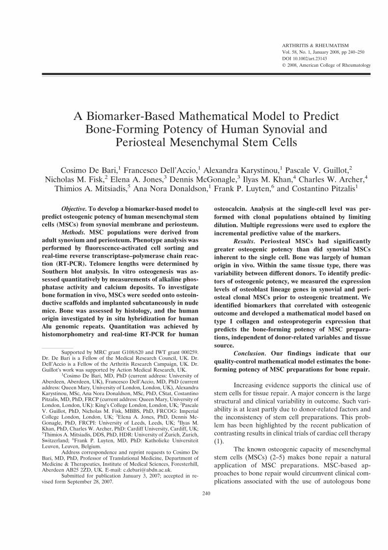
Figure 1. Osteogenic potency of synovial membrane (SM) mesenchymal stem cells (MSCs) and periosteal (Pe) MSCs. A and B, Osteogenesis invitro. Human SM MSCs and periosteal MSCs were treated with osteogenic medium. Alkaline phosphatase (ALP) activity (A) was determined ondays 0, 4, 8, 12, and 20, and MSC calcium deposition (B) was determined (in parallel wells) on days 0 and 20. Values are the mean ! SD of 4 donors.C–L, Bone formation in vivo. MSCs from 3 donors (ages 24, 36, and 83 years) were seeded onto Collagraft scaffolds, and the constructs implantedsubcutaneously into nude mice. C, Hematoxylin and eosin (H&E) staining of SM MSCs 8 weeks after implantation. D, Higher-magnification viewof the boxed area in C. E, H&E staining of periosteal MSCs 8 weeks after implantation. F, Higher-magnification view of the boxed area in E. G,Representative photomicrograph of an in situ hybridization for human Alu genomic repeats of a periosteal MSC construct at 8 weeks, counterstainedwith eosin. Inset, In situ hybridization for human Alu genomic repeats of mouse bone tissue used as a control, counterstained with eosin. Humannuclei are stained black. No nuclear counterstaining was performed, and therefore, Alu-negative nuclei are not visible. Bars # 100 !m. H,Quantification of the results of real-time quantitative reverse transcriptase–polymerase chain reaction for human osteocalcin (hOC) normalized tohuman "-actin. Expression of human OC was monitored in MSC monolayers and at 4 and 8 weeks after implantation of the constructs. The primersused were specific for human cDNA. The bone marker OC was barely detectable in the monolayer cultures, but OC levels increased progressivelyafter 4 and 8 weeks in vivo. At both time points examined, the levels of human OC in periosteal MSC constructs were significantly higher than thelevels in SM MSC constructs. Values are the mean ! SD of 3 donors. I, Quantification of in vivo bone formation assessed by histomorphometry.Values are the mean ! SD of 3 donors. J, Correlation between percent bone area and expression of human OC (normalized to human "-actin) at8 weeks in vivo. K, Correlation between micrograms of calcium per microgram of protein content at 20 days in vitro and percent bone area at 8 weeksin vivo. L, Correlation between micrograms of calcium per microgram of protein content at 20 days in vitro and expression of human OC (normalizedto human "-actin) at 8 weeks in vivo. Values in J–L are the mean. ! # P $ 0.05; !! # P $ 0.01, versus SM MSCs.
MATHEMATICAL MODEL PREDICTING MSC OSTEOGENIC POTENCY 243
points except days 0 and 20, periosteal MSCs displayedsignificantly higher levels of alkaline phosphatase activ-ity than did SM MSCs (Figure 1A). By day 20, calciumdeposits were significantly higher in periosteal MSCsthan in SM MSCs (Figure 1B). Under similar conditions,human dermal fibroblasts failed to undergo osteogenesis(data not shown). These data indicate greater osteogenicpotential of periosteal MSCs than SM MSCs in vitro.
To investigate whether the difference in osteo-genic potential was dependent on in vitro conditions, weexamined the capacity of SM MSCs and periostealMSCs to form bone in vivo. We used an establishedassay consisting of seeding cells onto osteoinductiveCollagraft scaffolds (composed of hydroxyapatite, trical-cium phosphate, and type I collagen, routinely used inorthopedic clinical practice) and implanting the con-structs under the skin of immunodeficient nude mice(4,20). The seeding efficiency of SM MSCs and perios-teal MSCs into Collagraft scaffolds was comparable(%40–70%), with no correlation between MSC seedingefficiency and bone formation in vivo. Histologically, nobone was evident at 4 weeks. At 8 weeks, areas of wovenbone were observed after H&E staining of both SMMSC–seeded (Figures 1C and D) and periosteal MSC–seeded scaffolds (Figures 1E and F).
In contrast, no bone was retrieved from emptyscaffolds or scaffolds seeded with expanded humandermal fibroblasts (data not shown), indicating thathuman MSCs are necessary for bone formation in vivounder these conditions. In situ hybridization for humanAlu genomic repeats demonstrated that human nucleiwere present within bone areas in both SM MSC andperiosteal MSC constructs (Figure 1G).
To confirm differentiation of human MSCs intoan osteoblast phenotype at the molecular level, wemonitored the expression of human osteocalcin (OC) byquantitative real-time RT-PCR using human-specificprimers. The levels of human OC messenger RNA(mRNA), normalized to human "-actin, were signifi-cantly higher in the periosteal MSC constructs at both 4and 8 weeks (Figure 1H), indicating that periostealMSCs had greater osteogenic potential in vivo than didSM MSCs.
To quantify bone formation, we measured thebone area in serial sections of MSC constructs byhistomorphometry, following H&E staining. At 8 weeks,the percent area occupied by bone was significantlyhigher in periosteal MSC constructs than in SM MSCconstructs (Figure 1I). The levels of OC mRNA de-tected with human-specific primers were significantly
higher in periosteal MSC than in SM MSC implants(Figure 1H) and correlated significantly with the percentbone area (gradient 121.4 [95% CI 112, 131]; R2 #0.997, P # 0.000) (Figure 1J).
A second source of variability within each tissuewas related to the individual donor. The coefficients ofvariation of OC expression levels and percent bone areain vivo were 55% and 90% for SM MSCs and 36% and41% for periosteal MSCs, respectively. This interindi-vidual variability was not due to unreliable measure-ments, since within each sample, the values of OCexpression and percent bone area, obtained using 2independent systems, displayed a high degree of corre-lation (Figure 1J).
Calcium deposition (normalized to protein con-tent) after 20 days of osteogenic treatment in vitrocorrelated significantly with both percent bone area(gradient 0.015 [95% CI 0.01, 0.02]; R2 # 0.941, P #0.0000) (Figure 1K) and expression levels of human OC(normalized to human "-actin; gradient 0.0001 [95% CI0.0001, 0.0002]; R2 # 0.944, P # 0.001) (Figure 1L) at 8weeks in vivo. This indicates that, under our experimen-tal conditions and at the specified time points, calciumdeposition (normalized to protein content) in the invitro osteogenesis assay was sufficient to measure osteo-genic outcome quantitatively.
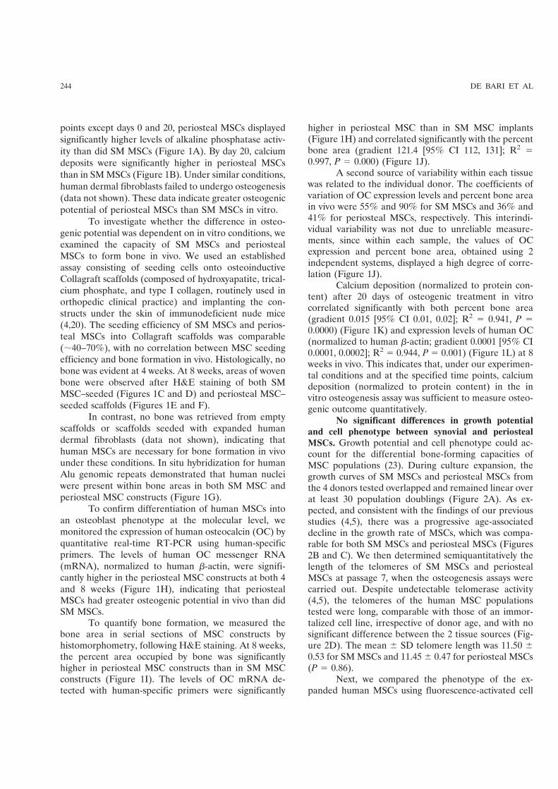
No significant differences in growth potentialand cell phenotype between synovial and periostealMSCs. Growth potential and cell phenotype could ac-count for the differential bone-forming capacities ofMSC populations (23). During culture expansion, thegrowth curves of SM MSCs and periosteal MSCs fromthe 4 donors tested overlapped and remained linear overat least 30 population doublings (Figure 2A). As ex-pected, and consistent with the findings of our previousstudies (4,5), there was a progressive age-associateddecline in the growth rate of MSCs, which was compa-rable for both SM MSCs and periosteal MSCs (Figures2B and C). We then determined semiquantitatively thelength of the telomeres of SM MSCs and periostealMSCs at passage 7, when the osteogenesis assays werecarried out. Despite undetectable telomerase activity(4,5), the telomeres of the human MSC populationstested were long, comparable with those of an immor-talized cell line, irrespective of donor age, and with nosignificant difference between the 2 tissue sources (Fig-ure 2D). The mean ! SD telomere length was 11.50 !0.53 for SM MSCs and 11.45 ! 0.47 for periosteal MSCs(P # 0.86).
Next, we compared the phenotype of the ex-panded human MSCs using fluorescence-activated cell
244 DE BARI ET AL

sorting. Both synovial and periosteal cell populationsdisplayed the conventional MSC phenotype (3,24,25),with no significant difference in the expression levels ofthe cell surface markers tested (Figure 2E). Histogramsare shown in Supplementary Figure 1, available on theArthritis & Rheumatism Web site at http://www.mrw.interscience.wiley.com/suppmat/0004-3591/suppmat/. Asexpected, CD45, a marker of hematopoietic lineagecells, was not detected in any of the MSC samples tested.Expression of CD73, CD166, CD105, CD13, and D7-FIB was detected uniformly in all MSC samples. CD146and CD106 displayed variable expression between do-nors, with no correlation with donor age. LNGFR was
undetectable in expanded MSCs (Figure 2E), consistentwith findings reported for MSCs from human bonemarrow (24). To exclude osteoblast contamination orearly osteogenic commitment of MSC populations, weanalyzed the cell surface expression levels of the osteo-blast lineage marker alkaline phosphatase (26,27). Bothsynovial and periosteal MSCs were negative for alkalinephosphatase (Figure 2E), consistent with the notion thatthey were undifferentiated MSCs with no detectablecommitment to the osteogenic lineage.
Osteogenic potency in single-cell–derived clonalpopulations. MSC populations were expanded as poly-clonal cultures. Thus, the different osteogenic capacities
Figure 2. Growth potential and phenotype of SM MSCs and periosteal MSCs. A, Kinetics of growth of MSCs from 4 donors. The kinetics of growthwere analyzed starting with the first passage. The growth curves overlapped and remained linear up to 30 population doublings, with no significantdifference between SM MSCs and periosteal MSCs. B and C, Age-associated decline in the growth rates of SM MSCs (B) and periosteal MSCs (C).D, Southern blot analysis of telomeres. Expanded MSCs from 3 donors of different ages were analyzed by Southern blot analysis to determine thelength of their telomeres. Regardless of donor age, the telomere lengths were comparable, with no significant difference between SM MSCs (S) andperiosteal MSCs (P). The U937 cell line was used as a positive control (high molecular weight [HMW]; mean telomere length 10.2 kb), and HL-60cells were used as a negative control (low molecular weight [LMW]; mean telomere length 3.9 kb). M # molecular weight marker. E, Surface markerphenotype of human MSCs following culture expansion. Values are the mean ! SD of 4 donors. Both SM MSCs and periosteal MSCs displayedhigh self-renewal capacity and a conventional MSC phenotype, and there was no significant difference between the 2 tissue sources. LNGFR #low-affinity nerve growth factor receptor (see Figure 1 for other definitions).
MATHEMATICAL MODEL PREDICTING MSC OSTEOGENIC POTENCY 245
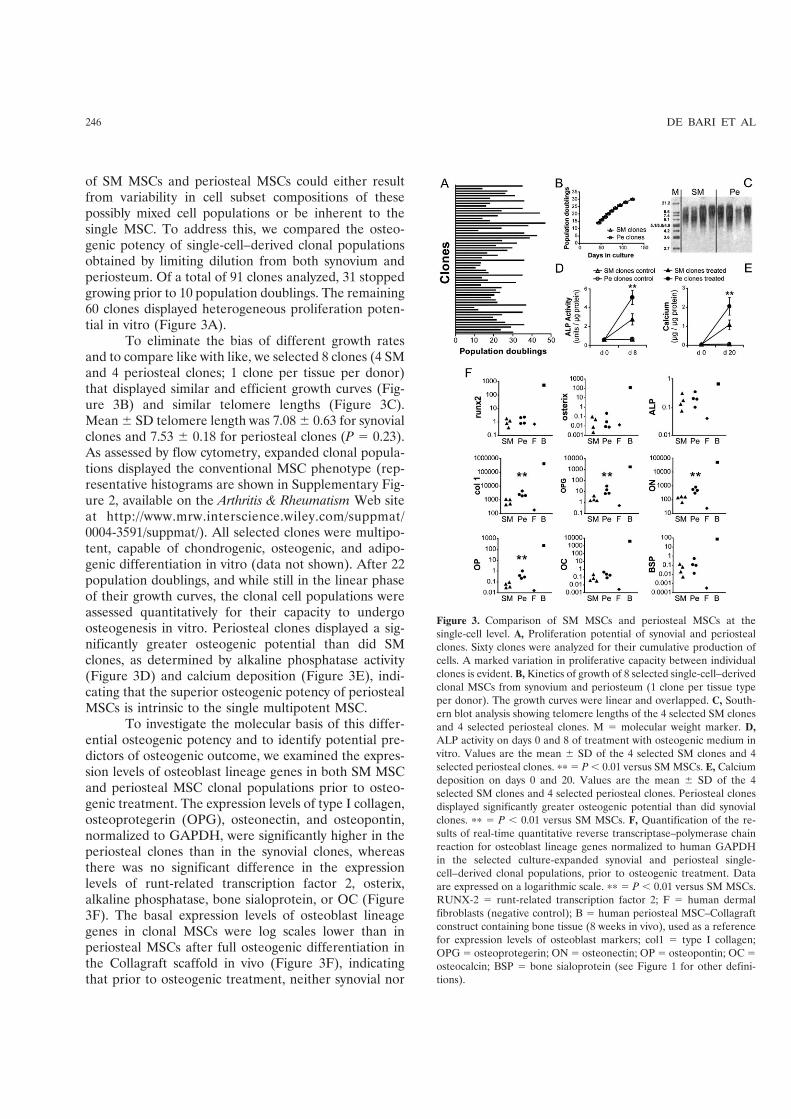
of SM MSCs and periosteal MSCs could either resultfrom variability in cell subset compositions of thesepossibly mixed cell populations or be inherent to thesingle MSC. To address this, we compared the osteo-genic potency of single-cell–derived clonal populationsobtained by limiting dilution from both synovium andperiosteum. Of a total of 91 clones analyzed, 31 stoppedgrowing prior to 10 population doublings. The remaining60 clones displayed heterogeneous proliferation poten-tial in vitro (Figure 3A).
To eliminate the bias of different growth ratesand to compare like with like, we selected 8 clones (4 SMand 4 periosteal clones; 1 clone per tissue per donor)that displayed similar and efficient growth curves (Fig-ure 3B) and similar telomere lengths (Figure 3C).Mean ! SD telomere length was 7.08 ! 0.63 for synovialclones and 7.53 ! 0.18 for periosteal clones (P # 0.23).As assessed by flow cytometry, expanded clonal popula-tions displayed the conventional MSC phenotype (rep-resentative histograms are shown in Supplementary Fig-ure 2, available on the Arthritis & Rheumatism Web siteat http://www.mrw.interscience.wiley.com/suppmat/0004-3591/suppmat/). All selected clones were multipo-tent, capable of chondrogenic, osteogenic, and adipo-genic differentiation in vitro (data not shown). After 22population doublings, and while still in the linear phaseof their growth curves, the clonal cell populations wereassessed quantitatively for their capacity to undergoosteogenesis in vitro. Periosteal clones displayed a sig-nificantly greater osteogenic potential than did SMclones, as determined by alkaline phosphatase activity(Figure 3D) and calcium deposition (Figure 3E), indi-cating that the superior osteogenic potency of periostealMSCs is intrinsic to the single multipotent MSC.
To investigate the molecular basis of this differ-ential osteogenic potency and to identify potential pre-dictors of osteogenic outcome, we examined the expres-sion levels of osteoblast lineage genes in both SM MSCand periosteal MSC clonal populations prior to osteo-genic treatment. The expression levels of type I collagen,osteoprotegerin (OPG), osteonectin, and osteopontin,normalized to GAPDH, were significantly higher in theperiosteal clones than in the synovial clones, whereasthere was no significant difference in the expressionlevels of runt-related transcription factor 2, osterix,alkaline phosphatase, bone sialoprotein, or OC (Figure3F). The basal expression levels of osteoblast lineagegenes in clonal MSCs were log scales lower than inperiosteal MSCs after full osteogenic differentiation inthe Collagraft scaffold in vivo (Figure 3F), indicatingthat prior to osteogenic treatment, neither synovial nor
Figure 3. Comparison of SM MSCs and periosteal MSCs at thesingle-cell level. A, Proliferation potential of synovial and periostealclones. Sixty clones were analyzed for their cumulative production ofcells. A marked variation in proliferative capacity between individualclones is evident. B, Kinetics of growth of 8 selected single-cell–derivedclonal MSCs from synovium and periosteum (1 clone per tissue typeper donor). The growth curves were linear and overlapped. C, South-ern blot analysis showing telomere lengths of the 4 selected SM clonesand 4 selected periosteal clones. M # molecular weight marker. D,ALP activity on days 0 and 8 of treatment with osteogenic medium invitro. Values are the mean ! SD of the 4 selected SM clones and 4selected periosteal clones. !! # P $ 0.01 versus SM MSCs. E, Calciumdeposition on days 0 and 20. Values are the mean ! SD of the 4selected SM clones and 4 selected periosteal clones. Periosteal clonesdisplayed significantly greater osteogenic potential than did synovialclones. !! # P $ 0.01 versus SM MSCs. F, Quantification of the re-sults of real-time quantitative reverse transcriptase–polymerase chainreaction for osteoblast lineage genes normalized to human GAPDHin the selected culture-expanded synovial and periosteal single-cell–derived clonal populations, prior to osteogenic treatment. Dataare expressed on a logarithmic scale. !! # P $ 0.01 versus SM MSCs.RUNX-2 # runt-related transcription factor 2; F # human dermalfibroblasts (negative control); B # human periosteal MSC–Collagraftconstruct containing bone tissue (8 weeks in vivo), used as a referencefor expression levels of osteoblast markers; col1 # type I collagen;OPG # osteoprotegerin; ON # osteonectin; OP # osteopontin; OC #osteocalcin; BSP # bone sialoprotein (see Figure 1 for other defini-tions).
246 DE BARI ET AL

periosteal clonal MSCs displayed an osteoblast pheno-type.
A biomarker-based mathematical model to pre-dict MSC osteogenic potency. Capitalizing on theseresults, we endeavored to identify predictors of osteo-genic potency of MSCs that would be independent of thetissue source and of the donor. To this end, we evaluatedthe correlations between the basal gene expression levelsin the 8 selected (4 synovial and 4 periosteal) clonalMSCs prior to osteogenic treatment and the values ofcalcium deposition at 20 days of osteogenic treatment invitro (Table 2). In a multiple linear regression model(R2 # 98%; adjusted 98%) of the individual markersstudied, type I collagen proved to be the most importantpredictor, followed by OPG. Although the bivariatecorrelation between calcium and OPG was moderate(R2 # 0.36), a curve fitting exercise indicated a strongquadratic trend (R2 # 0.91), thus confirming OPG as avalid predictor. Calcium increased on average by 0.43(95% CI 0.3, 0.53; P # 0.000) for each unit increase intype I collagen and by 0.012 (95% CI 0.001, 0.02; P #0.04) for each unit increase in OPG. The derived modelwas as follows:
Calcium/proteins # 0.70 & 0.43 ' (type I collagen/GAPDH)
& 0.012 ' (OPG/GAPDH)
There was no interaction with the type of clonalMSC population, indicating that this model can predictosteogenic outcome regardless of the tissue of origin ofMSCs.
The model showed excellent agreement with theactual calcium scale (Figure 4A). The Bland and Altmanplot (Figure 4B) proved that the bias was independent ofthe level of calcium assessed (the ICC was 98%). The
estimate of the average discrepancy in relation to theactual calcium scale was 0.01, with an SD of 0.09,yielding 95% limits of agreement of (0.07 (95% CI(0.08, (0.06) to 0.09 (95% CI 0.08, 0.10).
DISCUSSION
Several studies have demonstrated the therapeu-tic utility of MSCs in humans (6–8,28–31). However,donor-related variability and inconsistency of MSCpreparations limit standardization and impede compar-isons of clinical study outcomes. In this proof-of-conceptstudy, we proposed a strategy for tackling this problem.We developed a simple mathematical model based onthe expression of only 2 biomarkers, type I collagen andOPG, which predicts the osteogenic potency of adulthuman MSC preparations from synovium and perios-teum, independently of donor-related variables and tis-sue source.
The mathematical model was obtained usingsingle-cell–derived clonal populations. As with clonalpopulations, periosteal polyclonal MSCs displayed asignificantly greater osteogenic potency than did syno-vial polyclonal MSCs. However, within each tissuesource of MSCs, there was no statistically significantdifference in the osteogenic outcome between poly-clonal and clonal cell populations. In spite of this, whenwe applied the mathematical model to polyclonal syno-vial or periosteal MSC populations, it yielded an ICC of25%, thus showing low predictive power. In addition, itwas not possible to generate by regression analysis anymodel predictive of the osteogenic outcome of poly-clonal cell populations, at least with the molecularmarkers tested. A possible explanation is that in poly-
Figure 4. Analysis of the mathematical model predicting osteogenicoutcome. A, Plot of calcium predicted with the model versus calciummeasured, and line of equality. B, Bland and Altman plot of thedifference (measured minus predicted; bias) versus the average (mea-sured and predicted). Triangles represent SM MSCs, and circlesrepresent periosteal MSCs. See Figure 1 for definitions.
Table 2. Correlation of gene expression levels with calciumdeposition*
Gene R P
Runx2 0.108 0.400Osterix 0.164 0.349ALP 0.463 0.124Type I collagen 0.973 0.000OPG 0.598 0.059Osteonectin 0.904 0.001Osteopontin 0.322 0.218Osteocalcin 0.923 0.001BSP 0.338 0.206
* Basal expression levels of osteoblast lineage genes in clonal mesen-chymal stem cells prior to osteogenic treatment, and levels of calciumdeposition (normalized to protein content) at 20 days of osteogenictreatment in vitro were measured, and correlations determined. SeeTable 1 for definitions.
MATHEMATICAL MODEL PREDICTING MSC OSTEOGENIC POTENCY 247
clonal mixed cell populations, noncompetent, nonclono-genic cells would influence the overall gene expressionprior to osteogenic treatment, but are subsequently lostduring selective osteogenic conditions. In support of thishypothesis, in the osteogenic cultures a large proportionof cells underwent apoptosis within the first few days oftreatment, resulting in the selection of a subpopulationthat underwent osteogenesis (data not shown).
As reported with single-cell–derived colonies ofbone marrow–derived stromal cells (32–34), synovialand periosteal clonal populations became hetero-geneous during culture expansion, as determined by flowcytometry for the cell surface markers tested (Supple-mentary Figure 2). Nonetheless, the growth curves andthe osteogenic outcomes of individual clonal popula-tions were distinct, and it was possible to predict theirosteogenic potential using molecular markers. The phe-notypic heterogeneity within each clonal population maybe due to the different functional status of the individualcells of the single-cell progeny (e.g., different phases ofthe cell cycle, symmetric or asymmetric cell division, ordifferent stages of differentiation). However, in spite ofthe phenotypic heterogeneity during culture expansion,clones would maintain distinct, possibly inherent bio-logic features. The coexistence, in different proportions,of clones with diverse osteogenic potential within thesame polyclonal population would result in a variabilitytoo complex to be predicted by our marker analysis.
We are aware that the use of clonal populationsin the clinic would be impractical and costly, and there-fore, we do not advocate the use of our model for qualitycontrol of MSC populations for clinical use with thecurrent technologies. However, our biomarker-basedmodel represents a biologic platform upon which todevise strategies for the prospective purification of MSCsubpopulations with predictable and consistent bone-forming potency independently of the donor and tissuesource. Such purification, which could be either fromfresh synovial/periosteal tissue digests or from expandedpolyclonal cell populations, would not only increase theconsistency of MSC preparations for clinical use, butalso facilitate the estimation of their potency by usingbiomarkers, thus allowing standardization of therapeuticprotocols using MSCs.
Periosteal MSCs formed more bone in vivo thandid SM MSCs, as determined by histomorphometry. Thebone tissue retrieved appeared to be largely of humanorigin. Although we do not exclude contribution of themouse host to bone formation in vivo, the difference inbone amount is unlikely to be solely due to periostealMSCs having a greater capacity for recruiting mouse
host osteoprogenitor cells, since the levels of OC mRNAdetected with human-specific primers were significantlyhigher in periosteal MSC implants than in SM MSCimplants and correlated significantly with the percentbone area. Notably, no bone was retrieved from emptyscaffolds or scaffolds seeded with expanded humandermal fibroblasts, indicating that human MSCs arenecessary for bone formation in vivo under these exper-imental conditions.
The development of a biomarker-based modelthat predicts the bone-forming potency of human MSCpreparations is of considerable clinical relevance. It mayfacilitate the selection of individuals that qualify for anMSC-based bone repair approach. It may also helpidentify the best source and preparation protocol ofhuman MSCs or adjust the dose in bone-forming MSCunits from patient to patient. It remains to be investi-gated whether the same formula can be applied to MSCpreparations from other tissue sources, such as bonemarrow, or to any other stem cell preparations, whetherembryonic, fetal, or adult. We predict that, in addition tothe properties intrinsic to the cell preparation, otherfactors such as inflammation, biomechanics, and patientcomorbidity will influence orthotopic bone formationand clinical outcome. Preclinical studies and clinicaltrials of bone repair using MSC-based protocols will thusbe necessary for prospective validation of our potencyassays and related model.
The generation of consistent MSC-based thera-peutic protocols is a prerequisite for translating thesetechnologies into routine clinical application. There isevidence that MSCs from mismatched donors could bepoorly immunogenic in recipients in some in vivo sys-tems (35,36). Also, the induction of tolerance to alloge-neic stem cells is a field of intense investigation (37).Once the immunologic barriers are overcome, potencyassays and related quality control measures will providea solid basis for establishing batches of certified stem cellproducts with specific clinical indications for allogeneictransplantation. This approach will increase consistencyand decrease costs of MSC therapies, thus acceleratingtranslation to routine clinical use.
ACKNOWLEDGMENT
We thank the staff of the mortuary at the UniversityHospitals, Katholieke Universiteit Leuven.
AUTHOR CONTRIBUTIONSDr. De Bari had full access to all of the data in the study and
takes responsibility for the integrity of the data and the accuracy of thedata analysis.
248 DE BARI ET AL
Study design. De Bari, Dell’Accio, Fisk, Luyten, Pitzalis.Acquisition of data. De Bari, Dell’Accio, Karystinou, Guillot, Jones,McGonagle, Khan, Archer.Analysis and interpretation of data. De Bari, Dell’Accio, Guillot, Fisk,Jones, Khan, Archer, Mitsiadis, Donaldson.Manuscript preparation. De Bari, Dell’Accio, Guillot, Fisk, Jones,McGonagle, Khan, Mitsiadis, Donaldson, Luyten, Pitzalis.Statistical analysis. De Bari, Dell’Accio, Jones, Khan, Donaldson.
REFERENCES
1. Rosenzweig A. Cardiac cell therapy—mixed results from mixedcells. N Engl J Med 2006;355:1274–7.
2. Prockop DJ. Marrow stromal cells as stem cells for nonhemato-poietic tissues. Science 1997;276:71–4.
3. Pittenger MF, Mackay AM, Beck SC, Jaiswal RK, Douglas R,Mosca JD, et al. Multilineage potential of adult human mesenchy-mal stem cells. Science 1999;284:143–7.
4. De Bari C, Dell’Accio F, Vanlauwe J, Eyckmans J, Khan IM,Archer CW, et al. Mesenchymal multipotency of adult humanperiosteal cells demonstrated by single-cell lineage analysis. Ar-thritis Rheum 2006;54:1209–21.
5. De Bari C, Dell’Accio F, Tylzanowski P, Luyten FP. Multipotentmesenchymal stem cells from adult human synovial membrane.Arthritis Rheum 2001;44:1928–42.
6. Quarto R, Mastrogiacomo M, Cancedda R, Kutepov SM, Mukh-achev V, Lavroukov A, et al. Repair of large bone defects with theuse of autologous bone marrow stromal cells. N Engl J Med2001;344:385–6.
7. Vacanti CA, Bonassar LJ, Vacanti MP, Shufflebarger J. Replace-ment of an avulsed phalanx with tissue-engineered bone. N EnglJ Med 2001;344:1511–4.
8. Warnke PH, Springer IN, Wiltfang J, Acil Y, Eufinger H, Weh-moller M, et al. Growth and transplantation of a custom vascu-larised bone graft in a man. Lancet 2004;364:766–70.
9. Wang T, Dang G, Guo Z, Yang M. Evaluation of autologous bonemarrow mesenchymal stem cell-calcium phosphate ceramic com-posite for lumbar fusion in rhesus monkey interbody fusion model.Tissue Eng 2005;11:1159–67.
10. Phinney DG, Kopen G, Righter W, Webster S, Tremain N,Prockop DJ. Donor variation in the growth properties and osteo-genic potential of human marrow stromal cells. J Cell Biochem1999;75:424–36.
11. Kuznetsov SA, Mankani MH, Gronthos S, Satomura K, Bianco P,Robey PG. Circulating skeletal stem cells. J Cell Biol 2001;153:1133–40.
12. Batouli S, Miura M, Brahim J, Tsutsui TW, Fisher LW, GronthosS, et al. Comparison of stem-cell-mediated osteogenesis anddentinogenesis. J Dent Res 2003;82:976–81.
13. Hui JH, Li L, Teo YH, Ouyang HW, Lee EH. Comparative studyof the ability of mesenchymal stem cells derived from bonemarrow, periosteum, and adipose tissue in treatment of partialgrowth arrest in rabbit. Tissue Eng 2005;11:904–12.
14. Matsubara T, Suardita K, Ishii M, Sugiyama M, Igarashi A, Oda R,et al. Alveolar bone marrow as a cell source for regenerativemedicine: differences between alveolar and iliac bone marrowstromal cells. J Bone Miner Res 2005;20:399–409.
15. Sakaguchi Y, Sekiya I, Yagishita K, Muneta T. Comparison ofhuman stem cells derived from various mesenchymal tissues:superiority of synovium as a cell source. Arthritis Rheum 2005;52:2521–9.
16. De Bari C, Pitzalis C, Dell’Accio F. Reparative medicine: fromtissue engineering to joint surface regeneration. Regen Med2006;1:59–69.
17. Luyten FP, Dell’Accio F, De Bari C. Skeletal tissue engineering:
opportunities and challenges. Best Pract Res Clin Rheumatol2001;15:759–69.
18. Harley CB, Futcher AB, Greider CW. Telomeres shorten duringageing of human fibroblasts. Nature 1990;345:458–60.
19. Dell’Accio F, De Bari C, Luyten FP. Molecular markers predictiveof the capacity of expanded human articular chondrocytes to formstable cartilage in vivo. Arthritis Rheum 2001;44:1608–19.
20. Dell’Accio F, De Bari C, Luyten FP. Microenvironment andphenotypic stability specify tissue formation by human articularcartilage-derived cells in vivo. Exp Cell Res 2003;287:16–27.
21. De Bari C, Dell’Accio F, Luyten FP. Failure of invitro–differentiated mesenchymal stem cells from the synovialmembrane to form ectopic stable cartilage in vivo. ArthritisRheum 2004;50:142–50.
22. De Bari C, Dell’Accio F, Vandenabeele F, Vermeesch JR, Ray-mackers JM, Luyten FP. Skeletal muscle repair by adult humanmesenchymal stem cells from synovial membrane. J Cell Biol2003;160:909–18.
23. Muraglia A, Cancedda R, Quarto R. Clonal mesenchymal progen-itors from human bone marrow differentiate in vitro according toa hierarchical model. J Cell Sci 2000;113(Pt 7):1161–6.
24. Jones EA, Kinsey SE, English A, Jones RA, Straszynski L,Meredith DM, et al. Isolation and characterization of bonemarrow multipotential mesenchymal progenitor cells. ArthritisRheum 2002;46:3349–60.
25. Jones EA, English A, Henshaw K, Kinsey SE, Markham AF,Emery P, et al. Enumeration and phenotypic characterization ofsynovial fluid multipotential mesenchymal progenitor cells ininflammatory and degenerative arthritis. Arthritis Rheum 2004;50:817–27.
26. Gronthos S, Zannettino AC, Graves SE, Ohta S, Hay SJ, SimmonsPJ. Differential cell surface expression of the STRO-1 and alkalinephosphatase antigens on discrete developmental stages in primarycultures of human bone cells. J Bone Miner Res 1999;14:47–56.
27. Stewart K, Walsh S, Screen J, Jefferiss CM, Chainey J, Jordan GR,et al. Further characterization of cells expressing STRO-1 incultures of adult human bone marrow stromal cells. J Bone MinerRes 1999;14:1345–56.
28. Wakitani S, Mitsuoka T, Nakamura N, Toritsuka Y, Nakamura Y,Horibe S. Autologous bone marrow stromal cell transplantationfor repair of full-thickness articular cartilage defects in humanpatellae: two case reports. Cell Transplant 2004;13:595–600.
29. Wakitani S, Imoto K, Yamamoto T, Saito M, Murata N, YonedaM. Human autologous culture expanded bone marrow mesenchy-mal cell transplantation for repair of cartilage defects in osteoar-thritic knees. Osteoarthritis Cartilage 2002;10:199–206.
30. Horwitz EM, Gordon PL, Koo WK, Marx JC, Neel MD, McNallRY, et al. Isolated allogeneic bone marrow-derived mesenchymalcells engraft and stimulate growth in children with osteogenesisimperfecta: implications for cell therapy of bone. Proc Natl AcadSci U S A 2002;99:8932–7.
31. Horwitz EM, Prockop DJ, Fitzpatrick LA, Koo WW, Gordon PL,Neel M, et al. Transplantability and therapeutic effects of bonemarrow-derived mesenchymal cells in children with osteogenesisimperfecta. Nat Med 1999;5:309–13.
32. Colter DC, Class R, DiGirolamo CM, Prockop DJ. Rapid expan-sion of recycling stem cells in cultures of plastic-adherent cellsfrom human bone marrow. Proc Natl Acad Sci U S A 2000;97:3213–8.
33. Colter DC, Sekiya I, Prockop DJ. Identification of a subpopulationof rapidly self-renewing and multipotential adult stem cells incolonies of human marrow stromal cells. Proc Natl Acad SciU S A 2001;98:7841–5.
34. Prockop DJ. Further proof of the plasticity of adult stem cells andtheir role in tissue repair. J Cell Biol 2003;160:807–9.
35. Arinzeh TL, Peter SJ, Archambault MP, van den Bos C, GordonS, Kraus K, et al. Allogeneic mesenchymal stem cells regenerate
MATHEMATICAL MODEL PREDICTING MSC OSTEOGENIC POTENCY 249
bone in a critical-sized canine segmental defect. J Bone Joint SurgAm 2003;85-A:1927–35.
36. Rodriguez AM, Pisani D, Dechesne CA, Turc-Carel C, KurzenneJY, Wdziekonski B, et al. Transplantation of a multipotent cellpopulation from human adipose tissue induces dystrophin expres-
sion in the immunocompetent mdx mouse. J Exp Med 2005;201:1397–405.
37. Fairchild PJ, Cartland S, Nolan KF, Waldmann H. Embryonicstem cells and the challenge of transplantation tolerance. TrendsImmunol 2004;25:465–70.
DOI 10.1002/art.23236
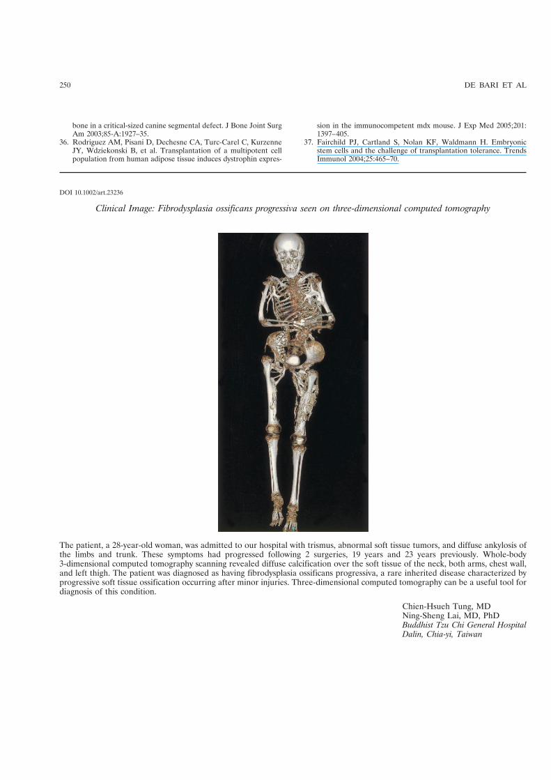
Clinical Image: Fibrodysplasia ossificans progressiva seen on three-dimensional computed tomography
The patient, a 28-year-old woman, was admitted to our hospital with trismus, abnormal soft tissue tumors, and diffuse ankylosis ofthe limbs and trunk. These symptoms had progressed following 2 surgeries, 19 years and 23 years previously. Whole-body3-dimensional computed tomography scanning revealed diffuse calcification over the soft tissue of the neck, both arms, chest wall,and left thigh. The patient was diagnosed as having fibrodysplasia ossificans progressiva, a rare inherited disease characterized byprogressive soft tissue ossification occurring after minor injuries. Three-dimensional computed tomography can be a useful tool fordiagnosis of this condition.
Chien-Hsueh Tung, MDNing-Sheng Lai, MD, PhDBuddhist Tzu Chi General HospitalDalin, Chia-yi, Taiwan
250 DE BARI ET AL
Copyright © 2022 FDOKUMEN