Evaluation of the Potency of the Pertussis Vaccine in ... - MDPI
Phytochemistry 69 (2008) 2627–2633
Contents lists available at ScienceDirect
Phytochemistry
journal homepage: www.elsevier .com/locate /phytochem
Non-cannabinoid constituents from a high potency Cannabis sativa variety
Mohamed M. Radwan a, Mahmoud A. ElSohly a,b,*, Desmond Slade a, Safwat A. Ahmed a, Lisa Wilson c,Abir T. El-Alfy c, Ikhlas A. Khan a,d, Samir A. Ross a,d,*
a National Center for Natural Products Research, School of Pharmacy, The University of Mississippi, University, MS 38677, USAb Department of Pharmaceutics, School of Pharmacy, The University of Mississippi, University, MS 38677, USAc Department of Pharmacology, School of Pharmacy, The University of Mississippi, University, MS 38677, USAd Department of Pharmacognosy, School of Pharmacy, The University of Mississippi, University, MS 38677, USA
a r t i c l e i n f o a b s t r a c t
Article history:Received 2 May 2008Received in revised form 3 June 2008Available online 4 September 2008
Keywords:Cannabis sativa L.CannabaceaeHigh potencyNon-cannabinoidAntimicrobialAntileishmanialAntimalarialAnti-oxidant
0031-9422/$ - see front matter � 2008 Elsevier Ltd. Adoi:10.1016/j.phytochem.2008.07.010
* Corresponding authors. Address: National Center fSchool of Pharmacy, The University of Mississippi, Un+1 662 915 1031; fax: +1 662 915 7989 (S.A. Ross), t662 915 5587 (M.A. ElSohly).
E-mail addresses: [email protected] (M.A. ElSoRoss).
Six new non-cannabinoid constituents were isolated from a high potency Cannabis sativa L. variety,namely 5-acetoxy-6-geranyl-3-n-pentyl-1,4-benzoquinone (1), 4,5-dihydroxy-2,3,6-trimethoxy-9,10-dihydrophenanthrene (2), 4-hydroxy-2,3,6,7-tetramethoxy-9,10-dihydrophenanthrene (3), 4,7-dime-thoxy-1,2,5-trihydroxyphenanthrene (4), cannflavin C (5) and b-sitosteryl-3-O-b-D-glucopyranoside-20-O-palmitate (6). In addition, five known compounds, a-cannabispiranol (7), chrysoeriol (8), 6-prenylap-igenin (9), cannflavin A (10) and b-acetyl cannabispiranol (11) were identified, with 8 and 9 beingreported for the first time from cannabis. Some isolates displayed weak to strong antimicrobial, antileish-manial, antimalarial and anti-oxidant activities. Compounds 2–4 were inactive as analgesics.
� 2008 Elsevier Ltd. All rights reserved.
1. Introduction
Cannabinoids are phenolic compounds possessing a C21 ter-penophenolic structure uniquely found in Cannabis sativa L. (ElS-ohly and Slade, 2005). Currently, 86 cannabinoids have beenisolated from cannabis (Ahmed et al., 2008; ElSohly and Slade,2005; Radwan et al., 2008). Non-cannabinoid constituents isolatedfrom cannabis include flavonoids, spiroindans, dihydrostilbenes,dihydrophenanthrenes, sterols and alkaloids, among others (Rossand ElSohly, 1995; Turner et al., 1980). As part of our program tostudy the constituents of high potency cannabis and their pharma-cology (Ahmed et al., 2008; Radwan et al., 2008), we herein reportthe isolation and structure elucidation of eleven non-cannabinoidconstituents including six new (1–6) and five known (7–11) com-pounds as well as their antimicrobial, antileishmanial, antimalarialand anti-oxidant activities. The analgesic activities of 2–4 are alsoreported.
ll rights reserved.
or Natural Products Research,iversity, MS 38677, USA. Tel.:el.: +1 662 915 5928; fax: +1
hly), [email protected] (S.A.
2. Results and discussions
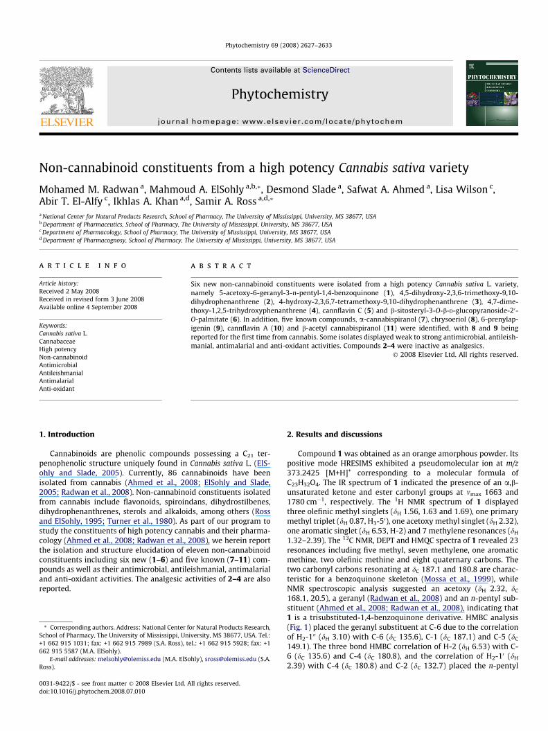
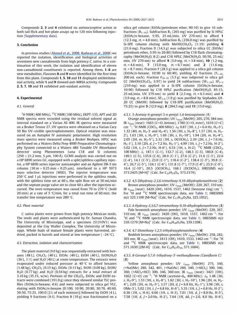
Compound 1 was obtained as an orange amorphous powder. Itspositive mode HRESIMS exhibited a pseudomolecular ion at m/z373.2425 [M+H]+ corresponding to a molecular formula ofC23H32O4. The IR spectrum of 1 indicated the presence of an a,b-unsaturated ketone and ester carbonyl groups at mmax 1663 and1780 cm�1, respectively. The 1H NMR spectrum of 1 displayedthree olefinic methyl singlets (dH 1.56, 1.63 and 1.69), one primarymethyl triplet (dH 0.87, H3-50), one acetoxy methyl singlet (dH 2.32),one aromatic singlet (dH 6.53, H-2) and 7 methylene resonances (dH
1.32–2.39). The 13C NMR, DEPT and HMQC spectra of 1 revealed 23resonances including five methyl, seven methylene, one aromaticmethine, two olefinic methine and eight quaternary carbons. Thetwo carbonyl carbons resonating at dC 187.1 and 180.8 are charac-teristic for a benzoquinone skeleton (Mossa et al., 1999), whileNMR spectroscopic analysis suggested an acetoxy (dH 2.32, dC
168.1, 20.5), a geranyl (Radwan et al., 2008) and an n-pentyl sub-stituent (Ahmed et al., 2008; Radwan et al., 2008), indicating that1 is a trisubstituted-1,4-benzoquinone derivative. HMBC analysis(Fig. 1) placed the geranyl substituent at C-6 due to the correlationof H2-100 (dH 3.10) with C-6 (dC 135.6), C-1 (dC 187.1) and C-5 (dC
149.1). The three bond HMBC correlation of H-2 (dH 6.53) with C-6 (dC 135.6) and C-4 (dC 180.8), and the correlation of H2-10 (dH
2.39) with C-4 (dC 180.8) and C-2 (dC 132.7) placed the n-pentyl
2628 M.M. Radwan et al. / Phytochemistry 69 (2008) 2627–2633
moiety at C-3. The location of the acetoxy group at C-5 wasdetermined by the four bond HMBC correlation between the acet-oxy methyl (dH 2.32) and C-5 (dC 149.1). The presence of the n-pen-tyl and geranyl groups were confirmed by COSY correlations(Fig. 1), establishing 1 as 5-acetoxy-6-geranyl-3-n-pentyl-1,4-benzoquinone.
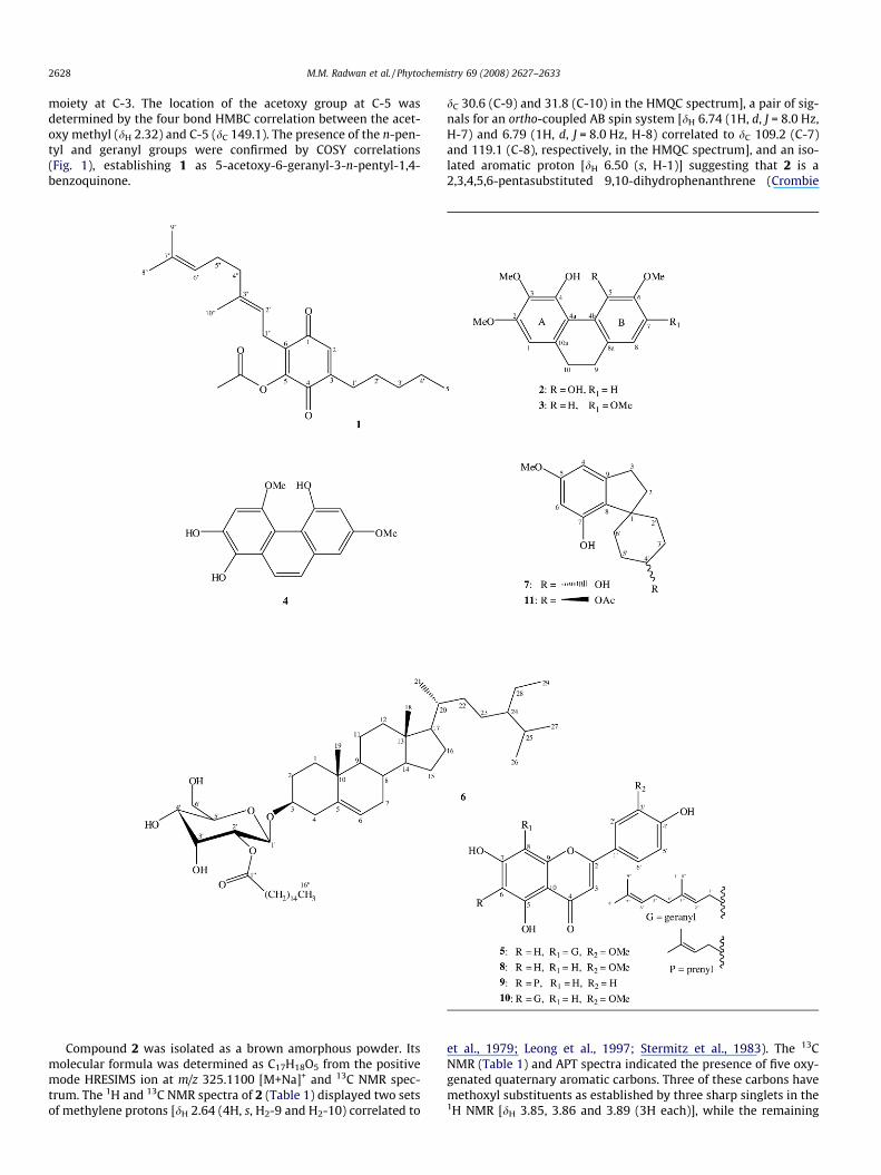
Compound 2 was isolated as a brown amorphous powder. Itsmolecular formula was determined as C17H18O5 from the positivemode HRESIMS ion at m/z 325.1100 [M+Na]+ and 13C NMR spec-trum. The 1H and 13C NMR spectra of 2 (Table 1) displayed two setsof methylene protons [dH 2.64 (4H, s, H2-9 and H2-10) correlated to
dC 30.6 (C-9) and 31.8 (C-10) in the HMQC spectrum], a pair of sig-nals for an ortho-coupled AB spin system [dH 6.74 (1H, d, J = 8.0 Hz,H-7) and 6.79 (1H, d, J = 8.0 Hz, H-8) correlated to dC 109.2 (C-7)and 119.1 (C-8), respectively, in the HMQC spectrum], and an iso-lated aromatic proton [dH 6.50 (s, H-1)] suggesting that 2 is a2,3,4,5,6-pentasubstituted 9,10-dihydrophenanthrene (Crombie
et al., 1979; Leong et al., 1997; Stermitz et al., 1983). The 13CNMR (Table 1) and APT spectra indicated the presence of five oxy-genated quaternary aromatic carbons. Three of these carbons havemethoxyl substituents as established by three sharp singlets in the1H NMR [dH 3.85, 3.86 and 3.89 (3H each)], while the remaining
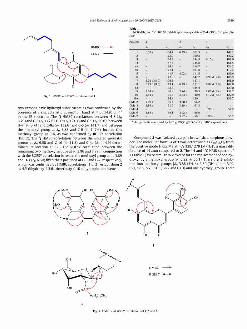
Table 11H (400 MHz) and 13C (100 MHz) NMR spectroscopic data of 2–4 (CDCl3, d in ppm, J inHz)a
Position 2 3 4
dH dC dH dC dH dC
1 6.50 s 104.4 6.39 s 103.9 – 140.22 – 152.0 – 150.3 – 140.23 – 136.4 – 134.5 6.15 s 107.64 – 147.2 – 146.8 – 161.5
4a – 114.9 – 114.7 – 124.24b – 121.1 – 107.8 – 117.4
5 – 141.7 8.02 s 111.5 – 156.66 – 147.6 – 147.2 6.93 d (2.0) 108.97 6.74 d (8.0) 109.2 – 147.1 – 161.08 6.79 d (8.0) 119.1 6.79 s 111.1 6.82 d (2.0) 102.0
8a – 132.6 – 125.4 – 129.09 2.64 s 30.6 2.74 s 29.5 8.06 d (8.4) 137.7
10 2.64 s 31.8 2.74 s 30.9 8.12 d (8.4) 122.910a – 136.4 – 130.1 – 132.7
OMe-2 3.89 s 56.3 3.88 s 56.2 – –OMe-3 3.86 s 61.0 3.89 s 61.3 – –OMe-4 – – – – 3.96 s 57.2OMe-6 3.85 s 56.1 3.92 s 56.0 – –OMe-7 – – 3.92 s 56.1 3.90 s 55.7
a Assignments confirmed by APT, gHMQC, gCOSY and gHMBC experiments.
HMBC
COSY
1
O
O
O
O
Fig. 1. HMBC and COSY correlations of 1.
M.M. Radwan et al. / Phytochemistry 69 (2008) 2627–2633 2629
two carbons have hydroxyl substituents as was confirmed by thepresence of a characteristic absorption band at mmax 3420 cm�1
in the IR spectrum. The 3J HMBC correlations between H-8 (dH
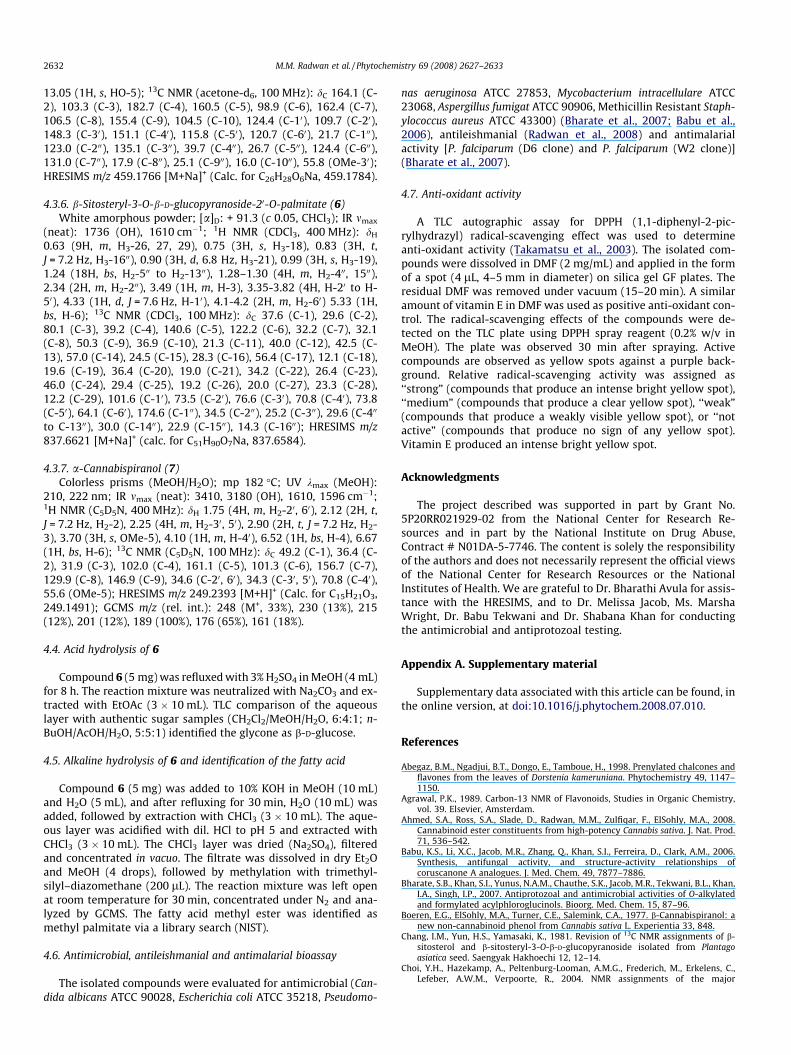
6.79) and C-6 (dC 147.6), C-4b (dC 121.1) and C-9 (dC 30.6); betweenH-7 (dH 6.74) and C-8a (dC 132.6) and C-5 (dC 141.7) and betweenthe methoxyl group at dH 3.85 and C-6 (dC 147.6), located thismethoxyl group at C-6, as was confirmed by ROESY correlation(Fig. 2). The 3J HMBC correlation between the isolated aromaticproton at dH 6.50 and C-10 (dC 31.8) and C-4a (dC 114.9) deter-mined its location at C-1. The ROESY correlation between theremaining two methoxyl groups at dH 3.86 and 3.89 in conjunctionwith the ROESY correlation between the methoxyl group at dH 3.89and H-1 (dH 6.50) fixed their positions at C-3 and C-2, respectively,which was confirmed by HMBC correlations (Fig. 2), establishing 2as 4,5-dihydroxy-2,3,6-trimethoxy-9,10-dihydrophenanthrene.
2
MeO
MeO OH HO OMe
H
HH
H
3
1'
2'3'
OOHO
OH
O
OH
O(CH2)14CH3
H
H
6
Fig. 2. HMBC and ROESY co
Compound 3 was isolated as a pale brownish, amorphous pow-der. The molecular formula of 3 was determined as C18H20O5 fromthe positive mode HRESIMS at m/z 339.1279 [M+Na]+, a mass dif-ference of 14 amu compared to 2. The 1H and 13C NMR spectra of3 (Table 1) were similar to 2 except for the replacement of one hy-droxyl by a methoxyl group (dH 3.92, dC 56.1). Therefore, 3 exhib-ited four methoxyl groups [dH 3.88 (3H, s), 3.89 (3H, s) and 3.92(6H, s); dC 56.0, 56.1, 56.2 and 61.3] and one hydroxyl group. Their
HMBC
ROESY
5
OO
OH O
OMe
OHH
rrelations of 2, 5 and 6.
2630 M.M. Radwan et al. / Phytochemistry 69 (2008) 2627–2633
locations on the dihydrophenanthrene skeleton were determinedby comparison to the 1H and 13C NMR data of 2 (Table 1) and con-firmed by HMBC and ROESY correlations, placing two methoxylgroups at C-2 and C-3 and the hydroxyl at C-4. The remainingmethoxyl groups are therefore attached to ring B. The presenceof a pair of isolated aromatic singlets [dH 6.79 (H-8) and 8.02 (H-5)] in the 1H NMR assigned the two methoxyl groups to C-6 (dC
147.2) and C-7 (dC 147.1) (Leong et al., 1997), which was confirmedby ROESY [dH 3.92 (OMe-6 and OMe-7)/H-5 and H-8; H-8/H2-9]and HMBC (H-5/C-7, C-4a; H-8/C-6, C-8a, C-9) correlations. Thus,the structure of 3 was established as 4-hydroxy-2,3,6,7-tetrameth-oxy-9,10-dihydrophenanthrene.
Compound 4 was isolated as a reddish brown powder. Itsmolecular formula was determined as C16H14O5 from negativemode HRESIMS (m/z 571.1630 [2M-H]�) and 13C NMR data. TheUV spectrum of 4 (kmax 258, 282 and 303 nm) is characteristic fora phenanthrene skeleton (Leong et al., 1997). The 1H NMR (Table1) displayed a pair of signals for an ortho-coupled AB spin system[dH 8.06 (1H, d, J = 8.4 Hz, H-9), 8.12 (1H, d, J = 8.4 Hz, H-10)], twometa-coupled protons [dH 6.82 (1H, d, J = 2.0 Hz, H-8), 6.93 (1H, d,J = 2.0 Hz, H-6)] and an isolated aromatic singlet [dH 6.15 (1H, s,H-3)] indicative of a pentasubstituted phenanthrene (Réthy et al.,2006; Leong et al., 1997). The 1H and 13C NMR spectra of 4 showedtwo aromatic methoxyl groups [dH 3.90 (3H, s) and 3.96 (3H, s)]and five oxygenated quaternary carbons, indicating that 4 hasthree hydroxyl groups. The ROESY correlation of the methoxylgroup at dH 3.90 with the protons at dH 6.93 (H-6) and 6.82 (H-8)established its location at C-7 (d 161.0), which was confirmed byHMBC [dH 3.90 (OMe-7)/C-7; H-8/C-7, C-6, C-8a, C-9; H-6/C-7, C-8] correlations. The ROESY and HMBC correlations of the methoxylgroup at dH 3.96 with H-3 (dH 6.15) and C-4 (dC 161.5), respectively,assigned its location at C-4. Therefore, the structure of 4 was deter-mined as 4,7-dimethoxy-1,2,5-trihydroxyphenanthrene.
Compound 5 was obtained as a yellow amorphous powder. Itspositive mode HRESIMS displayed an [M+Na]+ ion at m/z459.1766 suggesting C26H28O6 as the molecular formula and 13 de-grees of unsaturation. The IR spectrum showed absorption bands atmmax 3421 and 1662 cm�1 due to hydroxyl and carbonyl groups,respectively, while the UV absorption maxima at kmax 275 (bandI) and 340 (band II) nm were indicative of a flavone skeleton(Mabry et al., 1970). The 1H NMR spectrum of 5 established ahydrogen-bonded hydroxyl proton [dH 13.05 (s, HO-5)] whichwas confirmed by the bathochromic UV shift (+25 nm) of band IIupon the addition of AlCl3 to a methanolic solution of 5. Bathochro-mic UV shifts upon the addition of NaOMe (+61 nm) and NaOAc(+5 nm) suggested hydroxylation at C-40 and C-7, respectively(Mabry et al., 1970). The 1H NMR spectrum displayed two sharpsinglets at dH 6.66 (1H, H-3) and 6.36 (1H, H-6), one methoxylgroup (dH 3.98) and an ABX spin system of ring B [dH 7.01 (1H, d,J = 8.0 Hz, H-50), 7.58 (1H, d, J = 2.0 Hz, H-20), 7.64 (1H, dd, J = 2.0,8.0 Hz, H-60)]. The presence of a geranyl group was deduced fromthe three methyl singlets at dH 1.48, 1.53 and 1.82 and two olefinicproton triplets at dH 5.02 and 5.35 in the 1H NMR spectrum (Ahmedet al., 2008; Radwan et al., 2008). The 13C NMR, DEPT-135 andHMQC spectra displayed 26 resonances including three methyl,one methoxyl, three methylene, seven methine and 12 quaternarycarbons. The location of the methoxyl group was determined to beat C-30 from HMBC (OMe-30/C-30; H-50/C-30; H-20/C-30, C-40) andROESY (OMe-30/H-20) correlations (Fig. 2). The carbon resonanceat dC 98.9 corresponding to a proton singlet at dH 6.36 in the HMQCspectrum indicated an unsubstituted C-6 position (Agrawal, 1989).The location of the geranyl group at C-8 was confirmed by theHMBC correlation of the benzylic protons [dH 3.57 (2H, d, J = 6.8Hz, H2-100)] with C-8 (dC 106.5), C-7 (dC 162.4) and C-9 (dC 155.4)(Fig. 2). The spectroscopic data of 5 are similar to those reportedfor cannflavin A (10) (Agrawal, 1989; Choi et al., 2004) except for
the location of the geranyl group at C-8 instead of C-6, establishing5 as 8-geranyl-5,7,40-trihydroxy-30-methoxyflavone (cannflavin C).
Compound 6 was obtained as an optically active white amor-phous powder. Its molecular formula was deduced from the positivemode HRESIMS [M+Na]+ ion at m/z 837.6621 as C51H90O7. The 1HNMR spectrum displayed two tertiary [dH 0.75 (Me-18) and 0.99(Me-19)], three secondary [dH 0.63 (Me-26 and Me-27) and 0.90(Me-21)] and one primary [dH 0.63 (Me-29)] methyl groups in addi-tion to an olefinic proton at dH 5.33 (bs, H-6), indicating a sitosterolskeleton (Kovganko et al., 2000; Takemoto et al., 1967). The pres-ence of an anomeric proton [dH 4.33 (d, J = 7.6 Hz, H-10)] and carbon[dC 101.6 (C-10)] in the HMQC spectrum indicated monoglycosyla-tion at C-3 (dC 80.1) (Ishii et al., 1977) that was confirmed by HMBC(H-3/C-10; H-10/C-3) and ROESY (H-10/H-3) correlations (Fig. 2). Thesugar moiety was identified as b-D-glucopyranose by acid hydroly-sis of 6 and TLC comparison with authentic sugar samples. The 1Hand 13C NMR spectroscopic data of 6 were similar to those reportedfor b-sitosterol-3-O-b-D-glucopyranoside (Chang et al., 1981) withthe addition of 16 resonances characteristic for a palmitate moiety(Segre and Mannina, 1997), which was confirmed by methylationof the alkaline hydrolysis product of 6 followed by GCMS analysis.The downfield esterification shift of C-20 (+2 ppm), upfield shiftsof C-10 (�4 ppm) and C-30 (�3 ppm) and the four bond HMBC corre-lation of H-10 (dH 4.33) and C-100 (dC 174.6) (Fig. 2) placed the palmi-tate moiety at C-20 (Terui et al., 1976; Yamasaki et al., 1977). Thus,the structure of 6 was established as b-sitosteryl-3-O-b-D-glucopy-ranoside-20-O-palmitate.
Compound 7 was obtained as colorless prisms. Its positivemode HRESIMS gave an [M+H]+ ion at m/z 249.2393 correspondingto a molecular formula of C15H20O3. The GCMS showed a molecularion at m/z 248 (33%) and two characteristic ions at m/z 189 (100%)and 176 (65%) suggesting that 7 is a spiroindan derivative (El-Feraly et al., 1986). The 13C NMR, DEPT and HMQC spectra of 7 dis-played 15 resonances including one methoxyl, six methylene, twoaromatic methine, one sp3 oxymethine and five quaternary car-bons. The spectroscopic data of 7 are similar to those reportedfor b-cannabispiranol (Boeren et al., 1977; Shoyama and Nishioka,1978; Radwan et al., 2008) except for the downfield shift of theoxymethine carbon (+5.6 ppm), indicating a 40a-configuration.Although 7 is a known cannabis constituent (Crombie and Crom-bie, 1982), this is the first report of the full NMR assignments.
The flavones 8 and 9 were isolated as yellow amorphous pow-ders. Their molecular formulae were determined from the HRESIMSas C16H12O6 and C20H18O5, respectively. Their spectroscopic data(UV and NMR) were in agreement with reported values for chrysoe-riol (8) (Toth et al., 1980; Agrawal, 1989) and 6-prenylapigenin (9)(Abegaz et al., 1998). This is the first report of their isolation fromcannabis. The NMR spectra of 10 and 11 were identical with thoseof cannflavin A (Agrawal, 1989; Choi et al., 2004) and b-acetyl cann-abispiranol (Shoyama and Nishioka, 1978), respectively.
The antimicrobial, antileishmanial, antimalarial and anti-oxi-dant activities of the isolated compounds were tested. Compound1 displayed weak anti-MRSA (IC50 15.0 lg/mL), moderate antile-ishmanial (IC50 13.0 lg/mL) and mild antimalarial activity againstPlasmodium falciparum (D6 clone) and P. falciparum (W2 clone)with IC50 values of 2.8 and 2.6 lg/mL, respectively. Compound 5had moderate antileishmanial activity (IC50 17.0 lg/mL). Com-pound 9 showed moderate anti-MRSA (IC50 6.5 lg/mL), weak anti-candidal (IC50 20.0 lg/mL) and mild antimalarial activity against P.falciparum (D6 clone) and P. falciparum (W2 clone) with IC50 valuesof 2.8 and 2.0 lg/mL, respectively. Compound 10 exhibited strongantileishmanial activity (IC50 4.5 lg/mL). Compound 11 displayedweak antileishmanial activity (IC50 31.0 lg/mL).
Compounds 2, 5 and 11 displayed strong, 1, 7 and 10 moderateand 8 and 9 weak anti-oxidant activities in the DPPH assay, with 6being inactive.
M.M. Radwan et al. / Phytochemistry 69 (2008) 2627–2633 2631
Compounds 2, 3 and 4 exhibited no antinociceptive action inboth tail-flick and hot-plate assays up to 120 min following injec-tion (Supplementary data).
3. Conclusion
In previous studies (Ahmed et al., 2008; Radwan et al., 2008) wereported the isolation, identification and biological activities ofseventeen new cannabinoids from high potency C. sativa. In a con-tinuation of this work, the isolation and identification of elevennon-cannabinoid constituents is reported. Among them, 1–6 arenew metabolites. Flavones 8 and 9 were identified for the first timefrom this plant. Compounds 1, 5, 10 and 11 displayed antileishma-nial activity, while 1 and 9 showed anti-MRSA activity. Compounds2, 5, 7, 10 and 11 exhibited anti-oxidant activity.
4. Experimental
4.1. General
1H NMR (400 MHz), 13C NMR (100 MHz), DEPT-135, APT and 2DNMR spectra were recorded using the residual solvent signal asinternal standard on a Varian AS 400. IR spectra were measuredon a Bruker Tensor 27. UV spectra were obtained on a Varian Cary50 Bio UV–visible spectrophotometer. Optical rotation was mea-sured on an Autoplot IV automatic polarimeter. High resolutionmass spectra were measured using a Bruker BioApex. HPLC wasperformed on a Waters Delta Prep 4000 Preparative Chromatogra-phy System connected to a Waters 486 Tunable UV Absorbancedetector using Phenomenex Luna C18 and Si columns(250 � 21.2 mm, 5 lm, 100 Å). GCMS analysis was carried out ona HP 6890 series GC, equipped with a split/splitless capillary injec-tor, a HP 6890 series injector autosampler and an Agilent DB-5 mscolumn (30 m � 0.25 mm � 0.25 lm), interfaced to a HP 5973mass selective detector (MSD). The injector temperature was250 �C and 1 lL injections were performed in the splitless mode,with the splitless time set at 60 s, the split flow set at 50 mL/minand the septum purge valve set to close 60 s after the injection oc-curred. The oven temperature was raised from 70 to 270 �C (hold20 min) at a rate of 5 �C/min, for a total run time of 60 min; thetransfer line temperature was 280 �C.
4.2. Plant material
C. sativa plants were grown from high potency Mexican seeds.The seeds and plants were authenticated by Dr. Suman Chandra,The University of Mississippi, and a specimen (S1310V1) wasdeposited at the Coy Waller Complex, The University of Missis-sippi. Whole buds of mature female plants were harvested, air-dried, packed in barrels and stored at low temperature (�24 �C).
4.3. Extraction, isolation and characterization
The plant material (9.0 kg) was sequentially extracted with hex-anes (48 L), CH2Cl2 (40 L), EtOAc (40 L), EtOH (40 L), EtOH/H2O(36 L, 1:1) and H2O (40 L) at room temperature. The extracts wereevaporated under reduced pressure at 40 �C to afford hexanes(1.48 kg), CH2Cl2 (0.15 kg), EtOAc (0.13 kg), EtOH (0.09 kg), EtOH/H2O (0.77 kg) and H2O (0.54 kg) extracts for a total extract of3.16 kg (35.1%, w/w). Portions of the CH2Cl2, EtOAc and EtOH ex-tracts were combined (191.0 g) since they showed similar TLC pro-files (EtOAc/n-hexane, 4:6) and were subjected to silica gel VLC,eluting with EtOAc/n-hexane [0:100, 10:90, 20:80, 30:70, 40:60,50:50, 75:25, 100:0 (2 L of each mixture)] followed by EtOH (4 L),yielding 9 fractions (A-I). Fraction B (10 g) was fractionated on a
silica gel column (EtOAc/petroleum ether, 90:10) to give 16 sub-fractions (B1–16). Subfraction B1 (265 mg) was purified by Si HPLC(EtOAc/n-hexane, 5:95, 25 mL/min, UV 270 nm) to afford 1(12.3 mg, rt. = 4.0 min). Subfraction B3 (236.0 mg) was purified bySi-SPE column eluting with MeOH/CH2Cl2 (1:19) yielding 6(21.6 mg). Fraction D (14.3 g) was subjected to silica CC (EtOAc/petroleum ether, 5:95 to 20:80) followed by C18 flash chromatog-raphy (MeOH/H2O, 8:2) and C18 HPLC (MeCN/H2O, 50:50, 25 mL/min, UV 270 nm) to afford 9 (5.0 mg, rt. = 3.8 min), 10 (1.2 mg,rt. = 4.6 min), 7 (15.0 mg, rt. = 6.7 min) and 2 (11.6 mg,rt. = 8.7 min). Fraction F (28.5 g) was applied to a silica gel column(EtOAc/n-hexane, 10:90 to 60:40), yielding 42 fractions (F1–42,
200 mL each). Fraction F32–35 (5.5 g) was subjected to silica gelCC (MeOH/CH2Cl2, 3:97) to yield 24 subfractions (SF1–24). SF2–3
(210 mg) was applied to a Si-SPE column (EtOAc/n-hexane,10:90) followed by C18 HPLC purification (MeOH/H2O, 85:15,25 mL/min, UV 279 nm) to yield 3 (2.3 mg, rt. = 6.3 min) and 4(3.9 mg, rt. = 8.8 min). SF3–6 (1.9 g) was purified by Sephadex LH-20 CC (MeOH) followed by C18-SPE purification (MeOH/H2O,75:25) to give 5 (12.9 mg), 8 (284.5 mg) and 11 (15.0 mg).
4.3.1. 5-Acetoxy-6-geranyl-3-n-pentyl-1,4-benzoquinone (1)Orange amorphous powder; UV kmax (MeOH): 205, 270, 384 nm;
IR mmax (neat): 1663 (C@O, ketone), 1780 (C@O, ester), 1610 (C@C)cm�1; 1H NMR (CDCl3, 400 MHz): dH 0.87 (3H, t, J = 6.4 Hz, H3-50),1.32 (4H, m, H2-30 and H2-40), 1.56 (3H, s, H3-800), 1.57 (2H, m, H2-20), 1.63 (3H, s, H3-900), 1.69 (3H, s, H3-1000), 1.94 (2H, m, H2-400),2.05 (2H, m, H2-500), 2.32 (3H, s, OCOCH3), 2.39 (2H, t, J = 7.6 Hz,H2-10), 3.10 (2H, d, J = 7.2 Hz, H2-100), 4.99 (1H, t, J = 7.2 Hz, H-200),5.02 (1H, t, J = 7.2 Hz, H-600), 6.53 (1H, s, H-2); 13C NMR (CDCl3,100 MHz): dC 187.1 (C-1), 132.7 (C-2), 148.3 (C-3), 180.8 (C-4),149.1 (C-5), 135.6 (C-6), 28.9 (C-10), 27.6 (C-20), 31.6 (C-30), 22.6(C-40), 14.1 (C-50), 23.0 (C-100), 118.4 (C-200), 138.4 (C-300), 39.8 (C-400), 26.7 (C-500), 124.1 (C-600), 131.8 (C-700), 17.9 (C-800), 25.9 (C-900),16.4 (C-1000), 20.5 (OCOCH3), 168.1 (OCOCH3); HRESIMS m/z373.2425 [M+H]+ (Calc. for C23H33O4, 373.2379).
4.3.2. 4,5-Dihydroxy-2,3,6-trimethoxy-9,10-dihydrophenanthrene (2)Brown amorphous powder; UV kmax (MeOH): 220, 267, 310 nm;
IR mmax (neat): 3420 (OH), 1610, 1537, 1462 (benzene ring) cm�1;For 1H and 13C NMR spectroscopic data, see Table 1; HRESIMSm/z 325.1100 [M+Na]+ (Calc. for C17H18O5Na, 325.1052).
4.3.3. 4-Hydroxy-2,3,6,7-tetramethoxy-9,10-dihydrophenanthrene (3)Pale brownish amorphous powder; UV kmax (MeOH): 220, 267,
310 nm; IR mmax (neat): 3420 (OH), 1610, 1537, 1462 cm�1: For1H and 13C NMR spectroscopic data, see Table 1; HRESIMS m/z339.1279 [M+Na]+ (Calc. for C18H20O5Na, 339.1208).
4.3.4. 4,7-Dimethoxy-1,2,5-trihydroxyphenanthrene (4)Reddish brown amorphous powder; UV kmax (MeOH): 258, 282,
303 nm; IR mmax (neat): 3413 (OH), 1610, 1533, 1462 cm�1: For 1Hand 13C NMR spectroscopic data, see Table 1; HRESIMS m/z571.1630 [2M-H]� (Calc. for C32H27O10, 571.1604).
4.3.5. 8-Geranyl-5,7,40-trihydroxy-30-methoxyflavone (Cannflavin C)(5)
Yellow amorphous powder; UV kmax (MeOH): 275, 340,(+NaOMe) 280, 342, 401, (+NaOAc) 280, 340, (+AlCl3) 300, 346,360, (+AlCl3+HCl) 300, 346, 360 nm; IR mmax (neat): 3421 (OH),1662 (C@O) cm�1; 1H NMR (acetone-d6, 400 MHz): dH 1.48 (3H,s, H3-900), 1.53 (3H, s, H3-800), 1.82 (3H, s, H3-1000), 1.96 (2H, m, H2-400), 2.05 (2H, m, H2-500), 3.57 (2H, d, J = 6.8 Hz, H2-100), 3.98 (3H, s,OMe-30), 5.02 (1H, t, J = 6.8 Hz, H-600), 5.35 (1H, t, J = 6.8 Hz, H-200),6.36 (1H, s, H-6), 6.66 (1H, s, H-3), 7.01 (1H, d, J = 8.0 Hz, H-50),7.58 (1H, d, J = 2.0 Hz, H-20), 7.64 (1H, dd, J = 2.0, 8.0 Hz, H-60),
2632 M.M. Radwan et al. / Phytochemistry 69 (2008) 2627–2633
13.05 (1H, s, HO-5); 13C NMR (acetone-d6, 100 MHz): dC 164.1 (C-2), 103.3 (C-3), 182.7 (C-4), 160.5 (C-5), 98.9 (C-6), 162.4 (C-7),106.5 (C-8), 155.4 (C-9), 104.5 (C-10), 124.4 (C-10), 109.7 (C-20),148.3 (C-30), 151.1 (C-40), 115.8 (C-50), 120.7 (C-60), 21.7 (C-100),123.0 (C-200), 135.1 (C-300), 39.7 (C-400), 26.7 (C-500), 124.4 (C-600),131.0 (C-700), 17.9 (C-800), 25.1 (C-900), 16.0 (C-1000), 55.8 (OMe-30);HRESIMS m/z 459.1766 [M+Na]+ (Calc. for C26H28O6Na, 459.1784).
4.3.6. b-Sitosteryl-3-O-b-D-glucopyranoside-20-O-palmitate (6)White amorphous powder; [a]D: + 91.3 (c 0.05, CHCl3); IR mmax
(neat): 1736 (OH), 1610 cm�1; 1H NMR (CDCl3, 400 MHz): dH
0.63 (9H, m, H3-26, 27, 29), 0.75 (3H, s, H3-18), 0.83 (3H, t,J = 7.2 Hz, H3-1600), 0.90 (3H, d, 6.8 Hz, H3-21), 0.99 (3H, s, H3-19),1.24 (18H, bs, H2-500 to H2-1300), 1.28–1.30 (4H, m, H2-400, 1500),2.34 (2H, m, H2-200), 3.49 (1H, m, H-3), 3.35-3.82 (4H, H-20 to H-50), 4.33 (1H, d, J = 7.6 Hz, H-10), 4.1-4.2 (2H, m, H2-60) 5.33 (1H,bs, H-6); 13C NMR (CDCl3, 100 MHz): dC 37.6 (C-1), 29.6 (C-2),80.1 (C-3), 39.2 (C-4), 140.6 (C-5), 122.2 (C-6), 32.2 (C-7), 32.1(C-8), 50.3 (C-9), 36.9 (C-10), 21.3 (C-11), 40.0 (C-12), 42.5 (C-13), 57.0 (C-14), 24.5 (C-15), 28.3 (C-16), 56.4 (C-17), 12.1 (C-18),19.6 (C-19), 36.4 (C-20), 19.0 (C-21), 34.2 (C-22), 26.4 (C-23),46.0 (C-24), 29.4 (C-25), 19.2 (C-26), 20.0 (C-27), 23.3 (C-28),12.2 (C-29), 101.6 (C-10), 73.5 (C-20), 76.6 (C-30), 70.8 (C-40), 73.8(C-50), 64.1 (C-60), 174.6 (C-100), 34.5 (C-200), 25.2 (C-300), 29.6 (C-400
to C-1300), 30.0 (C-1400), 22.9 (C-1500), 14.3 (C-1600); HRESIMS m/z837.6621 [M+Na]+ (calc. for C51H90O7Na, 837.6584).
4.3.7. a-Cannabispiranol (7)Colorless prisms (MeOH/H2O); mp 182 �C; UV kmax (MeOH):
210, 222 nm; IR mmax (neat): 3410, 3180 (OH), 1610, 1596 cm�1;1H NMR (C5D5N, 400 MHz): dH 1.75 (4H, m, H2-20, 60), 2.12 (2H, t,J = 7.2 Hz, H2-2), 2.25 (4H, m, H2-30, 50), 2.90 (2H, t, J = 7.2 Hz, H2-3), 3.70 (3H, s, OMe-5), 4.10 (1H, m, H-40), 6.52 (1H, bs, H-4), 6.67(1H, bs, H-6); 13C NMR (C5D5N, 100 MHz): dC 49.2 (C-1), 36.4 (C-2), 31.9 (C-3), 102.0 (C-4), 161.1 (C-5), 101.3 (C-6), 156.7 (C-7),129.9 (C-8), 146.9 (C-9), 34.6 (C-20, 60), 34.3 (C-30, 50), 70.8 (C-40),55.6 (OMe-5); HRESIMS m/z 249.2393 [M+H]+ (Calc. for C15H21O3,249.1491); GCMS m/z (rel. int.): 248 (M+, 33%), 230 (13%), 215(12%), 201 (12%), 189 (100%), 176 (65%), 161 (18%).
4.4. Acid hydrolysis of 6
Compound 6 (5 mg) was refluxed with 3% H2SO4 in MeOH (4 mL)for 8 h. The reaction mixture was neutralized with Na2CO3 and ex-tracted with EtOAc (3 � 10 mL). TLC comparison of the aqueouslayer with authentic sugar samples (CH2Cl2/MeOH/H2O, 6:4:1; n-BuOH/AcOH/H2O, 5:5:1) identified the glycone as b-D-glucose.
4.5. Alkaline hydrolysis of 6 and identification of the fatty acid
Compound 6 (5 mg) was added to 10% KOH in MeOH (10 mL)and H2O (5 mL), and after refluxing for 30 min, H2O (10 mL) wasadded, followed by extraction with CHCl3 (3 � 10 mL). The aque-ous layer was acidified with dil. HCl to pH 5 and extracted withCHCl3 (3 � 10 mL). The CHCl3 layer was dried (Na2SO4), filteredand concentrated in vacuo. The filtrate was dissolved in dry Et2Oand MeOH (4 drops), followed by methylation with trimethyl-silyl–diazomethane (200 lL). The reaction mixture was left openat room temperature for 30 min, concentrated under N2 and ana-lyzed by GCMS. The fatty acid methyl ester was identified asmethyl palmitate via a library search (NIST).
4.6. Antimicrobial, antileishmanial and antimalarial bioassay
The isolated compounds were evaluated for antimicrobial (Can-dida albicans ATCC 90028, Escherichia coli ATCC 35218, Pseudomo-
nas aeruginosa ATCC 27853, Mycobacterium intracellulare ATCC23068, Aspergillus fumigat ATCC 90906, Methicillin Resistant Staph-ylococcus aureus ATCC 43300) (Bharate et al., 2007; Babu et al.,2006), antileishmanial (Radwan et al., 2008) and antimalarialactivity [P. falciparum (D6 clone) and P. falciparum (W2 clone)](Bharate et al., 2007).
4.7. Anti-oxidant activity
A TLC autographic assay for DPPH (1,1-diphenyl-2-pic-rylhydrazyl) radical-scavenging effect was used to determineanti-oxidant activity (Takamatsu et al., 2003). The isolated com-pounds were dissolved in DMF (2 mg/mL) and applied in the formof a spot (4 lL, 4–5 mm in diameter) on silica gel GF plates. Theresidual DMF was removed under vacuum (15–20 min). A similaramount of vitamin E in DMF was used as positive anti-oxidant con-trol. The radical-scavenging effects of the compounds were de-tected on the TLC plate using DPPH spray reagent (0.2% w/v inMeOH). The plate was observed 30 min after spraying. Activecompounds are observed as yellow spots against a purple back-ground. Relative radical-scavenging activity was assigned as‘‘strong” (compounds that produce an intense bright yellow spot),‘‘medium” (compounds that produce a clear yellow spot), ‘‘weak”(compounds that produce a weakly visible yellow spot), or ‘‘notactive” (compounds that produce no sign of any yellow spot).Vitamin E produced an intense bright yellow spot.
Acknowledgments
The project described was supported in part by Grant No.5P20RR021929-02 from the National Center for Research Re-sources and in part by the National Institute on Drug Abuse,Contract # N01DA-5-7746. The content is solely the responsibilityof the authors and does not necessarily represent the official viewsof the National Center for Research Resources or the NationalInstitutes of Health. We are grateful to Dr. Bharathi Avula for assis-tance with the HRESIMS, and to Dr. Melissa Jacob, Ms. MarshaWright, Dr. Babu Tekwani and Dr. Shabana Khan for conductingthe antimicrobial and antiprotozoal testing.
Appendix A. Supplementary material
Supplementary data associated with this article can be found, inthe online version, at doi:10.1016/j.phytochem.2008.07.010.
References
Abegaz, B.M., Ngadjui, B.T., Dongo, E., Tamboue, H., 1998. Prenylated chalcones andflavones from the leaves of Dorstenia kameruniana. Phytochemistry 49, 1147–1150.
Agrawal, P.K., 1989. Carbon-13 NMR of Flavonoids, Studies in Organic Chemistry,vol. 39. Elsevier, Amsterdam.
Ahmed, S.A., Ross, S.A., Slade, D., Radwan, M.M., Zulfiqar, F., ElSohly, M.A., 2008.Cannabinoid ester constituents from high-potency Cannabis sativa. J. Nat. Prod.71, 536–542.
Babu, K.S., Li, X.C., Jacob, M.R., Zhang, Q., Khan, S.I., Ferreira, D., Clark, A.M., 2006.Synthesis, antifungal activity, and structure-activity relationships ofcoruscanone A analogues. J. Med. Chem. 49, 7877–7886.
Bharate, S.B., Khan, S.I., Yunus, N.A.M., Chauthe, S.K., Jacob, M.R., Tekwani, B.L., Khan,I.A., Singh, I.P., 2007. Antiprotozoal and antimicrobial activities of O-alkylatedand formylated acylphloroglucinols. Bioorg. Med. Chem. 15, 87–96.
Boeren, E.G., ElSohly, M.A., Turner, C.E., Salemink, C.A., 1977. b-Cannabispiranol: anew non-cannabinoid phenol from Cannabis sativa L. Experientia 33, 848.
Chang, I.M., Yun, H.S., Yamasaki, K., 1981. Revision of 13C NMR assignments of b-sitosterol and b-sitosteryl-3-O-b-D-glucopyranoside isolated from Plantagoasiatica seed. Saengyak Hakhoechi 12, 12–14.
Choi, Y.H., Hazekamp, A., Peltenburg-Looman, A.M.G., Frederich, M., Erkelens, C.,Lefeber, A.W.M., Verpoorte, R., 2004. NMR assignments of the major
M.M. Radwan et al. / Phytochemistry 69 (2008) 2627–2633 2633
cannabinoids and cannabiflavonoids isolated from flowers of Cannabis sativa.Phytochem. Anal. 15, 345–354.
Crombie, L., Crombie, W.M.L., Jamieson, S.V., 1979. Isolation of cannabispiradienoneand cannabidihydrophenanthrene biosynthetic relationships between thespirans and dihydrostilbenes of Thailand cannabis. Tetrahedron Lett. 20, 661–664.
Crombie, L., Crombie, W.M.L., 1982. Natural products of Thailand high D1-THC-strain Cannabis the bibenzyl-spiran-dihydrophenanthrene group: relations withcannabinoids and canniflavones. J. Chem. Soc., Perkin Trans. 1, 1455–1466.
El-Feraly, F.S., El-Sherei, M.M., Al-Muhtadi, F.J., 1986. Spiro-indans from Cannabissativa. Phytochemistry 25, 1992–1994.
ElSohly, M.A., Slade, D., 2005. Chemical constituents of marijuana: the complexmixture of natural cannabinoids. Life Science 78, 539–548.
Ishii, H., Seo, S., Tori, K., Tozyo, T., Yoshimura, Y., 1977. The structures ofsaikosaponin-E and acetylsaikosaponins, minor components isolated fromBupleurum falcatum L. determined by 13C NMR spectroscopy. TetrahedronLett. 18, 1227–1230.
Kovganko, N.V., Nashkan, Z.N., Borisov, E.V., 2000. 13C NMR spectra of functionallysubstituted 3b-chloro derivatives of cholesterol and b-sitosterol. Chem. Nat.Compd. 36, 595–598.
Leong, Y.W., Kang, C.C., Harrison, L.J., Powell, A.D., 1997. Phenanthrenes,dihydrophenanthrenes and bibenzyls from the orchid Bulbophyllumvaginatum. Phytochemistry 44, 157–165.
Mabry, T.J., Markham, K.R., Thomas, M.B., 1970. The Systematic Identification ofFlavonoids. Springer-Verlag, New York.
Mossa, J.S., Muhammad, I., Ramadan, A.F., Mirza, H.H., El-Feraly, F.S., Hufford, C.D.,1999. Alkylated benzoquinone derivatives from Maesa lanceolata.Phytochemistry 50, 1063–1068.
Radwan, M.M., Ross, S.A., Ahmed, S.A., Slade, D., Zulfiqar, F., ElSohly, M.A., 2008.Isolation and characterization of new cannabis constituents from a high-potency variety. Planta Med. 74, 267–272.
Ross, S.A., ElSohly, M.A., 1995. Constituents of Cannabis sativa L XXVII. A review ofthe natural constituents: 1980–1994. Zagazig J. Pharm. Sci. 4, 1–10.
Réthy, B., Kovács, A., Zupkó, I., Forgo, P., Vasas, A., Falkay, G., Hohmann, J., 2006.Cytotoxic phenathrenes from the rhizome of Tamus communis. Planta Med. 72,767–770.
Segre, A., Mannina, L., 1997. 1H NMR study of edible oils. Recent Res. Develop. OilChem. 1, 297–308.
Shoyama, Y., Nishioka, I., 1978. Cannabis. XIII. Two new spiro-compounds, cannabispirol and acetyl cannabispirol. Chem. Pharm. Bull. 26,3641–3646.
Stermitz, F.R., Suess, T.R., Schauer, C.K., Anderson, O.P., Bye, R.A., 1983. New and oldphenanthrene derivatives from Oncidium cebolleta, a peyote-replacement plant.J. Nat. Prod. 46, 417–423.
Takamatsu, S., Hodges, W.T., Rajbhandari, I., Gerwick, W.H., Hamann, M.T., Nagle,D.G., 2003. Marine natural products as novel anti-oxidant prototypes. J. Nat.Prod. 66, 605–608.
Takemoto, T., Hikino, Y., Nomoto, K., Hikino, H., 1967. Structure of cyasterone, anovel C29 insect-moulting substance from Cyathula capitata. Tetrahedron Lett.33, 3191–3194.
Terui, Y., Tori, K., Tsuji, N., 1976. Esterification shifts in carbon-13 NMR spectra ofalcohols. Tetrahedron Lett. 8, 621–622.
Toth, L., Bulyaki, M., Bujtas, G., 1980. Flavonoids from Odontites rubra (Baumg). Opiz.Pharmazie. 35, 335–336.
Turner, C.E., ElSohly, M.A., Boeren, E.G., 1980. Constituents of Cannabis sativa L XVII.A review of the natural constituents. J. Nat. Prod. 43, 169–234.
Yamasaki, K., Kasai, R., Masaki, Y., Okihara, M., Tanaka, O., Oshio, H., Takagi, S.,Yamaki, M., Masuda, K., Nonaka, G., Tsuboi, M., Nishioka, I., 1977. Application of13C NMR to the structural elucidation of acylated plant glycosides. TetrahedronLett. 14, 1231–1234.
Copyright © 2022 FDOKUMEN