Hepatitis C Virus Induces the Cannabinoid Receptor 1
10
Hepatitis C Virus Induces the Cannabinoid Receptor 1 David van der Poorten 1 , Mahsa Shahidi 1 , Enoch Tay 1 , Jayshree Sesha 1 , Kayla Tran 2 , Duncan McLeod 2 , Jane S. Milliken 2 , Vikki Ho 1 , Lionel W. Hebbard 1 , Mark W. Douglas 1,3 , Jacob George 1 * 1 Storr Liver Unit, Westmead Millennium Institute, University of Sydney at Westmead Hospital, Sydney, Australia, 2 Department of Anatomical Pathology, Institute of Clinical Pathology and Medical Research (ICPMR), Westmead Hospital, Sydney, Australia, 3 Centre for Infectious Diseases and Microbiology, Westmead Hospital, Sydney, Australia Abstract Background: Activation of hepatic CB 1 receptors (CB 1 ) is associated with steatosis and fibrosis in experimental forms of liver disease. However, CB 1 expression has not been assessed in patients with chronic hepatitis C (CHC), a disease associated with insulin resistance, steatosis and metabolic disturbance. We aimed to determine the importance and explore the associations of CB 1 expression in CHC. Methods: CB 1 receptor mRNA was measured by real time quantitative PCR on extracted liver tissue from 88 patients with CHC (genotypes 1 and 3), 12 controls and 10 patients with chronic hepatitis B (CHB). The Huh7/JFH1 Hepatitis C virus (HCV) cell culture model was used to validate results. Principal Findings: CB 1 was expressed in all patients with CHC and levels were 6-fold higher than in controls (P,0.001). CB 1 expression increased with fibrosis stage, with cirrhotics having up to a 2 fold up-regulation compared to those with low fibrosis stage (p,0.05). Even in mild CHC with no steatosis (F0-1), CB 1 levels remained substantially greater than in controls (p,0.001) and in those with mild CHB (F0-1; p,0.001). Huh7 cells infected with JFH-1 HCV showed an 8-fold upregulation of CB 1 , and CB 1 expression directly correlated with the percentage of cells infected over time, suggesting that CB 1 is an HCV inducible gene. While HCV structural proteins appear essential for CB 1 induction, there was no core genotype specific difference in CB 1 expression. CB 1 significantly increased with steatosis grade, primarily driven by patients with genotype 3 CHC. In genotype 3 patients, CB 1 correlated with SREBP-1c and its downstream target FASN (SREBP-1c; R = 0.37, FASN; R = 0.39, p,0.05 for both). Conclusions/Significance: CB 1 is up-regulated in CHC and is associated with increased steatosis in genotype 3. It is induced by the hepatitis C virus. Citation: van der Poorten D, Shahidi M, Tay E, Sesha J, Tran K, et al. (2010) Hepatitis C Virus Induces the Cannabinoid Receptor 1. PLoS ONE 5(9): e12841. doi:10.1371/journal.pone.0012841 Editor: Jean-Pierre Vartanian, Institut Pasteur, France Received March 8, 2010; Accepted August 24, 2010; Published September 17, 2010 Copyright: ß 2010 van der Poorten et al. This is an open-access article distributed under the terms of the Creative Commons Attribution License, which permits unrestricted use, distribution, and reproduction in any medium, provided the original author and source are credited. Funding: This work was funded by the Gastroenterological Society of Australia (GESA) Postgraduate Medical Scholarship (DVDP), Australian National Health and Medical Research Council #402577, #219282 (JG), CJ Martin Fellowship (MD) and the Robert W. Storr Bequest to the University of Sydney (JG). The funders had no role in study design, data collection and analysis, decision to publish, or preparation of the manuscript. Competing Interests: Dr. David van der Poorten, Mark Douglas and Jacob George have filed a provisional patent on their findings of the relationship between CB 1 and the hepatitis C virus. * E-mail: [email protected] Introduction Chronic Hepatitis C (CHC) is one of the most common causes of hepatic fibrosis and cirrhosis with the World Health Organization (WHO) estimating that up to 3% (180 million people) of the world’s population are affected [1]. The pathogenic processes by which hepatitis C virus (HCV) causes liver fibrosis are incompletely understood, but include immune activation, direct cytopathic effects, activation of hepatic stellate cells, induction of insulin resistance and hepatic steatosis [2]. A number of clinical factors are associated with fibrosis progression in CHC including male gender, duration of infection, age at infection, excessive alcohol use [3] and most recently, daily cannabis smoking [4]. There are genotype-specific associations with steatosis: HCV genotype 1 induces steatosis in association with insulin resistance [5]; HCV genotype 3 directly induces steatosis [6] independent of other metabolic risk factors, which resolves following successful anti-viral therapy [7]. Steatosis in CHC is associated with liver fibrosis [8], an increased risk of liver cancer [9], and higher levels of viral replication [10]. Cannabis (Cannabis Sativa, marijuana) has been used for medicinal and ritual purposes for over 3 millennia, and remains the most commonly used recreational drug in the western world [11]. The identification of the cannabinoid receptor 1 (CB 1 ) in human brain some twenty years ago [12] and the subsequent discovery of endogenous cannabinoids, has led to an understand- ing of the importance of the endocannabinoid system in health and disease. There are two G protein-coupled cannabinoid receptors; CB 1 and CB 2 [13]. CB 1 is found in high concentrations in the brain, but is also present in many peripheral tissues such as the liver, adipose tissue and gut. CB 2 is found primarily in the immune system, but is also expressed in peripheral tissues including the liver [14]. The two best characterised endocanna- binoids (ECBs) are arachidonoylethanolamide (anandamide) and PLoS ONE | www.plosone.org 1 September 2010 | Volume 5 | Issue 9 | e12841
-
Upload
independent -
Category
Documents
-
view
7 -
download
0
Transcript of Hepatitis C Virus Induces the Cannabinoid Receptor 1
Hepatitis C Virus Induces the Cannabinoid Receptor 1David van der Poorten1, Mahsa Shahidi1, Enoch Tay1, Jayshree Sesha1, Kayla Tran2, Duncan McLeod2,
Jane S. Milliken2, Vikki Ho1, Lionel W. Hebbard1, Mark W. Douglas1,3, Jacob George1*
1 Storr Liver Unit, Westmead Millennium Institute, University of Sydney at Westmead Hospital, Sydney, Australia, 2 Department of Anatomical Pathology, Institute of
Clinical Pathology and Medical Research (ICPMR), Westmead Hospital, Sydney, Australia, 3 Centre for Infectious Diseases and Microbiology, Westmead Hospital, Sydney,
Australia
Abstract
Background: Activation of hepatic CB1 receptors (CB1) is associated with steatosis and fibrosis in experimental forms of liverdisease. However, CB1 expression has not been assessed in patients with chronic hepatitis C (CHC), a disease associated withinsulin resistance, steatosis and metabolic disturbance. We aimed to determine the importance and explore the associationsof CB1 expression in CHC.
Methods: CB1 receptor mRNA was measured by real time quantitative PCR on extracted liver tissue from 88 patients withCHC (genotypes 1 and 3), 12 controls and 10 patients with chronic hepatitis B (CHB). The Huh7/JFH1 Hepatitis C virus (HCV)cell culture model was used to validate results.
Principal Findings: CB1 was expressed in all patients with CHC and levels were 6-fold higher than in controls (P,0.001). CB1
expression increased with fibrosis stage, with cirrhotics having up to a 2 fold up-regulation compared to those with lowfibrosis stage (p,0.05). Even in mild CHC with no steatosis (F0-1), CB1 levels remained substantially greater than in controls(p,0.001) and in those with mild CHB (F0-1; p,0.001). Huh7 cells infected with JFH-1 HCV showed an 8-fold upregulation ofCB1, and CB1 expression directly correlated with the percentage of cells infected over time, suggesting that CB1 is an HCVinducible gene. While HCV structural proteins appear essential for CB1 induction, there was no core genotype specificdifference in CB1 expression. CB1 significantly increased with steatosis grade, primarily driven by patients with genotype 3CHC. In genotype 3 patients, CB1 correlated with SREBP-1c and its downstream target FASN (SREBP-1c; R = 0.37, FASN;R = 0.39, p,0.05 for both).
Conclusions/Significance: CB1 is up-regulated in CHC and is associated with increased steatosis in genotype 3. It is inducedby the hepatitis C virus.
Citation: van der Poorten D, Shahidi M, Tay E, Sesha J, Tran K, et al. (2010) Hepatitis C Virus Induces the Cannabinoid Receptor 1. PLoS ONE 5(9): e12841.doi:10.1371/journal.pone.0012841
Editor: Jean-Pierre Vartanian, Institut Pasteur, France
Received March 8, 2010; Accepted August 24, 2010; Published September 17, 2010
Copyright: � 2010 van der Poorten et al. This is an open-access article distributed under the terms of the Creative Commons Attribution License, which permitsunrestricted use, distribution, and reproduction in any medium, provided the original author and source are credited.
Funding: This work was funded by the Gastroenterological Society of Australia (GESA) Postgraduate Medical Scholarship (DVDP), Australian National Health andMedical Research Council #402577, #219282 (JG), CJ Martin Fellowship (MD) and the Robert W. Storr Bequest to the University of Sydney (JG). The funders hadno role in study design, data collection and analysis, decision to publish, or preparation of the manuscript.
Competing Interests: Dr. David van der Poorten, Mark Douglas and Jacob George have filed a provisional patent on their findings of the relationship betweenCB1 and the hepatitis C virus.
* E-mail: [email protected]
Introduction
Chronic Hepatitis C (CHC) is one of the most common causes
of hepatic fibrosis and cirrhosis with the World Health
Organization (WHO) estimating that up to 3% (180 million
people) of the world’s population are affected [1]. The pathogenic
processes by which hepatitis C virus (HCV) causes liver fibrosis are
incompletely understood, but include immune activation, direct
cytopathic effects, activation of hepatic stellate cells, induction of
insulin resistance and hepatic steatosis [2]. A number of clinical
factors are associated with fibrosis progression in CHC including
male gender, duration of infection, age at infection, excessive
alcohol use [3] and most recently, daily cannabis smoking [4].
There are genotype-specific associations with steatosis: HCV
genotype 1 induces steatosis in association with insulin resistance
[5]; HCV genotype 3 directly induces steatosis [6] independent of
other metabolic risk factors, which resolves following successful
anti-viral therapy [7]. Steatosis in CHC is associated with liver
fibrosis [8], an increased risk of liver cancer [9], and higher levels
of viral replication [10].
Cannabis (Cannabis Sativa, marijuana) has been used for
medicinal and ritual purposes for over 3 millennia, and remains
the most commonly used recreational drug in the western world
[11]. The identification of the cannabinoid receptor 1 (CB1) in
human brain some twenty years ago [12] and the subsequent
discovery of endogenous cannabinoids, has led to an understand-
ing of the importance of the endocannabinoid system in health
and disease. There are two G protein-coupled cannabinoid
receptors; CB1 and CB2 [13]. CB1 is found in high concentrations
in the brain, but is also present in many peripheral tissues such as
the liver, adipose tissue and gut. CB2 is found primarily in the
immune system, but is also expressed in peripheral tissues
including the liver [14]. The two best characterised endocanna-
binoids (ECBs) are arachidonoylethanolamide (anandamide) and
PLoS ONE | www.plosone.org 1 September 2010 | Volume 5 | Issue 9 | e12841

2-arachidonoyl-glycerol (2-AG). ECBs acting through CB1 have a
strong anabolic effect and play an important role in appetite
stimulation and normal energy homeostasis [14,15]. CB1 blockade
confers resistance to the development of diet-induced obesity [16],
increases adiponectin levels, reduces triglyceride levels and causes
weight loss independent of food intake [17,18]. CB1 activation
therefore is associated with obesity, insulin resistance and
dyslipidaemia.
Data detailing the importance of the endocannabinoid system to
hepatic disease remains limited. CB1 and CB2 receptors are weakly
expressed in normal liver, but are strongly up-regulated in
experimental liver injury and cirrhosis due to alcohol, hepatitis
B, and primary biliary cirrhosis [19]. CB1 inactivation has been
shown to inhibit the progression of fibrosis in three models of liver
injury [19]; conversely, CB2 blockade enhances experimental liver
fibrosis [20] and CB2 activation causes partial fibrotic reversal in
cirrhotic rats [21]. CB1 receptors have been shown to mediate
both alcohol [22] and diet [18] induced hepatic steatosis by up
regulating the lipogenic transcription factor SREBP-1c and
increasing de novo fatty acid synthesis [18]. In the obese Zucker
rat model, treatment with a CB1 antagonist abolished hepato-
megaly and steatosis, and caused normalisation of liver enzymes
[23]. Furthermore, in well characterised cohorts with CHC, daily
cannabis use, which was reported in 32% of patients, was
significantly associated with the progression of fibrosis and the
development of severe steatosis and fibrosis [4,24].
Thus, there is good reason to believe that the endocannabinoid
system is of importance in hepatitis C and may be involved in the
metabolic dysregulation, hepatic steatosis, hepatic fibrogenesis and
insulin resistance of CHC. To date however, the cannabinoid
receptors have not been definitively identified in association with
hepatitis C, nor has their significance been directly examined. In
this study we demonstrate for the first time that the CB1 receptor is
present in all patients with hepatitis C and is significantly up-
regulated when compared with controls. We show that CB1
receptor expression increases with fibrosis stage and is associated
with increased steatosis. Moreover, through the use of the Huh7/
JFH1 HCV cell culture model, we demonstrate that CB1 up-
regulation is also a viral effect, independent of hepatic inflamma-
tion and fibrosis.
Methods
Ethics statementThe study protocol was approved by the Human Ethics
Committee of the Western Sydney Area Health Service and
written informed consent was obtained from all participants.
Patient selectionStudy subjects were selected from a prospectively collected
database of over 400 patients with chronic HCV infection who
underwent liver biopsy at Westmead Hospital. All subjects had
antibodies against HCV (Monolisa anti-HCV; Sanofi Diagnostics
Pasteur, Marnes-la-Coquette, France) and detectable HCV RNA
by PCR (Amplicor HCV; Roche Diagnostics, Branchburg, NJ,
USA). Hepatitis C virus genotyping was performed with a second
generation reverse hybridization line probe assay (Inno-Lipa HCV
II; Innogenetics, Zwijndrecht, Belgium). Of 446 patients in total,
only the 372 with genotype 1 or 3 disease were included. Of these,
193 patients with additional risk factors for liver steatosis or fibrosis
other than HCV; ie those with diabetes, obesity (BMI.30 kg/m2),
significant alcohol intake (.20 g/day) or dyslipidaemia (Total
cholesterol .5.5 mmol/L, LDL .4 mmol/L, HDL ,1 mmol/L
or TG .2 mmol/L) were excluded. 87 were excluded due to lack
of stored liver tissue or serum, or poor quality RNA. 11% of the
cohort had smoked cannabis within the last year. Four patients
who used cannabis daily were excluded in line with recent data
showing that only regular daily use is a risk factor for the
progression of fibrosis and steatosis [4,24]. This left 88 study
participants. No patient had clinical evidence of hepatic
decompensation at the time of biopsy.
Clinical and Laboratory EvaluationA complete physical examination was performed on each
subject. On the morning of the liver biopsy, venous blood was
drawn after a 12 hour overnight fast to determine the serum levels
of alanine aminotransferase (ALT), albumin, bilirubin, platelet
count, international normalized ratio, glucose and insulin.
Hepatitis C viral load was measured by PCR (Amplicor HCV;
Roche Diagnostics, Branchburg, NJ, USA) with a dynamic range
of 100–850,000 IU/mL. Serum insulin was determined by radio-
immunoassay (Phadaseph insulin RIA; Pharmacia and Upjohn
Diagnostics AB, Uppsala, Sweden). Insulin resistance was
calculated by the homeostasis model (HOMA-IR) using the
following formula: HOMA-IR = fasting insulin (mU/L)6plasma
glucose (mmol/L)/22.5. All other biochemical tests were per-
formed using a conventional automated analyzer within the
Department of Clinical Chemistry at Westmead Hospital.
HistopathologyAll liver biopsy specimens were scored semi-quantitatively using
the Scheuer score [25] by an experienced hepatopathologist
blinded to clinical data. Portal/periportal inflammatory grade and
fibrosis stage was scored from 0 to 4. Steatosis was graded 0 to 3 as
follows; 0: ,2% fat, 1: 2–10% fat, 2: 10–30% fat, 3: .30% fat.
Patients with steatosis grades 2–3 were grouped together for
statistical purposes.
Control and Hepatitis B subjectsTwelve healthy controls had a core liver biopsy at the time of
cholecystectomy or benign tumor resection. All had normal liver
tests, negative serology for chronic viral hepatitis and no history of
liver disease or T2DM and normal liver histology. Ten patients with
chronic hepatitis B, low fibrosis and no steatosis on biopsy (F0-1)
were selected from a prospectively collected database. These patients
had a positive HBsAg, and raised ALT at the time of biopsy.
Huh7/JFH-1 (Japanese fulminant hepatitis) cell lineHuh7 cells were transfected with JFH-1 strain of hepatitis C by
electroporation and passaged in culture for 3 weeks until over 90%
of cells were infected, as previously described [26]. HCV infection
was confirmed by immunofluorescence, using antibodies against
HCV NS5A protein (provided by Prof Mark Harris, University of
Leeds). For the time course studies, Huh7 cells were infected by
incubating overnight with supernatant from JFH-1 infected Huh7
cells. Cells were then monitored for 26 days, with HCV infection
confirmed by immunofluorescence microscopy as above.
Subgenomic replicon and Genotype 1 and 3 Chimericvirus
Huh7 cells were transfected with a subgenomic replicon based
on the JFH-1 HCV strain, expressing nonstructural proteins NS3
to NS5B and containing a neomycin (G418) resistance gene [27].
Cells were passaged for 3 weeks in G418 (250 mg/mL) until only
transfected cells survived. Immunofluorescence confirmed that
over 90% of cells were infected. Chimeric viruses containing core
protein from genotype 1b (N strain) or genotype 3a (HCV3a-GLa)
Hep C Induces CB1
PLoS ONE | www.plosone.org 2 September 2010 | Volume 5 | Issue 9 | e12841

[28] were used to transfect Huh7 cells as described above. Cells
were passaged in culture until over 90% were infected.
RNA extraction and cDNA synthesisTotal RNA was isolated from liver and cell culture samples
using the RNeasy Mini Kit (Qiagen, Hilden, Germany) according
to the manufacturer’s protocol. RNA quality analysis was then
performed using an Agilent 2100 Bioanalyser (Agilent Technol-
ogies, Palo Alto, CA, USA) as per the manufacturer’s instructions.
Total RNA with an integrity number .7 was considered
acceptable. 200 ng and 1 ug of liver and cell RNA respectively
was then reverse transcribed to first strand complementary DNA
(cDNA) using Superscript III RT kit (Invitrogen, Carlsbad, CA,
USA) and random primers.
Gene expression by Real-time PCRReal-time quantitative PCR (qPCR) was performed using a
Corbett Rotor-gene 6000 (Corbett life sciences, Mortlake,
Australia). Amplifications were performed in a 10 uL reaction
containing 4 uL of cDNA, 5 uL of Platinum qPCR Super-mix
(Invitrogen, Carlsbad, CA, USA) and 0.25 uL of either CB1,
SREBP-1c or FASN Taqman primer probe (Applied Biosystems,
Foster City, CA, USA). Amplification conditions were according
to the manufacturer’s protocol. The housekeeper gene 18S was
used as an internal control. CB1 mRNA was quantitated using
Corbett Rotor-gene series software v1.7 (Corbett life sciences,
Mortlake, Australia) and values were expressed relative to 18S. For
all cell culture experiments, 3 replicates of control and infected
cells were assayed and the mean values reported.
Western Blot and ImmunohistochemistryThe relative tissue content of CB1 protein was assessed by western
blot analysis using CB1 receptor antibody (Sigma, product no.
C1233). Cells or liver biopsy tissue was processed using the Proteo-
extract TM sub cellular proteome extraction kit (Calbiochem, San
Diego, USA) to purify membrane fraction associated protein.
Protein (100 mg) was run on a 10% PAGE gel and blotted onto
nitrocellulose membranes. Membranes were blocked with 5% skim
milk powder in TBST (0.1% Tween) for 1 hour and incubated
overnight at 4uC with anti-CB1 antibody at a dilution of 1:1000
(diluted in 5% skim milk powder/TBST). Membrane were then
washed 3X in TBST and incubated with appropriate horse-radish
peroxidase conjugated secondary antibody and the resulting signal
detected using the Supersignal luminescent detection system
(Thermo Scientific, Rockford IL, USA). CB1 bands were further
quantitated by densitometry using Image J software (ImageJ, NIH,
Bethesda USA [29]), with values normalised to the loading control
dye (Amido Black). For immunohistochemistry, formalin fixed,
paraffin embedded 4 msections were stained using a Ventana
Benchmark Immunostainer (Ventana Medical Systems, Inc,
Arizona, USA). Detection was performed using Ventana’s Ultra
View DAB kit (Roche/Ventana 05269806001) using the following
protocol: The sections were dewaxed with Ventana EZ Prep.
Endogenous peroxidase activity was blocked using the Ventana
inhibitor in the kit. Anti cannabinoid receptor 1 antibody (Cayman,
product no. 10006590; Cayman Chemical, Ann Arbor, MI, USA)
was diluted in Biocare’s DaVinci Green diluent (Biocare Medical-
Concord, CA 94520) for 32 mins at 42uC. The site of the antigen
was visualised with Ventana’s Ultra View DAB kit. The sections
were counterstained with Ventana Haematoxylin and blued with
Ventana Blueing Solution. On completion of staining the sections
were dehydrated in alcohol, cleared in Xylene and mounted in
Permount. Negative controls where the primary antibody was
excluded confirmed the specificity of immunostaining.
Statistical AnalysisStatistical analysis was carried out using SPSS version 16.0
(SPSS Inc., Chicago, IL). Results are reported as mean 6 standard
deviation (SD). Univariate analysis of variance (ANOVA) was used
to examine factors associated with increasing histology grades/
stages as these were categorical variables with multiple end-points.
Student t-tests were used to compare means of continuous
variables. The strength of association between continuous
variables was reported using Spearman rank correlations due to
the non-parametric nature of certain variables. Multiple ordinal
regression analysis was performed to determine the independent
associations of viral load, steatosis grade and fibrosis stage. For the
steatosis and fibrosis models all variables significant on univariate
analysis were entered, and backward stepwise removal of variables
to create a best-fitting model was performed. An interaction term
(genotype multiplied by CB1) was used in the steatosis model to
determine if the association between CB1 and steatosis was
genotype dependent. P-values of ,0.05 were considered
significant.
Results
The baseline characteristics of the 88 patients with chronic
hepatitis C is presented in table 1. The mean age for these patients
was 42, with the majority male (64.8%) and of normal body mass.
56% had genotype 1 disease and 44% had genotype 3 infection.
Over a third had advanced fibrosis (F3–4; 37.5%) and steatosis was
present in 54.5%. Control patients are compared to the 31
hepatitis C patients with low fibrosis (F0-1) and no steatosis, and to
10 patients with chronic hepatitis B in table 2. Controls had a
Table 1. Baseline characteristics of patients with Chronichepatitis C.
Hepatitis C
(n = 88)
Age 42.6 (9.7)
Sex (male) 57 (64.8%)
BMI 24.9 (2.9)
Genotype 1 49 (56%)
Genotype 3 39 (44%)
Fibrosis Stage
0 12 (13.6%)
1 39 (44.3%)
2 4 (4.5%)
3 20 (22.7%)
4 13 (14.8%)
Steatosis Grade
0 40 (45.5%)
1 22 (25%)
2 22 (25%)
3 4 (4.5%)
Portal Inflammation Grade
1 11 (12.5%)
2 39 (44.3%)
3 22 (25%)
Variables are reported as mean (SD) or frequency (percentage) as appropriate.doi:10.1371/journal.pone.0012841.t001
Hep C Induces CB1
PLoS ONE | www.plosone.org 3 September 2010 | Volume 5 | Issue 9 | e12841
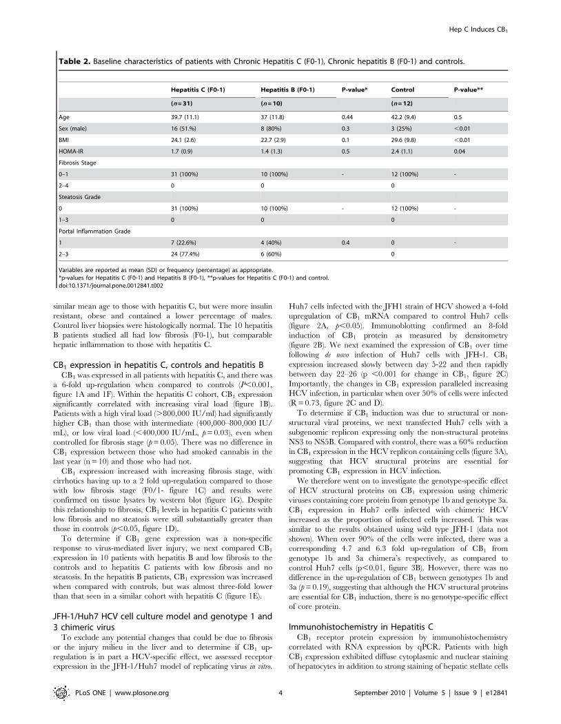
similar mean age to those with hepatitis C, but were more insulin
resistant, obese and contained a lower percentage of males.
Control liver biopsies were histologically normal. The 10 hepatitis
B patients studied all had low fibrosis (F0-1), but comparable
hepatic inflammation to those with hepatitis C.
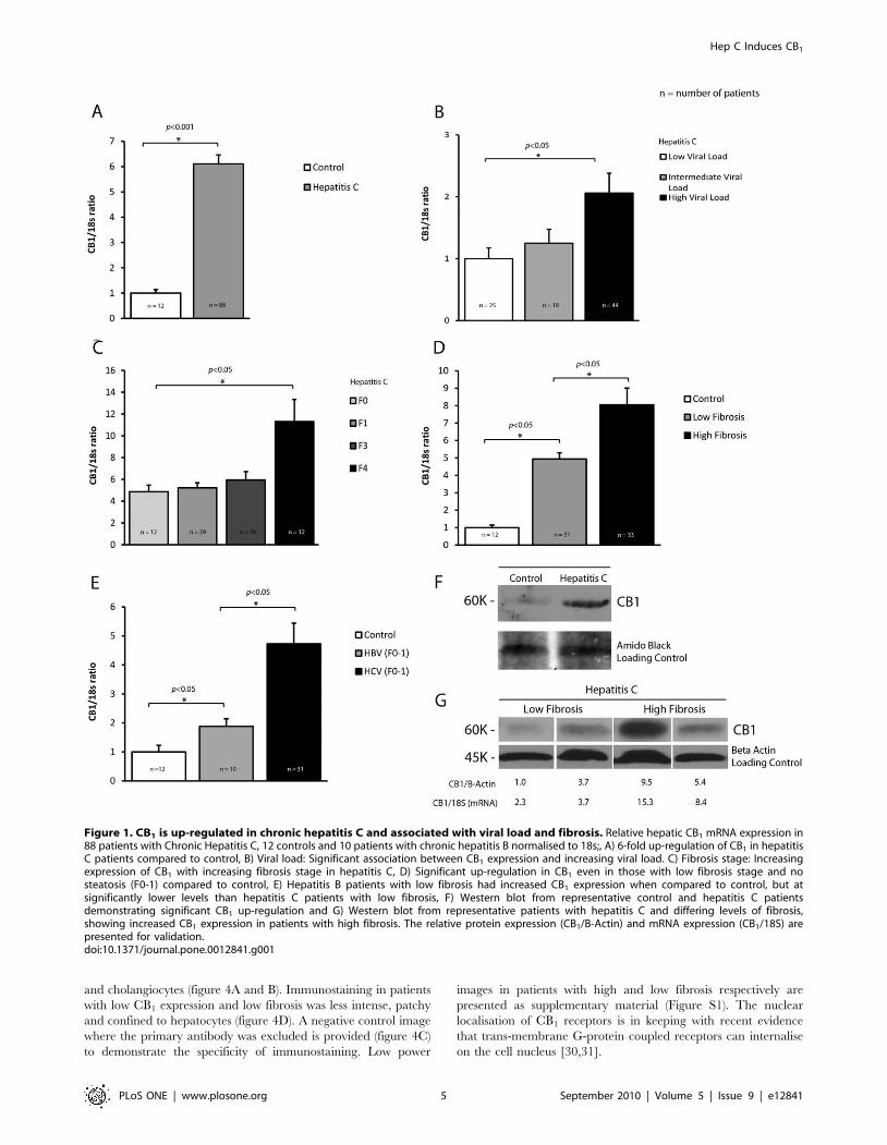
CB1 expression in hepatitis C, controls and hepatitis BCB1 was expressed in all patients with hepatitis C, and there was
a 6-fold up-regulation when compared to controls (P,0.001,
figure 1A and 1F). Within the hepatitis C cohort, CB1 expression
significantly correlated with increasing viral load (figure 1B).
Patients with a high viral load (.800,000 IU/ml) had significantly
higher CB1 than those with intermediate (400,000–800,000 IU/
mL), or low viral load (,400,000 IU/mL, p = 0.03), even when
controlled for fibrosis stage (p = 0.05). There was no difference in
CB1 expression between those who had smoked cannabis in the
last year (n = 10) and those who had not.
CB1 expression increased with increasing fibrosis stage, with
cirrhotics having up to a 2 fold up-regulation compared to those
with low fibrosis stage (F0/1- figure 1C) and results were
confirmed on tissue lysates by western blot (figure 1G). Despite
this relationship to fibrosis, CB1 levels in hepatitis C patients with
low fibrosis and no steatosis were still substantially greater than
those in controls (p,0.05, figure 1D).
To determine if CB1 gene expression was a non-specific
response to virus-mediated liver injury, we next compared CB1
expression in 10 patients with hepatitis B and low fibrosis to the
controls and to hepatitis C patients with low fibrosis and no
steatosis. In the hepatitis B patients, CB1 expression was increased
when compared with controls, but was almost three-fold lower
than that seen in a similar cohort with hepatitis C (figure 1E).
JFH-1/Huh7 HCV cell culture model and genotype 1 and3 chimeric virus
To exclude any potential changes that could be due to fibrosis
or the injury milieu in the liver and to determine if CB1 up-
regulation is in part a HCV-specific effect, we assessed receptor
expression in the JFH-1/Huh7 model of replicating virus in vitro.
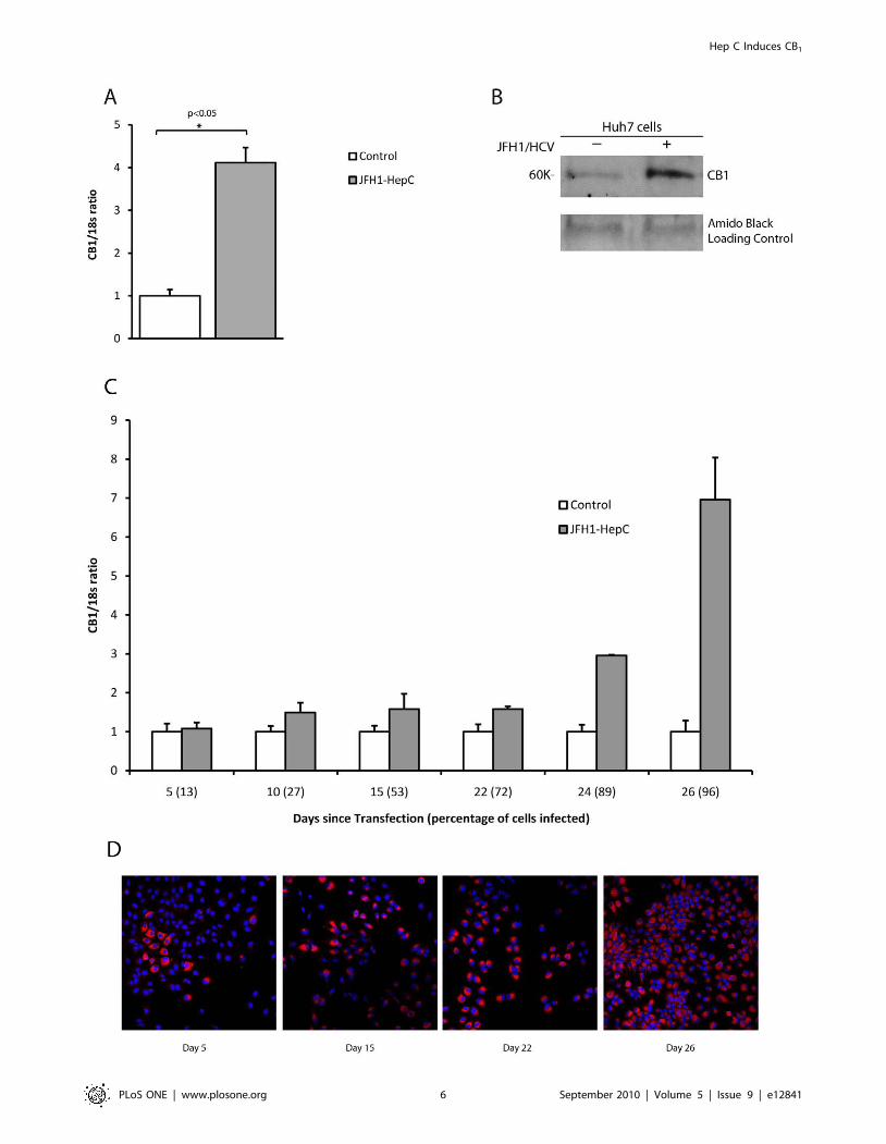
Huh7 cells infected with the JFH1 strain of HCV showed a 4-fold
upregulation of CB1 mRNA compared to control Huh7 cells
(figure 2A, p,0.05). Immunoblotting confirmed an 8-fold
induction of CB1 protein as measured by densitometry
(figure 2B). We next examined the expression of CB1 over time
following de novo infection of Huh7 cells with JFH-1. CB1
expression increased slowly between day 5-22 and then rapidly
between day 22–26 (p ,0.001 for change in CB1, figure 2C)
Importantly, the changes in CB1 expression paralleled increasing
HCV infection, in particular when over 50% of cells were infected
(R = 0.73, figure 2C and D).
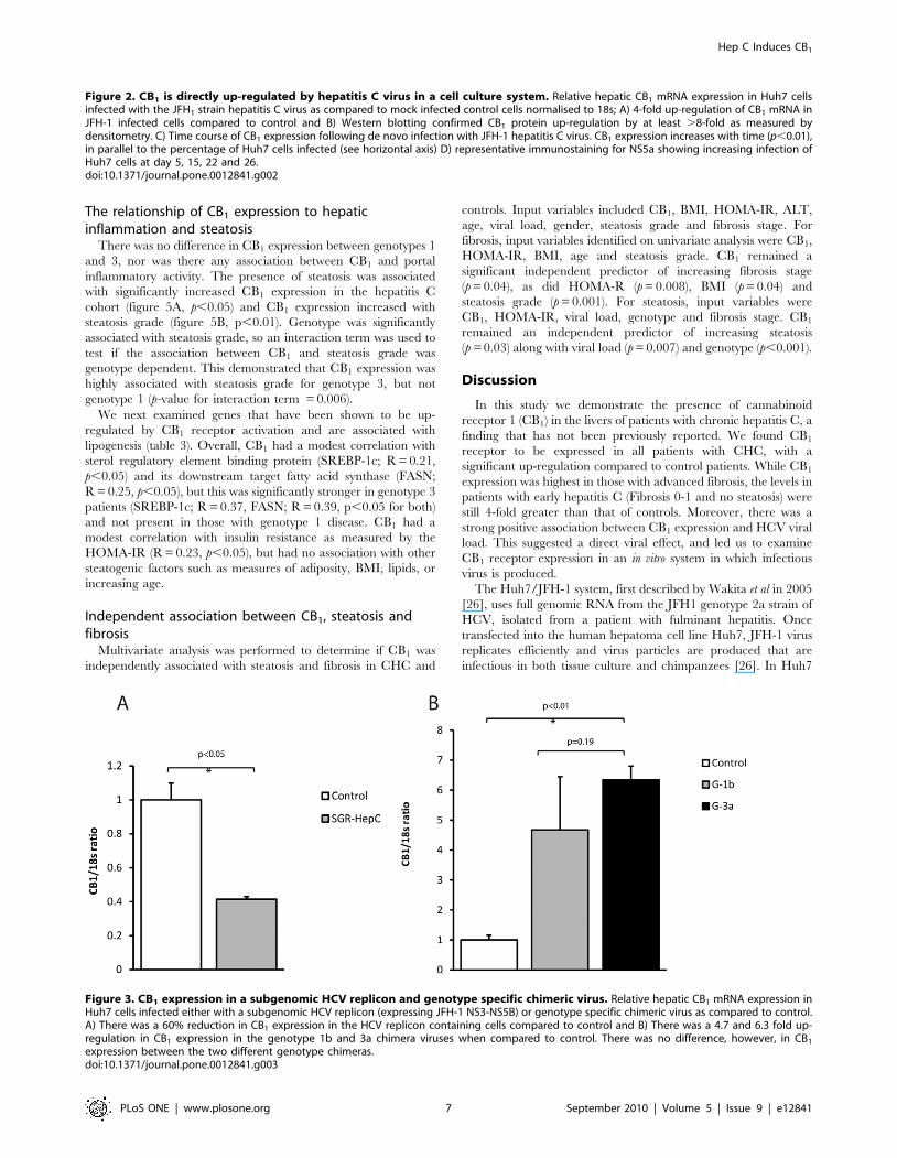
To determine if CB1 induction was due to structural or non-
structural viral proteins, we next transfected Huh7 cells with a
subgenomic replicon expressing only the non-structural proteins
NS3 to NS5B. Compared with control, there was a 60% reduction
in CB1 expression in the HCV replicon containing cells (figure 3A),
suggesting that HCV structural proteins are essential for
promoting CB1 expression in HCV infection.
We therefore went on to investigate the genotype-specific effect
of HCV structural proteins on CB1 expression using chimeric
viruses containing core protein from genotype 1b and genotype 3a.
CB1 expression in Huh7 cells infected with chimeric HCV
increased as the proportion of infected cells increased. This was
similar to the results obtained using wild type JFH-1 (data not
shown). When over 90% of the cells were infected, there was a
corresponding 4.7 and 6.3 fold up-regulation of CB1 from
genotype 1b and 3a chimera’s respectively, as compared to
control Huh7 cells (p,0.01, figure 3B). However, there was no
difference in the up-regulation of CB1 between genotypes 1b and
3a (p = 0.19), suggesting that although the HCV structural proteins
are essential for CB1 induction, there is no genotype-specific effect
of core protein.
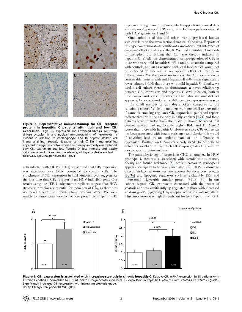
Immunohistochemistry in Hepatitis CCB1 receptor protein expression by immunohistochemistry
correlated with RNA expression by qPCR. Patients with high
CB1 expression exhibited diffuse cytoplasmic and nuclear staining
of hepatocytes in addition to strong staining of hepatic stellate cells
Table 2. Baseline characteristics of patients with Chronic Hepatitis C (F0-1), Chronic hepatitis B (F0-1) and controls.
Hepatitis C (F0-1) Hepatitis B (F0-1) P-value* Control P-value**
(n = 31) (n = 10) (n = 12)
Age 39.7 (11.1) 37 (11.8) 0.44 42.2 (9.4) 0.5
Sex (male) 16 (51.%) 8 (80%) 0.3 3 (25%) ,0.01
BMI 24.1 (2.6) 22.7 (2.9) 0.1 29.6 (9.8) ,0.01
HOMA-IR 1.7 (0.9) 1.4 (1.3) 0.5 2.4 (1.1) 0.04
Fibrosis Stage
0–1 31 (100%) 10 (100%) - 12 (100%) -
2–4 0 0 0
Steatosis Grade
0 31 (100%) 10 (100%) - 12 (100%) -
1–3 0 0 0
Portal Inflammation Grade
1 7 (22.6%) 4 (40%) 0.4 0 -
2–3 24 (77.4%) 6 (60%) 0
Variables are reported as mean (SD) or frequency (percentage) as appropriate.*p-values for Hepatitis C (F0-1) and Hepatitis B (F0-1), **p-values for Hepatitis C (F0-1) and control.doi:10.1371/journal.pone.0012841.t002
Hep C Induces CB1
PLoS ONE | www.plosone.org 4 September 2010 | Volume 5 | Issue 9 | e12841
and cholangiocytes (figure 4A and B). Immunostaining in patients
with low CB1 expression and low fibrosis was less intense, patchy
and confined to hepatocytes (figure 4D). A negative control image
where the primary antibody was excluded is provided (figure 4C)
to demonstrate the specificity of immunostaining. Low power
images in patients with high and low fibrosis respectively are
presented as supplementary material (Figure S1). The nuclear
localisation of CB1 receptors is in keeping with recent evidence
that trans-membrane G-protein coupled receptors can internalise
on the cell nucleus [30,31].
Figure 1. CB1 is up-regulated in chronic hepatitis C and associated with viral load and fibrosis. Relative hepatic CB1 mRNA expression in88 patients with Chronic Hepatitis C, 12 controls and 10 patients with chronic hepatitis B normalised to 18s;, A) 6-fold up-regulation of CB1 in hepatitisC patients compared to control, B) Viral load: Significant association between CB1 expression and increasing viral load. C) Fibrosis stage: Increasingexpression of CB1 with increasing fibrosis stage in hepatitis C, D) Significant up-regulation in CB1 even in those with low fibrosis stage and nosteatosis (F0-1) compared to control, E) Hepatitis B patients with low fibrosis had increased CB1 expression when compared to control, but atsignificantly lower levels than hepatitis C patients with low fibrosis, F) Western blot from representative control and hepatitis C patientsdemonstrating significant CB1 up-regulation and G) Western blot from representative patients with hepatitis C and differing levels of fibrosis,showing increased CB1 expression in patients with high fibrosis. The relative protein expression (CB1/B-Actin) and mRNA expression (CB1/18S) arepresented for validation.doi:10.1371/journal.pone.0012841.g001
Hep C Induces CB1
PLoS ONE | www.plosone.org 5 September 2010 | Volume 5 | Issue 9 | e12841
Hep C Induces CB1
PLoS ONE | www.plosone.org 6 September 2010 | Volume 5 | Issue 9 | e12841
The relationship of CB1 expression to hepaticinflammation and steatosis
There was no difference in CB1 expression between genotypes 1
and 3, nor was there any association between CB1 and portal
inflammatory activity. The presence of steatosis was associated
with significantly increased CB1 expression in the hepatitis C
cohort (figure 5A, p,0.05) and CB1 expression increased with
steatosis grade (figure 5B, p,0.01). Genotype was significantly
associated with steatosis grade, so an interaction term was used to
test if the association between CB1 and steatosis grade was
genotype dependent. This demonstrated that CB1 expression was
highly associated with steatosis grade for genotype 3, but not
genotype 1 (p-value for interaction term = 0.006).
We next examined genes that have been shown to be up-
regulated by CB1 receptor activation and are associated with
lipogenesis (table 3). Overall, CB1 had a modest correlation with
sterol regulatory element binding protein (SREBP-1c; R = 0.21,
p,0.05) and its downstream target fatty acid synthase (FASN;
R = 0.25, p,0.05), but this was significantly stronger in genotype 3
patients (SREBP-1c; R = 0.37, FASN; R = 0.39, p,0.05 for both)
and not present in those with genotype 1 disease. CB1 had a
modest correlation with insulin resistance as measured by the
HOMA-IR (R = 0.23, p,0.05), but had no association with other
steatogenic factors such as measures of adiposity, BMI, lipids, or
increasing age.
Independent association between CB1, steatosis andfibrosis
Multivariate analysis was performed to determine if CB1 was
independently associated with steatosis and fibrosis in CHC and
controls. Input variables included CB1, BMI, HOMA-IR, ALT,
age, viral load, gender, steatosis grade and fibrosis stage. For
fibrosis, input variables identified on univariate analysis were CB1,
HOMA-IR, BMI, age and steatosis grade. CB1 remained a
significant independent predictor of increasing fibrosis stage
(p = 0.04), as did HOMA-R (p = 0.008), BMI (p = 0.04) and
steatosis grade (p = 0.001). For steatosis, input variables were
CB1, HOMA-IR, viral load, genotype and fibrosis stage. CB1
remained an independent predictor of increasing steatosis
(p = 0.03) along with viral load (p = 0.007) and genotype (p,0.001).
Discussion
In this study we demonstrate the presence of cannabinoid
receptor 1 (CB1) in the livers of patients with chronic hepatitis C, a
finding that has not been previously reported. We found CB1
receptor to be expressed in all patients with CHC, with a
significant up-regulation compared to control patients. While CB1
expression was highest in those with advanced fibrosis, the levels in
patients with early hepatitis C (Fibrosis 0-1 and no steatosis) were
still 4-fold greater than that of controls. Moreover, there was a
strong positive association between CB1 expression and HCV viral
load. This suggested a direct viral effect, and led us to examine
CB1 receptor expression in an in vitro system in which infectious
virus is produced.
The Huh7/JFH-1 system, first described by Wakita et al in 2005
[26], uses full genomic RNA from the JFH1 genotype 2a strain of
HCV, isolated from a patient with fulminant hepatitis. Once
transfected into the human hepatoma cell line Huh7, JFH-1 virus
replicates efficiently and virus particles are produced that are
infectious in both tissue culture and chimpanzees [26]. In Huh7
Figure 3. CB1 expression in a subgenomic HCV replicon and genotype specific chimeric virus. Relative hepatic CB1 mRNA expression inHuh7 cells infected either with a subgenomic HCV replicon (expressing JFH-1 NS3-NS5B) or genotype specific chimeric virus as compared to control.A) There was a 60% reduction in CB1 expression in the HCV replicon containing cells compared to control and B) There was a 4.7 and 6.3 fold up-regulation in CB1 expression in the genotype 1b and 3a chimera viruses when compared to control. There was no difference, however, in CB1
expression between the two different genotype chimeras.doi:10.1371/journal.pone.0012841.g003
Figure 2. CB1 is directly up-regulated by hepatitis C virus in a cell culture system. Relative hepatic CB1 mRNA expression in Huh7 cellsinfected with the JFH1 strain hepatitis C virus as compared to mock infected control cells normalised to 18s; A) 4-fold up-regulation of CB1 mRNA inJFH-1 infected cells compared to control and B) Western blotting confirmed CB1 protein up-regulation by at least .8-fold as measured bydensitometry. C) Time course of CB1 expression following de novo infection with JFH-1 hepatitis C virus. CB1 expression increases with time (p,0.01),in parallel to the percentage of Huh7 cells infected (see horizontal axis) D) representative immunostaining for NS5a showing increasing infection ofHuh7 cells at day 5, 15, 22 and 26.doi:10.1371/journal.pone.0012841.g002
Hep C Induces CB1
PLoS ONE | www.plosone.org 7 September 2010 | Volume 5 | Issue 9 | e12841
cells infected with HCV (JFH-1) we showed that CB1 expression
was increased over 8-fold compared to control cells. The
enrichment of CB1 expression in JFH1-infected cells suggests for
the first time that CB1 receptor is an HCV-inducible gene. Our
results using the JFH-1 subgenomic replicon suggest that HCV
structural proteins are essential for induction of CB1, as there was
no increase seen with nonstructural proteins alone. We were
unable to demonstrate an effect of core protein genotype on CB1
expression using chimeric viruses, which supports our clinical data
showing no difference in CB1 expression between patients infected
with HCV genotypes 1 and 3.
One limitation of this and other liver biopsy-based human
studies relates to the cross-sectional nature of the data. Reports of
this type can demonstrate significant associations, but inference of
cause and effect are always difficult. We used a number of methods
to strengthen our finding that CB1 was directly induced by
hepatitis C. Firstly, we demonstrated an up-regulation of CB1 in
those with very mild hepatitis C (F0-1 and no steatosis) compared
with controls, and an association with viral load, which would not
be expected if this was a non-specific effect of fibrosis or
inflammation. We then went on to show that CB1 expression in
comparable patients with mild hepatitis B (F0-1) was significantly
lower (almost 3-fold) than those with mild hepatitis C. Finally, we
used a cell culture system to demonstrate a direct relationship
between CB1 expression and hepatitis C viral infection, both in
time course and static experiments. Cannabis smoking did not
appear to be a confounder as no difference in expression was seen
in the small number of cannabis smokers compared to the
remaining cohort. While the numbers were too small to determine
if cannabis smoking regulates CB1 expression, published reports
indicate that this is the case only in daily smokers [4,24] and these
patients were excluded from the study. It should be noted that
control subjects had significantly higher BMI and HOMA-IR
scores than those with hepatitis C. However, since CB1 expression
has been associated with insulin resistance and obesity, this would
if anything lead to an underestimate of the difference in
expression. Further work however clearly needs to be done to
define the mechanisms by which HCV up-regulates CB1 and the
specific viral proteins involved.
The pathophysiology of steatosis in CHC is complex. In HCV
genotype 1, steatosis is associated with metabolic disturbance,
obesity and insulin resistance [5], while steatosis in genotype 3
appears principally to be virally mediated [32]. HCV is known to
directly induce steatosis via interactions between core protein
[33,34] and lipogenic regulators such as SREBP-1c [35] and
microsomal triglyceride transfer protein (MTP) [36]. In our
cohort, hepatic CB1 expression correlated with the extent of
steatosis and was significantly up-regulated in those with increased
steatosis grade, suggesting CB1 receptor activation and signalling.
This association was highly significant for genotype 3, but not 1.
Figure 5. CB1 expression is associated with increasing steatosis in chronic hepatitis C. Relative CB1 mRNA expression in 88 patients withChronic Hepatitis C normalised to 18s; A) Steatosis: Significantly increased CB1 expression in hepatitis C patients with steatosis, B) Steatosis grades:Significantly increased CB1 expression with increasing steatosis grade.doi:10.1371/journal.pone.0012841.g005
Figure 4. Representative immunostaining for CB1 receptorprotein in hepatitis C patients with high and low CB1
expression. High CB1 expression and advanced fibrosis: A) strong,diffuse cytoplasmic and nuclear immunostaining of hepatocytes isevident in addition to cholangiocyte and B) hepatic stellate cellimmunostaining (arrows). Negative control: C) No immunostainingapparent in negative control where the primary antibody was excluded.Low CB1 expression and low fibrosis: D) low intensity and patchycytoplasmic and nuclear immunostaining of hepatocytes is evident.doi:10.1371/journal.pone.0012841.g004
Hep C Induces CB1
PLoS ONE | www.plosone.org 8 September 2010 | Volume 5 | Issue 9 | e12841

Moreover, in genotype 3 patients, CB1 expression correlated
strongly with the lipogenic transcription factor SREBP-1c and its
downstream target FASN, but in genotype 1 patients there was no
correlation. Clinical studies in CHC have hinted at the importance
of CB1 stimulation to steatosis, with daily cannabis use a risk factor
for steatosis severity in over 300 patients with CHC [24]. In
experimental work, endocannabinoid stimulation of CB1 mediates
diet-induced steatosis, since CB1 knockout mice fed a high fat diet
are resistant to steatosis [18]. Likewise, treatment of wild type mice
with a CB1 agonist induces de novo fatty acid synthesis via increased
hepatic expression of SREBP-1c and its downstream targets FASN
and acetyl coenzyme-A carboxylase-1 (ACC1) [18]. Finally, the
selective deletion of hepatocyte CB1 receptors alone is sufficient to
prevent diet [37] and alcohol-induced [22] hepatic steatosis
[22,37].
The endocannabinoid system plays an important role in liver
fibrosis. In three murine models of chronic liver injury, CB1
receptor antagonism by pharmacological or genetic means
reduced fibrosis area, TGF-b1 expression and the accumulation
of fibrogenic cells. It has also been shown that CB1 can mediate
liver fibrosis through effects on apoptosis and the growth of hepatic
myofibroblasts [19]. Clinical studies first demonstrated the likely
importance of this system in patients with CHC, showing daily
cannabis smoking to be independently associated with the
progression and severity of fibrosis [4]. In our study, CB1 was
expressed in all patients with CHC and increased with advancing
fibrosis, with the highest levels present in those with cirrhosis. We
were unable to show any relationship between CB1 receptor
expression and inflammatory grade, although this does not exclude
that the endocannabinoid system via CB1 may mediate fibrosis in
this way. Rather, it has been suggested that HCV can directly
activate and stimulate hepatic stellate cells through its core and
non-structural proteins, or via secretions from infected hepatocytes
[38,39]. In this context, activated HSCs not only secrete collagens
and cytokines, but also the endocannabinoid 2-AG, which in turn
up-regulates and activates hepatocyte CB1 [22]. Stimulation of
hepatocyte CB1 through this pathway or directly by HCV as we
demonstrate will serve to amplify the pathways by which liver
fibrosis develops in CHC [2,32]. It is interesting to note from our
immunohistochemistry that CB1 receptors were up-regulated on
hepatic stellate cells in CHC. One could therefore speculate that
the direct pro-fibrogenic interactions between HCV and stellate
cells demonstrated by Battaler and collegues [38] may in part, be
mediated through and exaggerated by the induction of CB1.
There has been much recent interest in the use of CB1
antagonists to treat both hepatic and metabolic disease and our
findings emphasize the likely usefulness of these compounds in
patients with hepatitis C. In addition to the amelioration of
steatosis and fibrosis, CB1 blockade reduces portal pressure and
can reverse mesenteric arterial dilation [40], making them useful
in end stage liver disease as well. We speculate that CB1
antagonism may also have an inhibitory effect on HCV
replication. This is prompted both by the significant, genotype
independent association we found between CB1 expression and
viral load, and by reports that blockade of the SREBP and FASN
signalling in hepatitis C cell culture models reduces HCV
replication [41,42]. Clearly this needs further study before any
conclusions can be drawn. Unfortunately, several CB1 receptor
antagonists were recently withdrawn from clinical development
including Rimonabant (SR141716A) and Taranabant (MK-0364).
These withdrawals followed an EU medicines safety edict that
Rimonabant be suspended due to excessive psychiatric side effects
including depression, anxiety and suicide [43]. Nonetheless, our
findings and those of others, showing the importance of hepatic
CB1 receptors in CHC, suggest that the development of a
peripherally selective CB1 antagonist with minimal neurotoxicity
remains a promising future option.
In conclusion, we have demonstrated that CB1 receptor is
widely expressed in the livers of patients with CHC and is
associated with advanced fibrosis and steatosis. Importantly, CB1
is also highly enriched in those with low fibrosis and is induced by
HCV in a cell culture system, findings that underscore the unique
susceptibility of patients with CHC to cannabis-induced liver
damage. CB1 has already been implicated in the genesis of fibrosis
and steatosis in experimental and metabolic liver disease. We have
now shown that it is important in CHC and may be a future target
for pharmacotherapy in this disease.
Supporting Information
Figure S1 Representative immunostaining for CB1 receptor
protein in hepatitis C patients with high and low CB1 expression.
A) and B) High CB1 expression and advanced fibrosis showing
strong, diffuse cytoplasmic and nuclear immunostaining primarily
of hepatocytes. C) and D) Low CB1 expression and low fibrosis
showing low intensity and patchy cytoplasmic and nuclear
immunostaining of hepatocytes.
Found at: doi:10.1371/journal.pone.0012841.s001 (6.68 MB
TIF)
Acknowledgments
The authors wish to thank Keshni Sharma and Lee Russell for assistance
with Data and sample collection, David Warton for assistance with
statistical methods and Dr John McLauchlan, MRC Virology Unit,
Glasgow for providing the Huh7 clone.
Author Contributions
Conceived and designed the experiments: DvdP LWH MWD JG.
Performed the experiments: DvdP MS ESET JS KT DM JSM VH.
Analyzed the data: DvdP KT LWH MWD. Contributed reagents/
materials/analysis tools: MWD. Wrote the paper: DvdP LWH MWD JG.
Table 3. Rank correlations between CB1 and factors associated with steatosis in HCV by genotype.
SREBP-1c FASN HOMA-IR BMI HDL TG Age
CB1 - HCV all 0.21* 0.25* 0.23* 0.10 0.03 0.01 0.15
CB1 - HCV G1 0.08 0.19 0.19 0.11 0.11 20.04 0.21
CB1 - HCV G3 0.37* 0.39* 0.24 0.20 0.01 0.02 0.14
*p-value ,0.05.SREBP-1c; Sterol regulatory element binding protein, FASN; fatty acid synthase, HOMA-IR; homeostasis model assessment of insulin resistance, BMI; body mass index,HDL; high density lipoprotein, TG; triglyceride.doi:10.1371/journal.pone.0012841.t003
Hep C Induces CB1
PLoS ONE | www.plosone.org 9 September 2010 | Volume 5 | Issue 9 | e12841
References
1. World Health Organisation Initiative for vaccine research (IVR); Hepatitis C
page. Available at: http://www.who.int/vaccine_research/diseases/viral_cancers/en/index2.html Accessed January 15, 2008.
2. van der Poorten D, George J (2008) Disease-specific mechanisms of fibrosis:hepatitis C virus and nonalcoholic steatohepatitis. Clin Liver Dis 12: 805–824,
ix.3. McCaughan GW, George J (2004) Fibrosis progression in chronic hepatitis C
virus infection. Gut 53: 318–321.
4. Hezode C, Roudot-Thoraval F, Nguyen S, Grenard P, Julien B, et al. (2005)Daily cannabis smoking as a risk factor for progression of fibrosis in chronic
hepatitis C. Hepatology 42: 63–71.5. Hui JM, Sud A, Farrell GC, Bandara P, Byth K, et al. (2003) Insulin resistance is
associated with chronic hepatitis C virus infection and fibrosis progression
[corrected]. Gastroenterology 125: 1695–1704.6. Hui JM, Kench J, Farrell GC, Lin R, Samarasinghe D, et al. (2002) Genotype-
specific mechanisms for hepatic steatosis in chronic hepatitis C infection.J Gastroenterol Hepatol 17: 873–881.
7. Kumar D, Farrell GC, Fung C, George J (2002) Hepatitis C virus genotype 3 is
cytopathic to hepatocytes: Reversal of hepatic steatosis after sustainedtherapeutic response. Hepatology 36: 1266–1272.
8. Asselah T, Rubbia-Brandt L, Marcellin P, Negro F (2006) Steatosis in ChronicHepatitis C: Why does it really matter? Gut 55: 123–130.
9. Ohata K, Hamasaki K, Toriyama K, Matsumoto K, Saeki A, et al. (2003)Hepatic steatosis is a risk factor for hepatocellular carcinoma in patients with
chronic hepatitis C virus infection.[see comment]. Cancer 97: 3036–3043.
10. Su AI, Pezacki JP, Wodicka L, Brideau AD, Supekova L, et al. (2002) Genomicanalysis of the host response to hepatitis C virus infection. PNAS 99:
15669–15674.11. Smart RG, Ogborne AC (2000) Drug use and drinking among students in 36
countries. Addict Behav 25: 455–460.
12. Devane WA, Hanus L, Breuer A, Pertwee RG, Stevenson LA, et al. (1992)Isolation and structure of a brain constituent that binds to the cannabinoid
receptor. Science 258: 1946–1949.13. Pacher P, Batkai S, Kunos G (2006) The endocannabinoid system as an
emerging target of pharmacotherapy. Pharmacol Rev 58: 389–462.14. Kunos G, Osei-Hyiaman D, Liu J, Godlewski G, Batkai S (2008) Endocanna-
binoids and the control of energy homeostasis. J Biol Chem 283: 33021–33025.
15. Di Marzo V, Goparaju SK, Wang L, Liu J, Batkai S, et al. (2001) Leptin-regulated endocannabinoids are involved in maintaining food intake. Nature
410: 822–825.16. Ravinet Trillou C, Delgorge C, Menet C, Arnone M, Soubrie P (2004) CB1
cannabinoid receptor knockout in mice leads to leanness, resistance to diet-
induced obesity and enhanced leptin sensitivity. Int J Obes Relat Metab Disord28: 640–648.
17. Despres JP, Golay A, Sjostrom L, Rimonabant in Obesity-Lipids Study G (2005)Effects of rimonabant on metabolic risk factors in overweight patients with
dyslipidemia.[see comment]. New England Journal of Medicine 353:2121–2134.
18. Osei-Hyiaman D, DePetrillo M, Pacher P, Liu J, Radaeva S, et al. (2005)
Endocannabinoid activation at hepatic CB1 receptors stimulates fatty acidsynthesis and contributes to diet-induced obesity.[see comment]. Journal of
Clinical Investigation 115: 1298–1305.19. Teixeira-Clerc F, Julien B, Grenard P, Tran Van Nhieu J, Deveaux V, et al.
(2006) CB1 cannabinoid receptor antagonism: a new strategy for the treatment
of liver fibrosis. Nat Med 12: 671–676.20. Julien, Grenard, Teixeira C, Van N, Li, et al. (2005) Antifibrogenic role of the
cannabinoid receptor CB2 in the liver. Gastroenterology 128: 742–755.21. Munoz-Luque J, Ros J, Fernandez-Varo G, Tugues S, Morales-Ruiz M, et al.
(2008) Regression of fibrosis after chronic stimulation of cannabinoid CB2
receptor in cirrhotic rats. J Pharmacol Exp Ther 324: 475–483.22. Jeong WI, Osei-Hyiaman D, Park O, Liu J, Batkai S, et al. (2008) Paracrine
activation of hepatic CB1 receptors by stellate cell-derived endocannabinoidsmediates alcoholic fatty liver. Cell Metab 7: 227–235.
23. Gary-Bobo M, Elachouri G, Gallas JF, Janiak P, Marini P, et al. (2007)
Rimonabant reduces obesity-associated hepatic steatosis and features of
metabolic syndrome in obese Zucker fa/fa rats. Hepatology 46: 122–129.
24. Hezode C, Zafrani ES, Roudot-Thoraval F, Costentin C, Hessami A, et al.
(2008) Daily cannabis use: a novel risk factor of steatosis severity in patients withchronic hepatitis C. Gastroenterology 134: 432–439.
25. Scheuer PJ (1991) Classification of chronic viral hepatitis: a need forreassessment. J Hepatol 13: 372–374.
26. Wakita T, Pietschmann T, Kato T, Date T, Miyamoto M, et al. (2005)Production of infectious hepatitis C virus in tissue culture from a cloned viral
genome. Nat Med 11: 791–796.
27. Kato T, Date T, Miyamoto M, Furusaka A, Tokushige K, et al. (2003) Efficient
replication of the genotype 2a hepatitis C virus subgenomic replicon.Gastroenterology 125: 1808–1817.
28. Shaw ML, McLauchlan J, Mills PR, Patel AH, McCruden EA (2003)Characterisation of the differences between hepatitis C virus genotype 3 and 1
glycoproteins. J Med Virol 70: 361–372.
29. Rasband W ImageJ. U.S. National Institutes of Health, Bethesda, Maryland,
USA, 1997-2007.
30. Boivin B, Vaniotis G, Allen BG, Hebert TE (2008) G protein-coupled receptors
in and on the cell nucleus: a new signaling paradigm? J Recept Signal Transduct
Res 28: 15–28.
31. Ellis J, Pediani JD, Canals M, Milasta S, Milligan G (2006) Orexin-1 receptor-
cannabinoid CB1 receptor heterodimerization results in both ligand-dependentand -independent coordinated alterations of receptor localization and function.
J Biol Chem 281: 38812–38824.
32. Negro F (2006) Mechanisms and significance of liver steatosis in hepatitis C virus
infection. World J Gastroenterol 12: 6756–6765.
33. Shi ST, Polyak SJ, Tu H, Taylor DR, Gretch DR, et al. (2002) Hepatitis C virus
NS5A colocalizes with the core protein on lipid droplets and interacts withapolipoproteins. Virology 292: 198–210.
34. Barba G, Harper F, Harada T, Kohara M, Goulinet S, et al. (1997) Hepatitis Cvirus core protein shows a cytoplasmic localization and associates to cellular lipid
storage droplets. PNAS 94: 1200–1205.
35. Kim KH, Hong SP, Kim K, Park MJ, Kim KJ, et al. (2007) HCV core proteininduces hepatic lipid accumulation by activating SREBP1 and PPARgamma.
Biochem Biophys Res Commun 355: 883–888.
36. Perlemuter G, Sabile A, Letteron P, Vona G, Topilco A, et al. (2002) Hepatitis C
virus core protein inhibits microsomal triglyceride transfer protein activity andvery low density lipoprotein secretion: a model of viral-related steatosis. FASEB
Journal 16: 185–194.
37. Osei-Hyiaman D, Liu J, Zhou L, Godlewski G, Harvey-White J, et al. (2008)
Hepatic CB1 receptor is required for development of diet-induced steatosis,dyslipidemia, and insulin and leptin resistance in mice. J Clin Invest 118:
3160–3169.
38. Bataller R, Paik YH, Lindquist JN, Lemasters JJ, Brenner DA (2004) Hepatitis C
virus core and nonstructural proteins induce fibrogenic effects in hepatic stellate
cells. Gastroenterology 126: 529–540.
39. Schulze-Krebs A, Preimel D, Popov Y, Bartenschlager R, Lohmann V, et al.
(2005) Hepatitis C virus-replicating hepatocytes induce fibrogenic activation ofhepatic stellate cells. Gastroenterology 129: 246–258.
40. Parfieniuk A, Flisiak R (2008) Role of cannabinoids in chronic liver diseases.World J Gastroenterol 14: 6109–6114.
41. Su AI, Pezacki JP, Wodicka L, Brideau AD, Supekova L, et al. (2002) Genomicanalysis of the host response to hepatitis C virus infection. Proc Natl Acad
Sci U S A 99: 15669–15674.
42. Yang W, Hood BL, Chadwick SL, Liu S, Watkins SC, et al. (2008) Fatty acid
synthase is up-regulated during hepatitis C virus infection and regulates hepatitisC virus entry and production. Hepatology 48: 1396–1403.
43. European Medicines Agency (2008) The European Medicines Agency
recommends suspension of the marketing authorisation of Acomplia. pp.London, 23 October, Doc. Ref. EMEA/CHMP/537777/532008.
Hep C Induces CB1
PLoS ONE | www.plosone.org 10 September 2010 | Volume 5 | Issue 9 | e12841