TG1050, an immunotherapeutic to treat chronic hepatitis B, induces robust T cells and exerts an...
27
ORIGINAL ARTICLE TG1050, an immunotherapeutic to treat chronic hepatitis B, induces robust T cells and exerts an antiviral effect in HBV-persistent mice Perrine Martin, 1 Clarisse Dubois, 1 Emilie Jacquier, 1 Sarah Dion, 2 Maryline Mancini-Bourgine, 2 Ophélie Godon, 2 Roland Kratzer, 1 Karine Lelu-Santolaria, 1 Alexei Evlachev, 1 Jean-François Meritet, 3 Yasmin Schlesinger, 4 Dominique Villeval, 4 Jean-Marc Strub, 5 Alain Van Dorsselaer, 5 Jean-Baptiste Marchand, 4 Michel Geist, 4 Renée Brandely, 4 Annie Findeli, 4 Houda Boukhebza, 1 Thierry Menguy, 4 Nathalie Silvestre, 4 Marie-Louise Michel, 2 Geneviève Inchauspé 1 ▸ Additional material is published online only. To view please visit the journal online (http://dx.doi.org/10.1136/ gutjnl-2014-308041). 1 Department of Infectious Diseases, Transgene SA, Lyon, France 2 Laboratoire de pathogénèse des virus de l’hépatite B Paris and INSERM U994, Institut Pasteur, Paris, France 3 Virology Unit, Cochin Hospital, Paris, France 4 Department of Vectors, Transgene SA, Strasbourg, France 5 Laboratoire de Spectrométrie de Masse BioOrganique, Strasbourg University, UMR 7178, Strasbourg, France Correspondence to Dr Geneviève Inchauspé, Transgene SA, 321 Avenue Jean Jaures, Lyon 69007, France; inchauspe@transgene. fr Received 15 July 2014 Revised 4 October 2014 Accepted 20 October 2014 To cite: Martin P, Dubois C, Jacquier E, et al. Gut Published Online First: [ please include Day Month Year] doi:10.1136/gutjnl- 2014-308041 ABSTRACT Objective To assess a new adenovirus-based immunotherapy as a novel treatment approach to chronic hepatitis B (CHB). Methods TG1050 is a non-replicative adenovirus serotype 5 encoding a unique large fusion protein composed of a truncated HBV Core, a modified HBV Polymerase and two HBV Envelope domains. We used a recently described HBV-persistent mouse model based on a recombinant adenovirus-associated virus encoding an over length genome of HBV that induces the chronic production of HBsAg, HBeAg and infectious HBV particles to assess the ability of TG1050 to induce functional T cells in face of a chronic status. Results In in vitro studies, TG1050 was shown to express the expected large polyprotein together with a dominant, smaller by-product. Following a single administration in mice, TG1050 induced robust, multispecific and long- lasting HBV-specific T cells detectable up to 1 year post- injection. These cells target all three encoded immunogens and display bifunctionality (ie, capacity to produce both interferon γ and tumour necrosis factor α as well as cytolytic functions). In addition, control of circulating levels of HBV DNA and HBsAg was observed while alanine aminotransferase levels remain in the normal range. Conclusions Injection of TG1050 induced both splenic and intrahepatic functional T cells producing cytokines and displaying cytolytic activity in HBV-naïve and HBV- persistent mouse models together with significant reduction of circulating viral parameters. These results warrant clinical evaluation of TG1050 in the treatment of CHB. INTRODUCTION Infection by HBV is one of the major causes and risk factors for developing liver cancer. 1 Over 2 billion people have been infected by HBV world- wide, and about 240 million of them are currently chronically infected and at high risk of developing cirrhosis and hepatocellular carcinoma. 2 Current chronic hepatitis B (CHB) therapies include nucleos (t)ide analogues (NUC) aimed at inhibiting viral replication 34 and pegylated interferon α (IFNα). 56 Significance of this study What is already known about the subject? ▸ HBV chronic infection can be controlled but is rarely cured. ▸ HBV immunotherapeutics have been described at preclinical and clinical levels with limited or no efficacy. ▸ This novel class of HBV therapeutics has included HBsAg in the majority, the Core antigen in some and only one coding for the HBV polymerase. ▸ HBV immunotherapeutics can induce functional T cells in HBV transgenic mice, typically mice transgenic for a single HBV antigen, but an effect on viral parameters has seldom been reported following their administration, particularly on levels of circulating HBsAg. What are the new findings? ▸ TG1050 is the only HBV immunotherapeutic covering in a single entity three HBV antigens/ domains, including polymerase. ▸ TG1050 induces persistence of multifunctional HBV-specific T cell responses up to 400 days after a single injection. ▸ Following single as well as multiple injections, TG1050 can educate functional T cells in a HBV chronic environment and display significant and persistent antiviral activity, in particular an impact on the level of HBsAg. ▸ TG1050 is the only adenovirus-based HBV immunotherapeutic currently planned for testing in the clinic. How might it impact on clinical practice in the foreseeable future? ▸ TG1050 will next be tested in the clinic in combination with nucleos(t)ide analogues in the treatment of chronic hepatitis B (CHB) and aims to increase the cure rate. ▸ If successful, TG1050 will bring a novel treatment paradigm to patients with CHB. Martin P, et al. Gut 2014;0:1–11. doi:10.1136/gutjnl-2014-308041 1 Hepatology Gut Online First, published on November 26, 2014 as 10.1136/gutjnl-2014-308041 Copyright Article author (or their employer) 2014. Produced by BMJ Publishing Group Ltd (& BSG) under licence. group.bmj.com on November 27, 2014 - Published by http://gut.bmj.com/ Downloaded from
Transcript of TG1050, an immunotherapeutic to treat chronic hepatitis B, induces robust T cells and exerts an...

ORIGINAL ARTICLE
TG1050, an immunotherapeutic to treat chronichepatitis B, induces robust T cells and exerts anantiviral effect in HBV-persistent micePerrine Martin,1 Clarisse Dubois,1 Emilie Jacquier,1 Sarah Dion,2
Maryline Mancini-Bourgine,2 Ophélie Godon,2 Roland Kratzer,1 Karine Lelu-Santolaria,1
Alexei Evlachev,1 Jean-François Meritet,3 Yasmin Schlesinger,4 Dominique Villeval,4
Jean-Marc Strub,5 Alain Van Dorsselaer,5 Jean-Baptiste Marchand,4 Michel Geist,4
Renée Brandely,4 Annie Findeli,4 Houda Boukhebza,1 Thierry Menguy,4
Nathalie Silvestre,4 Marie-Louise Michel,2 Geneviève Inchauspé1
▸ Additional material ispublished online only. To viewplease visit the journal online(http://dx.doi.org/10.1136/gutjnl-2014-308041).1Department of InfectiousDiseases, Transgene SA, Lyon,France2Laboratoire de pathogénèsedes virus de l’hépatite B Parisand INSERM U994, InstitutPasteur, Paris, France3Virology Unit, CochinHospital, Paris, France4Department of Vectors,Transgene SA, Strasbourg,France5Laboratoire de Spectrométriede Masse BioOrganique,Strasbourg University, UMR7178, Strasbourg, France
Correspondence toDr Geneviève Inchauspé,Transgene SA, 321 AvenueJean Jaures, Lyon 69007,France; [email protected]
Received 15 July 2014Revised 4 October 2014Accepted 20 October 2014
To cite: Martin P, Dubois C,Jacquier E, et al. GutPublished Online First:[please include Day MonthYear] doi:10.1136/gutjnl-2014-308041
ABSTRACTObjective To assess a new adenovirus-basedimmunotherapy as a novel treatment approach to chronichepatitis B (CHB).Methods TG1050 is a non-replicative adenovirusserotype 5 encoding a unique large fusion proteincomposed of a truncated HBV Core, a modified HBVPolymerase and two HBV Envelope domains. We used arecently described HBV-persistent mouse model based on arecombinant adenovirus-associated virus encoding an overlength genome of HBV that induces the chronic productionof HBsAg, HBeAg and infectious HBV particles to assessthe ability of TG1050 to induce functional T cells in face ofa chronic status.Results In in vitro studies, TG1050 was shown to expressthe expected large polyprotein together with a dominant,smaller by-product. Following a single administration inmice, TG1050 induced robust, multispecific and long-lasting HBV-specific T cells detectable up to 1 year post-injection. These cells target all three encoded immunogensand display bifunctionality (ie, capacity to produce bothinterferon γ and tumour necrosis factor α as well ascytolytic functions). In addition, control of circulating levelsof HBV DNA and HBsAg was observed while alanineaminotransferase levels remain in the normal range.Conclusions Injection of TG1050 induced both splenicand intrahepatic functional T cells producing cytokines anddisplaying cytolytic activity in HBV-naïve and HBV-persistent mouse models together with significantreduction of circulating viral parameters. These resultswarrant clinical evaluation of TG1050 in the treatmentof CHB.
INTRODUCTIONInfection by HBV is one of the major causes andrisk factors for developing liver cancer.1 Over 2billion people have been infected by HBV world-wide, and about 240 million of them are currentlychronically infected and at high risk of developingcirrhosis and hepatocellular carcinoma.2 Currentchronic hepatitis B (CHB) therapies include nucleos(t)ide analogues (NUC) aimed at inhibiting viralreplication3 4 and pegylated interferon α (IFNα).5 6
Significance of this study
What is already known about the subject?▸ HBV chronic infection can be controlled but is
rarely cured.▸ HBV immunotherapeutics have been described
at preclinical and clinical levels with limited orno efficacy.
▸ This novel class of HBV therapeutics hasincluded HBsAg in the majority, the Coreantigen in some and only one coding for theHBV polymerase.
▸ HBV immunotherapeutics can induce functionalT cells in HBV transgenic mice, typically micetransgenic for a single HBV antigen, but aneffect on viral parameters has seldom beenreported following their administration,particularly on levels of circulating HBsAg.
What are the new findings?▸ TG1050 is the only HBV immunotherapeutic
covering in a single entity three HBV antigens/domains, including polymerase.
▸ TG1050 induces persistence of multifunctionalHBV-specific T cell responses up to 400 daysafter a single injection.
▸ Following single as well as multiple injections,TG1050 can educate functional T cells in a HBVchronic environment and display significant andpersistent antiviral activity, in particular animpact on the level of HBsAg.
▸ TG1050 is the only adenovirus-based HBVimmunotherapeutic currently planned fortesting in the clinic.
How might it impact on clinical practice inthe foreseeable future?▸ TG1050 will next be tested in the clinic in
combination with nucleos(t)ide analogues inthe treatment of chronic hepatitis B (CHB) andaims to increase the cure rate.
▸ If successful, TG1050 will bring a noveltreatment paradigm to patients with CHB.
Martin P, et al. Gut 2014;0:1–11. doi:10.1136/gutjnl-2014-308041 1
Hepatology Gut Online First, published on November 26, 2014 as 10.1136/gutjnl-2014-308041
Copyright Article author (or their employer) 2014. Produced by BMJ Publishing Group Ltd (& BSG) under licence.
group.bmj.com on November 27, 2014 - Published by http://gut.bmj.com/Downloaded from
Despite the ability of these treatments to control HBV replica-tion in the great majority of patients and to improve liver hist-ology, complete cure of HBV is achieved in only 3–5% ofpatients. Therefore, most patients require costly life-longtreatments.
In patients resolving infection, development of broad androbust CD8+ and CD4+ T cell responses targeting multipleHBV antigens that produce cytokines and display cytolytic prop-erties have been observed7 8 and correlated with virus controland/or elimination.9 In contrast, patients with CHB displayweak, narrowed and dysfunctional HBV immune T cellresponses.10–12 Among the new therapeutic arsenal being devel-oped, immunotherapeutics aimed at inducing immune responsessimilar to those found in resolvers represent a growing field.Although first-generation HBV-specific immunotherapies have sofar had limited success in the clinic, a number have been capableof inducing HBV-specific responses in patients with CHB.13 14
Most HBV-specific immunotherapeutics currently tested in clin-ical trials involve only 1–2 antigens and, except for a poxvirus-based candidate, none of them is based on a viral vector.15 16
TG1050 is a novel immunotherapeutic based on a non-replicative adenovirus 5 vector encoding a unique and largefusion protein composed of modified HBV Core and Polymeraseand selected domains of the Env proteins. We show here that asingle injection of TG1050 is broadly immunogenic, including ina HBV-persistent model,17 and displays antiviral properties.
MATERIALS AND METHODSAnimalsBALB/c, C57BL/6J and HLA-A*0201 transgenic H-2 class I KOmice (HLA-A2 mice)18 were bred at Charles River Laboratories(L’Arbresle, France) and HLA-A*0201/DRB1*0101 transgenicH-2 class I/class II KO mice (HLA-A2/DR1 mice)19 were bred atthe Institut Pasteur (Paris, France). Animals receiving theadeno-associated virus (AAV)-HBV were manipulated in an A3confinement facility (see online supplement).
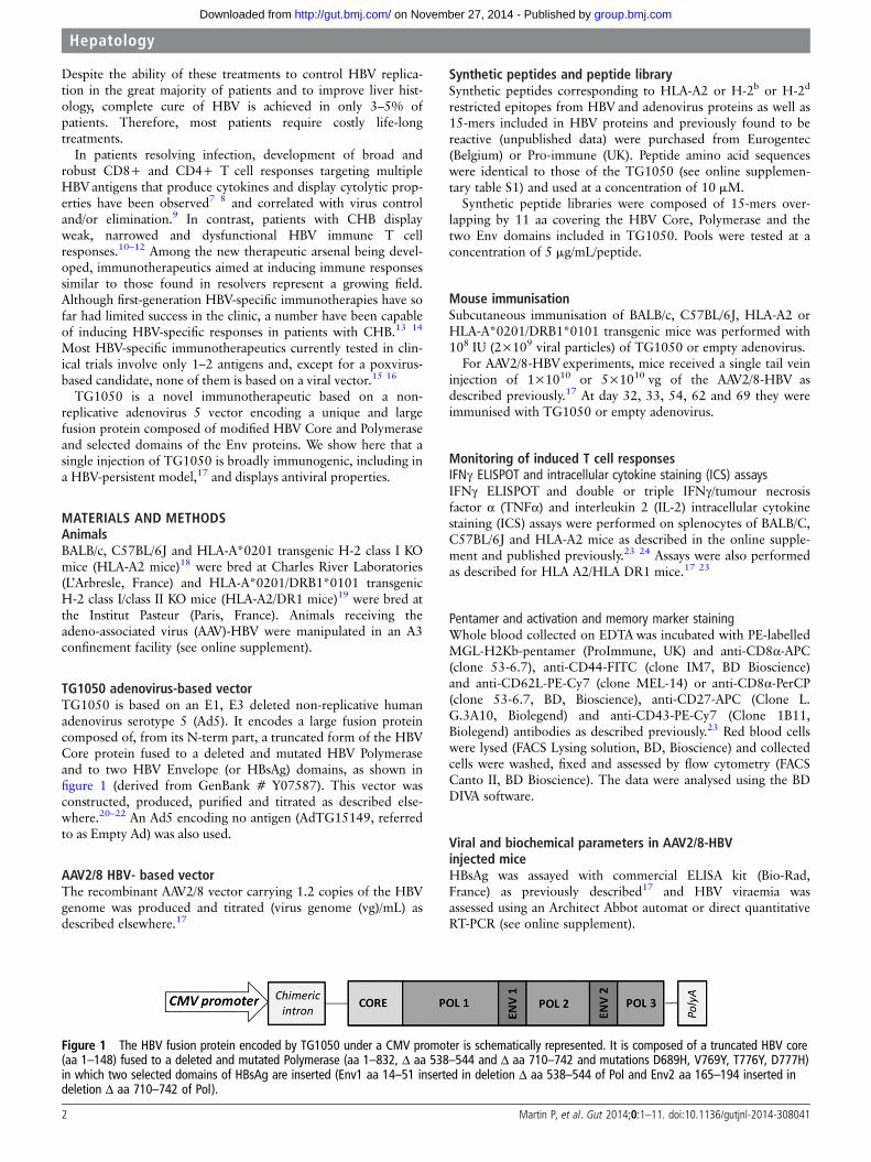
TG1050 adenovirus-based vectorTG1050 is based on an E1, E3 deleted non-replicative humanadenovirus serotype 5 (Ad5). It encodes a large fusion proteincomposed of, from its N-term part, a truncated form of the HBVCore protein fused to a deleted and mutated HBV Polymeraseand to two HBV Envelope (or HBsAg) domains, as shown infigure 1 (derived from GenBank # Y07587). This vector wasconstructed, produced, purified and titrated as described else-where.20–22 An Ad5 encoding no antigen (AdTG15149, referredto as Empty Ad) was also used.
AAV2/8 HBV- based vectorThe recombinant AAV2/8 vector carrying 1.2 copies of the HBVgenome was produced and titrated (virus genome (vg)/mL) asdescribed elsewhere.17
Synthetic peptides and peptide librarySynthetic peptides corresponding to HLA-A2 or H-2b or H-2d
restricted epitopes from HBV and adenovirus proteins as well as15-mers included in HBV proteins and previously found to bereactive (unpublished data) were purchased from Eurogentec(Belgium) or Pro-immune (UK). Peptide amino acid sequenceswere identical to those of the TG1050 (see online supplemen-tary table S1) and used at a concentration of 10 mM.
Synthetic peptide libraries were composed of 15-mers over-lapping by 11 aa covering the HBV Core, Polymerase and thetwo Env domains included in TG1050. Pools were tested at aconcentration of 5 mg/mL/peptide.
Mouse immunisationSubcutaneous immunisation of BALB/c, C57BL/6J, HLA-A2 orHLA-A*0201/DRB1*0101 transgenic mice was performed with108 IU (2×10⁹ viral particles) of TG1050 or empty adenovirus.
For AAV2/8-HBV experiments, mice received a single tail veininjection of 1×10¹⁰ or 5×10¹⁰ vg of the AAV2/8-HBV asdescribed previously.17 At day 32, 33, 54, 62 and 69 they wereimmunised with TG1050 or empty adenovirus.
Monitoring of induced T cell responsesIFNγ ELISPOT and intracellular cytokine staining (ICS) assaysIFNγ ELISPOT and double or triple IFNγ/tumour necrosisfactor α (TNFα) and interleukin 2 (IL-2) intracellular cytokinestaining (ICS) assays were performed on splenocytes of BALB/C,C57BL/6J and HLA-A2 mice as described in the online supple-ment and published previously.23 24 Assays were also performedas described for HLA A2/HLA DR1 mice.17 23
Pentamer and activation and memory marker stainingWhole blood collected on EDTA was incubated with PE-labelledMGL-H2Kb-pentamer (ProImmune, UK) and anti-CD8α-APC(clone 53-6.7), anti-CD44-FITC (clone IM7, BD Bioscience)and anti-CD62L-PE-Cy7 (clone MEL-14) or anti-CD8α-PerCP(clone 53-6.7, BD, Bioscience), anti-CD27-APC (Clone L.G.3A10, Biolegend) and anti-CD43-PE-Cy7 (Clone 1B11,Biolegend) antibodies as described previously.23 Red blood cellswere lysed (FACS Lysing solution, BD, Bioscience) and collectedcells were washed, fixed and assessed by flow cytometry (FACSCanto II, BD Bioscience). The data were analysed using the BDDIVA software.
Viral and biochemical parameters in AAV2/8-HBVinjected miceHBsAg was assayed with commercial ELISA kit (Bio-Rad,France) as previously described17 and HBV viraemia wasassessed using an Architect Abbot automat or direct quantitativeRT-PCR (see online supplement).
Figure 1 The HBV fusion protein encoded by TG1050 under a CMV promoter is schematically represented. It is composed of a truncated HBV core(aa 1–148) fused to a deleted and mutated Polymerase (aa 1–832, Δ aa 538–544 and Δ aa 710–742 and mutations D689H, V769Y, T776Y, D777H)in which two selected domains of HBsAg are inserted (Env1 aa 14–51 inserted in deletion Δ aa 538–544 of Pol and Env2 aa 165–194 inserted indeletion Δ aa 710–742 of Pol).
2 Martin P, et al. Gut 2014;0:1–11. doi:10.1136/gutjnl-2014-308041
Hepatology
group.bmj.com on November 27, 2014 - Published by http://gut.bmj.com/Downloaded from

RESULTSTG1050 composition and in vitro characterisation of HBVprotein expression via TG1050Figure 1 depicts the organisation of the large HBV encodedpolyprotein. This organisation resulted from the choice of HBVproteins known to be targeted by T cells during infection, inparticular resolutive infection (eg, Core) and/or containing clus-ters of T cell epitopes (eg, Env1 and Env2), together with theneed to generate a genetically stable vaccine fit for manufactur-ing (mutations in POL). In vitro expression of the HBV fusionprotein was analysed by western blotting (see online supplemen-tary figure S1A) and revealed a faint band corresponding to theCore-Pol-Env fusion protein (about 115 kDa, the calculatedmolecular weight (MW) being 113.9 kDa) as well as severaladditional bands corresponding to lower MW (ranging from 15to 50 kDa). Products between 15 and 26 kDa were onlydetected with a Core-specific antibody and with a Pol-specificantibody targeting the N-terminus of the protein, while theywere not detected with a Pol-specific antibody targeting aa 225–250 of the Polymerase (data not shown), suggesting that theobserved products correspond to N-terminus cleavage productsof the HBV fusion protein.
Analysis by bidimensional electrophoresis followed by immu-noblot together with LC-MS/MS analysis was performed tocharacterise the different protein species generated (see onlinesupplementary figure S1B and C). Data confirmed that productsaround 26 kDa are composed of the N-terminal part of thefusion protein—that is, the HBV Core protein and N-termamino acids of the Polymerase protein.
TG1050 induces high percentages of polyfunctional CD8+T cells producing IFNγ and TNFα and displaying cytolyticactivity in naïve mouse modelsThe ability of TG1050 to induce HBV-specific polyfunctionalT cells was assessed by ICS assay in three mouse strains follow-ing one injection.
In HLA-A2 mice (figure 2A), TG1050 induced IFNγ-producingCD8+ T cells targeting the Core and Polymerase proteins (up to7.5% and 9.5% of IFNγ+ CD8+T cells), most of them also pro-ducing TNFα. In C57BL/6J mice (figure 2B), IFNγ-producingCD8+ T cells targeting the three HBV antigens were detected(up to 2.4%, 1% and 3.2% of IFNγ+ CD8+ T cells specific forCore, Env and Polymerase respectively), with at least half ofthese cells producing both IFNγ and TNFα. Finally, in BALB/Cmice (figure 2C), TG1050 induced IFNγ-producing CD8+ Tcellsspecific of Env1 domain and Polymerase (up to 0.7% and 6.8%of IFNγ+CD8+ T cells specific for Env1 and Polymerase,respectively), most of them also producing TNFα. Overall, no(HLA-A2) or weak (C57BL/6J and BALB/c) CD4+ T cells weredetected in these experiments. Frequencies of TG1050-inducedIFNγ-producing cells were also assessed using IFNγ ELISPOTassay (see online supplementary figure S2). Responses targetingepitopes from all three encoded antigens were detected with vari-able magnitude and hierarchy depending on the mouse strain.
In vivo cytotoxic T lymphocyte (CTL) assays were performedto assess functional capacity of TG1050-induced CD8+ T cellsin vivo in the three different mouse lines (see online supplemen-tary figure S3). Cytolytic CD8+ T cells targeting Core andPolymerase (HLA-A2 and C57BL/6J, see online supplementaryfigure S3A and B) or Core, Pol and Env (BALB/c, see online sup-plementary figure S3C) were detected with percentages of lysisranging from 6% to 83%. Of note, all three Pol and CoreHLA-A2 epitopes described in patients with HBV were
recognised by the induced Tcells including the Core18–27 epitope(referred to as FLP) associated with resolution of infection.7 25 26
Overall, these data illustrate the capacity of TG1050 to inducepotent, multi-antigenic, polyfunctional CD8+ T cells displayingin vivo cytolytic activity specific to all encoded HBVantigens.
TG1050 induces long-lasting functional HBV-specific T cellsdisplaying recall potentialIn C57BL/6J mice, HBV-Core pentamer staining showed a peakof Core-specific T cells in blood 14 days post-injection (mean of3.4%), followed by a decrease up to day 96 (mean value around0.6% at day 96, figure 3A, left panel). The percentage ofCore-specific T cells progressively decreased but was still clearlydetectable when measured at day 310 and in some mice up today 400. Analysis of the CD44/CD62L phenotype—whichallows effector memory cells to be distinguished from centralmemory cells—on Core-specific T cells showed that a singleinjection of TG1050 induces mostly effector memory cells(CD44+/CD62L− cells, percentages ranging from 95% at day14 to 60% at day 400), but with the appearance of a significantproportion of central memory cells observed from day 310(28% and 35% of Core-specific T cells being CD44+/CD62L+at day 310 and day 400, respectively) (figure 3A right panel).Analysis of CD27/CD43 phenotypes on Core-specific T cells, inparticular the CD27+/CD43− population which has beendescribed as having good recall potential,27 showed that 20% ofinduced Core-specific cells were CD27+/CD43− during thefirst 100 days post-injection of TG1050. This proportionincreased to 29% and 36% at the latest analysed time points.
Functional HBV-specific T cells producing IFNγ remaineddetectable more than 1 year after one immunisation (figure 3B).The frequency of detected Pol-specific T cells was the highest(mean 290 spots/106 cells), followed by Core-specific T cells(mean 160 spots/106 cells) and Env1-specific T cells (mean 75spots/106 cells). Half or more of HBV-specific CD8+ T cellsdetected 400 days after one immunisation were still able toproduce both IFNγ and TNFα. These T cells targeted mainlyPolymerase and Core antigens (figure 3C).
TG1050 is able to induce functional T cells and antiviralresponses in HBV-persistent HLA-A2/DR1 transgenic micefollowing a single administrationTG1050 immunogenicity was evaluated in the recentlydescribed AAV-HBV-persistent model.17 Both short-term andlong-term studies were performed.
Short-term monitoringTwo experiments were performed in order to closely assess thekinetics of appearance of TG1050-induced T cells. The abilityof TG1050 to induce functional T cells producing cytokines wasassessed 2 weeks after TG1050 immunisation. Analysis per-formed on spleen cells of AAV-HBV mice showed that TG1050was able to induce IFNγ-producing cells specific for HLA-A2epitopes located within Polymerase, Core and Env to a lowerextent (figure 4A). The frequencies of IFNγ-producing cells andof responding mice were notably detectable but lower in thesemice (median values up to 105 and 73 spots/106 splenocytes inexperiments 1 (p<0.01) and 2 (p=NS), respectively), than inHBV-free mice (median values up to 1389 and 605 spots/106
splenocytes in experiments 1 and 2, respectively). SplenicCD8+ T cells producing at least one cytokine could be detectedin 60% and 40% of AAV-injected mice with percentages ofdetection reaching 0.32% and 0.7% (experiments 1 and 2respectively, figure 4B). In comparison, median values of CD8+
Martin P, et al. Gut 2014;0:1–11. doi:10.1136/gutjnl-2014-308041 3
Hepatology
group.bmj.com on November 27, 2014 - Published by http://gut.bmj.com/Downloaded from

Figure 2 Analysis of induced T cell responses following single injection of TG1050 using interferon γ (IFNγ)/tumour necrosis factor α (TNFα) ICSassay in (A) HLA-A2 transgenic mice, (B) C57BL/6J mice and (C) BALB/c mice. HLA-A2 transgenic mice or C57BL/6J or BALB/c mice were immunisedonce subcutaneously with TG1050 or an empty Adenovirus (negative control) and HBV-specific immune responses were monitored on spleen cellsusing IFNγ/TNFα intracellular cytokine staining assays using single peptides or pools of peptides derived from Core, Env or Polymerase HBV proteinsfor stimulation. The results are presented as the percentage of IFNγ and/or TNFα producing CD8+ or CD4+ T cells for each group. Each barcorresponds to an individual mouse. For each mouse, the white bar represents the percentage of CD8+ or CD4+ T cells producing TNFα alone, thegrey bar represents the percentage of CD8+ or CD4+ T cells producing IFNγ and TNFα and the black bar represents the percentage of CD8+ or CD4+ T cells producing IFNγ alone.
4 Martin P, et al. Gut 2014;0:1–11. doi:10.1136/gutjnl-2014-308041
Hepatology
group.bmj.com on November 27, 2014 - Published by http://gut.bmj.com/Downloaded from

Figure 3 Analysis of long-term HBV-specific T cells following a single injection of TG1050 up to 400 days post-injection. C57BL/6J mice wereimmunised once subcutaneously with TG1050 or an empty Ad (negative control). (A) HBV-specific cells were monitored and quantified along time inwhole blood of injected mice by pentamer staining of HBV Core-specific T cells. An example of a dot plot obtained for one representative mouseinjected with TG1050 at each time point is also shown on the right side of the graphs. Phenotypes of these Core-specific T cells were assessed byfollowing CD44 and CD62L as well as CD27 and CD43 markers. Examples of dot plots obtained for CD62 L/CD44 and CD27/CD43 stainingexperiments in TG1050-injected mice are also shown at the top of the figure (shown for day 400). Pie charts representative of the mean percentagesof HBV Core (MGL epitope)-specific CD8+ T cells displaying the indicated phenotypes are shown. (B, C). Interferon γ (IFNγ) ELISPOT (B) andintracellular cytokine staining (ICS) (C) assays realised at 400 days post-injection were performed using spleen cells and pools of overlappingpeptides covering, respectively, the HBV Core protein (PC1, PC2), the 2 Env domains (PE1, PE2) and the HBV polymerase protein (PP4) or irrelevantpeptides (IRR). For ELISPOT assay (3B), the horizontal dotted line represents the threshold of ELISPOT positivity. Each individual mouse is representedby an empty (empty Ad injected mice) or a plain (TG1050 injected mice) circle and the black line represents the mean of each group. For IFNγ/tumour necrosis factor α (TNFα) ICS assay (C), the results are presented as the percentage of IFNγ- and/or TNFα-producing CD8+ T cells for eachgroup. For each mouse, the white bar represents the percentage of CD8+ T cells producing TNFα alone, the grey bar represents the percentage ofCD8+ T cells producing IFNγ and TNFα and the black bar represents the percentage of CD8+ T cells producing IFNγ alone.
Martin P, et al. Gut 2014;0:1–11. doi:10.1136/gutjnl-2014-308041 5
Hepatology
group.bmj.com on November 27, 2014 - Published by http://gut.bmj.com/Downloaded from

T cells producing at least one cytokine were up to 9.1% and17.4% and found in up to 100% and 60% of HBV-freeresponding mice (experiments 1 and 2, respectively). In experi-ment 1, detected cells produced mainly one or two cytokines(mostly IFNγ+ or IFNγ+/TNFα+) in HBV-free and AAV-HBVinjected mice (data not shown). Liver-infiltrating HBV-specificT cells were also monitored (figure 4). In AAV-HBV injectedmice, cytokine-producing cells were detected in both experi-ments, albeit with distinct frequencies (percentage of respond-ing mice ranged from 20% to 60%). The percentages ofHBV-specific cells producing IFNγ and/or TNFα and/or IL-2and the percentages of responding mice were lower inAAV-HBV mice than in HBV-free mice (21% vs 0.46% of CD8+ T cells producing at least one cytokine; experiment 1, medianvalues). Some IL-2 producing cells were detected in the liver ofAAV-HBV injected mice, hence the overall proportion of mul-tiple cytokine producer cells (double and triple) approximated80%, with a slightly lower proportion in HBV-free mice (datanot shown). In experiment 2, due to the high backgroundobserved within the liver, no CD8+ T cells producing cytokines(single or multiple) could be detected in the control HBV-freemice. In AAV-HBV injected mice, one mouse injected withTG1050 displayed detectable CD8+ T cells producing at leastone cytokine (figure 4C).
As shown in figure 5, circulating HBV DNA remained stable(experiment 1) or increased (experiment 2) in the controlgroups that received an empty Ad vector (experiment 1, HBVDNA increased 0.7–1.7-fold from 6 to 14 days post-Empty Adinjection; experiment 2, HBV DNA increased 4-fold and 9-foldat 6 and 10 days post TG1050 injection, respectively;figure 5A). In contrast, TG1050-injected mice controlled HBV
DNA increase from 6 to 10 or 14 days post-injection. A strongtrend was observed in both experiments, and statistical signifi-cance was reached in experiment 2 at day 38 (p=0.0174).Initial levels of HBV DNA were different in both experiments, afactor that may have influenced the effect of TG1050.
Injection of TG1050 did not affect HBsAg levels in experi-ment 1, although the levels were consistently lower. A statistic-ally significant effect was seen in experiment 2 (figure 5B). Inthis experiment, a decrease was maintained over three consecu-tive time points, (p=0.0296, p=0.0319 and p=0.022 betweenAAV-HBV+Empty Ad and AAV-HBV+TG1050 groups for days38, 42 and 48, respectively).
No increase in ALAT levels over normal values was detectedin either experiment, independently of the analysed groups(data not shown).
Long-term monitoringMonitoring of spleen cells up to 4 months after TG1050 injec-tion (see online supplementary figure S4A, IFNγ ELISPOTassay) showed induction of high frequencies of IFNγ-producingcells targeting Core, Polymerase and Env domains. Frequenciesof IFNγ+ cells and percentages of responding mice were com-parable between TG1050 immunised HBV-free (PBS) andAAV-HBV mice. A high percentage of HBV-specific CD8+ Tcells producing IFNγ and/or TNFα and/or IL-2 was detected inHBV-free and AAV-HBV mice (see online supplementary figureS4B). The percentages of cells producing one, two and threecytokines were almost identical between the two groups,whether tested in the spleen or the liver (see online supplemen-tary figure S4C).
Figure 3 continued.
6 Martin P, et al. Gut 2014;0:1–11. doi:10.1136/gutjnl-2014-308041
Hepatology
group.bmj.com on November 27, 2014 - Published by http://gut.bmj.com/Downloaded from

Viral parameters following TG1050 or empty-Ad administra-tion were low in this experiment at the time of TG1050 adminis-tration (mean 7.11 mg/mL HBsAg and <300 IU/mL DNA in
50% of mice), probably due to the use of a lower dose ofAAV-HBV injected compared with that used in the short-termexperiments (5 times lower ie, 1×10¹⁰ vg). Hence, no impact on
Figure 4 Analysis of short-termHBV-specific functional T cells inHLA-A2/DR1 AAV-HBV-persistent micefollowing a single TG1050immunisation. HLA-A2/HLA-DR1transgenic mice were intravenouslyinjected with PBS or AAV-HBV (day 0)and immunised 1 month later (day 32)with either TG1050 or emptyadenovirus. Induced HBV-specificimmune responses were assessed2 weeks after TG1050 or empty Adimmunisation through (A) IFNγELISPOT assay on spleen cells andinterferon γ (IFNγ)/tumour necrosisfactor α (TNFα) intracellular cytokinestaining assays using (B) spleen cellsand (C) liver-infiltrating lymphocytes.The analytical conditions are asdescribed in figure 3, except thatHLA-A2-restricted peptides were used(see online supplementary table S1).Data from two experiments arepresented. In experiment 2, data fromthe group PBS+TG1050 could not beexploited due to high background. NE,non-evaluable.
Martin P, et al. Gut 2014;0:1–11. doi:10.1136/gutjnl-2014-308041 7
Hepatology
group.bmj.com on November 27, 2014 - Published by http://gut.bmj.com/Downloaded from

HBV DNA at day 47 or day160 could be seen (data not shown).Overall, comparison at all time points showed that HBsAg levelswere consistently lower in AAV-HBV mice immunised withTG1050 than in mice receiving an empty Ad, and significantlydifferent at day 47 (see online supplementary figure S5).
In conclusion, data collected in HLA-A2/DR1 mice show thatTG1050 can educate functional T cells in a HBV-persistentenvironment and can impact on both HBV DNA and HBsAglevels, albeit in a transient manner.
TG1050 is able to induce functional T cells and sustainedantiviral responses in HBV-persistent C57BL/6J micefollowing multiple administrationsThe AAV-HBV-persistent model was set up in C57BL/6J miceand TG1050 was injected up to four times.
Mice were immunised once with TG1050 at day 32 postAAV-HBV injection; half of them were monitored for viral para-meters and sacrificed for immune monitoring at day 46/47(figure 6A left panel and C). The remaining mice were keptalive, injected an additional three times at 1-week intervals andfollowed for viral parameters up to day 105 (figure 6A rightpanel and B). A strong reduction in HBV-DNA titres wasquickly observed (2 and 3 weeks after first TG1050 immunisa-tion) compared with control mice (26-fold and 44-fold,figure 6A). Following additional injections, the HBV-DNA titreremained significantly reduced until the end of monitoring.HBsAg titres also decreased 2–3 weeks after first TG1050
immunisation and remained low up to day 105 (10 weeks afterfirst immunisation) in mice receiving three additional injectionsof TG1050 compared with control mice.
The percentages of HBV-specific cells producing IFNγ and/orTNFα and/or IL-2 were monitored 2 weeks after the firstTG1050 injection and were found to be lower in HBV-persistentmice than in HBV-free mice (8.4% vs 1.9% of IFNγ+ CD8+Tcells, figure 6C). However, as a higher number of CD8+ Tcellswas observed in the liver of AAV-HBV mice (about three timesmore than in HBV-free mice), absolute numbers of HBVPolymerase-specific CD8T cells induced by TG1050 in theAAV-HBV and HBV-free mice were very close (data not shown).An IFNγ response was detected in all TG1050-injected mice,both HBV-free and persistent, whereas the percentage of HBVpersistent mice displaying TNFα+ and/or IL-2+ CD8+ T cellswas low (50% and 0%) compared with HBV-free mice (100%and 80%). The percentage of TG1050-induced cells secreting atleast two cytokines was higher in HBV-free mice (45%) than inHBV-persistent mice (19%).
Overall, data collected in C57BL/6J HBV-persistent miceshow that TG1050 induces a rapid and sustained antiviral effectfollowing multiple injections.
DISCUSSIONAdenovirus vectors are extremely potent at inducing cellular-based immune responses to the encoded immunogens and havebeen used to develop vaccines against a range of infectious
Figure 5 Monitoring of viral parameters in HLA-A2/DR1 AAV-HBV-persistent mice. HLA-A2/HLA-DR1 transgenic mice were intravenously injectedwith PBS or AAV-HBV (day 0) and immunised 1 month later (day 32) with either TG1050 or empty adenovirus. (A) HBV DNA and (B) HBsAg data arerepresented as mean fold changes (DNA and HBsAg) compared with their respective mean level at day 28 before TG1050 (plain circle curve) orempty Ad (empty circle curve) immunisation or in non-immunised mice (cross curve, for experiment 1 only). Statistical analyses were performedusing mixed models and ANOVA for repeated measures. *p<0.05.
8 Martin P, et al. Gut 2014;0:1–11. doi:10.1136/gutjnl-2014-308041
Hepatology
group.bmj.com on November 27, 2014 - Published by http://gut.bmj.com/Downloaded from

diseases and cancers.28 Ad serotype 5 has been the most widelyused Ad vector, displaying a good safety profile and a remark-able capacity to induce long-lasting and broad T cell-basedimmune responses, in particular those driven by CD8+.29
TG1050 is an Ad-5 vector expressing a unique fusion proteinthat includes the Core protein described as an effective target ofT cell responses in HBV resolvers.30 31 We show here in mousemodels that a single administration of TG1050 induces mainly
CD8 T cells (2–5%), although HBV-specific CD4 T cells werealso detected to a lesser extent (0.05–0.15%). While CD4T cells are important in HBV control,32 CD8 T cells have beenshown to be essential in clearance of HBV, as demonstrated indepletion studies performed in chimpanzees33 and in studiesdescribing functional exhaustion of HBV-specific CD8 T cells inpatients with CHB.34 The detection of multispecific T cells tar-geting all three TG1050-encoded immunogens and producing
Figure 6 Monitoring of viral and immunological parameters in C57BL/6J AAV-HBV-persistent mice. C57BL/6J naïve mice were intravenouslyinjected with PBS or AAV-HBV (day 0) and immunised 1 month later (day 32) with either TG1050 or empty adenovirus. (A) HBV DNA and (B) HBsAgdata are represented as mean fold changes (DNA and HBsAg) compared with their respective mean level before TG1050 (plain circle curve) orempty Ad (empty circle curve) immunisation. Statistical analyses were performed using mixed models and ANOVA for repeated measures. Theanalysis was done on changes from baseline in percentage. For DNA, the analysis was performed on ranks in order to fulfil the mixed modelhypothesis. *p<0.05. Induced HBV-specific immune responses were assessed 2 weeks after TG1050 or empty Ad immunisation (day 47) withinterferon γ (IFNγ)/tumour necrosis factor α (TNFα)/interleukin 2 (IL-2) intracellular cytokine staining assays using liver-infiltrating lymphocytes (C),using the H-2b restricted peptide VSA (polymerase) for in vitro stimulation. The results are presented as the percentage of CD8+ T cells producing atleast IFNγ or TNFα or IL-2. The percentage of mice displaying a positive response is indicated at the top of the graph.
Martin P, et al. Gut 2014;0:1–11. doi:10.1136/gutjnl-2014-308041 9
Hepatology
group.bmj.com on November 27, 2014 - Published by http://gut.bmj.com/Downloaded from

multiple cytokines was also observed. In addition, in vivo ana-lyses demonstrated the induction of CTL, with significant celllysis observed for Core- and Polymerase-specific peptides. CTLare expected to be a key component of the mechanism of actionof TG1050 as they aim at killing infected hepatocytes andhence clearing cccDNA.35 36 Finally, our study shows inductionof long-lasting HBV-specific T cells, detectable up to 400 dayspost-injection. Such induced Core-specific T cells are mainlyeffector memory cells (CD44+/CD62L−) and some of themdisplayed markers (CD27+/CD43−) associated with strongrecall capacity but also central memory cells (CD44+/CD62L+)that are detected until day 400 post-injection. In contrast toother vaccine vectors, adenoviruses are known to induce a long-lasting effector phase, possibly due to the persistence of a lowlevel of viral transcription in T lymphocytes37 together with asignificant memory response.38
Although anti-adenovirus immune responses were not mea-sured in this study, we have shown that a single subcutaneousadministration of HBV-specific Ad vectors (unpublished infor-mation and Boukhebza et al23) expectedly results in both T andB Ad-specific immune responses that do not preclude themounting of HBV-specific responses, including after multipleadministrations.23 Pre-existing immunity to Ad5 in humans hasbeen reported to hamper mounting of immunity to encodedimmunogens, in particular in the HIV field39 and whenE1-deleted Ad5 are used. However, recent papers on malariaand tuberculosis vaccine development have reported no impactof pre-Ad5 immunity to vaccine immunogens, particularly whenE1/E3 deleted vectors are used, such as for TG1050.40 41
Strategies to formulate Ad5 in order to minimise and/or preventthe impact of anti-Ad5 neutralising antibodies on vaccineimmunogenicity can be explored.42
First-generation HBV immunotherapeutics have been disap-pointing in the clinic. These were based on suboptimal vectorplatforms with respect to induction of strong T cell-basedimmunity. They have included peptides, adjuvanted recombinantantigens in single or complex mixtures including existing HBVprophylactic vaccines as well as DNA vaccines administered byconventional means.16 In contrast, TG1050 has a high antigeniccomplexity covering either full length or major domains ofthree HBV antigens and is based on an optimal platform forinduction of CD8-driven responses. Closest to TG1050,HB-110 is a combination of three plasmid DNAs expressingpre-S/S, Core/Polymerase and the IL-12 cytokine which showedpromising results in a therapy arrest clinical trial setting,43
although this was not confirmed in a later study.44
Among the few animal models supporting infection by aHBV,45 studies have been conducted in woodchucks infected bywoodchuck hepatitis virus (WHV) including evaluation of com-bined use of NUC and immunotherapeutics, a potent combin-ation of a DNA and an adenovirus-based vaccine.46 WHV andHBV genomic sequences display a high rate of diversity, rangingfrom 25% to 60%, limiting the potential of the woodchuckmodel in the evaluation of immunotherapeutics targeting thehuman virus. Here we used the recently described alternativeHBV-persistent murine model (AAV-HBV)17 to evaluate the cap-acity of TG1050 to educate HBV-specific functional T cells inface of ongoing HBV antigen expression. We also provide a firstinsight into the capacity of TG1050 to exert an antiviral effect.Remarkably, in this model a single subcutaneous administrationof TG1050 was able to educate broad HBV-specific CD8+T cells targeting Core, Pol and Env and capable of producingIFNγ, TNFα and, in some cases, IL-2, including in the liver ofimmunised mice. In parallel, some control of HBV DNA and
HBsAg levels in the serum of vaccinated mice was achieved,reaching statistical significance in two of the three reported short-term experiments, both in a HLA-A2/HLA-DR1 and in a C57BL/6J background. In a similar mouse model, Tay et al47 showed thatthe threshold of antigen expression within the liver is a dominantfactor determining the fate of T cells, with a high percentage ofcells being silenced. Interestingly, in C56BL/6J AAV-HBV mice, asingle administration resulted in a dramatic reduction in bothHBV DNA and HBsAg (>1 log decrease for the DNA), and a sig-nificant decrease was sustained until day 105. This persistenteffect may be linked to the three additional closely-spaced boost-ing injections administered. HLA-A2/DR1 mice have been shownto harbour a low percentage CD8+ T cells (reaching only 2–3%of total splenocytes48), a feature that may have been suboptimalwith respect to mounting of TG1050 antiviral efficacy. Inhumans a correlation has been reported between T cell exhaus-tion and viral load and/or antigenic load; the higher the levels ofviral parameters carried by the patients, the more immunedefects are observed.31 Together our observations suggest thatmultiple TG1050 administrations may have to be used in theclinic, particularly in patients with higher viral markers.Application of closely spaced administrations of viral-vectoredimmunotherapeutics, as performed here, has shown clinical effi-cacy in the treatment of patients with chronic hepatitis C.49
TG1050 is intended to be first developed in the clinic in combin-ation with NUC, hence the ultimate goal will be elimination ofHBsAg. At that stage, our preclinical data show the capacity ofTG1050 to affect the levels of HBsAg in the absence of a signifi-cant elevation of alanine aminotransferase. In patients, long-termtreatment with NUC has been shown in in vitro expansionstudies to restore some Tcell functionality.50 The combination ofTG1050 with NUC and possibly with blockers of inhibitorymolecules described in HBV Tcells of patients with CHB such asPD-1, TIM-3 and SLAM,51 52 should be explored.
In conclusion, we have developed a novel active targetedimmunotherapeutic that displays a number of immunologicalfeatures found in HBV-infected resolvers together with the cap-acity to exert antiviral activity on a stand alone-based approach.
Acknowledgements We thanks all employees of Plateau de BiologieExpérimentale de la souris in Lyon for taking care of the animals. This work waspartially funded by NATHEB program (FEDER/FUI).
Contributors All authors listed have contributed to the work described in thearticle. CD, EJ, AE and HB conducted immunological experiments in HBV naïve mice.RK, KL-S and AE conducted experiments in C57BL6 AAV-HBV mice. PM and GIsupervised the experiments and took a major part in writing the paper. SD, MM-Band OG conducted experiments in the HLA-A2/HLA-DR1 AAV-HBV model. M-LMsupervised these experiments and made an important contribution to writing thepaper. J-FM performed HBV DNA measurements. YS, DV, J-BM, MG, RB and AFperformed in vitro analyses. TM, NS, J-BM supervised these and took part in writingof the manuscript. J-MS and AVD performed mass spectrometry analysis.
Competing interests None.
Provenance and peer review Not commissioned; externally peer reviewed.
Data sharing statement Not relevant, no unpublishd data available.
Open Access This is an Open Access article distributed in accordance with theCreative Commons Attribution Non Commercial (CC BY-NC 4.0) license, whichpermits others to distribute, remix, adapt, build upon this work non-commercially,and license their derivative works on different terms, provided the original work isproperly cited and the use is non-commercial. See: http://creativecommons.org/licenses/by-nc/4.0/
REFERENCES1 Kim DY, Han KH. Epidemiology and surveillance of hepatocellular carcinoma. Liver
Cancer 2012;1:2–14.2 Ott JJ, Stevens GA, Groeger J, et al. Global epidemiology of hepatitis B virus
infection: new estimates of age-specific HBsAg seroprevalence and endemicity.Vaccine 2012;30:2212–19.
10 Martin P, et al. Gut 2014;0:1–11. doi:10.1136/gutjnl-2014-308041
Hepatology
group.bmj.com on November 27, 2014 - Published by http://gut.bmj.com/Downloaded from

3 Buti M. HBeAg-positive chronic hepatitis B: why do I treat my patients with nucleos(t)ide analogs? Liver Int 2014;34(Suppl 1):108–11.
4 Vigano M, Mangia G, Lampertico P. HBeAg-negative chronic hepatitis B: why do Itreat my patients with nucleos(t)ide analogues? Liver Int 2014;34(Suppl 1):120–6.
5 Kao JH. HBeAg-positive chronic hepatitis B: why do I treat my patients withpegylated interferon? Liver Int 2014;34(Suppl 1):112–19.
6 Vlachogiannakos J, Papatheodoridis GV. HBeAg-negative chronic hepatitis B: why do Itreat my patients with pegylated interferon-alfa?. Liver Int 2014;34(Suppl 1):127–32.
7 Bertoletti A, Ferrari C, Fiaccadori F, et al. HLA class I-restricted human cytotoxicT cells recognize endogenously synthesized hepatitis B virus nucleocapsid antigen.Proc Natl Acad Sci USA 1991;88:10445–9.
8 Rehermann B, Fowler P, Sidney J, et al. The cytotoxic T lymphocyte response tomultiple hepatitis B virus polymerase epitopes during and after acute viral hepatitis.J Exp Med 1995;181:1047–58.
9 Bertoletti A, Ferrari C. Innate and adaptive immune responses in chronic hepatitis Bvirus infections: towards restoration of immune control of viral infection. Gut2012;61:1754–64.
10 Das A, Hoare M, Davies N, et al. Functional skewing of the global CD8T cellpopulation in chronic hepatitis B virus infection. J Exp Med 2008;205:2111–24.
11 Lopes AR, Kellam P, Das A, et al. Bim-mediated deletion of antigen-specific CD8Tcells in patients unable to control HBV infection. J Clin Invest 2008;118:1835–45.
12 Maini MK, Schurich A. The molecular basis of the failed immune response inchronic HBV: therapeutic implications. J Hepatol 2010;52:616–19.
13 Godon O, Fontaine H, Kahi S, et al. Immunological and antiviral responses aftertherapeutic DNA immunization in chronic hepatitis B patients efficiently treated byanalogues. Mol Ther 2014;22:675–84.
14 Yang SH, Lee CG, Park SH, et al. Correlation of antiviral T-cell responses withsuppression of viral rebound in chronic hepatitis B carriers: a proof-of-concept study.Gene Ther 2006;13:1110–17.
15 Liu J, Kosinska A, Lu M, et al. New therapeutic vaccination strategies for thetreatment of chronic hepatitis B. Virol Sin 2014;29:10–16.
16 Michel ML, Deng Q, Mancini-Bourgine M. Therapeutic vaccines and immune-basedtherapies for the treatment of chronic hepatitis B: perspectives and challenges.J Hepatol 2011;54:1286–96.
17 Dion S, Bourgine M, Godon O, et al. Adeno-associated virus-mediated gene transferleads to persistent hepatitis B virus replication in mice expressing HLA-A2 andHLA-DR1 molecules. J Virol 2013;87:5554–63.
18 Pascolo S, Bervas N, Ure JM, et al. HLA-A2.1-restricted education and cytolyticactivity of CD8(+) T lymphocytes from beta2 microglobulin (beta2m) HLA-A2.1monochain transgenic H-2Db beta2m double knockout mice. J Exp Med1997;185:2043–51.
19 Pajot A, Michel ML, Fazilleau N, et al. A mouse model of human adaptive immunefunctions: HLA-A2.1-/HLA-DR1-transgenic H-2 class I-/class II-knockout mice. Eur JImmunol 2004;34:3060–9.
20 Chartier C, Degryse E, Gantzer M, et al. Efficient generation of recombinantadenovirus vectors by homologous recombination in Escherichia coli. J Virol1996;70:4805–10.
21 Fallaux FJ, Bout A, van der Velde I, et al. New helper cells and matched earlyregion 1-deleted adenovirus vectors prevent generation of replication-competentadenoviruses. Hum Gene Ther 1998;9:1909–17.
22 Erbs P, Regulier E, Kintz J, et al. In vivo cancer gene therapy byadenovirus-mediated transfer of a bifunctional yeast cytosine deaminase/uracilphosphoribosyltransferase fusion gene. Cancer Res 2000;60:3813–22.
23 Boukhebza H, Dubois C, Koerper V, et al. Comparative analysis of immunizationschedules using a novel adenovirus-based immunotherapeutic targeting hepatitis Bin naïve and tolerant mouse models. Vaccine. 2014;32:3256–63.
24 Fournillier A, Gerossier E, Evlashev A, et al. An accelerated vaccine schedule with apoly-antigenic hepatitis C virus MVA-based candidate vaccine induces potent, longlasting and in vivo cross-reactive T cell responses. Vaccine 2007;25:7339–53.
25 Maini MK, Boni C, Ogg GS, et al. Direct ex vivo analysis of hepatitis B virus-specificCD8(+) T cells associated with the control of infection. Gastroenterology1999;117:1386–96.
26 Penna A, Chisari FV, Bertoletti A, et al. Cytotoxic T lymphocytes recognize anHLA-A2-restricted epitope within the hepatitis B virus nucleocapsid antigen. J ExpMed 1991;174:1565–70.
27 Hikono H, Kohlmeier JE, Takamura S, et al. Activation phenotype, rather thancentral- or effector-memory phenotype, predicts the recall efficacy of memory CD8+T cells. J Exp Med 2007;204:1625–36.
28 Rollier CS, Reyes-Sandoval A, Cottingham MG, et al. Viral vectors as vaccineplatforms: deployment in sight. Curr Opin Immunol 2011;23:377–82.
29 Bassett JD, Swift SL, Bramson JL. Optimizing vaccine-induced CD8(+) T-cellimmunity: focus on recombinant adenovirus vectors. Expert Rev Vaccines2011;10:1307–19.
30 Lau GK, Suri D, Liang R, et al. Resolution of chronic hepatitis B and anti-HBsseroconversion in humans by adoptive transfer of immunity to hepatitis B coreantigen. Gastroenterology 2002;122:614–24.
31 Webster GJ, Reignat S, Brown D, et al. Longitudinal analysis of CD8+ T cellsspecific for structural and nonstructural hepatitis B virus proteins in patients withchronic hepatitis B: implications for immunotherapy. J Virol 2004;78:5707–19.
32 Asabe S, Wieland SF, Chattopadhyay PK, et al. The size of the viral inoculumcontributes to the outcome of hepatitis B virus infection. J Virol 2009;83:9652–62.
33 Thimme R, Wieland S, Steiger C, et al. CD8 + T cells mediate viral clearance anddisease pathogenesis during acute hepatitis B virus infection. J Virol 2003;77:68–76.
34 Rehermann B, Nascimbeni M. Immunology of hepatitis B virus and hepatitis C virusinfection. Nat Rev Immunol 2005;5:215–29.
35 Yang PL, Althage A, Chung J, et al. Immune effectors required for hepatitis B virusclearance. Proc Natl Acad Sci USA 2010;107:798–802.
36 Wieland SF, Spangenberg C, Thimme R, et al. Expansion and contraction of thehepatitis B virus transcriptional template in infected chimpanzees. Proc Natl AcadSci USA 2004;101:2129–34.
37 Tatsis N, Fitzgerald JC, Reyes-Sandoval A, et al. Adenoviral vectors persist in vivoand maintain activated CD8 T cells: implications for their use as vaccines. Blood2007;110:1916–23.
38 Steffensen MA, Holst PJ, Steengaard SS, et al. Qualitative and quantitative analysisof adenovirus-type 5 vector induced memory CD8T cells: not as bad as theirreputation. J Virol 2013;87:6283–95.
39 Buchbinder SP, Mehrotra DV, Duerr A, et al. Efficacy assessment of a cell-mediatedimmunity HIV-1 vaccine (the Step Study): a double-blind, randomised,placebo-controlled, test-of-concept trial. Lancet 2008;372:1881–93.
40 Tamminga C, Sedegah M, Regis D, et al. Adenovirus-5-vectored P. falciparumvaccine expressing CSP and AMA1. Part B: safety, immunogenicity and protectiveefficacy of the CSP component. PLoS ONE 2011;6:e25868.
41 Smaill F, Jeyanathan M, Smieja M, et al. A human type 5 adenovirus-basedtuberculosis vaccine induces robust T cell responses in humans despite preexistinganti-adenovirus immunity. Sci Transl Med 2013;5:205ra134.
42 Wortmann A, Vöhringer S, Engler T, et al. Fully detargeted polyethyleneglycol-coated adenoviru vectors are potent genetic vaccines and escape pre-existinganti-adenovirus antibodies. Mol Ther 2008;15:154–62.
43 Yang FQ, Lee CG, Park SH, et al. Correlation of antiviral T-cell responses withsuppression of viral rebound in chronic hepatitis V carriers: a proof-of-concept study.Gene Ther 2006;13:1110–17.
44 Yoon SK, Seo YB, Im SJ, et al. Safety and immunogenicity of therapeutic DNAvaccine with antiviral drug in chronic HBV patients and its immunogenicity in mice.Liver Int 2014:2–11.
45 Dandri M, Lütgehetmann M, Petersen J. Experimental animal models andtherapeutic approaches for HBV. Semin Immunopathol 2013;35:7–21.
46 Koshinka AD, Jordhen L, Zhang E. DNA prime-adenovirus boost immunizationinduces a vigorous and multifunctional T-cell response against hepadnaviral proteinsin mouse and woodchuck model. J Virol 2012;86:9297–310.
47 Tay SS, Wong YC, McDonald DM, et al. Antigen expression level threshold tunesthe fate of CD8T cells during primary hepatic immune responses. Proc Natl Acad SciUSA 2014;111:E2540–9.
48 Pajot A, Michel ML, Fazilleau N, et al. A mousse model of human adaptativeimmune function HLA-A2.1-/HLA-DR1-transgenic H-2 class I-/class II-knockout mice.Eur J Immunol 2004;34:3060–9.
49 Di Bisceglie AM, Janczweska-Kazek E, Habersetzer F, et al. Efficacy ofimmunotherapy with TG4040, peg-interferon, and ribavirin in a phase 2 study ofpatients with chronic hepatitis C infection. Gastroenterology 2014;147:119–31.
50 Boni C, Laccabue D, Lampertico P, et al. Restored function of HBV-specific T cellafter long-term effective therapy with nucleos(t)ides analogs. Gastroenterology2012;143:963–73.
51 Raziorrough B, Schraut W, Gerlach T, et al. The immunoregulatory role of CD244 inchronic hepatitis B virus infection and its inhibitory potential on virus-specific CD8+T-cell function. Hepatology 2010;52:1934–47.
52 Nebbia G, Peppa D, Schurich A, et al. Upregulation of the Tim3/galactin-9 pathwayof T-cell exhaustion in chronic hepatitis B infection. PLoS ONE 2012;7:e47648.
Martin P, et al. Gut 2014;0:1–11. doi:10.1136/gutjnl-2014-308041 11
Hepatology
group.bmj.com on November 27, 2014 - Published by http://gut.bmj.com/Downloaded from

HBV-persistent miceand exerts an antiviral effect inchronic hepatitis B, induces robust T cells TG1050, an immunotherapeutic to treat
Michel and Geneviève InchauspéHouda Boukhebza, Thierry Menguy, Nathalie Silvestre, Marie-Louise Jean-Baptiste Marchand, Michel Geist, Renée Brandely, Annie Findeli,Schlesinger, Dominique Villeval, Jean-Marc Strub, Alain Van Dorsselaer, Lelu-Santolaria, Alexei Evlachev, Jean-François Meritet, YasminMancini-Bourgine, Ophélie Godon, Roland Kratzer, Karine Perrine Martin, Clarisse Dubois, Emilie Jacquier, Sarah Dion, Maryline
published online November 26, 2014Gut
http://gut.bmj.com/content/early/2014/11/26/gutjnl-2014-308041Updated information and services can be found at:
MaterialSupplementary
htmlhttp://gut.bmj.com/content/suppl/2014/11/18/gutjnl-2014-308041.DC1.Supplementary material can be found at:
These include:
References#BIBLhttp://gut.bmj.com/content/early/2014/11/26/gutjnl-2014-308041
This article cites 50 articles, 19 of which you can access for free at:
Open Access
http://creativecommons.org/licenses/by-nc/4.0/non-commercial. See: provided the original work is properly cited and the use isnon-commercially, and license their derivative works on different terms, permits others to distribute, remix, adapt, build upon this workCommons Attribution Non Commercial (CC BY-NC 4.0) license, which This is an Open Access article distributed in accordance with the Creative
serviceEmail alerting
box at the top right corner of the online article. Receive free email alerts when new articles cite this article. Sign up in the
CollectionsTopic Articles on similar topics can be found in the following collections
(82)Hepatitis B (169)Open access
http://group.bmj.com/group/rights-licensing/permissionsTo request permissions go to:
http://journals.bmj.com/cgi/reprintformTo order reprints go to:
http://group.bmj.com/subscribe/To subscribe to BMJ go to:
group.bmj.com on November 27, 2014 - Published by http://gut.bmj.com/Downloaded from

Notes
http://group.bmj.com/group/rights-licensing/permissionsTo request permissions go to:
http://journals.bmj.com/cgi/reprintformTo order reprints go to:
http://group.bmj.com/subscribe/To subscribe to BMJ go to:
group.bmj.com on November 27, 2014 - Published by http://gut.bmj.com/Downloaded from

1
Supplementary
Supplementary Material and Methods
Cells
The human lung carcinoma A549 cell line (ATCC, CCL-185) and the human embryonic
kidney HEK 293 cell line (ATCC, CRL-1573) were used.
Western-blot analysis for HBV protein expression
Monolayers of A549 cells were mock-infected with AdTG15149 or infected at 250 Infectious
Unit (IU) /cell with TG1050. MG132 (proteasome inhibitor, 10 µM) was added 18 h post-
infection and 48 h post-infection, cells were lysed and analyzed by Western-blot using mouse
monoclonal antibody anti-Pol Hep B Pol 2C8 (that recognizes aa 8-20 of the Polymerase)
(Santa Cruz). Mouse monoclonal antibody anti-Pol Hep B 8D5 (that recognizes aa 225-250 of
the Polymerase) and mouse monoclonal anti-Core HepB C1-5 (that recognizes aa 70-80 of the
Core) (Santa Cruz) were also used (data not shown). A goat anti-mouse HRP conjugated
antibody (DakoCytomation) was used as secondary antibody and immunocomplexes were
detected using an enhanced HRP-luminol chemiluminescence system (Immune-Star Western-
C, BIO-RAD).
2D gel electrophoresis and LC-MS/MS
2D gel electrophoresis samples were overall prepared as for western blot analysis experiments
with few changes: HEK 293 cells were infected and lysed by 2D lysis buffer (9M Urea, 2M
Thiourea, 4% CHAPS and 60 mM DTT). Each strip (Biorad ReadStripTM IPG Strip 17cm pH
3-10 L) was loaded with one mg of protein, isofocalized and then transferred on a 12% SDS-
PAGE for a 2nd dimension separation.

2
Gels were either stained with Coomassie Instantblue™ overnight or blotted on PVDF
membranes, incubated with 2C8 monoclonal antibody as described above and developed with
colorimetric substrate 4CN (Biorad). Total protein spots on blots were developed using
colloidal gold staining (Biorad). Areas on Coomassie stained gels corresponding to
immunoreactive spots were located and 4 to 14 pieces of gel/area were excised for mass
spectrometry analysis. Gel pieces were treated by either trypsin or pepsin after reduction and
alkylation. Resulting peptides were directly analyzed by nanoLC-MS/MS on a
nanoACQUITY Ultra-Performance-LC (UPLC, Waters, Milford, MA) equipped with C18
precolumn (Waters Corp.), and an analytical BEH130 C18 column (Waters Corp.), 75 µm x
250 mm, 1.7 µm particle size. The MS and MS/MS analyzes were performed on the
SYNAPT™ (Waters, Milford, MA).26 Data were interpreted using Mascot (version 2.4.1,
Matrix science, London, England) and Scaffold 3 (version 3.6.5; Proteome Software Inc.,
Portland, OR, USA) software was used to identify proteins and hits corresponding to the
TG1050 primary structures (deduced from the 6 possible reading frames).
Mice. All mice were used following the requirement of the CEE directive 86/6009 and French
law. From 1st February 2013, the CEE directive 2010/63/UE of 22th September 2010 and the
French décret n° 2013-118 of 1st February 2013 were applied. BALB/c, C57BL/6J and HLA-
A*0201 transgenic, H-2 class I KO mice ( HLA-A2 mice),18 were housed for experiments at
the Plateau de Biologie Expérimentale de la Souris (PBES, Lyon, France) while HLA-
A*0201/DRB1*0101 transgenic, H-2 class I/class II KO mice (HLA-A2/DR1 mice),19 were
housed in the animal facility of Institut Pasteur (Paris, France).

3
Peptides. Synthetic peptide libraries were synthesized by ProImmune with Prospector™
LCMS Crude technique. Two pools of peptides covering the Core protein, 9 pools of peptides
covering the Polymerase, 1 pool covering Env1 and 1 pool covering Env2 were composed
(comprising from 9 to 25 peptides).
In vivo CTL assays
In vivo CTL assays were performed as described by Fournillier et al,24. Of note, for BALB/C
and C57BL/6J mouse experiments pulsed and unpulsed cells were respectively stained with 2
and 16 µM of CFSE.
HBV-DNA qRT-PCR
Viral DNA from 50 µL mouse plasma samples has been purified using the MagMAX™-96
Viral RNA/DNA Isolation Kit (Ambion) according to manufacturer’s specifications. Purified
viral DNA has been resuspended in 50 µL of elution buffer. Five µL (diluted 10x in H2O)
were tested for HBV-specific DNA by qRT-PCR in a 20 µL reaction using the GeneSig HBV
qPCR kit (PrimerDesign™Ltd) according to manufacturer’s instructions on a 7500HT Real-
Time PCR System (Applied Biosystems). Analysis was performed using
SDS v2.0.6 software (Applied Biosystems).
Triple Intracellular Cytokine Staining Assays (IFNγ/TNFα/IL2)
Livers were collected 15 days after adenovirus injection. Red blood cells were lysed and liver
infiltrating lymphocytes were incubated in supplemented MEM culture medium in presence
of 1μM of VSA (VSAAFYHLPL) peptide and GolgiPlug. After 5h at 37°C and overnight at
4°C cells were washed with 1 % FCS-PBS and incubated with anti-CD16/CD32 (clone

4
2.4G2) for 10min at 4°C. Then 1 % FCS-PBS containing Live/Dead Violet and monoclonal
antibodies against CD4-V500 (RM4.5) and CD8a-APC-H7 (53-6.7) were incubated 30min at
4°C. After washes, cells were fixed and permeabilized for 20min at room temperature with
Cytofix/Cytoperm and washed with Perm/Wash solution. Perm/Wash solution containing
monoclonal antibodies against CD3-PerCP (145-2C11), IFNγ-A488 (XMG1.2), IL2-PE
(JES6-5H4) and TNFα-APC (MP6-XT22) were incubated 30 min at 4°C. After washes, cells
were resuspended in 1% FCS-PBS and analyzed by flow cytometry using a BD FACS Canto
II cytometer. A technical cut-off value was determined as 25 x 100/average number of
CD3e+, CD8α+ or CD4+ cells. Additionally an experimental cut-off value was calculated as 3
times the standard deviation (SD) of values obtained without stimulation (medium). A
response was then considered as positive if the percentage of cytokine-positive cells was
higher than both cut-off values.
Supplementary Figures and Tables
Figure S1 (-A, -B and -C): TG1050 in vitro and biochemical characterization
Expression of the HBV fusion protein by TG1050 was assessed by Western Blot (S1-A) and
analysed using mouse monoclonal anti-Pol Hep B Pol 2C8 antibody. Arrow on the right
indicates the position of the full length Core-Pol-Env protein. Proteomic analysis of 2C8
immunoreactive protein species was performed (S1-B). Z1, Z2 and Z3 displayed respectively
apparent MWs of 24 kDa and an isoelectric point (pI) of ~5 +/-0.5, 13 kDa and pI of ~5.5 +/-
0.5 and 24 kDa and undetermined pI (no isofocalization). Sequences obtained after trypsin or
pepsin digestions and identified by LC-MS/MS analysis are shown (S1-C). Peptides
corresponding to TG1050 polyprotein were reported on the partial sequence in green, orange
and red for sequences respectively identified after pepsin treatment only, after pepsin and
trypsin treatments and after trypsin treatment only. Epitope recognized by the 2C8 antibody is

5
underlined. All peptides matched the sequence of the expected reading frame but only
covered the first 202 residues of the fusion protein. Peptides from Z1 and Z3 regions covered
similar parts of the TG1050 HBV fusion protein (i.e. from F24 and D29, respectively for Z1
and Z3, to K202) potentially representing the same protein isofocalized and non isofocalized
respectively. The difference of coverage observed between Z1 and Z3 could be explained by a
difference of both quantity and purity of the same immunoreactive protein in the two zones.
Z2-generated peptides covered a more restricted portion of the polyprotein than Z1/Z3 (from
W71 to K202). It could be hypothesized that the main 2C8 immunoreactive products in Z1 Z2
and Z3 correspond to stable degradation products of the polyprotein that accumulate in the
cell. The major immunoreactive 2C8 product in Z1 and Z3 regions could correspond to the
first ~210 residues of the fusion protein containing a nearly intact core and the 50-60 first
residues of Pol (calculated MW of 24 kDa and pI of 4.9). Z2 could be a more degraded
product than Z1/Z3, lacking the first 50-60 residues of the polyprotein (calculated MW of 15
kDa and pI of 5.3). This N-terminal degradation could explain both the vertical (24 to 15 kDa)
and horizontal (pI 4.9 to 5.3) migration shifts of Z2 versus Z1.
Figure S2: Analysis of induced T cell responses following single injection of TG1050
using IFN ELISPOT assay. HLA-A2 transgenic (2A), C57BL/6J (2B) and BALB/c (2C)
mice were immunized once subcutaneously with TG1050 or an empty Ad (negative control).
IFN ELISPOT assays were realized using spleen cells and pools of overlapping peptides
covering respectively the HBV Core protein (PC1, PC2), the 2 Env domains (PE1, PE2) and
the HBV polymerase protein (PP0 – PP8), or irrelevant peptides (IRR) or medium alone
(MED). Each individual mouse is represented by a dot, median values are represented by the
bar.

6
Figure S3: Analysis of induced in vivo cytolytic T cell responses following single injection
of TG1050 in HLA-A2 transgenic, C57BL/6J and BALB/C mice. HLA-A2 transgenic
mice or C57BL/6J or BALB/c mice were immunized once with TG1050 or an empty
Adenovirus (negative control) and induced HBV-specific immune responses were monitored
on spleen cells using in vivo CTL assays performed as described in Material and methods. An
example of overlay histograms obtained in each mouse strain for one of the tested peptide
(SLY for HLA-A2 mice (S3A), N13F for C57BL/6J mice (S3B) and HYF for BALB/c mice
(S3C)) is shown, the plain grey histogram corresponding to an empty Ad immunized mouse
injected with unloaded cells and HBV peptide loaded cells and the unfilled black line
histogram corresponding to a TG1050 immunized mouse injected with unloaded cells and
HBV peptide loaded cells. Percentages of specific in vivo cytolysis obtained for various HBV
peptides in each mouse strain are represented after TG1050 or empty Ad immunization. Each
empty or plain circle represents an individual mouse immunized respectively with empty Ad
or TG1050. Black lines represent the mean values of each group for each tested HBV peptide.
Dotted lines represent cut-off values for each peptide, being defined by mean value of the
percentages of specific lysis obtained with mice immunized with empty Ad for each peptide +
three times the standard deviation.
Figure S4: Induction of long term HBV-specific functional T cells in HLA-A2/HLA-DR1
AAV-HBV persistent mice following single TG1050 immunization. HLA-A2/HLA-DR1
transgenic mice were intravenously injected with PBS or AAV-HBV (day 0) and immunized
one month later (day 32) with either TG1050 or empty Ad. Long term induced HBV-specific
immune responses were then assessed 4 months post Adenovirus injection via IFN ELISPOT
assay on spleen cells (S4A) and IFN/TNFα/IL2 ICS assays using spleen cells and liver
infiltrating lymphocytes (S4B and S4C). HLA-A2 specific peptides were used for in vitro

7
stimulation : SLY for polymerase, ILC and FLP for Core and FLG, VLQ and GLS for Env
domains or an irrelevant peptide. Responses observed in the 3 tested groups are represented
on 3 separate graphs. For ICS assays, cells were stimulated with a mix of the above
mentioned peptides. Results are represented as the percentage of CD8+ T cells producing at
least IFN or TNFα or IL2.
On each graph (S4B), observed percentages of IFN or TNFα or IL2 producing cells is
represented by a box and whisker plot (encompassing the minimum value, the 25th percentile,
the median, the 75th percentile and the maximum value) The percentage of mice displaying
positive response is indicated at the top of the graph.. In addition mean percentage per group
of HBV-specific induced CD8+ T cells producing 1, 2 or 3 of the tested cytokine among cells
producing at least one cytokine was represented by pie charts for the 2 groups displaying
positive responses ie PBS+TG1050 and AAV-HBV+TG1050 (S4C).
Figure S5: Monitoring of HBsAg in HLA-A2/HLA-DR1AAV-HBV mice. HBsAg levels in
sera of mice were assessed at different time points post AAV-HBV injection. Data are
represented as mean fold changes compared to their respective mean level before TG1050
(Plain circle curve) or empty Ad (empty circle curve) or buffer (cross curve) injection.
Statistical differences are indicated by the star (p<0.05; two-way RM ANOVA/ Bonferonni).
Table S1: Peptide sequences used to recall specific T cells targeting epitopes from the
Core Polymerase and Envelope proteins of HBV

Supplementary
Table S1
Protein Start Position End Position Sequence Short name
Core 18 27 FLPSDFFPSV FLP
59 68 ILCWGELMTL ILC
Env
14 22 VLQAGFFLL VLQ
20 28 FLL TRI LTI FLL
41 49 FLGGTTVCL FLG
185 194 GLSPTVWLSV GLS
Pol 803 811 SLYADSPSV SLY
HLA-A2 restricted peptides
H2b restricted peptides
Protein Start Position End Position Sequence Short name
Core 93 100 MGLKFRQL MGL
129 143 PPAYRPPNAPILSTL P13L
Env 179 186 FVQWFVGL FVQ
163 177 FLWEWASARFSWLSL F13L
Pol
396 404 FAVPNLQSL FAV
419 428 VSAAFYHLPL VSA
44 58 NLNVSIPWTHKVGNF N13F
Protein Start Position End Position Sequence Short name
Core 87 85 SYVNTNMGL SYV
Env 28 39 IPQSLDSWWTSL IPQ
Pol 140 148 HYFQTRHYL HYF
H2d restricted peptides

Figure S1
A B
Emp
ty A
d
TG1
05
0
mo
ck
95 72
55
43
34
26
17
10
170 130
kDa
Core-Pol-Env Z1
Z2
Z3 Z1
Z2
Z3
130 95 72 55 43 34 26
17
10
MW (kDa) pH3---------------------10 pH3-------------------------10
1 MDIDPYKEFG ATVELLSFLP SDFFPSVRDL LDTASALYRE ALESPEHCSP
51 HHTALRQAIL CWGELMTLAT WVGGNLEDPI SRDLVVSYVN TNMGLKFRQL
101 LWFHISCLTF GRETVIEYLV SFGVWIRTPP AYRPPNAPIL STLPETTVPL
151 SYQHFRRLLL LDDEAGPLEE ELPRLADEGL NRRVAEDLNL GNLNVSIPWT
201 HKVGNFTGLY SSTVPVFNPH …………………
1 MDIDPYKEFG ATVELLSFLP SDFFPSVRDL LDTASALYRE ALESPEHCSP
51 HHTALRQAIL CWGELMTLAT WVGGNLEDPI SRDLVVSYVN TNMGLKFRQL
101 LWFHISCLTF GRETVIEYLV SFGVWIRTPP AYRPPNAPIL STLPETTVPL
151 SYQHFRRLLL LDDEAGPLEE ELPRLADEGL NRRVAEDLNL GNLNVSIPWT
201 HKVGNFTGLY SSTVPVFNPH …………………
1 MDIDPYKEFG ATVELLSFLP SDFFPSVRDL LDTASALYRE ALESPEHCSP
51 HHTALRQAIL CWGELMTLAT WVGGNLEDPI SRDLVVSYVN TNMGLKFRQL
101 LWFHISCLTF GRETVIEYLV SFGVWIRTPP AYRPPNAPIL STLPETTVPL
151 SYQHFRRLLL LDDEAGPLEE ELPRLADEGL NRRVAEDLNL GNLNVSIPWT
201 HKVGNFTGLY SSTVPVFNPH …………………
Z1
Z2
Z3
Supplementary
C

Empty Ad TG1050 IF
Ng-
pro
du
cin
g ce
lls/
10
6 c
ells
IF
Ng-
pro
du
cin
g ce
lls/
10
6 c
ells
IF
Ng-
pro
du
cin
g ce
lls/
10
6 c
ells
HLA
-A2
mic
e C
57
BL/
6J
mic
e B
ALB
/c m
ice
A
B
C
IRRELMED PC1 PC2 PE1 PE2 PP0 PP1 PP2 PP3 PP4 PP5 PP6 PP7 PP80
500
1000
1500
2000
2500Controls CORE ENV POLYMERASE
IRRELMED PC1 PC2 PE1 PE2 PP0 PP1 PP2 PP3 PP4 PP5 PP6 PP7 PP80
500
1000
1500
2000
2500
3000Controls CORE ENV POLYMERASE
IRRELMED PC1 PC2 PE1 PE2 PP0 PP1 PP2 PP3 PP4 PP5 PP6 PP7 PP80
500
1000
1500
2000Controls CORE ENV POLYMERASE
IRRELMED PC1 PC2 PE1 PE2 PP0 PP1 PP2 PP3 PP4 PP5 PP6 PP7 PP80
500
1000
1500
2000Controls CORE ENV POLYMERASE
IRRELMED PC1 PC2 PE1 PE2 PP0 PP1 PP2 PP3 PP4 PP5 PP6 PP7 PP80
500
1000
1500
2000
2500
3000Controls CORE ENV POLYMERASE
IRRELMED PC1 PC2 PE1 PE2 PP0 PP1 PP2 PP3 PP4 PP5 PP6 PP7 PP80
500
1000
1500
2000
2500Controls CORE ENV POLYMERASE
Supplementary
Figure S2

Figure S3
HLA-A2 mice C57BL/6J mice BALB/c mice
A B
% o
f in
viv
o s
pec
ific
cyt
oly
sis
0
20
40
60
80
100
Em
pty
Ad
TG
10
50
FLP ILC GLSGLS VLQ SLYCORE ENV POL
Em
pty
Ad
TG
10
50
Em
pty
Ad
TG
10
50
Em
pty
Ad
TG
10
50
Em
pty
Ad
TG
10
50
0
20
40
60
80
100
Em
pty
Ad
TG
10
50
MGL VSAF13L N13FCORE ENV POL
Em
pty
Ad
TG
10
50
Em
pty
Ad
TG
10
50
Em
pty
Ad
TG
10
50
Co
un
t
CFSE CFSE CFSE
Co
un
t
Co
un
t
N13F loaded cells
Unloaded cells
HYF loaded cells
Unloaded cells
SLY loaded cells
Unloaded cells
Supplementary
C
0
20
40
60
80
100
Em
pty
Ad
TG
1050
SYV IPQ HYF CORE ENV POL
Em
pty
Ad
TG
1050
Em
pty
Ad
TG
1050

PBS + TG1050 AAV-HBV + Empty Ad
B
A
AAV-HBV + TG1050
SL
Y
ILC
FL
P
FL
G
VL
Q
GL
S
Irre
lev
an
t0
500
1000
1500
2000
SL
Y
ILC
FL
P
FL
G
VL
Q
GL
S
Irre
lev
an
t0
500
1000
1500
2000
SL
Y
ILC
FL
P
FL
G
VL
Q
GL
S
Irre
lev
an
t0
500
1000
1500
IFN
g p
rod
uci
ng
cells
/ 1
06 c
ells
IFN+ TNF+ IL2+0
1
2
3
4
5
10
15
20
25
IFN+ TNF+ IL2+0
1
2
3
4
5
10
15
20
25
IFN+ TNF+ IL2+0
1
2
3
4
5
10
15
20
25
IFN+ TNF+ IL2+0
1
2
3
4
10
20
30
IFN+ TNF+ IL2+0
1
2
3
4
10
20
30
IFN+ TNF+ IL2+0
1
2
3
4
10
20
30
SPLE
EN
LIV
ER
% responders 100 100 100 16 50 33 0 0 0 0 0 0 0 0 100 100 100 33 50 66 0
% responders 100 100 100 100 100 100 0 0 33
100 100 100 100 100 83 0 0 0 % responders
%o
f C
D8
+ T
cells
pro
du
cin
g at
le
ast
1 c
yto
kin
e
%o
f C
D8
+ T
cells
pro
du
cin
g at
le
ast
1 c
yto
kin
e
Average HBV DNA titers at D28 : 588 IU/mL Average HBsAg titers at D28 : 7 µg/mL
Figure S4
Supplementary

1 2 3
cytokines
PBS + TG1050 AAV-HBV + TG1050
SPLE
ENS
LIV
ERS
Lon
g-t
erm
exp
erim
ent
C
Figure S4 (continuation)
Supplementary

Figure S5
Supplementary
0.0
0.5
1.0
1.5
2.0
2.5
28 47 61 75 89 117 140 160
Fold
ch
ange
(m
ean
+/-
SEM
) HBsAg