Diosmetin Exerts Synergistic Effects in Combination with 5 ...
Upload
independentCategory
view
5download
0
Development/Plasticity/Repair
Ephrin-A5 Exerts Positive or Inhibitory Effects on DistinctSubsets of EphA4-Positive Motor Neurons
Johann Eberhart,1 Jason Barr,1 Sinead O’Connell,1 Alleda Flagg,1 Mary E. Swartz,1 Karina S. Cramer,2
Kathryn W. Tosney,3 Elena B. Pasquale,4 and Catherine E. Krull1
1Division of Biological Sciences, University of Missouri-Columbia, Columbia, Missouri 65211, 2Department of Neurobiology and Behavior, University ofCalifornia-Irvine, Irvine, California 92697, 3Departments of Molecular, Cellular, and Developmental Biology, University of Michigan, Ann Arbor, Michigan48109, and 4The Burnham Institute, La Jolla, California 92037
Eph receptor tyrosine kinases and ephrins are required for axon patterning and plasticity in the developing nervous system. Typically,Eph– ephrin interactions promote inhibitory events; for example, prohibiting the entry of neural cells into certain embryonic territories.Here, we show that distinct subsets of motor neurons that express EphA4 respond differently to ephrin-A5. EphA4-positive LMC(l) axonsavoid entering ephrin-A5-positive hindlimb mesoderm. In contrast, EphA4-positive MMC(m) axons extend through ephrin-A5-positiverostral half-sclerotome. Blocking EphA4 activation in MMC(m) neurons or expanding the domain of ephrin-A5 expression in the somiteresults in the aberrant growth of MMC(m) axons into the caudal half-sclerotome. Moreover, premature expression of EphA4 in MMC(m)neurons leads to a portion of their axons growing into novel ephrin-A5-positive territories. Together, these results indicate that EphA4-ephrin-A5 signaling acts in a positive manner to constrain MMC(m) axons to the rostral half-sclerotome. Furthermore, we show that Ephactivation localizes to distinct subcellular compartments of LMC(l) and MMC(m) neurons, consistent with distinct EphA4 signalingcascades in these neuronal subpopulations.
Key words: motor axon; Eph; ephrin; mesoderm; electroporation; chick
IntroductionNeural circuits that control locomotion are composed of distinctmotor neurons that extend their axons precisely to innervatetarget muscles (Landmesser, 1978). Motor neurons that projectto particular muscles are organized into columns in the neuraltube, expressing a unique combination of transcription factors(TFs) that likely specifies motor neuron identity (Landmesser,1978; Tsuchida et al., 1994). The lateral motor column (LMC)forms only at limb levels; the position of motor neurons in theLMC reliably predicts their topographic pattern of innervation oflimb muscles. Motor neurons in the lateral LMC [LMC(l)]project to dorsal limb muscles, whereas motor neurons that lie inthe medial LMC [LMC(m)] extend to ventral limb muscles(Landmesser, 1978; Tosney and Landmesser, 1985). In contrast,motor neurons in the medial portion of the medial motor col-umn [MMC(m)] at hindlimb levels extend their axons ventrallyfrom the neural tube with LMC axons but take a sharp dorsal turn
and innervate epaxial muscle, derived from the somitic myotome(Tosney, 1987). Initially, MMC(m) axons are highly defascicu-lated in the rostral half-sclerotome, in contrast to the tightly bun-dled LMC axons in the spinal nerve. As development proceeds,MMC(m) axons form tight fascicles in the rostral half-sclerotome.
The mechanisms that control axon guidance and patterningof these motor neurons are beginning to be understood. EphA4 isrequired and sufficient for LMC(l) motor neurons to project dor-sally in the hindlimb, to innervate dorsal muscles (Helmbacher etal., 2000; Eberhart et al., 2002; Kania and Jessell, 2003). More-over, the Lim1 TF controls the expression of EphA4 in LMC(l)neurons, tying cell specification and axon guidance together mo-lecularly (Kania and Jessell, 2003). Mice that lack the TFs Lhx3and Lhx4 in MMC motor neurons exhibit defects in cell specifi-cation and axon pathway selection (Sharma et al., 1998, 2000).However, other studies have implicated signals in hindlimb me-soderm and the somitic dermomyotome, targets for LMC andMMC(m) axons, respectively, in axon patterning and guidance(Lance-Jones and Landmesser, 1980, 1981; Summerbell andStirling, 1981; Whitelaw and Hollyday, 1983; Tosney, 1987; Kaniaet al., 2000).
Certain members of the Eph family of receptor tyrosine ki-nases and their ephrin ligands are expressed dynamically by mo-tor neurons, limb mesoderm, and somites during axon pathfind-ing (see Fig. 1) (Ohta et al., 1996; Eberhart et al., 2000; Swartz etal., 2001b). Divided into A and B subclasses, EphA receptorsinteract primarily with ephrin-As, which are anchored to the
Received Oct. 19, 2003; revised Dec. 8, 2003; accepted Dec. 10, 2003.This work was supported by United States Public Health Service Grant MH59894 to C.E.K., National Science
Foundation Grant 0212326 to K.W.T., and a Muscular Dystrophy Association fellowship to S.O. J.E. was supported byNational Institutes of Health training Grant 2T32GMO8396-11.
Correspondence should be addressed to C. E. Krull at her present address (until April 2004), 108 Lefevre, Divisionof Biological Sciences, University of Missouri-Columbia, Columbia, MO 65202. E-mail: [email protected]; perma-nent address: Cell and Developmental Biology, University of Michigan Medical School, Ann Arbor, MI 48109.
J. Eberhart’s and M. E. Swartz’s present address: Institute of Neuroscience, University of Oregon, Eugene, OR97403.
A. Flagg’s present address: Committee of Developmental Biology, University of Chicago, Chicago, IL 60637.DOI:10.1523/JNEUROSCI.4719-03.2004
Copyright © 2004 Society for Neuroscience 0270-6474/04/241070-09$15.00/0
1070 • The Journal of Neuroscience, February 4, 2004 • 24(5):1070 –1078

plasma membrane by a glycosylphosphatidylinositol linkage;EphB receptors bind transmembrane ephrin-B proteins (Gale etal., 1996). Recent studies show that bidirectional signaling be-tween Eph receptors and ephrin-Bs is required for guidance andpatterning events in the developing nervous system (Kullanderand Klein, 2002). Typically, interactions between Ephs andephrins mediate inhibitory events, although recent evidence sug-gests that ephrins also have positive effects (Knoll et al., 2001;McLaughlin et al., 2003). The expression of these factors in mul-tiple tissues and their capacity for bidirectional signaling hasmade functional analyses challenging.
We are interested in delineating the mechanisms that influ-ence the patterning of EphA4-positive motor axons as theyproject to their targets. Using in ovo electroporation, we alteredthe activation or expression of EphA4 or its ligand, ephrin-A5, inmotor neurons, or hindlimb or somitic mesoderm, leaving adja-cent tissues unperturbed. Surprisingly, we found that EphA4-positive motor neurons in the LMC(l) and MMC(m) responddistinctly to ephrin-A5. Furthermore, Eph phosphorylation lo-calizes to different subcellular domains of these neuron subpopu-lations, suggesting distinct signaling machinery downstream ofEphA4.
Materials and MethodsEmbryos. Fertilized White Leghorn chicken eggs (Hy-Line International)were incubated to stages 14 –28 of development (Hamburger and Ham-ilton, 1951), before in ovo electroporation or embryo collection andpreparation for vibratome sectioning.
Immunocytochemistry. Embryos were fixed in 4% paraformaldehydefor 2 hr to overnight, and chicken-specific antibodies to EphA4 (Soans etal., 1994) and ephrin-A5 were applied, as previously described (Eberhartet al., 2000). Neurofilament antibody (RMO 270.3) was used to label allaxons (Lee et al., 1987). Islet 1/2 (39.4D5) and Lhx3 antibodies wereobtained from the Developmental Studies Hybridoma Bank (Iowa City,IA), under the auspices of the National Institute of Child Health andHuman Development, and maintained by the University of Iowa, De-partment of Biological Sciences (Iowa City, IA). Anti-phosphorylatedEph antibody (0.4 �g/ml; Shamah et al., 2001), kindly provided by MikeGreenberg (Harvard, Boston, MA), was applied to embryo sections. Allantibodies were applied as previously described (Eberhart et al., 2000).
In ovo electroporation. All EphA4 and ephrin-A5 DNA inserts werecloned into the pMES vector, which contains a chick � actin promoter/cytomegalovirus IE enhancer with an internal ribosomal entry site-enhanced green fluorescent protein (EGFP) (Swartz et al., 2001a,b; Eber-hart et al., 2002). Plasmid DNA encoding kinase-inactive EphA4/EGFP(4 �g/�l), full-length EphA4/EGFP (3 �g/�l), or full-length ephrin-A5/EGFP (3 �g/�l) was electroporated at hindlimb levels into the ventralneural tube at stages 15–17, the somitic sclerotome at stage 16, or lateralplate mesoderm that generates the hindlimb at stages 16/17, according toestablished procedures (Swartz et al., 2001a,b; Eberhart et al., 2002; Krull,2004). For electroporation of lateral plate mesoderm, DNA was injectedinto the coelom, which underlies the splanchnic mesoderm that will formthe limb at lumbosacral (hindlimb) levels, as previously described(Swartz et al., 2001b). Control embryos were transfected with pCAX (3�g/�l), which contains the same promoter/enhancer as pMES, to expressEGFP alone (Osumi and Inoue, 2001; Swartz et al., 2001a). After electro-poration, embryos were reincubated to stages 23–28 of development.Successful neural tube transfection was verified in ovo using an Olympus(Tokyo, Japan) fluorescence dissecting microscope equipped with EGFPoptics. Embryos were then collected and prepared for retrograde labeling(see below) or fixed for 2 hr to overnight in 4% paraformaldehyde, andprepared for vibratome sectioning where transverse or sagittal sectionswere acquired.
Retrograde labeling. Embryos electroporated with the full-lengthEphA4/EGFP or kinase-inactive EphA4/EGFP construct in the ventralneural tube or full-length ephrin-A5/EGFP construct in the hindlimbmesoderm were grown to stages 26 –28 and collected in Ringer’s solution.
Retrograde labeling of MMC(m) cell bodies via the dorsal ramus or theLMC cell bodies via the dorsal and ventral nerve trunks was performed,as previously described (see Fig. 5) (Sharma et al., 1998; Eberhart et al.,2002). To quantify the number of MMC(m) axons that had misprojectedinto the DRG, MMC(m) neuron cell bodies that were EphA4/EGFP- andLhx3-positive and dextran-backfilled from the DRG were counted, incomparison with all EphA4/EGFP-positive MMC(m) neurons. Twenty-seven percent (59 neurons) of a total of 220 MMC(m) neurons thatectopically expressed EphA4/EGFP misprojected into the DRG. In con-trols, where EGFP was expressed in MMC(m) neurons, no axon mis-projections occurred (n � 235 neurons/10 embryos).
Confocal microscopy. Optical sections at 2 �m intervals were collectedfrom vibratome sections labeled with EGFP and antibodies using a Bio-Rad (Hercules, CA) Radiance 2000 laser-scanning confocal microscope(Molecular Cytology Core, University of Missouri-Columbia), as previ-ously described (Eberhart et al., 2002). Z series stacks of 10 –30 �m wereassembled from sectioned material using Metamorph software. Imageswere processed and compiled into figures using Adobe Photoshop 7.0.
Cell culture. For cell culture experiments, embryos at stage 26 –28 werebackfilled at hindlimb levels from the dorsal ramus to label MMC(m)neurons, from the femoral nerve to label LMC(l) neurons, and the ven-tral nerve trunk to label LMC(m) neurons at the crural plexus usingeither Alexa 488 or Alexa 568 10,000 molecular weight dextrans, as pre-viously described (see Fig. 5) (Sharma et al., 1998; Eberhart et al., 2002).This approach results in the unequivocal labeling of distinct MMC(m),LMC(l), and LMC(m) neurons (Ensini et al., 1998). After retrogradelabeling, embryos were visually inspected to directly verify cell body lo-cations, coincident with dextran labeling, in the mediolateral and antero-posterior axes before neural tube dissection. Neural tubes from backfilledembryos were isolated, trypsinized for 10 min, and mechanically disso-ciated into single cells. These single cells were then plated at low density toavoid cell– cell contact on nitrocellulose-coated 35 mm tissue culturedishes containing substrate-bound, clustered ephrin-A5-Fc (5 �g/ml)and laminin (100 �g/ml). Control cells that were backfilled previouslywere plated on Fc (5 �g/ml) and laminin. Neurite lengths on all sub-strates were then measured for MMC(m) neurons, LMC(l) neurons,LMC(m) neurons, and control neurons using Openlab software (Impro-vision, Lexington, MA).
ResultsMotor neurons in the ventral neural tube that project to targetmuscles express a unique combination of Lim transcription fac-tors that are thought to specify motor neuron identity (Fig. 1)(Tsuchida et al., 1994). For example, motor neurons in the
Figure 1. The expression of Eph family members and the Lim code during the developmentof motor axon projections to target muscle. All diagrams illustrate expression patterns at thelevel of the crural plexus in the hindlimb. Left, All motor axons express EphA4, ephrin-A2, andephrin-A5 (gold) when they reach the base of the hindlimb at stage 21. Ephrin-A2 is diffuselyexpressed across the hindlimb mesoderm, whereas ephrin-A5 is restricted to the ventral hind-limb. Middle, As motor axons initiate sorting in the plexus at stage 23, EphA4 is segregated tothe forming dorsal nerve trunk (red) and presumably downregulated on the forming ventralnerve trunk. Ephrin-A5 protein is absent on motor axons whereas ephrin-A2 remains on allmotor axons (red, green). Right, When the adult pattern of axon projections have formed,motor neurons in the LMC(l) (green) express Lim1, EphA4, and ephrin-A2 and enter the dorsalhindlimb, which lacks ephrins. Motor neurons in the LMC(m) (red) project to the ventral hind-limb, which is rich in ephrins (blue) and express Isl1/2 and ephrin-A2, but not EphA4. MMC(m)neurons (purple) project to epaxial muscle and express Lhx3, Isl1/2, and EphA4 (this study).
Eberhart et al. • Effects of Ephrin-A5 on EphA4-Positive Motor Neurons J. Neurosci., February 4, 2004 • 24(5):1070 –1078 • 1071

LMC(l) express Lim1 and Islet 2 and in-nervate dorsal muscles in the hindlimb,whereas LMC(m) motor neurons expressIslet1/2 and innervate ventral hindlimbmuscles. In contrast, MMC(m) neuronsexpress Lhx3 and Islet1/2 and extend toepaxial muscle, derived from the somiticmyotome. In addition, these motor neu-rons and their axon projections expresscertain members of the Eph family in adynamic manner (Fig. 1) (Kilpatrick et al.,1996; Ohta et al., 1996; Eberhart et al.,2000). EphA4 becomes localized graduallyto LMC(l) neurons and their axon projec-tions at the level of the crural plexus,whereas all LMC neurons and their axonsexpress ephrin-A2. In addition, the hind-limb mesoderm exhibits a complicatedpattern of ephrin expression during thestages when motor axons project to theirlimb targets. When motor axons stall at thebase of the limb, ephrin-A2 protein is dif-fusely localized across the hindlimb meso-derm. As motor axons sort in the plexus toform dorsal and ventral nerve trunks,ephrin-A2 and -A5 proteins are restrictedto the ventral portion of the hindlimb me-soderm. Thus, EphA4-positive, dorsal ax-ons enter dorsal limb territory that is de-void of ephrins, and EphA4-negative,ventral axons enter ventral limb territoryrich in ephrins. Recent studies have shownthat EphA4 is sufficient to guide LMC(m)axons along a dorsal trajectory in the hind-limb (Eberhart et al., 2002; Kania and Jes-sell, 2003). However, it is not understoodwhether EphA4-positive axons useephrins in hindlimb mesoderm to selecttheir axon pathways.
LMC(l) axons avoid entering ephrin-A5domains in limb mesodermTo determine whether EphA4-positiveLMC(l) axons use ephrin-A5 expressed inventral hindlimb mesoderm to extend ac-curately into the limb, we ectopically ex-pressed ephrin-A5 broadly in hindlimbmesoderm and examined subsequent ef-fects on motor axon projections at stages24 –28 (Swartz et al., 2001b). In controls,where EGFP was expressed in hindlimbmesoderm at the base of the limb, dorsal and ventral nerve trunksformed correctly (Fig. 2A,B) (n � 8 of 8 embryos). In experimen-tal embryos, EphA4-positive, dorsal motor axons stopped at thebase of the hindlimb when confronted with a swath of ectopicephrin-A5, avoiding entry, whereas an EphA4-negative nervetrunk entered the hindlimb on its normal timetable (Fig. 2C,D)(n � 14 of 14 embryos). At later stages, as limb morphogenesisprogressed, EphA4-positive axons entered the limb by apparentlynavigating around regions of ectopic ephrin-A5 expression (datanot shown). To determine whether dorsoventral axon sortingwas disrupted in embryos where EphA4 axons stalled at the baseof the hindlimb when ephrin-A5 was ectopically expressed, dex-
tran amines were injected into the ventral nerve trunk to label motorneuron cell bodies retrogradely. Motor neurons that projected axonsventrally into the hindlimb had cell bodies positioned correctly inthe LMC(m), showing that axon sorting into the ventral nerve trunkwas normal (Fig. 2F) (n � 6 of 6 embryos). These results indicatethat EphA4-positive LMC(l) motor axons are strongly inhibitedfrom entry into the hindlimb by ephrin-A5.
Expression of EphA4 and ephrin-A5 by MMC(m) axonsand sclerotomeAs we examined EphA4 expression by LMC(l) neurons, we foundthat MMC(m) axons at hindlimb levels also expressed EphA4.
Figure 2. EphA4� LMC(l) axons are prohibited from limb entry by ectopic expression of ephrin-A5 in limb mesoderm. A, B,Schematic diagram and image showing that EphA4� dorsal axons (red) and neurofilament antibody-labeled axons (blue) of thecrural plexus enter the limb normally when EGFP (green) is expressed in limb mesoderm, controls. C, When ephrin-A5/EGFP isectopically expressed in limb mesoderm, dorsal EphA4� axons do not enter the limb. D, Neurofilament (NF) antibody reveals thataxons in the ventral nerve trunk enter normally in the presence of ectopic ephrin-A5/EGFP. E, Schematic diagram showing theviews of LMC axons depicted in A–D, in boxed area. F, Backfills of the ventral nerve trunk in embryos where ephrin-A5 wasectopically expressed in limb mesoderm reveals that LMC(m) cell bodies are labeled, indicating that axons in the ventral nervetrunk have sorted properly.
1072 • J. Neurosci., February 4, 2004 • 24(5):1070 –1078 Eberhart et al. • Effects of Ephrin-A5 on EphA4-Positive Motor Neurons

Previous studies have shown that EphA4 is limited to motor neu-rons at limb levels (Fukushima et al., 1996; Eberhart et al., 2000),whereas EphA3 localizes to MMC neurons at non-limb levels(Kilpatrick et al., 1996). Expression on MMC(m) axons initiatedat stage 25, when these axons have begun to accumulate lateral tothe dorsal root ganglia (DRG) to innervate axial muscle (data notshown). EphA4 was highly expressed on MMC(m) axon shafts atstage 26 (Fig. 3A,B) and persisted beyond stage 28, whereas ex-pression of EphA4 was weak on the distal tips of MMC(m) axons.To determine the ligand distribution for EphA4, we analyzed theexpression of ephrin-A2, ephrin-A5, and ephrin-A6 proteins.Ephrin-A5 localized primarily to the rostral half-sclerotome ofthe somite, where MMC(m) axons travel as they extend to targetaxial muscles (Fig. 3C). In contrast, neither ephrin-A2 norephrin-A6 localized to MMC(m) neurons or their axon pathways(data not shown). Collectively, these data indicate that MMC(m)axons normally enter ephrin-A5-positive territories, in contrastto LMC(l) axons which do not enter ephrin-A5 domains in thelimb mesoderm.
Blocking EphA4 signaling in MMC(m) neurons: axons enterthe caudal half-sclerotomeTo determine the contribution of EphA4 signaling to the pattern-ing of MMC(m) axon projections in the rostral half-sclerotome,kinase-inactive EphA4 (kiEphA4)/EGFP was expressed inMMC(m) neurons at stages 15–17 to disrupt EphA4 signaling,using in ovo electroporation (Eberhart et al., 2002). KiEphA4 actsas a dominant negative, to abolish phosphorylation that drivesactivation of the WT EphA4 receptor in the presence of ephrinstimulation (Ethell et al., 2001). In control embryos transfectedwith plasmid DNA encoding EGFP alone, MMC(m) axons wereconfined to the rostral half-sclerotome (Fig. 4A) (n � 14 of 14embryos). In experimental embryos in which EphA4 signaling
was blocked by expression of kiEphA4, MMC(m) axons grewaberrantly into the caudal half-sclerotome (Fig. 4B–D) (n � 10 of10 embryos). MMC(m) axons did not project aberrantly into thehindlimb, when EphA4 signaling was blocked (data not shown).These results indicate that EphA4 activation is required inMMC(m) neurons to maintain their axon projections in the ros-tral half-sclerotome.
Prematurely expressing EphA4 in MMC(m) neurons: axonsenter novel ephrin-A5-positive domainsTo analyze further the function of EphA4, full-length EphA4/EGFP was expressed ectopically in MMC(m) neurons before thedevelopment of their axon projections (i.e., stages 15–17), usingin ovo electroporation (Eberhart et al., 2002). Each transfectedembryo had at least one axon that inappropriately projected intonovel dorsal territories, including the neural tube (Fig. 5A) andthe forming DRG (Fig. 5C,D). Interestingly, ephrin-A5 wasstrongly expressed at the stage when MMC(m) axons mis-projected into these new territories (Fig. 5B). At times, the cellbody of a misprojecting axon could be identified visually as aMMC(m) neuron (Fig. 5A). Furthermore, dextrans were appliedto retrogradely label neuronal cell bodies that had misprojectedinto the DRG; cell bodies located in the MMC(m) were retro-gradely labeled (Fig. 5E,F). We quantified the number ofMMC(m) axons that projected aberrantly into the DRG, using acombination of retrograde labeling with dextrans, Lhx3 antibodylabeling, and EGFP signal. Approximately 27% of the totalMMC(m) neurons (n � 220 neurons/10 embryos) that expressedEphA4/EGFP projected aberrantly into the DRG. MMC(m) ax-ons that prematurely expressed EphA4 never projected incor-
Figure 3. EphA4 localizes predominantly to MMC(m) axon shafts; ephrin-A5 is expressed inrostral half-sclerotome. A, B, Transverse section showing that EphA4 protein (green) stronglymarks MMC(m) axon shafts (arrow) that lie lateral to the dorsal root ganglia (drg), at stage 26.Neurofilament (NF) antibody staining labels all axons (red). C, Sagittal section showing thatephrin-A5 protein (green) localizes primarily to the rostral half-sclerotome, occupied byMMC(m) axons that are labeled by NF antibody (red). nt, Neural tube; r, rostral half-sclerotome;c, caudal half-sclerotome.
Figure 4. MMC(m) axons grow aberrantly into the caudal half-sclerotome when EphA4signaling is blocked. All images are sagittal sections through MMC(m) axons, at the level of asingle somite. A, At stage 26, MMC(m) axons grow normally in the rostral half-sclerotome whenEGFP is expressed in MMC(m) neurons, controls. B, At stage 26, some MMC(m) axons extendabnormally (arrows) into the caudal half-sclerotome when kinase-inactive EphA4/EGFP is ex-pressed in MMC(m) neurons to block EphA4 phosphorylation. C, D, At stage 28, MMC(m) axonsare localized inappropriately in the caudal half-sclerotome when EphA4 signaling is blocked.Green, EGFP signal; red, NF antibody labeling. r, rostral half-sclerotome, c, caudalhalf-sclerotome.
Eberhart et al. • Effects of Ephrin-A5 on EphA4-Positive Motor Neurons J. Neurosci., February 4, 2004 • 24(5):1070 –1078 • 1073
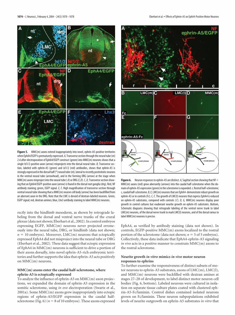
rectly into the hindlimb mesoderm, as shown by retrograde la-beling from the dorsal and ventral nerve trunks of the cruralplexus (data not shown; Eberhart et al., 2002). In control embryosexpressing EGFP, MMC(m) neurons never projected errone-ously into the neural tube, DRG, or hindlimb (data not shown;n � 10 embryos). Moreover, LMC(m) neurons that ectopicallyexpressed EphA4 did not misproject into the neural tube or DRG(Eberhart et al., 2002). These data suggest that ectopic expressionof EphA4 in MMC(m) neurons is sufficient to drive a portion oftheir axons dorsally, into novel ephrin-A5-rich embryonic terri-tories and further supports the idea that ephrin-A5 acts positivelyon MMC(m) neurons.
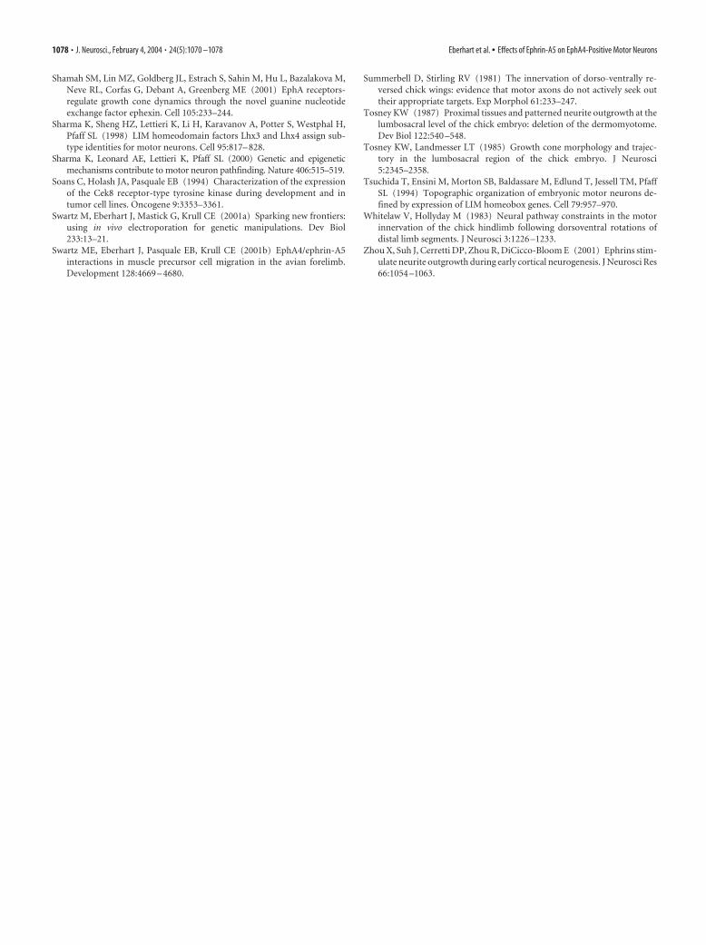
MMC(m) axons enter the caudal half-sclerotome, whereephrin-A5 is ectopically expressedTo analyze the influence of ephrin-A5 on MMC(m) axon projec-tions, we expanded the domain of ephrin-A5 expression in thesomitic sclerotome, using in ovo electroporation (Swartz et al.,2001a). Some MMC(m) axons grew inappropriately into ectopicregions of ephrin-A5/EGFP expression in the caudal half-sclerotome (Fig. 6) (n � 8 of 10 embryos). These axons expressed
EphA4, as verified by antibody staining (data not shown). Incontrols, EGFP-positive MMC(m) axons localized to the rostralportion of the sclerotome (data not shown; n � 5 of 5 embryos).Collectively, these data indicate that EphA4-ephrin-A5 signalingin vivo acts in a positive manner to constrain MMC(m) axons tothe rostral sclerotome.
Neurite growth in vitro mimics in vivo motor neuronresponses to ephrinsTo further examine the responsiveness of distinct subsets of mo-tor neurons to ephrin-A5 substrates, axons of LMC(m), LMC(l),and MMC(m) neurons were backfilled with dextran amines atstages 27–28 of development, to label distinct motor neuron cellbodies (Fig. 6, bottom). Labeled neurons were cultured in isola-tion on separate tissue culture plates coated with clustered eph-rin-A5-Fc/laminin. Control dishes contained isolated neuronsgrown on Fc/laminin. These neuron subpopulations exhibitedlevels of neurite outgrowth on ephrin-A5 substrates in vitro that
Figure 5. MMC(m) axons extend inappropriately into novel, ephrin-A5-positive territorieswhen EphA4/EGFP is prematurely expressed. A, Transverse section through the neural tube (nt)2 d after electroporation of EphA4/EGFP construct (green) into MMC(m) neurons shows that asingle Isl1/2-positive axon (arrow) misprojects into the dorsal neural tube. B, Transverse sec-tion, labeled with ephrin-A5 (green) and isl1/2 (red) antibodies, shows that ephrin-A5 isstrongly expressed in the dorsal half (*) neural tube (nt), lateral to recently postmitotic neuronsin the ventral neural tube (arrowhead), and in the forming DRG (arrow) at the stage whenMMC(m) axons misproject into the neural tube ( A) or DRG (C,D). C, D, Transverse section show-ing that an EphA4/EGFP-positive axon (arrow) is found in the dorsal root ganglia (drg). Red, NFantibody staining; green, EGFP signal. E, F, High magnification of transverse section throughventral neural tube showing that a MMC(m) neuron cell body (arrow) has been backfilled froman aberrant axon in the DRG. Note that the LMC is devoid of dextran-labeled neurons. Green,EGFP signal; red, dextran amines; blue, Lhx3 antibody staining to label MMC(m) neurons.
Figure 6. Neuron responses to ephrin-A5 are distinct. A, Sagittal section showing that NF�MMC(m) axons (red) grow aberrantly (arrows) into the caudal half-sclerotome when the do-main of ephrin-A5 expression (green) in the sclerotome is expanded. r, Rostral half-sclerotome;c, caudal half-sclerotome. B, E, LMC(m) neurons that are EphA4- demonstrate robust growth onephrin-A5 or in controls (Fc). C, F, The growth of LMC(l) neurons that express EphA4 is reducedon ephrin-A5 substrates, compared with controls ( C). D, G, MMC(m) neurons display poorgrowth in control cultures but exuberant neurite growth on ephrin-A5 substrates. Bottom,Schematic diagrams showing that retrograde labeling of the ventral nerve trunk to labelLMC(m) neurons, of the dorsal nerve trunk to mark LMC(l) neurons, and of the dorsal ramus tolabel MMC(m) neurons is precise.
1074 • J. Neurosci., February 4, 2004 • 24(5):1070 –1078 Eberhart et al. • Effects of Ephrin-A5 on EphA4-Positive Motor Neurons

correlated with their in vivo responsiveness: LMC(l) neurons thatexpressed EphA4 exhibited weak neurite growth on ephrin-A5(x � 46 �m; n � 17 cells) (Fig. 6F), comparable to controlLMC(l) neurons grown on Fc (x � 54 �m; n � 10 cells) (Fig. 6C).In contrast, MMC(m) neurons that expressed EphA4 displayedextensive neurite outgrowth on ephrin-A5 (x � 218 �m; n � 38cells) (Fig. 6G), compared with control MMC(m) neurons grownon Fc (x � 58 �m; n � 15 cells) (Fig. 6D). LMC(m) neurons thatdo not express EphA4 displayed moderate levels of neuritegrowth when grown on ephrin-A5 (x � 126 �m; n � 14 cells)(Fig. 6E), comparable to control LMC(m) neurons grown on Fc(x � 122 �m; n � 12 cells) (Fig. 6B). No significant differences inneurite numbers were present under any treatment condition.These data demonstrate that regenerating neurons in vitro exhibitsimilar responses to ephrin-A5 as described in vivo, supportingthe notion that distinct subsets of motor neurons that expressEphA4 respond differentially to ephrin-A5.
Eph activation localizes to discrete subcellular domains ofLMC(l) and MMC(m) neuronsTo examine whether the subcellular localization of Eph activa-tion could contribute to the differential responses of these axonpopulations, we examined the phosphorylation status or activa-tion of Eph receptors on LMC and MMC(m) axons in vivo, usingan anti-phosphorylated Eph antibody (Shamah et al., 2001).LMC axon shafts at the level of the spinal nerve (Fig. 7A,B) (n �2 embryos), crural plexus (Fig. 6C,D) (n � 2 embryos), and thedistal tips of the dorsal nerve trunk occupied by LMC(l) axons(Fig. 7E–H) (n � 6 embryos) exhibited Eph phosphorylation. Incontrast, MMC(m) axons displayed Eph activation localized totheir axon shafts (Fig. 8) (n � 6 embryos) but phosphorylation attheir distal tips was very weak (Fig. 8) (n � 6 embryos). Thispattern is consistent with that of EphA4 expression (Eberhart etal., 2000). These data argue that the EphA4 signaling machinerylocalizes to distinct domains of the axons of MMC(m) andLMC(l) neurons.
DiscussionFrom our findings, we conclude that EphA4-ephrin-A5 signalingcan exert positive and negative effects on the patterning of axonprojections from chick MMC(m) and LMC(l) motor neurons.Several lines of evidence support this conclusion. First, ectopicexpression of ephrin-A5 in the dorsal limb mesoderm preventsEphA4-positive LMC(l) axons from entry on their typical time-table. Normally, EphA4-positive axons project into the dorsallimb, where ephrin-A5 is absent (Eberhart et al., 2000). Second,blocking EphA4 signaling in MMC(m) neurons or ectopicallyexpressing ephrin-A5 in the caudal half-sclerotome allowsMMC(m) axons to extend inappropriately into the caudal half-sclerotome. Third, premature ectopic expression of EphA4 inMMC(m) neurons before their axons extend through the rostralhalf-sclerotome drives a portion of their projections into novel,ephrin-A5-positive regions. Fourth, isolated LMC(l) andMMC(m) neurons in vitro exhibit similar responses to ephrin-A5coated substrates as those shown in vivo. LMC(l) neurons exhibitlow levels of neurite growth, whereas MMC(m) neurons displayrobust neurite growth.
One mechanism that could contribute to altered axon projec-tions is that motor neuron identity or settling patterns in theneural tube are changed when EphA4 signaling is blocked orEphA4 is prematurely expressed in MMC(m) neurons. However,we have not observed such alterations: MMC(m) neurons arepositioned medial to the LMC at hindlimb levels and exhibit
Islet1/2 and Lhx 3 immunoreactivity, as is typical. Furthermore,recent studies have shown that Lim1 and Islet1 transcription fac-tors control motor neuron settling patterns, independent of Ephfamily members (Kania and Jessell, 2003).
What accounts mechanistically for these different responsesof EphA4-positive motor neurons to ephrin-A5? Three possibleexplanations emerge, which are not mutually exclusive: (1) thenormal inhibition of the caudal sclerotome for MMC(m) axonshas been altered in our manipulations, perhaps changing the ros-trocaudal polarity of the somites or masking the repulsion inher-
Figure 7. Eph phosphorylation localizes to the axon shafts and distal tips of LMC neurons invivo. All transverse sections were labeled with anti-phosphorylated Eph antibody to detect Ephactivation (red) and NF antibody to label all axons (green). A, B, At stage 21, before motor axonsarrive to the base of the limb, they exhibit Eph phosphorylation (arrow). nt, Neural tube. C, D, Atthe level of the crural plexus at stage 26, dorsal and ventral projecting axons display Eph phos-phorylation. E–H, Distal tips of LMC(l) axons (arrows) show Eph phosphorylation.
Eberhart et al. • Effects of Ephrin-A5 on EphA4-Positive Motor Neurons J. Neurosci., February 4, 2004 • 24(5):1070 –1078 • 1075

ent to this region of the somites, (2) dis-tinct signaling molecules downstream ofEphA4 are active in different subsets ofmotor neurons, or (3) EphA4 activationlocalizes to distinct regions of MMC(m)versus LMC(l) neurons.
To address the possibility that the ros-trocaudal polarity of the sclerotome mayhave been altered by ectopically expressingephrin-A5, allowing MMC(m) axons tonow enter this normally inhibitory terri-tory, we examined the expression of dis-tinct markers localized to the rostral andcaudal somitic domains, including cau-dally expressed ephrin-B1. In all cases, therostrocaudal polarity of the somites wasunaltered, indicating that this mechanismcould not account for the inappropriateentry of MMC(m) axons into the caudalhalf-sclerotome (data not shown). Further-more, EphA4 protein, a potentially positivesignal for MMC(m) axons, was not upregu-lated in the caudal half-sclerotome upon ex-pansion of the ephrin-A5 expression do-main (data not shown).
Interestingly, MMC(m) axons did notgrow exuberantly beyond the borders ofan individual somite when EphA4 signal-ing was blocked in MMC(m) neurons orwhen the domain of ephrin-A5 expression in the somite wasexpanded. This is most likely caused by the presence of an inter-somitic blood vessel between each somite that acts as a physicalbarrier to axon advance (K. Tosney, unpublished observations).
We cannot exclude the possibility that ectopic ephrin-A5 inthe somites abrogates the normal inhibition of the caudal half-sclerotome for MMC(m) axons, acting as a positive cue to makethat environment more seductive for growing axons. However,ephrin-A5 does not serve to mask any inhibitory effect of Ephfamily members localized to the caudal half-sclerotome onMMC(m) axons. Neither ephrin-B1 nor EphA7 in the caudal-half sclerotome are required to prohibit EphA4-positiveMMC(m) axons from entering this domain (Koblar et al., 2000)(C. E. Krull, unpublished data).
EphA4-positive MMC(m) and LMC(l) neurons may possess dis-tinct signaling components downstream of EphA4 that account fortheir different responsiveness to ephrin-A5. The strongest candidateto date is ephexin, which binds specifically to EphA4 and activatesRho, Rac, or Cdc42, dependent on cellular context (Shamah et al.,2001). Other EphA4 signaling components have not been identifiedthus far. Experiments are in progress to determine whether ephexincontributes to the different responses of LMC(l) and MMC(m) neu-rons to ephrin-A5.
Our results and others demonstrate that EphA4 forward sig-naling is required for both MMC(m) and LMC(l) axon projec-tions and patterning (Helmbacher et al., 2000; this study) (S.O’Connell and C. E. Krull, unpublished data). However, westrongly suspect that the molecular machinery that underliesEphA4 signaling is different, based on where EphA4 is activatedor phosphorylated on MMC(m) and LMC(l) axons. We foundthat Eph phosphorylation localizes to LMC axon shafts and thedistal tips of their axons, where growth cones are thought tosample the local environment and make pathway selection deci-sions. Eph phosphorylation on LMC(l) axons represents the ac-
tivation of EphA4, whereas Eph phosphorylation on LMC(m)axons is contributed presumably by EphB2 (Eberhart et al., 2000)(R. McLennan and C. E. Krull, unpublished observations). Incontrast, Eph phosphorylation is predominant on MMC(m)axon shafts and negligible on their distal tips. The distinct local-izations of activated EphA4 on growth cones and axon shaftscould indeed account for the differential responses of LMC(l)and MMC(m) neurons to ephrin-A5 in vivo. Previous studieshave shown that the upregulation of guidance factors on distalsegments of axons can influence guidance and axon responses(Keleman et al., 2002). Axons and growth cones are indeed capa-ble of protein translation and export to their cell surfaces (Brittiset al., 2002) and protein synthesis and degradation are critical foraxon guidance (Campbell and Holt, 2001). Moreover, axons canuse multiple guidance mechanisms including axon fasciculationor gradients of signaling factors to navigate to their targets (Dick-son, 2002). In the case of LMC(l) neurons, ephrin-A5 is inhibi-tory to axon advance, interacting with EphA4 receptors known tobe present on LMC(l) growth cones (Eberhart et al., 2000; thisstudy). In the case of MMC(m) neurons, ephrin-A5 would inter-act with EphA4 localized primarily to the surfaces of axon shafts,presumably influencing axon fasciculation. Experiments are inprogress to examine the underlying cell biology of EphA4 –ephrin-A5 signaling in these motor neuron populations.
Notably, there are distinct effects of premature or ectopic ex-pression of EphA4 on MMC(m) and LMC(m) neurons, respec-tively. Here, we show that 27% of MMC(m) neurons extend theiraxons into novel ephrin-A5 territories in the dorsal neural tube orDRG when EphA4 was prematurely expressed. In contrast, 76%of LMC(m) neurons that ectopically expressed EphA4 grew ab-errantly into the dorsal nerve trunk of the crural plexus in thehindlimb, instead of their normal ventral trajectory, but neverexited the hindlimb (Eberhart et al., 2002). Furthermore,LMC(m) axons that ectopically expressed EphA4 did not extend
Figure 8. MMC(m) neurons show Eph phosphorylation at their axon shafts but negligible signal at their distal axon tips in vivo.High-magnification views of transverse sections through MMC(m) axons labeled with anti-phosphorylated Eph antibody (red)and NF antibody (green). A–C, MMC(m) axon shafts (bracket in A) exhibit Eph activation, whereas distal tips show extremely weakEph activation (boxed area). D–F, In another embryo, MMC(m) axon shafts show strong activation of Eph (bracket in A) but veryweak signal localized to their distal tips (boxed area). drg, Dorsal root ganglia; m, myotome.
1076 • J. Neurosci., February 4, 2004 • 24(5):1070 –1078 Eberhart et al. • Effects of Ephrin-A5 on EphA4-Positive Motor Neurons

into ephrin-A5-rich territories. Together, these results suggestthat EphA4 does not act as a molecular switch to determinewhether motor axons will enter the hindlimb to innervate limbmuscle or the dorsal ramus to innervate epaxial muscle. Instead,EphA4-positive MMC(m) and LMC neurons exhibit distinct re-sponses to ephrin-A5.
It is conceivable that LMC(l) and MMC(m) neurons responddifferently to ephrin-A5 in our experiments because of dissimi-larities in ephrin-A5 protein concentrations that influence cellbehavior differently. However, this does not appear to be the case:the same ephrin-A5/EGFP plasmid concentration was used in allin ovo manipulations, generating comparable levels of cell trans-fection. Furthermore, isolated LMC(l) and MMC(m) neuronsrespond distinctly to identical concentrations of substrate-boundephrin-A5. It remains possible that the signaling componentsdownstream of ephrin-A5 are distinct in hindlimb mesodermand the somitic sclerotome. Other studies have argued that theaxonal coexpression of an Eph receptor and ephrin dampens theinhibitory or repellent effects of ephrins located in the targettissue (Hornberger et al., 1999). This mechanism is not likely toaccount for the differential responses to ephrin-A5 observedhere. Axons of LMC(l) neurons are decorated with EphA4 andephrin-A2 protein (Eberhart et al., 2000) and yet are inhibitedfrom entering ephrin-A5-positive domains in target limb meso-derm. With regard to MMC(m) neurons, we have not detectedephrins on these axons (J. Eberhart and C. E. Krull, unpublisheddata).
Results of previous studies suggest that ephrin-A5 can influ-ence axon projections and growth in a positive manner.Ephrin-A5 promotes branch formation in thalamic axons (Mannet al., 2002) and enhances the process outgrowth of cortical neu-rons (Zhou et al., 2001). Growing axons can alter their respon-siveness to extracellular cues that are encountered during naviga-tion to their targets. Another Eph family member, ephrin-B1, canact bifunctionally, serving as a repellent and an attractant forretinal ganglion cell axons (McLaughlin et al., 2003) and for dif-ferent populations of neural crest cells (Santiago and Erickson,2002). Our data support the view that EphA4-ephrin-A5 interac-tions can be either inhibitory or positive for motor neurons basedon the subcellular localization of their signaling machinery. In-triguingly, these data suggest the existence of distinct EphA4 sig-naling cascades in these neuron subpopulations. Using thesestrategies, Eph receptors and ephrins could indeed exert multipleinfluences on the patterning of axonal projections and other de-velopmental processes.
ReferencesBrittis PA, Qiang L, Flanagan JG (2002) Axonal protein synthesis provides a
mechanism for localized regulation at an intermediate target. Cell110:223–235.
Campbell DS, Holt CE (2001) Chemotropic responses of retinal growthcones mediated by rapid local protein synthesis and degradation. Neuron32:1013–1026.
Dickson BJ (2002) Molecular mechanisms of axon guidance. Science298:1959 –1964.
Eberhart J, Swartz M, Koblar SA, Pasquale EB, Tanaka H, Krull CE (2000)Expression of EphA4, ephrin-A2 and ephrin-A5 during axon outgrowthto the hindlimb indicates potential roles in pathfinding. Dev Neurosci22:237–250.
Eberhart J, Swartz ME, Koblar SA, Pasquale EB, Krull CE (2002) EphA4constitutes a population-specific guidance cue for motor neurons. DevBiol 247:89 –101.
Ensini M, Tsuchida TN, Belting HG, Jessell TM (1988) The control of ros-trocaudal pattern in the developing spinal cord: specification of motorneuron subtype identity is initiated by signals from paraxial mesoderm.Development 125:969 –982.
Ethell IM, Irie F, Kalo MS, Couchman JR, Pasquale EB, Yamaguchi Y (2001)EphB/syndecan-2 signaling in dendritic spine morphogenesis. Neuron31:1001–1013.
Fukushima M, Nakamura M, Ohta K, Okamura R, Negi A, Tanaka H (1996)Regional specification of motoneurons along the anterior-posterior axisis independent of the notochord. Development 122:905–914.
Gale NW, Holland SJ, Valenzuela DM, Flenniken A, Pan L, Ryan TE, Henke-meyer M, Strebhardt K, Hirai H, Wilkinson DG, Pawson T, Davis S,Yancopoulos GD (1996) Eph receptors and ligands comprise two majorspecificity subclasses and are reciprocally compartmentalized during em-bryogenesis. Neuron 17:9 –19.
Hamburger V, Hamilton HL (1951) A series of normal stages in the devel-opment of the chick embryo. J Morphol 88:49 –92.
Helmbacher F, Schneider-Maunoury S, Topilko P, Tiret L, Charnay P (2000)Targeting of the EphA4 tyrosine kinase receptor affects dorsal/ventralpathfinding of limb motor axons. Development 127:3313–3324.
Hornberger MR, Dutting D, Ciossek T, Yamada T, Handwerker C, Lang S,Weth F, Huf J, Wessel R, Logan C, Tanaka H, Drescher U (1999) Mod-ulation of EphA receptor function by coexpressed ephrinA ligands onretinal ganglion cell axons. Neuron 22:731–742.
Kania A, Jessell TM (2003) Topographic motor projections in the limb im-posed by LIM homeodomain protein regulation of ephrin-A:EphA inter-actions. Neuron 38:581–596.
Kania A, Johnson RL, Jessell TM (2000) Coordinate roles for LIM ho-meobox genes in directing the dorsoventral trajectory of motor axons inthe vertebrate limb. Cell 102:161–173.
Keleman K, Rajagopalan S, Cleppien D, Teis D, Paiha K, Huber LA, TechnauGM, Dickson BJ (2002) Comm sorts robo to control axon guidance atthe Drosophila midline. Cell 110:415– 427.
Kilpatrick TJ, Brown A, Lai C, Gassman M, Goulding M, Lemke G (1996)Expression of the Tyro4/Mek4/Cek4 gene specifically marks a subset ofembryonic motor neurons and their muscle targets. Mol Cell Neurosci7:62–74.
Knoll B, Zarbalis K, Wurst W, Drescher U (2001) A role for the EphA familyin the topographic targeting of vomeronasal axons. Development128:895–906.
Koblar SA, Krull CE, Pasquale EB, McLennan R, Peale FD, Cerretti DP, Both-well M (2000) Spinal motor axons and neural crest cells use differentmolecular guides for segmental migration through the rostral half-somite. J Neurobiol 42:437– 447.
Krull CE (2004) A primer on using in ovo electroporation to analyze genefunction. Dev Dyn, in press.
Kullander K, Klein R (2002) Mechanisms and functions of Eph and ephrinsignaling. Nat Rev Mol Cell Biol 3:475– 486.
Lance-Jones C, Landmesser LT (1980) Motoneuron projection patterns inthe chick hind limb following early partial reversals of the spinal cord.J Physiol (Lond) 302:581– 602.
Lance-Jones C, Landmesser LT (1981) Pathway selection by embryonicchick motoneurons in an experimentally altered environment. Proc R SocLond B Biol Sci 214:19 –52.
Landmesser L (1978) The development of motor projection patterns in thechick hind limb. J Physiol (Lond) 284:391– 414.
Lee VM, Carden MJ, Schlaepfer WW, Trojanowski JQ (1987) Monoclonalantibodies distinguish several differentially phosphorylated states of thetwo largest rat neurofilament subunits (NF-H and NF-M) and demon-strate their existence in the normal nervous system of adult rats. J Neuro-sci 7:3474 –3488.
Mann F, Peuckert C, Dehner F, Zhou R, Bolz J (2002) Ephrins regulate theformation of terminal axonal arbors during the development of thalamo-cortical projections. Development 129:3945–3955.
McLaughlin T, Hindges R, Yates PA, O’Leary DD (2003) Bifunctional ac-tion of ephrin-B1 as a repellent and attractant to control bi-directionalbranch extension in dorsal-ventral retinotopic mapping. Development130:2407–2418.
Ohta K, Nakamura M, Hirokawa K, Tanaka S, Iwama A, Suda T, Ando M,Tanaka H (1996) The receptor tyrosine kinase Cek8 is transiently ex-pressed on subtypes of motoneurons in the spinal cord during develop-ment. Mech Dev 54:59 – 69.
Osumi N, Inoue T (2001) Gene transfer into cultured mammalian embryosby electroporation. Methods 24:35– 42.
Santiago A, Erickson CA (2002) Ephrin-B ligands play a dual role in thecontrol of neural crest cell migration. Development 129:3621–3632.
Eberhart et al. • Effects of Ephrin-A5 on EphA4-Positive Motor Neurons J. Neurosci., February 4, 2004 • 24(5):1070 –1078 • 1077
Shamah SM, Lin MZ, Goldberg JL, Estrach S, Sahin M, Hu L, Bazalakova M,Neve RL, Corfas G, Debant A, Greenberg ME (2001) EphA receptors-regulate growth cone dynamics through the novel guanine nucleotideexchange factor ephexin. Cell 105:233–244.
Sharma K, Sheng HZ, Lettieri K, Li H, Karavanov A, Potter S, Westphal H,Pfaff SL (1998) LIM homeodomain factors Lhx3 and Lhx4 assign sub-type identities for motor neurons. Cell 95:817– 828.
Sharma K, Leonard AE, Lettieri K, Pfaff SL (2000) Genetic and epigeneticmechanisms contribute to motor neuron pathfinding. Nature 406:515–519.
Soans C, Holash JA, Pasquale EB (1994) Characterization of the expressionof the Cek8 receptor-type tyrosine kinase during development and intumor cell lines. Oncogene 9:3353–3361.
Swartz M, Eberhart J, Mastick G, Krull CE (2001a) Sparking new frontiers:using in vivo electroporation for genetic manipulations. Dev Biol233:13–21.
Swartz ME, Eberhart J, Pasquale EB, Krull CE (2001b) EphA4/ephrin-A5interactions in muscle precursor cell migration in the avian forelimb.Development 128:4669 – 4680.
Summerbell D, Stirling RV (1981) The innervation of dorso-ventrally re-versed chick wings: evidence that motor axons do not actively seek outtheir appropriate targets. Exp Morphol 61:233–247.
Tosney KW (1987) Proximal tissues and patterned neurite outgrowth at thelumbosacral level of the chick embryo: deletion of the dermomyotome.Dev Biol 122:540 –548.
Tosney KW, Landmesser LT (1985) Growth cone morphology and trajec-tory in the lumbosacral region of the chick embryo. J Neurosci5:2345–2358.
Tsuchida T, Ensini M, Morton SB, Baldassare M, Edlund T, Jessell TM, PfaffSL (1994) Topographic organization of embryonic motor neurons de-fined by expression of LIM homeobox genes. Cell 79:957–970.
Whitelaw V, Hollyday M (1983) Neural pathway constraints in the motorinnervation of the chick hindlimb following dorsoventral rotations ofdistal limb segments. J Neurosci 3:1226 –1233.
Zhou X, Suh J, Cerretti DP, Zhou R, DiCicco-Bloom E (2001) Ephrins stim-ulate neurite outgrowth during early cortical neurogenesis. J Neurosci Res66:1054 –1063.
1078 • J. Neurosci., February 4, 2004 • 24(5):1070 –1078 Eberhart et al. • Effects of Ephrin-A5 on EphA4-Positive Motor Neurons
Copyright © 2022 FDOKUMEN