CSPG4-Specific Immunity and Survival Prolongation in Dogs with Oral Malignant Melanoma Immunized...
11
Cancer Therapy: Preclinical CSPG4-Specific Immunity and Survival Prolongation in Dogs with Oral Malignant Melanoma Immunized with Human CSPG4 DNA Federica Riccardo 1 , Selina Iussich 2 , Lorella Maniscalco 2 , Saray Lorda Mayayo 2 , Giuseppe La Rosa 3 , Maddalena Arigoni 1 , Raffaella De Maria 2 , Francesca Gattino 2 , Stefania Lanzardo 1 , Elena Lardone 2 , Marina Martano 2 , Emanuela Morello 2 , Simone Prestigio 1 , Alessandra Fiore 1 , Elena Quaglino 1 , Sara Zabarino 2 , Soldano Ferrone 4 , Paolo Buracco 2 , and Federica Cavallo 1 Abstract Purpose: Due to the many similarities with its human counterpart, canine malignant melanoma (cMM) is a valuable model in which to assess the efficacy of novel therapeutic strategies. The model is herein used to evaluate the immunogenicity, safety, and therapeutic efficacy of a human chondroitin sulfate proteoglycan- 4 (hCSPG4) DNA-based vaccine. The fact that homology between hCSPG4 and cCSPG4 amino-acidic sequences stands at more than 80% provides the rationale for using an hCSPG4 DNA vaccine in the cMM model. Experimental Design: Dogs with stage II–III surgically resected CSPG4-positive oral MM were subjected to monthly intramuscular plasmid administration, which was followed immediately by electroporation (electrovaccination) for at least 6, and up to 20, months. The immunogenicity, safety, and therapeutic efficacy of the vaccine have been evaluated. Results: hCSPG4 electrovaccination caused no clinically relevant local or systemic side effects and resulted in significantly longer overall and disease-free survival times in 14 vaccinated dogs as compared with 13 nonvaccinated controls. All vaccinated dogs developed antibodies against both hCSPG4 and cCSPG4. Seven vaccinated dogs were also tested for a cCSPG4-specific T-cell response and only two gave a detectable interferon (IFN)g response. Conclusion: Xenogeneic electrovaccination against CSPG4 is able to overcome host unresponsiveness to the "self" antigen and seems to be effective in treating cMM, laying the foundation for its translation to a human clinical setting. Clin Cancer Res; 20(14); 3753–62. Ó2014 AACR. Introduction Melanoma is the sixth most common cancer worldwide and has seen the highest rise in incidence in recent decades (1). In its early stages, melanoma is highly curable with surgery, but approximately one third of all patients with melanoma experience disease recurrence and metastasis; once metastasis has occurred, it is often fatal (2). Patients whose tumors contain a BRAF gene mutation may benefit from treatment with mitogen-activated protein kinase path- way inhibitors; however, the development of resistance through multiple distinct mechanisms is a major problem (3). An alternative therapeutic strategy is found in immunotherapeutic approaches using cytokines, such as interferon (IFN)a and IL2 (4, 5), and the administration of antagonist monoclonal antibodies (mAb) that target CTLA- 4 (6) and/or PD-1 and PD-L1 (7). Inherent immunogenicity and the potential role played by immunologic events in melanoma natural history, have stimulated interest in the development and application of cancer vaccines (8). Despite numerous successful studies in murine models, the therapeutic efficacy of cancer vaccines has so far been disappointing (9). This is a reflection of the difficulty met in translating the results from mice to men. To overcome these limitations, the National Cancer Institute’s Center for Cancer Research launched the Comparative Oncology Program, in 2003, to foster the use of naturally occurring cancer in pet animals as models of human cancer Authors' Affiliations: 1 Department of Molecular Biotechnology and Health Sciences, Molecular Biotechnology Center, University of Torino; 2 Depart- ment of Veterinary Science, University of Torino, Grugliasco, Torino, Italy; 3 Veterinary practitioner; and 4 Department of Surgery, Massachusetts Gen- eral Hospital, Harvard Medical School, Boston, Massachusetts Note: Supplementary data for this article are available at Clinical Cancer Research Online (http://clincancerres.aacrjournals.org/). P. Buracco and F. Cavallo contributed equally to this article. Corresponding Authors: Federica Cavallo, Molecular Biotechnology Cen- ter, University of Torino, Via Nizza 52, Torino, Italy. Phone: 39-011-670- 6457; Fax: 39-011-236-5417; E-mail: [email protected]; and Paolo Buracco, Department of Veterinary Science, Largo Paolo Braccini 2, 10095 Grugliasco, Torino, Italy. Phone: 39-011-670-9097; E-mail: [email protected] doi: 10.1158/1078-0432.CCR-13-3042 Ó2014 American Association for Cancer Research. Clinical Cancer Research www.aacrjournals.org 3753 on March 25, 2015. © 2014 American Association for Cancer Research. clincancerres.aacrjournals.org Downloaded from Published OnlineFirst May 29, 2014; DOI: 10.1158/1078-0432.CCR-13-3042
-
Upload
independent -
Category
Documents
-
view
0 -
download
0
Transcript of CSPG4-Specific Immunity and Survival Prolongation in Dogs with Oral Malignant Melanoma Immunized...

Cancer Therapy: Preclinical
CSPG4-Specific Immunity and Survival Prolongation in Dogswith Oral Malignant Melanoma Immunized with HumanCSPG4 DNA
Federica Riccardo1, Selina Iussich2, Lorella Maniscalco2, Saray Lorda Mayayo2, Giuseppe La Rosa3,Maddalena Arigoni1, Raffaella De Maria2, Francesca Gattino2, Stefania Lanzardo1, Elena Lardone2,Marina Martano2, Emanuela Morello2, Simone Prestigio1, Alessandra Fiore1, Elena Quaglino1, Sara Zabarino2,Soldano Ferrone4, Paolo Buracco2, and Federica Cavallo1
AbstractPurpose:Due to the many similarities with its human counterpart, canine malignant melanoma (cMM)
is a valuablemodel inwhich to assess the efficacy of novel therapeutic strategies. Themodel is herein used to
evaluate the immunogenicity, safety, and therapeutic efficacy of a human chondroitin sulfate proteoglycan-
4 (hCSPG4) DNA-based vaccine. The fact that homology between hCSPG4 and cCSPG4 amino-acidic
sequences stands at more than 80% provides the rationale for using an hCSPG4 DNA vaccine in the cMM
model.
Experimental Design:Dogs with stage II–III surgically resected CSPG4-positive oral MMwere subjected
to monthly intramuscular plasmid administration, which was followed immediately by electroporation
(electrovaccination) for at least 6, and up to 20, months. The immunogenicity, safety, and therapeutic
efficacy of the vaccine have been evaluated.
Results: hCSPG4 electrovaccination caused no clinically relevant local or systemic side effects and
resulted in significantly longer overall and disease-free survival times in 14 vaccinated dogs as compared
with 13 nonvaccinated controls. All vaccinated dogs developed antibodies against both hCSPG4 and
cCSPG4. Seven vaccinated dogs were also tested for a cCSPG4-specific T-cell response and only two gave a
detectable interferon (IFN)g response.
Conclusion:Xenogeneic electrovaccination against CSPG4 is able to overcome host unresponsiveness to
the "self" antigen and seems to be effective in treating cMM, laying the foundation for its translation to a
human clinical setting. Clin Cancer Res; 20(14); 3753–62. �2014 AACR.
IntroductionMelanoma is the sixth most common cancer worldwide
and has seen the highest rise in incidence in recent decades(1). In its early stages, melanoma is highly curable withsurgery, but approximately one third of all patients with
melanoma experience disease recurrence and metastasis;once metastasis has occurred, it is often fatal (2). Patientswhose tumors contain a BRAF gene mutation may benefitfrom treatmentwithmitogen-activated protein kinase path-way inhibitors; however, the development of resistancethrough multiple distinct mechanisms is a major problem(3). An alternative therapeutic strategy is found inimmunotherapeutic approaches using cytokines, such asinterferon (IFN)a and IL2 (4, 5), and the administration ofantagonist monoclonal antibodies (mAb) that target CTLA-4 (6) and/or PD-1 and PD-L1 (7).
Inherent immunogenicity and the potential role playedby immunologic events in melanoma natural history, havestimulated interest in the development and application ofcancer vaccines (8). Despite numerous successful studies inmurine models, the therapeutic efficacy of cancer vaccineshas so far been disappointing (9). This is a reflection of thedifficultymet in translating the results frommice tomen. Toovercome these limitations, the National Cancer Institute’sCenter for Cancer Research launched the ComparativeOncology Program, in 2003, to foster the use of naturallyoccurring cancer in pet animals as models of human cancer
Authors' Affiliations: 1Department of Molecular Biotechnology and HealthSciences, Molecular Biotechnology Center, University of Torino; 2Depart-ment of Veterinary Science, University of Torino, Grugliasco, Torino, Italy;3Veterinary practitioner; and 4Department of Surgery, Massachusetts Gen-eral Hospital, Harvard Medical School, Boston, Massachusetts
Note: Supplementary data for this article are available at Clinical CancerResearch Online (http://clincancerres.aacrjournals.org/).
P. Buracco and F. Cavallo contributed equally to this article.
CorrespondingAuthors:FedericaCavallo,Molecular BiotechnologyCen-ter, University of Torino, Via Nizza 52, Torino, Italy. Phone: 39-011-670-6457; Fax: 39-011-236-5417; E-mail: [email protected]; and PaoloBuracco, Department of Veterinary Science, Largo Paolo Braccini 2, 10095Grugliasco, Torino, Italy. Phone: 39-011-670-9097; E-mail:[email protected]
doi: 10.1158/1078-0432.CCR-13-3042
�2014 American Association for Cancer Research.
ClinicalCancer
Research
www.aacrjournals.org 3753
on March 25, 2015. © 2014 American Association for Cancer Research. clincancerres.aacrjournals.org Downloaded from
Published OnlineFirst May 29, 2014; DOI: 10.1158/1078-0432.CCR-13-3042
(10). A European initiative with a similar purpose—theLUPA project—has recently been launched (11).
Canine malignant melanoma (cMM) shares many char-acteristics with its human counterpart, including histologicphenotype, tumor genetics, and clinical biologic behavioras well as the development of recurrent or resistant diseaseand far-reaching metastasis (12). cMM, thus, represents anattractive translational model for the assessment of theefficacy of novel immunotherapies for the treatment ofhuman MM (13).
A suitable immunotherapeutic target shouldhavea strongoncogenic role, be expressed in malignant cells in a highpercentage of patients, and show limited distribution innormal tissues (14, 15). Chondroitin sulfate proteoglycan-4(CSPG4), an early cell surface progression marker involvedin tumor cell proliferation, migration, and invasion meetsthese criteria (16). The induction of CSPG4-specific anti-bodies in patients with melanoma that have been immu-nized with CSPG4 mimics has been associated with signif-icantly longer survival andmetastases regression, highlight-ing the potential clinical relevance of this tumor antigen as atarget for antibody-based immunotherapy (17, 18). Inter-estingly, CSPG4 is highly conserved in its structural andfunctional properties through phylogenetic evolution. Inparticular, human CSPG4 (hCSPG4) displays 82% homol-ogy and 88% similarity to its canine counterpart in itsamino-acidic sequence. Moreover, the frequency of CSPG4expression in canine and humanmelanoma lesions is quitesimilar, being about 60% (19) and 80% (20), respectively.
DNA vaccination has been extensively explored in thefield of melanoma immunotherapy, and a number of DNAvaccination clinical trials have been performed in humanpatients with melanoma; trial results have all been disap-pointing, probably because of the lack of immunogenicityin DNA vaccines, even when administered with Salmonellatyphimurium (21–24). Besides, the United States Depart-
ment of Agriculture (USDA) licensed in2010aDNAvaccine(ONCEPT, Merial) in the veterinary field. This representsthe first approved anticancer vaccine and is meant for thetreatment of cMM. However, its therapeutic efficacy hasbeen recently questioned (25).
The introduction of electroporation to DNA vaccinedelivery (electrovaccination) has strongly increased immu-nogenicity and therapeutic efficacy in both mice andhumans (26, 27). Electrovaccination combines the advan-tages of DNA vaccination and electroporation. Specifically,the former is easy to handle, applicable to a broad popu-lation, safe, and induces both cellular and humoralimmune responses, whereas the latter enhances the expres-sion of the protein encoded by the immunizing DNA andprolongs the duration of the immune response (28, 29).
These findings and the translational power of veterinaryclinical trials have prompted us to test the safety and efficacyof intramuscular electrovaccination of a plasmid encodingfor CSPG4 in client-owned dogs with surgically resectedstage II–III CSPG4-positive, natural occurring oral MM.Because CSPG4 is a self-antigen with poor, or a lack of,immunogenicity in autologous hosts, we immunized dogswith hCSPG4.
Materials and MethodsDog enrollment
Dogs were treated according to the European guidelinesestablished in the Principles of Laboratory Animal Care(directive 86/609/EEC). The Ethical Committee of the Uni-versity Veterinary Teaching Hospital (Grugliasco, Italy)approved the study; written consent for entry into the studywas obtained from dog owners.
Pretreatment work-up included physical examination,blood count, serum biochemistry, and urinalysis. Fineneedle aspiration and/or biopsy were used for preoperativetumor diagnosis. Cytology was the initial preoperativeprocedure adopted to clinically stage the palpable regionallymph nodes (LN), even in case of a not apparent clinico-pathologic enlargement; in fact, size has not been consid-ered sufficiently predictive (30). A more objective stagingwas achieved via the surgical removal of all palpable region-al LN at the time of primary tumor resection and their fullhistologic evaluation. Full tumor staging also included askull and three-view chest radiography and abdominalultrasound examination; alternatively, a total body CT scanwas performed.
Dogs without concurrent life-threatening diseases andwith histologically confirmed oral stage II (2–4-cm diam-eter, negative LN) and III (>4-cm diameter and negative LNor any tumor size with ipsilateral-positive LN; ref. 31)surgically resected MM with a minimum of 6 monthsfollow-up on June 30, 2013, were included. Primary tumoren bloc resection (maxillectomy, mandibulectomy, lip/cheek excision, etc.), with the inclusion—if feasible—of atleast 2 cm of macroscopically normal tissue around thetumor, and regional lymphadenectomy were performed.For excision margin evaluation, the cut surface was stained
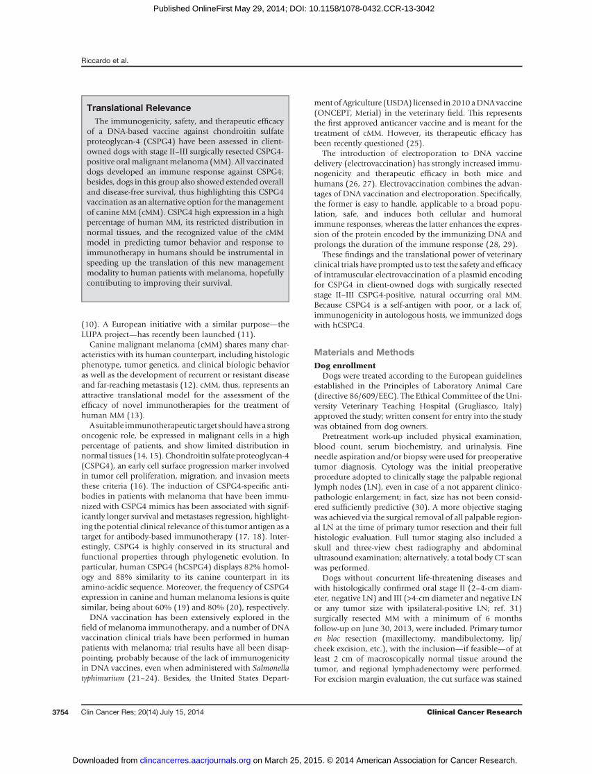
Translational RelevanceThe immunogenicity, safety, and therapeutic efficacy
of a DNA-based vaccine against chondroitin sulfateproteoglycan-4 (CSPG4) have been assessed in client-owned dogs with stage II–III surgically resected CSPG4-positive oralmalignantmelanoma (MM). All vaccinateddogs developed an immune response against CSPG4;besides, dogs in this group also showed extended overalland disease-free survival, thus highlighting this CSPG4vaccination as an alternative option for themanagementof canine MM (cMM). CSPG4 high expression in a highpercentage of human MM, its restricted distribution innormal tissues, and the recognized value of the cMMmodel in predicting tumor behavior and response toimmunotherapy in humans should be instrumental inspeeding up the translation of this new managementmodality to human patients with melanoma, hopefullycontributing to improving their survival.
Riccardo et al.
Clin Cancer Res; 20(14) July 15, 2014 Clinical Cancer Research3754
on March 25, 2015. © 2014 American Association for Cancer Research. clincancerres.aacrjournals.org Downloaded from
Published OnlineFirst May 29, 2014; DOI: 10.1158/1078-0432.CCR-13-3042

with a specific dye (TMD Tissue Marking Dye, TriangleBiomedical Sciences) just after surgery; the sample was thenfixed in 10% formalin. The same pathologist evaluated allsamples. Those margins with tumor cells reaching the dyewere considered as incomplete, whereas those with noevidence of tumor cells within at least 1 mm from the cutsurface were considered as "clean" margins. Samples werealso immunohistochemically tested for Ki67 expression(polyclonal Ki67 antibody—A-047; DAKO), mitotic index,and nuclear atypia (25, 32, 33). Immunohistochemicalanalyses of CSPG4 expression on MM samples were per-formed as previously described (19). Briefly, a total scoreranging from 0 to 8 was assigned to each MM sample byadding the value that represented the proportion of CSPG4positively stained tumor cells (score from 0 to 5) and theaverage staining intensity of CSPG4-positive tumor cells(score from 0 to 3).
In vivo electroporationOnly dogs with an MM characterized by a CSPG4 score
�3/8 (19) were considered as suitable vaccination candi-dates. Dogs were vaccinated with a pcDNA3.1 plasmidcoding for hCSPG4 generated as previously described(34). Vaccination started 3 to 4 weeks after surgery andwas repeated after 2 weeks and then monthly. For vaccina-tion, dogs were anesthetized with fentanyl IV 2 mg/kg(FentanestR; Pfizer) and propofol IV 4 to 7mg/kg (PropofolKabi, Fresenius Kabi), and then intubated; anesthesia wasmaintained with isoflurane (IsofluraneVet, Merial) and100% oxygen. The hCSPG4 plasmid (500 mg in 200 mL of0.03% NaCl) was injected into the muscles of the caudalthigh. Two minutes after plasmid injection, nine electricpulses (1 high voltage, amplitude 450 V, length 50 ms,frequency 3 HZ; 1 second pause; eight low-voltage ampli-tude 110V, length 20ms, pause 300ms)were applied to theinjection site using the CLINIPORATOR (Igea). Dogs weremonitored for acute, late local or systemic side effects.At each vaccination, clinical examinations, blood-work,
three-view chest radiographs, sera, and peripheral bloodmononuclear cells (PBMC) were obtained. Sera were ali-quoted and cryopreserved at�80�C until used. PBMCwereisolated using density gradient centrifugation columns(Accuspin; Sigma).
Cell linesB16 cells were stably transfected with either hCSPG4 or
cCSPG4 (pcDNA3.1 containing the cDNA coding forcCSPG4 XM_544783.2; GeneScript) plasmids using Lipo-fectamine (Invitrogen) according to the manufacturer’sinstructions; these cells are referred to as hCSPG4-B16 andcCSPG4-B16, respectively, and were cultured in the RPMI-1640 medium (Life Technologies), supplemented with10% heat-inactivated FBS (Life Technologies) and G418(0.5 mg/mL; Sigma). hCSPG4-positive SK-MEL-28 mela-noma cells were purchased from the American TypeCultureCollection and authenticated by short tandem repeat pro-filing. SK-MEL-28 were cultured in DMEM (Life Technolo-gies) supplemented with 10% FBS. Cell lines were routinely
checked for contamination by Mycoplasma using theMycoalert (Lonza) Detection Kit and consistently found tobe negative.
CSPG4-specific antibody detection in vaccinated dogsera
Sera collected from dogs at each vaccination were ana-lyzed by ELISA for their ability to selectively bind CSPG4isolated from the lysates of hCSPG4-B16 and cCSPG4-B16.Cell lysates were obtained by suspending 1 � 107 cells in 1mL of lysis buffer (50mmol/L Tris-HCl, 150mmol/L NaCl,pH 7.4) containing 1% Triton X-100, and an EDTA-freeprotease inhibitor mix (Roche). Suspensions were exten-sively vortexed and incubated on ice for 15 minutes andcentrifuged for 5 minutes at 13,000 rpm at 4�C. Super-natants were collected and stored at �80�C until used.Protein concentrationwasdeterminedusing an acid proteinassay (Pierce Biotechnology).
Of note, 96-well microtiter plates were coated overnight,at 4�C, with a pool of VF1-TP34, VF4-TP108, VF20-T87.41mAb (4 mg/mL of a 50 mmol/L NaHCO3 buffer, pH 9.6;ref. 35). Plates were blocked in 5% milk in TBST (1X TBS,0,5% Tween 20) and incubated for 2 hours at room tem-perature with 1 mg/mL cell lysate from hCSPG4-B16 orcCSPG4-B16 cells diluted in TBST/1% milk powder. Plateswere incubated at 4�C overnight at optimal serum dilution(1:50) in TBST/1% milk powder. After washing, boundantibodies were detected using horseradish peroxidase–conjugated sheep anti-dog IgG (AAI32P; Abd Serotech).Color was developed via the addition of 3,30,5,50-tetra-methylbenzidine substrate and absorbance was measuredat 450 nm on an ELISA microplate reader (Bio-Rad).
Immunofluorescence staining of hCSPG4-B16 andcCSPG4-B16 cells with sera was performed by seeding cellson glass coverslips, fixing them with 4% formalin (Sigma)and blocking with PBS supplemented with 1% bovineserum albumin. After 1 hour incubation at room temper-ature, with an optimal dilution of dog sera (1:10), cells wererinsed twice in PBS and bound antibodies were revealedwith FITC rabbit anti-dog IgG antibody (F7884; Sigma).After three rinses with PBS, coverslips were air-dried,mounted with fluoromount mounting medium (Sigma),and visualized using a confocal laser scanning microscopysystem equipped with an argon-ion laser (LSM510; Zeiss).Photographs were taken using a CCD-300-RC camera.Images were processed using Adobe Photoshop and Micro-soft PowerPoint softwares.
Statistical analysisThe Shapiro–Wilk normality test was used to analyze age
and body weight. Normally distributed data are reported asmeans� SD. Nonnormally distributed data are reported asmedian and range. Other variables are expressed as percen-tages. All other quantitative evaluations were carried outusing the Student t test. TheKaplan–Meiermethodwas usedto estimate disease-free and survival times. Cross tabula-tions were performed for the evaluation of excision marginstatus versus disease outcome in the three groups using a
CSPG4 Immunotherapy for Canine Malignant Melanoma
www.aacrjournals.org Clin Cancer Res; 20(14) July 15, 2014 3755
on March 25, 2015. © 2014 American Association for Cancer Research. clincancerres.aacrjournals.org Downloaded from
Published OnlineFirst May 29, 2014; DOI: 10.1158/1078-0432.CCR-13-3042

Fisher exact test. A Wilcoxon rank-sum test was used tocompare values of the cCSPG4 expression between groupsin disease outcome. Differences in survival distributionwere analyzed using the log-rank test. Statistical signifi-cance was set at P < 0.05. All analyses were conducted inR (36).
ResultsCanine population characteristics
Twenty males (60.6%) and 13 females (39.4%) wereincluded (Supplementary Table S1). Twenty-seven hadCSPG4-positive and 6 CSPG4-negative MM. FourteenCSPG4-positive dogs were vaccinated (group I), whereasthe remaining CSPG4-positive (group II, 13 dogs) and theCSPG4-negative (group III, 6 dogs) candidates did notundergo vaccination or any other post-surgery treatment.Age and body weight at presentation are reported in Sup-plementary Table S2. Age did not significantly differbetween groups (P > 0.05). Postoperative clinical stage dataare summarized in Table 1, which shows that stages weresimilarly distributed in the groups. Histology revealedincomplete excision margins in 3 of 14 group I dogs(21.4%), in 4 of 13 (30.8%) from group II, and 2 of 6(33.3%) from group III. No significant correlation between
the excision margin status and the disease outcome wasidentified in any of the groups.
The Ki67 index was available for 27 of 33 analyzed cMM(Table 2). It was more than 19.5% in all dogs but one(13.8%) from group I, in all but 2 (18.2 and 19.0%) fromgroup II and in 3 from group III (4, 16.7 and 14.4%). Themitotic indexmean in groups I, II and III was 15/10, 28/10,and 17/10 hpf, respectively (Table 2). A nuclear atypia scoreof more than 30% was recorded in 64.28%, 61.54%, and33.33% of cMM from groups I, II, and III, respectively(Table 2). The percentages of Ki67 positivity, mitotic index,and nuclear atypia scores showed no statistically significantdifferences in the three groups and were not correlated withsurvival. The range of CSPG4 expression in groups I and II(Table 3) was between 3 of 8 and 8 of 8.
hCSPG4 electrovaccination was safe and well toleratedNo local or systemic side effects, except a transient ery-
thema at the injection site, were observed after electrovac-cination. Considerable muscle contraction occurred duringelectric pulse delivery, which subsided immediately; therewas an occasional increase in respiratory and heart rates,which subsided after 1 minute. Recovery from anesthesiawas uneventful in all dogs. Hematologic and biochemical
Table 1. Clinical stage of oral MM from dogsincluded in the study
Group Stage II Stage III
Overall population (n ¼ 33) 10 (30.3)a 23 (69.7)a
Group Ib (n ¼ 14) 5 (35.7) 9 (64.3)Group IIc (n ¼ 13) 3 (23.1) 10 (76.9)Group IIId (n ¼ 6) 2 (33.3) 4 (66.7)Group IIþIII (n ¼ 19) 5 (26.3) 14 (73.7)
aPercentage in brackets.bCSPG4-positive MM; vaccinated dogs.cCSPG4-positive MM; nonvaccinated dogs.dCSPG4-negative MM; nonvaccinated dogs.
Table 2. Histologic and immunohistochemical analysis of oral MM from dogs included in the study
Threshold Overall population Group I Group II Group III Group IIþ III
Ki67a <19.5 18.5% (27/33)b 9.1% (11/14)b 18.2% (11/13)b 40.0% (5/6)b 25.0% (16/19)b
�19.5 81.5% (27/33) 90.9% (11/14) 81.8% (11/13) 60.0% (5/6) 75.0% (16/19)Mitotic Index (MI)a <4/10 hpf 3 (9.1%) 3 (21.4%) 0 (0.0%) 0 (0.0%) 0 (0.0%)
�4/10 hpf 30 (90.9%) 11 (78.6%) 13 (100.0%) 6 (100.0%) 19 (100.0%)Nuclear Atypia <30.0% 14 (42.4%) 5 (35.7%) 5 (38.5%) 4 (66.7%) 9 (47.4%)(PLEOM)a �30.0% 19 (57.6%) 9 (64.3%) 8 (61.5%) 2 (33.3%) 10 (52.6%)
aPrognostic factors for cMM as proposed by Bergin et al. (32), Smedley et al. (33), Ottnod et al. (25).bNumber of patients for which the data were available.
Table 3. The immunohistochemical score forCSPG4 expression in oral MM from dogsincluded in the study
CSPG4score
CSPG4þ
population(n ¼ 27)
Group I(n ¼ 14)
Group II(n ¼ 13)
3/8 3 (11.1)a 1 (7.1)a 2 (15.4)a
4/8 6 (22.2) 3 (21.4) 3 (23.1)5/8 5 (18.5) 3 (21.4) 2 (15.4)6/8 2 (7.4) 1 (7.1) 1 (7.7)7/8 10 (37.0) 5 (35.7) 5 (38.5)8/8 1 (3.7) 1 (7.1) 0 (0.0)
aPercentage in brackets.
Riccardo et al.
Clin Cancer Res; 20(14) July 15, 2014 Clinical Cancer Research3756
on March 25, 2015. © 2014 American Association for Cancer Research. clincancerres.aacrjournals.org Downloaded from
Published OnlineFirst May 29, 2014; DOI: 10.1158/1078-0432.CCR-13-3042

parameters did not change over the entire observationperiod. Moreover, the clinical status, appetite, water intake,and general behavior of all animals remained unaltereduntil disease progression occurred.
hCSPG4 electrovaccination induced both hCSPG4- andcCSPG4-specific antibodies in all vaccinated dogsVaccination-induced immune response was evaluated in
PBMC and sera. PBMC collected before vaccination andafter the third, were stimulated ex vivo with the six peptide-pools from cCSPG4 in ELISPOT for 24 hours, but nosignificant increases in IFNg-secreting cells were detectedafter vaccination (not shown). In the 7 dogs (#2, #3, #4, #7,#8, #10, and #11), from whom it was possible to obtainfresh dendritic cells (DC), PBMC were restimulated withmature DC (mDC) pulsed with cCSPG4-derived peptidepools. The ELISPOT assay only showed a detectable IFNgresponse in 2 dogs (#2 and #7) after vaccination, but thisresponse was significantly different from that observedwhen PBMCswere stimulatedwith unpulsedmDCormDCpulsed with an unrelated peptide (Supplementary Fig. S1).These data indicate that vaccination induced a low frequen-cy, if any, of circulating T cells that were cCSPG4 reactive. Incontrast, an hCSPG4- and a cCSPG4-specific antibodyresponse was found in the sera of all vaccinated dogs. Serawere tested by ELISA for their ability to bind lysates from
hCSPG4-B16 (Fig. 1A) and cCSPG4-B16 (Fig. 1B) cells.Eight (#1, #3, #4, #5, #6, #10, #11, and #13) of 14 dogsshowed ameasurable increase in hCSPG4-specific antibodytiter even after the first vaccination; all but two (#12 and#14) displayed a response after the second vaccination(Supplementary Fig. S2, black lines). The cCSPG4-specificresponse was detected in 7 (#1, #3, #4, #5, #6, #8, and #11)dogs after the first, and in all but 3 (#7, #12, and #14), afterthe second vaccination (Supplementary Fig. S2, gray lines).A strong hCSPG4- and cCSPG4-specific antibody responsewas detected in all vaccinated dogs after the third vaccina-tion. To confirm the ability of sera from vaccinated dogs torecognize native CSPG4 expressed on cells, sera collectedafter the fourth vaccination were tested, bymeans of immu-nofluorescence, for their ability to stain hCSPG4-B16 andcCSPG4-B16 cells. Results were consistent with those foundby ELISA; all sera were able to stain the cell membrane ofboth hCSPG4-B16 (Fig. 1C) and cCSPG4-B16 (Fig. 1D)cells and induce a certain degree of CSPG4 internalization.
An MTT proliferation assay was used to verify whethersera from vaccinated dogs were able to inhibit the pro-liferation of the CSPG4-positive SK-MEL-28 human mel-anoma cells. Sera collected after the third and fourthvaccinations were used for this assay, as flow cytometryanalysis showed that they contained antibodies ableto specifically bind SK-MEL-28 cells (Supplementary
Pre-vaccination
1.41.21.00.80.60.40.20.0
hCSPG4A B
C DPre
1°vax
2°vax
3°vax
4°vax
Pre1°va
x2°va
x3°va
x4°va
x
cCSPG4
O.D
. 450
nm
1.41.21.00.80.60.40.20.0
O.D
. 450
nm
Post-vaccination
Figure 1. CSPG4-specific antibodyresponse inducedby hCSPG4DNAvaccination. Sera from dogs beforeand after vaccinations, up to thefourth, were analyzed (dilution 1:50)for the presence of specific anti-human (A) or anti-canine (B)CSPG4antibodies by ELISA. Results,meanOD � SD values of all vaccinateddogs. Student t test; �, 0.01;���, <0.0001. Representativeimmunofluorescent staining ofhCSPG4-B16 (C) andcCSPG4-B16(D) cells incubated with pre-vaccination serum (top) or post-vaccination serum (bottom) fromcanine patients; magnification,�63.
CSPG4 Immunotherapy for Canine Malignant Melanoma
www.aacrjournals.org Clin Cancer Res; 20(14) July 15, 2014 3757
on March 25, 2015. © 2014 American Association for Cancer Research. clincancerres.aacrjournals.org Downloaded from
Published OnlineFirst May 29, 2014; DOI: 10.1158/1078-0432.CCR-13-3042

Fig. S3A). Post-vaccination sera from all vaccinated dogs,except #1 and #14, significantly inhibited SK-MEL-28 cellproliferation as compared with prevaccination sera (Sup-plementary Fig. S3B). Nevertheless, no statistical correla-tion was found between the clinical outcome and theanti-CSPG4 serum antibody titer or the ability of theserum to inhibit SK-MEL-28 proliferation.
The lack of a cMM cell line that highly expresses cCSPG4precludes the assessment of whether sera from vaccinateddogs inhibited cCSPG4-positive melanoma cell prolifera-tion. We have recently generated a cMM cell line startingfrom the bioptic material obtained from a metastatic LN(OLGA cells). Nevertheless, only about 60% of OLGA cellsexpressed low amounts of cCSPG4 (not shown). Confocalmicroscopy on OLGA cells allowed cCSPG4 decorated byhCSPG4-specific mAb (VF1-TP34, VF4-TP108, and VF20-T87.41) and, to a lesser extent, by sera from vaccinated dogsto be detected (Supplementary Fig. S4A and S4B). However,inhibition of OLGA cell proliferation after incubation withanti-CSPG4 mAb was seen only when OLGA cells werepretreated with a DNA methyltransferase inhibitor. Thisresulted in a transient increase in CSPG4 expression (datanot shown).
Prolongation of overall and disease-free survival inhCSPG4 electrovaccinated dogs
The 6- and 12-month survival rates of CSPG4-positivevaccinated dogs (group I) are 100% and 64.3%, respective-ly. The CSPG4 positivity score was not statistically corre-latedwith survival. Seven of 14 dogs are still alive at the timeof writing (273–781 days). Of the remaining 7 dogs, 4 wereeuthanatized because of MM, 2 because of second tumors(prostatic carcinoma and perianal adenocarcinoma withsublumbar LNmetastases), and 1, even though still disease-free, because of unrelated reasons (degenerative orthopedicproblems). The 6- and 12-month survival rates of CSPG4-positive nonvaccinated dogs (group II) are 69.2% and15.3%, respectively. The survival range is 152 to 621 days.Eleven of 13 dogs died because of MM, 1 was euthanatized,even though still disease-free, because of larynx paralysisand 1 was lost to follow-up at 294 days when still disease-free. The 6- and 12-month survival rates of CSPG4-negativenonvaccinated dogs (group III) are 83.3% and 33.3%,respectively. The survival range is 180 to 1,173 days. At thetime of writing only 1 of 6 dogs is still alive at 1,173 days,whereas 4 died because of MM and 1 because of multipleorgan failure due to advanced age.
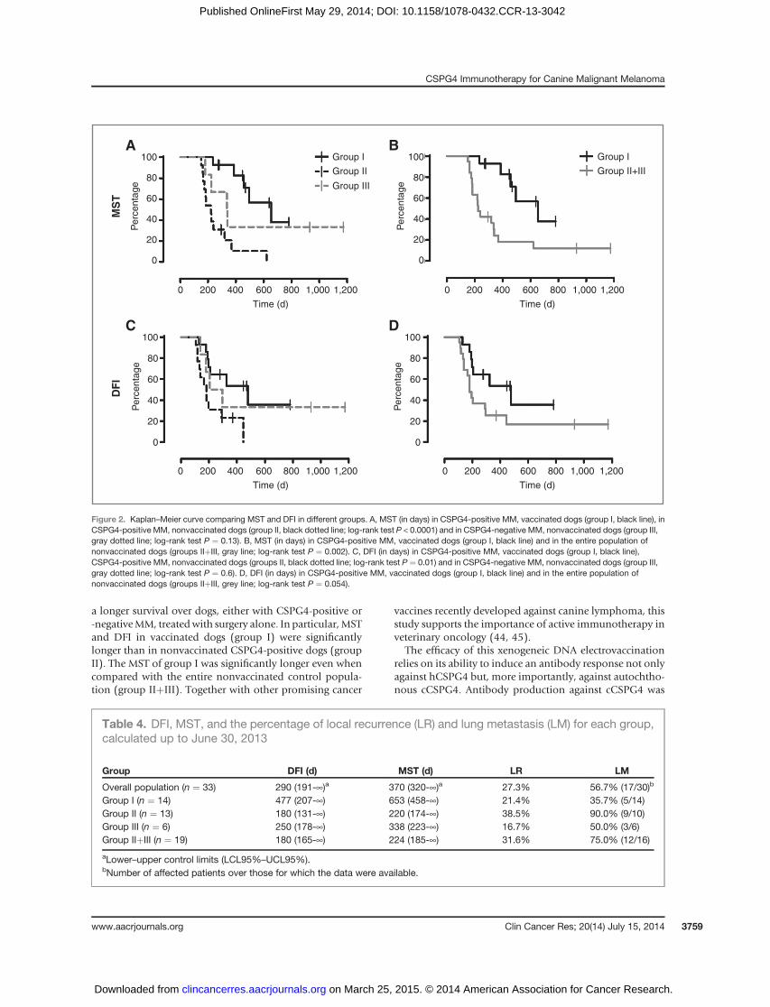
Kaplan–Meier curves for median survival time (MST;Fig. 2A and B) and disease-free interval (DFI; Fig. 2C andD) were analyzed. Group I exhibited significantly longerMST than group II (P < 0.0001; Fig. 2A), whereas it was notsignificantly different from group III (P ¼ 0.13; Fig. 2A).However, group I MST was significantly longer when com-pared with the overall nonvaccinated population (groupIIþIII; P ¼ 0.002; Fig. 2B). The group I DFI value wassignificantly longer than group II (P ¼ 0.01) but not thangroup III (P¼ 0.6; Fig. 2C) and group IIþIII (P¼ 0.054; Fig.2D). Group II MST and DFI were not significantly different
from those in group III (P¼ 0.14 and P¼ 0.2, respectively).A summary of DFI, MST, local recurrences, and lung metas-tasis is shown in Table 4.
No statistically significant correlationwas found betweenthe disease outcome and the excision margin status, thepercentages of Ki67 positivity, themitotic index, the nuclearatypia scores, and the CSPG4 expression.
DiscussionOral MM is the most common oral malignancy in dogs
and is generally locally aggressive and highly metastatic.Regional lymphatic metastases are present in 70% to 80%of dogs at presentation, even in case of clinically normalLN (37). Primary tumor en bloc resection and regionallymphadenectomy, and/or radiotherapy are the preferredtreatment methods and result in locoregional control ofMM in up to 75% of dogs. Disappointingly, the 1-yearsurvival rate is less than 30%, even after adjuvant che-motherapy (38); besides, the addition of radiotherapy tosurgery and adjuvant chemotherapy did not result in asignificant increase of the disease-free period (39). TheUSDA has licensed a xenogeneic DNA vaccine againsttyrosinase, ONCEPT (Merial), but its efficacy has beenrecently questioned (25). In fact, although the study byGrosenbaugh and colleagues (40) supported the ability ofthis vaccine to extend MST in dogs with locally controlledstage II–III oral MM, a further retrospective study byOttnod and colleagues (25) failed to validate the previousresults. This discrepancy could be partially related to thefeatures of the antigen targeted by ONCEPT, tyrosinase,which is expressed by a limited percentage of oral cMM(41) and which does not have a well-recognized onco-genic role in melanoma progression.
The present study has evaluated the immunotargeting ofCSPG4, a useful diagnostic biomarker of cMM (19) and anattractive oncoantigen (15), in dogs with surgically resectedstage II–III CSPG4-positive oral MM. The age and maleprevalence of the canine population presented here were inagreement with the literature (42, 43). Of the 27 CSPG4-positive oral MM-bearing dogs, 14 (group I) were vaccinat-ed, whereas the remaining (group II) and the 6 affected byCSPG4-negative oral MM (group III) were not. The rate ofprimary tumor local control (noninfiltrated excision mar-gins) at the first surgery was adequately high (72.7% in thewhole canine population, 78.6% in group I, 69.2% in groupII, and 66.6% in group III). All cMM were immunohisto-chemically tested for Ki67,mitotic index, andnuclear atypia(32, 33), which have been proposed as markers of poorprognosis (25). These markers were highly expressed inalmost all samples, thus indicating the aggressive clinicalbehavior of most cMM included in the study. However, theexpression of these markers was not associated with that ofCSPG4 and do not correlate with outcome.
Our results demonstrate the safety of the electrovaccina-tion with a plasmid that codes for hCSPG4 and its ability toinduce a specific immune response against both hCSPG4and the canine ortholog. This immune response resulted in
Riccardo et al.
Clin Cancer Res; 20(14) July 15, 2014 Clinical Cancer Research3758
on March 25, 2015. © 2014 American Association for Cancer Research. clincancerres.aacrjournals.org Downloaded from
Published OnlineFirst May 29, 2014; DOI: 10.1158/1078-0432.CCR-13-3042
a longer survival over dogs, either with CSPG4-positive or-negativeMM, treatedwith surgery alone. In particular,MSTand DFI in vaccinated dogs (group I) were significantlylonger than in nonvaccinated CSPG4-positive dogs (groupII). The MST of group I was significantly longer even whencompared with the entire nonvaccinated control popula-tion (group IIþIII). Together with other promising cancer
vaccines recently developed against canine lymphoma, thisstudy supports the importance of active immunotherapy inveterinary oncology (44, 45).
The efficacy of this xenogeneic DNA electrovaccinationrelies on its ability to induce an antibody response not onlyagainst hCSPG4 but, more importantly, against autochtho-nous cCSPG4. Antibody production against cCSPG4 was
Table 4. DFI, MST, and the percentage of local recurrence (LR) and lung metastasis (LM) for each group,calculated up to June 30, 2013
Group DFI (d) MST (d) LR LM
Overall population (n ¼ 33) 290 (191-¥)a 370 (320-¥)a 27.3% 56.7% (17/30)b
Group I (n ¼ 14) 477 (207-¥) 653 (458-¥) 21.4% 35.7% (5/14)Group II (n ¼ 13) 180 (131-¥) 220 (174-¥) 38.5% 90.0% (9/10)Group III (n ¼ 6) 250 (178-¥) 338 (223-¥) 16.7% 50.0% (3/6)Group IIþIII (n ¼ 19) 180 (165-¥) 224 (185-¥) 31.6% 75.0% (12/16)
aLower–upper control limits (LCL95%–UCL95%).bNumber of affected patients over those for which the data were available.
100
80
60
40
20
0
0 200 400 600
Time (d)
Group IA B
C D
Group II
Group I
Group II+III
Group III
800 1,000 1,200 0 200 400 600
Time (d)
800 1,000 1,200
0 200 400 600
Time (d)
800 1,000 1,200 0 200 400 600
Time (d)
800 1,000 1,200
Per
cent
age
MS
TD
FI
100
80
60
40
20
0
Per
cent
age
100
80
60
40
20
0
Per
cent
age
100
80
60
40
20
0
Per
cent
age
Figure 2. Kaplan–Meier curve comparing MST and DFI in different groups. A, MST (in days) in CSPG4-positive MM, vaccinated dogs (group I, black line), inCSPG4-positive MM, nonvaccinated dogs (group II, black dotted line; log-rank test P < 0.0001) and in CSPG4-negative MM, nonvaccinated dogs (group III,gray dotted line; log-rank test P ¼ 0.13). B, MST (in days) in CSPG4-positive MM, vaccinated dogs (group I, black line) and in the entire population ofnonvaccinated dogs (groups IIþIII, gray line; log-rank test P ¼ 0.002). C, DFI (in days) in CSPG4-positive MM, vaccinated dogs (group I, black line),CSPG4-positive MM, nonvaccinated dogs (groups II, black dotted line; log-rank test P ¼ 0.01) and in CSPG4-negative MM, nonvaccinated dogs (group III,gray dotted line; log-rank test P ¼ 0.6). D, DFI (in days) in CSPG4-positive MM, vaccinated dogs (group I, black line) and in the entire population ofnonvaccinated dogs (groups IIþIII, grey line; log-rank test P ¼ 0.054).
CSPG4 Immunotherapy for Canine Malignant Melanoma
www.aacrjournals.org Clin Cancer Res; 20(14) July 15, 2014 3759
on March 25, 2015. © 2014 American Association for Cancer Research. clincancerres.aacrjournals.org Downloaded from
Published OnlineFirst May 29, 2014; DOI: 10.1158/1078-0432.CCR-13-3042

not synchronous in all dogs, but after the fourth vaccinationit was detectable in all patients. A possible mechanism bywhich antibodies can interfere with the disease is theirability to inhibit CSPG4-dependent proliferation of resid-ual melanoma cells in MM-bearing dogs, which no longerdisplay clinically evident disease. The capacity of vaccina-tion-induced antibodies to inhibit hCSPG4-positive cellproliferation in vitro has been displayed in this work.Anti-cCSPG4 antibodies might carry out a similar mecha-nism in vivo because sera from vaccinated dogs can bindcCSPG4 on cMM-derived cells. The availability of a cell linethat highly express cCSPG4 would be instrumental indemonstrating that vaccination-induced antibodies inter-fere with cCSPG4 signaling and, thus, inhibit cMM cellproliferation.
The results here presented overlap those achieved inhumans with advanced melanoma that had been immu-nized with a different type of CSPG4 mimic (18), i.e., themouse anti-idiotypic mAb MK2-23, which bears the inter-nal image of the mouse mAb 763.74 against a definedCSPG4 epitope (46). A cause–effect relationship betweenthe induction of a CSPG4-specific immune response and astatistically significant DFI and survival prolongation hasnot been proved in either patients with human or caninemelanoma. Nevertheless, CSPG4-specific mAb have beenshown to inhibit the proliferation and migration of mela-noma cells in vitro (20). Furthermore, CSPG4-specific mAbadministration to SCID mice grafted with CSPG4-positivehuman tumors has been found to significantly prolong theirsurvival. These effects reflect the inhibition by CSPG4-specific mAb of signaling pathways associated with cellproliferation (20).
The direct activity of antibodies on melanoma cells maynot be the only mechanism that causes the improvedclinical outcome observed in our dogs. The ability of eachpatient to activate antibody indirect activities, such as anti-body-dependent cellular and complement-mediated cyto-toxicity, may be involved and deserve further investigation.
The downregulation of major histocompatibility com-plex (MHC) class Imolecules is a commonmechanismusedby melanoma cells to avoid cytolytic attack from fullyactivated antigen-specific T cells (47), making the role ofthe T-cell cytotoxic response in fighting melanoma contro-versial and strictly dependent on the MHC class-I status ofcMM in each patient. Here, no specific IFNg production inresponse to stimulation with 15-mer peptides derived fromcCSPG4 extracellular domains was found. However, it ispossible that other T-cell parameters, such as vaccine-eli-cited T-cell phenotype, the ability to release other cytokinesand the presence of cells negatively regulating the immuneresponse, may have an impact. The immunosuppressivemicroenvironment at the tumor site and draining LN(48, 49) may have locally inhibited the vaccine-inducedimmune response, allowing local recurrences and/ormetas-tasis to other satellite LN to occur in some dogs.
These data represent a starting point for further investi-gation into the potential of anti-CSPG4 vaccination againstcMM. However, a study involving more dogs is needed to
make firm conclusions. A limitation of this study is also thatit may seem that more attention was paid to vaccinated asopposed to nonvaccinated dogs; 3 dogs from group I weresubjected to a second surgical intervention in the follow-upperiod. CSPG4 adjuvant vaccination, thanks to its ability todelay expected widespread metastasis development and,therefore, to prolong survival, may have allowed the occur-rence of recurrences and further regional lymphatic metas-tases in group I, despite the initial surgery had apparentlylocally controlled the tumor in most dogs. These recur-rences and LN metastasis were still amenable to surgicalresection. This was not the case for nonvaccinated dogs thatsuccumbed to lung or widespread metastasis more rapidly,before they could develop local recurrences. It may also bespeculated that the change in the standard progression oforal MM induced by vaccination also resulted in the expo-sure of long-surviving dogs to a greater risk of dying of asecond tumor of a different histotype, as it was observed in 2patients in group I. However, all these speculations requireconfirmation through the enrollment of more cases in alltreatment groups. Further evaluation is also needed toconfirm the lack of statistical correlation between clinicaloutcome and incomplete excisionmargin status andCSPG4expression as this may be due to the low number ofdogs included in the study. Finally, the development ofrecurrences and regional lymphatic metastases despite vac-cination emphasizes the need to neutralize the tumorimmunosuppressive microenvironment and enhance vac-cine efficacy (50). Other adjuvant treatments, such as theadministration of mAb against immune check points (7),and even radiotherapy, metronomic chemotherapy, andantiangiogenic therapy may allow CSPG4 vaccination tobetter control the disease both locally and systemically. Thelast item to which attention will be addressed in future caseenrollmentwill be the timingof adjuvant vaccination:whento start, when to boost, and how many times.
In conclusion, our data provide the first evidence of theability of anti-CSPG4 electrovaccination to prolong overallsurvival in client-owned dogs with oral MM. The extensionof follow-up observation, the inclusion of more cases and adeeper evaluation of the induced immune response willhelp to better evaluate vaccination potential. Nevertheless,our data show CSPG4 vaccination as an alternative optionfor the management of cMM. Moreover, the strong trans-lational value of the cMM model points to the role thatCSPG4 can play as a good immunotherapy target for themanagement of the high percentage of human melanomasthat express this molecule.
Disclosure of Potential Conflicts of InterestNo potential conflicts of interest were disclosed.
Authors' ContributionsConception and design: F. Riccardo, S. Ferrone, P. Buracco, F. CavalloDevelopment of methodology: F. Riccardo, S. Iussich, L. Maniscalco,S. Lorda Mayayo, R. De Maria, F. Gattino, S. Lanzardo, E. Lardone,S. Prestigio, A. Fiore, S. Zabarino, S. FerroneAcquisitionofdata (provided animals, acquired andmanagedpatients,provided facilities, etc.): F. Riccardo, S. Iussich, L. Maniscalco, M. Arigoni,S. Lanzardo, M. Martano, E. Morello, P. Buracco, F. Cavallo
Riccardo et al.
Clin Cancer Res; 20(14) July 15, 2014 Clinical Cancer Research3760
on March 25, 2015. © 2014 American Association for Cancer Research. clincancerres.aacrjournals.org Downloaded from
Published OnlineFirst May 29, 2014; DOI: 10.1158/1078-0432.CCR-13-3042
Analysis and interpretation of data (e.g., statistical analysis, biosta-tistics, computational analysis): F. Riccardo, S. Iussich, G. La Rosa,S. Ferrone, P. Buracco, F. CavalloWriting, review, and/or revision of the manuscript: F. Riccardo,E. Quaglino, S. Ferrone, P. Buracco, F. CavalloAdministrative, technical, or material support (i.e., reporting or orga-nizing data, constructing databases): F. Riccardo, F. CavalloStudy supervision: P. Buracco, F. Cavallo
AcknowledgmentsThe authors thank Dr. Dale Lawson for his revision and editing of the
article.
Grant SupportThis workwas supported by grants from the Italian Association forCancer
Research (IG 5377), University of Torino, Fondazione Ricerca MolinetteOnlus and by PHS grant RO1CA138188 awarded by the National CancerInstitute.
The costs of publication of this article were defrayed in part by thepayment of page charges. This article must therefore be hereby markedadvertisement in accordance with 18 U.S.C. Section 1734 solely to indicatethis fact.
Received November 5, 2013; revised May 5, 2014; accepted May 6, 2014;published OnlineFirst May 29, 2014.
References1. Jemal A, Siegel R, Ward E, Hao Y, Xu J, Murray T, et al. Cancer
statistics, 2008. CA Cancer J Clin 2008;58:71–96.2. Jilaveanu LB, Aziz SA, Kluger HM. Chemotherapy and biologic
therapies for melanoma: do they work? Clin Dermatol 2009;27:614–25.
3. Eigentler TK, Meier F, Garbe C. Protein kinase inhibitors in melanoma.Expert Opin Pharmacother 2013;14:2195–201.
4. Atkins MB. Cytokine-based therapy and biochemotherapy foradvanced melanoma. Clin Cancer Res 2006;12:2353s–8s.
5. Gogas H, Polyzos A, Kirkwood J. Immunotherapy for advanced mel-anoma: fulfilling the promise. Cancer Treat Rev 2013;39:879–85.
6. Mocellin S, Nitti D. CTLA-4 blockade and the renaissance of cancerimmunotherapy. Biochim Biophys Acta 2013;1836:187–96.
7. Sznol M. Advances in the treatment of metastatic melanoma: newimmunomodulatory agents. Semin Oncol 2012;39:192–203.
8. Kaufman HL. Vaccines for melanoma and renal cell carcinoma. SeminOncol 2012;39:263–75.
9. Blanchard T, Srivastava PK, Duan F. Vaccines against advancedmelanoma. Clin Dermatol 2013;31:179–90.
10. Paoloni MC, Khanna C. Comparative oncology today. Vet Clin NorthAm Small Anim Pract 2007;37:1023–32.
11. Lequarre AS, Andersson L, AndreC, FredholmM,Hitte C, Leeb T, et al.LUPA: a European initiative taking advantage of the canine genomearchitecture for unravelling complex disorders in both human anddogs. Vet J 2011;189:155–9.
12. Bergman PJ, McKnight J, Novosad A, Charney S, Farrelly J, Craft D,et al. Long-term survival of dogs with advanced malignant melanomaafter DNA vaccination with xenogeneic human tyrosinase: a phase Itrial. Clin Cancer Res 2003;9:1284–90.
13. Khanna C, London C, Vail D, Mazcko C, Hirschfeld S. Guiding theoptimal translation of new cancer treatments from canine to humancancer patients. Clin Cancer Res 2009;15:5671–7.
14. Cavallo F, Calogero RA, Forni G. Are oncoantigens suitable targets foranti-tumour therapy? Nat Rev Cancer 2007;7:707–13.
15. Iezzi M, Quaglino E, Amici A, Lollini PL, Forni G, Cavallo F. DNAvaccination against oncoantigens: a promise. Oncoimmunology 2012;1:316–25.
16. Price MA, Colvin Wanshura LE, Yang J, Carlson J, Xiang B, Li G,et al. CSPG4, a potential therapeutic target, facilitates malignantprogression of melanoma. Pigment Cell Melanoma Res 2011;24:1148–57.
17. MittelmanA,ChenGZ,WongGY, LiuC,Hirai S, FerroneS.Humanhighmolecular weight-melanoma associated antigen mimicry by mouseanti-idiotypic monoclonal antibody MK2-23: modulation of the immu-nogenicity in patients with malignant melanoma. Clin Cancer Res1995;1:705–13.
18. Mittelman A, Chen ZJ, Kageshita T, Yang H, Yamada M, Baskind P,et al. Active specific immunotherapy in patients with melanoma. Aclinical trial with mouse antiidiotypic monoclonal antibodies elicitedwith syngeneic anti–high molecular weight melanoma-associatedantigen monoclonal antibodies. J Clin Invest 1990;86:2136–44.
19. Mayayo SL, Prestigio S,Maniscalco L, La RosaG, Arico A, DeMaria R,et al. Chondroitin sulfate proteoglycan-4: a biomarker and a potentialimmunotherapeutic target for caninemalignantmelanoma. Vet J 2011;190:e26–30.
20. Campoli M, Ferrone S, Wang X. Functional and clinical relevance ofchondroitin sulfate proteoglycan 4. AdvCancer Res 2010;109:73–121.
21. Toso JF, Gill VJ, Hwu P, Marincola FM, Restifo NP, SchwartzentruberDJ, et al. Phase I study of the intravenous administration of attenuatedSalmonella typhimurium to patients with metastatic melanoma. J ClinOncol 2002;20:142–52.
22. Triozzi PL, Aldrich W, Allen KO, Carlisle RR, LoBuglio AF, Conry RM.Phase I study of a plasmid DNA vaccine encoding MART-1 in patientswith resected melanoma at risk for relapse. J Immunother 2005;28:382–8.
23. Weber J, Boswell W, Smith J, Hersh E, Snively J, Diaz M, et al.Phase 1 trial of intranodal injection of a Melan-A/MART-1 DNAplasmid vaccine in patients with stage IV melanoma. J Immunother2008;31:215–23.
24. Yuan J, Ku GY, Gallardo HF, Orlandi F, Manukian G, Rasalan TS, et al.Safety and immunogenicity of a human and mouse gp100 DNAvaccine in a phase I trial of patients with melanoma. Cancer Immun2009;9:5.
25. Ottnod JM, Smedley RC, Walshaw R, Hauptman JG, Kiupel M, Obra-dovich JE. A retrospective analysis of the efficacy of Oncept vaccinefor the adjunct treatment of canine oral malignant melanoma. VetComp Oncol 2013;11:219–29.
26. Bodles-Brakhop AM, Heller R, Draghia-Akli R. Electroporation for thedelivery of DNA-based vaccines and immunotherapeutics: currentclinical developments. Mol Ther 2009;17:585–92.
27. Heller LC, Heller R. Electroporation gene therapy preclinical andclinical trials for melanoma. Curr Gene Ther 2010;10:312–7.
28. Sardesai NY, Weiner DB. Electroporation delivery of DNA vaccines:prospects for success. Curr Opin Immunol 2011;23:421–9.
29. Impellizeri JA, Ciliberto G, Aurisicchio L. Electro-gene-transfer as anew tool for cancer immunotherapy in animals. Vet Comp Oncol. 2012Oct 25. [Epub ahead of print].
30. Williams LE, Packer RA. Association between lymph node size andmetastasis in dogs with oral malignant melanoma: 100 cases (1987–2001). J Am Vet Med Assoc 2003;222:1234–6.
31. Owen LN. TNMclassification of tumours in domestic animals. Geneva:World Health Organization; 1980.
32. Bergin IL, Smedley RC, Esplin DG, Spangler WL, Kiupel M. Prognosticevaluation of Ki67 threshold value in canine oral melanoma. Vet Pathol2011;48:41–53.
33. Smedley RC, Spangler WL, Esplin DG, Kitchell BE, Bergman PJ, HoHY, et al. Prognostic markers for canine melanocytic neoplasms: acomparative review of the literature and goals for future investigation.Vet Pathol 2011;48:54–72.
34. Yang J, Price MA, Neudauer CL, Wilson C, Ferrone S, Xia H, et al.Melanoma chondroitin sulfate proteoglycan enhances FAK andERK activation by distinct mechanisms. J Cell Biol 2004;165:881–91.
35. Wilson BS, Imai K, Natali PG, Ferrone S. Distribution and molecularcharacterization of a cell-surface andacytoplasmic antigendetectablein human melanoma cells with monoclonal antibodies. Int J Cancer1981;28:293–300.
36. R Development Core Team. R: a language and environment forstatistical computing. Vienna, Austria: R Fundation for StatisticalComputing; 2010.
CSPG4 Immunotherapy for Canine Malignant Melanoma
www.aacrjournals.org Clin Cancer Res; 20(14) July 15, 2014 3761
on March 25, 2015. © 2014 American Association for Cancer Research. clincancerres.aacrjournals.org Downloaded from
Published OnlineFirst May 29, 2014; DOI: 10.1158/1078-0432.CCR-13-3042
37. Bergman PJ. Canine oral melanoma. Clin Tech Small Anim Pract2007;22:55–60.
38. Brockley LK, CooperMA, Bennett PF.Malignant melanoma in 63 dogs(2001–2011): the effect of carboplatin chemotherapy on survival. N ZVet J 2013;61:25–31.
39. DankG, Rassnick KM, Sokolovsky Y, Garrett LD, Post GS, Kitchell BE,et al. Use of adjuvant carboplatin for treatment of dogs with oralmalignant melanoma following surgical excision. Vet Comp Oncol2014;12:78–84.
40. Grosenbaugh DA, Leard AT, Bergman PJ, Klein MK, Meleo K, Susa-neck S, et al. Safety and efficacy of a xenogeneic DNA vaccineencoding for human tyrosinase as adjunctive treatment for oral malig-nant melanoma in dogs following surgical excision of the primarytumor. Am J Vet Res 2011;72:1631–8.
41. Smedley RC, Lamoureux J, Sledge DG, Kiupel M. Immunohistochem-ical diagnosis of canine oral amelanotic melanocytic neoplasms. VetPathol 2011;48:32–40.
42. Liptak JM, Lascelles BDX. Oral tumors. In: Kudnig ST, S�eguin B,editors. Veterinary surgical oncology. Wiley-Blackwell: Ames, Iowa,2012. p. 119–178.
43. Liptak JM,Withrow SJ. Oral tumors. In: Withrow SJ, Vail DM, Page RL,editors. Small animal clinical oncology: Elsevier: St. Louis, Missouri,2013. p. 381–397.
44. Gavazza A, Lubas G, Fridman A, Peruzzi D, Impellizeri JA, Luberto L,et al. Safety and efficacy of a genetic vaccine targeting telomerase plus
chemotherapy for the therapy of canine B-cell lymphoma. Hum GeneTher 2013;24:728–38.
45. Marconato L, Frayssinet P, Rouquet N,Comazzi S, Leone VF, LagangaP, et al. Randomized, placebo-controlled, double-blinded chemoim-munotherapy clinical trial in a pet dog model of diffuse large B-celllymphoma. Clin Cancer Res 2014;20:668–77.
46. Kusama M, Kageshita T, Chen ZJ, Ferrone S. Characterization ofsyngeneic antiidiotypic monoclonal antibodies to murine anti-humanhigh molecular weight melanoma-associated antigen monoclonalantibodies. J Immunol 1989;143:3844–52.
47. RayS, Chhabra A,Mehrotra S, Chakraborty NG, Ribas A, Economou J,et al. Obstacles to and opportunities for more effective peptide-basedtherapeutic immunization in human melanoma. Clin Dermatol 2009;27:603–13.
48. Tominaga M, Horiuchi Y, Ichikawa M, Yamashita M, Okano K, Jiku-maru Y, et al. Flow cytometric analysis of peripheral blood and tumor-infiltrating regulatory T cells in dogs with oral malignant melanoma.J Vet Diagn Invest 2010;22:438–41.
49. Wasserman J, Diese L, VanGundy Z, London C, Carson WE, Papen-fuss TL. Suppression of canine myeloid cells by soluble factors fromcultured canine tumor cells. Vet Immunol Immunopathol 2012;145:420–30.
50. Polak ME, Borthwick NJ, Jager MJ, Cree IA. Melanoma vaccines:the problems of local immunosuppression. Hum Immunol 2009;70:331–9.
Clin Cancer Res; 20(14) July 15, 2014 Clinical Cancer Research3762
Riccardo et al.
on March 25, 2015. © 2014 American Association for Cancer Research. clincancerres.aacrjournals.org Downloaded from
Published OnlineFirst May 29, 2014; DOI: 10.1158/1078-0432.CCR-13-3042
2014;20:3753-3762. Published OnlineFirst May 29, 2014.Clin Cancer Res Federica Riccardo, Selina Iussich, Lorella Maniscalco, et al. Oral Malignant Melanoma Immunized with Human CSPG4 DNACSPG4-Specific Immunity and Survival Prolongation in Dogs with
Updated version
10.1158/1078-0432.CCR-13-3042doi:
Access the most recent version of this article at:
Material
Supplementary
http://clincancerres.aacrjournals.org/content/suppl/2014/07/18/1078-0432.CCR-13-3042.DC1.html
Access the most recent supplemental material at:
Cited Articles
http://clincancerres.aacrjournals.org/content/20/14/3753.full.html#ref-list-1
This article cites by 45 articles, 12 of which you can access for free at:
E-mail alerts related to this article or journal.Sign up to receive free email-alerts
Subscriptions
Reprints and
To order reprints of this article or to subscribe to the journal, contact the AACR Publications Department at
Permissions
To request permission to re-use all or part of this article, contact the AACR Publications Department at
on March 25, 2015. © 2014 American Association for Cancer Research. clincancerres.aacrjournals.org Downloaded from
Published OnlineFirst May 29, 2014; DOI: 10.1158/1078-0432.CCR-13-3042