Correlates of serum resistin in elderly, non-diabetic patients with chronic kidney disease
Upload
khangminh22Category
view
3download
0
veterinarysciences
Article
Erythrogram Patterns in Dogs with Chronic Kidney Disease
Ilaria Lippi, Francesca Perondi , George Lubas , Eleonora Gori * , Alessio Pierini , Alessandra D’Addettaand Veronica Marchetti
�����������������
Citation: Lippi, I.; Perondi, F.; Lubas,
G.; Gori, E.; Pierini, A.; D’Addetta, A.;
Marchetti, V. Erythrogram Patterns in
Dogs with Chronic Kidney Disease.
Vet. Sci. 2021, 8, 123. https://doi.org/
10.3390/vetsci8070123
Received: 18 May 2021
Accepted: 29 June 2021
Published: 30 June 2021
Publisher’s Note: MDPI stays neutral
with regard to jurisdictional claims in
published maps and institutional affil-
iations.
Copyright: © 2021 by the authors.
Licensee MDPI, Basel, Switzerland.
This article is an open access article
distributed under the terms and
conditions of the Creative Commons
Attribution (CC BY) license (https://
creativecommons.org/licenses/by/
4.0/).
Department of Veterinary Science, University of Pisa, 56122 San Piero a Grado (Pisa), Italy;[email protected] (I.L.); [email protected] (F.P.); [email protected] (G.L.);[email protected] (A.P.); [email protected] (A.D.); [email protected] (V.M.)* Correspondence: [email protected]
Abstract: Anemia is considered a common finding in dogs with chronic kidney disease (CKD),typically as normochromic, normocytic, and non-regenerative. Although anemia can occur at anyCKD IRIS (International Renal Interest Society) stage, its severity is related with the loss of kidneyfunction. The aim of the present study was to retrospectively evaluate quantitative and morphologicalabnormalities of the erythrogram in dogs at different CKD IRIS stages. A total of 482 CBCs from3648 initially screened were included in the study. Anemia was present in 302/482 (63%) dogs, inthe majority of which it was normochromic, normocytic, and non-regenerative (295/302; 98%). Thenumber of reticulocytes was <60,000/µL in the majority of dogs (248/295; 84%), with a correlationbetween poor regeneration rate and progression of CKD (p = 0.0001). The frequency of anemiasignificantly differed (p = 0.0001) among the IRIS stages: 108/231 (47%) in IRIS 2, 77/109 (71%)in IRIS 3, and 117/142 (82%) in IRIS 4. Dogs at IRIS stages 3 and 4 were more likely to havemoderate to severe anemia, compared to dogs at IRIS stage 2 (p = 0.0001). Anisocytosis was themost frequent morphological abnormality (291/482; 60%), whereas the presence of poikilocytosisshowed an association with progression of IRIS stages (p = 0.009). Among different morphologicalabnormalities, the frequency of fragmented red blood cells and Howell–Jolly bodies showed asignificant association with the progression of CKD. Anemia was a frequent finding in CKD dogs,mostly associated with none to poor regeneration rate. Similar to human medicine, advanced CKDstages are more frequently characterized by morphological alterations, such as fragmented red bloodcells and Howell–Jolly bodies, which may suggest a more severe condition of reduced bone marrowactivity and microangiopathy.
Keywords: dog; CKD; anemia; erythrogram
1. Introduction
Anemia is considered a common finding in dogs with chronic kidney disease (CKD) [1],which typically occurs as normochromic, normocytic, and non-regenerative [2]. Althoughno published data are available regarding anemia in CKD dogs at different stages, clinicalexperiences seem to show an association between frequency of anemia and progression ofCKD. Anemia in CKD dogs is mainly related to the decreased production of erythropoietinas well as other mechanisms, such as inflammation, iron deficiency or inappropriate utiliza-tion, decreased red blood cell (RBC) survival due to metabolic or mechanical injuries, andthe anti-proliferative effects of uremic toxins [3]. The severity of anemia has been reportedto correlate positively with serum creatinine concentrations [1] and to become clinicallyrelevant in patients with CKD International Renal Interest Society (IRIS) stages 3 and 4 [4].Morphological abnormalities of RBCs, such as spiculated (echinocyte) and deformed RBCs(acanthocytes), have been occasionally reported in the blood smear evaluation of CKDdogs, although their association with the severity of CKD has not been investigated [2].
The aim of the present study was to retrospectively evaluate quantitative and mor-phological abnormalities of erythrogram in dogs at different CKD IRIS stages.
Vet. Sci. 2021, 8, 123. https://doi.org/10.3390/vetsci8070123 https://www.mdpi.com/journal/vetsci

Vet. Sci. 2021, 8, 123 2 of 9
2. Materials and Methods2.1. Case Selection
Medical records of CKD dogs, presented to the Veterinary Teaching Hospital “MarioModenato” of Pisa University, between January 2014 and December 2020, were retrospec-tively evaluated. Inclusion criteria for CKD included complete case-log with history, andlaboratory and ultrasonographic findings consistent with CKD. Specifically, diagnosisof CKD was based on chronic history of azotemia (more than 3–4 months), progressiveweight loss, poor appetite, polyuria–polydipsia (PU/PD), and renal ultrasound findings ofirregular shape, reduced cortico-medullary distinction, and hyperechoic cortices [5].
Dogs were excluded if (1) historical, laboratory, and/or ultrasonographic findingswere consistent with acute kidney injury (AKI); (2) there was a unavailable report of theblood smear evaluation; (3) use of alpha-darbepoetin or red blood cell transfusion priorto the presentation; (4) severe comorbidities, which might affect erythrogram, such ashyperadrenocorticism, hypothyroidism, and neoplasia; and (5) finally, since the same dogwas included in the case selection only once, rechecks of the same patients were excluded.
Included dogs were classified according to the IRIS guidelines for CKD, based onserum creatinine concentration as follows: stage 2 (serum creatinine 1.4 to 2.8 mg/dL);stage 3 (serum creatinine 2.9 to 5.0 mg/dL); and stage 4 (serum creatinine > 5.0 mg/dL) [6].
2.2. Hematological Parameters
Complete blood counts (CBCs) were performed using a laser cell counter (ProcyteDX, IDEXX Laboratories, Westbrook, ME, USA), and a blood smear stained with May–Grünwald Giemsa (Aerospray Wescor, Delcon, Milan, Italy) was microscopically examinedby experienced and trained clinical pathologists. For each dog included in the study,the following parameters were evaluated: red blood cell count (RBC), hematocrit (HCT)and hemoglobin (HGB) values, mean corpuscular volume (MCV), mean corpuscularhemoglobin (MCH), mean corpuscular hemoglobin concentration (MCHC), red blood celldistribution width (RDW), and absolute reticulocytes count (RET). Anemia was consideredmild if the HCT range was 37–30%, moderate if the HCT range was 29–20%, and severe ifthe HCT was below 19% [7,8]. Anemia was considered microcytic for MCV < 61 fL, normocyticfor MCV between 61 and 73 fL, and macrocytic for MCV > 73 fL. Anemia was consideredhypochromic for MCHC < 32 g/dL, normochromic for MCHC between 32 and 38 g/dL,and hyperchromic for MCHC > 38 g/dL. Regeneration rate was considered absent for RET< 60,000/µL, mild for RET ranging between 61,000 and 150,000/µL, and moderate for RET> 150,000/µL [7]. The degree of the different morphological abnormalities found on RBCs(poikilocytosis) at the microscopic evaluation was assessed according to Weiss (1984) as follows:weak ranging from −/+ to 1+, moderate for 2+, and marked ranging from 3+ to 4+ [9].
Serum creatinine and urea were assessed in liquid chemistry (instrument SAT450 orLiasys, with dedicated reagent kits, Analyzer Medical System—AMS, Rome, Italy).
2.3. Statistical Analysis
Continuous variables were tested for normality through the Kolmogorov–Smirnov test.Kruskal–Wallis test and Dunn’s multiple comparisons test were used to compare CBC
parameters among different CKD groups (IRIS 2, IRIS 3, and IRIS 4). Correlation analysisbetween RBC, HCT, and HGB values, and serum creatinine and urea was performed usingSpearman’s correlation test. Chi squared test of independence was used to compare thefrequency and the degree of anemia, the frequency of different types of anemia accordingto MCV and MCHC, and the frequency and the degree of different morphological abnor-malities of erythrograms among dogs at different stages of CKD. Statistical analysis wasperformed using commercial statistical software (IBM, SPSS v.25, IBM Corp., New York,NY, USA), and all data were considered statistically significant for p value < 0.05.

Vet. Sci. 2021, 8, 123 3 of 9
3. Results
The retrospective medical record evaluation of electronic clinical database of theUniversity of Pisa Veterinary Teaching Hospital gave initially a total of 3648 azotemic dogswithin the selected time interval. Afterwards, among the initial case-load, 3166 dogs wereexcluded due to the following reasons: 441 dogs had AKI, 2511 were rechecks of the samedog, in 127 dogs the blood smear evaluation was unavailable, and 87 dogs were previouslytreated with alpha-darbepoetin or blood transfusion. Finally, a total of 482 dogs with CKDwere included in the study.
Of the 482 enrolled dogs, 231 dogs (47.9%) were in CKD IRIS stage 2, 109 dogs (22.6%)were in IRIS stage 3, and 142 dogs (29.5%) were in IRIS stage 4. According to sex, 214 dogs(44.4%) were intact males, 20 dogs (4.1%) were neutered males, 159 dogs (32.9%) wereintact females, and 89 dogs (18.6%) were spayed females. Median age was 9.6 years (range5.9–12.4 years), and the most represented breeds were mix-breed (n = 145; 30%), Boxers(n = 31; 6.4%), Labrador retrievers (n = 30; 6.2%), German shepherds (n = 20; 4.1%), andGolden Retrievers (n = 17; 3.5%).
Anemia was present in 302/482 dogs (63%). The frequency of anemia was of 47%(108/231) in IRIS 2, 71% (77/109) in IRIS 3, and 82% (117/142) in IRIS 4. A Chi squared testshowed a statistical association between frequency of anemia and progression of the IRISstage (p = 0.0001). Characterization of anemia according to MCV and MCHC in dogs atdifferent IRIS stages is reported in Table 1. Normochromic and normocytic anemia was themost frequent type of anemia, which was found in 209/302 (69%) dogs. According to theregeneration rate, the majority of dogs (239/302; 79%) showed non-regenerative anemia(RET < 60,000/µL) (Table 2).
Table 1. Chi squared comparison of different types of anemia among dogs at different CKD IRIS stages.
IRIS 2(n = 108)
IRIS 3(n = 77)
IRIS 4(n = 117) p-Value
Frequency of different types of anemia 0.203Microcytic–Hypochromic 1/108 (1%) 2/77 (3%) 0/117 (0%)Microcytic–Normochromic 26/108 (24%) 11/77 (14%) 22/117 (19%)Microcytic–Hyperchromic 1/108 (1%) 3/77 (4%) 6/117 (5%)Normocytic–Normochromic 74/108 (69%) 57/77 (74%) 78/117 (67%)Normocytic–Hyperchromic 2/108 (2%) 0/77 (0%) 2/117 (2%)Macrocytic–Hypochromic 3/108 (3%) 0/77 (0%) 3/117 (3%)Macrocytic–Normochromic 1/108 (1%) 4/77 (5%) 6/117 (5%)
Table 2. Chi squared comparison of the frequency and degree of anemia and regeneration rate amongdogs at different CKD IRIS stages.
IRIS 2 IRIS 3 IRIS 4 p-Value
Frequency of anemia 0.0001
Anemia(n = 108)108/231
(47%)
(n = 77)77/109(71%)
(n = 117)117/142
(82%)Degree of anemia 0.0001
Mild (HCT 37–30%)(n = 104)49/104(47%)
(n = 76)34/76(45%)
(n = 115)27/115(23%)
Moderate (HCT 29–20%) 34/104(33%)
34/76(45%)
62/115(54%)
Severe (HCT < 19%) 21/104(20%)
8/76(10%)
26/115(23%)
Degree of regeneration 0.0001
None (RET < 60,000/µL)(n = 104)78/104(75%)
(n = 76)61/76(80%)
(n = 115)109/115
(95%)
Mild (RET 60,000–150,000/µL) 20/104 (19%) 12/76(16%)
5/115(4%)
Moderate (RET > 150,000/µL) 6/104(6%)
3/7(4%)
1/115(0.8%)
Degree of anemia was defined as mild (HCT 37–30%), moderate (HCT 29–20%), and severe (HCT < 19%);degree of regeneration was defined as none (RET < 60,000/µL), mild (RET 60,000–150,000/µL), and moderate(RET > 150,000/µL).

Vet. Sci. 2021, 8, 123 4 of 9
Median values of erythrogram parameters of dogs at different CKD IRIS stages arereported in Table 3.
Table 3. Kruskal-Wallis comparison of erythrogram parameters of dogs at different IRIS stages.
Parameter ReferenceRange
CKD(n = 482)
IRIS 2(n = 231)
IRIS 3(n = 109)
IRIS 4(n = 142) p-Value
RBC (M/µL) 5.6–8.8 5.17 ± 1.64 5.86(4.76–6.7)
5.0(4.0–6.0)
4.2(3.3–5.2) <0.0001
HCT (%) 37.3–61.7 33.2 ± 10.7 37.6(30.4–43.9)
32.1(26.0–39.2)
27.3(21.2–335.0) <0.0001
HGB (g/dL) 13.1–20.5 11.9(9.2–14.4)
13.3(10.8–15.2)
11.2(13.8–9.5)
9.8(12.1–7.7) <0.0001
MCV (fL) 61.6–73.5 64.6(61.5–67.5)
64.8(61.6–67.2)
64.8(61.8–68.0)
64(60.8–67.7) 0.53
MCH (pg) 21.2–25.9 22.8(21.8–23.7)
22.6(21.6–23.6)
23(22.0–23.7)
23(21.9–24) 0.11
MCHC (g/dL) 32.0–37.9 35.3(34.1–36.3)
35.1(34.0–35.9)
35.3(33.9–36.4)
35.7(34.5–36.7) 0.0002
RDW (%) 13.6–21.7 16.2(14.9–18.2)
16.5(15.0–18.8)
16(14.7–18.3)
15.8(14.7–17.7) 0.024
RET (K/µL) 10–110 24(1–166)
34(3–166)
23(12–56)
16(1–78) 0.003
RBC, red blood cells; HCT, hematocrit; HGB, hemoglobin; MCV, mean corpuscular volume; MCH, mean cellhemoglobin; MCHC, mean corpuscular hemoglobin concentration; RDW, red blood cell distribution width; RET,reticulocyte number. CKD anemic dogs (n = 482) were distributed as IRIS 2 (n = 231), IRIS 3 (n = 109), andIRIS 4 (n = 142). Normally distributed data are represented as mean ± standard deviation, and non-normallydistributed data are presented as median and range. The majority of dogs at IRIS stage 2, 3, and 4 showednormocytic–normochromic anemia. No statistically significant difference in the frequency of different types ofanemia was found at different IRIS stages.
Spearman’s correlation analysis showed a negative, linear correlation between serumurea and creatinine and the following erythrogram parameters: RBC, HCT, and HGB(respectively, Figures 1–3).
Figure 1. Spearman’s correlation analysis between RBC and serum creatinine and urea. Statisticallysignificant, negative linear correlation (p < 0.0001; r −0.3470) between RBC (M/µmol) and serumcreatinine (mg/dL), and statistically significant negative linear correlation (p < 0.0001; r −0.2840)between RBC (M/µmol) and serum urea (mg/dL) in the totality of anemic CKD dogs (n = 302).

Vet. Sci. 2021, 8, 123 5 of 9
Figure 2. Spearman’s correlation analysis between HCT and serum creatinine and urea. Statisticallysignificant, negative linear correlation (p < 0.0001; r −0.3439) between HCT (%) and serum creatinine(mg/dL), and statistically significant negative linear correlation (p < 0.0001; r −0.2194) between HCT(%) and serum urea (mg/dL) in the totality of anemic CKD dogs (n = 302).
Figure 3. Spearman’s correlation analysis between HGB and serum creatinine and urea. Statisticallysignificant, negative linear correlation (p < 0.0001; r −0.3286) between HGN (g/dL) and serumcreatinine (mg/dL), and statistically significant negative linear correlation (p < 0.0001; r −0.2194)between HGB (g/dL) and serum urea (mg/dL) in the totality of anemic CKD dogs (n = 302).
Morphological abnormalities of the erythrogram are reported in Table 4.
Table 4. Chi squared comparison of morphological abnormalities of the erythrogram in dogs atdifferent CKD IRIS stages.
Degree IRIS 2 IRIS 3 IRIS 4 p-Value
Anisocytosis(291/482; 60%)
weak 90 45 560.77moderate 40 15 25
marked 7 5 8
Polychromasia(113/482; 23%)
weak 59 1 180.001moderate 9 1 2
marked 1 1 1
Howell–Jolly bodies(87/482; 18%)
weak 30 11 310.186moderate 7 0 5
marked 3 0 0
Poikilocytosis (135/482;28%)
weak 33 23 290.315moderate 15 14 7
marked 3 4 7
Echinocyte(73/135; 54%)
weak 16 11 170.464moderate 7 9 12
marked 0 1 0
Fragmented cells(54/135; 40%)
weak 21 7 200.008moderate 2 4 0
marked 0 0 0
Acantocytes(45/135; 33%)
weak 15 5 150.05moderate 3 5 2
marked 0 0 0
Dacriocytes(13/135, 10%)
weak 4 8 00.488moderate 0 1 0
marked 0 0 0
Eccentrocytes(5/135; 4%)
weak 1 1 00.441moderate 0 0 1
marked 1 0 1
Vet. Sci. 2021, 8, 123 6 of 9
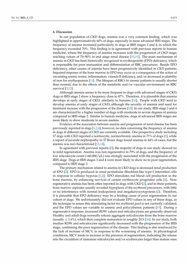
4. Discussion
In our population of CKD dogs, anemia was a very common finding, which washighlighted in approximatively 60% of dogs, especially in more advanced IRIS stages. Thefrequency of anemia increased particularly in dogs at IRIS stages 3 and 4, in which thefrequency exceeded 70%. This finding is in agreement with previous reports in humanmedicine, where the frequency of anemia increases with the progression of CKD stage,reaching values of 70–90% in end stage renal disease [10,11]. The main mechanism ofanemia in CKD has been historically recognized in erythropoietin (EPO) deficiency, whichis responsible for poor maturation and differentiation of RBC precursors. Beside EPOdeficiency, other causes of anemia have been progressively identified in CKD patients.Impaired response of the bone marrow to EPO may occur as a consequence of the action ofcirculating uremic toxins, inflammation, vitamin B deficiency, and/or decreased availabilityof iron for erythropoiesis [11]. The lifespan of RBCs in uremic patients is usually shorterthan normal, due to the effects of the metabolic and/or vascular environment on RBCsurvival [3,12].
Although anemia seems to be more frequent in dogs with advanced stages of CKD,dogs at IRIS stage 2 show a frequency close to 47%. Therefore, it is plausible that anemiadevelops at early stages of CKD, similarly to humans [10]. People with CKD tend todevelop anemia at early stages of CKD, although the severity of anemia and need fortreatment increase with the progression of the disease [10]. In our study, IRIS stage 3 and 4are characterized by a higher number of dogs with moderate to severe degrees of anemiacompared to IRIS stage 2. Similar to human medicine, dogs at advanced IRIS stages aremore likely to show moderate to severe anemia.
Evidence of the association between anemia and progression of renal disease has beenpreviously reported in dogs [1,2,4]; however, no data concerning the frequency of anemiain dogs at different stages of CKD are currently available. One prospective study including17 dogs with CKD reported a normocytic, normochromic anemia in 71% of dogs [1], whilea report of juvenile nephropathy in 37 Boxer dogs found anemia in 44% of cases, but theanemia was not characterized [13,14].
In agreement with previous reports [2], the majority of dogs in our study showed noto mild regeneration. Anemia was non regenerative in 79% of dogs, and the frequency oflow reticulocyte count (<60,000/µL) was strongly associated with the progression of theIRIS stage. Dogs at IRIS stages 3 and 4 were more likely to show no to poor regeneration,compared to IRIS stage 2.
The primary mechanism related to anemia in CKD dogs is decreased renal productionof EPO [2]. EPO is produced in renal peritubular fibroblast-like type-1 interstitial cellsin response to cellular hypoxia [1,2]. EPO stimulates red blood cell production in thebone marrow, by enhancing survival of certain erythrocyte progenitor cells [2]. Non-regenerative anemia has been often reported in dogs with CKD [2], and in these patients,bone marrow aspirates usually revealed hypoplasia of the erythroid precursors, with littleor no interference with normal leukopoiesis and megakaryocytopoiesis [2]. Therefore,it is plausible that EPO deficiency may be a leading cause of poor regeneration in ourcohort of dogs. We unfortunately did not evaluate EPO values in any of these dogs, asthe technique to assess this stimulating factor for erythroid pool is not currently validated,and the EPO values are variable in anemic and polycythemic patients [15]. During aregenerative response, increased RDW values and reticulocytosis are generally observed.Healthy and adult dogs normally release aggregate reticulocytes from the bone marrow(usually ≤ 1.0%), which then complete maturation in roughly 24 h [16]. In our study, bothmedian RDW and reticulocytes significantly decreased with the progression of the IRISstage, confirming the poor regeneration of the disease. This finding is also reinforced bythe lack of increase of MCV, in response to the worsening of anemia. In physiologicalconditions, MCV tends to increase in the presence of regeneration, indicating the releaseinto the circulation of immature reticulocytes and/or erythrocytes larger than mature ones.

Vet. Sci. 2021, 8, 123 7 of 9
In our cohort, no significant increase in the median MCV was noticed in IRIS stage 3 and 4,despite the worsening of anemia.
Another confirmation of the reduced regeneration process is the lower frequency ofpolychromasia with the progression of CKD. The finding of a higher degree of polychroma-sia in IRIS stage 2 compared to stages 3 and 4 may reflect more intense activity of the bonemarrow in the earlier stages. Polychromasia is commonly associated with a prematurerelease of immature forms of RBCs. It is plausible that the progression of CKD depressesthe activity of the bone marrow, reducing the release of immature RBCs.
Typically, the severity of anemia is roughly proportional to the loss of kidney func-tion [2,17], as reported by the study of King et al. (1992), where the hematocrit correlatednegatively with serum creatinine concentrations [1]. Our findings seemed to confirmhuman data, as RBC, HCT, and HGB showed a negative linear correlation with serumcreatinine and urea. The worsening of anemia with the progression of the IRIS stage maybe related to several mechanisms beside EPO deficiency. Serum of uremic patients has beenassociated with inhibition of hematopoietic progenitor growth. Among these factors, theuremic toxin indoxyl sulphate may play a significant role, interfering with erythropoiesisand limiting EPO gene transcription during hypoxia. On the other hand, indoxyl sulphatemay also shorten RBC survival by triggering suicidal erythrocyte death [10].
According to morphological abnormalities, anisocytosis was the most frequent findingfound. Although anisocytosis was found in the 60% of the dogs of the present study, itsfrequency and degree were not associated with the progression of the disease. Moreover,the median RDW of our dogs was within the reference range and did not change signif-icantly among the IRIS stages. Anisocytosis is a parameter commonly associated with ahigh variability of the sizes of RBCs, which has been historically associated with renalfailure [18,19]. In the course of anemia, the finding of anisocytosis, as well as elevated RDW,may reflect different pathological conditions, such as deficiency of iron, folic acid, andvitamin B12, or inflammation, as well as hemolytic uremic syndrome [20]. Although noinformation on the iron panel was available in our study, a potential role of iron metabolismimpairment on the frequency of anisocytosis cannot be rule-out. As is known, systemic ironbalance is maintained by regulating dietary iron absorption and iron release from storagesites. In CKD, iron metabolism may be significantly affected, on one hand, by ongoingiron losses due to chronic bleeding, frequent blood samples, and losses during dialysissessions; and on the other hand, by a reticuloendothelial cell iron blockade. In particular,CKD patients may present a significantly elevated serum concentration of hepcidin, whichis responsible for impaired intestinal absorption of iron, and reticuloendothelial cell ironblockade [21].
In our cohort of dogs, poikilocytosis was found in 28% of the CKD population, and itsfrequency increased significantly in dogs at IRIS stages 3 and 4. Echinocytes were the mostfrequent form of poikilocytosis in our population (53%), followed by fragmented red cells(39%) and acanthocytosis (34%). Interestingly, acanthocytosis has been previously reportedas one of the most frequent abnormalities in rabbits with renal failure [22]. Although thefinding of echinocytes has also been associated with non-pathological conditions (such asprolonged blood storage, artifacts due to excessive EDTA, or slow drying of the smear),its association with acanthocytosis may be related to cholesterol enrichment of the RBCmembrane. Elevated serum cholesterol concentrations would be responsible for formationof RBC membrane projections and increased membrane rigidity. As dyslipidemia andelevated serum cholesterol are very common findings in human CKD, it is possible thatthey may a play a key role also in CKD dogs [22,23]. The authors’ unpublished clinicalexperience reported a frequency of hypercholesterolemia and hypertriglyceridemia ofapproximately 70–80% of CKD dogs. Interestingly, in our cohort of dogs, the frequencyof fragmented red cells increased significantly with the progression of the IRIS stage. Thefinding of fragmented red cells together with acanthocytes is not surprising, as fragmentedRBC may arise from the fragmentation process of acanthocytes. Physical damage ofRBC and fragmentation have been documented in dogs with glomerulonephritis, due

Vet. Sci. 2021, 8, 123 8 of 9
to microangiopathy. Turbulent blood flow, related to endothelial fibrin deposition andmicrothrombi, may promote microangiopathic damage [24]. Uremic toxins may also play asignificant role in promoting RBC fragmentation by increasing the inlet of ionized calciumand the outlet of phosphatidylserine in the lipid bilayer, and by increasing the cellularrigidity [22].
Howell–Jolly bodies (HJBs) were another morphological abnormality found in ourcohort, which frequency increased with the progression of CKD stage. On the other hand,polychromasia was significantly reduced from stage 2 to stage 4. The presence of HJBsduring renal failure may be caused by a possible alteration of the blood–bone–marrowbarrier, due to the presence of uremic toxins. The increased number of HJBs in advancedstages of CKD may also be related to a condition of systemic inflammation and elevatedoxidative damage, in which the removal of HJBs from the bloodstream is reduced due tothe lower phagocytosis activity of macrophages [18,25].
Given the association between progression of CKD and severity of anemia, a prospec-tive study evaluating the role of uremic toxins (such as indoxyl sulphate) in anemic CKDdogs would be advisable. In particular, the serum levels of uremic toxins may be comparedto serum markers of systemic inflammation, as well as oxidative damage. Prospectivestudies should also investigate factors that may affect RBC membrane integrity, such as thelipid profile.
The present study has several limitations. First of all, the retrospective nature of thestudy over a wide time interval did not allow us to include IRIS stage 1 dogs. Serum SDMAhas been only recently introduced in the routine blood work screening of CKD. Therefore,it is possible that dogs with IRIS stage 1 were undiagnosed prior to SDMA evaluation.As the number of dogs in IRIS stage 1 was significantly lower that the number of dogs inthe other stages, we opted not to include them in the statistical analysis. Unfortunately,iron profiles were available only in a limited number of dogs, which did not allow us toinclude iron profiles in the statistics. Finally, although the study population was selectedbase on the presence of uremia due to CKD, it was not possible to recognize the presenceof concomitant comorbidities as they can also affect the erythrogram profile.
5. Conclusions
In conclusion, anemia seems a very frequently found disorder in CKD dogs, mostlyassociated with poor regeneration rate. Although anemia may be present at any CKDIRIS stage, the frequency of this hematological disorder and its degree of severity seem tobe proportional to the loss of kidney function, with low reticulocyte count (<60,000/µL)associated with the progression of the IRIS stage. Similar to human medicine, advancedCKD IRIS stages are more frequently characterized by RBC morphological alterations, suchas fragmented red blood cells and Howell–Jolly bodies, which may suggest a more severecondition of reduced bone marrow activity and microangiopathy.
Author Contributions: Conceptualization, F.P., I.L. and G.L.; methodology, I.L., F.P. and E.G.; formalanalysis, I.L., F.P., E.G. and A.D.; data curation, I.L., F.P., A.D. and E.G.; writing—original draftpreparation, I.L.; writing—review and editing, I.L., V.M., G.L., E.G. and A.P.; supervision, I.L., V.M.and G.L. All authors have read and agreed to the published version of the manuscript.
Funding: This research received no external funding.
Institutional Review Board Statement: Not applicable.
Informed Consent Statement: Not applicable.
Data Availability Statement: Data supporting reported results can be found on the electronicdatabase (OCIROE) of the Vet-erinary Teaching Hospital of the University of Pisa.
Conflicts of Interest: None of the authors of this paper has a financial or personal relationship withother people or organizations that could inappropriately influence or bias the content of the paper.

Vet. Sci. 2021, 8, 123 9 of 9
References1. King, L.G.; Giger, U.; Diserens, D.; Nagode, L.A. Anemia of chronic renal failure in dogs. JVIM 1992, 6, 264–270. [CrossRef]
[PubMed]2. Polzin, D.J. Chronic Kidney Disease. In Nephrology and Urology of Small Animals, 1st ed.; Bartges, J., Polzin, D.J., Eds.; Blackwell
Publishing Ltd.: Chicester, UK, 2011; pp. 433–471.3. Fry, M.M. Anemia of inflammatory, neoplastic, renal and endocrine diseases. In Schalm’s Veterinary Hematology, 6th ed.; Weiss,
D.J., Wardrop, K.J., Eds.; Wiley-Blackwell: Ames, IA, USA, 2010; pp. 246–250.4. Fiocchi, E.H.; Cowgill, L.D.; Brown, D.C.; Markovich, J.E.; Tucker, S.; Labato, M.A.; Callan, M.B. The use of darbepoetin to
stimulate erythropoiesis in the treatment of anemia of chronic kidney disease in dogs. JVIM 2017, 31, 476–485. [CrossRef][PubMed]
5. Bragato, N.; Borges, N.C.; Soares Fioravanti, M.C. B-mode and Doppler ultrasound of chronic kidney disease in dogs and cats.Vet. Res. Commun. 2017, 41, 307–315. [CrossRef] [PubMed]
6. IRIS Staging of CKD (Modified 2019). Available online: http://www.iris-kidney.com (accessed on 1 January 2021).7. Tvedten, H. Laboratory and clinical diagnosis of anemia. In Schalm’s Veterinary Hematology, 6th ed.; Weiss, D.J., Wardrop, K.J.,
Eds.; Wiley-Blackwell: Ames, IA, USA, 2010; pp. 152–161.8. Villiers, E. Disorders of erythrocytes. In BSAVA Manual of Canine and Feline Clinical Pathology, 3rd ed.; Villers, E., Ristic, J., Eds.;
BSAVA: Gloucester, UK, 2016; pp. 38–66.9. Weiss, D.J. Uniform evaluation and semiquantitative reporting of hematologic data in veterinary laboratories. Vet. Clin. Pathol.
1984, 13, 27–31. [CrossRef] [PubMed]10. Gluba-Brzozka, A.; Franczyk, B.; Olszewski, R.; Rysz, J. The influence of inflammation on anemia in CKD patients. Int. J. Mol. Sci.
2020, 21, 725. [CrossRef] [PubMed]11. Cases, A.; Egocheaga, M.I.; Tranche, S.; Pallares, V.; Ojeda, R.; Gorriz, J.L.; Portoles, J.M. Anemia of chronic kidney disease:
Protocol of study, management and referral to nephrology. Nefrologia 2018, 38, 8–12. [CrossRef] [PubMed]12. Bowry, S.K.; Gatti, E. Impact of hemodialysis therapy on anemia of chronic kidney disease: The potential mechanisms. Blood
Purif. 2011, 32, 210–219. [CrossRef] [PubMed]13. Chandler, M.L.; Elwood, C.; Murphy, K.F.; Gajanayake, I.; Syme, H.M. Juvenile nephropathy in 37 boxer dogs. J. Small Anim.
Pract. 2007, 48, 690–694. [CrossRef] [PubMed]14. Bartges, J.W. Chronic kidney disease in dogs and cats. Vet. Clin. N. Am. Small Anim. Pract. 2012, 42, 669–692. [CrossRef] [PubMed]15. Cook, S.M.; Lothrop, C.D. Serum erythropoietin concentrations measured by radiommunoassay in normal, polycythemic, and
anemic dogs and cats. JVIM 1994, 8, 18–25.16. Christian, J.A. Erythrokinetics and erythrocyte destruction. In Schalm’s Veterinary Hematology, 6th ed.; Weiss, D.J., Wardrop, K.J.,
Eds.; Wiley-Blackwell: Ames, IA, USA, 2010; pp. 136–143.17. Dorgalaleh, A.; Mahmudi, M.; Tabibian, S.; Kashani Khatib, Z.; Hossein Tamaddon, G.; Sanei Moghaddam, E.; Bamedi, T.;
Alizadeh, S.; Moradi, E. Anemia and thrombocytopenia in acute and chronic renal failure. Int. J. Hematol. Oncol. Stem Cell Res.2013, 7, 34–39. [PubMed]
18. Paltrinieri, S.; Bertazzolo, W.; Giordano, A. Ematologia ed Emostasi. In Patologia Clinica del Cane e del Gatto, Approccio Pratico allaDiagnostica di Laboratorio, 1st ed.; Paltrinieri, S., Bertazzolo, W., Giordano, A., Eds.; Elsevier: Milano, Italy, 2017; pp. 22–64.
19. Salvagno, G.L.; Sanchis-Gomar, F.; Picanza, A.; Lippi, G. Red blood cell distribution width: A simple parameter with multipleclinical applications. Crit. Rev. Clin. Lab. Sci. 2015, 52, 85–105. [CrossRef] [PubMed]
20. Duchnowski, P.; Hryniewiecki, T.; Kusmierczyk, M.; Szymanski, P. Anisocytosis predicts postoperative renal replacement therapyin patients undergoing heart valve surgery. Cardiol. J. 2020, 27, 362–367. [CrossRef] [PubMed]
21. Babitt, J.L.; Lin, H.Y. Mechanisma of anemia in CKD. J. Am. Soc. Nephrol. 2012, 23, 1631–1634. [CrossRef] [PubMed]22. Christopher, M.M.; Hawkins, M.G.; Burton, A.G. Poikilocytosis in rabbits: Prevalence, type, and association with disease. PLoS
ONE 2014, 9, e112455. [CrossRef] [PubMed]23. Reiss, A.B.; Voloshyna, I.; DeLeon, J.; Miyawaki, N.; Mattana, J. Cholesterol metabolism in CKD. Am. J. Kidney Dis. 2015, 66,
1071–1082. [CrossRef] [PubMed]24. Warry, E.; Bohn, A.; Emanuelli, M.; Thamm, D.; Lana, S. Disease distribution in canine patients with acanthocytosis: 123 cases.
Vet. Clin. Pathol. 2013, 42, 465–470. [CrossRef] [PubMed]25. Harvey, J.W. Evaluation of erythrocytes; Abnormal erythtocyte morphology. In Veterinary Hematology, a Diagnostic Guide and Color
Atlas, 2nd ed.; Harvey, J.W., Ed.; Elsevier: St. Louis, MO, USA, 2012; pp. 59–70.
Copyright © 2022 FDOKUMEN