Effect of ranitidine on acetaminophen-induced hepatotoxicity in dogs
13
Effect of Ranitidine on Acetaminophen-Induced Hepatotoxicity in Dogs C. Panella, L. Makowka, M. Barone, L. Polimeno, S. Rizzi, J. Demetris, S. Bell, F. W. Guglielmi, J. G. Prelich, D. H. Van Thiel, T. E. Starzl, and A. Francavilla Departments of Gastroenterology, University of Bari, Bari, Italy; Surgery, Pathology, and Gastroenterology, University Health Center of Pittsburgh, and Epidemiology, Graduate School of Public Health, University of Pittsburgh, Pittsburgh, Pennsylvania. Abstract The effect of ranitidine administration upon the hepatotoxic effect produced by a multidose acetaminophen administration regimen was examined. Seventy-two dogs received three subcutaneous injections of acetaminophen (750, 200, 200 mg/kg body wt) in DMSO (600 mg/ml) at time zero, 9 hr later, and 24 hr after the first dose. Ten control animals (group I) were not given ranitidine, the remaining 62 dogs received an intramuscular injection of ranitidine 30 min before each acetaminophen dose. Three different doses of ranitidine were used (mg/kg body wt): 50 mg, group II (33 dogs); 75 mg, group III (14 dogs); 120 mg, group IV (15 dogs). Ranitidine reduced the expected acetaminophen-induced hepatoxicity in a dose–response manner. Moreover, a significant correlation was found between the ranitidine dose and the survival rate, as evidenced by transaminase levels in the serum and histology of the liver. This model of fulminant hepatic failure induced by acetaminophen and its modulation with ranitidine provides clinical investigators with a research tool that will be useful in the future investigation of putative medical and surgical therapies being investigated for use in the clinical management of fulminant hepatic failure. Because of the size of the animal used in this model, frequent and serial analyses of blood and liver were available for study to determine the effect of therapy within a given animal as opposed to within groups of animals. Keywords hepatotoxicity; acetaminophen; ranitidine Recently, a large-animal model of acute hepatic failure using acetaminophen was described (1). The two major innovations of this model include the use of DMSO (which does not affect transaminase levels or liver and kidney histology) as the vehicle for the administered acetaminophen and a serial dosage schedule which maintains a hepatotoxic level of acetaminophen in the animals’ blood for several hours, thereby producing a more severe and consistent hepatic necrosis with a mortality rate of 90% (1). All other models of fulminant hepatic failure, particularly those using rats (2), rabbits (3) have been difficult to standardize, are not reproducible, and do not allow for serial observations to be made in animals that may be recovering. The use of large and single doses of ranitidine prior to the administration of acetaminophen in the commonly used rat model of fulminant hepatic failure (7) appears to reduce the subsequent mortality and improve the survival rate of the animals (8). This effect depends on the ability of this H 2 -receptor antagonist to bind cytochrome P-450 (9) and inhibit © 1990 Plenum Publishing Corporation Address for reprint requests: Dr. Antonio Francavilla, Veterans Administration Medical Center, University Drive C, Building 6, Pittsburgh, Pennsylvania 15240. NIH Public Access Author Manuscript Dig Dis Sci. Author manuscript; available in PMC 2010 October 17. Published in final edited form as: Dig Dis Sci. 1990 March ; 35(3): 385–391. NIH-PA Author Manuscript NIH-PA Author Manuscript NIH-PA Author Manuscript
Transcript of Effect of ranitidine on acetaminophen-induced hepatotoxicity in dogs

Effect of Ranitidine on Acetaminophen-Induced Hepatotoxicity inDogs
C. Panella, L. Makowka, M. Barone, L. Polimeno, S. Rizzi, J. Demetris, S. Bell, F. W.Guglielmi, J. G. Prelich, D. H. Van Thiel, T. E. Starzl, and A. FrancavillaDepartments of Gastroenterology, University of Bari, Bari, Italy; Surgery, Pathology, andGastroenterology, University Health Center of Pittsburgh, and Epidemiology, Graduate School ofPublic Health, University of Pittsburgh, Pittsburgh, Pennsylvania.
AbstractThe effect of ranitidine administration upon the hepatotoxic effect produced by a multidoseacetaminophen administration regimen was examined. Seventy-two dogs received threesubcutaneous injections of acetaminophen (750, 200, 200 mg/kg body wt) in DMSO (600 mg/ml) attime zero, 9 hr later, and 24 hr after the first dose. Ten control animals (group I) were not givenranitidine, the remaining 62 dogs received an intramuscular injection of ranitidine 30 min beforeeach acetaminophen dose. Three different doses of ranitidine were used (mg/kg body wt): 50 mg,group II (33 dogs); 75 mg, group III (14 dogs); 120 mg, group IV (15 dogs). Ranitidine reduced theexpected acetaminophen-induced hepatoxicity in a dose–response manner. Moreover, a significantcorrelation was found between the ranitidine dose and the survival rate, as evidenced by transaminaselevels in the serum and histology of the liver. This model of fulminant hepatic failure induced byacetaminophen and its modulation with ranitidine provides clinical investigators with a research toolthat will be useful in the future investigation of putative medical and surgical therapies beinginvestigated for use in the clinical management of fulminant hepatic failure. Because of the size ofthe animal used in this model, frequent and serial analyses of blood and liver were available for studyto determine the effect of therapy within a given animal as opposed to within groups of animals.
Keywordshepatotoxicity; acetaminophen; ranitidine
Recently, a large-animal model of acute hepatic failure using acetaminophen was described(1). The two major innovations of this model include the use of DMSO (which does not affecttransaminase levels or liver and kidney histology) as the vehicle for the administeredacetaminophen and a serial dosage schedule which maintains a hepatotoxic level ofacetaminophen in the animals’ blood for several hours, thereby producing a more severe andconsistent hepatic necrosis with a mortality rate of 90% (1). All other models of fulminanthepatic failure, particularly those using rats (2), rabbits (3) have been difficult to standardize,are not reproducible, and do not allow for serial observations to be made in animals that maybe recovering. The use of large and single doses of ranitidine prior to the administration ofacetaminophen in the commonly used rat model of fulminant hepatic failure (7) appears toreduce the subsequent mortality and improve the survival rate of the animals (8). This effectdepends on the ability of this H2-receptor antagonist to bind cytochrome P-450 (9) and inhibit
© 1990 Plenum Publishing CorporationAddress for reprint requests: Dr. Antonio Francavilla, Veterans Administration Medical Center, University Drive C, Building 6,Pittsburgh, Pennsylvania 15240.
NIH Public AccessAuthor ManuscriptDig Dis Sci. Author manuscript; available in PMC 2010 October 17.
Published in final edited form as:Dig Dis Sci. 1990 March ; 35(3): 385–391.
NIH
-PA Author Manuscript
NIH
-PA Author Manuscript
NIH
-PA Author Manuscript

microsomal hepatic mixed-function oxidase activity. Although cimetidine is known to be themost powerful agent among H2-receptor antagonists with respect to its effect on cytochromeP-450 (10), we used ranitidine because it does not affect hepatic regeneration, as previouslyshown by us and by others (11–13).
In this paper, the effect of different doses of ranitidine on acetaminophen-inducedhepatotoxicity in dogs was evaluated to develop a model for acute hepatic failure with apredetermined mortality rate.
MATERIALS AND METHODSChemicals
Acetaminophen and DMSO were purchased from Sigma Chemical Co., St. Louis, Missouri;pentothal was purchased from Abbott Laboratories, North Chicago, Illinois; ranitidine wasobtained from Glaxo, Inc., Research Triangle Park, North Carolina; xylocaine was purchasedfrom Astra Pharmaceutical Products, Ind., Worchester, Massachusetts; and formaldehyde wasobtained from Fisher Scientific, Pittsburgh, Pennsylvania.
AnimalsSeventy-two male beagles were purchased from Russel B. Hutton Farms, St. Thomas,Pennsylvania. The dogs were housed in a large animal care facility, kept at a constanttemperature of 20 ± 1° C and with a 0600- to 1800-hr light–dark schedule. Dogs werequarantined and allowed to acclimate to their surroundings for a minimum of one week beforeuse. All dogs were given dry dog food and water ad libitum. Prior to study, their body weightsranged from 9–14 kg.
Study DesignThe animals were randomly divided into four different groups.
Group I consisted of 10 dogs that received a total of three subcutaneous injections ofacetaminophen in DMSO at a concentration of 600 mg/ml. The first injection of acetaminophen(750 mg/kg body wt) was given at noon; the second injection (200 mg/kg body wt) was given9 hr later; the third dose (200 mg/kg body wt) was given 24 hr after the initial dose.
Groups II–IV consisted of 62 dogs that were administered the acetaminophen using the sameschedule as the dogs in group I but, in addition, were given ranitidine intramuscularly 30 minbefore each dose of acetaminophen at one of the following dosages: 50 mg/kg body wt, groupII (33 dogs); 75 mg/kg body wt, group III (14 dogs); 120 mg/kg body wt, group IV (15 dogs).All animals were provided with intravenous glucose in amounts necessary to maintain theirblood sugar within normal limits as defined as ± 15% of the basal.
Biochemical DeterminationsIn all dogs (groups I–IV), serum glutamic-pyruvic transaminase levels (SGPT-ALT) weredetermined before and at scheduled times after acetaminophen administration using standardlaboratory methods. Acetaminophen blood levels were determined in groups I and IV animalsusing the method described by Routh et al (14).
HistologyAll animals that died underwent a full necropsy, which included a histologic evaluation of boththe liver and kidneys.
Panella et al. Page 2
Dig Dis Sci. Author manuscript; available in PMC 2010 October 17.
NIH
-PA Author Manuscript
NIH
-PA Author Manuscript
NIH
-PA Author Manuscript

Three dogs from group IV were sacrificed at 72 hr for histological evaluation becauseexperience with the model had shown the histologic injury to be maximal at this time pointand the probability of a long-term survival in an animal surviving to this point in time is greaterthan 90%.
The histology assessment of the degree of tissue necrosis present was performed using asemiquantitative scale. This scale was based on two observations: the extent of necrosis withinindividual lobules, which was classified as (1) mild = less than ⅓ of the lobule, (2) moderate⅓–⅔ of the lobule, and (3) more than ⅔ of the lobules being involved; and the number ofhepatic lobules affected with (1) being less than ⅓, (2) ⅓–⅔, and (3) greater than ⅔ of thelobules being affected. A score for each variable assessed was obtained and a mean of the twovariables was used as the component score for the biopsy. These scores could range from 0–6with no injury in any lobule to massive necrosis in greater than ⅔ of all lobules. Tissues werefixed in 10% neutral buffered formalin, sectioned at 5 µm and stained with hematoxylin andeosin.
Statistical AnalysisThe statistical analysis for the biochemical determinations was performed using one-wayanalysis of variance (SPCC/PC statistical software, SPSS, Inc., Chicago, Illinois). The unpairedStudent’s t test was used for the analysis of the survival rates in groups I–IV. The relationshipbetween long-term survival and the dose of ranitidine administered was examined using probitanalysis (15). A weighted regression equation was used with an adjustment for naturalmortality. A value of P < 0.05 was considered to be significant.
RESULTSAnimal Survival
The survival rate and time course of the experiment for the dogs in groups I–IV are summarizedin Figure 1. No deaths occurred within the first 24 hr. A 10% mortality was seen in all groupsexcept group IV within the next 24 hr. From 48 through 72 hr, a progressive increase inmortality was observed in all four groups. A mortality of 90% at 72 hr was achieved in theanimals in group I. In group II, animals given ranitidine at a dose of 50 mg/kg body wt, a small(from 90% to 82%) but insignificant reduction in the mortality rate at 72 hr was observed. Ingroup III, animals given ranitidine at a dose of 75 mg/kg body wt, a substantial reduction inthe mortality rate (50%) at 72 hr was observed (P < 0.05). In group IV, animals given ranitidineat a dose of 120 mg/kg body wt, the mortality rate was reduced to only 25% at 72 hr (P < 0.05).Thus, a statistically significant dose–response in terms of survival was observed withincreasing doses of ranitidine from 50 to 120 mg/kg body wt.
Biochemical ParametersTable 1 lists the serum ALT (SGPT) values in the animals at each time point. No differencefor ALT values between groups of non surviving dogs was evident. Similarly, no differencebetween groups of surviving animals for transaminase values was seen. However, a significantdifference was observed between the transaminase values of surviving and nonsurvivinganimals across time. Surviving animals appeared to have both a reduced and delayed releaseof transaminase into serum than did the nonsurviving animals.
Figure 2 demonstrates the plasma levels of acetaminophen observed in the animals of groupsI and IV across time. A significant difference (P < 0.05) in acetaminophen plasma levels after28 hr was seen between the animals in groups I and IV and persisted through 40 hr after theinitial dosing of the animals. The actual acetaminophen levels present in the blood of dogs ingroup I are shown in the inset.
Panella et al. Page 3
Dig Dis Sci. Author manuscript; available in PMC 2010 October 17.
NIH
-PA Author Manuscript
NIH
-PA Author Manuscript
NIH
-PA Author Manuscript

HistopathologyThe liver tissue obtained from the nonsurviving animals of each group demonstrated severecentrilobular necrosis involving 100% of the hepatic lobules, reticulin collapse (Figure 3A andB), and occasional areas of central–central bridging necrosis. The extent of the necrosis withinindividual lobules varied from a low of 40% in the least severely affected animals to essentially100% necrosis with only a thin rim of viable periportal hepatocytes remaining in the moreseverely affected animals. The hepatocellular necrosis in each case studied was associated withcentrilobular congestion. Individual necrotic cells demonstrated cytoaggregation, eosinophilia,nuclear pyknosis, karyorrhexsis, and karyolysis. The few residual viable periportal hepatocytesdemonstrated cellular swelling and microvacuolization.
The histopathology of the three randomly selected dogs in group IV that were sacrificed forstudy having a predicted 90% change of survival demonstrated a zonal necrosis involving nomore than 70% of the lobules (Figure 4). In addition, the degree of necrosis within individuallobules varied from a low of 10% to a high of 70%. The cells in the necrotic areas demonstratedcytological alterations similar to those described in the liver specimens obtained from thenonsurviving dogs. No histologic alterations were evident in the kidneys obtained from eitherthe surviving or nonsurviving dogs in any of the groups studied.
Relationship between Survival and Ranitidine DosageFigure 5 illustrates the relationship between survival at 72 hr and thereafter and the ranitidinedosage used. The curve obtained fits the following function: probit = −2.48 + 3.94 (log10 dose).This function predicts the expected mortality at different dosages of ranitidine between 50 and150 mg/kg body wt. Table 2 lists the survival at each dosage of ranitidine used using the probitanalysis. Note that except for the higher dose of ranitidine (>100 mg/kg body wt), little overlapexists between the figures. Moreover, despite some overlap, a group enhancement of survivaloccurred with each incremented dose of ranitidine used.
DISCUSSIONA large animal model (1,8) of acetaminophen-induced hepatic failure using a protocol ofmultidose acetaminophen administration is described. This model is highly reproducible andprovides clinical investigators with a much needed large-animal model of fulminant hepaticfailure that will allow within-animal assessments of liver injury using serial blood samplingand liver biopsy methods of assessment.
The results reported demonstrate that it is possible to ameliorate acetaminophen-inducedhepatotoxicity in this large animal model in a dose-dependent manner with the use of ranitidineadministered at doses ranging from 50 to 120 mg/kg body wt. Using a probit analysis, it ispossible to predict the expected mortality for each dose of ranitidine. This should provide futureinvestigators a predictable model of fulminant hepatic failure for any given experimentalpurpose that might be investigated. This model should be particularly useful for investigatorsplanning to evaluate the effects of putative therapies for fulminant hepatic failure in that itprovides animals with predictably different mortality rates for study.
The protective effect of ranitidine observed in this animal model is due to its capacity to bindcytochrome P-450 and prevent the oxidation of acetaminophen to its toxic metabolites (16–18). Ranitidine was chosen for these experiments instead of cimetidine, which is a morepowerful inhibitor of P-450 enzyme systems, in an effort to avoid the “hormonal”antiandrogenic influence of cimetidine upon the expected hepatic regeneration (11–13). Thedata presented in Figure 2 are consistent with such a hypothesis, but do not prove it. The amount
Panella et al. Page 4
Dig Dis Sci. Author manuscript; available in PMC 2010 October 17.
NIH
-PA Author Manuscript
NIH
-PA Author Manuscript
NIH
-PA Author Manuscript

of acetaminophen administered to the animals in group I was sufficient to induce a mortalityrate of 90%.
Ranitidine, particularly at very high doses, is known to bind to P-450 and inhibits themetabolism of acetaminophen. Such an effect was observed as ranitidine reduced both the rateof acetaminophen disappearance from blood (Figure 2) and the mortality experienced (Figure1).
It is of some interest that no correlation between the degree of hepatic injury and the dose ofranitidine administered to surviving animals was evident. The surviving animals all appearedto have a delayed peak in transaminase levels, probably reflecting a delayed production ofputative acetaminophen toxic metabolites. The histopathologic studies performed in the dogsfrom group IV, which were sacrificed, clearly demonstrates the problems that arise inattempting to prognosticate using small tissue samples. Despite the low mortality rate of thisparticular group, 60 ± 20% of the hepatic lobules were involved in the necrotic process andthe degree of necrosis within individual lobules varied widely (42 ± 20%).
On the basis of the data presented here, the following four major conclusions can be drawn:(1) a multidose acetaminophen injection protocol used in conjunction with ranitidineadministration is a reproducible method for inducing acute hepatic failure in dogs with apredetermined mortality rate, depending upon the dose of ranitidine used; (2) with increasingdoses of ranitidine (0–120 mg/kg body wt), the mortality can be reduced predictably from 90%to 25%; (3) probit analysis allows one to predict the expected mortality in this model for anydose of ranitidine used by the investigation; and (4) this model of acute hepatic failure shouldprovide clinical investigators with a malleable tool to test the effect of putative therapies forfulminant hepatic failure.
AcknowledgmentsSupported by Research Grants from the Veterans Administration and Project Grant AM-29961 from the NationalInstitutes of Health, Bethesda, Maryland, and by grant 87/0129144 from the Consiglio Nazionale dell Ricerche, Italy.
REFERENCES1. Francavilla A, Makowka L, Polimeno L, Barone M, Demetris J, Guglielmi F, Ambrosino G, Van Thiel
DH, Starzl T. A new model of acute hepatic failure in dogs with implication for transplantationresearch. Transplant Proc 1988;20(1):714–718.
2. Zieve L, Dozeman R, LaFontaine D, Draves K. Kinase activity and ornithine decarboxylase activityafter massive necrosis with acetaminophen in the rat. J Lab Clin Med 1985;106:583–588. [PubMed:4056569]
3. Blitzer BL, Waggoner JG, Jones AE, Gralnik H, Towne D, Butler J, Weise V, Koplin I, Walters I,Teychenne P, Goodman D, Berk N. A model of fulminant hepatic failure in the rabbit.Gastroenterology 1978;74:664–671. [PubMed: 631503]
4. Ortega L, Landa Garcia JI, Torres Garcia A, Silecchia G, Arenas J, Suarez A, Moreno Azcoitia M,Sanz Esponera J, Moreno Gonzalez E, Balibrea Cantero JL. Acetaminophen-induced fulminant hepaticfailure in dogs. Hepatology 1985;5(4):673–676. [PubMed: 4018740]
5. Miller DJ, Hickman R, Fratter R, Terblanche J, Saunders SJ. An animal model of fulminant hepaticfailure: A feasibility study. Gastroenterology 1976;71:109–113. [PubMed: 1278635]
6. Gazzard BG, Hughes RD, Mellon PJ, Portmann B, Williams R. A dog model of fulminant hepaticfailure produced by paracetamol administration. Br J Exp Pathol 1975;56:408–411. [PubMed:1212424]
7. Leonard TB, Morgan DG, Dent JG. Ranitidine-acetaminophen interaction: Effects on acetaminophen-induced hepatotoxicity in Fischer 344 rats. Hepatology 1985;5(3):480–487. [PubMed: 3997076]
Panella et al. Page 5
Dig Dis Sci. Author manuscript; available in PMC 2010 October 17.
NIH
-PA Author Manuscript
NIH
-PA Author Manuscript
NIH
-PA Author Manuscript

8. Francavilla A, Makowka L, Polimeno L, Barone M, Demetris J, Prelich J, Van Thiel D, Starzl T. Anovel model for acetaminophen-induced fulminant hepatic failure in the dog. Gastroenterology1989;96:470–478. [PubMed: 2910762]
9. Rendic S, Kajfez F, Ruf H-H. Characterization of cimetidine, ranitidine and related structuresinteraction with cytochrome P-450. Drug Metab Disp 1982;11:137–142.
10. Powell JR. The pharmacokinetic basis for H2-antagonist drug interactions: concepts and implications.J Clin Gastroenterol 1983;5:95–113. [PubMed: 6140286]
11. Kanashima R, Nagasue N, Furusawa M, Inokuchi K. Inhibitory effect of cimetidine on liverregeneration after two-thirds hepatectomy in rats. Am J Surg 1983;146:293–298. [PubMed: 6614315]
12. Francavilla A, Polimeno L, Di Leo A, Makowka L, Barone M, Starzl TE. Effect of cimetidine onhepatic proliferation in vitro. Hepatology 1986;6(5):1130.
13. Francavilla A, Panella C, Polimeno L, Di Leo A, Makowka L, Barone M, Amoruso A, Ingrosso M,Starzl T. Effect of cimetidine, ranitidine, famotidine and omeprazole on hepatocyte proliferation invitro. J Hepatol 1989;8(1):32–41. [PubMed: 2564010]
14. Routh JI, Shane NA, Arredondo EG, Paul WD. Determination of N-acetyl-p-aminophenol in plasma.Clin Chem 1968;14:882–899. [PubMed: 5675309]
15. Finney, DJ. Probit Analysis: A Statistical Treatment of the Sigmoid Response Curve with a Foreword.2nd ed.. Tattersfield, F., editor. Cambridge (England): University Press; 1964.
16. Atwood SJ. The laboratory in the diagnosis and management of acetaminophen and salicylateintoxication. Pediatr Clin North Am 1980;27:871–879. [PubMed: 7470213]
17. Rendic S, Alebic Kolbah T, Kafez F, Ruf H. Interaction of ranitidine with liver microsomes.Xenobiotica 1982;12:9–17. [PubMed: 6124064]
18. Speeg KV, Christian DC, Mitchell MC. Ranitidine and acetaminophen hepatoxicity. Ann Inter Med1984;100:315–316.
Panella et al. Page 6
Dig Dis Sci. Author manuscript; available in PMC 2010 October 17.
NIH
-PA Author Manuscript
NIH
-PA Author Manuscript
NIH
-PA Author Manuscript

Fig 1.Survival rate versus time in group I–IV dogs. All the animals received the same acetaminophentreatment. In addition they received a different intramuscular dose of ranitidine 30 min beforeeach acetaminophen administration (mg/kg). Group I (control), 0 mg; group II, 50 mg; groupIII, 75 mg; group IV, 120 mg.
Panella et al. Page 7
Dig Dis Sci. Author manuscript; available in PMC 2010 October 17.
NIH
-PA Author Manuscript
NIH
-PA Author Manuscript
NIH
-PA Author Manuscript

Fig 2.Acetaminophen plasmatic levels of dogs in groups I and IV. Each value (mean ± SD) representsthe value obtained in four dogs at each time. *Significantly different from the value obtainedin the control animals (group I) at the same time (P < 0.05). Inset: acetaminophen plasma levelsin the animals of group I after initial injection.
Panella et al. Page 8
Dig Dis Sci. Author manuscript; available in PMC 2010 October 17.
NIH
-PA Author Manuscript
NIH
-PA Author Manuscript
NIH
-PA Author Manuscript

Fig 3.Characteristic histopathologic appearance of liver in dogs from group I. (A) Note severecentrilobular necrosis and hemorrhage with some preservation of periportal hepatocytes (portaltract at bottom center) (H & E, 200×). (B) The damage resulted in collapse of the perivenularreticulin architecture with preservation at periportal areas (Reticulin stain, 200×). PT = portaltract; CV = central vein.
Panella et al. Page 9
Dig Dis Sci. Author manuscript; available in PMC 2010 October 17.
NIH
-PA Author Manuscript
NIH
-PA Author Manuscript
NIH
-PA Author Manuscript

Fig 4.Histopathologic appearance of liver in dogs from group IV (H & E. 400×, PT = portal tract,CV = central vein). Note that there is centrilobular congestion, but centrilobular hepatocytesare intact and viable (arrow). Microvesicular stenosis was also detected in hepatocytes(arrowhead).
Panella et al. Page 10
Dig Dis Sci. Author manuscript; available in PMC 2010 October 17.
NIH
-PA Author Manuscript
NIH
-PA Author Manuscript
NIH
-PA Author Manuscript
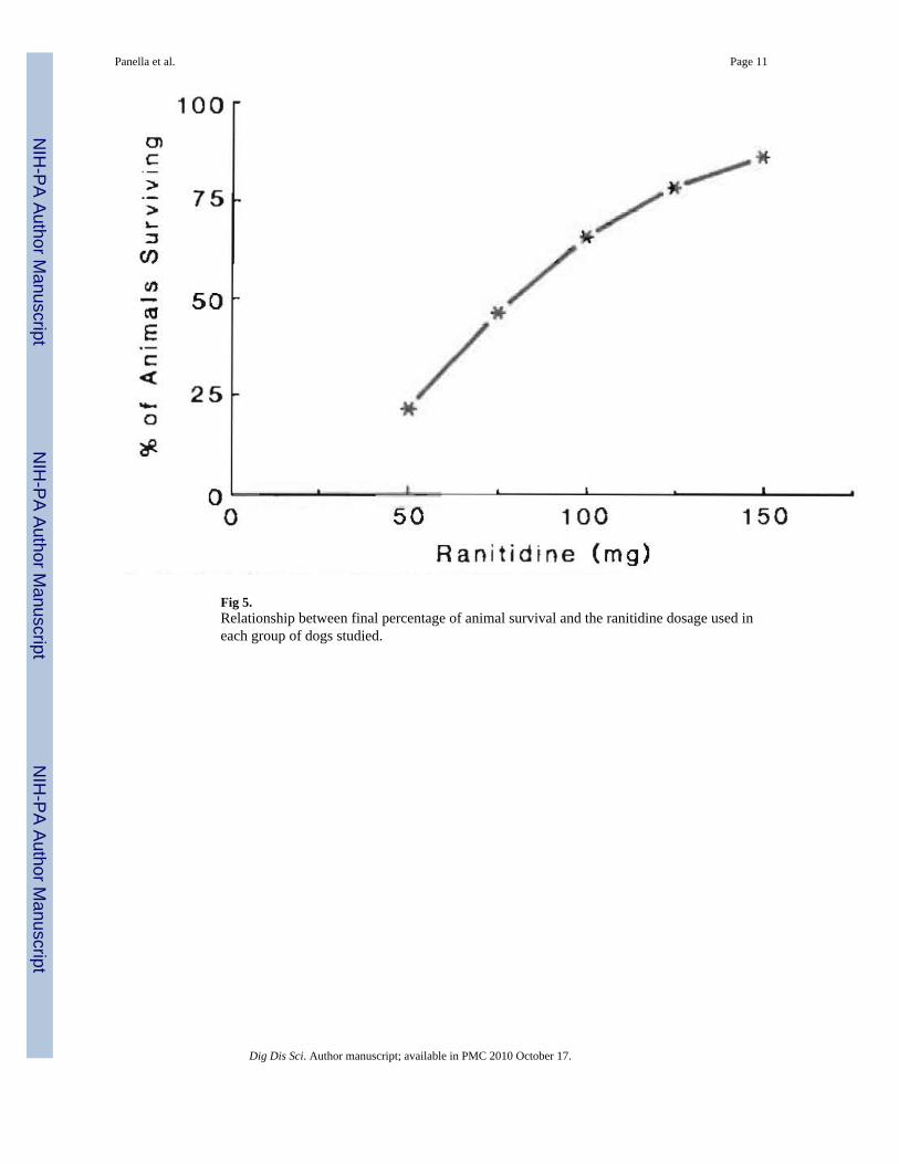
Fig 5.Relationship between final percentage of animal survival and the ranitidine dosage used ineach group of dogs studied.
Panella et al. Page 11
Dig Dis Sci. Author manuscript; available in PMC 2010 October 17.
NIH
-PA Author Manuscript
NIH
-PA Author Manuscript
NIH
-PA Author Manuscript

NIH
-PA Author Manuscript
NIH
-PA Author Manuscript
NIH
-PA Author Manuscript
Panella et al. Page 12
TAB
LE 1
Seru
m g
luta
mic
-pyr
uvic
tran
sam
inas
es (A
LT-S
GPT
) in
nons
urvi
ving
and
surv
ivin
g do
gs fr
om g
roup
s I–I
V a
t diff
eren
t tim
es a
fter i
ntox
icat
ion.
*
AL
T-S
GPT
(uni
ts/li
ter)
0.0
hr24
hr
48 h
r72
hr
96 h
r12
0 hr
Non
surv
ivin
g
G
roup
I40
± 1
475
± 5
02,
631a
b ±
2,69
619
,980
ab ±
4,2
00
G
roup
II38
± 1
769
± 4
33,
507a
b ±
1,49
318
,601
ab ±
5,4
2617
,500
ab ±
6,1
00
Gro
up II
I29
± 1
081
± 4
63,
001a
b ±
703
12,9
70ab
± 4
,521
G
roup
IV42
± 1
547
± 2
13,
204a
b ±
1,77
612
,401
ab ±
3,2
0313
,601
ab ±
2,0
00
Surv
ivin
g
G
roup
I31
± 1
464
± 3
158
0a ±
278
1,70
6a ±
401
1,80
0a ±
503
1,02
4a ±
426
G
roup
II37
± 1
561
± 3
071
1a ±
200
2,48
9a ±
1,9
002,
907a
± 8
217,
709a
± 7
93
G
roup
III
38 ±
17
65 ±
30
810a
± 6
003,
279a
± 3
,250
3,12
6a ±
2,6
132,
017a
± 1
,975
G
roup
IV40
± 1
559
± 3
379
9a ±
601
2,90
0a ±
2,7
803,
214a
± 2
,789
7,90
5a ±
9,5
82
* The
valu
es a
re e
xpre
ssed
as m
ean
± SD
.
a P <
0.05
whe
n th
e va
lues
are
com
pare
d to
the
basa
l lev
els.
b P <
0.05
whe
n th
e va
lues
of n
onsu
rviv
ing
dogs
are
com
pare
d w
ith th
e va
lue
of th
e su
rviv
ing
dogs
of t
he sa
me
grou
p.
Dig Dis Sci. Author manuscript; available in PMC 2010 October 17.

NIH
-PA Author Manuscript
NIH
-PA Author Manuscript
NIH
-PA Author Manuscript
Panella et al. Page 13
TABLE 2
Confidence intervals for estimated probits of survival rate at different doses of ranitidine
Ranitidine dose (mg) Estimated survival % 95% of confidence interval for % survival
50 21.8 12.6 – 34.0
75 46.6 36.5 – 57.0
100 65.8 51.3 – 78.4
125 78.6 60.6 – 90.5
150 86.5 67.4 – 96.0
Dig Dis Sci. Author manuscript; available in PMC 2010 October 17.