Synthesis and Characterizations of Macroporous...
15
This article was downloaded by: [UNIVERSITY OF KWAZULU-NATAL] On: 10 April 2013, At: 06:16 Publisher: Taylor & Francis Informa Ltd Registered in England and Wales Registered Number: 1072954 Registered office: Mortimer House, 37-41 Mortimer Street, London W1T 3JH, UK International Journal of Polymeric Materials and Polymeric Biomaterials Publication details, including instructions for authors and subscription information: http://www.tandfonline.com/loi/gpom20 Synthesis and Characterizations of Macroporous Poly(acrylamide-2- acrylamido-2-methyl-1-propanesulfonic acid) Hydrogels for In Vitro Drug Release of Ranitidine Hydrochloride K. Varaprasad a , N. Narayana Reddy a , S. Ravindra a , K. Vimala a & K. Mohana Raju a a Synthetic Polymer Laboratory, Department of Polymer Science & Technology, Sri Krishnadevaraya University, Anantapur, India Version of record first published: 09 Jun 2011. To cite this article: K. Varaprasad , N. Narayana Reddy , S. Ravindra , K. Vimala & K. Mohana Raju (2011): Synthesis and Characterizations of Macroporous Poly(acrylamide-2-acrylamido-2-methyl-1- propanesulfonic acid) Hydrogels for In Vitro Drug Release of Ranitidine Hydrochloride, International Journal of Polymeric Materials and Polymeric Biomaterials, 60:7, 490-503 To link to this article: http://dx.doi.org/10.1080/00914037.2010.531816 PLEASE SCROLL DOWN FOR ARTICLE Full terms and conditions of use: http://www.tandfonline.com/page/terms-and-conditions This article may be used for research, teaching, and private study purposes. Any substantial or systematic reproduction, redistribution, reselling, loan, sub-licensing, systematic supply, or distribution in any form to anyone is expressly forbidden. The publisher does not give any warranty express or implied or make any representation that the contents will be complete or accurate or up to date. The accuracy of any instructions, formulae, and drug doses should be independently verified with primary sources. The publisher shall not be liable for any loss, actions, claims, proceedings, demand, or costs or damages whatsoever or howsoever caused arising directly or indirectly in connection with or arising out of the use of this material.
-
Upload
independent -
Category
Documents
-
view
1 -
download
0
Transcript of Synthesis and Characterizations of Macroporous...
This article was downloaded by: [UNIVERSITY OF KWAZULU-NATAL]On: 10 April 2013, At: 06:16Publisher: Taylor & FrancisInforma Ltd Registered in England and Wales Registered Number: 1072954 Registeredoffice: Mortimer House, 37-41 Mortimer Street, London W1T 3JH, UK
International Journal of PolymericMaterials and Polymeric BiomaterialsPublication details, including instructions for authors andsubscription information:http://www.tandfonline.com/loi/gpom20
Synthesis and Characterizations ofMacroporous Poly(acrylamide-2-acrylamido-2-methyl-1-propanesulfonicacid) Hydrogels for In Vitro Drug Releaseof Ranitidine HydrochlorideK. Varaprasad a , N. Narayana Reddy a , S. Ravindra a , K. Vimala a &K. Mohana Raju aa Synthetic Polymer Laboratory, Department of Polymer Science &Technology, Sri Krishnadevaraya University, Anantapur, IndiaVersion of record first published: 09 Jun 2011.
To cite this article: K. Varaprasad , N. Narayana Reddy , S. Ravindra , K. Vimala & K. Mohana Raju(2011): Synthesis and Characterizations of Macroporous Poly(acrylamide-2-acrylamido-2-methyl-1-propanesulfonic acid) Hydrogels for In Vitro Drug Release of Ranitidine Hydrochloride, InternationalJournal of Polymeric Materials and Polymeric Biomaterials, 60:7, 490-503
To link to this article: http://dx.doi.org/10.1080/00914037.2010.531816
PLEASE SCROLL DOWN FOR ARTICLE
Full terms and conditions of use: http://www.tandfonline.com/page/terms-and-conditions
This article may be used for research, teaching, and private study purposes. Anysubstantial or systematic reproduction, redistribution, reselling, loan, sub-licensing,systematic supply, or distribution in any form to anyone is expressly forbidden.
The publisher does not give any warranty express or implied or make any representationthat the contents will be complete or accurate or up to date. The accuracy of anyinstructions, formulae, and drug doses should be independently verified with primarysources. The publisher shall not be liable for any loss, actions, claims, proceedings,demand, or costs or damages whatsoever or howsoever caused arising directly orindirectly in connection with or arising out of the use of this material.
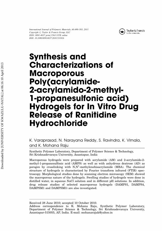
Synthesis andCharacterizations ofMacroporousPoly(acrylamide-2-acrylamido-2-methyl-1-propanesulfonic acid)Hydrogels for In Vitro DrugRelease of RanitidineHydrochloride
K. Varaprasad, N. Narayana Reddy, S. Ravindra, K. Vimala,
and K. Mohana Raju
Synthetic Polymer Laboratory, Department of Polymer Science & Technology,Sri Krishnadevaraya University, Anantapur, India
Macroporous hydrogels were prepared with acrylamide (AM) and 2-acrylamido-2-methyl-1-propanesulfonic acid (AMPS) as well as with anhydrous dextrose (AD) asporogen by crosslinking with N,N1-methylenebisacrylamide (MBA). The chemicalstructure of hydrogels is characterized by Fourier transform infrared (FTIR) spec-troscopy. Morphological studies done by scanning electron microscopy (SEM) showedthe macroporous nature of the hydrogels. Swelling studies of hydrogels were done indistilled water, in aqueous NaCl solution and in different pH solutions. In addition,drug release studies of selected macroporous hydrogels (DAMPS1, DAMPS4,DAMPSM1 and DAMPSM3) are also investigated.
Received 28 June 2010; accepted 10 October 2010.Address correspondence to K. Mohana Raju, Synthetic Polymer Laboratory,Department of Polymer Science & Technology, Sri Krishnadevaraya University,Anantapur-515055, AP, India. E-mail: [email protected]
International Journal of Polymeric Materials, 60:490–503, 2011
Copyright # Taylor & Francis Group, LLC
ISSN: 0091-4037 print=1563-535X online
DOI: 10.1080/00914037.2010.531816
Dow
nloa
ded
by [
UN
IVE
RSI
TY
OF
KW
AZ
UL
U-N
AT
AL
] at
06:
16 1
0 A
pril
2013
Keywords 2-acrylamido-2-methyl-l-propanesulfonic acid, anhydrous dextrose,macroporous hydrogels
INTRODUCTION
Generally, macroporous hydrogels (MPH) are post response materials when
compared to normal hydrogels, because MPH have pores. The pores of macro-
porous hydrogels hold an enormous amount of water when allowed to remain
in a water reservoir for a long period of time. Because of their unique proper-
ties these hydrogels are used in a number of applications [1] such as in cellular
and tissue engineering [2,3] and drug delivery [4].
The swelling properties of MPHs mainly depend on the composition of
co-monomers and the porosity of the hydrogel. These hydrogels release the
drug entrapped inside the pores through a mechanism dependent on drug dif-
fusion coefficient [5]. MPHs are prepared by various methods such as phase
separation [6,7], cryogelation technique [8] and other methods [9]. However,
we have chosen the free radical polymerization process in the presence of sol-
uble substances such as dextrose anhydrous, and sucrose. These materials
should not react with other reactants (vinyl monomers, crosslinkers, initiators,
activators and other polymers) in the polymerization system and which are
washed out from the hydrogel after polymerization [10,11]. Superabsorbent
hydrogels such as macroporous poly(acrylamide-co-sodium methacrylate) can
be obtained by use of the crosslinking polymerization method [12]. The rapid
de-swelling of porous poly(N-isopropylacrylamide) hydrogels prepared using
a free radical polymerization [13], Jia-Ming Chern and his coworkers reported
preparation and swelling characterization of poly(N-isopropylacrylamide)-
based porous hydrogels [14] and also Hideaki Tokuyama et al. reported prep-
aration of porous poly(N-Isopropylacrylamide) gel [15].
This report describes the development and the characterization of a series of
poly(acrylamide-2-acrylamido-2-methyl-1-propanesulfonic acid) P(AM-AMPS)
hydrogel. The effects of AMPS, MBA ratio, pH and different concentrations of
NaCl on the hydrogels swelling properties were investigated. Additionally,
release profiles of ranitidine hydrochloride (RH) drug from test hydrogels were
studied in simulated gastric media pH (2.2) at 37�C.
EXPERIMENTAL
MaterialsAcrylamide (AM), and 2-acrylamido-2-methyl-1-propanesulfonic acid
(AMPS) were purchased from Aldrich Chemicals. N,N1-methylenebisacrylamide
(MBA), ammonium persulfate (APS), N,N,N1,N1-tetramethylethylenediamine
Macroporous Hydrogels 491
Dow
nloa
ded
by [
UN
IVE
RSI
TY
OF
KW
AZ
UL
U-N
AT
AL
] at
06:
16 1
0 A
pril
2013

(TMEDA) and anhydrous dextrose (AD), were purchased from S.D fine chemi-
cals. Ranitidine hydrochloride was obtained from Aurobindo Pharmaceutical
Limited, Hyderabad, India. All the chemicals were used without further purifi-
cation. Throughout the experiments double-distilled water was used.
Preparation of Macroporous Hydrogels14.6 mM of AM dissolved in 2 ml of double-distilled water and 0.24 mM of
AMPS is added into the 100 ml beaker. To this reaction mixture, 4.622 mM of
AD a porogen, 1 ml of MBA (1 g=100 ml) as a crosslinker and also 1 ml of APS
(5 g=100 ml), 1 ml of TMEDA (1 g=100 ml) as an initiating pair were sequen-
tially added by stirring at 100 rpm on a magnetic stir plate. After the reaction
has been completed, the hydrogel was immersed in distilled water at 25�C for
24 h to remove the unreacted materials present in the hydrogel network.
Finally, the hydrogel was dried at 25�C for 2 d. Similarly, other hydrogels were
prepared in a similar manner. The feed compositions of the hydrogels are pre-
sented in Table I.
CharacterizationsFTIR spectrophotometer is used to identify the hydrogel formation. To rec-
ord the FTIR spectra of hydrogel, the sample was completely dried in an oven
(Baheti Enterprises, Hyderabad, India) at 40�C for 6 h. These samples were
read between 600 and 4000 cm�1 on a Bruker IFS 66 V FTIR spectrometer
(Ettlingen, Germany) using the KBr disk method. UV-vis absorption spectra
of the samples were recorded on an ELICO SL 164 Model (The Elico Co.,
Hyderabad, India). The drug loading efficiency and releasing profiles of rani-
tidine hydrochloride in the hydrogels was determined by UV-spectral
measurement at kmax 315 nm. Scanning electron microscopy (SEM; JEOL
JSM 840A, Tokyo, Japan) is employed to study the internal structure of hydro-
gels under dry conditions. The dry samples were coated with a thin layer of
palladium gold alloy.
Swelling MeasurementsThe dried hydrogels are immersed in distilled water at 25�C until the swell-
ing equilibrium is reached (almost 26 h) and they are removed and blotted with
filter paper to remove the excess water solution present on the surface and then
weighed. The immersion time and drying procedure were repeated until the
weight of swollen samples is constant. The swelling ratio, equilibrium swelling
ratio and the swelling water content in percentage is defined as follows:
Swelling ratio ðSg=gÞ ¼ ½Wt � Wd=Wd�
492 K. Varaprasad et al.
Dow
nloa
ded
by [
UN
IVE
RSI
TY
OF
KW
AZ
UL
U-N
AT
AL
] at
06:
16 1
0 A
pril
2013

Table
1:Fe
ed
compositionand
swelling
data
ofmacroporouspoly(A
M-A
MPS)
hydrogels
Hydro
gels
code
AM
(mM)
(AMPS)
mM
DA
(mM)
(MBA)
mM
(APS)
mM
(TMED
A)
mM
Swelling
ratio
(g/g)
Equilibrium
water
content
(EWC)(%
)
Feed
compositionofhydrogels
DAMPS1
14.06
0.2412
4.6227
0.0648
0.21929
0.172
32.24
96.99225
DAMPS2
14.06
0.4824
4.6227
0.0648
0.21929
0.172
50.12
98.04412
DAMPS3
14.06
0.7237
4.6227
0.0684
0.21929
0.172
70.27
98.59698
DAMPS4
14.06
1.20625
4.6227
0.0684
0.21929
0.172
81.81
98.79245
DAMPSM1
14.06
1.20625
4.6227
0.0324
0.21929
0.172
61.5
98.4
DAMPSM2
14.06
1.20625
4.6227
0.0486
0.21929
0.172
59.12
98.33676
DAMPSM3
14.06
1.20625
4.6227
0.0810
0.21929
0.172
52.04
98.11481
Hydro
gels
code
Swelling
exponent(n
)
Diffusion
coefficient
(D)cm
2s�
1
Initialsw
elling
rate
(ri)
[(g
water/g
hydro
gel)/min]
Theore
tical
equilibrium
swelling
(TSeq)
[(g
water/g
hydro
gel)]
Swelling
rate
constant(k
s)
[(g
hydro
gel/g
water)/min]
Swelling
data
ofporousP(A
M-A
MPS)
hydrogels
DAMPS1
0.99791
2.04482
0.02759
36.24502
0.003046
DAMPS2
0.66662
2.29724
0.01791
55.83473
0.000948
DAMPS3
0.68249
2.56446
0.01222
81.83306
0.000493
DAMPS4
0.98504
3.7445
0.00981
101.9368
0.000301
DAMPSM1
0.81135
2.7836
0.01511
66.18134
0.000673
DAMPSM2
0.66518
2.46387
0.0158
63.29114
0.000797
DAMPSM3
0.7728
2.64043
0.0178
56.17978
0.000893
493
Dow
nloa
ded
by [
UN
IVE
RSI
TY
OF
KW
AZ
UL
U-N
AT
AL
] at
06:
16 1
0 A
pril
2013
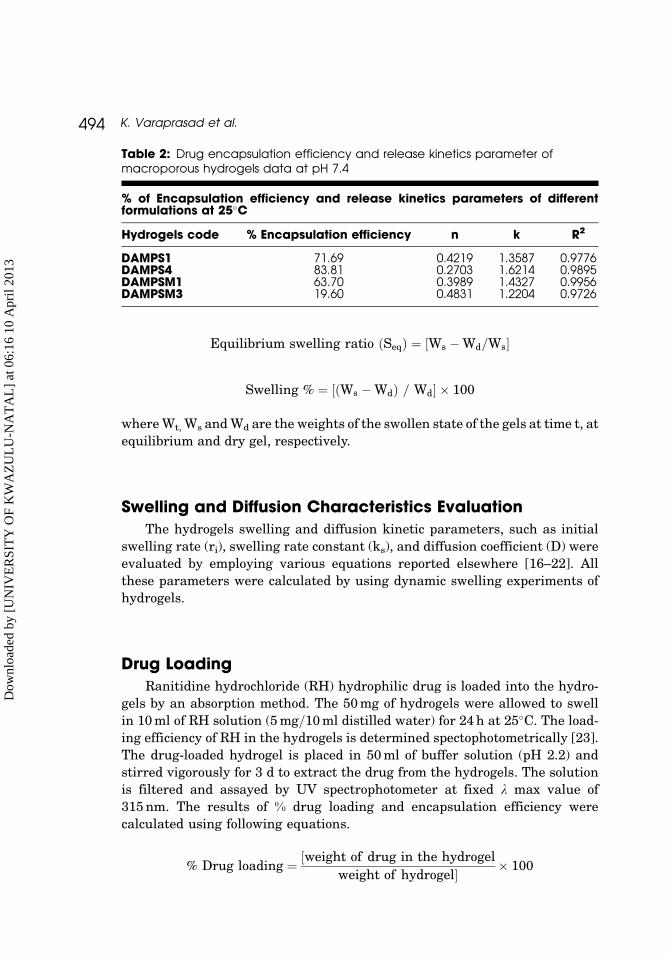
Equilibrium swelling ratio ðSeqÞ ¼ ½Ws � Wd=Ws�
Swelling % ¼ ½ðWs � WdÞ = Wd� � 100
where Wt, Ws and Wd are the weights of the swollen state of the gels at time t, at
equilibrium and dry gel, respectively.
Swelling and Diffusion Characteristics EvaluationThe hydrogels swelling and diffusion kinetic parameters, such as initial
swelling rate (ri), swelling rate constant (ks), and diffusion coefficient (D) were
evaluated by employing various equations reported elsewhere [16–22]. All
these parameters were calculated by using dynamic swelling experiments of
hydrogels.
Drug LoadingRanitidine hydrochloride (RH) hydrophilic drug is loaded into the hydro-
gels by an absorption method. The 50 mg of hydrogels were allowed to swell
in 10 ml of RH solution (5 mg=10 ml distilled water) for 24 h at 25�C. The load-
ing efficiency of RH in the hydrogels is determined spectophotometrically [23].
The drug-loaded hydrogel is placed in 50 ml of buffer solution (pH 2.2) and
stirred vigorously for 3 d to extract the drug from the hydrogels. The solution
is filtered and assayed by UV spectrophotometer at fixed k max value of
315 nm. The results of % drug loading and encapsulation efficiency were
calculated using following equations.
% Drug loading ¼ ½weight of drug in the hydrogel
weight of hydrogel� � 100
Table 2: Drug encapsulation efficiency and release kinetics parameter ofmacroporous hydrogels data at pH 7.4
% of Encapsulation efficiency and release kinetics parameters of differentformulations at 25�C
Hydrogels code % Encapsulation efficiency n k R2
DAMPS1 71.69 0.4219 1.3587 0.9776DAMPS4 83.81 0.2703 1.6214 0.9895DAMPSM1 63.70 0.3989 1.4327 0.9956DAMPSM3 19.60 0.4831 1.2204 0.9726
494 K. Varaprasad et al.
Dow
nloa
ded
by [
UN
IVE
RSI
TY
OF
KW
AZ
UL
U-N
AT
AL
] at
06:
16 1
0 A
pril
2013

% Encapsulation efficiency ¼ ½%actual loading
% theoretical loading� � 100
Release of Ranitidine Hydrochloride (RH)In order to study the release pattern of RH from the loaded hydrogels, a
known weight of RH-loaded hydrogel is placed in 100 ml of 2.2 pH phosphate
buffer at 37�C. The released amount of RH is determined at different time
intervals by recording the absorbance of the release medium by using UV-vis
spectrophotometer ELICO SL 164 Model (The Elico Co., Hyderabad, India).
The recorded absorbance is then related to the amount of RH released using
a calibration plot (0.12–5% concentration). The absorption of the solutions of
RH is measured at kmax 315 nm.
RESULTS AND DISCUSSION
Effect of Monomer on Swelling and DiffusionCharacteristics of HydrogelsThe incorporation of AMPS, a hydrophilic monomer, into the macromole-
cular network is expected to enhance the water uptake capacity as well as
the drug uptake capacity of the hydrogel, which, in turn, will facilitate release
of the entrapped drug. The concentration of AMPS is varied from
0.24–1.206 mM to study the effect of AMPS content on the swelling rations
of the hydrogels (Figure 1). With increasing the amount of AMPS, the swelling
ratios of the hydrogels are found to increase. The behavior is caused by hydro-
philic properties of the AMPS, which is derived from hydrophilic groups (-SO3
H) in its structure. The swelling ratios of the hydrogels increase from
32.34–81.81 g=g with increasing AMPS content. The higher the AMPS content
in the hydrogels the higher the content of hydrophilic groups (Acid groups)
[24]. Figure 2 shows the dry and swollen hydrogel photographs of DAMPS3
hydrogel.
The swelling and diffusion kinetic parameters, such as initial swelling rate
(ri, g water=g hydrogel=min) (0.012–0.0098), swelling rate constant (ks, g gel=g
water=min) (0.00304–0.0003), theoretical equilibrium swelling (Tseq g water=g
hydrogel) (36.245–101.936) (Figure 1(b)), swelling exponent(n) (0.66–0.99)
(Figure 1(c)) equilibrium water content (EWC%, 96.992 to 98.792), and dif-
fusion coefficient (cm2 s�1) (2.044–3.744) (Figure 1(d)) were calculated from
the dynamic swelling ratio values for the hydrogels prepared with different
amounts of AMPS. The swelling exponent (n) results (0.666–0.997) indicates
that the swelling transport mechanism is non-Fickian in nature. These values
are presented in Table I.
Macroporous Hydrogels 495
Dow
nloa
ded
by [
UN
IVE
RSI
TY
OF
KW
AZ
UL
U-N
AT
AL
] at
06:
16 1
0 A
pril
2013
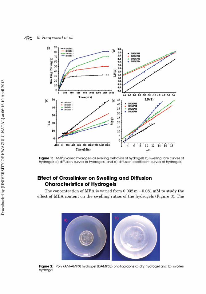
Effect of Crosslinker on Swelling and DiffusionCharacteristics of HydrogelsThe concentration of MBA is varied from 0.032 m �0.081 mM to study the
effect of MBA content on the swelling ratios of the hydrogels (Figure 3). The
Figure 2: Poly (AM-AMPS) hydrogel (DAMPS3) photographs a) dry hydrogel and b) swollenhydrogel.
Figure 1: AMPS varied hydrogels a) swelling behavior of hydrogels b) swelling rate curves ofhydrogels c) diffusion curves of hydrogels, and d) diffusion coefficient curves of hydrogels.
496 K. Varaprasad et al.
Dow
nloa
ded
by [
UN
IVE
RSI
TY
OF
KW
AZ
UL
U-N
AT
AL
] at
06:
16 1
0 A
pril
2013
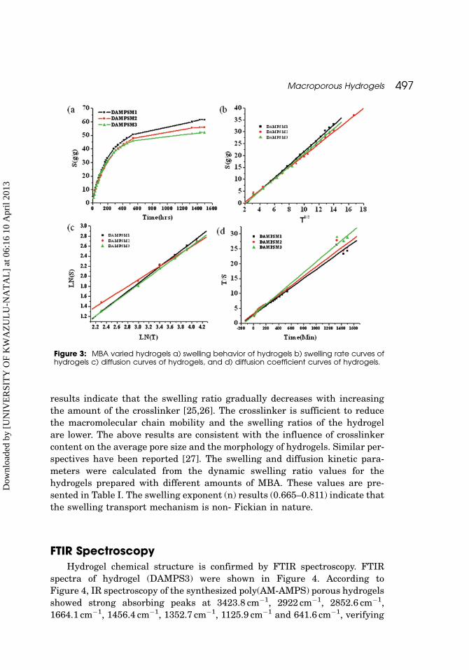
results indicate that the swelling ratio gradually decreases with increasing
the amount of the crosslinker [25,26]. The crosslinker is sufficient to reduce
the macromolecular chain mobility and the swelling ratios of the hydrogel
are lower. The above results are consistent with the influence of crosslinker
content on the average pore size and the morphology of hydrogels. Similar per-
spectives have been reported [27]. The swelling and diffusion kinetic para-
meters were calculated from the dynamic swelling ratio values for the
hydrogels prepared with different amounts of MBA. These values are pre-
sented in Table I. The swelling exponent (n) results (0.665–0.811) indicate that
the swelling transport mechanism is non- Fickian in nature.
FTIR SpectroscopyHydrogel chemical structure is confirmed by FTIR spectroscopy. FTIR
spectra of hydrogel (DAMPS3) were shown in Figure 4. According to
Figure 4, IR spectroscopy of the synthesized poly(AM-AMPS) porous hydrogels
showed strong absorbing peaks at 3423.8 cm�1, 2922 cm�1, 2852.6 cm�1,
1664.1 cm�1, 1456.4 cm�1, 1352.7 cm�1, 1125.9 cm�1 and 641.6 cm�1, verifying
Figure 3: MBA varied hydrogels a) swelling behavior of hydrogels b) swelling rate curves ofhydrogels c) diffusion curves of hydrogels, and d) diffusion coefficient curves of hydrogels.
Macroporous Hydrogels 497
Dow
nloa
ded
by [
UN
IVE
RSI
TY
OF
KW
AZ
UL
U-N
AT
AL
] at
06:
16 1
0 A
pril
2013
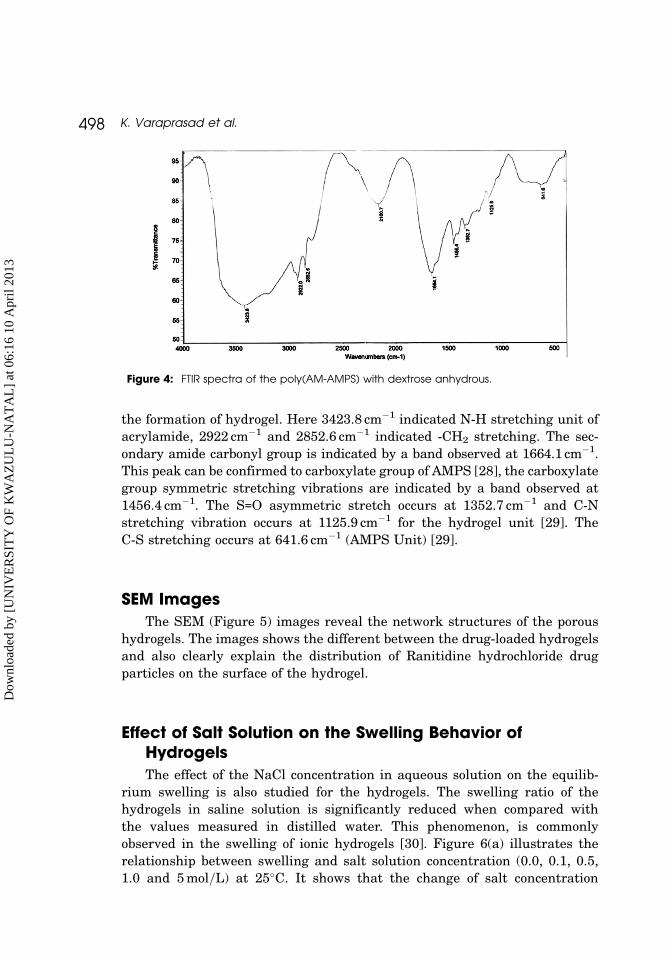
the formation of hydrogel. Here 3423.8 cm�1 indicated N-H stretching unit of
acrylamide, 2922 cm�1 and 2852.6 cm�1 indicated -CH2 stretching. The sec-
ondary amide carbonyl group is indicated by a band observed at 1664.1 cm�1.
This peak can be confirmed to carboxylate group of AMPS [28], the carboxylate
group symmetric stretching vibrations are indicated by a band observed at
1456.4 cm�1. The S=O asymmetric stretch occurs at 1352.7 cm�1 and C-N
stretching vibration occurs at 1125.9 cm�1 for the hydrogel unit [29]. The
C-S stretching occurs at 641.6 cm�1 (AMPS Unit) [29].
SEM ImagesThe SEM (Figure 5) images reveal the network structures of the porous
hydrogels. The images shows the different between the drug-loaded hydrogels
and also clearly explain the distribution of Ranitidine hydrochloride drug
particles on the surface of the hydrogel.
Effect of Salt Solution on the Swelling Behavior ofHydrogelsThe effect of the NaCl concentration in aqueous solution on the equilib-
rium swelling is also studied for the hydrogels. The swelling ratio of the
hydrogels in saline solution is significantly reduced when compared with
the values measured in distilled water. This phenomenon, is commonly
observed in the swelling of ionic hydrogels [30]. Figure 6(a) illustrates the
relationship between swelling and salt solution concentration (0.0, 0.1, 0.5,
1.0 and 5 mol=L) at 25�C. It shows that the change of salt concentration
Figure 4: FTIR spectra of the poly(AM-AMPS) with dextrose anhydrous.
498 K. Varaprasad et al.
Dow
nloa
ded
by [
UN
IVE
RSI
TY
OF
KW
AZ
UL
U-N
AT
AL
] at
06:
16 1
0 A
pril
2013

higher than about 1.0 mol=L has no appreciable influence on the swelling ratio
of the hydrogel.
Effect of Swelling Medium pHTo investigate the swelling behavior of the hydrogels at various pH solu-
tions, the hydrogels are swollen in different buffer solutions of pH 2, 3, 5, 7,
11 and 12 at 37�C. As shown in Figure 6(b) the swelling ratios of the hydrogels
decrease with increasing pH values. In all the hydrogels, maximum swelling is
Figure 5: SEM images of the hydrogels A) DAMPS4, B) DAMPS1 drug-loaded hydrogel,C) DAMPS4 drug-loaded hydrogel.
Macroporous Hydrogels 499
Dow
nloa
ded
by [
UN
IVE
RSI
TY
OF
KW
AZ
UL
U-N
AT
AL
] at
06:
16 1
0 A
pril
2013

observed at pH 2 and minimum at pH 12. The effect of pH on the swelling of
the AMPS hydrogels is explained on the basis of protonation of the amino
groups of AMPS. In the acidic medium, the protonation of the amino groups
leads to repulsion in the polymer chains, thus allowing more water in the
hydrogel network. At higher pH, deprotonation of the animo groups takes
place and repulsion in polymer chains is reduced.
In Vitro Release StudyPolyacrylamide-based hydrogels have received considerable attention
because of their use in many applications [31–33]. Among them porous hydro-
gels are most important in controlled-release applications, which received
greater attention in recent years as effective drug delivery devices in biomedi-
cal engineering [34]. Drug absorption from the gastrointestinal tract is a
complex procedure and is subject to many variables. In addition, porous hydro-
gels have also been developed for prolonging the residence time of delivery sys-
tems in the gastrointestinal tract. In this study, our intention is to develop
porosity in the copolymeric hydrogels of AM-AMPS by incorporating dextrose,
thereby the prolonged delivery of the drug can be achieved.
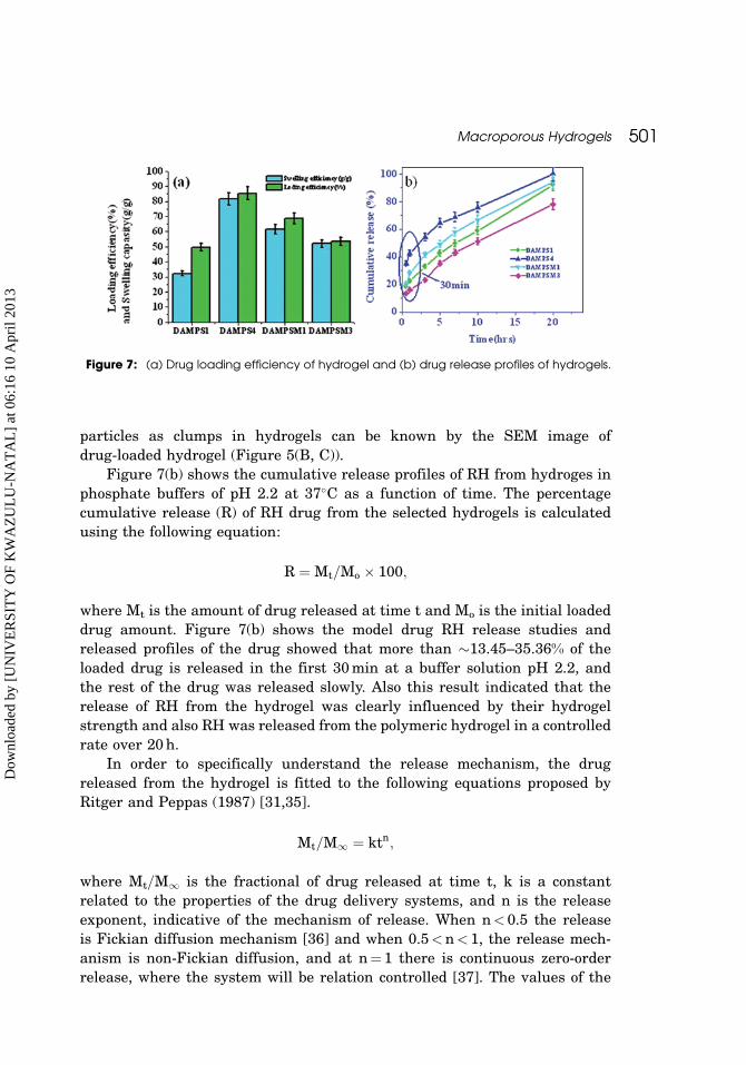
The loading efficiency of ranitidine hydrochloride (RH) is found to be high-
est in DAMPS4 (85.43), DAMPSM1 (68.7), DAMPSM4 (53.56) and DAMPS1
(49.69). This can be explained by the higher hydrophilic content of hydrogel
uptake with the higher amount of drug. This is due to the hydrophilic nature
(acid groups) of the AMPS monomer. But with MBA, varied hydrogels lower
the crosslink density of hydrogel uptake with a higher amount of drug. This
is due to the basis of the crosslink density of hydrogel (Figure 3).
However, drug-loading efficiency depends on the swelling behavior of
hydrogels [23]. It can be explained by Figure 7(a). The entrapments of drug
Figure 6: Swelling profiles of different hydrogels in (a) different concentrations of NaCl2solution, (b) different buffer solutions.
500 K. Varaprasad et al.
Dow
nloa
ded
by [
UN
IVE
RSI
TY
OF
KW
AZ
UL
U-N
AT
AL
] at
06:
16 1
0 A
pril
2013
particles as clumps in hydrogels can be known by the SEM image of
drug-loaded hydrogel (Figure 5(B, C)).
Figure 7(b) shows the cumulative release profiles of RH from hydroges in
phosphate buffers of pH 2.2 at 37�C as a function of time. The percentage
cumulative release (R) of RH drug from the selected hydrogels is calculated
using the following equation:
R ¼ Mt=Mo � 100;
where Mt is the amount of drug released at time t and Mo is the initial loaded
drug amount. Figure 7(b) shows the model drug RH release studies and
released profiles of the drug showed that more than �13.45–35.36% of the
loaded drug is released in the first 30 min at a buffer solution pH 2.2, and
the rest of the drug was released slowly. Also this result indicated that the
release of RH from the hydrogel was clearly influenced by their hydrogel
strength and also RH was released from the polymeric hydrogel in a controlled
rate over 20 h.
In order to specifically understand the release mechanism, the drug
released from the hydrogel is fitted to the following equations proposed by
Ritger and Peppas (1987) [31,35].
Mt=M1 ¼ ktn;
where Mt=M1 is the fractional of drug released at time t, k is a constant
related to the properties of the drug delivery systems, and n is the release
exponent, indicative of the mechanism of release. When n< 0.5 the release
is Fickian diffusion mechanism [36] and when 0.5<n< 1, the release mech-
anism is non-Fickian diffusion, and at n¼ 1 there is continuous zero-order
release, where the system will be relation controlled [37]. The values of the
Figure 7: (a) Drug loading efficiency of hydrogel and (b) drug release profiles of hydrogels.
Macroporous Hydrogels 501
Dow
nloa
ded
by [
UN
IVE
RSI
TY
OF
KW
AZ
UL
U-N
AT
AL
] at
06:
16 1
0 A
pril
2013
exponent of release n are calculated according to the above method. The
release rate characteristics of drug from hydrogels are presented in Table I.
Table values 0.27 to 0.48 clearly indicate that the hydrogels follows Fickan
diffusion-controlled release mechanism [35].
CONCLUSIONS
This study demonstrates the preparation of porous hydrogels in a free radical
polymerization process. The morphological and chemical structures of the
hydrogels were confirmed using SEM and FTIR analyses. The swelling
and deswelling kinetic parameters suggest that the hydrogels’ network is
non-Fickian in nature. The studies on the swelling ratio of hydrogels reveal
their sensitive response to environmental pH values change. The effects of dif-
ferent parameters on RH drug release behavior of hydrogels were studied. The
results of drug release kinetics for the hydrogels indicated a Fickian type
transport (at pH 2.2). All the results indicate that this hydrogel may serve
as a potential device for the delivery of drugs in which the primary target is
the gastrointestinal tract.
REFERENCES
[1] Murat Ozmen, M., Valentina Dinu, M., and Okay, O. Polym. Bull. 60, 169 (2008).
[2] Skelly, P., and Tighe, B. J. Polymer 20, 1051–1052 (1979).
[3] Haldon, R. A., and Lee, B. E. Br. Polym. J. 4491 (1972).
[4] Ashok, K., Pandey, M., Koshy, M. K., and Saraf, S. A. Inter J. Drug Delivery 2, 135(2010).
[5] Amin, S., Rajabnezhad, S., and Kohli, K. Scientific Research and Essay 3, 1175(2009).
[6] Okay, O. Prog. Polym. Sci. 25, 711 (2000).
[7] Sayil, C., and Okay, O. Polymer 42, 7639 (2001).
[8] Ozmen, M., Dinu, M., Valentina, M., Dragan, S., and Oguz Okay, E. J. Mac MolecSci. Part A: Pure and Appl. Chem. 44, 1195 (2007).
[9] Pradny, M., Lesny, P., Fiala, J., Vacik, J., Slouf, M., Micholck, T., and Sykova, E.Covect. Czech. Chem. Commun. 68, 812 (2003).
[10] Gonen Karakecıli, A., Satriano, C., Gumusderelıoglu, M., and Marletta, G. Radi-ation Physics and Chemistry 77, 154, (2008).
[11] Oxley, H., Corlchill, P. H., Fitton, J. H., and Tighe, B. J. Biomaterials. 14, 1064(1993).
[12] Mohan, Y. Murali, Keshava Murty, P. S., and Mohana Raju, K. J. Appl. Poly. Sci.101, 3202 (2006).
[13] Serizawa, T., Wakita, K., and Akashi, M. Macromolecules 35, 10 (2002).
502 K. Varaprasad et al.
Dow
nloa
ded
by [
UN
IVE
RSI
TY
OF
KW
AZ
UL
U-N
AT
AL
] at
06:
16 1
0 A
pril
2013

[14] Chern, J.-M., Lee, W.-F., and Hsieh, M.-Y. J. Appl. Polym. Sci., 92, 3651 (2004).
[15] Tokuyama, Hideki, Ishihara, Noriaki, and Sakohara, Shuji. Polym. Bull. 61, 399(2008).
[16] Yao, K. J., Zhou, W. J., Yao, K.-J., and Zhou, W.-J. J. Appl. Poly. Sci. 53, 153 (1994).
[17] Preniche, C., Cohen, M. E., Vazquez, B., and Romon, J. S. Polymer 38, 5977 (1997).
[18] Karadag, E., and Saraydin, D. Polym. Bull. 48, 299 (2002).
[19] Peppas, N. A., and Franson, N. M. J. Polym. Phys. Ed. 21, 983 (1983).
[20] Jabbari, E., and Nozari, S. Eur. Polym. Mater. 36, 2685 (2000).
[21] Varaprasad, K., Ravindra, S., Narayana Reddy, N., Vimala, K., and Mohana Raju,K. J. Appl. Polym. Sci. 116, 3593 (2010).
[22] Ende, M. T. A., and Peppas, N. A. J. Control Release 48, 47 (1997).
[23] Ravindra, S., Murali Mohan, Y., Varaprasad, K., Narayana Reddy, N., Vimala, K.,and Mohana Raju, K. J. Polymeric Materials 58, 278 (2009).
[24] Povrjavadi, A., Barzegar, Sh., and Zeidabadi, F. Polymer 67, 644 (2007).
[25] Peppas, L. B., and Harland, R. S. (1990). In Absorbent Polymer Technology,Elsevier: Amsterdam, p. 45.
[26] Peppas, N. A., and A. G. (1986). In Hydrogels in Medicine and Pharmacy, CRCPress: Boca Raton, Vol. 1, p. 1.
[27] Liu, C., Chen, Y., and Chen, J. Carbohydrate Poly 79, 500 (2010).
[28] Pourjavadi, A., Hosseinzadeh, H., and Mazidi, R. J. Appl. Polym. Sci. 98, 255(2005).
[29] Chi Zhang, Allan, and Easteal, J. J. Appl. Polym. Sci. 104, 1723 (2007).
[30] Flory, P. J. (1953). Principles of Polymer Chemistry, Cornell University, New York.
[31] Erdener Karadag, Semiha Kundakci, and Omer Baris, Uzum. Polym-Plasti.Techn. & Engi. 48, 1217 (2009).
[32] Bajpai, A. K., and Bhanu, S. Int. J. Polym. Mater. 53, 319 (2004).
[33] Bars, O., Uzum, and Karadag, E. Polym-Plasti. Techn. & Engi. 49, 609 (2010).
[34] Demirel, G. B., Caykara, T., Demiray, M., and Guru, M. J. Macro. Sci. R, Part A:Pure and Applied Chemistry 46, 58 (2009).
[35] Ritger, P. L., and Peppas, N. A. J. Control Release 5, 37 (1987).
[36] Lynch, I., and Dawson, K. A. J. Physical Chem. B 108, 10893 (2004).
[37] Above Mullarney, M. P., Seery, T. A. P., and Weiss, R. A. Polymer 47, 3845 (2006).
Macroporous Hydrogels 503
Dow
nloa
ded
by [
UN
IVE
RSI
TY
OF
KW
AZ
UL
U-N
AT
AL
] at
06:
16 1
0 A
pril
2013