Prophylactic and therapeutic effects of taurine against aluminum-induced acute hepatotoxicity in...
8
This article appeared in a journal published by Elsevier. The attached copy is furnished to the author for internal non-commercial research and education use, including for instruction at the authors institution and sharing with colleagues. Other uses, including reproduction and distribution, or selling or licensing copies, or posting to personal, institutional or third party websites are prohibited. In most cases authors are permitted to post their version of the article (e.g. in Word or Tex form) to their personal website or institutional repository. Authors requiring further information regarding Elsevier’s archiving and manuscript policies are encouraged to visit: http://www.elsevier.com/copyright
-
Upload
independent -
Category
Documents
-
view
0 -
download
0
Transcript of Prophylactic and therapeutic effects of taurine against aluminum-induced acute hepatotoxicity in...
This article appeared in a journal published by Elsevier. The attachedcopy is furnished to the author for internal non-commercial researchand education use, including for instruction at the authors institution
and sharing with colleagues.
Other uses, including reproduction and distribution, or selling orlicensing copies, or posting to personal, institutional or third party
websites are prohibited.
In most cases authors are permitted to post their version of thearticle (e.g. in Word or Tex form) to their personal website orinstitutional repository. Authors requiring further information
regarding Elsevier’s archiving and manuscript policies areencouraged to visit:
http://www.elsevier.com/copyright
Author's personal copy
Journal of Hazardous Materials 192 (2011) 880– 886
Contents lists available at ScienceDirect
Journal of Hazardous Materials
j our na l ho me p age: www.elsev ier .com/ locate / jhazmat
Prophylactic and therapeutic effects of taurine against aluminum-induced acutehepatotoxicity in mice
Wael M. El-Sayeda,b,∗, Mohamed A. Al-Kahtania, Ashraf M. Abdel-Moneima,c
a King Faisal University, Faculty of Science, Department of Biological Sciences, Al-Hufof 31982, Ahsaa, Saudi Arabiab University of Ain Shams, Faculty of Science, Department of Zoology, Abbassia 11566, Cairo, Egyptc Alexandria University, Faculty of Science, Department of Zoology, Alexandria 21511, Egypt
a r t i c l e i n f o
Article history:Received 5 November 2010Received in revised form 31 May 2011Accepted 31 May 2011Available online 6 June 2011
Keywords:AluminumHepatotoxicityTaurineGlutathione systemAntioxidant and cytoprotective enzymes
a b s t r a c t
Aluminum is a well known neurotoxin and a possible candidate of hepatotoxins to humans. Using naturalantioxidants against metal-induced hepatotoxicity is a modern approach. In the present study, Aluminum(AlCl3) intoxication (a single injection of 25 mg Al3+/kg, i.p.) for 24 h in mice resulted in elevations in serumalanine aminotransferase activity and serum tumor necrosis factor and hepatic malondialdehyde levels.Aluminum reduced the activities of glutathione peroxidase, glutathione S-transferase, quinone oxidore-ductase, and catalase in liver. In addition, Al caused hepatic hemorrhage, cellular degeneration as well asnecrosis of hepatocytes. Ultrastructure examination showed swelling of mitochondria, derangement ofrough endoplasmic reticulum cisternae and pleomorphic nuclei with abnormal chromatin distribution.Taurine, a sulfur-containing amino acid was administered to mice daily for 5 days before (at 100 mg/kg,i.p.) or 2 h after (a single dose of 1 g/kg, i.p.) aluminum administration. Treating mice with taurine at eitherdosing regimens, pre- or post-aluminum administration alleviated aluminum oxidative damaging effects.The rate of recovery was better when taurine was administered prior to Al. Taurine had anaphylactic andtherapeutic activity against hepatotoxicity induced by aluminum in mice.
© 2011 Elsevier B.V. All rights reserved.
1. Introduction
Aluminum (Al) is a toxic element to humans and animals.Human exposure to aluminum seems inevitable. In addition toenvironmental exposure from drinking water and cooking uten-sils, aluminum is a component of food additives, antacids, and renaldialysate [1]. Aluminum is a known neurotoxin associated withmany nervous disorders such as encephalopathy and Alzheimer’sdisease [2]. The toxicity and oxidative stress caused by aluminumis well investigated in nervous tissues [3]. On the other hand,very little is known about the hepatotoxic effects of aluminum.The mechanism of Al-toxicity is poorly understood but it is sug-gested that aluminum generates reactive oxygen species (ROS) thatcause lipid peroxidation (LPO) and oxidative damage to proteinsand DNA [4]. Therefore, chelating agents and natural antioxidantswhich alleviate the oxidative stress or induce the cellular antiox-idant milieu would most probably treat and/or protect againstaluminum poisoning. Taurine (2-aminoethanesulphonic acid), is asulfur-containing �-amino acid present in mammalian tissues [5].
∗ Corresponding author at: University of Ain Shams, Faculty of Science, Depart-ment of Zoology, Abbassia 11566, Cairo, Egypt. Tel.: +20 2 2482 1633;fax: +20 2 2684 2123.
E-mail address: [email protected] (W.M. El-Sayed).
Taurine is known to have antioxidant properties [6], stabilize thecell membranes and reduce LPO [7], and display scavenging activ-ity against free radicals [8]. The chemical similarity of taurine toacetylcysteine is an encouraging factor to use taurine against metal-induced hepatotoxicity. Taurine has reduced the toxic effects ofcopper, cadmium, and lead in rats [9–11] and chromium in mice[12]. Taurine has also been shown to protect against hepatotoxicityinduced by either tamoxifen [13] or acetaminophen [14].
In the present study, we investigated the protective and/ortherapeutic effects of taurine against aluminum-induced hepato-toxicity in mice. The mechanism of action of taurine is vague, butinduction of cellular antioxidants and inhibition of LPO in mice isa suggested way of action of this natural antioxidant. Therefore,a study was undertaken to evaluate the effects of this amino acidat two dosing regimens on the hepatic antioxidant and chemopro-tective enzymes as well as on the histopathology of murine liverchallenged with Al-intoxication.
2. Methods and materials
2.1. Animal treatment and biological sample preparation
Adult male CF-1 mice (25–35 g) were obtained from Faculty ofVeterinary Medicine, King Faisal University, and were maintainedin a humidity- and temperature-controlled environment on a
0304-3894/$ – see front matter © 2011 Elsevier B.V. All rights reserved.doi:10.1016/j.jhazmat.2011.05.100
Author's personal copy
W.M. El-Sayed et al. / Journal of Hazardous Materials 192 (2011) 880– 886 881
Table 1Effect of AlCl3 and taurine on alanine aminotransferase (sALT), malondialdehyde (MDA), and tumor necrosis factor-alpha (TNF-�).
Treatment Dose/kg sALT activitya (mU/ml serum) MDAa (nmol/mg protein) TNF-�a (pmol/ml serum)
Control – 76.6 ± 12.2 0.31 ± 0.03 30.6 ± 2.3AlCl3 25 mg Al3+ × 1 273.0 ± 18.8* 1.03 ± 0.08* 217.3 ± 16.3*
AlCl3/taurine 25 mg Al3+ × 1/1 g × 1 173.2 ± 32.1+ 0.47 ± 0.05+ 143.7 ± 10.4+
Taurine 1 g × 1 58.6 ± 9.2 0.33 ± 0.01 28.1 ± 2.5Taurine/AlCl3 100 mg × 5/25 mg Al3+ × 1 145.2 ± 8.8+ 0.48 ± 0.04+ 174.9 ± 11.6+
Taurine 100 mg × 5 58.3 ± 4.1 0.32 ± 0.03 32.2 ± 3.1
a Data are expressed as mean ± SEM (n = 5).* Indicates a significant difference (P < 0.05) from naïve.+ Indicates a significant difference (P < 0.05) from AlCl3-treated animals.
12-h light/dark cycle with continuous free access to food and water.The mice were divided into 6 groups, 5 mice each. The first group(naïve) was injected with isotonic saline. Aluminum chloride wasintraperitoneally (i.p.) injected into mice in groups 2 and 3 at a sin-gle dose of 25 mg Al3+/kg [15]. After 2 h, group 3 was further treatedwith taurine (i.p.) at a single dose of 1 g/kg [12]. Group 4 was onlytreated with a single injection of taurine (1 g/kg). Mice in groups 5and 6 were intraperitoneally treated with taurine 100 mg/kg dailyfor 5 days [16]. One day later after the last dose of taurine, group 5was injected with a single dose of 25 mg Al3+/kg. All animal pro-cedures were approved by the University of King Faisal AnimalCare and Use Committee and were conducted in agreement withNIH guidelines for the humane care of laboratory animals. Animalswere sacrificed 24 h after the final dose; blood was immediately col-lected for serum preparation and frozen at −80 ◦C. The livers werequickly perfused in situ (via the hepatic portal vein) with normalice cold saline. The gall bladder was then carefully dissected away,and the remaining liver was homogenized in ice cold buffer andsubjected to centrifugation (9000 × g for 15 min), and the super-natant was collected and stored at −80 ◦C until assayed for enzymeactivity. Protein content was immediately determined with Folin-Ciocalteu’s phenol reagent (Sigma; St. Louis, MO) according toLowry et al. [17].
2.2. Enzyme activity and biochemical analyses
Serum alanine aminotransferase (sALT) activity was deter-mined from the serum-dependent absorbance change of NADHoxidation at 340 nm in the presence of optimized concentrationsof l-alanine, �-ketoglutarate and purified lactic dehydrogenaseenzyme [18]. Malondialdehyde (MDA) was measured through esti-mation of a stable chromophore formed from the reaction ofa chromogenic reagent, N-methyl-2-phenylindole, with MDA at45 ◦C [19]. Tumor necrosis factor (TNF-�) level in serum wasmeasured by ELISA method using Bender Medsystems (Vienna,Austria) kit as described by the manufacturer. Glutathione perox-idase activity was determined from the azide insensitive rate ofoxidation of NADPH in the presence of hydrogen peroxide, glu-tathione, and glutathione reductase [20]. Glutathione S-transferaseactivity was determined from the conjugation of glutathione with1-chloro-2,4-dinitrobenzene [CDNB] detected by the change inabsorbance at 340 nm [21]. Through monitoring the inhibition rateof NADH-dependent reduction of 2,6-dichlorophenolindophenolby 3,3′-methylenebis[4-hydroxycoumarin], the activity of quinoneoxidoreductase was determined [22]. Catalase activity was mea-sured from the rate of dismutation of hydrogen peroxide (H2O2) towater and molecular oxygen in a two-step coupling reaction [23].Superoxide dismutase activity was measured in a coupling reactionthrough formation of formazan dye from tetrazolium salt by super-oxide radicals generated by xanthine oxidase and hypoxanthine[24]. Glutathione reductase activity was measured from the rateof reduction of oxidized glutathione by NADPH [25]. Glutathione
(GSH) reacts with 5,5′-dithiobis (2-nitrobenzoic acid) producingthe disulphide form or oxidized glutathione (GSSG) and 2-nitro-5-thiobenzoic acid (TNB) and under standardized assay conditions,the rate of formation of TNB was monitored and GSH level wasdetermined [26]. Any elevation in the activities of enzymes in thecurrent investigation does not distinguish between an activation ofexisting enzyme and an increase arising from increased amountsof enzyme protein.
2.3. Histopathological studies
For the histological examination, pieces of the liver were fixedin 10% neutral buffered formalin (pH 7.2), dehydrated in ascendingseries of ethyl alcohol (70–100%), cleared in xylene, and embeddedin paraffin wax. Paraffin sections of 5 �m in thickness were stainedwith hematoxylin and eosin (H&E).
2.4. Transmission electron microscopy studies
For electron microscopic examinations of liver tissues, primaryfixation was performed in 3% glutaraldehyde in sodium phosphatebuffer (200 mM, pH 7.2) for 3 h at 4 ◦C. Liver tissues were washedwith the same buffer and postfixed in 1% osmium tetroxide (AgarSci. Ltd., England) in sodium phosphate buffer, pH 7.2, for 1 h at4 ◦C. Tissue samples were washed with the same buffer for 3 h at4 ◦C and then embedded in Araldite (Agar Sci. Ltd., England). Thinsections were cut with Leica EM UC6 (Leica Co., Austria) ultrami-crotome. Samples were stained with 2% uranyl acetate and leadcitrate. The sections were viewed and photographed on Jeol JEM1011 transmission electron microscope (Jeol Ltd., Japan) at 80 kV.
2.5. Statistical analysis of data
Results are expressed as the mean ± SEM. Treated group sizewas 5 animals. Statistical analyses were performed using ANOVA,followed by Fisher’s protected least significant difference multiplerange test. The data were compared against those from the propercontrol animals. Differences were considered significant at P < 0.05.
3. Results
3.1. Antioxidants and biochemical study
The treatment of mice with AlCl3 increased serum alanineaminotransferase (sALT) activity compared to naïve animals(Table 1). Administration of taurine before or following AlCl3caused significant reductions in sALT activity when compared toAlCl3-treated mice. Treating mice with either doses of taurinealone (100 mg for 5 days or a single dose of 1 g) caused insignifi-cant decreases in sALT activity when compared with naïve animals(Table 1). The same scenario was repeated for hepatic malondi-aldehyde (MDA) and serum tumor necrosis factor (TNF-�), where
Author's personal copy
882 W.M. El-Sayed et al. / Journal of Hazardous Materials 192 (2011) 880– 886
Table 2Effect of AlCl3 and taurine on activities of some hepatic antioxidant and chemoprotective enzymes.
Treatment Dose/kg GPx activitya,b
(nmol/mg/min)GST activitya,b
(nmol/mg/min)(NQO) activitya,b
(nmol/mg/min)Catalase activitya
(mU/mg)
Control – 1397 ± 17 2722 ± 172 39.2 ± 1.5 722 ± 130AlCl3 25 mg Al3+ × 1 286 ± 24* 1191 ± 128* 18.4 ± 1.8* 375 ± 20*
AlCl3/taurine 25 mg Al3+ × 1/1 g × 1 952 ± 92+ 3113 ± 166+ 41.5 ± 2.2+ 720 ± 31+
Taurine 1 g × 1 1302 ± 103 3108 ± 232 46.0 ± 1.5 631 ± 60Taurine/AlCl3 100 mg × 5/25 mg Al3+ × 1 1419 ± 134+ 3037 ± 239+ 37.1 ± 1.8+ 685 ± 59+
Taurine 100 mg × 5 1194 ± 86 2763 ± 169 38.8 ± 3.2 660 ± 80
a Data are expressed as mean ± SEM (n = 5).b Abbreviations: GPx (glutathione peroxidase), GST (glutathione S-transferase), NQO (NAD(P)H-quinone oxidoreductase).* Indicates a significant difference (P < 0.05) from naïve.+ Indicates a significant difference (P < 0.05) from AlCl3-treated animals.
Table 3Effect of AlCl3 and taurine on activities of glutathione reductase (GR) and superoxide dismutase (SOD) in liver.
Treatment Dose/kg GR activitya (nmol/mg/min) SOD activitya (mU/mg)
Control – 1668 ± 37 1.42 ± 0.27AlCl3 25 mg Al3+ × 1 1589 ± 31 1.46 ± 0.16AlCl3/taurine 25 mg Al3+ × 1/1 g × 1 1826 ± 90 1.05 ± 0.09Taurine 1 g × 1 2195 ± 209 0.98 ± 0.07Taurine/AlCl3 100 mg × 5/25 mg Al3+ × 1 2279 ± 376 1.10 ± 0.05Taurine 100 mg × 5 2072 ± 195 1.07 ± 0.04
a Mean ± SEM, n = 5.
taurine at both dosing regimens had no effect. Aluminum intoxi-cation resulted in significant elevations in both hepatic MDA andserum TNF-� levels compared to naïve animals (Table 1). Taurinesignificantly reduced MDA and TNF-� levels in intoxicated ani-mals at both dosing regimens used; pre- and post-AlCl3 treatment(Table 1).
Treating mice with AlCl3 resulted in significant reductionsin the activities of glutathione peroxidase (GPx), glutathione S-transferase (GST), NADPH-quinone oxidoreductase (NQO), andcatalase in liver compared to naïve animals (Table 2). Treatmentwith taurine either following or after treatment with AlCl3 causedsignificant elevations in the activities of all enzymes listed abovewhen compared to AlCl3-intoxicated animals (Table 2). Taurinealone had no significant effect on the activities of previously men-tioned enzymes at both dosing regimens studied (Table 2). NeitherAlCl3 nor taurine at any treatment caused a significant change inthe activities of glutathione reductase (GR) or superoxide dismu-tase (SOD) in liver. The elevations seen in GR activity after taurineadministration either alone or in conjunction with AlCl3 did notachieve a statistical significance (Table 3). The reduction in hepaticreduced glutathione (GSH) level caused by AlCl3 did not achievea statistical significance either (P ∼ 0.07). Taurine at both regimensstudied increased GSH levels in AlCl3-insulted animals compared toeither naïve or AlCl3-treated animals (Table 4). Hepatic GSH contentwas significantly elevated in animals treated with taurine aloneat either dose regimens investigated when compared with naïveanimals (Table 4).
Table 4Effect of AlCl3 and taurine on hepatic glutathione (GSH) level.
Treatment Dose/kg GSHa (�mol/mg)
Control – 4.23 ± 0.09AlCl3 25 mg Al3+ × 1 3.10 ± 0.04AlCl3/taurine 25 mg Al3+ × 1/1 g × 1 6.48 ± 0.40*,+
Taurine 1 g × 1 6.02 ± 0.15*
Taurine/AlCl3 100 mg × 5/25 mg Al3+ × 1 7.12 ± 0.28*,+
Taurine 100 mg × 5 6.18 ± 0.29*
a Mean ± SEM, n = 5.* Indicates a significant difference (P < 0.05) from naïve.+ Indicates a significant difference (P < 0.05) from AlCl3-treated animals.
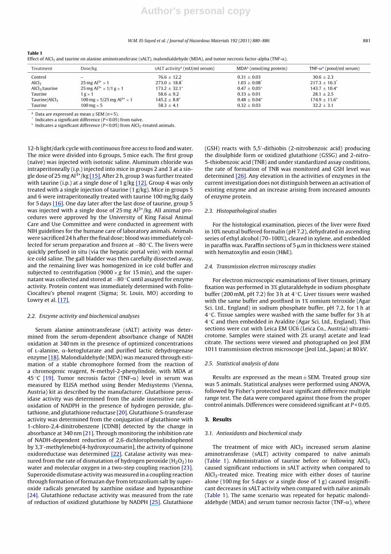
3.2. Histopathology
The histological profile of the normal liver sections showednormal hepatic cells with well preserved cytoplasm, prominentnucleus, nucleolus, central vein and compact arrangement of hep-atocytes (Fig. 1A). The liver tissues from mice that received AlCl3revealed extensive injuries, characterized by a loss of normal archi-tecture of the parenchymatous tissue, congested sinusoids andblood vessels, infiltration of inflammatory cells, cellular degenera-tion with nuclear pyknosis and presence of necrotic areas (Fig. 1B).Mice post-treated with taurine presented similar histopathologicalchanges compared with Al-treated mice with attenuated severity(Fig. 1C). In mice treated with taurine prior to Al, hemorrhage andinflammation were less in comparison to Al-treated group (Fig. 1D).Cellular degeneration and necrosis were rare. No histopathologicalalteration was observed in liver of mice from groups treated onlywith taurine.
3.3. Electron microscopy
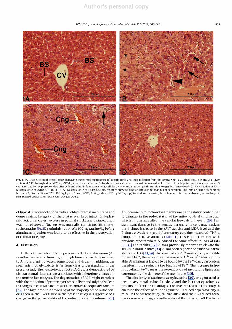
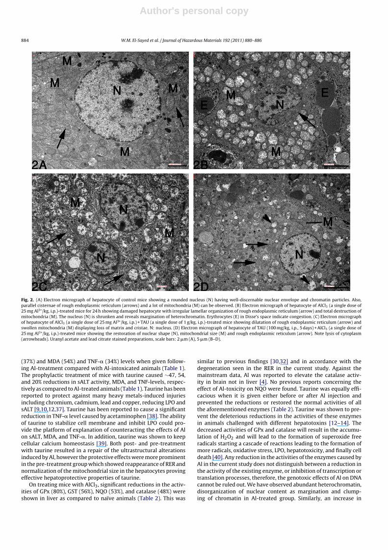
Control group had a round nucleus with a regular nuclear enve-lope and a very distinct nucleolus. Mitochondria and endoplasmicreticulum were abundant in the hepatocytes (Fig. 2A). Examina-tion of liver from Al-treated mice under transmission electronmicroscope (Fig. 2B) revealed a reduction in the amount of roughendoplasmic reticulum (RER) in the hepatocytes. RER was disor-ganized into isolated cisternae that were often closely associatedwith mitochondria surrounding it. Additional smooth endoplasmicreticulum appeared in the cytoplasm of hepatocytes in Al-treatedmice. A swollen appearance of the mitochondria was revealed in theliver along with breaking up of the mitochondrial cristae. Nucleusdisplayed an irregular outline and condensed heterochromatin waslocated on the margin of the nuclear envelope. In animals post-treated with taurine, deformation was occasionally seen in theshape of nucleus and there were some heterochromatin fields onthe margin of the nuclear envelope. RER cisternae were partiallydilated and disintegrated and mitochondria were swollen (Fig. 2C).Membranous structures of the hepatocytes were found to be bet-ter preserved in animals pre-treated with taurine. Most of themitochondria were characterized by the same basic architecture
Author's personal copy
W.M. El-Sayed et al. / Journal of Hazardous Materials 192 (2011) 880– 886 883
Fig. 1. (A) Liver section of control mice displaying the normal architecture of hepatic cords and their radiation from the central vein (CV), blood sinusoids (BS). (B) Liversection of AlCl3 (a single dose of 25 mg Al3+/kg, i.p.)-treated mice for 24 h exhibits marked disturbances of the normal architecture of the hepatic tissues, necrotic areas (*)characterized by the presence of Kupffer cells and other inflammatory cells, cellular degeneration (arrows) and sinusoidal congestion (arrowhead). (C) Liver section of AlCl3(a single dose of 25 mg Al3+/kg, i.p.) + TAU (a single dose of 1 g/kg, i.p.)-treated mice showing dilation and distinct features of congestion (Cng) and cellular degeneration(arrow). (D) Liver section of TAU (100 mg/kg, i.p., 5 days) + AlCl3 (a single dose of 25 mg Al3+/kg, i.p.)-treated mice showing the cellular architecture with nearly normal aspect.H&E stained preparations, scale bars: 200 �m (A–D).
of typical liver mitochondria with a folded internal membrane anddense matrix. Integrity of the cristae was kept intact. Endoplas-mic reticulum cisternae were in parallel stacks and disintegrationwas not observed. Nucleus was normally containing little hete-rochromatin (Fig. 2D). Administration of a 100 mg taurine/kg beforealuminum injection was found to be effective in the preservationof cellular integrity.
4. Discussion
Little is known about the hepatotoxic effects of aluminum (Al)in either animals or humans, although humans are daily exposedto Al from drinking water, some foods and drugs. In addition, themechanism of Al-toxicity is far from clear understanding. In thepresent study, the hepatotoxic effect of AlCl3 was demonstrated byultrastructural observations associated with deleterious changes inthe murine hepatocytes. The degeneration of RER might correlatewith the reduction of protein synthesis in liver and might also leadto changes in cellular calcium as RER is known to sequester calcium[27]. The high-amplitude swelling of the majority of the mitochon-dria seen in the liver tissue in the present study is suggestive of achange in the permeability of the mitochondrial membrane [28].
An increase in mitochondrial membrane permeability contributesto changes in the redox status of the mitochondrial thiol groupswhich in turn may affect the cellular free calcium levels [29]. Thissignificant damage to the hepatic parenchyma cells may explainthe 4-times increase in the sALT activity and MDA level and the7-times elevation in pro-inflammatory cytokine measured; TNF-�compared to naïve animals (Table 1). This is in accordance withprevious reports where Al caused the same effects in liver of rats[30,31] and rabbits [32]. Al was previously reported to elevate theTNF-� in brain in mice [33]. Al has been reported to cause oxidativestress and LPO [31,34]. The ionic radii of Al3+ most closely resemblethose of Fe3+, therefore the appearance of Al3+ in Fe3+ sites is prob-able. Aluminum is known to be bound by the Fe3+-carrying proteintransferrin thus reducing the binding of Fe2+. The increase in freeintracellular Fe2+ causes the peroxidation of membrane lipids andconsequently the damage of the membrane [35].
The similarity of taurine to acetylcysteine [36], an agent used totreat heavy metal-induced toxicity, and the fact that cysteine is aprecursor of taurine encouraged the research team in this study toexamine the effects of taurine against Al-induced hepatotoxicity inmice. In the present study, taurine alleviated the Al-induced acuteliver damage and significantly reduced the elevated sALT activity
Author's personal copy
884 W.M. El-Sayed et al. / Journal of Hazardous Materials 192 (2011) 880– 886
Fig. 2. (A) Electron micrograph of hepatocyte of control mice showing a rounded nucleus (N) having well-discernable nuclear envelope and chromatin particles. Also,parallel cisternae of rough endoplasmic reticulum (arrows) and a lot of mitochondria (M) can be observed. (B) Electron micrograph of hepatocyte of AlCl3 (a single dose of25 mg Al3+/kg, i.p.)-treated mice for 24 h showing damaged hepatocyte with irregular lamellar organization of rough endoplasmic reticulum (arrow) and total destruction ofmitochondria (M). The nucleus (N) is shrunken and reveals margination of heterochromatin. Erythrocytes (E) in Disse’s space indicate congestion. (C) Electron micrographof hepatocyte of AlCl3 (a single dose of 25 mg Al3+/kg, i.p.) + TAU (a single dose of 1 g/kg, i.p.)-treated mice showing dilatation of rough endoplasmic reticulum (arrow) andswollen mitochondria (M) displaying loss of matrix and cristae. N: nucleus. (D) Electron micrograph of hepatocyte of TAU (100 mg/kg, i.p., 5 days) + AlCl3 (a single dose of25 mg Al3+/kg, i.p.)-treated mice showing the restoration of nuclear shape (N), mitochondrial size (M) and rough endoplasmic reticulum (arrow). Note lysis of cytoplasm(arrowheads). Uranyl acetate and lead citrate stained preparations, scale bars: 2 �m (A), 5 �m (B–D).
(37%) and MDA (54%) and TNF-� (34%) levels when given follow-ing Al-treatment compared with Al-intoxicated animals (Table 1).The prophylactic treatment of mice with taurine caused ∼47, 54,and 20% reductions in sALT activity, MDA, and TNF-levels, respec-tively as compared to Al-treated animals (Table 1). Taurine has beenreported to protect against many heavy metals-induced injuriesincluding chromium, cadmium, lead and copper, reducing LPO andsALT [9,10,12,37]. Taurine has been reported to cause a significantreduction in TNF-� level caused by acetaminophen [38]. The abilityof taurine to stabilize cell membrane and inhibit LPO could pro-vide the platform of explanation of counteracting the effects of Alon sALT, MDA, and TNF-�. In addition, taurine was shown to keepcellular calcium homeostasis [39]. Both post- and pre-treatmentwith taurine resulted in a repair of the ultrastructural alterationsinduced by Al, however the protective effects were more prominentin the pre-treatment group which showed reappearance of RER andnormalization of the mitochondrial size in the hepatocytes provingeffective hepatoprotective properties of taurine.
On treating mice with AlCl3, significant reductions in the activ-ities of GPx (80%), GST (56%), NQO (53%), and catalase (48%) wereshown in liver as compared to naïve animals (Table 2). This was
similar to previous findings [30,32] and in accordance with thedegeneration seen in the RER in the current study. Against themainstream data, Al was reported to elevate the catalase activ-ity in brain not in liver [4]. No previous reports concerning theeffect of Al-toxicity on NQO were found. Taurine was equally effi-cacious when it is given either before or after Al injection andprevented the reductions or restored the normal activities of allthe aforementioned enzymes (Table 2). Taurine was shown to pre-vent the deleterious reductions in the activities of these enzymesin animals challenged with different hepatotoxins [12–14]. Thedecreased activities of GPx and catalase will result in the accumu-lation of H2O2 and will lead to the formation of superoxide freeradicals starting a cascade of reactions leading to the formation ofmore radicals, oxidative stress, LPO, hepatotoxicity, and finally celldeath [40]. Any reduction in the activities of the enzymes caused byAl in the current study does not distinguish between a reduction inthe activity of the existing enzyme, or inhibition of transcription ortranslation processes, therefore, the genotoxic effects of Al on DNAcannot be ruled out. We have observed abundant heterochromatin,disorganization of nuclear content as margination and clump-ing of chromatin in Al-treated group. Similarly, an increase in
Author's personal copy
W.M. El-Sayed et al. / Journal of Hazardous Materials 192 (2011) 880– 886 885
chromatin condensation with discontinuity in nuclear membranein both the cerebrum and cerebellum of rats orally treated withAlCl3 was revealed [41]. At a molecular level, any effects of Alwould be most likely to involve DNA damage. Al does have agenotoxic profile and has been shown to bind to DNA [42]. Aftertreating animals with taurine at either dosing regimens investi-gated, severely degenerated nuclei were rarely detected. Nuclearcontent was almost normal in appearance and organization withtaurine treatment, which was previously reported to prevent DNAdamage [43].
Neither SOD nor GR activity in liver was affected by any treat-ment (Table 3), although a previous report showed that Al reducedboth enzyme activities in liver but after 70 days of treatment [31].On contrary, it was reported that Al-intoxication resulted in eleva-tions in the activities of SOD and GR in rat but in brain [4]. Similarto our findings, taurine has been shown to exert no effect on SOD[44].
Hepatic GSH level was not significantly affected by Al treatment.Some reports have been shown that Al caused a reduction in thelevel of GSH [30,32], yet it has been showed that Al-intoxication hadno effect on hepatic GSH [45]. On the other hand, treatment of nor-mal or Al-intoxicated animals with taurine resulted in significantelevations in hepatic GSH levels (Table 4) in accordance with a pre-vious report [13]. GSH was the only parameter studied affected bytaurine in normal mice. The thiol group of GSH could be used in neu-tralizing the toxic products produced by Al-intoxication. Taurinewas suggested to stimulate the nitrosylation of GSH into nitrosog-lutathione [46]. The latter is much potent antioxidant than GSHitself. Increased hepatic GSH level, and GPx and catalase activitieswere suggested to be attributed to the role of taurine in maintainingnormal insulin-like growth factor 1 (IGF-I) level [47].
Taurine (2-aminoethanesulfonic acid) is a unique amino acidthat has amino group and sulfonate group. Both groups can bindheavy metals and stimulate excretion of such hazardous met-als [10]. Is taurine alleviating Al-induced hepatotoxicity throughreducing the bioavailability of Al? This question needs furtherinvestigation. Taurine not only can act as a direct antioxidant byscavenging free radicals and inhibiting LPO but, it can also stimulatethe activity of glutathione-metabolizing and other cytoprotectiveenzymes when the cell is exposed to stressful conditions. The toxiceffects of Al may be mitigated by taurine through improving thecellular antioxidant defense system, stabilization of cell membrane,and prevention of LPO. Taurine also seems to have direct beneficialeffects on liver parenchymal cells, therefore taurine supplementa-tion may be helpful in abrogation of Al-hepatotoxicity.
References
[1] J.L. Greger, C.F. Powers, Assessment of exposure to parenteral and oral alu-minum with and without citrate using a desferrioxamine test in rats, Toxicology76 (1992) 119–132.
[2] M. Kawahara, Effects of aluminum on the nervous system and its possible linkwith neurodegenerative diseases, J. Alzheimers Dis. 8 (2005) 171–182.
[3] P. Bhalla, D.K. Dhawan, Protective role of lithium in ameliorating thealuminium-induced oxidative stress and histological changes in rat brain, Cell.Mol. Neurobiol. 29 (2009) 513–521.
[4] M.J. Strong, R.M. Garruto, J.G. Joshi, W.R. Mundy, T.J. Shafer, Can the mechanismsof aluminum neurotoxicity be integrated into a unified scheme? J. Toxicol.Environ. Health 48 (1996) 599–613.
[5] R.W. Chesney, Taurine: its biological role and clinical implications, Adv. Pediatr.32 (1985) 1–42.
[6] J. Hayes, K.F. Tipton, L. Bianchi, L.D. Corte, Complexities in the neurotoxic actionsof 6-hydroxydopamine in relation to the cytoprotective properties of taurine,Brain Res. Bull. 55 (2001) 239–245.
[7] R.J. Huxtable, Physiological actions of taurine, Physiol. Rev. 72 (1992) 101–163.[8] L. Nittynen, M.L. Nurminen, R. Korpela, H. Vapaatalo, Role of arginine, taurine
and homocysteine in cardiovascular diseases, Ann. Med. 31 (1999) 318–326.[9] D.F. Hwang, L.C. Wang, H.M. Cheng, Effect of taurine on toxicity of copper in
rats, Food Chem. Toxicol. 36 (1998) 239–244.[10] D.F. Hwang, L.C. Wang, Effect of taurine on toxicity of cadmium in rats, Toxi-
cology 167 (2001) 173–180.
[11] L. Patrick, Lead toxicity part II: the role of free radical damage and the use ofantioxidants in the pathology and treatment of lead toxicity, Altern. Med. Rev.11 (2006) 114–127.
[12] I.I. Bos gelmez, T. Söylemezoglu, G. Güvendik, The protective and antidotaleffects of taurine on hexavalent chromium-induced oxidative stress in miceliver tissue, Biol. Trace Elem. Res. 125 (2008) 46–58.
[13] H. Tabassum, H. Rehman, B.D. Banerjee, S. Raisuddin, S. Parvez, Attenuationof tamoxifen-induced hepatotoxicity by taurine in mice, Clin. Chim. Acta 370(2006) 129–136.
[14] J. Das, J. Ghosh, P. Manna, P.C. Sil, Acetaminophen induced acute liverfailure via oxidative stress and JNK activation: protective role of taurineby the suppression of cytochrome P450 2E1, Free Radic. Res. 44 (2010)340–355.
[15] D. Viezeliene, H. Rodovicius, L. Ivanov, Effect of aluminum ions on the activitiesof tRNALeu and leucyl-tRNA synthetase in mouse liver in vivo and in vitro,Biologija 4 (2006) 28–30.
[16] M. Sinha, P. Manna, P.C. Sil, Taurine protects the antioxidant defense system inthe erythrocytes of cadmium treated mice, BMB Rep. 41 (2008) 657–663.
[17] O.H. Lowry, N.J. Rosebrough, A.L. Farr, R.J. Randall, Protein measurement withthe Folin phenol reagent, J. Biol. Chem. 193 (1951) 265–275.
[18] F. Wroblewski, J.S. LaDue, Serum glutamic pyruvic transaminase in cardiac withhepatic disease, Proc. Soc. Exp. Biol. Med. 91 (1956) 569–571.
[19] H. Esterbauer, R.J. Schaur, H. Zollner, Chemistry and biochemistry of 4-hydroxynonenal, malondialdehyde and related aldehydes, Free Radic. Biol.Med. 11 (1991) 81–128.
[20] L. Flore, W.A. Gunzler, Assays of glutathione pereoxidase, Methods Enzymol.105 (1984) 114–121.
[21] W.H. Habig, W.B. Jakoby, Glutathione S-transferases (rat and human), MethodsEnzymol. 77 (1981) 218–231.
[22] A.M. Benson, M.J. Hunkeler, P. Talalay, Increase of NAD(P)H:quinine reductaseby dietary antioxidants: possible role in protection against carcinogenesis andtoxicity, Proc. Natl. Acad. Sci. U.S.A. 77 (1980) 5216–5220.
[23] H. Aebi, Catalase in vitro, Methods Enzymol. 105 (1984) 121–126.[24] B. Malstrom, L. Andreasson, B. Reinhammaer, Measurement of superoxide dis-
mutase activity, in: P. Boyer (Ed.), The Enzymes, vol. XIIB, Academic, New York,1975, p. 533.
[25] I.K. Smith, T.L. Vierheller, C.A. Thorne, Assay of glutathione reductase in crudetissue homogenates using 5 5′-dithiobis(2-nitrobenzoic acid), Anal. Biochem.175 (1988) 408–413.
[26] G.L. Ellman, Tissue sulfhydryl groups, Arch. Biochem. Biophys. 82 (1959)70–77.
[27] J.W. Putney, C.M. Ribeiro, Signaling pathways between the plasma mem-brane and endoplasmic reticulum calcium stores, Cell. Mol. Life Sci. 57 (2000)1272–1286.
[28] U. De Marchi, M. Mancon, V. Battaglia, S. Ceccon, P. Cardellini, A. Toninello,Influence of reactive oxygen species production by monoamine oxidase activityon aluminum-induced mitochondrial permeability transition, Cell. Mol. Life Sci.61 (2004) 2664–2671.
[29] T. Kristián, Metabolic stages, mitochondria and calcium in hypoxic/ischemicbrain damage, Cell Calcium 36 (2004) 221–233.
[30] F.M El-Demerdash, Antioxidant effect of vitamin E and selenium on lipid per-oxidation, enzyme activities and biochemical parameters in rats exposed toaluminum, J. Trace Elem. Med. Biol. 18 (2004) 113–121.
[31] A.S. Newairy, A.F. Salama, H.M. Hussien, M.I. Yousef, Propolis alleviatesaluminium-induced lipid peroxidation and biochemical parameters in malerats, Food Chem. Toxicol. 47 (2009) 1093–1098.
[32] M.I. Yousef, Aluminium-induced changes in hemato-biochemical parameters,lipid peroxidation and enzyme activities of male rabbits: protective role ofascorbic acid, Toxicology 199 (2004) 47–57.
[33] M.J Gonzalez-Munoz, I. Meseguer, M.I. Sanchez-Reus, A. Schultz, R. Olivero,J. Benedí, F.J. Sánchez-Muniz, Beer consumption reduces cerebral oxidationcaused by aluminum toxicity by normalizing gene expression of tumor necroticfactor alpha and several antioxidant enzymes, Food Chem. Toxicol. 46 (2008)1111–1118.
[34] R. Dua, Aluminium phosphide exposure: implications on rat brain lipid peroxi-dation and antioxidant defence system, Pharmacol. Toxicol. 89 (2001) 315–319.
[35] B. Nehru, P. Anand, Oxidative damage following chronic aluminium expo-sure in adult and pup rat brains, J. Trace Elem. Med. Biol. 19 (2005)203–208.
[36] Y. Yen-Hung, L. Ya-Ting, H. Hung-Sheng, H. Deng-Fwu, Effect of taurine ontoxicity of aluminum in rats, Eur. e-J. Clin. Nutr. Metabol. (2009) e187–e192.
[37] L.C. Wang, D.F. Hwang, S.S. Jeng, H.M. Cheng, Effect of high dose of dietarytaurine on toxicity of lead in rats, J. Chin. Agric. Chem. Soc. 35 (1997) 612–618.
[38] E. Waters, J.H. Wang, H.P. Redmond, Q.D. Wu, E. Kay, D. Bouchier-Hayes, Roleof taurine in preventing acetaminophen-induced hepatic injury in the rat, Am.J. Physiol. Gastrointest. Liver Physiol. 280 (2001) 1274–1279.
[39] S. Schaffer, J. Azuma, K. Takahashi, M. Mozaffari, Why is taurine cytoprotective?Adv. Exp. Med. Biol. 526 (2003) 307–321.
[40] R. Kakkar, S. Mantha, J. Radhi, K. Prasad, J. Karla, Antioxidant defense system indiabetic kidney, Life Sci. 60 (1997) 667–679.
[41] P. Bhalla, N. Singla, D.K. Dhawan, Potential of lithium to reduce aluminium-induced cytotoxic effects in rat brain, BioMetals 23 (2010) 197–206.
[42] W.J. Lukiw, Aluminium and Alzheimer’s disease, in: C. Exley (Ed.), The Sciencethat Describes the Link, Elsevier, London, 2001, pp. 147–168.
[43] S.A. Messina, R. Dawson Jr., Attenuation of oxidative damage to DNA by taurineand taurine analogs, Adv. Exp. Med. Biol. 483 (2000) 355–367.
Author's personal copy
886 W.M. El-Sayed et al. / Journal of Hazardous Materials 192 (2011) 880– 886
[44] J. Balkan, F.H. Parldar, S. Dogru-Abbasoglu, G. Aykac -Toker, M. Uysal, The effectof taurine or betaine pretreatment on hepatotoxicity and prooxidant statusinduced by lipopolysaccharide treatment in the liver of rats, Eur. J. Gastroen-terol. Hepatol. 17 (2005) 917–921.
[45] E.H. Jeffery, H.T. Jansen, J.A. Dellinger, In vivo interactions of aluminum withhepatic cytochrome P-450 and metallothionein, Fundam. Appl. Toxicol. 8(1987) 541–548.
[46] C.C. Chiueh, P. Rauhala, The redox pathway of S-nitrosoglutathione, glutathioneand nitric oxide in cell to neuron communications, Free Radic. Res. 31 (1999)641–650.
[47] R. Dawson Jr., S. Liu, B. Eppler, T. Patterson, Effects of dietary taurine supple-mentation or deprivation in aged male Fischer 344 rats, Mech. Ageing Dev. 107(1999) 73–91.