Essential Oil Composition and Traditional Uses of Salvia ...
Upload
independentCategory
view
0download
0
1
Drinking of Salvia officinalis tea increases
CCl4-induced hepatotoxicity in mice
Authors: Cristovao F. Lima, Manuel Fernandes-Ferreira, Cristina Pereira-Wilson*
Address: Department/Centre of Biology, School of Sciences, University of Minho,
4710-057 Braga, Portugal.
Corresponding author: * Cristina Pereira-Wilson; telephone +351 253604318; fax
+351 253678980; e-mail [email protected]
Running title: Sage tea increases CCl4-induced hepatotoxicity
Keywords: Salvia officinalis L. Infusion; Mice; CCl4-induced Hepatotoxicity; Herb-
Drug Interaction; Gender Differences
Abbreviations: CCl4 – carbon tetrachloride; CYP – cytochrome P450; CYPR –
NADPH-cytochrome P450 reductase; EROD – ethoxyresorufin-O-dealkylation; GSH –
glutathione (reduced form); GST – glutathione-s-transferase; GPox – glutathione
peroxidase; GR – glutathione reductase; H&E – hematoxylin and eosin; PNP-H –
paranitrophenol hydroxylation ; PROD – pentoxyresorufin-O-dealkylation; t-BHP –
tert-butyl hydroperoxide

2
Abstract 1
In a previous study, the drinking of a Salvia officinalis tea (prepared as an 2
infusion) for 14 days improved liver antioxidant status in mice and rats where, among 3
other factors, an enhancement of glutathione-S-transferase (GST) activity was observed. 4
Taking in consideration these effects, in the present study the potential protective effects 5
of sage tea drinking against a situation of hepatotoxicity due to free radical formation, 6
such as that caused by carbon tetrachloride (CCl4), were evaluated in mice of both 7
genders. Contrary to what was expected, sage tea drinking significantly increased the 8
CCl4-induced liver injury, as seen by increased plasma transaminase levels and 9
histology liver damage. In accordance with the previous study, sage tea drinking 10
enhanced significantly GST activity. Additionally, glutathione peroxidase was also 11
significantly increased by sage tea drinking. Since CCl4 toxicity results from its 12
bioactivation mainly by cytochrome P450 (CYP) 2E1, the expression level of this 13
protein was measured by Western Blot. An increase in CYP 2E1 protein was observed 14
which may explain, at least in part, the potentiation of CCl4-induced hepatotoxicity 15
conferred by sage tea drinking. The CCl4-induced hepatotoxicity was higher in females 16
than males. In conclusion, our results indicate that, although sage tea did not have toxic 17
effects of its own, herb-drug interactions are possible and may affect the efficacy and 18
safety of concurrent medical therapy with drugs that are metabolized by phase I 19
enzymes.20

3
1. Introduction 21
Chronic liver diseases are common worldwide and are characterized by a 22
progressive evolution from steatosis to chronic hepatitis, fibrosis, cirrhosis, and 23
hepatocellular carcinoma (Loguercio and Federico, 2003; Vitaglione et al., 2004). There 24
are increasing evidences that free radicals and reactive oxygen species play a crucial 25
role in the various steps that initiate and regulate the progression of liver diseases 26
independently of the agent in its origin (Loguercio and Federico, 2003; Vitaglione et al., 27
2004). By virtue of its unique vascular and metabolic features, the liver is exposed to 28
absorbed drugs and xenobiotics in concentrated form. Detoxification reactions (phase I 29
and phase II) metabolize xenobiotics aiming to increase substrate hydrophilicity for 30
excretion. Drug-metabolizing enzymes detoxify many xenobiotics but bioactivate or 31
increase the toxicity of others (Jaeschke et al., 2002). In case of bioactivation, the liver 32
is the first organ exposed to the damaging effects of the newly formed toxic substance. 33
Therefore, protective mechanisms relevant to the liver are of particular interest. 34
Because free radicals and reactive oxygen species play a central role in liver 35
diseases pathology and progression, dietary antioxidants have been proposed as 36
therapeutic agents to counteract liver damage (Vitaglione et al., 2004). Additionally, 37
recent studies have suggested that natural antioxidants in complex mixtures ingested 38
with the diet are more efficacious than pure compounds in preventing oxidative stress-39
related pathologies due to particular interactions and synergisms (Vitaglione et al., 40
2004). Natural antioxidants may act as protectors of several pathologies not only as 41
conventional hydrogen-donating compounds (antiradical activity) but, more 42
importantly, may exert modulatory effects in cells through actions in antioxidant, drug-43
metabolizing and repairing enzymes as well as working as signaling molecules in 44
important cascades for cell survival (Ferguson et al., 2004; Williams et al., 2004). 45

4
Salvia officinalis L. (common sage) is a medicinal plant well known for its 46
reputation of being a panacea and for its strong antioxidant properties attributed to its 47
constitution in phenolic compounds (rosmarinic acid being the most representative) 48
(Cuvelier et al., 1994; Baricevic and Bartol, 2000). In an in vivo study using rats, 49
treatment with a sage water extract for 5 weeks protected against the hepatotoxicity of 50
azathioprine, a drug that acts by reducing GSH levels, revealing the antioxidant 51
properties of this extract (Amin and Hamza, 2005). Drinking of sage tea (prepared as an 52
infusion) for 14 days also improved liver antioxidant status in mice and rats. It 53
significantly increased the activity of a phase II detoxifying enzyme, glutathione-S-54
transferase (GST), and protected against lipid peroxidation and GSH depletion induced 55
by an oxidant insult (tert-butyl hydroperoxide) in rat hepatocytes in primary culture 56
(Lima et al., 2005). In view of these observations we hypothesised that sage tea would 57
have protective effects in an in vivo situation of free radical-mediated hepatotoxicity, 58
such as that caused by the well known hepatotoxin carbon tetrachloride (CCl4). 59
Therefore, in the present study, we evaluate the potential hepatoprotective effects of 60
sage tea drinking for 14 days against a subsequent acute toxic dose of CCl4 in mice. 61
In the liver, CCl4 metabolism begins with the formation of the trichloromethyl 62
radical (CCl3˙) through the action of cytochrome P450 (CYP) enzymes, phase I drug-63
metabolizing or detoxifying enzymes. This radical can also react with oxygen to form 64
its highly reactive derivative trichloromethylperoxy radical (CCl3OO˙). Both radicals 65
initiate chain reactions of direct and indirect bond formation with cellular molecules 66
(nucleic acids, proteins, lipids and carbohydrates) impairing crucial cellular processes 67
that may ultimately culminate in extensive cell damage and death (Weber et al., 2003). 68
The bioactivation of CCl4 is mainly executed by the CYP 2E1 isozyme, but at higher 69

5
concentrations CYP 2B1, CYP 2B2 and CYP 3A (only in humans) are capable of 70
attacking this haloalkane (Weber et al., 2003). 71
Because the bioactivation of the drug needs to occur in this model of 72
hepatotoxicity, effects on the activity of CYP enzymes and in particular the expression 73
of CYP 2E1 should be considered when studying effects on CCl4 toxicity. It is well 74
known today that the inhibition of CYP 2E1 decreases CCl4 hepatotoxicity. On the 75
other hand, the induction of this cytochrome increases the drug’s hepatotoxicity (Weber 76
et al., 2003). Since pharmaceutical drugs may also be metabolized by CYP enzymes, 77
drug-drug interactions are possible and have been recognized between herbal medicines 78
and conventional drugs, which may affect the safety of phytomedicine users (Ioannides, 79
2002; Izzo, 2005; Hu et al., 2005). 80
Finally, gender is another factor that should be studied. Because CYP enzyme 81
activities are known to be gender dependent (Kato and Yamazoe, 1992; Clewell et al., 82
2002; Meibohm et al., 2002), the extension of cell damage caused by toxicants that are 83
metabolized by phase I enzymes may be significantly different in males and females. 84
We therefore evaluated the gender effect on the potential protection against CCl4-85
induced hepatotoxicity conferred by sage tea drinking in mice. 86
87
2. Materials and methods 88
2.1. Chemicals 89
Glutathione reductase (EC 1.6.4.2.), glucose-6-phosphate dehydrogenase (EC 90
1.1.1.49.), aprotinine, tert-butyl hydroperoxide (t-BHP), 7-ethoxyresorufin, 7-91
pentoxyresorufin and Bradford reagent were purchased from Sigma (St. Louis, MO, 92
USA). The rabbit polyclonal antibody against CYP 2E1 protein was purchased from 93
StressGen (Victoria, Canada). All other reagents were of analytical grade. 94

6
95
2.2. Plant material, preparation of sage tea and composition in phenolic and volatile 96
compounds 97
Salvia officinalis L. plants were cultivated in an experimental farm located in 98
Arouca, Portugal, and were collected in April, 2001. The aerial parts of plants were 99
lyophilized and kept at -20 ºC. Considering that sage is traditionally used as a tea, an 100
infusion of sage was routinely prepared as in a previous study by pouring 150 ml of 101
boiling water onto 2 g of the dried plant material and allowing to steep for 5 min (Lima 102
et al., 2005). This preparation produced a 3.5 ± 0.1 mg of dry weight extract per ml of 103
infusion, with rosmarinic acid (362 µg/ml of infusion) and luteolin-7-glucoside (115.3 104
µg/ml of infusion) as a major phenolic compounds and 1,8-cineole, cis-thujone, trans-105
thujone, camphor and borneol as major volatile compounds (4.8 µg/ml of infusion) 106
(Lima et al., 2005). 107
108
2.3. Animals 109
Twenty male and twenty female Balb/c mice, 6-8 weeks (male: 20.3 ± 2.4; 110
female: 17.6 ± 1.9), were purchased from Charles River Laboratories (Spain) and 111
acclimated to our laboratory animal facilities for at least one week before the start of the 112
experiments. During this period, the animals were maintained on a natural light/dark 113
cycle at 20 ± 2 ºC and given food and tap water ad libitum. The animals used in this 114
experiment were kept and handled in accordance to our University regulations that 115
follows the Guidelines for the Humane Use and Care of Laboratory Animals. 116
117
2.4. CCl4-induced hepatotoxicity in mice 118

7
Twenty male Balb/c mice were randomly divided into two groups (five per 119
cage), given food ad libitum and either drinking water (tap) or sage tea ad libitum for 14 120
days (beverage was renewed daily). Twenty four hours before the end of the 121
experiment, half the animals of each drinking group received an ip injection of CCl4 in 122
order to observe the hepatic injury effects (Chung et al., 2005). CCl4 was administered 123
ip at 20 µl/kg in olive oil (8 ml/kg) to induce liver injury as previously described (Chen 124
et al., 2004), and controls received vehicle only. At the end of the experiment, animals 125
were sacrificed by cervical dislocation and plasma collected for measurement of 126
transaminase activities (ALT-alanine aminotransferase and AST-aspartate 127
aminotransferase). The livers were also collected, frozen in liquid nitrogen and kept at -128
80 ºC for later analysis and measurement of several liver parameters. 129
The same experimental outline was used for the twenty female Balb/c mice. 130
131
2.5. Biochemical analysis 132
Histological examinations 133
A fresh piece of the liver from each mouse, previously trimmed to 134
approximately 2 mm thickness, was rapidly immersed in Bouin’s solutionand kept for 135
24 h at 4 ºC. Fixed tissues were then processed routinely for embedding in paraffin, 136
sectioned (5 µm), deparaffinized and rehydrated using standard techniques. The extent 137
of CCl4-induced liver damage was evaluated based on morphological changes in liver 138
sections stained with hematoxylin and eosin (H&E) using standard techniques. 139
Histological damage was expressed using the following score system: 0 - absent; + - 140
few; + + - mild; + + + - moderate; + + + + - severe; and, + + + + + - extremely severe. 141
Liver homogenates and microsome isolation 142

8
For measurement of the activities of GST, glutathione peroxidase (GPox), 143
glutathione reductase (GR) and NADPH-cytochrome P450 reductase (CYPR) in mice 144
liver, a piece of tissue was homogenized individually in a phosphate/glycerol buffer pH 145
7.4 (Na2HPO4 20 mM; β-mercaptoethanol 5 mM; EDTA 0.5 mM; BSA 0.2% (w/v); 146
aprotinine 10µg/ml and glycerol 50% (v/v)) and centrifuged at 10,000 × g at 4 ºC for 10 147
min and the supernatant collected. 148
For measurement of the activities of cytochromes P450 and analysis of the 149
expression level of CYP 2E1 protein, liver microsomes were isolated by differential 150
centrifugation as described elsewhere (Barbier et al., 2000). In brief, a piece of the liver 151
was homogenized in homogenization buffer (80 mM K2HPO4, 80 mM KH2PO4 (pH 152
7.4), 20% (v/v) glycerol, 1 mM EDTA, 1 mM dithiothreitol and 0.1 mM 153
phenylmethanesulfonyl fluoride) and centrifuged at 12,000 × g at 4 ºC for 20 min. The 154
supernatant was collected and centrifuged at 105,000 × g at 4 ºC for 1 h. Microsomal 155
pellets were resuspended in homogenization buffer, rapidly frozen in liquid nitrogen 156
and stored at -80 ºC. 157
Enzyme activities 158
Alanine aminotransferase (ALT), aspartate aminotransferase (AST), GST and 159
GR activities were measured spectrophotometrically as previously described (Lima et 160
al., 2005). GPox activity was also measured as previously described by Lima et al. 161
(2006). 162
The CYPR activity was determined indirectly by measuring its NADPH-163
cytochrome c reductase activity as previously described (Phillips and Langdon, 1962) 164
with the modifications introduced by Plaa and Hewitt (1982) and the results expressed 165
as nmol cytochrome c reduced per minute per mg of protein (mU/mg). 166
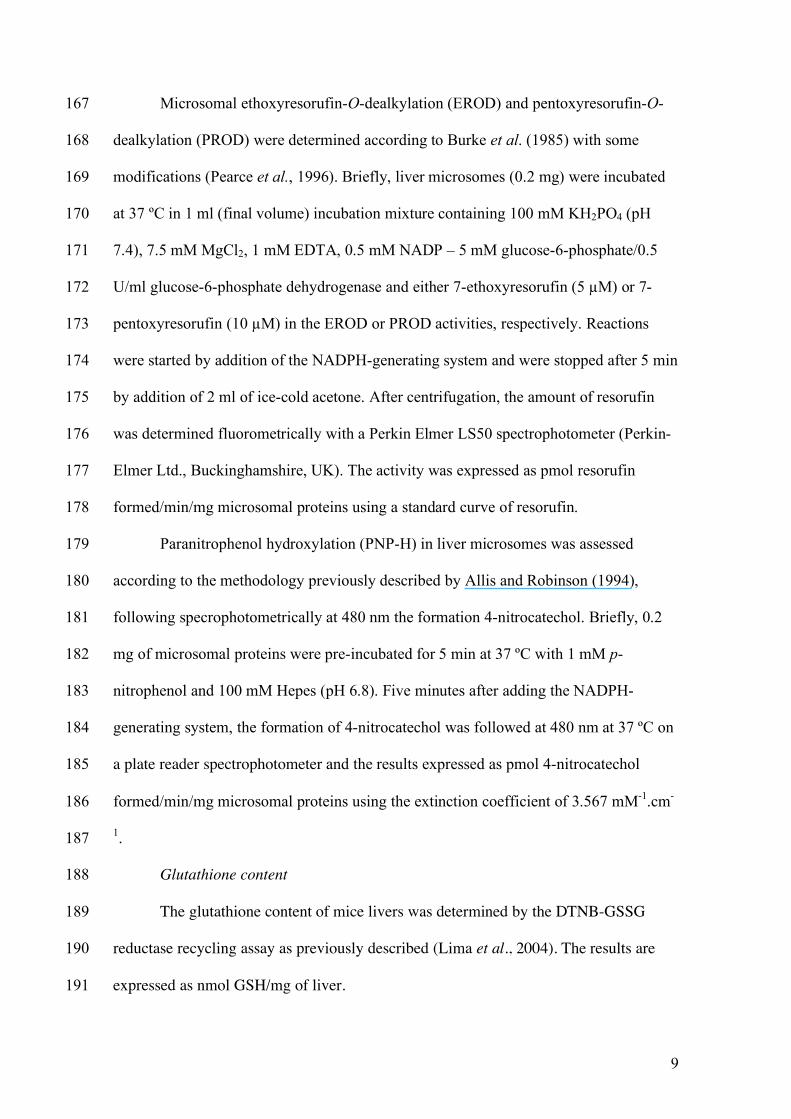
9
Microsomal ethoxyresorufin-O-dealkylation (EROD) and pentoxyresorufin-O-167
dealkylation (PROD) were determined according to Burke et al. (1985) with some 168
modifications (Pearce et al., 1996). Briefly, liver microsomes (0.2 mg) were incubated 169
at 37 ºC in 1 ml (final volume) incubation mixture containing 100 mM KH2PO4 (pH 170
7.4), 7.5 mM MgCl2, 1 mM EDTA, 0.5 mM NADP – 5 mM glucose-6-phosphate/0.5 171
U/ml glucose-6-phosphate dehydrogenase and either 7-ethoxyresorufin (5 µM) or 7-172
pentoxyresorufin (10 µM) in the EROD or PROD activities, respectively. Reactions 173
were started by addition of the NADPH-generating system and were stopped after 5 min 174
by addition of 2 ml of ice-cold acetone. After centrifugation, the amount of resorufin 175
was determined fluorometrically with a Perkin Elmer LS50 spectrophotometer (Perkin-176
Elmer Ltd., Buckinghamshire, UK). The activity was expressed as pmol resorufin 177
formed/min/mg microsomal proteins using a standard curve of resorufin. 178
Paranitrophenol hydroxylation (PNP-H) in liver microsomes was assessed 179
according to the methodology previously described by Allis and Robinson (1994), 180
following specrophotometrically at 480 nm the formation 4-nitrocatechol. Briefly, 0.2 181
mg of microsomal proteins were pre-incubated for 5 min at 37 ºC with 1 mM p-182
nitrophenol and 100 mM Hepes (pH 6.8). Five minutes after adding the NADPH-183
generating system, the formation of 4-nitrocatechol was followed at 480 nm at 37 ºC on 184
a plate reader spectrophotometer and the results expressed as pmol 4-nitrocatechol 185
formed/min/mg microsomal proteins using the extinction coefficient of 3.567 mM-1.cm-186
1. 187
Glutathione content 188
The glutathione content of mice livers was determined by the DTNB-GSSG 189
reductase recycling assay as previously described (Lima et al., 2004). The results are 190
expressed as nmol GSH/mg of liver. 191

10
Protein 192
Protein content of liver homogenates was determined with Bradford Reagent 193
using bovine serum albumin as a standard. Protein content of liver microsomes was 194
determined by the Lowry method (Lowry et al., 1951). 195
196
2.6. CYP 2E1 expression analysis 197
The expression of CYP 2E1 protein was analyzed by Western Blot. 198
Electrophoretic separation of microsomal proteins (15 µg) was performed in 12% 199
sodium dodecyl sulfate–polyacrylamide gels (SDS–PAGE) using the mini-PROTEAN 3 200
electrophoresis cell (Bio-Rad Laboratories, Inc., Hercules, California, USA) according 201
to the method of Laemmli (1970). The separated proteins were electrotransferred to 202
polyvinylidene difluoride (PVDF) membranes (Amersham Biosciences, 203
Buckinghamshire, UK) using the method of Towbin and collaborators (1979). The 204
PVDF membranes were blocked with 5% nonfat dry milk overnight at 4°C and the 205
immunoblots exposed to rabbit polyclonal antibody against CYP 2E1 protein. 206
Immunodetection was performed using horseradish peroxidase–labeled donkey anti-207
rabbit IgG antibody (Amersham Biosciences, Buckinghamshire, UK) and developed 208
with ECL reagents (Amersham Biosciences) according to manufacturer’s instructions. 209
The amount of protein was quantified by densitometry analysis on the SigmaScan Pro 5 210
program (SPSS Inc., San Rafael, CA, USA) and expressed as percentage of the protein 211
level present in control situation. 212
213
2.7. Statistical Analysis 214
Data are expressed as means ± SEM (n=5). Statistical significances (P values < 215
0.05) were evaluated by two-way ANOVA based on gender and treatment group (water 216

11
drinking + saline ip; water drinking + CCl4 ip; sage tea drinking + saline ip; sage tea 217
drinking + CCl4 ip) followed by the Student-Newman-Keuls post hoc test. ALT and 218
AST data were natural logarithm transformed prior to statistical analysis in order to 219
stabilize the variance. 220
221
3. Results 222
The effect of drinking of sage tea for 14 days (instead of water) on the 223
hepatotoxicity of CCl4 was evaluated in mice of both genders challenged with a single 224
dose of CCl4 (20 µl/kg, ip). Plasma transaminase activities were measured 24 h after 225
CCl4 administration as markers of liver injury (Fig. 1). Elevated ALT and AST activities 226
were observed due to CCl4 administration, which is always higher in females compared 227
with males. Both males and females that had been drinking sage tea were significantly 228
more sensitive to the hepatotoxic effects of CCl4 than their control counterparts, as 229
indicated by increased plasma transaminase activities. 230
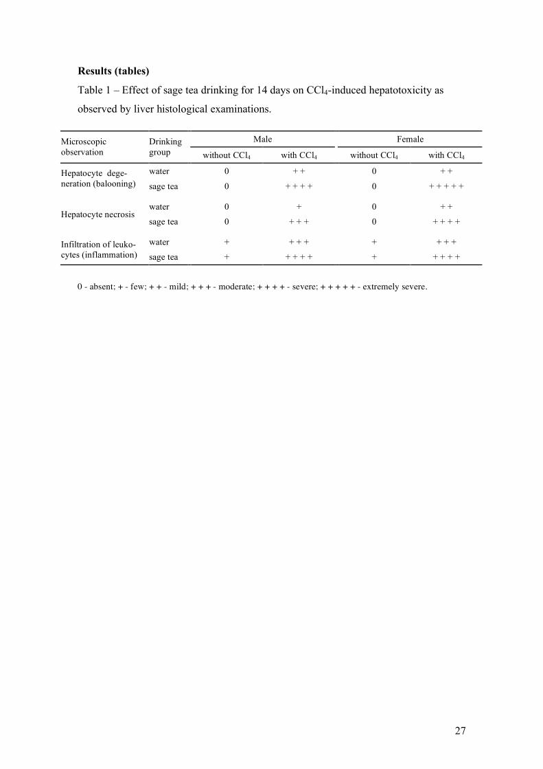
CCl4 is a hepatotoxicant known to produce a characteristic centrilobular pattern 231
of degeneration and necrosis (Weber et al., 2003). Histological examination of H&E-232
stained liver sections was conducted 24 h after CCl4 administration to confirm the 233
pattern of hepatotoxicity and compare the extent of liver injury between control and 234
sage tea drinking animals (Table 1). Morphological findings were consistent with 235
plasma transaminase observations. The CCl4 induced histopathological changes in the 236
liver with significant degeneration and necrosis of hepatocytes in the centrilobular 237
region and with perivenular inflammatory infiltrates. These CCl4-induced 238
histopathological changes were significantly potentiated in the sage tea drinking group 239
of mice with about 50-60% of total area presenting signs of degeneration, necrotic 240
<Insert figure 1 here>
<Insert table 1 here>

12
regions and higher leukocyte infiltration. Also histologically, the liver damage induced 241
by the CCl4 in mice appear to be higher in females than in males. 242
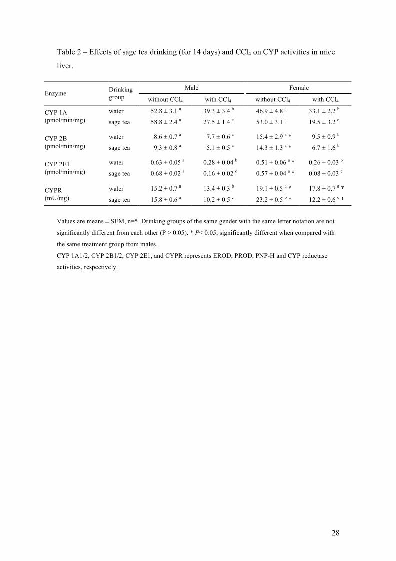
CCl4 is a hepatotoxic chemical that requires metabolic activation by phase I 243
drug-metabolizing enzymes and therefore it was important to monitor the effects of sage 244
tea drinking on the activity of some CYP enzymes. For that, EROD, PROD and PNP-H 245
were measured in liver microsomal fractions (Table 2). Comparing the groups where 246
CCl4 was not administered, although not statistically significant, sage tea drinking 247
increased slightly, between 8% and 13%, the activity of CYP 1A and CYP 2E1 in both 248
genders. The activities of CYP 2B and CYP 2E1 in females was lower and higher, 249
respectively, when compared with males. Twenty four hours after administration, CCl4 250
hepatotoxicity was also reflected in the decrease observed for the activities of the CYP’s 251
measured as well as in the majority of the others enzyme activities (Table 3). 252
Comparing drinking groups, the decrease in these enzyme activities after CCl4 253
administration was also consistent with the higher toxicity in sage tea groups, since it 254
was in general significantly higher in sage tea than water drinking mice. 255
The CYPR is an essential enzyme for microssomal P450-mediated 256
monooxygenase activity, which by interaction with the different CYP’s transfers the 257
essential electron from NADPH (Backes and Kelley, 2003; Henderson et al., 2003). 258
Therefore, its activity was measured (Table 2), and was found to be significantly higher 259
in female mice, which indirectly may contributed to higher toxicity of CCl4 in females. 260
Sage tea drinking induced 21% the activity of this cytochrome, but only in female mice. 261
The bioactivation of CCl4 is mainly executed by CYP 2E1 (Weber et al., 2003). 262
It is also known that modulatory effects on the expression of CYP 2E1 affects the CCl4-263
induced hepatotoxicity (Weber et al., 2003). Therefore, in addition to the measurement 264
of some CYP enzyme activities which included the CYP 2E1, the expression of this 265
<Insert table 2 here>
<Insert table 3 here>

13
cytochrome was evaluated by Western Blot (Fig. 2). Sage tea drinking for 14 days 266
increased significantly the amount of CYP 2E1 protein in females (24%) abut it only 267
slightly increased in males (8%). In sage tea drinking mice, the decrease on CYP 2E1 268
protein induced by CCl4 was most severe in females. 269
After bioactivation, CCl4-induced hepatotoxicity is mediated by primary and 270
secondary bond formation of reactive species to critical cellular molecules such as 271
DNA, lipids, proteins or carbohydrates (Weber et al., 2003). Thus, detoxifying enzymes 272
(such as GST) and antioxidant enzymes (such as the pair GPox/GR) are important 273
against the cell stress situation caused by CCl4. To monitor effects at this level, three 274
glutathione-related enzymes were measured (Table 3) and gender differences were 275
observed in all of them. The activity of GST in males was significantly increased by 276
sage tea drinking, as previously observed in other study (Lima et al., 2005). GPox 277
activity was also increased by sage tea drinking but significantly only in females. 278
Hepatic GSH is an important intracellular antioxidant that can scavenge free radicals 279
and could be important in the defense against radical-mediated hepatotoxicity. 280
Alterations in GSH and oxidized glutathione (GSSG) levels are therefore an important 281
indicator of oxidative stress. Comparing the groups where CCl4 was not administered, 282
there was no effect of sage tea drinking on GSH and GSSG levels in male and female 283
mice (Table 3). Twenty four hours after CCl4 administration, GSH levels decreased 284
significantly only in females from the sage tea drinking group. GSSG levels increased 285
significantly after CCl4 administration in both genders but only in the sage tea drinking 286
groups (Table 3). This increase was significantly higher in females than males. As a 287
result, glutathione data also suggest higher cell damage induced by CCl4 in the sage tea 288
drinking groups in females. 289
<Insert figure 2 here>

14
Finally, soluble protein measured after 10,000 × g centrifugation (Table 3) 290
corroborates the previous results. Comparing the groups where CCl4 was not 291
administered, the higher soluble protein found in the sage tea drinking groups suggests 292
induction of protein expression. The decrease in soluble protein, with concomitant 293
precipitation of damaged proteins, found after the haloalkane administration suggests 294
higher toxicity of CCl4 in the sage tea drinking groups and in females. 295
296
4. Discussion 297
In a previous study, sage tea drinking significantly increased (rat and mouse) 298
liver GST activity and protected against GSH depletion and lipid peroxidation induced 299
by an oxidant agent (Lima et al., 2005). Considering these beneficial effects on liver 300
antioxidant status the present study was carried out in order to evaluate whether sage tea 301
drinking would reduce the extent of hepatic injury induced by CCl4 in male and female 302
mice. In a recently published work, GST was implicated as an important defence 303
mechanism during the early stages (1–6 h) of the CCl4-induced liver injury (Dwivedi et 304
al., 2006). GST is a phase II enzyme that plays a key role in cellular detoxification of 305
xenobiotics, electrophiles and reactive oxygen species through their conjugation to GSH 306
(Mates, 2000). Besides an essential substrate to GST and GPox, GSH is also an 307
important intracellular antioxidant (hydrogen-donating compound) that spontaneously 308
neutralizes several electrophiles and reactive oxygen species (Lu, 1999). After 309
bioactivation of CCl4, in addition to dangerous free radical formation and subsequent 310
reactive oxygen species formation, a sequence of chain reactions can be initiated that 311
leads to lipid peroxidation (Weber et al., 2003). Since sage tea drinking has also been 312
shown to decrease lipid peroxidation induced by tert-butyl hydroxide in rat hepatocyte 313
primary cultures (Lima et al, 2005), this also suggested here possible beneficial effects 314

15
against CCl4. However, contrary to our hypothesis, sage tea drinking increased 315
significantly the CCl4-induced hepatotoxicity in mice. 316
CCl4 becomes toxic upon activation mainly through CYP 2E1, and an induction 317
or an over-expression of this cytochrome correlates with higher CCl4 toxicity (Weber et 318
al., 2003; Chan et al., 2005). Sage tea drinking for 14 days increased the expression 319
level of CYP 2E1. In agreement with this, the activity of this cytochrome was also 320
slightly increased by sage tea drinking. This could provide an explanation for the higher 321
CCl4 toxicity in tea drinking mice. CYP 2E1 protein is localized predominantly in the 322
central zone of the liver lobule (Forkert et al., 1991), which explains the typical 323
centrilobular region of hepatocyte injury observed after CCl4 administration. This 324
pattern of centrilobular toxicity was more extensive in sage tea versus water drinking 325
mice. After CCl4 bioactivation, the resulting CCl3˙ radical binds covalently to CYP 2E1, 326
either to the active site of the enzyme or to the heme group, thereby causing suicide 327
inactivation (Weber et al., 2003). After drug administration to sage tea drinking mice, 328
CYP 2E1 levels, originally higher, decreased to significant lower levels. A decrease in 329
CYP 2E1 expression and activity after CCl4 exposure seem to reflect inactivation of the 330
protein, which is consistent with the increased CCl4 hepatotoxicity in this drinking 331
group. However, to confirm increased CCl4 bioactivation through CYP 2E1 in sage tea 332
drinking mice than the water drinking cohorts, measurement of covalent binding of 333
14CCl4-derived radiolabel to liver tissue would have to be done. The simultaneous 334
increases in GST and GPox activities by sage tea drinking, and possibly other 335
detoxifying and antioxidant enzymes, seem to have been incapable of neutralizing 336
increased CCl4 toxicity. Also, the previously observed beneficial effect of sage tea 337
against lipid peroxidation (Lima et al., 2005) seemed to be insufficient to block CCl4-338
induced damage. The increased levels of CYP 2E1 protein and activity induced by sage 339

16
tea drinking may, thus, at least in part, provide an explanation for the obtained results – 340
an herb-toxicant interaction between sage tea and CCl4 that potentiated the haloalkane’s 341
toxicity. 342
Herb-drug interactions have been described for a variety of plants used as 343
phytomedicines, many of them by case reports of interactions between herbs and 344
pharmaceutical drugs (Izzo, 2005; Hu et al., 2005). CYP isozymes are particularly 345
vulnerable to modulation by the diverse active constituents of herbs (Zhou et al., 2003). 346
This important phase I drug-metabolizing enzyme system is responsible for the 347
metabolism of a variety of xenobiotics and some important endogenous substances such 348
as steroids and prostaglandins (Anzenbacher and Anzenbacherova, 2001; Tamasi et al., 349
2003). Although CYP-mediated reactions are primarily detoxification processes, certain 350
substrates are metabolically activated resulting in the generation of reactive 351
intermediates with increased toxicity and mutagenicity (Jaeschke et al., 2002; Tamasi et 352
al., 2003). Many pharmaceutical drugs are also metabolized by these phase I enzymes 353
and modulation of CYPs by herbs may either exacerbate the undesirable effects (by 354
increasing toxicity) or antagonize the actions (by increasing clearance) of concurrent 355
medical therapy (Stedman, 2002). In addition, severe hepatic injury may be caused by 356
chemicals or natural toxins metabolically activated by drug-metabolizing enzymes as a 357
result of occupational, household or environmental exposure, emphasizing the need for 358
understanding mechanisms of action of herbal extracts. Thus, although interspecies 359
differences in xenobiotic metabolism are well documented (Caldwell, 1992), the drug-360
toxicant interaction between sage tea and CCl4 reported here highlight possible herb-361
drug interactions between this extract and drugs metabolized by the liver. However, as 362
far as we know, there were no reports of drug-drug interactions between sage tea and 363
pharmaceutical drugs or environmental contaminants. In this particular study, where a 364

17
herb-drug interaction was observed, sage tea replaced almost 100% the water that the 365
animal consumed, since food is provided as dry pellets. Therefore, by taking 1 or 2 cups 366
of sage tea, a person never reaches the dose of sage extract ingested by mice in this 367
study. So, it seems that the moderate, traditional drinking of sage tea by people most 368
likely does not result in adverse interactions with other drugs. It should, however, be 369
kept in mind that, if a phytomedicine with a higher dose of sage is taken over an 370
extended period of time, an opportunity for enzyme induction could occur and 371
undesirable interactions take place. Additionally, interindividual differences in drug 372
metabolism, for example due to genetic polymorphism of CYP genes (Tamasi et al., 373
2003; Wu and Cederbaum, 2005), could increase the susceptibility of different 374
populations or individuals for herb-drug interactions. 375
Many of these drug-metabolizing enzymes and also antioxidant enzymes are 376
known to be gender dependent (Chaubey et al., 1994; Clewell et al., 2002; Sverko et al., 377
2004), which may ultimately differentially affect the toxicity of drugs between male and 378
female individuals of the same specie (Kato and Yamazoe, 1992; Meibohm et al., 2002; 379
Chanas et al., 2003). The hepatotoxicity of CCl4 to females was higher than to males in 380
both drinking groups. Looking to all measured parameters, several gender differences 381
were observed which can explain the higher toxicity to female mice. In terms of drug 382
bioactivation, although the activity of CYP 2E1 was lower in females, the expression of 383
CYP 2E1, the activity of CYP 2B family and the activity of CYPR were higher in 384
females which seems to indicate an increased ability to metabolise CCl4 in females. In 385
terms of cell defences against drug-induced injury, although GPox activity was higher 386
in females, GST activity is significantly higher in males. At least during the initial stage 387
of CCl4-induced hepatotoxicity, GST is more likely to confer protection, since CCl4 388
toxicity is mediated by strong free radicals. 389

18
These CYP modulatory as well as antioxidant effects of plant extracts have often 390
been attributed to phenolic and monoterpenic compounds (Elegbede et al., 1993; 391
Banerjee et al., 1995; Birt et al., 2001; Ren et al., 2003; Ferguson et al., 2004). 392
Flavonoids are a diverse group of polyphenols that are produced by several plants 393
(Havsteen, 2002). In relation to phase I and phase II drug-metabolizing enzymes, 394
flavonoids have been reported to possess several modulatory effects, either inducing or 395
decreasing the expression of these enzymes and also either as potent inhibitors or 396
stimulators of enzyme activities, depending on structure, concentration, and assay 397
conditions (Zhou et al., 2003; Ferguson et al., 2004). Rosmarinic acid is the predominat 398
phenolic compound in sage tea (Lima et al., 2005). The oral administration of 399
rosmarinic acid in rats was previously shown not to induce phase I and phase II 400
enzymes (Debersac et al., 2001), and, therefore, was possibly not the responsible for the 401
effects observed in our study. Luteolin-7-glucoside, the major flavonoid present in sage 402
tea, and also monoterpenes present in the essential oil fraction, could, on the other hand, 403
be good candidates. However, pre-treatment of rats with luteolin-7-glucoside was 404
recently found to protect significantly against CCl4-induced toxicity, and its effects 405
attributed to the compound’s antioxidant properties acting as scavenger of reactive 406
oxygen species (Zheng et al., 2004). Most likely, the sage tea effects observed here were 407
a result of interactions and synergisms among the different compounds and metabolites 408
present, which makes it difficult to attribute them to any particular compound or family 409
of compounds. 410
In conclusion, the present work showed that sage tea drinking for 14 days 411
significantly potentiated CCl4-induced hepatic injury in mice, to a higher degree in 412
females, as a result, at least in part, of an induction of CYP 2E1. In addition, although 413
sage tea did not have toxic effects of its own and in fact seemed to improve the 414

19
antioxidant status of the liver, the observed herb-toxicant interaction may affect the 415
efficacy and safety of concurrent medical therapy with drugs that are metabolized by 416
phase I enzymes. 417
418
Acknowledgments 419
We would like to thank Dr. Jonathan Wilson, Alice Ramos and Marisa F. Azevedo for 420
the help provided with the histological examinations. CFL was supported by the 421
Foundation for Science and Technology, Portugal, grant SFRH/BD/6942/2001. This 422
work was supported by FCT research grant POCI/AGR/62040/2004.423

20
References 336 337
Allis, J.W. and Robinson, B.L., 1994. A kinetic assay for p-nitrophenol hydroxylase in 338
rat liver microsomes. Anal. Biochem. 219, 49-52. 339
Amin, A. and Hamza, A.A. (2005) Hepatoprotective effects of Hibiscus, Rosmarinus 340
and Salvia on azathioprine-induced toxicity in rats. Life Sci. 77, 266-278. 341
Anzenbacher, P. and Anzenbacherova, E. (2001) Cytochromes P450 and metabolism of 342
xenobiotics. Cell. Mol. Life Sci. 58, 737-747. 343
Backes, W.L. and Kelley, R.W. (2003) Organization of multiple cytochrome P450s with 344
NADPH-cytochrome P450 reductase in membranes. Pharmacol. Ther. 98, 221-345
233. 346
Banerjee, S., Welsch, C.W. and Rao, A.R. (1995) Modulatory influence of camphor on 347
the activities of hepatic carcinogen metabolizing enzymes and the levels of 348
hepatic and extrahepatic reduced glutathione in mice. Cancer Lett. 88, 163-169. 349
Barbier, O., Lapointe, H., El Alfy, M., Hum, D.W. and Belanger, A. (2000) Cellular 350
localization of uridine diphosphoglucuronosyltransferase 2B enzymes in the 351
human prostate by in situ hybridization and immunohistochemistry. J. Clin. 352
Endocrinol. Metab. 85, 4819-4826. 353
Baricevic, D. and Bartol, T. (2000) The biological/pharmacological activity of the 354
Salvia genus. In: S.E. Kintzios (Ed), SAGE - The Genus Salvia. Harwood 355
Academic Publishers, Amsterdam, pp. 143-184. 356
Birt, D.F., Hendrich, S. and Wang, W. (2001) Dietary agents in cancer prevention: 357
flavonoids and isoflavonoids. Pharmacol. Ther. 90, 157-177. 358

21
Burke, M.D., Thompson, S., Elcombe, C.R., Halpert, J., Haaparanta, T. and Mayer, R.T., 359
1985. Ethoxy-, pentoxy- and benzyloxyphenoxazones and homologues: a series of 360
substrates to distinguish between different induced cytochromes P-450. Biochem. 361
Pharmacol. 34, 3337-3345. 362
Caldwell, J. (1992) Problems and opportunities in toxicity testing arising from species 363
differences in xenobiotic metabolism. Toxicol. Lett. 64/65, 651-659. 364
Chan, W.H., Sun, W.Z. and Ueng, T.H. (2005) Induction of rat hepatic cytochrome P-365
450 by ketamine and its toxicological implications. Journal of Toxicology and 366
Environmental Health. Part A. 68, 1581-1597. 367
Chanas, B., Wang, H. and Ghanayem, B.I. (2003) Differential metabolism of 368
acrylonitrile to cyanide is responsible for the greater sensitivity of male vs 369
female mice: role of CYP2E1 and epoxide hydrolases. Toxicol. Appl. Pharmacol. 370
193, 293-302. 371
Chaubey, M., Singhal, S.S., Awasthi, S., Saxena, M., Dyer, R.B., Awasthi, Y.C. and 372
Herzog, N.K. (1994) Gender-related differences in expression of murine 373
glutathione S-transferases and their induction by butylated hydroxyanisole. 374
Comp. Biochem. Physiol. C 108, 311-319. 375
Chen, J.H., Tipoe, G.L., Liong, E.C., So, H.S.H., Leung, K.M., Tom, W.M., Fung, 376
P.C.W. and Nanji, A.A. (2004) Green tea polyphenols prevent toxin-induced 377
hepatotoxicity in mice by down-regulating inducible nitric oxide-derived 378
prooxidants. Am. J. Clin. Nutr. 80, 742-751. 379
Chung, H., Hong, D.P., Jung, J.Y., Kim, H.J., Jang, K.S., Sheen, Y.Y., Ahn, J.I., Lee, 380
Y.S. and Kong, G. (2005) Comprehensive analysis of differential gene 381
22
expression profiles on carbon tetrachloride-induced rat liver injury and 382
regeneration. Toxicol. Appl. Pharmacol. 206, 27-42. 383
Clewell, H.J., Teeguarden, J., McDonald, T., Sarangapani, R., Lawrence, G., Covington, 384
T., Gentry, R. and Shipp, A. (2002) Review and evaluation of the potential 385
impact of age- and gender-specific pharmacokinetic differences on tissue 386
dosimetry. Crit. Rev. Toxicol. 32, 329-389. 387
Cuvelier, M.E., Berset, C. and Richard, H. (1994) Antioxidant constituents in sage 388
(Salvia officinalis). J. Agric. Food Chem. 42, 665-669. 389
Debersac, P., Vernevaut, M.F., Amiot, M.J., Suschetet, M. and Siess, M.H. (2001) 390
Effects of a water-soluble extract of rosemary and its purified component 391
rosmarinic acid on xenobiotic-metabolizing enzymes in rat liver. Food Chem. 392
Toxicol. 39, 109-117. 393
Dwivedi, S., Sharma, R., Sharma, A., Zimniak, P., Ceci, J.D., Awasthi, Y.C. and Boor, 394
P.J. (2006) The course of CCl4 induced hepatotoxicity is altered in mGSTA4-4 395
null (-/-) mice. Toxicology 218, 58-66. 396
Elegbede, J.A., Maltzman, T.H., Elson, C.E. and Gould, M.N. (1993) Effects of 397
anticarcinogenic monoterpenes on phase II hepatic metabolizing enzymes. 398
Carcinogenesis 14, 1221-1223. 399
Ferguson, L.R., Philpott, M. and Karunasinghe, N. (2004) Dietary cancer and 400
prevention using antimutagens. Toxicology 198, 147-159. 401

23
Forkert, P.G., Massey, T.E., Jones, A.B., Park, S.S., Gelboin, H.V. and Anderson, L.M. 402
(1991) Distribution of cytochrome CYP2E1 in murine liver after ethanol and 403
acetone administration. Carcinogenesis 12, 2259-2268. 404
Havsteen, B.H. (2002) The biochemistry and medical significance of the flavonoids. 405
Pharmacol. Ther. 96, 67-202. 406
Henderson, C.J., Otto, D.M.E., Carrie, D., Magnuson, M.A., McLaren, A.W., Rosewell, 407
I. and Wolf, C.R. (2003) Inactivation of the hepatic cytochrome P450 system by 408
conditional deletion of hepatic cytochrome P450 reductase. J. Biol. Chem. 278, 409
13480-13486. 410
Hu, Z.P., Yang, X.X., Ho, P.C.L., Chan, S.Y., Heng, P.W.S., Chan, E., Duan, W., Koh, 411
H.L. and Zhou, S.F. (2005) Herb-drug interactions - A literature review. Drugs 412
65, 1239-1282. 413
Ioannides, C. (2002) Pharmacokinetic interactions between herbal remedies and 414
medicinal drugs. Xenobiotica 32, 451-478. 415
Izzo, A.A. (2005) Herb-drug interactions: an overview of the clinical evidence. Fundam. 416
Clin. Pharmacol. 19, 1-16. 417
Jaeschke, H., Gores, G.J., Cederbaum, A.I., Hinson, J.A., Pessayre, D. and Lemasters, 418
J.J. (2002) Forum - Mechanisms of hepatotoxicity. Toxicol. Sci. 65, 166-176. 419
Kato, R. and Yamazoe, Y. (1992) Sex-specific cytochrome P450 as a cause of sex- and 420
species-related differences in drug toxicity. Toxicol. Lett. 64/65, 661-667. 421
Laemmli, U.K., 1970. Cleavage of structural proteins during the assembly of the head 422
of bacteriophage T4. Nature 227, 680-685. 423

24
Lima, C.F., Andrade, P.B., Seabra, R.M., Fernandes-Ferreira, M. and Pereira-Wilson, C. 424
(2005) The drinking of a Salvia officinalis infusion improves liver antioxidant 425
status in mice and rats. J. Ethnopharmacol. 97, 383-389. 426
Lima, C.F., Carvalho, F., Fernandes, E., Bastos, M.L., Santos-Gomes, P.C., Fernandes-427
Ferreira, M. and Pereira-Wilson, C. (2004) Evaluation of toxic/protective effects 428
of the essential oil of Salvia officinalis on freshly isolated rat hepatocytes. 429
Toxicol. In Vitro 18, 457-465. 430
Lima, C.F., Fernandes-Ferreira, M. and Pereira-Wilson, C. (2006) Phenolic compounds 431
protect HepG2 cells from oxidative damage: Relevance of glutathione levels. 432
Life Sci. (in press, DOI: 10.1016/j.lfs.2006.06.042). 433
Loguercio, C. and Federico, A. (2003) Oxidative stress in viral and alcoholic hepatitis. 434
Free Radic. Biol. Med. 34, 1-10. 435
Lowry, O.H., Rosebrough, N.J., Farr, A.L. and Randall, R.J. (1951) Protein 436
Measurement with the Folin Phenol Reagent. J. Biol. Chem. 193, 265-275. 437
Lu, S.C. (1999) Regulation of hepatic glutathione synthesis: current concepts and 438
controversies. FASEB J. 13, 1169-1183. 439
Mates, J.M. (2000) Effects of antioxidant enzymes in the molecular control of reactive 440
oxygen species toxicology. Toxicology 153, 83-104. 441
Meibohm, B., Beierle, I. and Derendorf, H. (2002) How important are gender 442
differences in pharmacokinetics? Clin. Pharmacokinet. 41, 329-342. 443
Pearce, R.E., McIntyre, C.J., Madan, A., Sanzgiri, U., Draper, A.J., Bullock, P.L., Cook, 444
D.C., Burton, L.A., Latham, J., Nevins, C. and Parkinson, A. (1996) Effects of 445

25
freezing, thawing, and storing human liver microsomes on cytochrome P450 446
activity. Arch. Biochem. Biophys. 331, 145-169. 447
Phillips, A.H. and Langdon, R.G. (1962) Hepatic triphosphopyridine nucleotide-448
cytochrome c reductase: isolation, characterization, and kinetic studies. J. Biol. 449
Chem. 237, 2652-2660. 450
Plaa, G.L. and Hewitt, W.R., 1982. Detection and evaluation of chemically induced 451
liver injury. In: Hayes, A.H. (Ed.), Principles and Methods of Toxicology. Raven 452
Press, New York, pp. 407-634. 453
Ren, W., Qiao, Z., Wang, H., Zhu, L. and Zhang, L. (2003) Flavonoids: promising 454
anticancer agents. Med. Res. Rev. 23, 519-534. 455
Stedman, C. (2002) Herbal hepatotoxicity. Semin. Liver Dis. 22, 195-206. 456
Sverko, V., Sobocanec, S., Balog, T. and Marotti, T. (2004) Age and gender differences 457
in antioxidant enzyme activity: potential relationship to liver carcinogenesis in 458
male mice. Biogerontology 5, 235-242. 459
Tamasi, V., Vereczkey, L., Falus, A. and Monostory, K. (2003) Some aspects of 460
interindividual variations in the metabolism of xenobiotics. Inflamm. Res. 52, 461
322-333. 462
Towbin, H., Staehelin, T. and Gordon, J., 1979. Electrophoretic transfer of proteins 463
from polyacrylamide gels to nitrocellulose sheets: procedure and some applications. 464
Proc. Natl. Acad. Sci. U. S. A 76, 4350-4354. 465
Vitaglione, P., Morisco, F., Caporaso, N. and Fogliano, V. (2004) Dietary antioxidant 466
compounds and liver health. Crit. Rev. Food Sci. Nutr. 44, 575-586. 467

26
Weber, L.W.D., Boll, M. and Stampfl, A. (2003) Hepatotoxicity and mechanism of 468
action of haloalkanes: Carbon tetrachloride as a toxicological model. Crit. Rev. 469
Toxicol. 33, 105-136. 470
Williams, R.J., Spencer, J.P.E. and Rice-Evans, C. (2004) Flavonoids: Antioxidants or 471
signalling molecules? Free Radic. Biol. Med. 36, 838-849. 472
Wu, D. and Cederbaum, A.I. (2005) Oxidative stress mediated toxicity exerted by 473
ethanol-inducible CYP2E1. Toxicol. Appl. Pharmacol. 207, 70-76. 474
Zheng, Q.S., Sun, X.L., Xubo, Li, G., Song, M. and Wang, C.H. (2004) Protective 475
effects of luteolin-7-glucoside against liver injury caused by carbon tetrachloride 476
in rats. Pharmazie 59, 286-289. 477
Zhou, S.F., Gao, Y.H., Jiang, W.Q., Huang, M., Xu, A.L. and Paxton, J.W. (2003) 478
Interactions of herbs with cytochrome P450. Drug Metab. Rev. 35, 35-98. 479
480
481
27
Results (tables)
Table 1 – Effect of sage tea drinking for 14 days on CCl4-induced hepatotoxicity as
observed by liver histological examinations.
0 - absent; + - few; + + - mild; + + + - moderate; + + + + - severe; + + + + + - extremely severe.
Male Female Microscopic observation
Drinking group without CCl4 with CCl4 without CCl4 with CCl4
water 0 + + 0 + + Hepatocyte dege-neration (balooning) sage tea 0 + + + + 0 + + + + +
water 0 + 0 + + Hepatocyte necrosis
sage tea 0 + + + 0 + + + +
water + + + + + + + + Infiltration of leuko-cytes (inflammation) sage tea + + + + + + + + + +
28
Table 2 – Effects of sage tea drinking (for 14 days) and CCl4 on CYP activities in mice
liver.
Values are means ± SEM, n=5. Drinking groups of the same gender with the same letter notation are not
significantly different from each other (P > 0.05). * P< 0.05, significantly different when compared with
the same treatment group from males.
CYP 1A1/2, CYP 2B1/2, CYP 2E1, and CYPR represents EROD, PROD, PNP-H and CYP reductase
activities, respectively.
Male Female Enzyme Drinking
group without CCl4 with CCl4 without CCl4 with CCl4
water 52.8 ± 3.1 a 39.3 ± 3.4 b 46.9 ± 4.8 a 33.1 ± 2.2 b CYP 1A (pmol/min/mg)
sage tea 58.8 ± 2.4 a 27.5 ± 1.4 c 53.0 ± 3.1 a 19.5 ± 3.2 c
water 8.6 ± 0.7 a 7.7 ± 0.6 a 15.4 ± 2.9 a * 9.5 ± 0.9 b
CYP 2B
(pmol/min/mg) sage tea 9.3 ± 0.8 a 5.1 ± 0.5 a 14.3 ± 1.3 a * 6.7 ± 1.6 b
water 0.63 ± 0.05 a 0.28 ± 0.04 b 0.51 ± 0.06 a * 0.26 ± 0.03 b
CYP 2E1
(pmol/min/mg) sage tea 0.68 ± 0.02 a 0.16 ± 0.02 c 0.57 ± 0.04 a * 0.08 ± 0.03 c
water 15.2 ± 0.7 a 13.4 ± 0.3 b 19.1 ± 0.5 a * 17.8 ± 0.7 a * CYPR
(mU/mg) sage tea 15.8 ± 0.6 a 10.2 ± 0.5 c 23.2 ± 0.5 b * 12.2 ± 0.6 c *

29
Table 3 – Effects of sage tea drinking (for 14 days) and CCl4 on glutathione-related
enzymes, glutathione levels and soluble protein in mice livers.
1 Liver soluble proteins measured in the supernatant after a centrifugation of 10,000 × g for 10 min at 4 ºC
by the Bradford reagent using bovine serum albumin as a standard.
Values are means ± SEM, n=5. Drinking groups of the same gender with the same letter notation are not
significantly different from each other (P > 0.05). * P< 0.05, significantly different when compared with
the same treatment group from males.
GST: glutathione-s-transferase; GPox: glutathione peroxidase; GR: glutathione reductase; GSH:
glutathione (reduced form); GSSG: glutathione: oxidized form.
Male Female Parameter Drinking
group without CCl4 with CCl4 without CCl4 with CCl4
water 305 ± 15 a 307 ± 12 a 128 ± 5 a * 115 ± 6 a * GST (mU/mg)
sage tea 369 ± 30 b 237 ± 23 c 144 ± 4 a * 76 ± 8 b *
water 432 ± 20 a 456 ± 14 a 779 ± 12 a * 772 ± 12 a * GPox (mU/mg)
sage tea 493 ± 30 a 570 ± 25 b 888 ± 19 b * 694 ± 25 c *
water 24.1 ± 0.9 a 24.0 ± 0.3 a 20.1 ± 0.6 a,b * 21.1 ± 0.2 a * GR (mU/mg)
sage tea 25.2 ± 1.0 a 20.9 ± 1.1 b 22.4 ± 0.4 a * 18.3 ± 0.5 b *
water 7.61 ± 0.24 a 7.48 ± 0.22 a 7.46 ± 0.33 a 8.36 ± 0.16 a GSH (nmol/mg liver) sage tea 6.53 ± 0.34 a 8.18 ± 0.56 a 6.71 ± 0.14 a 4.53 ± 1.09 b *
water 0.26 ± 0.02 a,b 0.23 ± 0.02 a,b 0.19 ± 0.02 a 0.27 ± 0.06 a GSSG (nmol/mg liver) sage tea 0.19 ± 0.02 b 0.31 ± 0.03 a 0.22 ± 0.03 a 1.35 ± 0.20 b *
water 195.7 ± 4.3 a 200.4 ± 2.8 a 194.1 ± 3.6 a 180.1 ± 1.2 b * Protein (mg protein/g liver)1 sage tea 215.8 ± 4.3 b 171.7 ± 4.2 c 214.3 ± 2.9 c 154.8 ± 4.9 d *

30
Results (figures)
Fig. 1 – Effects of sage tea drinking for 14 days on CCl4-induced increase in plasma
transaminase activities. (A) ALT: alanine aminotransferase; (B) AST: aspartate
aminotransferase. Values are means ± SEM, n=5. For statistical evaluation, these data
were natural logarithm transformed in order to stabilize the variance. Drinking groups
of the same gender with the same letter notation are not significantly different from
each other (P > 0.05). * P< 0.05, significantly different when compared with the same
treatment group from males.

31
Fig. 2 – Effects of sage tea drinking (for 14 days) and CCl4 on expression of CYP 2E1
in the liver of male and female mice. Each gel lane was loaded with fifteen µg of
microsome proteins for the Western blotting analysis. (A) Results obtained from five
mice of each group. Mean ± SEM. Groups of the same gender with the same letter
notation are not significantly different from each other (P > 0.05). * P< 0.05,
significantly different when compared with the same treatment group from males. (B)
Representative images of the imunodetection of CYP 2E1 by Western Blot from 2
animals for each group.
Copyright © 2022 FDOKUMEN