An integrative analysis of ethanol tolerance and withdrawal in zebrafish (Danio rerio)
Upload
independentCategory
view
0download
0
ORIGINAL ARTICLE
The Effect of Taurine on Hepatic Steatosis Inducedby Thioacetamide in Zebrafish (Danio rerio)
Thais Ortiz Hammes • Gabriela Lima Pedroso • Carolina Rigatti Hartmann •
Thayssa Dalla Costa Escobar • Laisa Beduschi Fracasso • Darlan Pase da Rosa •
Norma Possa Marroni • Marilene Porawski • Themis Reverbel da Silveira
Received: 12 May 2011 / Accepted: 20 September 2011 / Published online: 14 October 2011
� Springer Science+Business Media, LLC 2011
Abstract
Background Nonalcoholic fatty liver disease is one of the
most prevalent forms of chronic liver disease in the Wes-
tern world. Taurine is a conditionally essential amino acid
in humans that may be a promising therapy for treating this
disease.
Aim To evaluate the effect of taurine on hepatic steatosis
induced by thioacetamide in Danio rerio.
Methods Animals were divided into four groups: control
(20 ll of saline solution), taurine (1,000 mg/kg), thioace-
tamide (300 mg/kg), and the taurine–thioacetamide group
(1,000 ? 300 mg/kg). Thioacetamide was injected intra-
peritoneally three times a week for 2 weeks. The mRNA
expression, lipoperoxidation, antioxidant enzymatic
activity, and histological analyses were evaluated in the
liver and the triglyceride content was assessed in the
serum.
Results Thioacetamide injection induced steatosis, as
indicated by histological analyses. The lipoperoxidation
showed significant lipid damage in the thioacetamide group
compared to the taurine–thioacetamide group (p \ 0.001).
Superoxide dismutase (SOD) activity in the taurine–thioac-
etamide group (5.95 ± 0.40) was significantly increased
compared to the thioacetamide group (4.14 ± 0.18 U SOD/
mg of protein) (p \ 0.001). The mRNA expression of SIRT1
(0.5-fold) and Adiponectin receptor 2 (0.39-fold) were lower
in the thioacetamide group than the control (p \ 0.05). TNF-
a mRNA expression was 6.4-fold higher in the thioacetamide
group than the control (p \ 0.05). SIRT1 mRNA expression
was 2.6-fold higher in the taurine–thioacetamide group than
in the thioacetamide group.
Conclusions Taurine seems to improve hepatic steatosis by
reducing oxidative stress and increasing SIRT1 expression.
Keywords Nonalcoholic fatty liver disease � Taurine �Thioacetamide � Zebrafish � SIRT1
Introduction
Nonalcoholic fatty liver disease (NAFLD) has been
increasingly recognized as the most common form of
chronic liver disease in both adults and children [1]. In
humans, it is histologically characterized by the accumu-
lation of fat, which is predominantly comprised of mainly
triglycerides in more than 5% of hepatocytes [2]. It is one
of the most prevalent forms of chronic liver disease, and its
prevalence is estimated to be 31% in American adults [3].
In addition, the prevalence increases in risk populations,
T. O. Hammes (&) � G. L. Pedroso � C. R. Hartmann �T. D. C. Escobar � L. B. Fracasso � D. P. da Rosa �N. P. Marroni � M. Porawski � T. R. da Silveira
Laboratorio de Hepatologia e Gastroenterologia Experimental,
Centro de Pesquisa Experimental, Hospital de Clınicas de Porto
Alegre, Rua Ramiro Barcelos 2350, Porto Alegre,
RS 90035-903, Brazil
e-mail: [email protected]
T. O. Hammes � G. L. Pedroso � T. R. da Silveira
Faculdade de Medicina, Programa de Pos-Graduacao, Ciencias
em Gastroenterologia e Hepatologia, Universidade Federal do
Rio Grande do Sul (UFRGS), Porto Alegre, RS, Brazil
D. P. da Rosa � N. P. Marroni
Programa de Pos-Graduacao em Medicina: Ciencias Medicas,
Universidade Federal do Rio Grande do Sul (UFRGS), Porto
Alegre, RS, Brazil
N. P. Marroni
Programa de Pos-Graduacao em Genetica e Toxicologia,
Universidade Luterana do Brasil (ULBRA), Canoas, RS, Brazil
123
Dig Dis Sci (2012) 57:675–682
DOI 10.1007/s10620-011-1931-4

such as persons with a large waist circumference, obesity,
insulin resistance, or diabetes. In Brazil, more than 80% of
patients admitted for bariatric surgery have hepatic stea-
tosis [4, 5]. The pathogenesis of hepatic steatosis is still
poorly understood. There is no consensus regarding ther-
apies for NAFLD, and many pharmacological agents are
being tested for this purpose [6]. Nevertheless, it is known
that free fatty acids (FFA) and adipocytokines, such as
TNF-a (tumor necrosis factor a) and adiponectin have a
central role in its development.
Taurine (2-aminoethanesulfur acid) is a conditionally
essential amino acid in humans and important for the
development of the nervous system, xenobiotic conjuga-
tion, osmoregulation, and other processes [7]. Taurine also
presents hypolipidemic and antioxidant effects, which is
the main reason why it has been tested as a therapeutic
agent against steatosis [8]. Interestingly, Chen et al. [9]
observed a reduction of oxidative stress, an increase in
expression of adiponectin, a decrease in expression of
TNF-a, and an improvement of histological parameters of
steatosis in rats with nonalcoholic steatohepatitis treated
with taurine compared to controls.
Among the animal models currently available for the
study of liver diseases, the zebrafish (Danio rerio) has
gained prominence for its easy and cheap culture system,
short life cycle and molecular homology with humans [10].
Several authors have used this freshwater fish in experi-
mental studies [11, 12]. Amali et al. [13] recently proposed
a model of hepatic steatosis in zebrafish with intraperito-
neal injections of thioacetamide (300 mg/kg), which is a
potent hepatotoxic agent. Disease models induced by drugs
in zebrafish are cheaper than mice, because zebrafish are
small organisms that only require limited amounts of drugs
to induce disease development [14].
The aim of this study was to evaluate whether taurine
treatment has a protective effect on hepatic steatosis
induced by thioacetamide in zebrafish.
Materials and Methods
Animals
One hundred twenty adults Danio rerio (3–6 months-old;
weight: 334 ± 23.75 mg) of both sexes were purchased
from a commercial supplier (Delphis, Brazil) and accli-
mated to soft water (25–28�C; pH 6.8) over a 14-day period
in an aerated 40-l aquarium. After acclimation, the zebra-
fish were housed in four 10-l aquariums containing 30 fish
each. Fish were fed daily with a commercial tropical fish
food (Alcon Basic, Brazil) and maintained on a 14:10 h
light–dark photoperiod regimen. Zebrafish were fasted for
24 h prior to the beginning of experimentation. All
procedures used were approved by the Ethics Committee of
the Hospital de Clınicas de Porto Alegre (protocol number
09 393).
Experimental Design
Animals were divided into the following four groups:
control (Ctrl—20 ll of saline solution), taurine (TAU—
1,000 mg/kg diluted in 20 ll of saline solution), thioace-
tamide (TAA—300 mg/kg diluted in 20 ll of saline solu-
tion) [12], and a thioacetamide–taurine group (TAU ?
TAA—1,000 ? 300 mg/kg diluted in 20 ll of saline
solution). The experiment duration was 2 weeks and the
drugs were injected intraperitoneally three times a week.
Zebrafish were cryo-anesthetized and killed at the end of
the experiment.
Histological Analysis
Livers from the zebrafish were fixed in 10% formalin and
embedded in paraffin wax, sectioned (6 lm), and stained
with hematoxylin and eosin (H&E). The liver tissue was
also frozen at -20�C, cryosectioned (6–8 lm thick), and
stained with Oil Red O to assess the fatty droplet accu-
mulation [15]. The lipid content of pictures of Oil Red
slides was quantified with Adobe� Photoshop� CS3
(Adobe Systems, USA) and expressed as pixels per 1,000.
To quantify the fat positivity by Oil Red O staining, we
selected the color representing fat positivity as the fore-
ground color, and the color representing the liver as the
background. The value of the foreground and of the sum of
the first and second plans were recorded and converted into
a percentage.
Biochemical Analysis
To evaluate serum triglycerides, 12 animals of each group
were cryo-anesthetized and had their blood collected as
previously described [16]. Briefly, a transverse incision
was made just before the tail, and the blood was immedi-
ately collected with an automatic pipette pre-washed with
5 M EDTA. The blood of three pools of four animals each
was centrifuged for 10 min at 3,200 rpm (Eppendorf
Centrifuge 5415D, Eppendorf, Germany). The serum was
collected and triglycerides were analyzed using a colori-
metric test (Labtest Diagnostica, Brazil).
Nine milliliters of phosphate buffer (140 mM KCL,
20 mM sodium phosphate, pH 7.4) per liver tissue gram
was added and the tissue was homogenized in an Ultra
Turrax (IKA-WERK, Germany) for 40 s at 4�C. It was then
centrifuged for 10 min at 4,000 rpm (SORVALL RC-5B
Refrigerated Superspeed Centrifuge, Du Pont Company,
USA). The supernatant was placed into Eppendorf tubes
676 Dig Dis Sci (2012) 57:675–682
123

and the precipitate was discarded. The samples were stored
at -80�C for posterior analyses.
The amount of aldehydes generated by lipid peroxida-
tion was measured by the thiobarbituric acid reactive
substances assay (TBARS). Briefly, the samples were
incubated at 100�C for 30 min after addition of 0.37%
thiobarbituric acid in 15% trichloroacetic acid and centri-
fuged at 3,000 rpm for 10 min at 4�C. Absorbance was
determined by a spectrophotometer at 535 nm [17].
The superoxide dismutase (SOD) catalyzes the reaction
of two superoxide anions to form hydrogen peroxide,
which is less reactive and can be degraded by enzymes
such as catalase (CAT). The analysis of SOD is based on
the ability of the sample to inhibit the superoxide-mediated
adrenaline oxidation [18]. One unit of SOD activity was
defined as the amount of enzyme required to inhibit the
reaction of oxidation by 50%, and was measured by
absorbance at 480 nm and expressed as unit mg protein-1.
The analysis of CAT activity is based on the sample ability
to consume hydrogen peroxide. One unit of CAT activity
was defined as the amount of enzyme required to consume
1 lmol H2O2 in 1 s, and was measured by absorbance at
240 nm and expressed as unit mg protein-1 [19]. Four
pools of five livers were used for all biochemical analyses.
The protein content was evaluated by the Bradford method
using bovine albumin as the standard (Sigma–Aldrich,
USA). The absorbance was measured with a spectropho-
tometer at 595 nm and all biochemical analyses were
normalized by protein content [20].
Quantitative Real-Time Polymerase Chain Reaction
(qRT-PCR)
The liver samples (n = 8) from each fish were obtained by
microsurgery and immediately immersed in TRIZOL
reagent (Invitrogen, USA). Total RNA was isolated
according to the manufacturer’s instructions and the con-
centrations were quantified by UV spectrophotometry
(Nanodrop, Thermo Fisher Scientific, USA) at 260 nm.
RNA purity was verified by a 260/280 nm ratio of 1.8 or
greater. First-strand cDNA was synthesized from 800 ng of
total RNA using the SuperscriptTM II RT system (Invitro-
gen, USA). Gene expression analysis was performed in
duplicate on a Stratagene MX3000P real-time PCR
machine using TaqMan Gene Expression Assays (Applied
Biosystems, USA) for the following genes: Tumor
Necrosis Factor Alpha (TNF-a; NM_212859.2), Adipo-
nectin Receptor 2 (ADIPOR2; NM_001025506.2), Sirtuin
1 (SIRT1; ENSDART00000098209) and Elongation Factor
1-alpha (EF1-a; NM_131263.1). PCR was performed with
10 ll of TaqMan Gene Expression PCR Master Mix
(Applied Biosystems, Foster City, CA), 5 lM of the probe,
18 lM of each primer, 4 ll of diluted cDNA (100 ng), and
5 ll of RNAse-free water in a 20-ll final reaction mixture.
The two-step PCR conditions were as follows: 2 min at
50�C, 10 min at 95�C, and 40 cycles with 15 s at 95�C and
1 min at 60�C.
Gene expression was quantified using the 2-DDCt
(threshold cycle) method [21]. Each sample was analyzed
in duplicate and the DCT value was obtained by subtracting
the elongation factor-a CT value from the CT value of the
gene of interest. To calculate the difference between
groups, the DCT mean value obtained for the control group
was used to calculate the DDCt of each gene (2-DDCt).
Statistical Analysis
Statistical analyses were performed by using Prism 5
(GraphPad, San Diego, USA). Distributions were first tested
for normality using the Shapiro–Wilk test. Multiple compar-
isons between Gaussian distributions were performed using
one-way ANOVA with Tukey–Kramer post hoc tests. For
non-normal distributions, the Kruskal–Wallis test was used
with Dunn post hoc tests for multiple comparisons. Results
with p \ 0.05 were considered statistically significant.
Results
Taurine Promotes Change in Hepatic Lipid Content
There was an increase of lipid content in the livers of
animals receiving a TAA injection (Figs. 1, 2), which was
determined by measuring the number of pixels that corre-
sponded to fat positivity by Oil Red staining (n = 3;
p \ 0.0001). The TAA group had a significantly greater
content of lipids in the liver than the control (p \ 0.001),
TAU (p \ 0.001), and TAU ? TAA (p \ 0.001) groups.
There was no difference in serum triglycerides between the
groups (n = 3).
Taurine Prevents Hepatic Lipoperoxidation
To evaluate the peroxidative damage in hepatic lipids, we
used the TBARS method (Fig. 3a). After 15 days, lipid
peroxidation was significantly different between the groups
(n = 4; p \ 0.0001). The TAA group showed higher levels
of lipoperoxidation compared to the control (p \ 0.001)
and taurine group (p \ 0.001). Importantly, the TAU ?
TAA group was effective in preventing TAA-induced lipid
oxidation (p \ 0.001).
Taurine Alters SOD but Not CAT Activity
The antioxidant defenses were evaluated by determining
SOD (Fig. 3b) and CAT (Fig. 3c) enzyme activities.
Dig Dis Sci (2012) 57:675–682 677
123
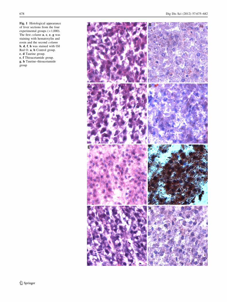
Fig. 1 Histological appearance
of liver sections from the four
experimental groups (91,000).
The first column a, c, e, g was
staining with hematoxylin and
eosin and the second columnb, d, f, h was stained with Oil
Red O. a, b Control group.
c, d Taurine group.
e, f Thioacetamide group.
g, h Taurine–thioacetamide
group
678 Dig Dis Sci (2012) 57:675–682
123

Exposure to TAA for 2 weeks significantly reduced the
SOD activity (n = 5) when compared to the control
(p \ 0.001) and TAU group (p \ 0.01). Treatment of these
fish with taurine (the TAU ? TAA group) prevented the
TAA-mediated SOD inhibition (p \ 0.001). In contrast,
CAT showed no statistically significant changes in its
activity profile between the groups (n = 4).
The Effect of Taurine on the Expression of Cytokine
mRNA
The relative mRNA expression of ADIPOR2 (Fig. 4a),
SIRT1 (Fig. 4b), and TNF-a (Fig. 4c) in hepatic tissue was
analyzed by qRT-PCR. The Kruskal–Wallis test revealed a
statistically significant difference in mRNA expression of
ADIPOR2 (p = 0.0014), SIRT1 (p = 0.0038), and TNF-a(p = 0.0079) between the groups. The expression of
ADIPOR2 was lower in the TAA group than in the control
(p \ 0.05) and TAU (p \ 0.01) groups; however, no dif-
ference was observed with the TAU ? TAA group. In
contrast, SIRT1 showed a reduction in mRNA expression
in the TAA group when compared to the control
(p \ 0.05), TAU (p \ 0.05), and TAU ? TAA (p \ 0.01)
groups. The mRNA expression of TNF-a was increased in
the TAA group compared to the control (p \ 0.05) and
TAU (p \ 0.05) groups; but, no difference was observed
with the TAU ? TAA group (p [ 0.05).
Discussion
NAFLD is a spectrum of disorders that ranges from
asymptomatic steatosis to nonalcoholic steatohepatitis
(NASH) and cirrhosis. Many factors are involved in the
pathogenesis and progression of NAFLD, such as insulin
resistance, oxidative stress, and inflammatory cascade [22].
A two-hit theory has been accepted as the hypothesis
for the chain of events that has been implicated in the
progression of steatosis to NASH [23]. This theory postu-
lates that steatosis, the first hit, increases the liver sensi-
tivity to oxidative stress and to proinflammatory cytokines,
which characterizes the second hit [24]. Recently, a third
hit—hepatocyte death and lack of repair—was included to
explain the NAFLD progression [25].
TAA is a selective and potent hepatotoxin widely used
in research as an inducer of hepatic damage [26]. To exert
its hepatotoxic effect, TAA needs to undergo two steps of
bioactivation mediated by CYP2E1. The metabolites of
TAA then form covalent bonds with liver macromolecules
[27]. Moreover, the oxidative stress seems to have an
important role in TAA-induced liver injury [28]. In
humans, greater than 5% accumulation of lipids in hepa-
tocytes is characterized as NAFLD. However, in zebrafish,
there is no cut-off percentage for this disease. Because of
this, we considered the group with a significant difference
in hepatic lipid content compared to the control group as
the definition of hepatic steatosis. The results of this study
showed that TAA treatment of zebrafish induced hepatic
steatosis in zebrafish similar to that previously described
[12].
Hepatic triglyceride formation is a protective strategy
for the liver to defend itself from lipotoxicity caused by an
overload of FFA from adipose tissue [29, 30]. These FFA
also undergo b-oxidation or become esterified with glyc-
erol to form triglycerides, which are then packaged and
exported as very low density lipoproteins (VLDL).
Therefore, hepatic fat accumulation can occur as a result of
fat synthesis or increase in delivery, reduction of fat oxi-
dation, or decrease in fat exportation [31]. Nevertheless,
hypertriglyceridemia is an independent risk factor associ-
ated with NAFLD [32]. Contrary to our expectations, this
study did not find a significant difference in serum tri-
glycerides between the groups. However, TAA-treated
zebrafish that received taurine showed an improvement in
fat lipid accumulation compared to TAA-treated zebrafish
alone. This result is in agreement with others studies that
Fig. 2 The effect of taurine and thioacetamide on the hepatic
accumulation of lipids (a) and serum triglycerides (b). To test
differences between groups, ANOVA followed by the Tukey test
were used (p \ 0.05). Data are presented as mean and SE.
a Difference between control (Ctrl) and thioacetamide (TAA) group.
b Difference between the TAA group and the co-treatment of taurine
and thioacetamide (TAA ? TAU) group
Dig Dis Sci (2012) 57:675–682 679
123

observed a reduction in hepatic steatosis after treatment
with taurine in models of hepatic injury [9, 33, 34].
Several studies have demonstrated the antioxidant role
of taurine [35, 36]. In this study, we found an increase of
lipoperoxidation in the group exposed to TAA and a pro-
tective effect of taurine on lipoperoxide formation in zeb-
rafish. We have not found any study to date that has
analyzed the effect of taurine on oxidative stress in the
zebrafish liver. Taurine has been previously evaluated in
the zebrafish brain exposed to ethanol and was shown to
prevent the increase of lipid peroxidation induced by eth-
anol [37]. Our finding is consistent with previous studies in
rats, which also observed a higher content of malondial-
dehydes in the TAA-induced hepatic damage group as well
as a reduction of hepatic lipoperoxidation when taurine was
co-administered with TAA [8, 28]. Considering that taurine
is a very stable molecule, it may have a scavenger-type role
against reactive oxygen species (ROS). In fact, it has been
demonstrated that taurine inhibits lipoperoxidation induced
by tert-butyl hydroperoxide in slices of rat liver and can
protect thiol groups from oxidation [38].
SOD and CAT act coordinately to control ROS levels.
SOD catalyzes the reaction of two superoxide anions,
resulting in the formation of hydrogen peroxide, which is
less reactive and can be degraded by enzymes such as
CAT. Our results indicated a decrease in SOD activity in
the group with TAA-induced liver damage compared to the
control; however, there were no changes in CAT activity. It
is well known that an imbalance in antioxidant defenses
promotes lipoperoxidation and oxidative stress, which is an
important factor in the progression of NAFLD, and some
studies have reported a reduction of SOD activity in the
liver of patients with NAFLD [39–41]. On the other hand,
the TAU ? TAA group presented higher levels of SOD
activity compared to the TAA group. This result differs
from some previous studies that have shown that SOD
activity was not altered after taurine treatment, however
this analysis was done in cirrhosis [8, 28]. In contrast,
Chang et al. [42] found an enhancement of SOD activity in
hamsters with steatosis induced by a high-fat/cholesterol
diet that were treated with taurine. The increase in SOD
activity and decrease in lipid peroxidation in the
TAU ? TAA group in our study confirms the hypothesis
that taurine exerts a protective effect on the oxidant-anti-
oxidant imbalance, thereby preventing oxidative stress.
Many genes are the focus of studies that attempt to
elucidate the molecular mechanisms involved in NAFLD
development and progression [43]. In this study, we found
an enhancement in TNF-a and decrease in ADIPOR2 and
SIRT1 mRNA expression in the TAA group compared to
the Ctrl group. In fact, an increase in TNF-a and a reduc-
tion in ADIPOR2 levels has been observed in individuals
with NAFLD [44, 45]. Adiponectin is an anti-inflammatory
adipokine that is antagonized by TNF-a. Low circulating
levels of adiponectin seem to contribute to the progression
of NAFLD [44]. The adiponectin effects in the liver are
predominantly mediated by the receptor ADIPOR2 and the
Fig. 3 The effect of taurine and thioacetamide on hepatic lipoperox-
idation a, SOD b, and CAT c activity. To test differences between
groups, ANOVA and the Tukey test were used (p \ 0.05). Data are
presented as mean and SE. a Difference between the control (Ctrl) and
thioacetamide (TAA) group. b Difference between the TAA group and
the co-treatment of taurine and thioacetamide (TAA ? TAU) group
680 Dig Dis Sci (2012) 57:675–682
123

mRNA expression of this receptor negatively correlates
with hepatic aminotransferases in NAFLD patients [45].
Higher ADIPOR2 expression in the liver was shown in
transgenic mice with moderate overexpression of SIRT1
[46]. In this study, we found an increase in SIRT1
expression in the TAU ? TAA group compared to the
TAA group alone.
SIRT1 is an NAD?-dependent protein deacetylase that
has been recently associated with a protective effect in
NAFLD [47]. No published study to date has evaluated the
effect of taurine on SIRT1 expression in hepatic steatosis in
adult zebrafish. The expression of SIRT1 is highest in the
male gonads and liver compared to other tissues, and has the
highest expression among other isoforms in zebrafish [48].
Deng et al. [49] found a reduction of SIRT1 expression in rats
with NAFLD caused by high-fat diets. In addition, Costa
et al. [50] found that SIRT1 expression was decreased in
visceral adipose tissue of morbidly obese patients with
severe steatosis compared to patients with slight or moderate
steatosis. Moreover, the function of SIRT1 in hepatic stea-
tosis seems to be due to the induction of SOD and lower
activation of TNF-a and interleukin 6 (IL-6), which confers a
protective effect during the second hit of NASH [46].
In conclusion, the results of this study revealed that
taurine can improve oxidative stress parameters in a model
of hepatic steatosis induced by TAA in adult wild-type
zebrafish. Furthermore, we found that taurine prevented the
decrease of SIRT1 mRNA expression caused by TAA
exposure. Based on our results, taurine may be a promising
therapy for treating hepatic steatosis.
Acknowledgments This work was financially supported by Fundo
de Incentivo a Pesquisa e Eventos of Hospital de Clınicas de Porto
Alegre (FIPE-HCPA).
References
1. Roberts EA. Pediatric nonalcoholic fatty liver disease (NAFLD):
a ‘‘growing’’ problem? J Hepatol. 2007;46:1133–1142.
2. Angulo P. Nonalcoholic fatty liver disease. N Engl J Med.
2002;346:1221–1231.
3. Browning JD, Szczepaniak LS, Dobbins R, et al. Prevalence of
hepatic steatosis in an urban population in the United States:
impact of ethnicity. Hepatology. 2004;40:1387–1395.
4. Moretto M, Kupski C, Mottin CC, et al. Hepatic steatosis in
patients undergoing bariatric surgery and its relationship to body
mass index and co-morbidities. Obes Surg. 2003;13:622–624.
5. Cotrim HP, Parise ER, Oliveira CP, et al. Nonalcoholic fatty liver
disease in Brazil. Clinical and histological profile. Ann Hepatol.2011;10:33–37.
6. OH MK, Winn J, Poordad F. Review article: diagnosis and
treatment of non-alcoholic fatty liver disease. Aliment PharmacolTher. 2008;28:503–522.
Fig. 4 The effect of taurine and thioacetamide on the mRNA
expression of ADIPOR2 (a), SIRT1 (b), and TNF-a (c). To test
differences between groups, the Kruskal–Wallis test followed by the
Dunn test were used (p \ 0.05). Each box represents the median and
interquartile range of values, with the ends of the vertical linesindicating the minimum and maximum data values. a Difference
between the control (Ctrl) and thioacetamide (TAA) group. bDifference between the TAA group and the co-treatment of taurine
and thioacetamide (TAA ? TAU) group
Dig Dis Sci (2012) 57:675–682 681
123

7. Huxtable RJ. Physiological actions of taurine. Physiol Rev.
1992;72:101–163.
8. Balkan J, Dogru-Abbasoglu S, Kanbagli O, et al. Taurine has a
protective effect against thioacetamide-induced liver cirrhosis by
decreasing oxidative stress. Hum Exp Toxicol. 2001;20:251–254.
9. Chen SW, Chen YX, Shi J, et al. The restorative effect of taurine
on experimental nonalcoholic steatohepatitis. Dig Dis Sci.2006;51:2225–2234.
10. Lam SH, Gong Z. Modeling liver cancer using zebrafish: a
comparative oncogenomics approach. Cell Cycle. 2006;5:
573–577.
11. Passeri MJ, Cinaroglu A, Gao C, et al. Hepatic steatosis in
response to acute alcohol exposure in zebrafish requires sterol
regulatory element binding protein activation. Hepatology.
2009;49:443–452.
12. Rekha RD, Amali AA, Her GM, et al. Thioacetamide accelerates
steatohepatitis, cirrhosis and HCC by expressing HCV core
protein in transgenic zebrafish Danio rerio. Toxicology.
2008;243:11–22.
13. Amali AA, Rekha RD, Lin CJ, et al. Thioacetamide induced liver
damage in zebrafish embryo as a disease model for steatohepa-
titis. J Biomed Sci. 2006;13:225–232.
14. McGrath P, Li CQ. Zebrafish: a predictive model for assessing
drug-induced toxicity. Drug Discov Today. 2008;13:394–401.
15. Kinkel AD, Fernyhough ME, Helterline DL, et al. Oil red-O
stains non-adipogenic cells: a precautionary note. Cytotechnol-ogy. 2004;46:49–56.
16. Jagadeeswaran P, Sheehan JP, Craig FE, et al. Identification and
characterization of zebrafish thrombocytes. Br J Haematol.1999;107:731–738.
17. Buege JA, Aust SD. Microsomal lipid peroxidation. MethodsEnzymol. 1978;52:302–310.
18. Misra HP, Fridovich I. The role of superoxide anion in the
autoxidation of epinephrine and a simple assay for superoxide
dismutase. J Biol Chem. 1972;247:3170–3175.
19. Boveris A, Chance B. The mitochondrial generation of hydrogen
peroxide. General properties and effect of hyperbaric oxygen.
Biochem J. 1973;134:707–716.
20. Bradford MM. A rapid and sensitive method for the quantitation
of microgram quantities of protein utilizing the principle of
protein-dye binding. Anal Biochem. 1976;72:248–254.
21. Livak KJ, Schmittgen TD. Analysis of relative gene expression
data using real-time quantitative PCR and the 2[-delta delta
C(T)] method. Methods. 2001;25:402–408.
22. Lewis J, Mohanty S. Nonalcoholic fatty liver disease: a review
and update. Dig Dis Sci. 2010;55:560–578.
23. Day CP. Pathogenesis of steatohepatitis. Best Pract Res ClinGastroenterol. 2002;16:663–678.
24. Day CP, James OF. Steatohepatitis: a tale of two ‘‘hits’’?
Gastroenterology. 1998;114:842–845.
25. Tiniakos DG, Vos MB, Brunt EM. Nonalcoholic fatty liver dis-
ease: pathology and pathogenesis. Annu Rev Pathol. 2010;5:
145–171.
26. Chilakapati J, Shankar K, Korrapati MC, et al. Saturation toxic-
okinetics of thioacetamide: role in initiation of liver injury. DrugMetab Dispos. 2005;33:1877–1885.
27. Tunez I, Munoz MC, Villavicencio MA, et al. Hepato- and
neurotoxicity induced by thioacetamide: protective effects of
melatonin and dimethylsulfoxide. Pharmacol Res. 2005;52:
223–228.28. Dogru-Abbasoglu S, Kanbagli O, Balkan J, et al. The protective
effect of taurine against thioacetamide hepatotoxicity of rats.
Hum Exp Toxicol. 2001;20:23–27.
29. Law K, Brunt EM. Nonalcoholic fatty liver disease. Clin LiverDis. 2010;14:591–604.
30. Yamaguchi K, Yang L, McCall S, et al. Inhibiting triglyceride
synthesis improves hepatic steatosis but exacerbates liver damage
and fibrosis in obese mice with nonalcoholic steatohepatitis.
Hepatology. 2007;45:1366–1374.
31. Dowman JK, Tomlinson JW, Newsome PN. Pathogenesis of non-
alcoholic fatty liver disease. Qjm. 2010;103:71–83.
32. Assy N, Kaita K, Mymin D, et al. Fatty infiltration of liver in
hyperlipidemic patients. Dig Dis Sci. 2000;45:1929–1934.
33. Tasci I, Mas N, Mas MR, et al. Ultrastructural changes in
hepatocytes after taurine treatment in CCl4 induced liver injury.
World J Gastroenterol. 2008;14:4897–4902.
34. Chen X, Sebastian BM, Tang H, et al. Taurine supplementation
prevents ethanol-induced decrease in serum adiponectin and
reduces hepatic steatosis in rats. Hepatology. 2009;49:
1554–1562.
35. Schaffer SW, Azuma J, Mozaffari M. Role of antioxidant activity
of taurine in diabetes. Can J Physiol Pharmacol. 2009;87:91–97.
36. Lakshmi Devi S, Anuradha CV. Mitochondrial damage, cyto-
toxicity and apoptosis in iron-potentiated alcoholic liver fibrosis:
amelioration by taurine. Amino Acids. 2010;38:869–879.
37. Rosemberg DB, da Rocha RF, Rico EP, et al. Taurine prevents
enhancement of acetylcholinesterase activity induced by acute
ethanol exposure and decreases the level of markers of oxidative
stress in zebrafish brain. Neuroscience. 2010;171:683–692.
38. Oliveira MW, Minotto JB, de Oliveira MR, et al. Scavenging and
antioxidant potential of physiological taurine concentrations
against different reactive oxygen/nitrogen species. PharmacolRep. 2010;62:185–193.
39. Oliveira CP, Coelho AM, Barbeiro HV, et al. Liver mitochondrial
dysfunction and oxidative stress in the pathogenesis of experi-
mental nonalcoholic fatty liver disease. Braz J Med Biol Res.
2006;39:189–194.
40. Videla LA, Rodrigo R, Orellana M, et al. Oxidative stress-related
parameters in the liver of non-alcoholic fatty liver disease
patients. Clin Sci (Lond). 2004;106:261–268.
41. Yesilova Z, Yaman H, Oktenli C, et al. Systemic markers of lipid
peroxidation and antioxidants in patients with nonalcoholic fatty
liver disease. Am J Gastroenterol. 2005;100:850–855.
42. Chang YY, Chou CH, Chiu CH, et al. Preventive effects of
taurine on development of hepatic steatosis induced by a high-fat/
cholesterol dietary habit. J Agric Food Chem. 2011;59:450–457.
43. Malaguarnera M, Di Rosa M, Nicoletti F, et al. Molecular
mechanisms involved in NAFLD progression. J Mol Med.
2009;87:679–695.
44. Jou J, Choi SS, Diehl AM. Mechanisms of disease progression in
nonalcoholic fatty liver disease. Semin Liver Dis. 2008;28:
370–379.
45. Kaser S, Moschen A, Cayon A, et al. Adiponectin and its
receptors in non-alcoholic steatohepatitis. Gut. 2005;54:117–121.
46. Banks AS, Kon N, Knight C, et al. SirT1 gain of function
increases energy efficiency and prevents diabetes in mice. CellMetab. 2008;8:333–341.
47. Colak Y, Ozturk O, Senates E, et al. SIRT1 as a potential ther-
apeutic target for treatment of nonalcoholic fatty liver disease.
Med Sci Monit. 2011;17:HY5–HY9.
48. Pereira TCB, Rico EP, Rosemberg DB, et al. Zebrafish as a
model organism to evaluate drugs potentially able to modulate
sirtuin expression. Zebrafish. 2011;8:9–16.
49. Deng XQ, Chen LL, Li NX. The expression of SIRT1 in nonal-
coholic fatty liver disease induced by high-fat diet in rats. LiverInt. 2007;27:708–715.
50. dos Costa CS, Hammes TO, Rohden F, et al. SIRT1 transcription
is decreased in visceral adipose tissue of morbidly obese patients
with severe hepatic steatosis. Obes Surg. 2010;20:633–639.
682 Dig Dis Sci (2012) 57:675–682
123
Copyright © 2022 FDOKUMEN


![[Hepatic steatosis, visceral fat and metabolic alterations in apparently healthy overweight/obese individuals]](https://static.fdokumen.com/doc/165x107/6324f8237fd2bfd0cb03375f/hepatic-steatosis-visceral-fat-and-metabolic-alterations-in-apparently-healthy.jpg)