Phyllanthus urinaria extract attenuates acetaminophen induced hepatotoxicity: Involvement of...
10
Phytomedicine 16 (2009) 751–760 Phyllanthus urinaria extract attenuates acetaminophen induced hepatotoxicity: Involvement of cytochrome P450 CYP2E1 Desmond Kwok Po Hau a , Roberto Gambari b , Raymond Siu Ming Wong c , Marcus Chun Wah Yuen d , Gregory Yin Ming Cheng c , Cindy Sze Wai Tong c , Guo Yuan Zhu a , Alexander Kai Man Leung a , Paul Bo San Lai e , Fung Yi Lau c , Andrew Kit Wah Chan c , Wai Yeung Wong f , Stanton Hon Lung Kok d , Chor Hing Cheng d , Chi Wai Kan d , Albert Sun Chi Chan d , Chung Hin Chui c, , Johnny Cheuk On Tang d, , David Wang Fun Fong a, a Research and Development Division, School of Chinese Medicine, Hong Kong Baptist University, Hong Kong, China b BioPharmaNet, Department of Biochemistry and Molecular Biology, The University of Ferrara, Ferrara, Italy c Department of Medicine and Therapeutics, Li Ka Shing Medical Sciences Building, Prince of Wales Hospital, The Chinese University of Hong Kong, Hong Kong, China d Institute of Textiles and Clothing and Applied Biology, The Hong Kong Polytechnic University, Hong Kong, China e Department of Surgery, Li Ka Shing Medical Sciences Building, Prince of Wales Hospital, The Chinese University of Hong Kong, Hong Kong, China f Department of Chemistry, Hong Kong Baptist University, Hong Kong, China Abstract Acetaminophen is a commonly used drug for the treatment of patients with common cold and influenza. However, an overdose of acetaminophen may be fatal. In this study we investigated whether mice, administered intraperitoneally with a lethal dose of acetaminophen, when followed by oral administration of Phyllanthus urinaria extract, may be prevented from death. Histopathological analysis of mouse liver sections showed that Phyllanthus urinaria extract may protect the hepatocytes from acetaminophen-induced necrosis. Therapeutic dose of Phyllanthus urinaria extract did not show any toxicological phenomenon on mice. Immunohistochemical staining with the cytochrome P450 CYP2E1 antibody revealed that Phyllanthus urinaria extract reduced the cytochrome P450 CYP2E1 protein level in mice pre- treated with a lethal dose of acetaminophen. Phyllanthus urinaria extract also inhibited the cytochrome P450 CYP2E1 enzymatic activity in vitro. Heavy metals, including arsenic, cadmium, mercury and lead, as well as herbicide residues were not found above their detection limits. High performance liquid chromatography identified corilagin and gallic acid as the major components of the Phyllanthus urinaria extract. We conclude that Phyllanthus urinaria extract is effective in attenuating the acetaminophen induced hepatotoxicity, and inhibition of cytochrome P450 CYP2E1 enzyme may be an important factor for its therapeutic mechanism. r 2009 Elsevier GmbH. All rights reserved. Keywords: Acetaminophen; Cytochrome 450 CYP2E1; Hepatoprotection; Hepatotoxicity; Phyllanthus urinaria ARTICLE IN PRESS www.elsevier.de/phymed 0944-7113/$ - see front matter r 2009 Elsevier GmbH. All rights reserved. doi:10.1016/j.phymed.2009.01.008 Corresponding author. Tel.: +852 2632 3120; fax: +852 2637 5396 (C.H. Chui). Corresponding author. Tel.: +852 3400 8727; fax: +852 2364 9932 (J.C.O. Tang). Corresponding author. Tel.: +852 3411 5308; fax: +852 3411 2902 (D.W.F. Fong). E-mail addresses: [email protected] (C.H. Chui), [email protected] (J.C.O. Tang), [email protected] (D.W.F. Fong).
Transcript of Phyllanthus urinaria extract attenuates acetaminophen induced hepatotoxicity: Involvement of...
ARTICLE IN PRESS
0944-7113/$ - se
doi:10.1016/j.ph
�Correspond�Correspond�CorrespondE-mail addr
Phytomedicine 16 (2009) 751–760
www.elsevier.de/phymed
Phyllanthus urinaria extract attenuates acetaminophen induced
hepatotoxicity: Involvement of cytochrome P450 CYP2E1
Desmond Kwok Po Haua, Roberto Gambarib, Raymond Siu Ming Wongc,Marcus Chun Wah Yuend, Gregory Yin Ming Chengc, Cindy Sze Wai Tongc,Guo Yuan Zhua, Alexander Kai Man Leunga, Paul Bo San Laie, Fung Yi Lauc,Andrew Kit Wah Chanc, Wai Yeung Wongf, Stanton Hon Lung Kokd,Chor Hing Chengd, Chi Wai Kand, Albert Sun Chi Chand, Chung Hin Chuic,�,Johnny Cheuk On Tangd,�, David Wang Fun Fonga,�
aResearch and Development Division, School of Chinese Medicine, Hong Kong Baptist University, Hong Kong, ChinabBioPharmaNet, Department of Biochemistry and Molecular Biology, The University of Ferrara, Ferrara, ItalycDepartment of Medicine and Therapeutics, Li Ka Shing Medical Sciences Building, Prince of Wales Hospital,
The Chinese University of Hong Kong, Hong Kong, ChinadInstitute of Textiles and Clothing and Applied Biology, The Hong Kong Polytechnic University, Hong Kong, ChinaeDepartment of Surgery, Li Ka Shing Medical Sciences Building, Prince of Wales Hospital, The Chinese University of Hong Kong,
Hong Kong, ChinafDepartment of Chemistry, Hong Kong Baptist University, Hong Kong, China
Abstract
Acetaminophen is a commonly used drug for the treatment of patients with common cold and influenza. However,an overdose of acetaminophen may be fatal. In this study we investigated whether mice, administered intraperitoneallywith a lethal dose of acetaminophen, when followed by oral administration of Phyllanthus urinaria extract, may beprevented from death. Histopathological analysis of mouse liver sections showed that Phyllanthus urinaria extract mayprotect the hepatocytes from acetaminophen-induced necrosis. Therapeutic dose of Phyllanthus urinaria extract did notshow any toxicological phenomenon on mice. Immunohistochemical staining with the cytochrome P450 CYP2E1antibody revealed that Phyllanthus urinaria extract reduced the cytochrome P450 CYP2E1 protein level in mice pre-treated with a lethal dose of acetaminophen. Phyllanthus urinaria extract also inhibited the cytochrome P450 CYP2E1enzymatic activity in vitro. Heavy metals, including arsenic, cadmium, mercury and lead, as well as herbicide residueswere not found above their detection limits. High performance liquid chromatography identified corilagin and gallicacid as the major components of the Phyllanthus urinaria extract. We conclude that Phyllanthus urinaria extract iseffective in attenuating the acetaminophen induced hepatotoxicity, and inhibition of cytochrome P450 CYP2E1enzyme may be an important factor for its therapeutic mechanism.r 2009 Elsevier GmbH. All rights reserved.
Keywords: Acetaminophen; Cytochrome 450 CYP2E1; Hepatoprotection; Hepatotoxicity; Phyllanthus urinaria
e front matter r 2009 Elsevier GmbH. All rights reserved.
ymed.2009.01.008
ing author. Tel.: +852 2632 3120; fax: +852 2637 5396 (C.H. Chui).
ing author. Tel.: +852 3400 8727; fax: +852 2364 9932 (J.C.O. Tang).
ing author. Tel.: +852 3411 5308; fax: +852 3411 2902 (D.W.F. Fong).
esses: [email protected] (C.H. Chui), [email protected] (J.C.O. Tang), [email protected] (D.W.F. Fong).
ARTICLE IN PRESSD.K.P. Hau et al. / Phytomedicine 16 (2009) 751–760752
Introduction
Acetaminophen (APAP) has been widely used as amedicine for pain and fever relief (Whitcomb 1994).Since APAP can be purchased easily from anypharmaceutical outlet and even from supermarkets,without prescriptions from clinicians, it is commonlyconsidered as a ‘‘safe drug’’ when taken within thesuggested therapeutic dose. However, APAP can behepatotoxic when an overdose is administered and,warning messages are present in the package. Clinically,APAP has been demonstrated to be nephrotoxic andhepatotoxic from animal experiments and in humanbeings (Curry et al. 1982; Keaton 1988; Vermeulen et al.1992; Bonkovsky et al. 1994).
The use of herbal extracts in the treatment of humandiseases is becoming very popular worldwide. Scientificapproaches further magnify the reliability of the use ofherbal extracts as complimentary medicine. Extracts andmolecules from medicinal plants have been demon-strated to be important in the development of agentsagainst human viruses, such as herpes simplex viruses Iand II (Khan et al. 2005) and antitumor drugs(Lampronti et al. 2003). They are also compounds ofinterest in the treatment of genetic diseases, includingthalassemia (Bianchi et al. 2008). The ethanol extractfrom anomalous fruit of Gleditsia sinensis has been welldocumented for its anticancer properties on humancancer cell lines (Chui et al. 2005; Tang et al. 2007).
In this respect, Phyllanthus urinaria (P. urinaria) hasbeen extensively investigated for its possible anticanceractivity. The boiled water extract from the whole plant ofP. urinaria has been reported to induce apoptosis in anumber of human cancer cell lines, including leukaemia,hepatoblastoma, nasopharyngeal carcinoma and fibrosar-coma but not the normal human endothelial cells andliver cells (Huang et al. 2004a). The bcl-2 anti-apoptoticprotein was further shown to be down-regulated aftertreatment of Lewis lung carcinoma cells with this boiledwater extract of P. urinaria (Huang et al. 2003). Furthermechanistic investigation using HL-60 human acutepromyelocytic leukaemia cell line suggested that inductionof apoptosis by the boiled water extracts of P. urinaria isassociated with the activation of the CD95 Fas receptor/ligand expression and ceramide-mediated pathways(Huang et al. 2004b). This boiled water extract ofP. urinaria was further shown to exhibit anti-tumor andanti-angiogenic effects in mice bearing Lewis lungcarcinoma. P. urinaria can reduce the blood vessel densityand decrease the matrix induced tube formation of humanumbilical cord endothelial cells as well as the theirmigration (Huang et al. 2006). The medicinal use ofP. urinaria, however, is not restricted to the treatment ofneoplastic diseases. The use of P. urinaria as hepatopro-tective agent in tetrachloromethane induced hepatic injuryhas been previously documented (Lee et al. 2006).
In the present study, we conducted experimentsdesigned to explore the hepatoprotective activity ofP. urinaria in a mice model twenty four hours after theadministration of a lethal dose of APAP (550mg/kg).This APAP dosage causes, in C57Bl6 mice, a very lowsurvival rate, since the majority of them succumb withintwo weeks (Wong et al., personal communication). Theemployed experimental model system simulates aclinical condition similar to those of patients admittedfor acute liver injury to the emergency departments ofhospitals. Our studies were designed to determinewhether P. urinaria extracts may be effective inattenuating the APAP induced hepatotoxicity; cyto-chrome P450 CYP2E1 enzyme was chosen as abiochemical marker and may be an important factorby which to further explore the mechanisms of potentialtherapeutic relevance in our animal experimental modelsystem.
Materials and methods
Chemicals and reagents
Unless otherwise stated, all the reagents, includingAPAP, were purchased from Sigma chemicals. Thephysiological saline for APAP injection was obtainedfrom Baxter. Silymarin was purchased from Sigmachemical. Primary antibody conjugated with biotinagainst mouse cytochrome P450 CYP2E1 and substratefor peroxidase were purchased from US Biologicals,while secondary antibody and the subsequent signaldetection reagents were purchased from Dako. Thein vitro cytochrome P450 CYP2E1 kit was purchasedfrom In Vitrogen.
Preparation of the P. urinaria extract
P. urinaria in powdered form was kindly provided bythe Bioactive Technologies Ltd. (Hong Kong). Briefly,whole plants of P. urinaria were identified, confirmedand a voucher sample was kept by the company. Theplants were then excised and 5 kilograms (dry weight)was extracted with 30 litres of 80% ethanol for 90min.The percentage of yield was 11%. Afterwards, the driedpowder was dissolved completely in distilled water andadjusted to a final concentration of 10mg/ml. Thegreenish yellow mixture was sterile filtered and stored at�20 1C until future use.
Animal care
Eight weeks old C57Bl6 mice, weighing approxi-mately 20–25 g, were purchased from the animal unit ofThe Chinese University of Hong Kong and maintained

ARTICLE IN PRESSD.K.P. Hau et al. / Phytomedicine 16 (2009) 751–760 753
in a conventional sanitary facility, in accordance withthe institutional guidelines on animal care, with therequired consistent temperature and relative humidity.All the procedures were approved by the AnimalResearch Ethics Committee.
APAP treatment on mice
APAP was dissolved in physiological saline. A total of37 mice were included in the study. On day one, acuteliver injury was induced by intraperitoneally (i.p.)administered APAP at a dose of 550mg/kg of bodyweight. From day two to day four, treatment groupsreceived various doses of P. urinaria extract or silymarin(positive reference) once daily while APAP groupreceived water. Two additional groups consisted of micetreated with (i) buffer i.p. at day 1 and P. urinaria
extract at the dose of 200mg/kg daily for three days, and(ii) buffer i.p. at day 1 and water for three days. Themortality rate and change of body weight in the animalswere monitored and recorded. On day 5, all the micewere sacrificed and autopsies was performed to collectvital organs.
Haematoxylin and eosin (H and E) histochemistry
staining
Sections of mouse liver from autopsy samples weredewaxed, washed with phosphate buffered saline (PBS)and then stained with H and E for nucleus andcytoplasm staining using the conventional protocolreported elsewhere. Slides were then premounted andinspected under a light microscope.
Immunohistochemistry analysis of cytochrome 450
CYP2E1
Sections of mouse liver from autopsy samples weredewaxed with xylene and gradient concentrations ofethanol. Possible endogenous peroxidase was blockedand slides were washed with PBS. Slides were thenblocked again and treated with diluted primary anti-body (rabbit anti-rat cytochrome P450 CYP2E1) inPBS. Slides were washed with PBS and then treated withthe secondary antibody CSA II rabbit link. Afterwashing, slides were further treated with amplificationreagent and anti-fluorescein-HRP. Afterwards, slideswere incubated with DAB substrate. Nuclei were stainedwith haematoxylin and finally slides were inspectedunder a light microscope.
In vitro cytochrome P450 CYP2E1 enzyme assay
Detailed procedures can be found in the user guidemanual supplied with the reagent kit. Briefly, various
concentrations of P. urinaria extract were mixed withthe reagent buffer, the cytochrome P450 CYP2E1enzyme and the regeneration system. Before the finaladdition of the substrate for the cytochrome P450CYP2E1 enzyme, an excitation emission reading wasrecorded for any possible background fluorescence. Thesubstrate was added, and after 30min incubation,diethyldithiocarbamate, at a final concentration of100 mM, was added to terminate the reaction. A secondexcitation emission reading was recorded, and resultswere analysed.
Analytical chemistry analysis for P. urinaria extract
Any possible contaminations of heavy metal andherbicide from P. urinaria extract were examined.The herbicide list included aldrin, cis-chlordane, trans-chlordane, oxychlordane, p,p0-DDD (4,40-DDD), p,p0-DDE, (4,40-DDE), o,p0-DDT (2,40-DDT), p,p0-DDT(4,40-DDT), dieldrin, endrin, heptachlor, heptachlorepoxide isomer B, hexachlorobenzene, hexachlorocyclo-hexane (BHC) a-isomer, hexachlorocyclohexane (BHC)b-isomer, hexachlorocyclohexane (BHC) g-isomer (lin-dane), hexachlorocyclohexane (BHC) d-isomer, methylpentachlorophenyl sulphide (MPCPS), pentachloroani-line and pentachloronitrobenzene (quintozene). Threeindependent tests were performed and results wereexpressed as mean 7 standard deviations from threeindependent experiments.
Method for heavy metals analysis
Individual stock standard solution of arsenic (As),cadmium (Cd), mercury (Hg) and lead (Pb; 1000mg/l)were purchased from The National Institute of Metrol-ogy, PR China, and were of purity X99.99%. Stocksolutions containing As, Cd and Pb each at 200 mg/l andHg at 100 mg/l were prepared in 2% v/v nitric acidsolution. The working solutions ranged from 0.2 to20 mg/l for As, Cd and Pb and from 0.2 to 20 mg/l for Hgand were obtained daily by appropriate dilutions with2% v/v nitric acid solution. Indium (In) and bismuth(Bi) at 2mg/l and germanium (Ge) at 20mg/l wereadded to the working solutions as an internal standard.Water (18MO) was prepared with a Milli-Q system(Millipore, US). It was further analyzed by ICP-MS(PerkinElmer Sciex Elan 6100 inductive couple plasmamass spectrometer equipped with a concentric quartznebulizer).
Method for pesticide residues analysis
Individual stock standard solutions of organochlorinepesticide (500mg/l) were purchased from Supercol andChem Service, and were of 498% purity. Stock
ARTICLE IN PRESS
Table 1. HPLC mobile phase condition for the ingredients
identification in P. urinaria extract.
Time (min) A (%) B (%)
0 3 97
10 3 97
15 10 90
40 20 80
45 30 70
50 3 97
60 3 97
A: Acetonitrile. B: 0.1% trifluoroacetic acid. The percentage is in terms
of total volume.
D.K.P. Hau et al. / Phytomedicine 16 (2009) 751–760754
solutions containing 20 pesticides each at concentrationsranging from 2 to 10mg/l were prepared in iso-octaneand stored at �4 1C in amber glass bottles. The workingsolutions ranging from 0.02 to 0.15mg/l were obtaineddaily by appropriate dilutions with iso-octane. 1-Bromo-2-nitrobenzene at 0.2mg/l was added to the workingsolutions as an internal standard. All the solvents(acetone, dichloromethane, ethyl acetate, n-hexane andiso-octane (LabScan, Thailand)) were of pesticide grade.Anhydrous sodium sulphate (Sigma-Aldrich, US) was ofanalytical grade. Analytical regent grade chemicals andwater (18MO) were prepared with a Milli-Q system(Millipore, US).
GC-ECD condition
Gas chromatographic analyses were performed on anAligent 6890 gas chromatograph equipped with anelectron capture detector. A DB-17MS fused silicacapillary column of 30m� 0.25mm i.d. and 0.25m filmthickness from J&W Scientific was used. Helium(purityX99.999%) was used as a carrier gas at a flowrate of 1ml/min. A one liter extract was injected insplitless mode. The injection temperature was 210 1C.The oven temperature was programmed from initialtemperature 100 1C (held for 2min) to 165 1C at20 1C/min, followed by 200 1C at 10 1C/min, 230 1C at2 1C/min, 265 1C at 3 1C/min and finally at 280 1C (heldfor 10min.). The detector was 300 1C. Results werefurther confirmed by GC-MS.
Pesticide residues sample preparation
Sample was mixed with 100ml ethyl acetate and 4 ganhydrous sodium chloride. The mixture was sonicatedfor 15min and the solids were allowed to settle. Afterdouble extraction of residue each with 50ml ethylacetate, the supernatant was combined and evaporatedto near dryness. The residual extract was diluted to10ml with dichloromethane for subsequent purificationin GPC.
HPLC analysis for P. urinaria extract
To identify major components from the P. urinaria,HPLC analysis was performed. Corilagin and gallic acidwere used as markers and serial combinations wereprepared. Agilent 1100 series HPLC and Symmetry C18(5 mm, 4.6mm� 250mm) column was used and detectedwith DAD detection using wavelength of 270 nm.
Mobile phase consisted of acetonitrile and trifluor-oacetic acid (0.1%) while the gradient is shown inTable 1. Flow rate was adjusted to 0.8ml per minute andinjection volume was 10 ml. Then calibration curves for
both markers were set where peak areas were correlatedto the corresponding concentrations. According to theretention time, markers were identified from theP. urinaria extract again and the relative concentrationswere further estimated according to their correspondingpeak areas. The calculated results for the concentrationof major components were expressed as mean7standard derivation obtained from three independentexperiments.
Results
P. urinaria extract inhibits APAP induced
hepatotoxicity in vivo
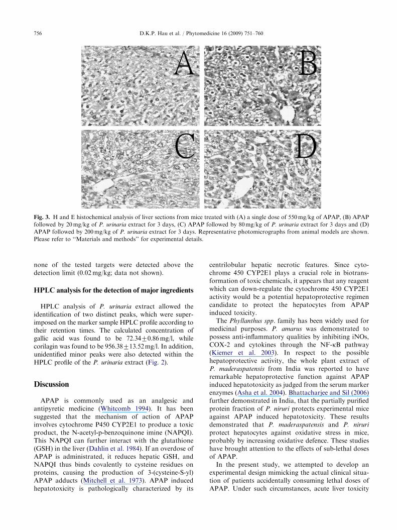
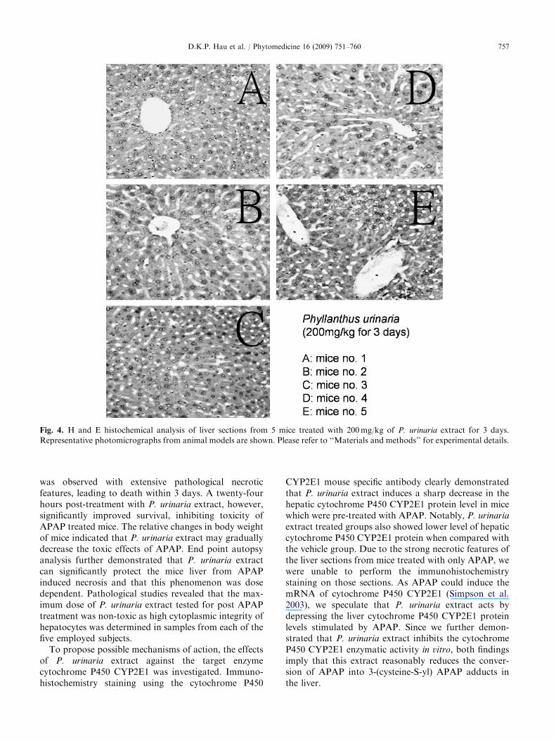
Table 2 shows that when APAP was administratedintraperitoneally to mice at a dose of 550mg/kg withoutfurther treatments, a high mortality rate and rapid dropin body weight (Fig. 1) were observed. Hematoxylin (H)and Eosin (E) staining of the liver autopsy samplesshowed extensive necrotic features (Fig. 3A). Whenmice were treated with APAP together with oraladministration of P. urinaria extract, an improvementof survival rate (Table 2) as well as body weightvariation (Fig. 1) was observed and noticeably, itsimprovement of survival rate is similar to the positivereference, silymarin (Table 2). After increasing the doseof P. urinaria extracts from 20 to 200mg/kg per day fora continuous treatment of three days, liver autopsiesalso showed significant improvement in cytoplasmintegrity (Figs. 3B to D). Each of the five control miceonly treated with 200mg/kg of P. urinaria for 3 daysexhibited no evidence of necrotic feature with highintegrity of cytoplasm, as was evident from the H and Estaining of liver autopsy sections (Fig. 4). The bodyweight of animals which have been treated with onlyvehicle or P. urinaria were found to be increased (datanot shown).

ARTICLE IN PRESS
Fig. 1. Mean decrease in body weight of mice from days 2 to 5
(compared with day 1) after single dose treatment of APAP
(550mg/kg) on day 1. Results are shown as mean7standard
derivations from individual animal group (please refer to
Table 2 for corresponding number).
Table 2. Survival percentage of mice treated with various
combinations of APAP and P. urinaria extract.
Number of day
1 2 3 4 5
Percentage of mice survival
0 (n ¼ 5) 100 100 100 100 100
APAP only (n ¼ 5) 100 80 0 0 0
APAP+20mg/kga (n ¼ 5) 100 100 100 100 100
APAP+40mg/kga (n ¼ 5) 100 100 100 100 80
APAP+80mg/kga (n ¼ 7) 100 100 100 100 85
APAP+200mg/kga (n ¼ 5) 100 100 100 100 100
APAP+100mg/kgb (n ¼ 7) 100 100 100 100 100
APAP+200mg/kgb (n ¼ 7) 100 100 100 100 100
200mg/kga (n ¼ 5) 100 100 100 100 100
APAP (single dose at 550mg/kg on day 1).
‘‘n’’—the number of mice involved in the corresponding experimental
group.aP. urinaria extract (single dose daily for 3 consecutive days from
day 2 to 4 after a single dose of APAP at 550mg/kg on day 1).bSilymarin (single dose daily for 3 consecutive days from day 2 to 4
after a single dose of APAP at 550mg/kg on day 1).
Fig. 2. HPLC analysis for (A) P. urinaria extract and (B) two
markers including gallic acid (retention time �11.062min) and
corilagin (retention time �31.118min). Three independent
experiments were performed and similar results obtained.
Shown are representative results from one experiment. Please
refer to ‘‘Materials and methods’’ for experimental details.
D.K.P. Hau et al. / Phytomedicine 16 (2009) 751–760 755
Cytochrome P450 CYP2E1 is involved in the
protective activity of P. urinaria extract against
APAP induced hepatotoxicity
Sections of liver autopsy samples from treated micewere further investigated for any possible changes in theprotein level of cytochrome P450 CYP2E1 by using aspecific antibody. As shown in Fig. 5, samples fromvehicle treated control mice showed relatively higherprotein level of cytochrome P450 CYP2E1 whencompared with samples obtained from mice treatedwith only P. urinaria extract (Figs. 5A and B). The
inhibitory effects of P. urinaria extract were confirmedin the liver autopsy sections from mice treated withAPAP followed by P. urinaria extract; in this case aneven higher decrease in the protein level of cytochromeP450 CYP2E1 was observed (Fig. 5C). In agreementwith the data shown in Fig. 5, the in vitro enzymaticassay for the effect of P. urinaria extract on cytochromeP450 CYP2E1 showed a dose dependent inhibition(Fig. 6). The 50% inhibitory concentration was about500 mg/ml under this experimental system.
Analytical chemistry for the detection of heavy
metals and herbicides
Analytical chemistry assays of P. urinaria extractshowed that heavy metals, including arsenic, cadmium,mercury and lead, were not present above the detectionlimit (0.05mg/kg). With respect to herbicide residues,
ARTICLE IN PRESS
Fig. 3. H and E histochemical analysis of liver sections from mice treated with (A) a single dose of 550mg/kg of APAP, (B) APAP
followed by 20mg/kg of P. urinaria extract for 3 days, (C) APAP followed by 80mg/kg of P. urinaria extract for 3 days and (D)
APAP followed by 200mg/kg of P. urinaria extract for 3 days. Representative photomicrographs from animal models are shown.
Please refer to ‘‘Materials and methods’’ for experimental details.
D.K.P. Hau et al. / Phytomedicine 16 (2009) 751–760756
none of the tested targets were detected above thedetection limit (0.02mg/kg; data not shown).
HPLC analysis for the detection of major ingredients
HPLC analysis of P. urinaria extract allowed theidentification of two distinct peaks, which were super-imposed on the marker sample HPLC profile according totheir retention times. The calculated concentration ofgallic acid was found to be 72.3470.86mg/l, whilecorilagin was found to be 956.38713.52mg/l. In addition,unidentified minor peaks were also detected within theHPLC profile of the P. urinaria extract (Fig. 2).
Discussion
APAP is commonly used as an analgesic andantipyretic medicine (Whitcomb 1994). It has beensuggested that the mechanism of action of APAPinvolves cytochrome P450 CYP2E1 to produce a toxicproduct, the N-acetyl-p-benzoquinone imine (NAPQI).This NAPQI can further interact with the glutathione(GSH) in the liver (Dahlin et al. 1984). If an overdose ofAPAP is administrated, it reduces hepatic GSH, andNAPQI thus binds covalently to cysteine residues onproteins, causing the production of 3-(cysteine-S-yl)APAP adducts (Mitchell et al. 1973). APAP inducedhepatotoxicity is pathologically characterized by its
centrilobular hepatic necrotic features. Since cyto-chrome 450 CYP2E1 plays a crucial role in biotrans-formation of toxic chemicals, it appears that any reagentwhich can down-regulate the cytochrome 450 CYP2E1activity would be a potential hepatoprotective regimencandidate to protect the hepatocytes from APAPinduced toxicity.
The Phyllanthus spp. family has been widely used formedicinal purposes. P. amarus was demonstrated topossess anti-inflammatory qualities by inhibiting iNOs,COX-2 and cytokines through the NF-kB pathway(Kiemer et al. 2003). In respect to the possiblehepatoprotective activity, the whole plant extract ofP. maderaspatensis from India was reported to haveremarkable hepatoprotective function against APAPinduced hepatotoxicity as judged from the serum markerenzymes (Asha et al. 2004). Bhattacharjee and Sil (2006)further demonstrated in India, that the partially purifiedprotein fraction of P. niruri protects experimental miceagainst APAP induced hepatotoxicity. These resultsdemonstrated that P. maderaspatensis and P. niruri
protect hepatocytes against oxidative stress in mice,probably by increasing oxidative defence. These studieshave brought attention to the effects of sub-lethal dosesof APAP.
In the present study, we attempted to develop anexperimental design mimicking the actual clinical situa-tion of patients accidentally consuming lethal doses ofAPAP. Under such circumstances, acute liver toxicity
ARTICLE IN PRESS
Fig. 4. H and E histochemical analysis of liver sections from 5 mice treated with 200mg/kg of P. urinaria extract for 3 days.
Representative photomicrographs from animal models are shown. Please refer to ‘‘Materials and methods’’ for experimental details.
D.K.P. Hau et al. / Phytomedicine 16 (2009) 751–760 757
was observed with extensive pathological necroticfeatures, leading to death within 3 days. A twenty-fourhours post-treatment with P. urinaria extract, however,significantly improved survival, inhibiting toxicity ofAPAP treated mice. The relative changes in body weightof mice indicated that P. urinaria extract may graduallydecrease the toxic effects of APAP. End point autopsyanalysis further demonstrated that P. urinaria extractcan significantly protect the mice liver from APAPinduced necrosis and that this phenomenon was dosedependent. Pathological studies revealed that the max-imum dose of P. urinaria extract tested for post APAPtreatment was non-toxic as high cytoplasmic integrity ofhepatocytes was determined in samples from each of thefive employed subjects.
To propose possible mechanisms of action, the effectsof P. urinaria extract against the target enzymecytochrome P450 CYP2E1 was investigated. Immuno-histochemistry staining using the cytochrome P450
CYP2E1 mouse specific antibody clearly demonstratedthat P. urinaria extract induces a sharp decrease in thehepatic cytochrome P450 CYP2E1 protein level in micewhich were pre-treated with APAP. Notably, P. urinaria
extract treated groups also showed lower level of hepaticcytochrome P450 CYP2E1 protein when compared withthe vehicle group. Due to the strong necrotic features ofthe liver sections from mice treated with only APAP, wewere unable to perform the immunohistochemistrystaining on those sections. As APAP could induce themRNA of cytochrome P450 CYP2E1 (Simpson et al.2003), we speculate that P. urinaria extract acts bydepressing the liver cytochrome P450 CYP2E1 proteinlevels stimulated by APAP. Since we further demon-strated that P. urinaria extract inhibits the cytochromeP450 CYP2E1 enzymatic activity in vitro, both findingsimply that this extract reasonably reduces the conver-sion of APAP into 3-(cysteine-S-yl) APAP adducts inthe liver.

ARTICLE IN PRESS
Fig. 5. Immunohistochemical analysis for the expression of
cytochrome P450 CYP2E1 on liver sections from mice treated
with (A) buffer control for 3 days, (B) 200mg/kg of P. urinaria
extract for 3 days and (C) a single dose of 550mg/kg of APAP
on day 1 followed by 200mg/kg of P. urinaria extract for 3
days. Representative photomicrographs from animal models
are shown. Please refer to ‘‘Materials and methods’’ for
experimental details.
Fig. 6. In vitro enzymatic activity study of P. urinaria extract
on cytochrome P450 CYP2E1. Three individual experiments
were performed and each with triplicate tests. Shown are
representative results from one experiment where similar
results were obtained. Results are shown as mean7standard
derivations. Please refer to ‘‘Materials and methods’’ for
experimental details.
D.K.P. Hau et al. / Phytomedicine 16 (2009) 751–760758
The acceptability and reliability of herbal extractsused for medicinal purpose always face difficulties ofheavy metal and herbicides contamination. In addition,the presence of unwanted contaminants within plantextracts may cause errors in the interpretation of theresults on biological activity; in the case when con-taminants retain biological effects. In order to addressthis issue, panels of heavy metals and herbicides werescreened, but none of the targeted heavy metals orherbicides was found to be present above the corre-sponding detection limits.
It is always argued that the study of herbal extractswhich makes definitive conclusions about specificcompounds is impossible. However, such extracts havebeen used in traditional medicine. Therefore, we wereinterested in determining the presence of characterizedmolecules within the P. urinaria extract. To this aim,HPLC was performed, and the obtained spectrum wascompared with two control markers, corilagin and gallicacid. These molecules were chosen as markers becausethey are expected to be present in P. urinaria. Asanticipation, both corilagin and gallic acid wereidentified as major peaks within the P. urinaria extract.Notably, gallic acid is the major component of thetannic acid. Previous research has shown that tannicacid reduces the hepatic cytochrome P450 CYP2E1protein twenty four hours after a single dose i.p.injection from 20 to 80mg/kg (Krajka-Kuzniak andBaer-Dubowska 2003). Is gallic acid also an inhibitor ofcytochrome P450 CYP2E1 enzyme as tannic acid? Parket al. (2005) reported that gallic acid isolated fromOrostachys japonicus may also attenuate the hepatictoxicity from mice induced by an i.p. injection ofbromobenzene. Orally administrated gallic acid at a

ARTICLE IN PRESSD.K.P. Hau et al. / Phytomedicine 16 (2009) 751–760 759
dose of 20mg/kg/day may reduce the aniline hydro-xylase activity (cytochrome P450 CYP 2E1 activity).Gallic acid may further restore the activity of epoxidehydrolase which was decreased by bromobenzene.Furthermore, the hepatic lipid peroxidation inducedby bromobenzene was prevented with gallic acid. Theirresults suggest that gallic acid of O. japonicus mayprotect liver from bromobenzene toxicity by, at least inpart, inhibiting the cytochrome P450-dependent mono-oxygenase activities and by enhancing the epoxidehydrolase activity. Since the theoretically calculatedamount of gallic acid received by the mice from ourP. urinaria extract was only approximately equalto 1.5mg/kg/day, we speculate that gallic acid fromour P. urinaria might only be in part responsible for themechanisms involved in the hepatoprotective functionas well. Further experimental work is still on-goingto elucidate whether other components from ourP. urinaria also participate in hepatoprotection.
Recently, the protective effects of Pycnogenol oncarbon tetrachloride-induced hepatotoxicity in Sprague-Dawley rats was reported (Yang et al. 2008). Here, ourresults demonstrate that P. urinaria extract is effective inallowing survival of mice after receiving an overdoseof APAP by protecting the hepatocytes from necrosis.The underlying mechanism involves the down-regula-tion of hepatic cytochrome P450 CYP2E1 protein afterstimulation from a lethal dose of APAP. Chemicalcomposition analysis showed that corilagin and gallicacid are the major components where gallic acid may bepartly responsible for the therapeutic action ofP. urinaria extract. We assume that P. urinaria extractcan be potentially used as a complementary medicine inemergency treatment for the overdose of APAP in thefuture provided that more favourable pre-clinical andclinical data are available to support our hypothesis.
Acknowledgements
We acknowledge a Niche area grant offered by theHong Kong Polytechnic University to Dr. C.H. Chui(HK$200,000; BB8Q) and a postgraduate research fundto Mr. D.K.P. Hau from The Baptist University ofHong Kong (40-40-173 RDD Development Fund).Professor R. Gambari is sponsored by AIRC (ItalianAssociation for Cancer Research). Lastly, Mr. D.K.P.Hau would like to thank the supervision from ProfessorW.F. Fong and Bioactive Technologies Limited(Hong Kong) for the supply of P. urinaria extract.
References
Asha, V.V., Akhila, S., Wills, P.J., Subramoniam, A., 2004.
Further studies on the antihepatotoxicity activity of
Phyllanthus maderaspatensis. Linn. J. Ethnopharmacol.
92, 67–70.
Bhattacharjee, R., Sil, P.C., 2006. The protein fraction of
Phyllanthus niruri plays a protective role against acetami-
nophen induced hepatic disorder via its antioxidant proper-
ties. Phytother. Res. 20, 595–601.
Bianchi, N., Zuccato, C., Lampronti, I., Borgatti, M.,
Gambari, R., 2008. Fetal hemoglobin inducers from the
natural world: a novel approach for identification of drugs
for the treatment of b-thalassemia and sickle cell anemia.
Evidence-based Complementary Alternative Med., eCAM
2007, in press, doi:10.1093/ecam/nem139.
Bonkovsky, H.L., Kane, R.E., Jones, D.P., Galinsky, R.E.,
Banner, B., 1994. Acute hepatic and renal toxicity from low
doses of acetaminophen in the absence of alcohol abuse or
malnutrition: evidence for increased susceptibility to drug
toxicity due to cardiopulmonary and renal insufficiency.
Hepatology 19, 1141–1148.
Chui, C.H., Lau, F.Y., Chan, A.S.C., Cheng, G.Y.M., Wong,
R.S.M., Lai, K.B., Kok, S.H.L., Yeung, T.T.L., Teo,
I.T.N., Yau, M.Y.C., Cheung, F., Cheng, C.H., Tang,
J.C.O., 2005. Gleditsia sinensis fruit extract-induced
apoptosis involves changes of reactive oxygen species level,
mitochondrial membrane depolarization and caspase 3
activation. Int. J. Mol. Med. 15, 539–543.
Curry Jr., R.W., Robinson, J.D., Sughrue, M.J., 1982. Acute
renal failure after acetaminophen ingestion. J. Am. Med.
Assoc. 247, 1012–1014.
Dahlin, D.C., Miwa, G.T., Lu, A.Y., Nelson, S.D., 1984.
N-acetyl-p-benzoquinone imine: a cytochrome P-450-
mediated oxidation product of acetaminophen. Proc. Natl.
Acad. Sci. USA 81, 1327–1331.
Huang, S.T., Yang, R.C., Yang, L.J., Lee, P.N., Pang, J.H.S.,
2003. Phyllanthus urinaria triggers the apoptosis and Bcl-2
down-regulation in Lewis lung carcinoma cells. Life Sci. 72,
1705–1716.
Huang, S.T., Yang, R.C., Pang, J.H.S., 2004a. Aqueous
extract of Phyllanthus urinaria induces apoptosis in human
cancer cells. Am. J. Chin. Med. 32, 175–183.
Huang, S.T., Yang, R.C., Chan, M.Y., Pang, J.HS., 2004b.
Phyllanthus urinaria induces the Fas receptor/ligand
expression and cermide-medicated apoptosis in HL-60
cells. Life Sci. 75, 339–351.
Huang, S.T., Yang, R.C., Lee, P.N., Yang, S.H., Liao, S.K.,
Chan, T.Y., Pang, J.H.S., 2006. Anti-tumor and anti-
angiogenic effects of Phyllanthus urinaria mice bearing
Lewis lung carcinoma. Int. Immunopharmacol. 6, 870–879.
Keaton, M.R., 1988. Acute renal failure in an alcoholic during
therapeutic acetaminophen ingestion. South. Med. J. 81,
1163–1166.
Khan, M.T., Ather, A., Thompson, K.D., Gambari, R., 2005.
Extracts and molecules from medicinal plants against
herpes simplex viruses. Antiviral. Res. 67, 107–119.
Kiemer, A.K., Hartung, T., Huber, C., Vollmar, A.M., 2003.
Phyllanthus amarus has anti-inflammatory potential by
inhibition of iNOS, COX-2, and cytokines via the NF-kBpathway. J. Hepatol. 38, 289–297.
Krajka-Kuzniak, V., Baer-Dubowska, W., 2003. The effects of
tannic acid on cytochrome P450 and phase II enzymes in
mouse liver and kidney. Toxicol. Lett. 143, 209–216.

ARTICLE IN PRESSD.K.P. Hau et al. / Phytomedicine 16 (2009) 751–760760
Lampronti, I., Martello, D., Bianchi, N., Borgatti, M.,
Lambertini, E., Piva, R., Jabbar, S., Choudhuri, M.S.,
Khan, M.T., Gambari, R., 2003. In vitro antiproliferative
effects on human tumor cell lines of extracts from the
Bangladeshi medicinal plant Aegle marmelos Correa.
Phytomedicine 10, 300–308.
Lee, C.Y., Peng, W.H., Cheng, H.Y., Chen, F.N., Lai, M.T.,
Chiu, T.H., 2006. Hepatoprotective effect of Phyllanthus in
Taiwan on acute liver damage induced by carbon tetra-
chloride. Am. J. Chin. Med. 34, 471–482.
Mitchell, J.R., Jollow, D.J., Potter, W.Z., Gillette, J.R.,
Brodie, B.B., 1973. Acetaminophen-induced hepatic necro-
sis. IV. Protective role of glutathione. J. Pharmacol. Exp.
Ther. 187, 211–217.
Park, J.C., Han, W.D., Park, J.R., Choi, S.H., Choi, J.W., 2005.
Changes in hepatic drug metabolizing enzymes and lipid
peroxidation by methanol extract and major compound of
Orostachys japonicus. J. Ethnopharmacol. 102, 313–318.
Simpson, K., Hogaboam, C.M., Kunkel, S.L., Harrison, D.J.,
Bone-Larson, C., Lukacs, N.W., 2003. Stem cell factor
attenuates liver damage in a murine model of acetamino-
phen-induced hepatic injury. Lab. Invest. 83, 199–206.
Tang, W.K., Chui, C.H., Fatima, S., Kok, S.H.L., Pak, K.C.,
Ou, T.M., Hui, K.S., Wong, M.M., Wong, J., Law, S.,
Tsao, S.W., Lam, K.Y., Beh, P.S., Srivastava, G., Ho,
K.P., Chan, A.S., Tang, J.C.O., 2007. Inhibitory effects of
Gleditsia sinensis fruit extract on telomerase activity and
oncogenic expression in human esophageal squamous cell
carcinoma. Int. J. Mol. Med. 19, 953–960.
Vermeulen, N.P., Bessems, J.G., Van-de-Straat, R., 1992.
Molecular aspects of paracetamol-induced hepatotoxicity
and its mechanism-based prevention. Drug Metab. Rev. 24,
367–407.
Whitcomb, D.C., 1994. Acetaminophen poisoning and liver
function. N. Engl. J. Med. 331, 1311–1312.
Yang, Y.S., Ahn, T.H., Leem, J.C., Moon, C.J., Kim, S.H.,
Jun, W., Park, S.S., Kim, H.C., Kim, J.C., 2008. Protective
effects of Pycnogenol on carbon tetrachloride-induced
hepatotoxicity in Sprague–Dawley rats. Food. Chem.
Toxicol. 46, 380–387.