Characterization of a Xenopus laevis mitochondrial protein with a high affinity for supercoiled DNA
12
Volume 14 Number 15 1986 Nucleic Acids Research Characterization of a Xenopus laevis mitochondrial protein with a high affinity for supercoiled DNA Bernard Mignotte and Monique Barat Laboratoire de Biologie Gdn6rale, Batiment 400, Universite de Paris-Sud, F-91405 Orsay Cedex, France Received 3 June 1986; Revised and Accepted 16 July 1986 ABSTRACT A DNA binding protein of 31 Kd -mtDBPC- has been isolated from X. laevis oocyte mitochondria. It is present in large amounts in the organelle and does not show any enzymatic activity. Its binding to the superhelical form of a DNA is higher than for any other form, or for RNA. No sequence specificity could be found for any mtDNA fragments tested, including both origins of replication. It is able to introduce superhelical turns into relaxed circular DNA in the presence of a topoisomerase I activity. It could be a component of the mitochondrial nucleoids. INTRODUCTION Mitochondrial nucleoids (mtDNA-protein complexes) have been found in various organisms but the proteins bound to the mtDNA have never been clearly identified. A histone-like protein of 20 Kd, HM, has been reported in yeast mitochondria, it is able to introduce superhelical turns in relaxed circular DNA molecules (1) but it was not recovered from purified nucleoids (2) and it is not yet known if it could have a structural role in the organization of the nucleoid. In Xenopus laevis oocyte mitochondria we have shown that mtDNA is packaged in a compact beaded structure which is probably associated with the membrane (3). The characterization of the protein components of the beads was made difficult by the presence of membrane fragments; however one acid- soluble protein (28 Kd) was only present in the nucleoid fractions and there- fore seemed to be a specific constituent of the nucleoid structure. When mtDNA molecules are in a replicative form the single-stranded part of the D- loop is covered with a protein, mtSSB, which could be isolated from both nucleoid fractions and whole mitochondria (4, 5). This mtSSB is probably similar to the P16 protein described in rat liver mitochondria (6). This work was carried out to purify from X. laevis oocyte mitochondrial extracts DNA-binding protein(s) having properties expected for the protein component(s) of the beaded nucleoid and most importantly a specific affinity © I RL Press Limited, Oxford, England. 5969
Transcript of Characterization of a Xenopus laevis mitochondrial protein with a high affinity for supercoiled DNA

Volume 14 Number 15 1986 Nucleic Acids Research
Characterization of a Xenopus laevis mitochondrial protein with a high affinity for supercoiledDNA
Bernard Mignotte and Monique Barat
Laboratoire de Biologie Gdn6rale, Batiment 400, Universite de Paris-Sud, F-91405 Orsay Cedex,France
Received 3 June 1986; Revised and Accepted 16 July 1986
ABSTRACTA DNA binding protein of 31 Kd -mtDBPC- has been isolated from X. laevis
oocyte mitochondria. It is present in large amounts in the organelle and doesnot show any enzymatic activity. Its binding to the superhelical form of aDNA is higher than for any other form, or for RNA. No sequence specificitycould be found for any mtDNA fragments tested, including both origins ofreplication. It is able to introduce superhelical turns into relaxed circularDNA in the presence of a topoisomerase I activity. It could be a component ofthe mitochondrial nucleoids.
INTRODUCTION
Mitochondrial nucleoids (mtDNA-protein complexes) have been found in
various organisms but the proteins bound to the mtDNA have never been clearly
identified. A histone-like protein of 20 Kd, HM, has been reported in yeast
mitochondria, it is able to introduce superhelical turns in relaxed circular
DNA molecules (1) but it was not recovered from purified nucleoids (2) and it
is not yet known if it could have a structural role in the organization of
the nucleoid. In Xenopus laevis oocyte mitochondria we have shown that mtDNA
is packaged in a compact beaded structure which is probably associated with
the membrane (3). The characterization of the protein components of the beads
was made difficult by the presence of membrane fragments; however one acid-
soluble protein (28 Kd) was only present in the nucleoid fractions and there-
fore seemed to be a specific constituent of the nucleoid structure. When
mtDNA molecules are in a replicative form the single-stranded part of the D-
loop is covered with a protein, mtSSB, which could be isolated from both
nucleoid fractions and whole mitochondria (4, 5). This mtSSB is probablysimilar to the P16 protein described in rat liver mitochondria (6).
This work was carried out to purify from X. laevis oocyte mitochondrial
extracts DNA-binding protein(s) having properties expected for the proteincomponent(s) of the beaded nucleoid and most importantly a specific affinity
© I RL Press Limited, Oxford, England. 5969
Nucleic Acids Research
for supercoiled DNA. We report here the isolation of a DNA-binding protein,
present in a rather large amount in the mitochondria, which binds preferen-
tially to supercoiled DNA molecules and is able to introduce superhelical
turns into relaxed circular DNA in the presence of a topoisomerase I
activity.
MATERIALS AND METHODS
Materials
DEAE cellulose (DE52) and phosphocellulose (Pll) were obtained from
Whatman. DNA cellulose was prepared using native calf thymus DNA according to
the procedure of Litman (7). The hybrid plasmids containing the whole D-loop
region (pXlmBl4) or the light strand origin of replication (pXlmB3) were iso-
lated as described in (8). pXlmE2 corresponds to pUC18 with the Eco Rl-Hpa 1
fragment (nucleotides 2089 - 3995 according to Dunon-Bluteau et al. (9))
which include the promotors and the 0 replication origin. pXlmH5 corresponds
to pUC18 with the Hpa 1 fragment (nucleotides 520-2088) which contains the3 3
region surrounding the 3' end of the D-loop. [ H]poly(dG-dC) and [ H]poly(dG-
BrdC) were a gift from Dr. B. Malfoy (Orl6ans, France). X. laevis mtRNA was
provided by N. Dennebouy.
Isolation of the mitochondrial DNA binding proteinMitochondria from Xenopus laevis oocytes were purified on a linear
sucrose gradient as described in (5). The outer membrane was removed by
digitonin according to the procedure of G. Brun et al. (10). Purified
mitoplasts (-500 mg of proteins) were lysed with 1 % Triton X100 in 20 mM
Tris pH 7.5, 1 M NaCl, 2 mM dithioerythritol (DTE), 0.1 mM phenylmethyl-
sulphonyl fluoride (PMSF) at a protein concentration of about 10 mg/ml. The
lysate was centrifuged at 200 000 g for 165 min, the clear supernatant was
dialysed against 20 mM KPO pH 7.5, 2 mM 2-mercaptoethanol, 20 % glycerol and
applied on a DEAE cellulose column equilibrated with the same buffer.
Adsorbed proteins were eluted with a linear KPO gradient from 20 to 500 mM.
The binding activity was measured by filter binding assays as previouslydescribed (8) using an in vivo labelled supercoiled DNA. The corresponding
fractions were pooled and dialysed against 20 mM Tris pH 7.5, 1 mM EDTA, 2 mM
2-mercaptoethanol, 200 mM NaCl (buffer A 200), 20 % glycerol and loaded on a
DNA cellulose column. The column was eluted stepwise with 550 mM and 1 M
NaCl. The DNA binding activity was recovered in the 1 M eluate. The fractions
were dialysed against 20 mM Tris pH 7.5, 0.1 mM EDTA, 2 mM 2-mercaptoethanol,
50 mM NaCl, 60 % glycerol and kept at -20°C.
5970

Nucleic Acids Research
Except when mentioned in the text the filter binding assays were perfor-
med in 200 A1 of the following standard buffer 20 mM KPO pH 5,7, 1 mM EDTA,
2 mM 2-mercaptoethanol, 500 mM NaCl, 5 % glycerol.
Sedimentation analysis
The protein solution in 60 % glycerol was diluted in the standard
buffer, immediately layered over a 10 to 30 % glycerol gradient performed in
the same buffer and centrifuged at 53000 rpm in a Beckman VTi 65 rotor at 3°C
for 3 hours. The fractions were collected and their binding activity mea-
sured. The markers BSA and horse cytochrome c were run in a parallel gradient
and their distribution was determined spectrophotometrically at 280 nm and
410 nm respectively.
When the preformed DNA-protein complex was run on a glycerol gradient the
centrifugation was performed at 49000 rpm in a Beckman SW60 rotor for 2
hours.
Protein gel electroDhoresisProteins were electrophoresed on an 11 % polyacrylamide slab gel contai-
ning 0.1 % NaDod SO using a discontinuous buffer system (11). Protein bands
were visualyzed using the silver staining method (12). The protein amount was
estimated by comparison of the intensity of the band with those of markers.
DNA Drotein association and agarose gel electrophoresis
The topoisomerase I activity was purified from oocyte cytosol according
to the procedure of E. Mattocia et al. (13). Relaxed pBR328 was obtained by
incubation of the supercoiled form with topoisomerase I in buffer A 200, 5 %
glycerol, for 90 min at 30°C. The DNA was then extracted with phenol-chloro-
form-isoamyl alcohol, then chloroform-isoamyl alcohol and precipitated with
isopropanol in the presence of 0.5 M NaCl. The precipitate was washed, dried
and dissolved in 10 mM Tris pH 7.5, 1 mM EDTA. 200 ng of this relaxed DNA
were incubated in buffer A 200 with topoisomerase I and various amounts of
the protein for 90 min at 30°C ; the reaction was stopped by ethanol precipi-
tation. The precipitated DNA was then redisolved in 1% SDS in order to
separated it from proteins and analysed by electrophoresis on 1 % agarose gelin 36 mM Tris, 30 mm NaH2PO4, 1 mM EDTA pH 7.7.
RESULTS
Isolation of the protein
The cleared mitoplast lysate was chromatographed on a DEAE cellulose
column, the eluted fractions were assayed for their binding activity to
supercoiled DNA at 150 mM NaCl (fig. 1). While most of the proteins adsorbed
5971

Nucleic Acids Research
C
- 0C
3 C 0C r0
=20-'0
10 20 30 40 5060 7500E4o
S 0.
z a~~~~~~
10 20 30 40 50 60 70Fractions
Figure 1 - DEAE cellulose chromatography.Proteins adsorbed on the column were eluted with a linear 20-500 mM KPO4
gradient 10 pl of fractions (6 ml) were used to measure the binding activityat 150 mM NaCl with 1.6 pg of supercoiled H pBR328.
on the column were eluted at a rather low ionic strength, those showing the
ability to bind supercoiled DNA were eluted at - 230 mM K Phosphate (or - 270
mM NaCl) ; the retention on filters of heat denatured DNA by the same frac-
tions was very low. No binding activity for supercoiled DNA was found in the
flow-through fraction. The fractions of the peak were then adsorbed on a DNA
cellulose-column, the corresponding activity was eluted at 1M NaCl. The
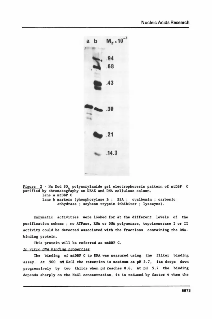
electrophoretic pattern is shown on fig. 2, a major band is found at 31 Kd
and two faint ones at 28 and 26 Kd. The addition to the purification proce-
dure of a phosphocellulose column (from which the activity was eluted at - 1M
KCl) after the DEAE cellulose column and a heparin ultrogel column following
the DNA cellulose step did not modify significantly the electrophoretic pat-
tern. Whatever the purification scheme the 31 Kd band was present and
usually much more important than the 28 and 26 Kd ones ; however in some
preparations the relative ratio was different, the 28 Kd band being the major
one. The incubation of the purified fraction did not change the initial
ratio. The 28 and 26 Kd polypeptides could be degradation products of the 31
Kd protein, the critical step being, perhaps, the lysis in spite of the
presence of PMSF.
From the binding activity in the DEAE peak and asssuming that one mole-
cule of protein allows the retention on a filter of one molecule of DNA the
amount of protein has been estimated: a minimum of 100 molecules per mtDNA
molecule would be present in the mitochondria.
5972
Nucleic Acids Research
a b Mr xlo-
94
^. 68
%_ 130
XV 21
v% .14.3
Figure 2 - Na Dod S04 polyacrylamide gel electrophoresis pattern of mtDBP Cpurified by chromatography on DEAE and DNA cellulose column.
lane a mtDBP Clane b markers (phosphorylase B BSA ovalbumin ; carbonic
anhydrase ; soybean trypsin inhibitor ; lysozyme).
Enzymatic activities were looked for at the different levels of the
purification scheme ; no ATPase, RNA or DNA polymerase, topoisomerase I or II
activity could be detected associated with the fractions containing the DNA-
binding protein.
This protein will be referred as mtDBP C.
In vitro DNA binding nropertiesThe binding of mtDBP C to DNA was measured using the filter binding
assay. At 500 mM NaCl the retention is maximum at pH 5.7, its drops down
progressively by two thirds when pH reaches 8.6. At pH 5.7 the binding
depends sharply on the NaCl concentration, it is reduced by factor 4 when the
5973

Nucleic Acids Research
Table 1: Stability of the mtDBP C - DNA complex.
a b cbinding Dissociation Control
Time 1 0.5 1 2 10 10+1 10+5 10+10 10+60 10(min.)
DNA bound(percent l 21 20 19 22 1 20 23 18 211 1of input) I
_ _ _ _ I _ _ _ _ _ I _ _ _ _ _ _ _ I _ _
a) Supercoiled H-pUC18 (0.9 ug) was incubated with a non-saturatingamount of proqein for 0.5 to 10 min. at 30°C.
b) Supercoiled H-pUC18 was incubated with mtDBP C for 10 min ; Aten-fold excess of cold DNA was then added and the labelled DNAretained on a filter was measured after the additionnal incubationtime.
c) In the control cold and labelled pUC18 DNA were added at the sametime and incubated for 10 min at 30°C.
salt concentration is 100 mM and by factor 20 when it is 1 M. On the contrary
at pH 7.9 the optimum retention plateaus between 200 and 500 mM NaCl. The44 4
addition to the assay mixture of Ca Mg or ATP has no effect on the
binding efficiency.
The binding reaction is very rapid, the maximunm retention is obtained
within half a minute. The stability of the complex was determined by studying3
the fate of the preformed H DNA-protein complex after the addition of a
large excess (10 fold) of unlabelled homologous DNA: no significative disso-
ciation could be found after an hour as judged by the filter binding assay
(Table 1).
GlXcerol gradient analysisFrom glycerol gradients performed at 500 mM NaCl pH 5.7 a sedimentation
coefficient of approximately 2S was found. In such gradients the protein was
monitored by its binding activity. Fractions corresponding to the peak of
activity as well as those where no activity was found were pooled separately
and analyzed on an SDS polyacrylamide gel. The 31 Kd polypeptide and the
minor 28 and 26 Kd ones where only found in the first pool. Moreover the
three polypeptides were recovered in their initial relative amount.It should3
be added that when a preformed H DNA-protein complex was run on a glycerol
gradient only the fractions of the radioactive peak contained polypeptides,
their molecular weights were 31, 28, 26 Kd and the bands were in the same
ratio as in the initial protein fraction. These results show that the three
5974

Nucleic Acids Research
C<
050
*0
z
0.5 1 2 4 8Protein ng
Figure 3 - Affinity for different forms of DNA.DNA binding activity was assayed in the standard buffer with 300 ng of
supercoiled * . , linearized o...o and heat-denatured o---o H pBR328DNA.
polypeptides do not correspond to different subunits of a single protein.
Either they are three different DNA-binding proteins with similar
chromatographic and binding properties, or the 28 and 26 Kd polypeptides are
degradation products of the 31 Kd one.
Differential affinity of the protein for various DNAs
Figure 3 shows that the supercoiled form of the pBR328 DNA was retained
on a filter (in the standard conditions) with a higher efficiency than the
double or single-strand linearized form. This preference for supercoiled DNA
molecules was confirmed by competition experiments, (fig. 4-I). pBR328 is
about 10 times more effective as a competitor in its supercoiled form than in
linearized double or single strand form. The relative affinity of mtDBP C for
supercoiled plasmids with or without fragments of mitochondrial DNA overlap-
ping either the 0 (pXlmBl4) or the 0 (pXlmB3) origin of replication was
very similar indicating that it does not show sequence specificity for these
regions, fig. 4-I. It should be noticed that in these experiments the control
DNA was the plasmid in its dimeric form to reduce a possible effect due to
the size of the plasmids. When the D-loop region was subcloned in shorter
fragments and inserted in the pUC18 plasmid no sequence specificity was found
(fig. 4-II) this figure also shows that mtDBP C does not bind efficientlyto mitochondrial RNAs.
It has been shown (14) that supercoiled DNA molecules having a natural
5975

Nucleic Acids Research
-a0-
0
10
I
I-
C
c
Ratio of competitor to 3H DNA
Figure 4 - Affinity for various DNAs and RNA. 3I - DNA binding activity was measured with supercoiled H pBR328 in the
presence of increasing amounts of competitive unlabelled DNAs linearpBR328 o- , denatured pBR328 00 , supercoiled pBR328 * , super-coiled pXlmBl4 s--u , supercoiled pXlmB3 A-A . 3
II - DNA binding activity was assayed with supercoiled H pUC18 in thepresence of total mitochondrial X. laevis RNA v , linear pUC18 o-o ,
supercoiled pXlmH5 A- , supercoiled pUC18 *- , supercoiled pXlmE2 A-A.Results are expressed as % of DNA bound in the absence of competitor.
negative superhelical density can contain sequences in the Z form under
physiological conditions. Such left-handed segment could be found in mito-
chondrial DNA and consequently one could anticipate a mitochondrial protein
showing a specific binding for Z DNA. In this respect mtDPB C was tested for
its binding to Z DNA poly(dG-BrdC) by comparison with the B form poly(dG-dC).
Figure 5 shows that it binds to both forms with the same efficiency at 200
mM NaCl. A similar result was obtained at other ionic strengths in the range
50 to 300 mM.
5976

Nucleic Acids Research
0
CL
w4-
0c
0
4)
z0co
2 4 8Protein ng
16
Figure 5 - Affinity for the B and Z forms of a polynucleotide.Binling activity was measured at 2R0 mM NaCl, pH 7.5 with 100 pg of
either [ H]poly(dG-dC) (B form) A,- or [ H]poly(dG-5BrdC) (Z form) *-ein the presence of 200 ng of unlabelled E. coli DNA as a competitor forunspecific binding.
I "X.,.
a Ib C: t I f O I A.I .) i;c I
FIr
.F. ..
Figure 6 - Modification of the superhelicity of a DNA by mtDBP C in thepresence of topoisomerase I.
I - Generation of superhelical turns.200 ng of purified relaxed pBR328 were incubated with increasing amounts
of protein in the presence of purified X. laevis topoisomerase I at 200 mMNaCl at 30°C for 90 min. DNA was then analyzed by agarose gel electrophoresis(see material and methods). Lanes a to e correspond to protein-DNA associa-tion in the presence of topoisomerase I, the wt/wt protein to DNA ratio was0.0, 0.1, 0.2, 0.4, 0.8 - lane f. relaxed pBR328 - lane g. same as e withouttopoisomerase I - lane h. native pBR328.II. Inhibition of topoisomerase I activity.
200 ng of supercoiled pBR328 were incubated with X. laevis topoisomeraseI in the presence of increasing amounts of mtDBP C at 200 mM NaCl at 30°C for90 min. Lanes a to d correspond to protein/DNA ratio of 0.0, 0.02, 0.05 and0.1.
5977

Nucleic Acids Research
DNA protein association
If mtDBP C participates in the beaded structure of the nucleoids one
could imagine that by its association to relaxed circular DNA, it would
introduce negative superhelical turns in the molecule as histones do in
nucleosomes. The superhelical turns can be measured in the presence of the
topoisomerase I. The purified relaxed pBR328 DNA was incubated with the
protein at different protein-DNA ratios in the presence of topoisomerase I
for 90 min and analysed on an agarose gel after deproteinisation (fig. 6-I).
The number of superhelical turns increases with the amount of protein added
reaching a maximum at an approximate weight ratio of 1 (lane e). The DNA
still present at the position of form II and relaxed form I ( FIr ) molecules
corresponds to the nicked molecules which were present in the initial DNA
preparation (compare lanes e and h). Furthermore the presence of mtDBP C
protects supercoiled DNA against the action of the topoisomerase I, fig.
6-II. Both these results show that the association of this protein with cova-
lently closed DNA induces a topological constraint in the molecule. It had
been shown previously that mtDBP C had no topoisomerase I or II activity.
DISCUSSION
Using as a selective property a preferential binding to supercoiled DNA
at high ionic strength we have isolated from oocyte mitochondrial extracts a
DNA binding protein showing a sedimentation coefficient of 2S measured on a
glycerol gradient in 0.5 M NaCl which is consistent with a monomer of 31 Kd.
The protein adsorbs to both DEAE and phosphocellulose columns from which it
is eluted at 0.25 and 1 M NaCl respectively. At low ionic strength (< 200 mM
NaCl) it undergoes self association and tends to form aggregates. These
properties could suggest that it contains distinct domains involved in bin-
ding to DNA and in protein-protein interactions (preliminary experimentsindicate a cooperative binding).
The high salt concentration (1 4 NaCl) necessary to elute the protein
from a DNA cellulose column and an optimum ionic strength of 0.5 M NaCl in
the binding assays suggest a rather strong affinity for DNA. The affinity of
mtDBP C for supercoiled DNA appear higher than for linear or double-stranded
DNA. It was tempting to assign some role to this protein in the specific
recognition of supercoiled DNA containing the D-loop region we have
previously found in mitochondrial extracts (8). The lack of specific affinity
for any DNA sequence precludes the sole involvement of mtDBP C in this
preferential binding.
The association of mtDBP C with relaxed circular DNA ( FIr ) introduces
5978

Nucleic Acids Research
superhelical turns in the molecules in the presence of topoisomerase I, the
maximum was reached for an approximate weight ratio of 1. It also protects
supercoiled DNA against the action of topoisomerase I. In this way thisprotein compares to the yeast HM protein, however no cross reaction of the HM
antibodies with mtDBP C has been found ( C. Jacq, personnal communication ).The amount of mtDBP C necessary to convert FIr DNA into FI is about 8 times
higher than the one necessary to protect a same amount of form I DNA against
the activity of topoisomerase I. Assuming the affinity of mtDBP C is the same
for any non-constrained molecule ( linear (FIII) or FIr ) this reflects the
higher affinity (- 10 times) of mtDBP C for supercoiled DNA. This effect on
the superhelicity of a DNA suggests that mtDBP C could be a component of the
beaded structures of the nucleoid.
From studies done in the absence of protease inhibitor one acid soluble
protein of 28 Kd seemed to be a specific component of nucleoid structures
(3). It might be suggested that this polypeptide is the degraded form of the
31 Kd protein characterized in this paper. Antibodies raised against the
isolated mtDBP C will be of interest to determine whether the 28 Kd
polypeptide of the nucleoid and the 28 Kd degradation product of mtDBP C are
identical.
In conclusion we have isolated a mitochondrial DBP which shows the pro-
perties expected for the (one of the) structural protein(s) of the nucleoid.
ACKNOWLEDGEMENTSWe thank J.C. Mounolou for helpful discussions and C.J. Herbert for a
critical reading of the manuscript. We are grateful to C. Dufresne andJ. Marsault for excellent technical assistance. We also wish to thankM. Decraene for typing this manuscript.
Financial support was provided by the CNRS (ATP Structure et Expressiondu Genome and U.A 86) and the Ecole Pratique des Hautes Etudes.
REFERENCES1. Caron, F., Jacq, C. and Rouviere-Yaniv, J. (1979) Proc. Natl. Acad.
Sci. USA. 76, 4265-4269.2. Rickwood, D., Chambers, J.A.A. and Barat, M. (1981) Exp. Cell Res. 133,
1-13.3. Barat., M., Rickwood, D., Dufresne, C. and Mounolou, J.C. (1985) Exp.
Cell Res. 157, 207-217.4. Barat, M. and Mignotte, B. (1981) Chromosoma (Berl.) 82, 583-593.5. Mignotte, B., Barat, M. and Mounolou, J.C. (1985) Nuc. Acids Res. 13,
(5), 1703-1716.6. Pavco, A.P. and Van Thuyle, G.C. (1985) J. Cell. Biol. 100, 258-264.7. Litman, R.M. (1968) J. Biol. Chem. 243, 6222-6233.8. Mignotte, B., Barat, M., Marsault, J. and Mounolou, J.C. (1983) Biochem.
Biophys. Res. Commun. 1127, 99-107.9. Dunon-Bluteau, D., Volovitch, M. and Brun, G. (1985) Gene. 36, 65-78.
5979

Nucleic Acids Research
10. Brun, G., Vannier, P., Scovassi, I. and Callen, J.C. (1981) Eur. J.Biochem. 118, 407-415.
11. Laemmli, U.K. (1970) Nature (Lond.) 227, 680-685.12. Oakley, B.R., Kirsch, D.R. and Morris, N.R. (1980) Anal. Biochem. 105,
361-363.13. Mattocia, E., Gandini-Attardi, D. and Tocchini-Valentini, G.P. (1976)
Proc. Natl. Acad. Sci. USA. 78, 4551-4554.14. Rich, A., Nordheim, A. and Wang, A.H.J. (1984). Ann. Rev. Biochem. 53,
791-846.
5980