National will overhaul Immigration but unsure of stranded migrants
Upload
independentCategory
view
0download
0
Mechanism of RecO recruitment to DNA bysingle-stranded DNA binding proteinMikhail Ryzhikov, Olga Koroleva, Dmitri Postnov, Andrew Tran and Sergey Korolev*
Department of Biochemistry and Molecular Biology, St. Louis University School of Medicine,1100 S Grand Blvd., St Louis, MO 63021, USA
Received January 7, 2011; Revised March 18, 2011; Accepted March 21, 2011
ABSTRACT
RecO is a recombination mediator protein (RMP) im-portant for homologous recombination, replicationrepair and DNA annealing in bacteria. In allpathways, the single-stranded (ss) DNA binding pro-tein, SSB, plays an inhibitory role by protectingssDNA from annealing and recombinase binding.Conversely, SSB may stimulate each reactionthrough direct interaction with RecO. We present acrystal structure of Escherichia coli RecO bound tothe conserved SSB C-terminus (SSB-Ct). SSB-Ctbinds the hydrophobic pocket of RecO in a conform-ation similar to that observed in the ExoI/SSB-Ctcomplex. Hydrophobic interactions facilitate bindingof SSB-Ct to RecO and RecO/RecR complex inboth low and moderate ionic strength solutions. Incontrast, RecO interaction with DNA is inhibited byan elevated salt concentration. The SSB mutantlacking SSB-Ct also inhibits RecO-mediated DNAannealing activity in a salt-dependent manner.Neither RecO nor RecOR dissociates SSB fromssDNA. Therefore, in E. coli, SSB recruits RMPs tossDNA through SSB-Ct, and RMPs are likely to alterthe conformation of SSB-bound ssDNA without SSBdissociation to initiate annealing or recombination.Intriguingly, Deinococcus radiodurans RecO doesnot bind SSB-Ct and weakly interacts with thepeptide in the presence of RecR, suggesting thediverse mechanisms of DNA repair pathwaysmediated by RecO in different organisms.
INTRODUCTION
Homologous recombination (HR) is the most efficientpathway for chromosomal damage repair in all organisms(1–4). Deficient HR renders cells unable to repair chromo-some breaks and to reinitiate stalled replication (2), whilehyperactivity leads to illegitimate recombination and
chromosomal abnormalities (5,6). Ubiquitous recombin-ation mediator proteins (RMPs) together with single-stranded (ss) DNA binding proteins regulaterecombinational DNA repair. HR is initiated by bindingof the RecA-like recombinase to ssDNA, forming theextended nucleoprotein filament called the presynapticcomplex. The ssDNA-binding proteins inhibit this step,while the RMPs overcome their inhibitory effect. Inaddition to HR, RMPs are also important for translesionsynthesis (TLS) (7,8) and DNA annealing (9–11). RMPsinclude phage UvsY (12), prokaryotic RecBCD andRecFOR proteins (7,13–15), and numerous eukaryoticmembers (16). Mutations of human RMPs are associatedwith cancer predisposition, mental retardation, UV sensi-tivity and premature aging (17–20). The mechanism ofRMP’s interaction with DNA, recombinases and ssDNAbinding proteins is poorly understood.RecF, -O and -R proteins are the most common RMPs
in bacteria. They are involved in the restart of stalled rep-lication, in ssDNA gaps (SSGs) repair, in TLS and indouble-strand (ds) DNA breaks (DSB) repair (21–24).RecFOR are the only RMPs found in the radiation resist-ant bacteria, Deinococcus radiodurans (25,26). InEscherichia coli, both RecO and RecR are essential forthe RecA loading on SSB-protected ssDNA (27), whileRecF is likely to play a regulatory role (28,29) or isinvolved in an alternative pathway of HR initiation (30).RecO interacts with SSB and DNA, while RecR bindseither RecO or RecF (27,31–33). RecO anneals compli-mentary ssDNA strands protected by cognate SSB(34,35), resembling the properties of the eukaryoticRMPs, Rad52 and BRCA2 (9,10). The crystal structuresof all three proteins from D. radiodurans have been previ-ously reported, as well as a structure of the RecORcomplex (36–40). The crystal structure of RecR revealeda DNA clamp-like tetramer architecture (36); however,the role of a potential DNA clamp in RMPs-mediatedreaction is unknown. The crystal structure of RecO didnot resemble any structural features of the functional eu-karyotic analog, Rad52 (38,40,41). RecO is a basic proteinwith an extensive, positively charged surface area. It is
*To whom correspondence should be addressed. Tel: 314 977 9261; Fax: 314 977 9205; Email: [email protected]
Published online 18 April 2011 Nucleic Acids Research, 2011, Vol. 39, No. 14 6305–6314doi:10.1093/nar/gkr199
� The Author(s) 2011. Published by Oxford University Press.This is an Open Access article distributed under the terms of the Creative Commons Attribution Non-Commercial License (http://creativecommons.org/licenses/by-nc/2.5), which permits unrestricted non-commercial use, distribution, and reproduction in any medium, provided the original work is properly cited.
by guest on August 8, 2016
http://nar.oxfordjournals.org/D
ownloaded from

comprised of three domains: the N-terminal OB-folddomain, which is characteristic for DNA binding proteins,the central a-helical domain and the zinc binding domain.RecO binds both ss- and dsDNA substrates, likely by theOB-fold domain and, potentially, positively chargedsurface areas of other domains. The RecR binding inter-face is also localized on the OB-fold domain of RecO (39).Interestingly, in the BRCA2 structure there are threeOB-fold domains, which bind either ssDNA or otherproteins (42).Overall, the mechanism of RecFOR interactions with
recombinase, DNA and SSB during presynaptic complexformation remains unknown. In addition to binding RecRand ss- and dsDNA, RecO also interacts with SSB (27).The SSB lacking eight C-terminal residues (SSB�C8)inhibits RecOR-mediated HR initiation in E. coli, suggest-ing that RecO binds the C-terminus of SSB (SSB-Ct) (43).In addition to protecting ssDNA, the SSB specificallyinteracts with multiple proteins of various DNA metabol-ism events through the conserved SSB-Ct (44,45).Therefore, SSB plays two seemingly opposing roles: pre-venting recombinase and RMPs from interaction withssDNA and potentially recruiting RMPs to ssDNA. It isunknown whether the interaction of RecOR(F) with SSBshould either completely or partially remove SSB fromssDNA (46) or alter the conformation of SSB-boundssDNA, making it available for RecA nucleation and an-nealing. The structure of RecO from D. radiodurans didnot reveal any potential SSB-Ct binding sites resemblingthose previously reported in other SSB-Ct bindingproteins. To understand the mechanism of RMPs’ inter-actions with SSB and DNA, which is likely to describe thecommon features of SSB interactions with multiple DNAmetabolism proteins, we solved a high resolution crystalstructure of the E. coli RecO bound to SSB-Ct, anddemonstrated that SSB recruits RecO to ssDNA in bothannealing and recombination initiation reactions.Intriguingly, D. radiodurans RecO does not bind SSB-Ct,suggesting that the role of RecO interaction with SSB maydiffer in the two organisms.
MATERIALS AND METHODS
DNA, peptides and proteins
For simplicity, E. coli homologs will be referred to byprotein names without prefixes in the rest of the paper.DNA oligonucleotides were purchased from IntegratedDNA Technologies, Inc. (Coralville, IA, USA). Peptideswere initially purchased from GenScript USA, Inc.(Piscataway, NJ, USA) and later from LifeTein LLC(South Plainfield, NJ, USA). SSB-Ct peptide had an add-itional N-terminal Trp residue for quantification(WYMDFDDDIPF-177). SSB-Ct-113 had a Pro176Sermutation, SSB-Ct-3�D had Asp172Ala, Asp173Ala,Asp174Ala mutations and SSB-Ct-�F lacks theC-terminal Phe177. RecO protein was cloned and purifiedas previously described for DrRecO with an additionalpurification step on a heparin column (38). SSB proteinwas purified from a plasmid provided by Dr M. Cox(University of Wisconsin) according to a previously
described protocol (43). All RecO mutants were char-acterized by the comparable wild-type protein solubilityand affinity toward the heparin resin. A few RecOmutants were tested for DNA annealing. The E. coliSSB mutant lacking the last eight amino acids(SSB�C8) was a gift from Dr M. Cox.
Crystallization and structure determination
RecO protein was concentrated in solution with 0.2MNaCl, 10mM Tris–HCl, pH 7.5, 0.5mM TCEP, 5%DMSO, 0.2mM CHAPS and with 1.5molar excess ofSSB-Ct, up to a concentration of 4 g/l. Crystals weregrown by the vapor diffusion method with sitting dropsusing 96-well Corning plates. Initial crystals diffractingto 3.5 A resolution were obtained with buffer containing20–25% PEG 4K and Bis–Tris Propane, pH 5.5. Initialattempts to solve the structure with the molecular replace-ment method were not successful, partially due to crystaltwinning. Later, crystals were also obtained with1.9–2.2M DL-Malic acid, pH 7.0, using both nativeprotein and selenomethionine derivative. Final data setswere collected on a GM/CA-CAT beam line at APS,ANL. Initial phases were obtained using 2.8 A resolutionsingle wavelength data set (SAD) collected from Se-Methderivative crystal and 2.3 A native data set using Phenixsoftware (47) (Supplementary Table S1). The initial mapobtained from the SAD experiment followed by the phasesextension to 2.3 A using the native data set was of excel-lent quality, sufficient for automated building of �80% ofthe structure with the Arp/Warp program (48). The modelbuilding and refinement were completed using the Coot(49) and Refmac (50) programs. The CNS program wasimplemented to calculate composite omit maps duringmodel building (51). There were two RecO molecules perasymmetric unit. Analysis of electron density mapsrevealed the presence of SSB-Ct bound to both moleculesand a CHAPS molecule (detergent used in crystallization)bound to one subunit only. All substrates were built usingexperimental electron density maps. Non-crystallographyaveraging was not utilized during initial model buildingand refinement steps.
Peptide and DNA binding assays
Proteins were dialyzed from storage buffer againstassay buffers with the addition of 50mM Arg-HCl and50mM NaGlu. Concentrations were determined by ab-sorbance at 280 nm and extinction coefficients of24 595M�1 cm�1 (RecO), 22 600M�1 cm�1 (DrRecO),6335M�1 cm�1 (RecR), 9000M�1 cm�1 (DrRecR) and27 960M�1 cm�1 (SSB). Fluorescein-labeled (FAM)peptides were solubilized in DMF (dimethylformamide),diluted in assay buffer and concentrations determinedby absorbance at 280 nm and extinction coefficient of5,500M�1 cm�1. Fluorescein-labeled DNA was solubil-ized in Mili-Q H2O. The reaction volume of all bindingassays was 100 ml. All reactions were run in triplicate andindependently repeated two or more times. Two differentreaction buffers were utilized for various assays: buffer Awith 25% glycerol, 50mM NaCl, 20mM Bis–TrisPropane, pH 8.0, 1mM TCEP and similar buffer B with
6306 Nucleic Acids Research, 2011, Vol. 39, No. 14
by guest on August 8, 2016
http://nar.oxfordjournals.org/D
ownloaded from
200mM NaCl. Fluorescent assays were read at 485/528nmusing a BioTek Synergy 2 plate reader. Fluorescent polar-ization anisotropy values were normalized using the fol-lowing equation: A=(Ai�A0)/A0, where A0 and Ai arefluorescence anisotropy values for free substrate and foreach titration point correspondingly. Binding constantswere determined with BioKin DynaFit using single-sitebinding approximation for all assays. Isotherms wereplotted with the Systat SigmaPlot program.
Competition assay
RecO at 1 mMwas pre-incubated with 0.9 mM of F-SSB-Ctin buffer B for 30min at room temperature and wastitrated by increasing concentrations of peptides, SSBand SSB bound to dT35 and dT70.
DNA pull down
Biotin-labeled dT45 was immobilized on streptavidinbeads and 50 ml of beads were incubated with 100 ml of a0.5 mM solution of SSB or SSB�C8 for 30min, washedwith excess buffer a few times and then incubated withRecO and RecOR proteins for 30–60min (200ml of totalvolume with 0.2 mM of RecO and 2 mM of RecR, whenpresent, with 0.02mM SSB and 0.1mg/ml BSA). Thebeads were washed 2 times with 500 ml of the correspond-ing buffer and samples of all solutions and beads wereanalyzed on SDS–PAGE. The pull-down experiment wasperformed multiple times while varying the presence ofBSA, detergent and small amount (5%) of SSB togetherwith RecO and RecOR to compensate for potential dis-sociation. Neither of these conditions was found to alterfinal results.
DNA annealing
Oligonucleotide 1 with the sequence TCCTTTTGATAAGAGGTCATTTTTGCGGATGGCTTAGAGCTTAATTGC was labeled by fluorescein (FAM) at the 50-end, andthe complimentary oligonucleotide 2 was labeled withDabsyl at the 30-end. Each oligonucleotide at the concen-tration of 25 nM was incubated separately with 250 nM ofeither SSB or SSB�C8 in different buffers for 20min andthen solutions with complimentary DNA were mixedtogether. RecO equilibrated in corresponding buffer wasadded to the mixture with the final concentration of250 nM. The concentrations of NaCl were 10, 20, 60 and100mM. Annealing of DNA was monitored by a decrease(quenching) of FAM emission as a function of time. Theintensities were read on the BioTek plate reader (pre-heated to 27�C). Fluorescence intensity values wereinverted and normalized using the following equation:I=�1*(Ii� I0) /I0, where I0 and Ii are fluorescence inten-sity values at initial time point and the next time point,respectively.
RESULTS
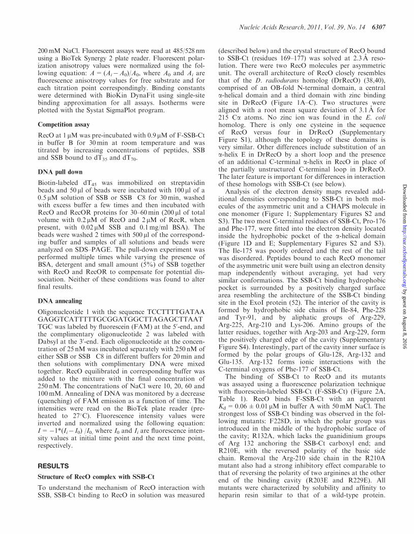
Structure of RecO complex with SSB-Ct
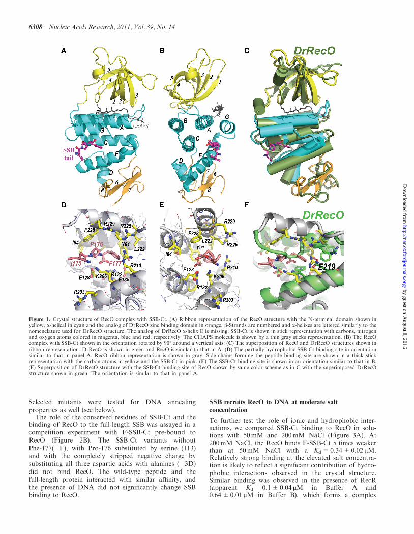
To understand the mechanism of RecO interaction withSSB, SSB-Ct binding to RecO in solution was measured
(described below) and the crystal structure of RecO boundto SSB-Ct (residues 169–177) was solved at 2.3 A reso-lution. There were two RecO molecules per asymmetricunit. The overall architecture of RecO closely resemblesthat of the D. radiodurans homolog (DrRecO) (38,40),comprised of an OB-fold N-terminal domain, a centrala-helical domain and a third domain with zinc bindingsite in DrRecO (Figure 1A–C). Two structures werealigned with a root mean square deviation of 3.1 A for215 Ca atoms. No zinc ion was found in the E. colihomolog. There is only one cysteine in the sequenceof RecO versus four in DrRecO (SupplementaryFigure S1), although the topology of these domains isvery similar. Other differences include substitution of ana-helix E in DrRecO by a short loop and the presenceof an additional C-terminal a-helix in RecO in place ofthe partially unstructured C-terminal loop in DrRecO.The later feature is important for differences in interactionof these homologs with SSB-Ct (see below).Analysis of the electron density maps revealed add-
itional densities corresponding to SSB-Ct in both mol-ecules of the asymmetric unit and a CHAPS molecule inone monomer (Figure 1; Supplementary Figures S2 andS3). The two most C-terminal residues of SSB-Ct, Pro-176and Phe-177, were fitted into the electron density locatedinside the hydrophobic pocket of the a-helical domain(Figure 1D and E; Supplementary Figures S2 and S3).The Ile-175 was poorly ordered and the rest of the tailwas disordered. Peptides bound to each RecO monomerof the asymmetric unit were built using an electron densitymap independently without averaging, yet had verysimilar conformations. The SSB-Ct binding hydrophobicpocket is surrounded by a positively charged surfacearea resembling the architecture of the SSB-Ct bindingsite in the ExoI protein (52). The interior of the cavity isformed by hydrophobic side chains of Ile-84, Phe-228and Tyr-91, and by aliphatic groups of Arg-229,Arg-225, Arg-210 and Lys-206. Amino groups of thelatter residues, together with Arg-203 and Arg-229, formthe positively charged edge of the cavity (SupplementaryFigure S4). Interestingly, part of the cavity inner surface isformed by the polar groups of Glu-128, Arg-132 andGlu-135. Arg-132 forms ionic interactions with theC-terminal oxygens of Phe-177 of SSB-Ct.The binding of SSB-Ct to RecO and its mutants
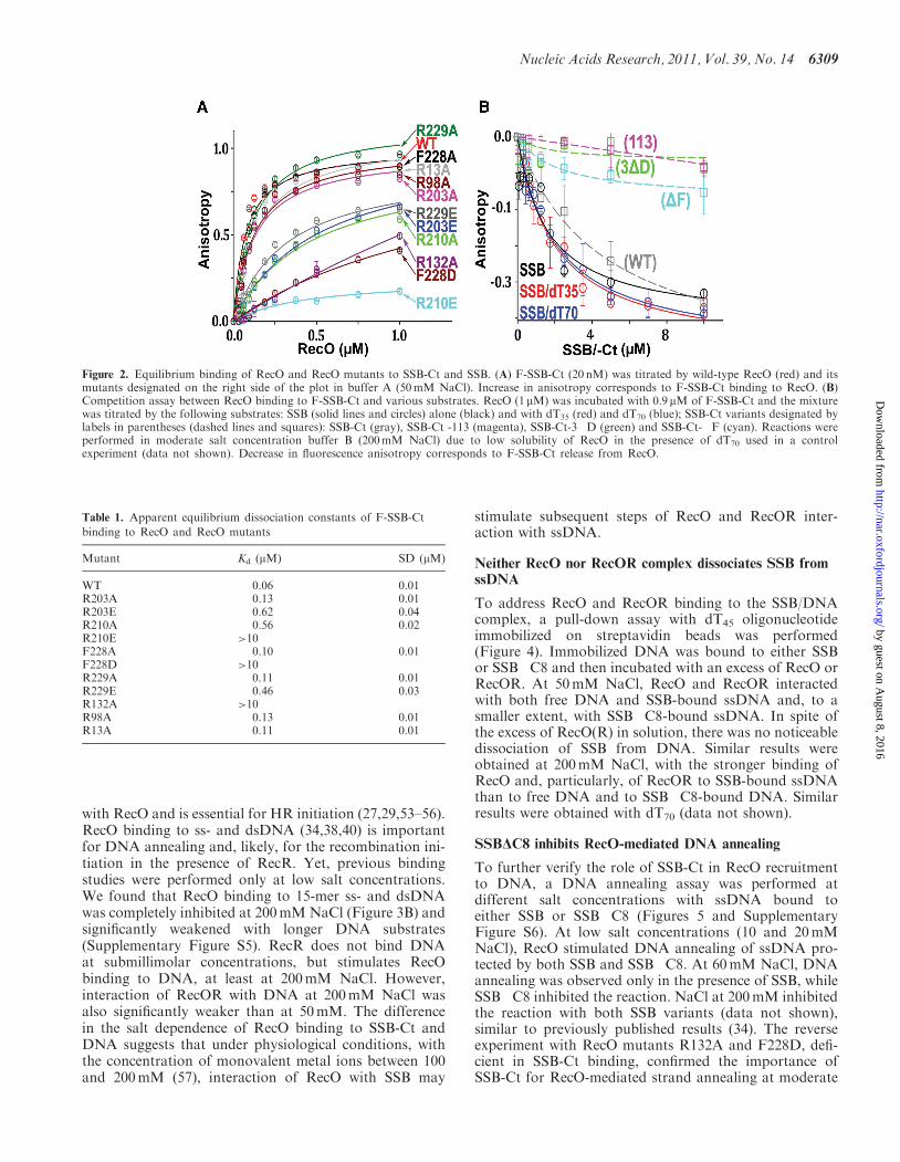
was assayed using a fluorescence polarization techniquewith fluorescein-labeled SSB-Ct (F-SSB-Ct) (Figure 2A,Table 1). RecO binds F-SSB-Ct with an apparentKd=0.06±0.01 mM in buffer A with 50mM NaCl. Thestrongest loss of SSB-Ct binding was observed in the fol-lowing mutants: F228D, in which the polar group wasintroduced in the middle of the hydrophobic surface ofthe cavity; R132A, which lacks the guanidinium groupsof Arg 132 anchoring the SSB-Ct carboxyl end; andR210E, with the reversed polarity of the basic sidechain. Removal the Arg-210 side chain in the R210Amutant also had a strong inhibitory effect comparable tothat of reversing the polarity of two arginines at the otherend of the binding cavity (R203E and R229E). Allmutants were characterized by solubility and affinity toheparin resin similar to that of a wild-type protein.
Nucleic Acids Research, 2011, Vol. 39, No. 14 6307
by guest on August 8, 2016
http://nar.oxfordjournals.org/D
ownloaded from
Selected mutants were tested for DNA annealingproperties as well (see below).The role of the conserved residues of SSB-Ct and the
binding of RecO to the full-length SSB was assayed in acompetition experiment with F-SSB-Ct pre-bound toRecO (Figure 2B). The SSB-Ct variants withoutPhe-177(�F), with Pro-176 substituted by serine (113)and with the completely stripped negative charge bysubstituting all three aspartic acids with alanines (�3D)did not bind RecO. The wild-type peptide and thefull-length protein interacted with similar affinity, andthe presence of DNA did not significantly change SSBbinding to RecO.
SSB recruits RecO to DNA at moderate saltconcentration
To further test the role of ionic and hydrophobic inter-actions, we compared SSB-Ct binding to RecO in solu-tions with 50mM and 200mM NaCl (Figure 3A). At200mM NaCl, the RecO binds F-SSB-Ct 5 times weakerthan at 50mM NaCl with a Kd=0.34±0.02 mM.Relatively strong binding at the elevated salt concentra-tion is likely to reflect a significant contribution of hydro-phobic interactions observed in the crystal structure.Similar binding was observed in the presence of RecR(apparent Kd=0.1±0.04mM in Buffer A and0.64±0.01mM in Buffer B), which forms a complex
Figure 1. Crystal structure of RecO complex with SSB-Ct. (A) Ribbon representation of the RecO structure with the N-terminal domain shown inyellow, a-helical in cyan and the analog of DrRecO zinc binding domain in orange. b-Strands are numbered and a-helixes are lettered similarly to thenomenclature used for DrRecO structure. The analog of DrRecO a-helix E is missing. SSB-Ct is shown in stick representation with carbons, nitrogenand oxygen atoms colored in magenta, blue and red, respectively. The CHAPS molecule is shown by a thin gray sticks representation. (B) The RecOcomplex with SSB-Ct shown in the orientation rotated by 90� around a vertical axis. (C) The superposition of RecO and DrRecO structures shown inribbon representation. DrRecO is shown in green and RecO is similar to that in A. (D) The partially hydrophobic SSB-Ct binding site in orientationsimilar to that in panel A. RecO ribbon representation is shown in gray. Side chains forming the peptide binding site are shown in a thick stickrepresentation with the carbon atoms in yellow and the SSB-Ct in pink. (E) The SSB-Ct binding site is shown in an orientation similar to that in B.(F) Superposition of DrRecO structure with the SSB-Ct binding site of RecO shown by same color scheme as in C with the superimposed DrRecOstructure shown in green. The orientation is similar to that in panel A.
6308 Nucleic Acids Research, 2011, Vol. 39, No. 14
by guest on August 8, 2016
http://nar.oxfordjournals.org/D
ownloaded from
with RecO and is essential for HR initiation (27,29,53–56).RecO binding to ss- and dsDNA (34,38,40) is importantfor DNA annealing and, likely, for the recombination ini-tiation in the presence of RecR. Yet, previous bindingstudies were performed only at low salt concentrations.We found that RecO binding to 15-mer ss- and dsDNAwas completely inhibited at 200mMNaCl (Figure 3B) andsignificantly weakened with longer DNA substrates(Supplementary Figure S5). RecR does not bind DNAat submillimolar concentrations, but stimulates RecObinding to DNA, at least at 200mM NaCl. However,interaction of RecOR with DNA at 200mM NaCl wasalso significantly weaker than at 50mM. The differencein the salt dependence of RecO binding to SSB-Ct andDNA suggests that under physiological conditions, withthe concentration of monovalent metal ions between 100and 200mM (57), interaction of RecO with SSB may
stimulate subsequent steps of RecO and RecOR inter-action with ssDNA.
Neither RecO nor RecOR complex dissociates SSB fromssDNA
To address RecO and RecOR binding to the SSB/DNAcomplex, a pull-down assay with dT45 oligonucleotideimmobilized on streptavidin beads was performed(Figure 4). Immobilized DNA was bound to either SSBor SSB�C8 and then incubated with an excess of RecO orRecOR. At 50mM NaCl, RecO and RecOR interactedwith both free DNA and SSB-bound ssDNA and, to asmaller extent, with SSB�C8-bound ssDNA. In spite ofthe excess of RecO(R) in solution, there was no noticeabledissociation of SSB from DNA. Similar results wereobtained at 200mM NaCl, with the stronger binding ofRecO and, particularly, of RecOR to SSB-bound ssDNAthan to free DNA and to SSB�C8-bound DNA. Similarresults were obtained with dT70 (data not shown).
SSB"C8 inhibits RecO-mediated DNA annealing
To further verify the role of SSB-Ct in RecO recruitmentto DNA, a DNA annealing assay was performed atdifferent salt concentrations with ssDNA bound toeither SSB or SSB�C8 (Figures 5 and SupplementaryFigure S6). At low salt concentrations (10 and 20mMNaCl), RecO stimulated DNA annealing of ssDNA pro-tected by both SSB and SSB�C8. At 60mM NaCl, DNAannealing was observed only in the presence of SSB, whileSSB�C8 inhibited the reaction. NaCl at 200mM inhibitedthe reaction with both SSB variants (data not shown),similar to previously published results (34). The reverseexperiment with RecO mutants R132A and F228D, defi-cient in SSB-Ct binding, confirmed the importance ofSSB-Ct for RecO-mediated strand annealing at moderate
Figure 2. Equilibrium binding of RecO and RecO mutants to SSB-Ct and SSB. (A) F-SSB-Ct (20 nM) was titrated by wild-type RecO (red) and itsmutants designated on the right side of the plot in buffer A (50mM NaCl). Increase in anisotropy corresponds to F-SSB-Ct binding to RecO. (B)Competition assay between RecO binding to F-SSB-Ct and various substrates. RecO (1mM) was incubated with 0.9 mM of F-SSB-Ct and the mixturewas titrated by the following substrates: SSB (solid lines and circles) alone (black) and with dT35 (red) and dT70 (blue); SSB-Ct variants designated bylabels in parentheses (dashed lines and squares): SSB-Ct (gray), SSB-Ct -113 (magenta), SSB-Ct-3�D (green) and SSB-Ct-�F (cyan). Reactions wereperformed in moderate salt concentration buffer B (200mM NaCl) due to low solubility of RecO in the presence of dT70 used in a controlexperiment (data not shown). Decrease in fluorescence anisotropy corresponds to F-SSB-Ct release from RecO.
Table 1. Apparent equilibrium dissociation constants of F-SSB-Ct
binding to RecO and RecO mutants
Mutant Kd (mM) SD (mM)
WT 0.06 0.01R203A 0.13 0.01R203E 0.62 0.04R210A 0.56 0.02R210E >10F228A 0.10 0.01F228D >10R229A 0.11 0.01R229E 0.46 0.03R132A >10R98A 0.13 0.01R13A 0.11 0.01
Nucleic Acids Research, 2011, Vol. 39, No. 14 6309
by guest on August 8, 2016
http://nar.oxfordjournals.org/D
ownloaded from

salt concentration (Figure S6). Both mutants annealedSSB-bound ssDNA at low salt concentrations, but wereunable to anneal SSB-bound ssDNA at 60 and 100mMNaCl.
Deinococcus radiodurans RecO does not bind SSB tail
Structural comparison of E. coli and D. radiodurans RecOrevealed a different conformation of the SSB tail bindingsite (Figure 1C and F). In DrRecO, the potential peptidebinding cavity is occupied by the C-terminus. Moreover,the residues forming the SSB-Ct binding pocket are notconserved. For example, there is Glu219 in DrRecOinstead of the Arg210 in RecO. This substitution(R210E) completely inhibited SSB-Ct binding by RecO(Figure 2 and Table 1). Correspondingly, DrRecO didnot bind DrSSB-Ct (Figure 6). Both SSB-Ct are highlyconserved, and the E. coli homolog binds peptides fromboth organisms with comparable affinity, in agreementwith earlier studies which demonstrated the functionalityof DrSSB in recombination initiation with E. coli proteins(58). The only weak interaction was observed with theDrRecOR complex in the presence of magnesium at50mM NaCl, leaving open the possibility of similar inter-actions between DrRecOR and DrSSB-Ct during recom-bination initiation. Due to the lack of a hydrophobiccavity on DrRecO surface, this binding is likely to bemediated by ionic interactions between the acidic part ofSSB-Ct and the DrRecOR complex, which may resemblethat of ssDNA binding to DrRecOR.
DISCUSSION
SSB protects ssDNA and specifically binds numerousproteins (44). These two functions are somewhat exclusive
since protein interactions eventually lead to SSB removalfrom ssDNA. The relationship between these events is es-sential for understanding the mechanisms of DNA repli-cation, recombination and repair. During recombination,SSB plays a dual role by inhibiting the initiation andfacilitating the extension of RecA nucleoproteinfilament. RecOR overcomes the first inhibitory step ofloading RecA on SSB-protected ssDNA, and thisfunction depends on SSB recognition by RecO. The struc-tural and equilibrium binding data presented here describethe mechanism of RecO interaction with SSB mediated bySSB-Ct and suggest that SSB facilitates the RecO bindingto ssDNA through SSB-Ct under physiological conditionsin both DNA annealing and RecR-mediated recombin-ation initiation pathways.
Hydrophobic interactions facilitate RecO recruitment tossDNA by SSB at physiological conditions
SSB-Ct binding to RecO depends on both hydrophobicand electrostatic interactions. The importance of hydro-phobic interactions is underlined by the structural data,where only the hydrophobic part of SSB-Ct is well orderedin structures of RecO and ExoI complexes, by mutagenesisand by the moderate salt dependence of RecO binding toSSB-Ct. The acidic residues of SSB-Ct are disordered inboth structures. Only substitution of all three aspartatesdecreases interaction in a similar degree to that of a singlemutation of Pro176 or Phe177. Thus, hydrophobic inter-actions should play a major role in SSB-Ct binding toRecO and other proteins. SSB binds ssDNA at a widerange of salt concentrations (59), suggesting that it facili-tates RecO interaction with ssDNA at physiological con-ditions. A similar recruitment mechanism may be commonfor other DNA replication and repair proteins known to
Figure 3. Different salt dependence of RecO binding to SSB-Ct and DNA. (A) The binding of F-SSB-Ct to RecO alone (solid lines, circles) or in thepresence of 10 mM of RecR (dashed lines, squares) in low salt buffer A (red) and in moderate salt buffer B (blue) measured as a function of thefluorescent anisotropy change. Binding to the RecOR complex resulted in a larger signal than in the case of RecO due to the larger size of thecomplex. However, the beginning of both isotherms coincides, reflecting the similar binding constants of SSB-Ct to RecO and RecOR. (B) DNAbinding by RecO at low (red) and moderate (blue) salt concentrations. FAM-labeled dT15 (5 nM) (solid lines and solid symbols) and 5 nM of 15 bpdsDNA labeled with FAM (dashed lines and open symbols) were titrated by RecO alone (circles) and in the presence of 10 mM of RecR (squares).Note the shift of red isotherms for the RecR complex to the left, reflecting stimulation of RecO DNA binding by RecR. The shape of the isothermfor the RecOR binding at 200mM NaCl is likely to reflect a two-step mechanism of RecO binding to RecR and binding of the complex to DNA,rather than cooperative binding to DNA.
6310 Nucleic Acids Research, 2011, Vol. 39, No. 14
by guest on August 8, 2016
http://nar.oxfordjournals.org/D
ownloaded from

bind SSB and which are characterized by a strong depend-ence of DNA binding on the ionic strength of solution.For example, the � subunit of the DNA polymerase IIIbinds SSB-Ct, and this interaction stimulates both clamploading and polymerase processivity only at elevated ionicstrength (60,61). Similarly, we found that RecO-mediatedDNA annealing depends on the presence of SSB-Ct onlyat moderate salt concentrations.Previously, SSB was proposed to limit interaction of
RecO with ssDNA due to a time delay in the RecAloading reaction mediated by RecOR in the presence ofSSB (43). In pull-down and DNA annealing assays, wealso observed an inhibitory role of the SSB�C8 coreprotein on RecO interaction with ssDNA. At the sametime, the presence of SSB-Ct clearly stimulated both an-nealing and ssDNA binding by RecO at elevated salt con-centrations. The relationship between the protective roleof the SSB core structure and the recruitment role ofSSB-Ct should be further investigated with a more quan-titative approach. Interestingly, overexpression of RecOpartially compensated temperature sensitivity of SSB�35Bacillus subtilis cells expressing SSB without theC-terminus (45).The recruitment mechanism suggests a synergy between
DNA and SSB binding. PriA binds ssDNA-bound SSB 10times stronger than free SSB (62). Interaction of � withSSB was not affected by the presence of DNA, but � doesnot bind DNA. However, DNA stimulates � binding toSSB by 1000-fold in the presence of DNA binding subunitof Pol III (61). No significant enhancement RecO bindingto DNA-bound SSB versus free SSB was observed in acompetition assay, at least at 200mM NaCl (Figure 2B).However, in a pull-down assay at 200mM NaCl, thebinding of RecO and RecOR to DNA was stronger inthe case of SSB-bound DNA (Figure 4).
Figure 4. Binding of RecO and RecOR to ssDNA in the presence ofSSB and SSB�C8. Streptavidin beads with immobilized dT45 wereincubated first with SSB or SSB�C8, then with either RecO orRecOR proteins, and immobilized proteins were detected onsilver-stained 15% SDS–PAGE. (A) Pull down assay was performedin buffer A (50mM NaCl) with the addition of 0.1mM CHAPS and0.1mg/ml BSA. Proteins immobilized on DNA-bound beads are shownin the following lanes: 1, SSB; 2, SSB�C8; 4, RecO; 5, RecO in thepresence of SSB; 6, RecO in the presence of SSB�C8; 8, RecOR; 9,RecOR in the presence of SSB; 10, RecOR in the presence of SSB�C8.The loading solutions for RecO and RecOR are shown on lanes 3 and7, respectively. (B) Similar assay performed in buffer B (200mM NaCl)with similar order of lanes. Protein bands are marked on the right sidewith O corresponding to RecO, R to RecR; S to SSB; �C to SSB�C8;str to streptavidin. Unlike the results from the equilibrium binding ex-periments (Figure 3 and Supplementary Figure S5), in the absence ofRecR, RecO binds to DNA immobilized on beads relatively strong atmoderate salt concentration (B, lane 4), which may be an artifact ofprotein aggregation due to high local concentration of immobilizedDNA. However, SSB-bound DNA still remains the preferred substratefor RecO in contrast to low salt conditions in A.
Figure 5. SSB�C8 inhibits RecO-mediated annealing at an elevatedsalt concentration. Complementary ssDNA oligonucleotides labeledwith FAM and Dabsyl were incubated with SSB or SSB�C8 inbuffers of variable NaCl concentration and mixed together. FAMquenching was plotted as a function of time after addition of RecO.Blue, cyan and green colors correspond to SSB-bound DNA at 10, 20and 60mM NaCl; purple, orange and red colors correspond toSSB�C8-bound DNA at 10, 20 and 60mM NaCl. Gray and blacklabels correspond to the incubation of SSB and SSB�C8 at 10mMNaCl without RecO.
Figure 6. DrRecO does not bind SSB-Ct. Equilibrium binding ofDrSSB-Ct to DrRecO and RecO assayed using FAM-labeledDrSSB-Ct (10 nM). Binding to RecO (EcO) in buffer A is shown inred, to DrRecO (DrO) in buffer A in gray, and in buffer A with 5mMMgCl2 in black, to DrRecOR in the presence of 20 mM of DrRecR(DrOR) in buffer A in green, in buffer A with 5mM MgCl2 in blue,and in buffer B in cyan.
Nucleic Acids Research, 2011, Vol. 39, No. 14 6311
by guest on August 8, 2016
http://nar.oxfordjournals.org/D
ownloaded from
RecO and RecOR form a complex with DNA-bound SSB
Another consequence of a synergy or cooperativity be-tween SSB and DNA binding is that the SSB-mediatedrecruitment of proteins to ssDNA should not cause itsown removal from DNA. In the case of recombination,RMPs facilitate loading of recombinase on SSB-protectedssDNA, which is likely accompanied by at least partialremoval of SSB from ssDNA. Previously, it was suggestedthat RecOR either form a complex with ssDNA-boundSSB (27) or remove SSB from ssDNA (46). Our datademonstrated that RecO binding to SSB-Ct and DNAare not exclusive (Figure 2B). None of our pull-downassays revealed a noticeable dissociation of SSB as wellas SSB�C8 from DNA, even when they were incubatedwith an excess of RecO and RecOR (Figure 4). We hy-pothesize that RecO and RecOR do not remove SSB fromssDNA, but instead alter the conformation of ssDNA.SSB binds ssDNA in two different modes: SSB65, whereall four SSB monomers are wrapped by 65-mer ssDNA,and SSB35, with only two monomers bound to 35-merssDNA (63,64). Even removing the unstructuredC-termini affects DNA binding (65) and the equilibriumbetween these two modes (66). Binding of SSB-Ct to posi-tively charged RecO is likely to change the conformationof bound ssDNA. Such an altered conformation should becompatible with the DNA annealing process in thepresence of complimentary ssDNA. The completion ofDNA annealing can force the dissociation of SSB fromDNA. RecR inhibits DNA annealing by RecO and is es-sential for RecA loading, even though its interaction withRecA had never been shown. Therefore, RecR should alsochange the conformation of ssDNA or of the whole RecO/SSB/ssDNA assembly to prevent DNA from annealingand to make it available for RecA binding. This hypoth-esis is supported by our data showing similar binding ofRecO and RecOR to SSB-Ct and a stimulatory effect ofRecR on RecO binding to DNA. Theoretically, evenpartial unwrapping of ssDNA from the SSB tetramer bythe RecOR complex, for example, by changing SSB65 tothe SSB35 mode, may free ssDNA of a sufficient length forRecA nucleation to occur (67).
Differences of SSB interaction with RecO inD. radiodurans and E. coli
The bacterium, D. radiodurans, is characterized by aunique ability to efficiently repair thousands of chromo-some breaks caused by radiation and desiccation (68–71).RecFOR are important for the rapid reconstitution of ashredded genome, which is thought to occur through asynthesis-dependent strand annealing process followedby DNA recombination (26). Previously, we demonstratedthat DrRecO does stimulate DNA annealing in theabsence of SSB (38). The lack of DrRecO bindingto DrSSB-Ct suggests that such annealing is SSB-independent in this organism, which may be importantfor annealing of relatively short ssDNA fragments insuf-ficient for SSB binding. In the case of recombination ini-tiation, we cannot rule out DrRecOR interaction withSSB-Ct. However, previous observations imply the exist-ence of alternative pathways of recombination initiation.
First, significant differences in properties of RecA fromtwo organisms have been reported (72). Second, RecFwas shown to rescue RecOR function in the presence ofSSB�C8 mutant in E. coli (30), suggesting an alternativeSSB-Ct independent pathway of RecOR-mediated RecAloading. This data also suggests that, in addition to SSBbinding, other functions of RecO are essential for recom-bination initiation.
The prediction of similar SSB-Ct binding properties ofRecO homologs is complicated by the overall low sequencehomology and the lack of conserved motifs (38). Eventhe identification of RecO homologs in some genomes ischallenging (73,74). Likewise, the prediction of an SSB-Ctbinding site formed mostly by C-terminal a-helixes is dif-ficult, unless overall sequences are considerably shorter, asis the case of Helicobacter pylori. Thus, in some species,the RecO-mediated strand annealing may be independentof interaction with SSB-Ct. Strand annealing and HR ini-tiation activities may also reside in separate proteins.Interestingly, DdrB was recently identified as a ssDNAannealing protein in D. radiodurans as well (75). TheRPA-dependent strand annealing property is not con-served between Brh2 and BRCA2 homologs in eukaryotes(10,76).
Overall, a large number of known SSB binding partnersimplies a multifunctional role for SSB-Ct. In addition toits role in stabilization of DNA–protein complexesreported here, SSB-Ct was suggested to perform a switchfunction for two proteins exchanging places on DNA via amutually exclusive interaction with SSB-Ct (77).Interestingly, other RecF pathway proteins, includingRecQ and RecJ, also bind SSB-Ct (78–80), suggestingthat a similar switch mechanism regulates the transitionfrom the RecQJ-mediated step of DNA processing to theRecFOR-mediated step of RecA loading (78).
ACCESSION NUMBER
PDB ID 3Q8D.
SUPPLEMENTARY DATA
Supplementary Data are available at NAR Online.
ACKNOWLEDGEMENTS
We thank M. Cox for providing plasmid for SSB purifi-cation and purified SSB�C8, GM/CA-CAT beam line atAPS, ANL for assistance in data collection, T. Lohmanfor advice, A. Kozlov for critical review and T. Baird andE. Los for help in manuscript preparation. The coordin-ates of RecO-SSB-Ct complex and structure factors weredeposited to PBD with ID 3Q8D.
FUNDING
Funding for project, including Open Access charge:National Institutes of Health (grant GM073837).
Conflict of interest statement. None declared.
6312 Nucleic Acids Research, 2011, Vol. 39, No. 14
by guest on August 8, 2016
http://nar.oxfordjournals.org/D
ownloaded from
REFERENCES
1. Cox,M.M. (1999) Recombinational DNA repair in bacteriaand the RecA protein. Prog Nucleic Acid Res. Mol. Biol., 63,311–366.
2. Cox,M.M., Goodman,M.F., Kreuzer,K.N., Sherratt,D.J.,Sandler,S.J. and Marians,K.J. (2000) The importance of repairingstalled replication forks. Nature, 404, 37–41.
3. Kowalczykowski,S.C. (2000) Initiation of genetic recombinationand recombination-dependent replication. Trends Biochem. Sci.,25, 156–165.
4. Kuzminov,A. (2001) DNA replication meets genetic exchange:chromosomal damage and its repair by homologousrecombination. Proc Natl Acad. Sci. USA, 98, 8461–8468.
5. Stark,J.M., Pierce,A.J., Oh,J., Pastink,A. and Jasin,M. (2004)Genetic steps of mammalian homologous repair with distinctmutagenic consequences. Mol. Cell. Biol., 24, 9305–9316.
6. Luo,G., Santoro,I.M., McDaniel,L.D., Nishijima,I., Mills,M.,Youssoufian,H., Vogel,H., Schultz,R.A. and Bradley,A. (2000)Cancer predisposition caused by elevated mitotic recombination inBloom mice. Nat. Genet., 26, 424–429.
7. Fujii,S., Isogawa,A. and Fuchs,R.P. (2006) RecFOR proteins areessential for Pol V-mediated translesion synthesis andmutagenesis. EMBO J., 25, 5754–5763.
8. Jiang,Q., Karata,K., Woodgate,R., Cox,M.M. andGoodman,M.F. (2009) The active form of DNA polymerase V isUmuD’(2)C-RecA-ATP. Nature, 460, 359–363.
9. Sugiyama,T., New,J.H. and Kowalczykowski,S.C. (1998) DNAannealing by RAD52 protein is stimulated by specific interactionwith the complex of replication protein A and single-strandedDNA. Proc. Natl Acad. Sci. USA, 95, 6049–6054.
10. Mazloum,N., Zhou,Q. and Holloman,W.K. (2007) DNA binding,annealing, and strand exchange activities of Brh2 protein fromUstilago maydis. Biochemistry, 46, 7163–7173.
11. Mortensen,U.H., Bendixen,C., Sunjevaric,I. and Rothstein,R.(1996) DNA strand annealing is promoted by the yeast Rad52protein. Proc. Natl Acad. Sci. USA, 93, 10729–10734.
12. Sweezy,M.A. and Morrical,S.W. (1999) Biochemical interactionswithin a ternary complex of the bacteriophage T4 recombinationproteins uvsY and gp32 bound to single-stranded DNA.Biochemistry, 38, 936–944.
13. Lloyd,R.G. and Thomas,A. (1983) On the nature of the RecBCand RecF pathways of conjugal recombination in Escherichia coli.Mol. Gen. Genet., 190, 156–161.
14. Wang,T.C. and Smith,K.C. (1983) Mechanisms forrecF-dependent and recB-dependent pathways of postreplicationrepair in UV-irradiated Escherichia coli uvrB. J. Bacteriol., 156,1093–1098.
15. Kolodner,R., Fishel,R.A. and Howard,M. (1985) Geneticrecombination of bacterial plasmid DNA: effect of RecF pathwaymutations on plasmid recombination in Escherichia coli.J. Bacteriol., 163, 1060–1066.
16. Symington,L.S. (2002) Role of RAD52 epistasis group genes inhomologous recombination and double-strand break repair.Microbiol. Mol. Biol. Rev., 66, 630–670.
17. Ouyang,K.J., Woo,L.L. and Ellis,N.A. (2008) Homologousrecombination and maintenance of genome integrity: cancer andaging through the prism of human RecQ helicases.Mech. Ageing Dev., 129, 425–440.
18. Tal,A., Arbel-Goren,R. and Stavans,J. (2009) Cancer-associatedmutations in BRC domains of BRCA2 affect homologousrecombination induced by Rad51. J. Mol. Biol., 393, 1007–1012.
19. Powell,S.N., Willers,H. and Xia,F. (2002) BRCA2 keeps Rad51 inline. High-fidelity homologous recombination prevents breast andovarian cancer? Mol. Cell., 10, 1262–1263.
20. Thompson,L.H. and Schild,D. (2002) Recombinational DNArepair and human disease. Mutat. Res., 509, 49–78.
21. Whitby,M.C. and Lloyd,R.G. (1995) Altered SOS inductionassociated with mutations in recF, recO and recR. Mol. Gen.Genet., 246, 174–179.
22. Kusano,K., Nakayama,K. and Nakayama,H. (1989)Plasmid-mediated lethality and plasmid multimer formation in anEscherichia coli recBC sbcBC mutant. Involvement of RecFrecombination pathway genes. J. Mol. Biol., 209, 623–634.
23. Clark,A.J. (1991) rec genes and homologous recombinationproteins in Escherichia coli. Biochimie, 73, 523–532.
24. Kidane,D., Sanchez,H., Alonso,J.C. and Graumann,P.L. (2004)Visualization of DNA double-strand break repair in live bacteriareveals dynamic recruitment of Bacillus subtilis RecF, RecO andRecN proteins to distinct sites on the nucleoids. Mol. Microbiol.,52, 1627–1639.
25. Cox,M.M., Keck,J.L. and Battista,J.R. (2010) Rising from theAshes: DNA Repair in Deinococcus radiodurans. PLoS Genet., 6,e1000815.
26. Bentchikou,E., Servant,P., Coste,G. and Sommer,S. (2010) Amajor role of the RecFOR pathway in DNA double-strand-breakrepair through ESDSA in Deinococcus radiodurans. PLoS Genet.,6, e1000774.
27. Umezu,K. and Kolodner,R.D. (1994) Protein interactions ingenetic recombination in Escherichia coli. Interactions involvingRecO and RecR overcome the inhibition of RecA bysingle-stranded DNA-binding protein. J. Biol. Chem., 269,30005–30013.
28. Sandler,S.J. and Clark,A.J. (1994) RecOR suppression of recFmutant phenotypes in Escherichia coli K-12. J. Bacteriol., 176,3661–3672.
29. Morimatsu,K. and Kowalczykowski,S.C. (2003) RecFOR ProteinsLoad RecA Protein onto Gapped DNA to Accelerate DNAStrand Exchange. A Universal Step of Recombinational Repair.Mol. Cell., 11, 1337–1347.
30. Sakai,A. and Cox,M.M. (2009) RecFOR and RecOR as distinctRecA loading pathways. J. Biol. Chem., 284, 3264–3272.
31. Webb,B.L., Cox,M.M. and Inman,R.B. (1997) RecombinationalDNA repair: the RecF and RecR proteins limit the extension ofRecA filaments beyond single-strand DNA gaps. Cell, 91,347–356.
32. Webb,B.L., Cox,M.M. and Inman,R.B. (1995) An interactionbetween the Escherichia coli RecF and RecR proteins dependenton ATP and double-stranded DNA. J. Biol. Chem., 270,31397–31404.
33. Makharashvili,N., Mi,T., Koroleva,O. and Korolev,S. (2009)RecR-mediated modulation of RecF dimer specificity for single-and double-stranded DNA. J. Biol. Chem., 284, 1425–1434.
34. Luisi-DeLuca,C. and Kolodner,R. (1994) Purification andcharacterization of the Escherichia coli RecO protein.Renaturation of complementary single-stranded DNA moleculescatalyzed by the RecO protein. J. Mol. Biol., 236, 124–138.
35. Kantake,N., Madiraju,M.V., Sugiyama,T. andKowalczykowski,S.C. (2002) Escherichia coli RecO proteinanneals ssDNA complexed with its cognate ssDNA-bindingprotein: A common step in genetic recombination.Proc. Natl Acad. Sci. USA, 99, 15327–15332.
36. Lee,B.I., Kim,K.H., Park,S.J., Eom,S.H., Song,H.K. andSuh,S.W. (2004) Ring-shaped architecture of RecR: implicationsfor its role in homologous recombinational DNA repair.EMBO J., 23, 2029–2038.
37. Koroleva,O., Makharashvili,N., Courcelle,C.T., Courcelle,J. andKorolev,S. (2007) Structural conservation of RecF and Rad50:implications for DNA recognition and RecF function. EMBO J.,26, 867–877.
38. Makharashvili,N., Koroleva,O., Bera,S., Grandgenett,D.P. andKorolev,S. (2004) A novel structure of DNA repair protein RecOfrom Deinococcus radiodurans. Structure, 12, 1881–1889.
39. Timmins,J., Leiros,I. and McSweeney,S. (2007) Crystal structureand mutational study of RecOR provide insight into its mode ofDNA binding. EMBO J., 26, 3260–3271.
40. Leiros,I., Timmins,J., Hall,D.R. and McSweeney,S. (2005) Crystalstructure and DNA-binding analysis of RecO from Deinococcusradiodurans. EMBO J., 24, 906–918.
41. Singleton,M.R., Wentzell,L.M., Liu,Y., West,S.C. andWigley,D.B. (2002) Structure of the single-strand annealingdomain of human RAD52 protein. Proc. Natl Acad. Sci. USA,99, 13492–13497.
42. Yang,H., Jeffrey,P.D., Miller,J., Kinnucan,E., Sun,Y.,Thoma,N.H., Zheng,N., Chen,P.L., Lee,W.H. and Pavletich,N.P.(2002) BRCA2 function in DNA binding and recombination froma BRCA2-DSS1-ssDNA structure. Science, 297, 1837–1848.
Nucleic Acids Research, 2011, Vol. 39, No. 14 6313
by guest on August 8, 2016
http://nar.oxfordjournals.org/D
ownloaded from
43. Hobbs,M.D., Sakai,A. and Cox,M.M. (2007) SSB protein limitsRecOR binding onto single-stranded DNA. J. Biol. Chem., 282,11058–11067.
44. Shereda,R.D., Kozlov,A.G., Lohman,T.M., Cox,M.M. andKeck,J.L. (2008) SSB as an organizer/mobilizer of genomemaintenance complexes. Crit. Rev. Biochem. Mol. Biol., 43,289–318.
45. Costes,A., Lecointe,F., McGovern,S., Quevillon-Cheruel,S. andPolard,P. (2010) The C-Terminal Domain of the Bacterial SSBProtein Acts as a DNA Maintenance Hub at Active ChromosomeReplication Forks. PLoS Genet., 6, e1001238.
46. Inoue,J., Honda,M., Ikawa,S., Shibata,T. and Mikawa,T. (2008)The process of displacing the single-stranded DNA-bindingprotein from single-stranded DNA by RecO and RecR proteins.Nucleic Acids Res., 36, 94–109.
47. Adams,P.D., Afonine,P.V., Bunkoczi,G., Chen,V.B., Davis,I.W.,Echols,N., Headd,J.J., Hung,L.W., Kapral,G.J., Grosse-Kunstleve,R.W. et al. (2010) PHENIX: a comprehensivePython-based system for macromolecular structure solution.Acta. Crystallogr. D Biol. Crystallogr., 66, 213–221.
48. Langer,G., Cohen,S.X., Lamzin,V.S. and Perrakis,A. (2008)Automated macromolecular model building for X-raycrystallography using ARP/wARP version 7. Nat. Protoc., 3,1171–1179.
49. Emsley,P., Lohkamp,B., Scott,W.G. and Cowtan,K. (2010)Features and development of Coot. Acta. Crystallogr.D Biol. Crystallogr., 66, 486–501.
50. Winn,M.D., Murshudov,G.N. and Papiz,M.Z. (2003)Macromolecular TLS refinement in REFMAC at moderateresolutions. Methods Enzymol., 374, 300–321.
51. Brunger,A.T. (2007) Version 1.2 of the Crystallography andNMR system. Nat. Protoc., 2, 2728–2733.
52. Lu,D. and Keck,J.L. (2008) Structural basis of Escherichia colisingle-stranded DNA-binding protein stimulation of exonucleaseI. Proc. Natl Acad. Sci. USA, 105, 9169–9174.
53. Chow,K.H. and Courcelle,J. (2004) RecO acts with RecF andRecR to protect and maintain replication forks blocked byUV-induced DNA damage in Escherichia coli. J. Biol. Chem.,279, 3492–3496.
54. Ivancic-Bace,I., Peharec,P., Moslavac,S., Skrobot,N., Salaj-Smic,E.and Brcic-Kostic,K. (2003) RecFOR Function Is Required forDNA Repair and Recombination in a RecA Loading-DeficientrecB Mutant of Escherichia coli. Genetics, 163, 485–494.
55. Umezu,K., Chi,N.W. and Kolodner,R.D. (1993) Biochemicalinteraction of the Escherichia coli RecF, RecO and RecR proteinswith RecA protein and single-stranded DNA binding protein.Proc. Natl Acad. Sci. USA, 90, 3875–3879.
56. Handa,N., Morimatsu,K., Lovett,S.T. and Kowalczykowski,S.C.(2009) Reconstitution of initial steps of dsDNA break repair bythe RecF pathway of E. coli. Genes Dev., 23, 1234–1245.
57. Cayley,S., Lewis,B.A., Guttman,H.J. and Record,M.T. Jr. (1991)Characterization of the cytoplasm of Escherichia coli K-12 as afunction of external osmolarity. Implications for protein-DNAinteractions in vivo. J. Mol. Biol., 222, 281–300.
58. Eggington,J.M., Haruta,N., Wood,E.A. and Cox,M.M. (2004)The single-stranded DNA-binding protein of Deinococcusradiodurans. BMC Microbiol., 4, 2.
59. Lohman,T.M., Overman,L.B. and Datta,S. (1986) Salt-dependentchanges in the DNA binding co-operativity of Escherichia colisingle-strand binding protein. J. Mol. Biol., 187, 603–615.
60. Kelman,Z., Yuzhakov,A., Andjelkovic,J. and O’Donnell,M. (1998)Devoted to the lagging strand-the subunit of DNA polymeraseIII holoenzyme contacts SSB to promote processive elongationand sliding clamp assembly. EMBO J., 17, 2436–2449.
61. Glover,B.P. and McHenry,C.S. (1998) The chi psi subunits ofDNA polymerase III holoenzyme bind to single-strandedDNA-binding protein (SSB) and facilitate replication of anSSB-coated template. J. Biol. Chem., 273, 23476–23484.
62. Kozlov,A.G., Jezewska,M.J., Bujalowski,W. and Lohman,T.M.(2010) Binding specificity of Escherichia coli single-stranded DNAbinding protein for the chi subunit of DNA pol III holoenzymeand PriA helicase. Biochemistry, 49, 3555–3566.
63. Bujalowski,W. and Lohman,T.M. (1986) Escherichia colisingle-strand binding protein forms multiple, distinct complexeswith single-stranded DNA. Biochemistry, 25, 7799–7802.
64. Raghunathan,S., Kozlov,A.G., Lohman,T.M. and Waksman,G.(2000) Structure of the DNA binding domain of E. coli SSBbound to ssDNA. Nat. Struct. Biol., 7, 648–652.
65. Savvides,S.N., Raghunathan,S., Futterer,K., Kozlov,A.G.,Lohman,T.M. and Waksman,G. (2004) The C-terminal domain offull-length E. coli SSB is disordered even when bound to DNA.Protein Sci., 13, 1942–1947.
66. Kozlov,A.G., Cox,M.M. and Lohman,T.M. (2010) Regulation ofsingle-stranded DNA binding by the C termini of Escherichia colisingle-stranded DNA-binding (SSB) protein. J. Biol. Chem., 285,17246–17252.
67. Roy,R., Kozlov,A.G., Lohman,T.M. and Ha,T. (2009) SSBprotein diffusion on single-stranded DNA stimulates RecAfilament formation. Nature, 461, 1092–1097.
68. Minton,K.W. (1994) DNA repair in the extremelyradioresistant bacterium Deinococcus radiodurans. Mol.Microbiol., 13, 9–15.
69. Zahradka,K., Slade,D., Bailone,A., Sommer,S., Averbeck,D.,Petranovic,M., Lindner,A.B. and Radman,M. (2006) Reassemblyof shattered chromosomes in Deinococcus radiodurans. Nature,443, 569–573.
70. Battista,J.R., Earl,A.M. and Park,M.J. (1999) Why isDeinococcus radiodurans so resistant to ionizing radiation?Trends Microbiol., 7, 362–365.
71. Mattimore,V. and Battista,J.R. (1996) Radioresistance ofDeinococcus radiodurans: functions necessary to survive ionizingradiation are also necessary to survive prolonged desiccation.J. Bacteriol., 178, 633–637.
72. Kim,J.I. and Cox,M.M. (2002) The RecA proteins ofDeinococcus radiodurans and Escherichia coli promote DNAstrand exchange via inverse pathways. Proc. Natl Acad. Sci. USA,99, 7917–7921.
73. Rocha,E.P., Cornet,E. and Michel,B. (2005) Comparative andevolutionary analysis of the bacterial homologous recombinationsystems. PLoS Genet., 1, e15.
74. Marsin,S., Mathieu,A., Kortulewski,T., Guerois,R. andRadicella,J.P. (2008) Unveiling novel RecO distant orthologuesinvolved in homologous recombination. PLoS Genet., 4,e1000146.
75. Xu,G., Lu,H., Wang,L., Chen,H., Xu,Z., Hu,Y., Tian,B. andHua,Y. (2010) DdrB stimulates single-stranded DNA annealingand facilitates RecA-independent DNA repair in Deinococcusradiodurans. DNA Repair, 9, 805–812.
76. Jensen,R.B., Carreira,A. and Kowalczykowski,S.C. (2010) Purifiedhuman BRCA2 stimulates RAD51-mediated recombination.Nature, 467, 678–683.
77. Yuzhakov,A., Kelman,Z. and O’Donnell,M. (1999) Tradingplaces on DNA–a three-point switch underlies primer handofffrom primase to the replicative DNA polymerase. Cell, 96,153–163.
78. Courcelle,J. and Hanawalt,P.C. (1999) RecQ and RecJ processblocked replication forks prior to the resumption of replication inUV-irradiated Escherichia coli. Mol. Gen. Genet., 262, 543–551.
79. Shereda,R.D., Bernstein,D.A. and Keck,J.L. (2007) A centralrole for SSB in Escherichia coli RecQ DNA helicase function.J. Biol. Chem., 282, 19247–19258.
80. Han,E.S., Cooper,D.L., Persky,N.S., Sutera,V.A. Jr.,Whitaker,R.D., Montello,M.L. and Lovett,S.T. (2006) RecJexonuclease: substrates, products and interaction with SSB.Nucleic Acids Res., 34, 1084–1091.
6314 Nucleic Acids Research, 2011, Vol. 39, No. 14
by guest on August 8, 2016
http://nar.oxfordjournals.org/D
ownloaded from
Copyright © 2022 FDOKUMEN