Secondary structure prediction and structure-specific sequence analysis of single-stranded DNA
Upload
independentCategory
view
0download
0
DNA recognition and the precleavage state duringsingle-stranded DNA transposition inD radiodurans
Alison Burgess Hickman1Jeffrey A James14 Orsolya Barabas15Cecile Pasternak2 Bao Ton-Hoang3Michael Chandler3 Suzanne Sommer2
and Fred Dyda11Laboratory of Molecular Biology National Institute of DiabetesDigestive and Kidney Diseases National Institutes of Health BethesdaMD USA 2CNRS UMR 8621 LRC CEA 42V Institut de Genetiqueet Microbiologie Universite Paris-Sud Orsay Cedex France and3Laboratoire de Microbiologie et Genetique Moleculaires CentreNational de la Recherche Scientifique Toulouse Cedex France
Bacterial insertion sequences (ISs) from the IS200IS605
family encode the smallest known DNA transposases and
mobilize through single-stranded DNA transposition
Transposition by one particular family member ISDra2
from Deinococcus radiodurans is dramatically stimulated
upon massive c irradiation We have determined the
crystal structures of four ISDra2 transposaseIS end com-
plexes combined with in vivo activity assays and fluores-
cence anisotropy binding measurements these have
revealed the molecular basis of strand discrimination
and transposase action The structures also show that
previously established structural rules of target site recog-
nition that allow different specific sequences to be targeted
are only partially conserved among family members
Furthermore we have captured a fully assembled active
site including the scissile phosphate bound by a divalent
metal ion cofactor (Cd2thorn ) that supports DNA cleavage
Finally the observed active site rearrangements when the
transposase binds a metal ion in which it is inactive
provide a clear rationale for metal ion specificity
The EMBO Journal (2010) 29 3840ndash3852 doi101038
emboj2010241 Published online 1 October 2010
Subject Categories genome stability amp dynamics structural
biology
Keywords Deinococcus insertion sequence IS mobile
element transposition
Introduction
Exposure to 10 gray (Gy) of ionizing radiation is enough to
kill a human yet Deinococcus radiodurans perhaps the
lsquoworldrsquos toughest bacteriumrsquo has the remarkable ability to
survive an instantaneous dose of 5000 Gy without loss of
viability (Moseley and Mattingly 1971) Although this dose
effectively shatters the bacterial genome by introducing about
200 double-stranded breaks within 4 or 5 h following irradia-
tion D radiodurans has accurately reassembled its genome
This remarkable task is accomplished mainly using a process
termed extended synthesis dependent strand annealing
which involves the extensive synthesis of single-stranded
DNA (ssDNA) (Zahradka et al 2006 Slade et al 2009)
Like that of essentially all microbes the genome of
D radiodurans contains a variety of mobile elements and
DNA transposition by insertion sequences (ISs) has been
shown to be a major source of spontaneous mutagenesis in
this bacterium (Mennecier et al 2006) ISs are the simplest
form of mobile DNA they can transpose from one genomic
location (donor site) to another (target site) assisted by a self-
encoded transposase enzyme that specifically recognizes and
synapses the ends of the IS Synapse formation is followed by
a regulated set of DNA cutting and joining reactions that first
liberate the transposon from donor DNA and then integrate
the transposon ends into target DNA All of these events are
carried out by the transposase and the exact mechanism by
which these are achieved is dependent on the particular IS
family
When IS activity in D radiodurans was monitored shortly
after irradiation one particular element ISDra2 stood out as
it showed an B100-fold increase in activity following 10 kGy
of g irradiation becoming the predominant transposition
event (Mennecier et al 2006) This is in contrast to non-
irradiated cells where ISDra2 insertions are rare The reason
for this increase in ISDra2 transposition during genomic
recovery from g irradiation became clear as ISDra2 belongs
to the recently described IS200IS605 family (Kersulyte et al
1998 2002) that unlike any other characterized DNA trans-
position system uses an asymmetric ssDNA-dependent path-
way for transposition (Guynet et al 2008 Pasternak et al
2010) This pathway has several unusual features Whereas
most ISs contain inverted repeat sequences at the two ends
in the IS200IS605 family the transposase recognizes its IS
ends by binding to subterminal palindromic sequences that
form imperfect hairpin structures (designed IPs for imperfect
palindromes Figures 1A and 2A) Recognition is also strand
specific as only one strand (the top strand) of the transposon
is bound and subsequently transposed Members of
the IS200IS605 family are also unusual from the point of
view of target specificity Whereas most ISs integrate randomly
or with minor sequence preferences ISs in this family
always insert just 30 of a well-defined tetra- or pentanucleo-
tide sequence (Kersulyte et al 1998 Islam et al 2003Received 7 May 2010 accepted 6 September 2010 published online1 October 2010
Corresponding author Laboratory of Molecular Biology NationalInstitute of Diabetes Digestive and Kidney Diseases National Institutesof Health Bethesda MD 20892 USA Tel thorn 1 301 402 4496Fax thorn 1 301 496 0201 E-mail freddydanihgov4Present address Office of Technology Transfer Johns HopkinsUniversity 100 North Charles Street 5th Floor BaltimoreMD 21201 USA5Present address European Molecular Biology LaboratoryMeyerhofstrasse 1 Heidelberg 69117 Germany
The EMBO Journal (2010) 29 3840ndash3852 | amp 2010 European Molecular Biology Organization | All Rights Reserved 0261-418910
wwwembojournalorg
The EMBO Journal VOL 29 | NO 22 | 2010 amp2010 European Molecular Biology Organization
EMBO
THE
EMBOJOURNAL
THE
EMBOJOURNAL
3840
httpwww-isbiotoulfr and P Siguier personal commu-
nication) The exact sequence of this target site is character-
istic of the particular family member
Although most ISs encode transposase enzymes that are
members of the retroviral integrase superfamily (Hickman
et al 2010) IS200IS605 transposases are ssDNA endo-
nucleases belonging to the vast HUH nuclease superfamily
Proteins with HUH nuclease domains are also involved in a
diverse variety of other processes such as the initiation of
rolling circle replication and conjugative DNA transfer and
replication initiation of certain ssDNA viruses (Koonin and
Ilyina 1993) HUH nucleases contain one or two active site
tyrosine residues that act as nucleophiles during DNA
cleavage Upon nucleophilic attack the transposase becomes
covalently attached to DNA through a 50-phosphotyrosine
intermediate and a free 30-OH group is released on the
other side of the DNA break DNA cleavage also requires a
divalent metal ion cofactor that is coordinated by the two
Figure 1 Model of transposon excision by members of the IS200IS605 family of insertion sequences (ISs) and sequence alignment of some oftheir transposases (A) The IS is shown as a thin black line with its left end (LE) in red and right end (RE) in blue The imperfect palindromes(IP) are shown schematically as hairpins which are flanked on their 50 sides by solid boxes representing the four nucleotides involved incleavage (or target) site recognition The transposase TnpA is shown as a dimer with orange subunits Step 1 TnpA synapses the two IS endsby binding the IPs and brings the cleavage sites (shown as white boxes) at the IS ends into the active sites through DNAndashDNA interactionsbetween the nucleotides 50 of each end (TTGATat LE TTCAA at RE in the case of ISDra2) and those at the 50 sides of the IPs For clarity the twoIS ends are shown binding in the same orientation to each monomer of the dimer however in the determined structures one IP is rotated by1801 relative to the other (vide infra) Step 2 End cleavage mediated by the active site Tyr on helix aD results in covalent attachment(represented by a red hexagon) of one TnpA subunit to the cleaved LE and the other subunit to the flanking DNA 30 of the cleaved RE Step 3 Atrans-to-cis movement of the aD helices brings the two ends of the donor DNA together in one active site (monomer on left) and the LE into theproximity of the cleaved RE (monomer on right) Step 4 Nucleophilic attack of the two 30-OH groups onto the covalent phosphotyrosineintermediates results in sealed donor DNA and a circular transposon intermediate Reversal of these steps (grey arrows) represents insertion ofthe transposon intermediate into target DNA (B) Sequence alignment of the transposases of ISDra2 the IS represented by PDB code 2fyx andIS608 superimposed on the secondary structure (b-strands in blue a-helices in red) of IS608 TnpA The amino acid numbering is for ISDra2transposase The active site His (on the fourth b-strand) and Tyr residues (on helix aD) are in bold and residues putatively involved in thetrans-to-cis transition in blue
ISDra2 transposase and its complexesAB Hickman et al
amp2010 European Molecular Biology Organization The EMBO Journal VOL 29 | NO 22 | 2010 3841
histidine residues that comprise the HUH motif (Datta et al
2003 Guasch et al 2003) DNA strand transfer is achieved
when a 30-OH end of a DNA strand attacks the 50-phospho-
tyrosine linkage resulting in a resealed phosphodiester
backbone
A mechanistic model of ssDNA transposition in the IS200
IS605 family has been derived from biochemical experiments
and structural data obtained for one of its members the
transposase TnpA of IS608 from Helicobacter pylori
(Ronning et al 2005 Ton-Hoang et al 2005 Barabas et al
2008 Guynet et al 2008) As IS200IS605 transposases
contain only one active site tyrosine (Figure 1B) in order to
coordinate cleavage at their left end (LE) and right end (RE)
they assemble as dimers in this way there are two functional
active sites and two binding sites for IP hairpins one for LE
and another for RE (shown schematically in Figure 1A)
Studies on IS608 TnpA revealed two important keys to
transposition The first is that the cleavage sites at both the LE
and RE are recognized through non-sequential base-pairing
interactions between nucleotides at the cleavage site (shown
as a white box at either end in Figure 1A) and four nucleo-
tides at the 50 base of the IPs (shown as solid orange and light
blue boxes) As the sequences of the 4-nt 50 extensions of the
IPs are different at LE and RE specific recognition of the
different LE and RE cleavage site sequences becomes possi-
ble Furthermore as the circular transposon intermediate to
be integrated contains the LE IP and its 4 nt 50 extension
target site recognition is mechanistically identical to the
recognition of the LE cleavage site The second key is
that after cleavage at LE and RE (step 2 in Figure 1A) a
conformational change is believed to occur that swaps
the 50-phosphotyrosine linkages between the two monomers
(step 3) in the dimer through the exchange of a mobile
a-helix Resolving these linkages (step 4) yields a circular
transposon and a resealed donor DNA strand Integration
occurs when the transposase dimer bound to the circular
intermediate captures an ssDNA that contains the target
sequence and the reaction cycle repeats two cleavages
one swap and two resolution steps later the result is an
integrated transposon
Although we now know a great deal about the mechanism
of IS608 transposition some aspects cannot be straightfor-
wardly generalized to other family members For example
the IS608 target sequence is a tetranucleotide (TTAC) whereas
the target sequence of ISDra2 is a pentanucleotide (TTGAT
Islam et al 2003) It is not clear how the recognition rules
derived from the IS608 structures appropriate for tetranu-
cleotides would allow recognition of a pentanucleotide
As the mechanism determined for IS608 TnpA can be
exploited to redirect insertions to preselected and specified
tetranucleotide sites (Guynet et al 2009) understanding how
longer target sequences are recognized may prove extremely
useful Furthermore the discrimination between the top- and
bottom-strand IPs of IS608 TnpA is influenced by nucleotides
present at the tip of the hairpin and an extrahelical base in the
stem of the IP there is no such base in the stem of the IPs of
ISDra2 leaving open the question of how the two strands are
distinguished Finally certain amino acids that we concluded
should be crucial for IS608 TnpA transposition are simply not
found in the ISDra2 transposase
We describe here a series of crystal structures of the
ISDra2 transposase bound to a variety of ssDNA substrates
representing either the LE or RE of ISDra2 In conjunction
with biochemical and in vivo experiments these structures
expand our understanding of how IS200IS605 transposases
work In particular we have captured the transposase with
a fully assembled active site including the scissile phosphate
a state that eluded us with IS608 TnpA We also provide
structural explanations for pentanucleotide recognition
strand discrimination and the metal ion specificity of
DNA cleavage
Results
We have determined crystal structures of four complexes of
the ISDra2 transposase (TnpADra2) with various bound DNAs
Figure 2 Structures of TnpADra2 bound to its transposon ends (A) ISDra2 LE and RE DNA sequences The overall structures of the TnpADra2
dimer bound to (B) the LE IP (in red and orange) and a short ssDNA (in black) representing the five nucleotides 50 of the LE cleavage site whichis equivalent to the target site and (C) the RE IP (in light and dark blue) extended to the transposon 30 end The DNA colour schemecorresponds to that shown in Figures 1A and 2A Residues of TnpADra2 highlighted in green are specifically referred to in the text (Y30 H67H69 W107 and Y132)
ISDra2 transposase and its complexesAB Hickman et al
The EMBO Journal VOL 29 | NO 22 | 2010 amp2010 European Molecular Biology Organization3842
representing several stages of the transposition reaction
(Table I) Recent biochemical work has confirmed that
TnpADra2 uses ssDNA substrates in in vitro transposition
reactions (Pasternak et al 2010) as does the IS608 transpo-
sase One structure (Figure 2B) is of a ternary complex
(designated TnpALE27T5) in which TnpADra2 is complexed
with an oligonucleotide containing the LE IP (designated
LE27 from LE nt thorn 11 to thorn 37 Figure 2A) and a 5-nt
oligonucleotide (T5) comprising the target site TTGAT We
have also solved a complex (designated TnpARE34) be-
tween TnpADra2 and a 34-nt oligonucleotide that contains
the RE IP and extends to the RE cleavage site (RE nt 34 to
1 Figure 2C) In addition we have solved two other LE
ternary complexes TnpACd is the complex between
TnpADra2 Y132F (the active site Tyr has been mutated to
Phe) LE27 and an oligonucleotide in which the target site
has been extended by 1 nt TTGATkG to include the first 50 nt
of the transposon (designated T5kG where k indicates the
cleavage site) TnpACd was crystallized in the presence of
Cd2thorn a divalent metal ion that robustly supports cleavage
The last complex TnpAZn contains wild-type TnpA and the
same oligonucleotides as TnpACd but was crystallized
in the presence of Zn2thorn which supports only a very low
level of cleavage TnpALE27T5 was solved with experi-
mental phasing the other structures were solved with mole-
cular replacement using TnpALE27T5 as the search model
Representative electron density is shown in Supplementary
Figure S1
IP recognition by TnpADra2
A characteristic feature of transposon DNA sequences in the
IS200IS605 family are subterminal IP sequences located
close to the cleavage sites As only the top strand is cut and
subsequently moved to a new genomic location the transpo-
sase must be able to distinguish the top-strand IP hairpins
from those on the bottom The previously determined struc-
tures of IS608 TnpA (Ronning et al 2005) demonstrated that
one important discriminating feature is the tip of the hairpin
loop where the 50-most T at the tip (shown in cyan in
Figure 3A) sits in a pocket on the transposase surface
which would not be able to accommodate the corresponding
adenine that is present at the tip of the bottom-strand IP
Table I Crystallographic structure determination and refinement
TnpALE27T5(native)
TnpALE27T5(I derivative)
TnpARE34 TnpACd TnpAZn
Data collectionSpace group P212121 P212121 P21 P212121 P212121
Cell dimensions a b c (A) 1047 1282 1406 1044 1286 1401 768 893 901 503 869 1283 498 927 1261b (deg) 1112Resolution (A) 23 24 23 19 19Total reflections 575 978 515 046 181 638 333 244 327 763Unique reflections 84171 74 480 50130 44 818 45 279Is(I) 159 (24) 112 (25) 164 (31) 134 (691) 164 (45)Completeness () 995 (976) 1000 (998) 996 (990) 998 (991) 967 (759)Rsym 007 (083) 0103 (0716) 0049 (0341) 0093 (0186) 0048 (0244)
SIRAS phasing at 30 ARCullis 064RKraut (isoano) 01630203Phasing power (isoano) 14919Combined FOM 0402
Molecular replacement at 40 ARotation function corr coeff 02090204 0146 01370127Rfactor after rigid body refinement 0417 0423 0368
RefinementResolution (A) 300ndash23 300ndash23 30ndash19 300ndash19No reflections (FX0) 78 520 47 910 44 616 44 429RworkRfree 02180249 02150239 01850214 01770210
No atomsProtein and nucleic acid 10 055 7236 3447 3444Solvent 548 164 401 473
B-factorsProtein and nucleic acid 3934 6463 2707 2282Solvent 3818 4530 3876 3349
RmsdBond lengths (A) 0009 001 0017 0013Bond angles (deg) 41 300 194 454Dimers per AU 3 2 1 1
Ramachandran plot statisticsMost favoured 921 927 931 944Disallowed 0 0 0 0
ISDra2 transposase and its complexesAB Hickman et al
amp2010 European Molecular Biology Organization The EMBO Journal VOL 29 | NO 22 | 2010 3843
hairpin The structures of TnpADra2 bound to the LE IP or the
RE IP indicate that this element of discrimination is retained
in all structures one thymine at the hairpin tip (shown in
blue in Figure 3A) is flipped back into a small pocket where it
stacks against the peptide plane of Gly89
The other IP-discriminating feature for IS608 TnpA is an
unpaired thymine within the IP stem which is flipped out
and is specifically recognized by the transposase (also shown
in cyan in Figure 3A) This structural feature is not conserved
in ISDra2 Instead each IP stem of ISDra2 contains a GT
mismatch (Gthorn 20Tthorn 33 on the LE G25T12 on the RE
Figure 2A) that is recognized and apparently stabilized by
TnpADra2
The key amino acid in the recognition of the GT mismatch
is Arg14 located at the tip of a b-hairpin that dives deep into
the major groove and stacks on the top of the mismatched T
(shown for Tthorn 33 on LE in Figure 3B and C) while forming
hydrogen bonds to the immediately adjacent G (Gthorn 34 on
LE) These interactions twist the DNA such that the Watsonndash
Crick faces of the mismatched nucleotides are turned away
from each other thereby avoiding a clash between the proto-
nated N1 of Gthorn 34 and the protonated N3 of Tthorn 33
A consequence of these DNA distortions is the accumulation
of negative charge on one side of Tthorn 33 as O4 of Tthorn 33
moves toward the sugar-phosphate backbone of the opposite
strand This electrostatic problem is thwarted by the creation
of a Mg2thorn -binding site (Figure 3C) The octahedrally coordi-
nated Mg2thorn ion also provides a hydrogen bond to O4 of
Tthorn 33 through one of its coordinated water molecules
It seems likely that this element of mismatch recognition
Figure 3 DNA binding by TnpADra2 GT mismatch recognition and discrimination between the top and bottom strand of the transposon(A) Comparison of the binding of the tip of the hairpin loop by IS608 TnpA (left PDB code 2A6O) and TnpADra2 (right) The distinguishingfeatures of the hairpins are shown in shades of blue the unpaired T in the stem of the IS608 hairpin and the T at the hairpin tip (left) and themismatched GT base pair and the T at the hairpin tip of the ISDra2 hairpin (right) (B) Space-filling representation of the complex betweenTnpADra2 (two subunits shown in shades of green) and LE27 (in orange) highlighting the position of Arg14 (C) A web of hydrogen-bondinginteractions and a bound Mg2thorn ion mediate the interaction between Arg14 and the Gthorn 20Tthorn 33 mismatch on the LE (D) Dissociationconstants for binding between the IS608 and ISDra2 transposases and their IPs and variants determined by fluorescence anisotropymeasurements
ISDra2 transposase and its complexesAB Hickman et al
The EMBO Journal VOL 29 | NO 22 | 2010 amp2010 European Molecular Biology Organization3844
also contributes to discrimination between the top and bot-
tom strands as such a water-mediated H-bond could not be
formed on the bottom-strand IP with the CA mismatch of
the bottom-strand IP the Mg2thorn -bound water molecule can-
not act as an H-bond acceptor with the protonated N4 of the
cytosine In all four TnpADra2DNA complexes the mode
of recognition and stabilization of the GT mismatch is
essentially identical
Relative to the IP the location of the GT mismatch in the
ISDra2 stem is exactly where the unpaired base yielding the
flipped out thymine is for IS608 The flipped out T is
recognized by IS608 TnpA by sequestration between the
side chains of Arg52 and Phe75 and by a set of H-bonds
(Ronning et al 2005) whereas the crucial residue for recog-
nizing and stabilizing the GT mismatch of ISDra2 is Arg14
None of these residues are conserved between the two
proteins (Figure 1B)
One final factor that influences IP recognition and strand
discrimination by TnpADra2 is the relative ease of hairpin
formation by the top- and bottom-strand IPs Whereas the
top-strand IPs on both the LE and RE readily fold into hairpin
structures (as assessed by size-exclusion chromatography
data not shown) attempts to form a hairpin with the bottom-
strand ISDra2 LE IP sequence were unsuccessful due to its
vast preference to form annealed dsDNA products Even
when the small amount of the bottom-strand hairpinned
form was chromatographically separated from the dsDNA
form the former rapidly converted to the latter even at 41C
Indeed thermodynamic data suggest that whereas GT mis-
matches are well tolerated in dsDNA oligonucleotides CA
mismatches are less so (SantaLucia and Hicks 2004)
Both the hairpin tip and the stem IP irregularity are
important for IP recognition
The structures of IS608 TnpA and TnpADra2 bound to their IPs
suggest that two IP features are important for recognition the
bases at the hairpin tip and an imperfection in the hairpin
stem To investigate the relative influence of these we
determined dissociation constants for IP binding both to
IS608 TnpA and TnpADra2 using fluorescence anisotropy
with various carboxyfluorescein (FAM)-modified oligonu-
cleotides
As shown in Figure 3D modification of either feature
resulted in a decrease in binding The effect was most
dramatic when pyrimidine bases at the tip of the IS608 and
ISDra2 IPs were changed to purines for both transposases
this resulted in a complete loss of detectable IP binding
(compare IPs 1ndash2 and 5ndash6) This is consistent with
structural observations that the 50-most T at the hairpin tips
sit in a pocket located on each transposase surface too small
for a larger purine base
The imperfections in the IP stems appear to be less
important contributors to binding than the bases at the hair-
pin tips For IS608 TnpA changing the flipped out T in the IP
stem to A (IP 3) reduced binding B40-fold relative to the
wild-type IP whereas correction of the mismatch in the stem
of the ISDra2 IP (IP 7) led to an approximately six-fold
reduction in binding
We also tested the ability of the two transposases to bind to
their bottom-strand IPs (IP 4 and 8) There was no detect-
able binding in either case demonstrating that the discrimi-
nation between the top and bottom strand of the transposon
occurs at the initial stage of transposase binding
The role of TnpADra2 Arg14
To evaluate the role of Arg14 in vivo we used a recently
developed genetic system in D radiodurans (Pasternak et al
2010) to measure the first stage in IS transposition excision of
the IS from the donor backbone In this system the unique
active copy of ISDra2 has been replaced by a mutated
derivative ISDra2-113 that is inserted at the unique 50-
TTGAT-30 target site present in the tetA gene of a TetR cassette
in this way precise excision of the IS restores a TetR pheno-
type In ISDra2-113 both tnpA encoding the transposase and
a second gene tnpB carried by the IS but which is not
necessary for transposition (Pasternak et al 2010) have
been deleted and the functional ends for transposition have
been retained TnpADra2 is expressed in trans from a plasmid
expression vector
As shown in Table II precise excision of ISDra2-113 was
strictly dependent on the presence of TnpADra2 In strains
devoid of plasmid or carrying an empty vector the frequency
of TetR colonies was undetectable but increased to B3103
when wild-type TnpADra2 was expressed When wild-type
TnpADra2 was replaced by the R14A mutant the excision
frequency dropped B60-fold supporting a crucial role of
Arg14 in IP recognition and subsequent transposon excision
We also measured the binding affinity of purified TnpADra2
R14A protein to its IP using our fluorescence anisotropy
assay The dissociation constant was B14 mM an B30-fold
loss in binding relative to wild type which is consistent with
the corresponding decrease in excision frequency
Helix aD is a mobile structural element that
can be in cis or trans
A key feature of the molecular model of ssDNA transposition
is a large conformational rearrangement that exchanges the
location of the two aD helices containing the tyrosine nu-
cleophiles between the two monomers of the transposase
dimer (Figure 1A step 3) Indeed biochemical data strongly
suggest that while a trans-active site arrangement (ie the
tyrosine of one monomer is close to the HUH motif of the
other) is catalytically competent for transposon end cleavage
the resolution steps require a transcis isomerization
(Barabas et al 2008) However we have not yet observed a
IS200IS605 family transposase in the cis configuration In all
the structures of IS608 TnpA determined to date the active
sites are arranged in trans and TnpADra2 is no different
Nevertheless there does appear to be a precedent for a
Table II Excision frequencies of the ISDra2-113 derivative in aDeinococcus radiodurans tester straina expressing TnpADra2 variants
Residentplasmid
TnpADra2 expressedin trans
Excision frequency
No No o108
pGY11559 No o108
pGY13203 TnpA wt 32 (plusmn06) 103
pGY13521 TnpAR14A 53 (plusmn16) 105
pGY13522 TnpAS122GE123G 17 (plusmn03) 103
aGY13115 genotype DDR1652DDR1651OtetAOISDra2-113 (CamR)(Pasternak et al 2010)
ISDra2 transposase and its complexesAB Hickman et al
amp2010 European Molecular Biology Organization The EMBO Journal VOL 29 | NO 22 | 2010 3845
cis-active site The coordinates of a high resolution crystal
structure of an uncomplexed IS200IS605 family transposase
that is 808 identical to TnpADra2 (Figure 1B) have been
deposited in the Protein Data Bank under the PDB ID 2fyx
and the protein is in the cis conformation As the 2fyx
transposase is a result of structural genomics efforts unfor-
tunately no biochemical data are available and it must be
noted that the HUH motif is not intact
For the trans-to-cis structural transition to take place
modelling suggested that a seven amino acid long loop just
upstream of helix aD must change its structure substantially
For IS608 TnpA this seems feasible as the loop contains two
adjacent Gly residues Gly117 and Gly118 that can act as
pivots as the main chain dihedral angle of glycine is less
restricted than that of all other amino acids However in
TnpADra2 and in the protein represented by 2fyx the equiva-
lently placed residues are Ser and Glu and there are no
glycines in the seven-residue loop (Figure 1B) raising the
question of how well these residues might permit the
conformational change
We therefore examined the effect in vivo of mutating these
two residues to glycine Expression of the S122GE123G
mutant TnpADra2 gave approximately half the excision
frequency observed for wild-type TnpADra2 protein
(Table II) Thus despite their bulkier side chains Ser122
and Glu123 do not appear to impede a rate-limiting step of
transposition andmdashdespite our expectationsmdashmutation of
both to Gly does not lead to an increase in activity
Examination of the main chain torsion angles of the seven-
loop residues of the 2fyx protein and TnpADra2 reveals that all
the residues are in the most favoured regions of the
Ramachandran plot (Supplementary Figure S2) However it
appears that in going from the cis conformation (2fyx) to the
trans conformation (all the TnpADra2 structures) the main
chain dihedrals of two sequential residues change from one
allowed phipsi region to another further supporting
the notion that a transcis isomerization is structurally
reasonable
Both transposase and transposon DNA contribute
to TnpADra2 target and cleavage site recognition
Perhaps the most surprising aspect of the mechanism of
ssDNA transposition carried out by IS608 TnpA is that
cleavage sites are not recognized by the transposase directly
but rather by non-sequential base pairing between the trans-
poson 50 extensions of the IPs and sequences just 50 of the
cleavage sites (Barabas et al 2008) How well are these
features of DNA recognition and cleavage recapitulated for
ISDra2 Figure 4A and B show cleavage site recognition of
the LE and RE by TnpADra2 In contrast to IS608 TnpA
TnpADra2 combines both DNAndashDNA and proteinndashDNA inter-
actions to recognize its cleavage sites Four of the 5 nt just 50
of the cleavage site (eg T4 G3 A2 T1 on the LE) are
recognized by the 4-nt 50 extension of the IP the fifth (T5) is
recognized by TnpADra2 itself The pattern of recognition
echoes that of IS608 that is the non-linear arrangement of
base pairs is the same (see inset in Figure 4) On the other
hand T5 is sandwiched between Tyr30 and Trp107 and
forms hydrogen bonds with surrounding residues including
His32 and main chain atoms of Lys106 and Trp107 Neither
Trp107 nor His32 is conserved between IS608 TnpA and
TnpADra2 and the transposase structures differ in this region
due to a shift of 5ndash6 A in the position of a short a-helix which
in TnpADra2 is between Gln101 and Trp107 and contains
residues that interact with T5
The first view of a fully assembled active site
To date none of the structures obtained for IS200IS605
family transposases have shown the nuclease catalytic site
in an active configuration and we suspect there are two
reasons for this First helix aD that contains the nucleophilic
tyrosine is extremely mobilemdashas it presumably must bemdashas
it is proposed to move from the trans to cis conformation to
complete strand transfer Correspondingly in the IS608 TnpA
structures helix aD does not pack tightly against the body of
the molecule and small forces such as those resulting from
lattice packing appear to influence its position A similar
phenomenon is observed here for TnpADra2 For example in
the TnpALE27T5 ternary complex there are three dimers in
the crystallographic asymmetric unit related to each other
mainly by non-crystallographic translations (Supplementary
Figure S3) Although the three TnpADra2 transposase dimers
and bound oligonucleotides are essentially structurally iden-
tical there are major differences in the positions of the six aD
helices Simulated annealed omit electron density is clear for
all of them indicating that the catalytically important helix
aD can be in several distinct positions depending on the
crystal packing environment
The second reason why we have not previously observed a
fully assembled active site is that the transposaseDNA
structures to date have captured a mechanistically inconsis-
tent state of the system That is whereas the product of the
cleavage reaction is a free 30-OH and a phosphotyrosine
intermediate the structures have all contained the 30-OH
group in the active site but without a corresponding phos-
photyrosine intermediate or even the scissile phosphate
Our attempts to obtain lsquomechanistically consistentrsquo com-
plexes with IS608 TnpA have been unsuccessful largely due
to poor biophysical properties of the resulting complexes
However the ISDra2 TnpALE27T5 structure suggested an
alternate approach As seen in Figure 4A the 30-end base of
the target sequence T1 is base paired to Athorn 16 which in
turn is stacked on top of Cthorn 15 As the base of Cthorn 15 is not
engaged in any hydrogen bonding the structure suggested
that if the target sequence were extended by 1 nt in the
30 direction (corresponding to the first nt of the transposon
on LE) a base pair might form between Cthorn 15 and this
additional nt thereby perhaps holding the scissile phosphate
in the appropriate place in the active site
In an attempt to capture the state in the transposition
pathway just prior to LE cleavage (which is mechanistically
equivalent to target cleavage) we used TnpADra2 with a
Y132F mutation bound to LE27 and T5kG The best diffract-
ing crystals were obtained in the presence of Cd2thorn which
was subsequently found to be bound at the active site
(Figure 5A) In the structure the entire T5kG substrate is
visible including Gthorn 1 (which is indeed base paired to
Cthorn 15) and the scissile phosphate Cd2thorn is octahedrally
coordinated by His69 and His67 of the HUH motif Gln136
on helix aD Sd of Met64 a water molecule and one of the
non-bridging oxygens of the scissile phosphate His23 and
Glu66 in the second coordination shell of the metal ion
presumably keep His69 and His67 in tautomerization states
in which the Nd atoms are protonated so that they can ligand
ISDra2 transposase and its complexesAB Hickman et al
The EMBO Journal VOL 29 | NO 22 | 2010 amp2010 European Molecular Biology Organization3846
the metal through their Ne atoms The position of His23 is
such that it could form a hydrogen bond to the next phos-
phate group if T5kG were extended by yet another nt into the
transposon end thus expanding the hydrogen-bonding
network
When the position of Tyr132 was modelled based on the
observed position of Phe132 it became clear that the oxygen
atom of the OH group would be B26 A from the phosphorus
atom of the scissile phosphate in-line for a nucleophilic
attack To confirm that this structure represents a catalytically
relevant state we evaluated the ability of various divalent
metal ions to support LE cleavage activity of the wild-type
protein As shown in Figure 5B Mg2thorn Mn2thorn and Cd2thorn all
support LE cleavage suggesting that the TnpACd complex
represents the precleavage state
Zn2thorn does not support DNA cleavage
We also obtained crystals of TnpADra2 bound to LE27 and
T5kG in the presence of Zn2thorn TnpADra2 is only slightly
active in 2 mM Zn2thorn (Figure 5B) and increasing the zinc
concentration to 25 mM does not lead to increased activity
(data not shown) Although this divalent metal ion occupies
the active site (Figure 5C) we observe a rearrangement of
ligands such that Zn2thorn is tetrahedrally coordinated its pre-
ferred coordination state (Dudev and Lim 2003) Three of the
four ligands are those that participated in Cd2thorn binding
His69 and His67 of the HUH motif and a non-bridging oxygen
atom of the scissile phosphate However completing the
tetrahedral arrangement is His137 Gln136 which in the
Cd2thorn structure provides a metal ligand is now hydrogen
bonded to the second non-bridging oxygen atom of the
scissile phosphate The net effect of Zn2thorn rsquos apparent pre-
ference for His137 as a ligand is a positional shift of helix aD
such that Tyr132 the active site nucleophile is pushed B6 A
away from the scissile phosphate providing a straightforward
explanation for the lack of cleavage activity in the presence
of Zn2thorn
Discussion
To probe the mechanism of transposition catalysed by mem-
bers of the prokaryotic IS200IS605 family we have been
investigating D radiodurans ISDra2 an element that presents
a number of contrasts to the well-characterized H pylori
IS608 We were initially drawn to the IS200IS605 family as
it possesses the smallest known transposases and as our
initial studies revealed they are single-domain proteins that
carry out all of the chemical steps of transposition without
the need for any accessory host proteins TnpADra2 pushes the
transposase size requirement even further as it does not have
the final C-terminal a-helix found in IS608 TnpA and consists
of a mere 140 amino acids Although the IS608 TnpA and
TnpADra2 transposases can be superimposed with a 14-A
rmsd over 123 Ca positions indicating that they are
structurally very similar despite only B34 sequence iden-
tity our structural data suggest that amino acid diversity
gives this family of proteins the flexibility to recognize and
stabilize different DNAs and to integrate into target sites of
differing length
H67 Y30
W107A 5primeprime
I XTAGTTndash1 ndash5
+16 +13ACAC
3prime5prime
Y132
H69
H67 Y30
ndash1
ndash2
ndash3
ndash4
ndash5
163prime
5prime3prime
Y132 +16+15 +14
+13
LE
H69
H67Y30
W107BIS608 CATT
ndash1 ndash4
XI
XI
+19 +16GAAA
TAAG
AACTTndash1 ndash5
ndash29 ndash32
3prime
5prime
Y132ndash1
ndash2ndash3
ndash4
ndash5
ndash29
ndash30ndash31 ndash32
5primeRE
Figure 4 Transposon end and cleavage site recognition by TnpADra2 On the left are the overall TnpADra2 dimer structures corresponding to thecomplexes TnpALE27T5 (A) and TnpARE34 (B) In the middle are close-up views of the active sites The cartoons on the right show thestate during the transposition pathway captured by the crystal structures corresponding to the model in Figure 1A In these cartoons thevertical black arrows correspond to the sites of cleavage and the pattern of base-pairing interactions at the cleavage sites at LE (A) and RE (B)are indicated by the black lines The inset box shows the corresponding pattern of base-pairing interactions for IS608 (Barabas et al 2008) Thedashed lines indicate portions of DNA not present in the structures
ISDra2 transposase and its complexesAB Hickman et al
amp2010 European Molecular Biology Organization The EMBO Journal VOL 29 | NO 22 | 2010 3847
One of the significant contrasts between these two family
members is that ISDra2 specifically integrates into a penta-
nucleotide target sequence (TTGAT) whereas IS608 is
targeted to a different sequence (TTAC) We previously
established that the IS608 target site is bound to IS608
TnpA through base-pairing interactions with an internal
segment of transposon DNA (Barabas et al 2008) By ana-
logy one rather obvious structural solution to selecting a 5-nt
target site might have been to use a 5-nt long internal
segment of DNA for recognition However this is not what
occurs for ISDra2 Rather the tetranucleotide-to-tetranucleo-
tide base-pairing solution is retained as a subset of the basis
of recognition and the fifth nt is specifically recognized by
the protein
This means that for the ISDra2 target sequence 50-TTGAT
the internal segment of DNA that is used for recognition is
30-ACAC and the pattern of base pairing seen for IS608 is
maintained The fifth nt T5 is sandwiched in between
residues Tyr30 and Trp107 and forms base-specific contacts
to three nearby residues with a hydrogen-bonding pattern
that suggests a strong preference for T at this position
Satisfyingly a T is also present at the 5 position at the RE
cleavage site
One consequence of this mode of pentanucleotide recogni-
tion is that it indicates that the notion of being able to redirect
IS200IS605 family members to even longer target sequences
is unlikely to be as straightforward as expanding the base-
pairing complementarity between the target site and the
50 4-nt extension Rather whereas the complementarity ap-
proach works well for redirecting to a 4-nt target sequence
(Guynet et al 2009) incorporating a fifth nucleotide into the
ISDra2 target site requires special accommodation in the form
of a specific protein-binding pocket This explains the ob-
servation that IS200IS605 family members appear to be
targeted generally only to tetra- and pentanucleotide se-
quences and implies that to accommodate an even longer
target site would require the dedication of even more of the
limited TnpADra2 protein surface to the task
A second point of contrast between IS608 and ISDra2 is
that the IPs at the transposon ends differ in their imperfec-
tions Whereas the IPs at the ends of IS608 have an unpaired
nt within the hairpin stem those of ISDra2 have a mis-
matched base pair Our fluorescence anisotropy binding
data indicate that these imperfections do not drive DNA
end binding but rather they enhance binding and presum-
ably account in part for strand discrimination In two differ-
ent approaches to selectivity the two related transposases
use non-conserved amino acids for hairpin imperfection
recognition For IS608 the unpaired T is bound within a
precisely contoured pocket on the surface of TnpA whereas
the GT mismatch of ISDra2 is dealt with by a set of interac-
tions organized by an Arg residue of TnpADra2
Although there are a number of enzymes that recognize
mismatched bases as part of their function the solution used
by TnpADra2 appears unique The GT mismatch does not
form a wobble base pair as seen for example in the case of
the Vsr endonuclease bound to DNA (Tsutakawa et al 1999)
Instead a combination of stacking and hydrogen-bonding
interactions cause the mismatched T to turn away from the
WatsonndashCrick face of the G The direction of this motion is
opposite to that seen when prokaryotic and eukaryotic MutS
bind a GT mismatch (Lamers et al 2000 Warren et al 2007)
for TnpADra2 the base turns away frommdashrather than intomdash
the minor groove of the DNA stem Furthermore MutS-
bound DNA is sharply bent at the mismatch resulting in
severe local narrowing of the major groove a deformation we
do not see for TnpADra2-bound hairpin DNA (although eval-
uating the DNA geometry is difficult as the stem is relatively
short) Another major difference in the mode of GT mis-
match recognition by TnpADra2 and MutS is the nature of the
amino acid side chain that stacks against the mismatched
Q136
E66F132
M64W
Cd2+
Andash2Tndash1H23
H69H67
G+1
C
Y132
H67
H137M64
E66
Q136
Zn2+
G+1
Tndash1 Andash2H23
H69
B
A
TnpATnpA-DNA
Metal none Mg Ca Zn CdMn
Figure 5 The TnpADra2 active site is fully assembled in the pre-sence of Cd2thorn but is distorted by Zn2thorn (A) The structure of thecomplex of TnpADra2 Y132F in the presence of Cd2thorn (white sphere)bound to LE27 (not visible) and an oligonucleotide T5kG thattraverses the LE cleavage site where target nucleotides are shown inblack and the first 50 transposon nucleotide in red (B) TnpADra2 isfully active for LE cleavage in the presence of Cd2thorn but not Zn2thorn Cleavage of a 13-mer target sequence results in a covalent complexbetween the product and TnpADra2 and is detected by Coomassie-stained SDSndashPAGE as the formation of a higher molecular weightband (C) The active site of TnpADra2 is distorted in the presenceof Zn2thorn (blue sphere) explaining the lack of cleavage activity
ISDra2 transposase and its complexesAB Hickman et al
The EMBO Journal VOL 29 | NO 22 | 2010 amp2010 European Molecular Biology Organization3848
T In MutS a Phe has sufficient room due to the sharp bend in
the DNA to reach into the widened minor groove to facilitate
stacking On the other hand Arg14 of TnpADra2 forms
hydrogen bonds with the base just 30 of the mismatched T
as it reaches into the major groove (Gthorn 34 in Figure 3C) this
guanine would normally stack against the mismatched T in
the standard B form DNA To our knowledge this is the first
observation of such a geometrical arrangement in the context
of mismatch recognition It is curious that the equivalent of
Arg14 is conserved in repetitive extragenic palindrome (REP)-
associated tyrosine transposases which are also members of
the IS200IS605 family (Nunvar et al 2010 P Siguier perso-
nal communication) suggesting a similar role for this residue
in recognizing the imperfect palindromes present in bacterial
REP sequences
In contrast to residues that are involved in recognition and
discrimination of the transposon IPs all of the important
active site residues are conserved between IS608 TnpA and
TnpADra2 The assembled active site of TnpADra2 was cap-
tured structurally in the presence of heavy metal ions and
although Cd2thorn is certainly not the physiological divalent
metal ion used in vivo its octahedral-coordinating environ-
ment produces a cofactor-bound enzyme that is competent
for DNA cleavage and presumably recapitulates the authen-
tic Mg2thorn -bound form The structures presented here are
consistent with a chemical mechanism proposed for HUH
nucleases (Hickman et al 2002) in which a metal ion both
localizes and polarizes the scissile phosphate setting it up for
nucleophilic attack by the catalytic tyrosine The active site
arrangement of TnpADra2 is similar to that seen for TraI an
HUH nuclease involved in the initiation of conjugation (Datta
et al 2003)
We captured a second precleavage state of the enzyme in
the presence of Zn2thorn in which a different array of ligands
was observed tetrahedrally arranged around the metal ion
Although TnpADra2 is not active in the presence of Zn2thorn the
related HUH nuclease domain of TrwC the plasmid R388
relaxase has been structurally characterized in the presence
of several divalent metal ions including Zn2thorn Cu2thorn and
Ni2thorn (Boer et al 2006) In all cases the metal ion-binding
environment is tetrahedral and TrwC was demonstrated to
be active in the presence of these three metals Thus the lack
of activity of TnpADra2 in the presence of Zn2thorn is not due to
the tetrahedral metal-binding environment per se nor is there
anything untoward about using three His residues at an
enzyme active site as metal-coordinating ligands (Dudev
and Lim 2003) as this is precisely what has been observed
for the relaxase domains of both TrwC (Guasch et al 2003)
and TraI (Datta et al 2003 Larkin et al 2005) Rather the
conformational state of TnpADra2 bound to Zn2thorn reflects the
fact that the third Zn2thorn ligand His 137 simply happens to be
at an inopportune position on helix aD
The shift in position of the mobile helix aD that accom-
panies Zn2thorn binding concomitantly moves the nucleophilic
tyrosine residue away from the scissile phosphate thus
explaining the lack of cleavage activity in the presence
of Zn2thorn A shift in position of a nucleophile-bearing a-helix
has been previously postulated for the prototypical HUH
nuclease the phi X174 gene A protein this time in the context
of a mechanistically critical movement of nucleophiles
(van Mansfeld et al 1986) In that case two tyrosine residues
spaced by three residues were proposed to reside on a helix
which moves during the course of rolling circle replication
alternating in access to different scissile phosphate groups
alternatively bound at the active site It is possible that helix
aD mobility is a common feature of HUH nucleases and that
DNA transposases with HUH domains have exploited this
inherent structural malleability in the context of transposition
to direct strand transfer by allowing a transcis-active site
isomerization
Among DNA transposases there seems to be a very limited
number of structurally unrelated enzyme folds capable of
carrying out the DNA cleavage and rejoining reactions re-
quired to move a segment of DNA from one location to
another The majority of ISs and transposons are believed
to have catalytic domains with an RNase H-related fold (ie
that of retroviral integrase Nowotny 2009 Hickman et al
2010) The wide range of sequences that can be recognized
and distinguished is readily understood by the fact that these
enzymes are multidomain proteins containing dedicated se-
quence-specific DNA-binding domains (Davies et al 2000
Watkins et al 2004 Rice 2005 Richardson et al 2009)
Therefore successfully identifying their own transposon
ends does not appear to be a conceptually sophisticated
matter In contrast IS200IS605 transposons encode small
single-domain proteins that have the ability to carry out all of
the chemical reactions of transposition on their own specific
transposon sequences An additional level of complexity is
that in contrast to most RNase H-like transposases they
insert into specific target sequences The series of protein
DNA complex structures presented here sheds light on the
types of structural differences that account for the ability of a
small protein to not only distinguish among transposon DNA
sequences but also to identify specific target sites in the
absence of any site-specific DNA-binding domains The varia-
bilitymdashan Arg14 here or an Arg52Phe75 pair theremdashis subtle
yet remarkable in its simplicity
Materials and methods
Protein purification and crystallizationOligonucleotides were either obtained desalted from IDT (Coral-ville IA) or synthesized using an Applied Biosystems 394 DNARNA synthesizer Iodinated LE27 in which Tthorn 18 Tthorn 21 Tthorn 25Tthorn 30 and Tthorn 33 were substituted with 5-I-dU was synthesizedusing 5-I-dU-CE phosphoramidite (Glen Research VA) in all casesoligonucleotides were used without further purification The geneencoding ISDra2 TnpA was cloned between the NcoI and XhoI sitesof pET-15b R14A and S122GE123G mutations were introduced bythe QuikChange method (Stratagene)
Protein was expressed in BL21 (DE3) cells upon induction byaddition of IPTG to a final concentration of 05 mM followed by cellgrowth overnight at 161C Cells were harvested resuspended in35 mM Tris pH 80 05 M NaCl and 10 (wv) glycerol and frozenat 801C until use Thawed cells were diluted 11 (vv) with 35 mMTris pH 80 05 M NaCl 5 glycerol 5 mM imidazole (Im) and4 mM b-mercaptoethanol sonicated and centrifuged to recoversoluble protein Protein was loaded onto a 5-ml Ni-affinity columnand washed sequentially with lysis buffer containing 5 mM Im then50 mM Im followed by gradient elution from 005 to 05 M ImFractions containing TnpADra2 were combined thrombin (Sigma)added to a final concentration of 2 Umg protein and the solutionwas dialyzed overnight at 41C against 35 mM Tris pH 80 05 MNaCl 5 glycerol and 05 mM TCEP Thrombin was removed bypassage over benzamidine Sepharose prior to gel filtration on aTSK-SW3000 column Fractions containing TnpADra2 were concen-trated combined with the appropriate oligonucleotide(s) anddialyzed against 35 mM Tris pH 80 015 M NaCl (LE IP) or 01 Msodium malonate (RE IP) 10 mM MgCl2 and 05 mM TCEP
ISDra2 transposase and its complexesAB Hickman et al
amp2010 European Molecular Biology Organization The EMBO Journal VOL 29 | NO 22 | 2010 3849
The TnpALE27T5 complex was formed by mixing protein at10 mgml with LE27 and T5 at a molar ratio of 11113 prior todialysis Crystals were obtained by the hanging drop method at191C by mixing the complex 11 with well solution containing 15ndash20 PEG 6000 and 50 mM sodium acetate pH 46 Crystals werecryoprotected by slow soak into 17 mM Tris pH 80 75 mM NaCl025 mM TCEP 5 mM MgCl2 50 mM sodium acetate pH 46 25PEG 6000 and 15ndash20 glycerol and frozen by immersion in liquidpropane
The TnpARE34 complex was formed by mixing protein at10 mgml with RE34 DNA at a molar ratio of 111 prior to dialysisCrystals were obtained by the hanging drop method at 191C bymixing the complex 11 with well solution containing 016 M MgCl201 M Tris pH 79 and 34 M 16-hexanediol Crystals were frozendirectly in liquid propane prior to data collection
The TnpACd complex was formed by mixing protein with aY132F mutation at 9 mgml with LE27 and T5kG at a molar ratio of11113 prior to dialysis Crystals were obtained overnight at 191Cby hanging drop vapor diffusion upon mixing the complex 11 withwell solution containing 10 mM CdCl2 01 M sodium acetate pH 48and 14 PEG 4000 Crystals were cryoprotected by serial transferinto solutions containing 17 mM Tris pH 80 75 mM NaCl 025 mMTCEP 5 mM MgCl2 5 mM CdCl2 50 mM sodium acetate pH 4814 PEG 4000 and increasing glycerol concentration to a finalconcentration of 20 (vv) Crystals were frozen by immersion inliquid propane
The TnpAZn complex was formed as described for TnpACdbut with the wild-type protein Crystals were obtained as for TnpACd but with a well solution containing 10 mM ZnCl2 01 M sodiumacetate pH 42 and 17 PEG 4000
Crystallographic data collection and structure determinationAll diffraction data were collected at 95K on a rotating anode sourceequipped with multilayer focusing optics using Cu Ka radiation andan Raxis IV image-plate detector Data were integrated and scaledwith Denzo and Scalepack (Otwinowski and Minor 1997 Table I)Attempts to solve the TnpALE27T5 structure with molecularreplacement using IS608 TnpA as a search model were onlypartially successful presumably due to the pseudo-translationalsymmetry between the three dimers in the asymmetric unitAlthough two dimers could be located clearly the orientation andposition of the third remained ambiguous Difference Fourier mapsfor the third dimer phased with the first two dimers were not ofsufficient quality to place it Therefore single isomorphousreplacement with anomalous scattering experimentally phasedmaps were calculated based on an iodine derivative obtained usingiodinated LE27 Iodine sites were located with Shelxd (Sheldrick1998) and their positional and occupancy parameters were refinedwith Phases-95 (Furey and Swaminathan 1997) using a phaseintegrating least squares procedure The TnpARE34 and TnpACdstructures were solved with molecular replacement using AMoRe(Navaza 2001) using the TnpALE27T5 dimer with the lowestaverage atomic displacement parameters (ADPs) as a search modelTnpAZn was solved with a monomeric search model Allmolecular models were built with O (Jones et al 1991) and refinedwith Cartesian simulated annealing Non-crystallographic symme-try restraints were used only in the case of TnpARE34 IndividualADP refinement and energy minimization was carried out with CNS11 (Brunger et al 1998) All structures were verified withcomposite simulated annealed omit maps Molecular figures weregenerated using Pymol (DeLano 2002) Coordinates have beendeposited in the Protein Data Bank with the accession codes 2xm3(TnpALE27T5) 2xma (TnpARE34) 2xo6 (TnpACd) and 2xqc(TnpAZn)
Fluorescence anisotropy measurementsHPLC-purified 30 FAM-labelled oligonucleotides were purchasedfrom IDT For IS608 TnpA all top-strand oligonucleotides were 27 ntlong and corresponded to LE nt thorn 16 to thorn 42 (50-AAAGCCCCTAGCTTTTAGCTATGGGGA numbering as in Barabas et al (2008)) withthe modifications shown in Figure 3D For TnpADra2 the oligo-nucleotides were 26 nt long and corresponded to LE nt thorn 13 tothorn 38 (50-CACACTCGTGACTTCAGTCATGAGTT) For bottom-strandoligonucleotides the appropriate reverse complement sequenceswere used
Oligonucleotides were annealed by resuspension in TE heatingto 951C followed by rapid cooling on ice or dry ice The extent of
hairpin formation versus double-stranded dimers was monitored bysize-exclusion chromatography on Superdex 200 using a PharmaciaSmartSystem In the case of the ISDra2 bottom-strand hairpin (theonly oligonucleotide where the amount of dimer was significant)this also allowed for partial separation of the two species for use insubsequent experiments
To measure dissociation constants for the interaction betweenIS608 TnpA or TnpADra2 and various modified IPs anisotropychanges of FAM-labelled oligonucleotides were monitored upontitration with protein Each oligonucleotide was added to thehighest protein concentration sample (generally 2ndash5 mgml) to afinal concentration of either 50 nM DNA (IS608 TnpA) or 40 nM(TnpADra2) Serial dilutions of the proteinndashDNA mixture wereperformed in low adsorption 96-well plates (Corning) with buffer(02 M NaCl 20 mM Tris pH 75 20 mM MgCl2 1 mM DTT)containing the appropriate amount of DNA to keep it constantthroughout the experiment Experiments were performed intriplicate using black clear flat bottom 384-well plates (Corning)using a SpectraMax M5 micro-plate reader (Molecular Devices)The excitation wavelength was 493 nm and emission wavelengthwas 525 nm (cutoff 515 nm)
For each experiment performed in triplicate fluorescenceanisotropy change values were calculated as observed anisotropyminus initial anisotropy (detected in the absence of protein) andplotted as a function of protein concentration Binding data were fitto a single-binding site model using Microcal Origin 60 and theequation Afrac14Afthorn (AbAf) (LTthornKdthorn x)O[(LTthornKdthorn x)24LTx]2LT where A is the measured anisotropy change Af is the anisotropyfor the free ligand Ab is the anisotropy for the bound ligand LT isthe total added ligand concentration and x is the proteinconcentration The single-binding site model was tested byanalysing Hill plots no allosteric behaviour was detected ReportedKd values correspond to the average value for the indicated numberof experiments and the uncertainties correspond to SD values ForKd values 4100 mM in Figure 3D at the highest protein concentra-tion used the binding curve did not reach saturation and the Kd wasestimated to be over 100mM (ie the apparent inflection point of thesigmoidal-binding curve on a log plot was above 100mM)
LE cleavage assayA TnpADra2LE24 (LE nt thorn 13 to thorn 36) complex was prepared bymixing purified protein and DNA both at 60 mM followed byovernight dialysis into 02 M NaCl 20 mM Tris pH 75 and 02 mMTCEP Cleavage was initiated by addition of 11 molar excess of a13-mer target sequence (50-TTGATGCTTGAGG) and the appropriatevolumes of stock solutions of MgCl2 MnCl2 CaCl2 ZnCl2 or CdCl2to a final concentration of 2 mM Reactions were quenched after 2 hat 221C with SDSndashPAGE sample buffer and the formation ofcovalently modified TnpADra2 was monitored by SDSndashPAGE using4ndash12 NuPAGE gels (Invitrogen)
D radiodurans in vivo excision assayBacterial strains media and growth conditions have beenpreviously described (Bonacossa de Almeida et al 2002 Pasternaket al 2010) as have the construction of the D radiodurans GY13115tester strain and the ISDra2-113 mini-transposon (Pasternak et al2010)
The D radiodurans ISDra2 tnpA mutant genes encodingTnpAR14A or TnpAS122G E123G were isolated from the correspondingpET-15b derivatives by NdeIndashXhoI restriction and then cloned intoplasmid pGY11559 (Mennecier et al 2004) to generate pGY13521and pGY13522 respectively Wild-type tnpA was cloned intopGY11559 to generate pGY13203 (Pasternak et al 2010) and allconstructs were verified by DNA sequencing
To measure the in vivo excision frequencies of ISDra2-113 10individual CamR TetS colonies purified from each derivative ofGY13115 strain expressing in trans wild type mutant or noTnpADra2 were inoculated into 3 ml of TGY2X supplemented withspectinomycin and grown to an OD650 of 13 Determination of thetotal number of viable cells was performed on TGY plates andexcision of ISDra2-113 from the tetA gene was selected on TGYplates containing tetracycline Colonies were counted after 4 days at301C The frequencies of the excision event per viable cell fromthese 10 independent measurements were used to calculate meanvalues and standard deviations
ISDra2 transposase and its complexesAB Hickman et al
The EMBO Journal VOL 29 | NO 22 | 2010 amp2010 European Molecular Biology Organization3850
Supplementary dataSupplementary data are available at The EMBO Journal Online(httpwwwembojournal org)
Acknowledgements
We thank A Bailone for fruitful discussions and G Coste for experttechnical assistance This work was supported by the IntramuralProgram of the National Institute of Diabetes Digestive and KidneyDiseases of the National Institutes of Health (FD) the CentreNational de la Recherche Scientifique (France MC and SS)
European contract LSHM-CT-2005-019023 (MC) and ANR grantMobigen (MC and SS) Some data were collected at the SoutheastRegional Collaborative Access Team 22-ID beamline at theAdvanced Photon Source (APS) Argonne National LaboratoryUse of the APS was supported by the US Department of EnergyBasic Energy Sciences Office of Science under Contract No W-31-109-Eng-38
Conflict of interest
The authors declare that they have no conflict of interest
References
Barabas O Ronning DR Guynet C Hickman AB Ton-Hoang BChandler M Dyda F (2008) Mechanism of IS200IS605 familyDNA transposases activation and transposon-directed target siteselection Cell 132 208ndash220
Boer R Russi S Guasch A Lucas M Blanco AG Perez-Luque R CollM de la Cruz F (2006) Unveiling the molecular mechanism ofa conjugative relaxase the structure of TrwC complexed with a27-mer DNA comprising the recognition hairpin and the cleavagesite J Mol Biol 358 857ndash869
Bonacossa de Almeida C Coste G Sommer S Bailone A (2002)Quantification of RecA protein in Deinococcus radiodurans re-veals involvement of RecA but not LexA in its regulation MolGenet Genom 268 28ndash41
Brunger AT Adams PD Clore GM Delano WL Gros P Grosse-kunstleve RW Jiang JS Kuszewski J Nilges M Pannu NS ReadRJ Rice LM Simonson T Warren GL (1998) Crystallography andNmr systemmdasha new software suite for macromolecular structuredetermination Acta Crystallogr D 54 905ndash921
Datta S Larkin C Schildbach JF (2003) Structural insights intosingle-stranded DNA binding and cleavage by F Factor TraIStructure 11 1369ndash1379
Davies DR Goryshin IY Reznikoff WS Rayment I (2000) Three-dimensional structure of the Tn5 synaptic complex transpositionintermediate Science 289 77ndash85
DeLano WL (2002) The PyMol molecular graphics system Availableat httpwwwpymolorg
Dudev T Lim C (2003) Principles governing Mg Ca and Zn bindingand selectivity in proteins Chem Rev 103 773ndash787
Furey W Swaminathan S (1997) PHASES-95 a program package forprocessing and analyzing diffraction data from macromoleculesMethods Enzymol 277 590ndash620
Guasch A Lucas M Moncalian G Cabezas M Perez-Luque RGomis-Ruth FX de la Cruz F Coll M (2003) Recognition andprocessing of the origin of transfer DNA by conjugative relaxaseTrwC Nat Struct Biol 10 1002ndash1010
Guynet C Achard A Ton-Hoang B Barabas O Hickman AB Dyda FChandler M (2009) Resetting the site redirecting integration of aninsertion sequence in a predictable way Mol Cell 34 612ndash619
Guynet C Hickman AB Barabas O Dyda F Chandler M Ton-HoangB (2008) In vitro reconstitution of a single-stranded transpositionmechanism of IS608 Mol Cell 29 302ndash312
Hickman AB Chandler M Dyda F (2010) Integrating prokaryotesand eukaryotes DNA transposases in light of structure Crit RevBiochem Mol Biol 45 50ndash69
Hickman AB Ronning DR Kotin RM Dyda F (2002) Structuralunity among viral origin binding proteins crystal structure ofthe nuclease domain of adeno-associated virus Rep Mol Cell 10327ndash337
Islam MS Hua Y Ohba H Satoh K Kikuchi M Yanagisawa TNarumi I (2003) Characterization and distribution of IS8301 in theradioresistant bacterium Deinococcus radiodurans Genes GenetSyst 78 319ndash327
Jones TA Zou JY Cowan SW Kjeldgaard M (1991) Improvedmethods for building protein models in electron density mapsand the location of errors in these models Acta Crystallogr A 47110ndash119
Kersulyte D Akopyants NS Clifton SW Roe BA Berg DE (1998)Novel sequence organization and insertion specificity of IS605and IS606 chimaeric transposable elements of Helicobacterpylori Gene 223 175ndash186
Kersulyte D Velapatino B Dailide G Mukhopadhyay AK Ito YCahuayme L Parkinson AJ Gilman RH Berg DE (2002)Transposable element ISHp608 of Helicobacter pylori nonran-dom geographic distribution functional organization and inser-tion specificity J Bacteriol 184 992ndash1002
Koonin EV Ilyina TV (1993) Computer-assisted dissection of rollingcircle DNA replication Biosystems 30 241ndash268
Lamers MH Perrakis A Enzlin JH Winterwerp HHK de Wind NSixma TK (2000) The crystal structure of DNA mismatchrepair protein MutS binding to a GT mismatch Nature 407711ndash717
Larkin C Datta S Harley MJ Anderson BJ Ebie A Hargreaves VSchildbach JF (2005) Inter- and intramolecular determinants ofthe specificity of single-stranded DNA binding and cleavage bythe F factor relaxase Structure 13 1533ndash1544
Mennecier S Coste G Servant P Bailone A Sommer S (2004)Mismatch repair ensures fidelity of replication and recombinationin the radioresistant organism Deinococcus radiodurans MolGenet Genom 272 460ndash469
Mennecier S Servant P Coste G Bailone A Sommer S (2006)Mutagenesis via IS transposition in Deinococcus radiodurans MolMicrobiol 59 317ndash325
Moseley BEB Mattingly A (1971) Repair of irradiated transformingdeoxyribonucleic acid in wild type and a radiation-sensitivemutant of Micrococcus radiodurans J Bacteriol 105 976ndash983
Navaza J (2001) Implementation of molecular replacement inAMoRe Acta Crystallogr D 57 1367ndash1372
Nowotny M (2009) Retroviral integrase superfamily the structuralperspective EMBO Rep 10 144ndash151
Nunvar J Huckova T Licha I (2010) Identification and character-ization of repetitive extragenic palindromes (REP)-associatedtyrosine transposases implications for REP evolution and dy-namics in bacterial genomes BMC Genom 11 44
Otwinowski Z Minor W (1997) Processing of X-ray diffraction datacollected in oscillation mode Meth Enzymol 276 307ndash326
Pasternak C Ton-Hoang B Coste G Bailone A Chandler MSommer S (2010) Irradiation-induced Deinococcus radioduransgenome fragmentation triggers transposition of a single residentinsertion sequence PLoS Genet 6 e1000799
Rice PA (2005) Visualizing Mu transposition assembling the puzzlepieces Genes Dev 19 773ndash775
Richardson JM Colloms SD Finnegan DJ Walkinshaw MD (2009)Molecular architecture of the Mos1 paired-end complex thestructural basis of DNA transposition in a eukaryote Cell 1381096ndash1108
Ronning DR Guynet C Ton-Hoang B Perez ZN Ghirlando RChandler M Dyda F (2005) Active site sharing and subterminalhairpin recognition in a new class of DNA transposases Mol Cell20 143ndash154
SantaLucia J Hicks D (2004) The thermodynamics ofDNA structural motifs Annu Rev Biophys Biomol Struct 33415ndash440
Sheldrick GM (1998) SHELX applications to macromolecules InDirect Methods for Solving Macromolecular Structures Fortier S(ed) pp 401ndash411 Dordrecht Kluwer Academic Publications
Slade D Lindner AB Paul G Radman M (2009) Recombination andreplication in DNA repair of heavily irradiated Deinococcusradiodurans Cell 136 1044ndash1055
Ton-Hoang B Guynet C Ronning DR Cointin-Marty B Dyda FChandler M (2005) Transposition of ISHp608 member of an
ISDra2 transposase and its complexesAB Hickman et al
amp2010 European Molecular Biology Organization The EMBO Journal VOL 29 | NO 22 | 2010 3851
unusual family of bacterial insertion sequences EMBO J 243325ndash3338
Tsutakawa SE Jingami H Morikawa K (1999) Recognition ofa TG mismatch the crystal structure of very short patchrepair endonuclease in complex with a DNA duplex Cell 99615ndash623
van Mansfeld ADM van Teeffelen HAAM Baas PD Jansz HS (1986)Two juxtaposed tyrosyl-OH groups participate in phiX174 gene Aprotein catalysed cleavage and ligation of DNA Nucleic Acids Res14 4229ndash4238
Warren JJ Pohlhaus TJ Changela A Iyer RR Modrich PLBeese LS (2007) Structure of the human MutS alpha DNA lesionrecognition complex Mol Cell 26 579ndash592
Watkins S van Pouderoyen G Sixma TK (2004) Structural analysisof the bipartite DNA-binding domain of Tc3 transposase bound totransposon DNA Nucleic Acids Res 32 4306ndash4312
Zahradka K Slade D Bailone A Sommer S Averbeck DPetranovic M Lindner AB Radman M (2006) Reassembly ofshattered chromosomes in Deinococcus radiodurans Nature443 569ndash573
ISDra2 transposase and its complexesAB Hickman et al
The EMBO Journal VOL 29 | NO 22 | 2010 amp2010 European Molecular Biology Organization3852
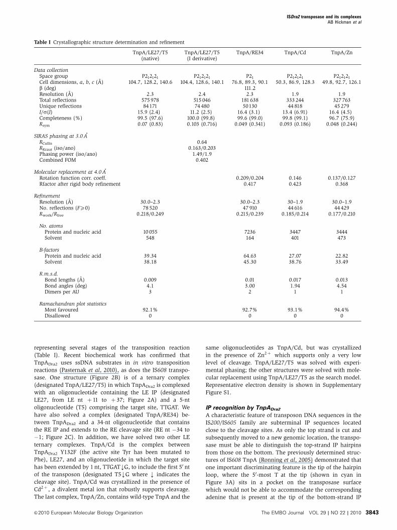
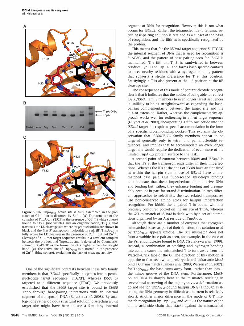
Copyright © 2022 FDOKUMEN