Human DNA Ligase III Recognizes DNA Ends by Dynamic Switching between Two DNA-Bound States
20
Human DNA Ligase III Recognizes DNA Ends by Dynamic Switching Between Two DNA Bound States † Elizabeth Cotner-Gohara 1,6 , In-Kwon Kim 1,6 , Michal Hammel 2 , John A. Tainer 3,4 , Alan E. Tomkinson 5 , and Tom Ellenberger 1,* 1 Department of Biochemistry and Molecular Biophysics, Washington University School of Medicine, St. Louis, MO 63110, USA 2 Physical Biosciences Division, Lawrence Berkeley National Laboratory, Berkeley, CA 94720, USA 3 Life Sciences Division, Lawrence Berkeley National Laboratory, Berkeley, CA 94720, USA 4 Department of Molecular Biology and The Skaggs Institute for Chemical Biology, The Scripps Research Institute, La Jolla, CA 92037, USA 5 University of Maryland School of Medicine, Baltimore, MD Abstract Human DNA ligase III has essential functions in nuclear and mitochondrial DNA replication and repair and contains a PARP-like zinc finger (ZnF) that increases DNA nick-joining and intermolecular DNA ligation. Yet, the bases for ligase III specificity and structural variation among human ligases are not understood. Here combined crystal structure and small angle x-ray scattering results reveal dynamic switching between two nick-binding components of ligase III: the ZnF-DNA binding domain (DBD) form a crescent-shaped surface used for DNA end recognition which switches to a ring formed by the nucleotidyl transferase (NTase) -OB-fold (OBD) domains for catalysis. Structural and mutational analyses indicate that high flexibility and distinct DNA binding domain features in ligase III assist both nick-sensing and the transition from nick-sensing by the ZnF to nick-joining by the catalytic core. The collective results support a “jackknife model” whereby the ZnF loads ligase III onto nicked DNA and conformational changes deliver DNA into the active site. This work has implications for the biological specificity of DNA ligases and functions of PARP-like zinc fingers. DNA ligase III is a vertebrate-specific protein functioning in DNA replication and repair pathways, including nucleotide excision repair, base excision repair, and single-strand break repair, plus mitochondrial replication and repair (1). DNA ligase III is furthermore † This work was supported in part by grants from the National Institutes of Health (NIH) including (5R01 GM052504; TE), and The Structural Cell Biology of DNA Repair Program (P01 CA92584; AET, TE, JAT). Funding for the SIBYLS beamline was provided in part by the Offices of Science and Biological and Environmental Research, U.S. Department of Energy, under Contract DE- AC02-05CH11231. This work includes research conducted at the Northeastern Collaborative Access Team beamlines of the Advanced Photon Source, supported by award RR-15301 from the National Center for Research Resources at the National Institute of Health. Use of the Advanced Photon Source is supported by the U.S. Department of Energy, Office of Basic Energy Sciences, under Contract No. DE-AC02-06CH11357. * Correspondence: (314) 362-0287 (phone); (314) 362-4432 (fax); [email protected]. 6 These authors contributed equally to this work Coordinates have been deposited with the Protein Data Bank as entry 3L2P. Supporting Information Available We provide additional figures and experimental procedures in the supporting information. This material is available free of charge via the Internet at: http://pubs.acs.org. NIH Public Access Author Manuscript Biochemistry. Author manuscript; available in PMC 2011 July 27. Published in final edited form as: Biochemistry. 2010 July 27; 49(29): 6165–6176. doi:10.1021/bi100503w. NIH-PA Author Manuscript NIH-PA Author Manuscript NIH-PA Author Manuscript
Transcript of Human DNA Ligase III Recognizes DNA Ends by Dynamic Switching between Two DNA-Bound States

Human DNA Ligase III Recognizes DNA Ends by DynamicSwitching Between Two DNA Bound States†
Elizabeth Cotner-Gohara1,6, In-Kwon Kim1,6, Michal Hammel2, John A. Tainer3,4, Alan E.Tomkinson5, and Tom Ellenberger1,*
1 Department of Biochemistry and Molecular Biophysics, Washington University School ofMedicine, St. Louis, MO 63110, USA2 Physical Biosciences Division, Lawrence Berkeley National Laboratory, Berkeley, CA 94720,USA3 Life Sciences Division, Lawrence Berkeley National Laboratory, Berkeley, CA 94720, USA4 Department of Molecular Biology and The Skaggs Institute for Chemical Biology, The ScrippsResearch Institute, La Jolla, CA 92037, USA5 University of Maryland School of Medicine, Baltimore, MD
AbstractHuman DNA ligase III has essential functions in nuclear and mitochondrial DNA replication andrepair and contains a PARP-like zinc finger (ZnF) that increases DNA nick-joining andintermolecular DNA ligation. Yet, the bases for ligase III specificity and structural variationamong human ligases are not understood. Here combined crystal structure and small angle x-rayscattering results reveal dynamic switching between two nick-binding components of ligase III:the ZnF-DNA binding domain (DBD) form a crescent-shaped surface used for DNA endrecognition which switches to a ring formed by the nucleotidyl transferase (NTase) -OB-fold(OBD) domains for catalysis. Structural and mutational analyses indicate that high flexibility anddistinct DNA binding domain features in ligase III assist both nick-sensing and the transition fromnick-sensing by the ZnF to nick-joining by the catalytic core. The collective results support a“jackknife model” whereby the ZnF loads ligase III onto nicked DNA and conformational changesdeliver DNA into the active site. This work has implications for the biological specificity of DNAligases and functions of PARP-like zinc fingers.
DNA ligase III is a vertebrate-specific protein functioning in DNA replication and repairpathways, including nucleotide excision repair, base excision repair, and single-strand breakrepair, plus mitochondrial replication and repair (1). DNA ligase III is furthermore
†This work was supported in part by grants from the National Institutes of Health (NIH) including (5R01 GM052504; TE), and TheStructural Cell Biology of DNA Repair Program (P01 CA92584; AET, TE, JAT). Funding for the SIBYLS beamline was provided inpart by the Offices of Science and Biological and Environmental Research, U.S. Department of Energy, under Contract DE-AC02-05CH11231. This work includes research conducted at the Northeastern Collaborative Access Team beamlines of theAdvanced Photon Source, supported by award RR-15301 from the National Center for Research Resources at the National Institute ofHealth. Use of the Advanced Photon Source is supported by the U.S. Department of Energy, Office of Basic Energy Sciences, underContract No. DE-AC02-06CH11357.*Correspondence: (314) 362-0287 (phone); (314) 362-4432 (fax); [email protected] authors contributed equally to this workCoordinates have been deposited with the Protein Data Bank as entry 3L2P.Supporting Information AvailableWe provide additional figures and experimental procedures in the supporting information. This material is available free of charge viathe Internet at: http://pubs.acs.org.
NIH Public AccessAuthor ManuscriptBiochemistry. Author manuscript; available in PMC 2011 July 27.
Published in final edited form as:Biochemistry. 2010 July 27; 49(29): 6165–6176. doi:10.1021/bi100503w.
NIH
-PA Author Manuscript
NIH
-PA Author Manuscript
NIH
-PA Author Manuscript

implicated in the repair of DNA double-strand breaks when nonhomologous end joining(NHEJ) activity is compromised (2). Upregulated ligase III expression in chronic myeloidleukemia cells, with concomitant decreases in the expression of the NHEJ proteins DNAligase IV and Artemis, may promote cell survival and disease progression, raising thepossibility of selectively inhibiting ligase III as a cancer treatment (3). Besides repairingnuclear DNA, ligase III is the only mitochondrial DNA ligase where it functions in DNArepair and replication.
Three DNA ligase III isoforms are generated by alternative mRNA splicing and translationinitiation, and expression of one or more of these is essential for the viability of mammaliancells and animals (4). The LigIIIα isoform interacts with XRCC1 through a C-terminalBRCA1-related C-terminal (BRCT) domain, and this protein complex functions in a varietyof DNA repair pathways, most prominently in the repair of DNA single-strand breaks (5,6).LigIIIβ lacks the C-terminal BRCT domain (6,7), and is expressed only in the male germline where it presumably repairs DNA strand breaks during meiotic recombination (7,8).The mitochondrial DNA ligase III (mtLigIII) isoform has an N-terminal mitochondriallocalization sequence besides the C-terminal BRCT interaction domain. However, XRCC1is absent from mitochondria and mtLigIII appears to function alone in mitochondrial DNAmaintenance (9,10).
Besides LigIII, two other DNA ligases are expressed in mammalian cells. DNA ligase I(LigI) is an essential enzyme that repairs Okazaki fragments during DNA replication andalso functions in long patch base excision repair. DNA ligase IV (LigIV) has specializedfunctions in the repair of DNA double-strand breaks by the NHEJ pathway, and in therearrangement of immunoglobulin genes (1,11). All three mammalian DNA ligases containa homologous catalytic core, consisting of two domains that are structurally conserved inprokaryotic DNA ligases and other members of a superfamily of nucleotidyl transferasesthat includes mRNA-capping enzymes and RNA ligases (12). Additional N- and C-terminalregions flanking the catalytic core of mammalian DNA ligases provide other functions,including interactions with other proteins that dictate the subcellular localization of eachenzyme. The nucleotidyl transferase (NTase) and OB-fold (OBD) domains comprise thecatalytic core of DNA ligases that harbors essential residues participating in a three-stepDNA end joining reaction (1). A third, noncatalytic domain located immediately N-terminalto the catalytic core of all three mammalian DNA ligases (13) extends the DNA interactionsurface of these enzymes. This DNA binding domain (DBD) is essential for DNArecognition and nick joining functions of human LigI (14) and LigIII (15). A crystalstructure of LigI bound to a nicked DNA revealed that the DBD and the adjoining NTaseand OBD domains form a compact, ring-shaped structure that sequesters the ends of thenicked strand in the active site (14). At present, it is unknown if this protein architecture isconserved in the other mammalian DNA ligases and how structural modifications specific toeach enzyme may contribute to their different biological functions.
In particular, the LigIII polypeptides are distinguished from the other human DNA ligasesby the presence of an N-terminal zinc finger (ZnF) domain that binds cooperatively inconjunction with the adjacent DBD domain to nicks and gaps in the backbone of duplexDNA (15). This DNA nick-sensing by the ZnF evidently contributes to substrate selectionand increases the catalytic efficiency of nick-joining by DNA ligase III (15–18).Furthermore, the ZnF has a profound effect on stimulating the intermolecular ligation of twoDNAs (15), an activity likely relevant to the involvement of LigIIIα/XRCC1 in the back-uppathway of nonhomologous end joining by ligase III (2). The DNA ligase III ZnF isstructurally related to the two N-terminal zinc-finger domains of poly(ADP-ribose)polymerase (16). Although a study of the LigIII ZnF domain by NMR identified residues ina β-hairpin motif that are strongly perturbed by the addition of DNA (17), the molecular
Cotner-Gohara et al. Page 2
Biochemistry. Author manuscript; available in PMC 2011 July 27.
NIH
-PA Author Manuscript
NIH
-PA Author Manuscript
NIH
-PA Author Manuscript

mechanisms by which DNA nick-sensing and catalytic activity are enhanced by the ZnF ofLigIII are unknown.
To characterize the DNA interaction and structure of human ligase III, we combined x-raycrystal structural analysis with small angle x-ray scattering (SAXS) to analyze solutionarchitectures and flexibility. These experiments suggest how DNA ligase III interactions andconformational changes contribute to DNA substrate selection and end joining activities.This multi-domain enzyme has a dynamic shape permitting alternative ensembles ofdomains to engage the DNA in competing configurations. Conformational switching canassist in loading DNA ligase III onto nicked DNA and promoting the juxtaposition of twoDNA molecules in the active site for ligation of two DNAs.
Experimental ProceduresProtein purification
LigIIIβ and ΔZnF-LigIII were purified as described previously (Cotner-Gohara et al., 2008),and purified protein was concentrated to 30–40 mg/ml and stored at −80°C.Selenomethionine labeled ΔZnF-LigIII protein was expressed in BL21(DE3) using aminoacids to suppress methionine biosynthesis, as described (19), and was purified by the sameprotocol as native protein. LigIII755 (residues 1–755) and ΔZnF755 (residues 170–755) werecloned into pET28a, and purified using same protocol as LigIIIβ and ΔZnF-LigIII,respectively.
Nicked DNA Substrate PreparationThe DNA strands were synthesized on an Applied Biosystems 394 DNA/RNA Synthesizerand were desalted using a SepPak cartridge (Waters, Inc). The nicked DNA substrate wasformed by annealing equimolar amounts of the three DNA strands: 5′CGGGATGCGTddC(upstream; ddC is 2′, 3′-dideoxycytidine monophosphate), 5′PO4-GTCGGACTGGC(downstream), 5′GCCAGTCCGACGACGCATCCCG (template) in 5 mM MES pH 6.5 and20 mM NaCl.
CrystallizationA LigIII-DNA complex was formed by incubating 0.6 mM nicked DNA substrate, 0.6 mMΔZnF-LigIIIβ, 10 mM ATP and 100 mM MgCl2. The ligase-DNA complex was mixed withan equal volume of well solution (1.8 M ammonium sulfate, 0.1 M sodium acetate pH 5.6).Crystals (P41212, a = 130.1 Å, b = 130.1 Å, c = 150.4 Å) grew at 22°C by hanging dropvapor diffusion. Prior to flash-cooling in liquid nitrogen, crystals were washed in wellsolution and transferred to a cryoprotectant solution containing 1.8 M ammonium sulfate,0.2 M sodium acetate pH 5.6, and 25% glycerol. Crystals diffracted beyond 3.5 Å usingsynchrotron radiation, and there is one LigIIIβ ΔZnF – DNA complex per asymmetric unit.
X-ray Data CollectionX-ray diffraction data extending to 3.0 Å resolution were collected from frozen crystals atthe NE-CAT beamline at the Advanced Photon Source, Argonne, Illinois and at the MBCbeamline at the Advanced Light Source, Berkeley, California. Data from two multi-wavelength anomalous dispersion experiments and one native experiment were used. X-raydata were processed using HKL2000 (20) or d*trek then scaled using Scalepack (20,21).Nineteen of twenty-one SeMet sites were located by automated Patterson searches usingSOLVE (22). Heavy-atom parameters were refined and experimental phases were calculatedin SHARP using the native dataset in combination with the MAD datasets (23).Experimentally phased maps had a well-defined solvent boundary and obvious electrondensity for both protein and nucleic acid. Phase improvement and density modification in
Cotner-Gohara et al. Page 3
Biochemistry. Author manuscript; available in PMC 2011 July 27.
NIH
-PA Author Manuscript
NIH
-PA Author Manuscript
NIH
-PA Author Manuscript

SOLOMON in SHARP greatly enhanced the interpretability of the electron density. Thebinding register of the DNA with respect to the protein was determined using the clearlyvisible nick in the DNA backbone, and purines were distinguishable from pyrimidines. TheSeMet sites, bulky amino acid side chains, and comparison to the DNA ligase I structure(14) helped define the amino acid register. The crystallographic model was constructedusing COOT (24), with refinement in REFMAC (25). TLS parameters were refined usingREFMAC, with the DBD, NTase and OBD domains and DNA treated as separate domains.Figures were generated using PYMOL (www.pymol.org), and molecular surfaceelectrostatics were calculated with APBS (26). Crystallographic data statistics are shown inTable 1.
Small Angle X-ray ScatteringLigIII was adenylated with 5 mM MgCl2 and 1 mM ATP for 1 hr at 4°C, and the reactionwas quenched by adding 10 mM EDTA. LigIII was dialyzed with a buffer containing 50mM Tris-HCl pH 7.5, 10% glycerol, 2 mM DTT and 250 mM NaCl. For protein-DNAcomplexes, 20-mer nicked DNA substrate (3′-OH nick for ZnF-lacking LigIII and 3′-ddCnick for ZnF-containing LigIII) was mixed with LigIII (protein : DNA = 1 : 1.3), and allprotein-DNA complexes were purified by gel-filtration. However, comparison of purifiedand unpurified mixtures of LigIIIβ and DNA in different molar ratios (from 1:0.8 to 1:1.2)gave comparable, although not identical results. LigIIIβ binds tightly to DNA in a bufferwith 50 mM Tris-HCl pH 7.5, 10% glycerol, 2 mM DTT and 250 mM NaCl, while ΔZnFshows salt-dependent DNA-binding affinity on gel-filtration, which is consistent with theoptimum near 100 mM NaCl for nick-joining and DNA-binding activities of ΔZnF (15,16).Thus, LigIIIβ/LigIII755-DNA complexes were purified at 250 mM NaCl to protect fromaggregation, whereas ΔZnF/ΔZnF755-DNA complexes were purified at 100 mM NaCl.
SAXS data were collected at the ALS beamline 12.3.1 LBNL Berkeley, California (27).Incident x-rays were tuned to a wavelength of λ = 1.0–1.5 Å at a sample-to-detector distanceof 1.5 m, resulting in scattering vectors (q) ranging from 0.007 Å−1 to 0.31 Å−1. Thescattering vector is defined as q = 4π sinθ/λ, where 2θ is the scattering angle. Allexperiments were performed at 20°C and data was processed as described (27). Briefly, thedata were acquired at short and long time exposures (0.5s, 5s) then scaled and merged forcalculations using the entire scattering spectrum. The experimental SAXS data wereevaluated for aggregation by inspection of Guinier plots (28). The radius of gyration Rg wasderived by the Guinier approximation I(q) = I(0) exp(−q2Rg
2/3) with the limits qRg<1.6.The program GNOM (29) was used to compute the pair-distance distribution functions, P(r).This approach also provided the maximum dimension of the macromolecule, Dmax. In ourrigid body modeling strategy BILBOMD, molecular dynamics simulations were used toexplore conformational space adopted by LigIIIβ constructs. A Minimal Ensemble Search(MES) is used to identify the minimal set of conformers required to accurately fit theexperimental data (30). For more details about data evaluation, see the Supplemental Data.
Site-directed mutagenesis and mutants purificationLigIIIβ was mutated using the Quickchange site-directed mutagenesis kit (Stratagene), andmutant proteins were expressed in E. coli Rosetta cells. Cells were grown in LB mediumcontaining ampicillin at 37 °C, and cultures were induced by 1 mM isopropylthiogalactoside at 16 °C. After 16 hours of incubation, cells were harvested. Cells wereresuspended in Buffer A (50 mM Tris-HCl pH 7.5, 150 mM NaCl, 0.2% NP-40, 5 mM β-mercaptoethanol, 1 mM PMSF, 1 mM benzamidine-HCl, 1 μg/ml aprotinin, 2 μg/mlleupeptin, 1 μg/ml pepstatin A, and μg/ml chymostatin) and lysed by sonication. The lysatewas clarified by centrifugation at 30,000 g for 1 hour at 4 °C and loaded onto aphosphocellulose column equilibrated with Buffer A. The fraction eluted with Buffer A with
Cotner-Gohara et al. Page 4
Biochemistry. Author manuscript; available in PMC 2011 July 27.
NIH
-PA Author Manuscript
NIH
-PA Author Manuscript
NIH
-PA Author Manuscript

1 M NaCl was pooled and loaded onto a 5 ml HiTrap Nickel Chelating Column (GEHealthcare) equilibrated with Buffer B (50 mM Tris-HCl pH 7.5, 250 mM NaCl, 5 mM β-mercaptoethanol, 1 mM PMSF, and 1 mM benzamidine-HCl). Following a wash with BufferB, the mutant proteins were eluted with stepwise with Buffer B plus 50 mM and 500 mMimidazole. Freshly purified protein was concentrated and used for the ligation assay.
DNA End Joining AssaysDNA ligation activities with nicked and blunt-ended DNA substrates were measured at 22°C as described previously (15). For ligation of an RNA:DNA heteroduplex containing asingle strand nick, the downstream (15 mer) RNA oligonucleotide was 5’-labeled withpolynucleotide kinase. RNA-containing substrates were reacted with full-length LigIIIβ at 4°C or 22 °C, and DNA-containing substrates were reacted at 4 °C with different time points.Reactions were quenched by adding 10 mM EDTA/formamide. The RNA oligonucleotide(15 mer), adenylated intermediate, and ligation product (28 mer) were separated bydenaturing PAGE, and quantified by a phosphorimaging device (Fuji BAS1000).
ResultsStructural Organization of DNA Ligase III Complexed to DNA
To define the molecular structure of LigIII and its DNA interactions, we crystallized theLigIII catalytic region comprising the DBD, NTase, and OBD domains (ΔZnF-LigIIIβprotein) in complex with a 22-mer nicked DNA substrate (Figure 1). The enzyme wascaptured prior to step 2 of the ligation reaction with the AMP cofactor covalently bonded toLys421 in the active site (Figure S1). The protein architecture and conformation of ΔZnF-LigIIIβ engaging nicked DNA resemble those of human LigI bound to nicked DNA (14),despite only 21% amino acid sequence identity. The three domains (DBD, NTase and OBD)completely encircle the DNA and sequester the 3′-OH and 5′-PO4 termini of the nickedDNA strand. Based upon this new structure, a clamp-like structure is a hallmark ofeukaryotic DNA ligases bound to their DNA substrates (14,31,32) that is mimicked byligases from lower organisms (14,31,32). The enclosed architecture of the enzyme-substratecomplex may serve to orient the ends of two DNA strands for ligation. The LigIII NTasedomain primarily engages the nicked DNA strand, whereas the OBD inserts into the minorgroove of the DNA duplex opposite the nick, helping to secure the bound DNA within theactive site of the NTase domain (Figure S2). A comparison of the crystal structures ofhuman DNA ligase III and ligase I shows conserved residues within the NTase and OBDdomains that interact with and stabilize the DNA in an underwound conformation, therebyexposing the ends of the nicked strand to the enzyme active site (Figure 1C) (14). Thesimilarities between the pre-step 2 conformation of LigIII and the post-step 2 conformationof LigI indicate that no change in conformation is required during the step 2 adenylation ofthe DNA 5′-end.
This new structure of DNA ligase III shows that the DBD is a structurally conservedelement of mammalian DNA ligases, despite only 14% sequence identity between the DBDsof LigIII and LigI (Figure 2) (13). Not surprisingly, these enzymes have different DNAbinding properties (14,15,33,34) that may reflect an adaptation of LigIII to its additionalZnF domain. The DBD's α-helical fold has a pseudo two-fold symmetry (Figure 1C) witheach half consisting of a helix-hairpin-helix DNA binding motif and an extended loop thatruns along the DNA backbone (Figure 1C). The DNA interaction surface of the DBD coversnearly one and one-half turns of the DNA double helix, contacting the DNA backbone andminor groove on the surface opposite to the nicked DNA strand. The connecting loopswithin each helix-hairpin-helix module are inserted into the minor groove where theyinteract with the edges of base pairs and the DNA phosphates. These interactions are
Cotner-Gohara et al. Page 5
Biochemistry. Author manuscript; available in PMC 2011 July 27.
NIH
-PA Author Manuscript
NIH
-PA Author Manuscript
NIH
-PA Author Manuscript

compatible with the underwound structure of the DNA and widened minor groove incomplex with LigIII.
The DBD also directly interacts with the NTase and OBD domains, resulting in a closedconformation of LigIII around the DNA nick (Figure 1A). In this conformation, the ZnF isprecluded from accessing the nicked DNA strand, implying that the enzyme must adopt adifferent conformation to enable DNA nick-sensing by the ZnF. However, the smallerburied surface area (850 Å2) at the interface between the DBD and NTase domains of LigIIIin comparison to LigI (1386 Å2; PDB ID: 1X9N) may provide greater flexibility to LigIII(Figure S3). This difference in buried surface area is attributed to a short loop in LigIII(residues Pro225-Asn235) that contacts the NTase domain and replaces a longer connectingloop of LigI (residues Asn336-Gly350). We suggest that the smaller, more hydrophilicinterface may create a flexible hinge between the DBD and NTase domains of LigIII thatfacilitates a transition in protein conformation to the closed, catalytic conformation seen inthe crystal structure from a nick-sensing conformation that enables the ZnF and DBD tobind cooperatively to DNA (15). Further evidence for the conformational flexibility ofLigIII is seen in another loop (residues 376 to 383) that connects the DBD and NTasedomains and is disordered in the crystal structure. The SAXS experiments described belowprovide additional evidence for the flexible structure of DNA ligase III.
Interactions of LigIII with DNA and Nucleotide SubstratesAlthough the structures of LigIII and LigI complexed with nicked DNA are similar, theseenzymes have significantly different DNA binding activities. LigI binds to DNA withmoderate affinity (KDNA = 0.4 μM) but without specificity for nicked DNA. The LigI DBDaccounts for most of this binding activity (14). LigIII binds to nicked DNA with comparableaffinity, but relies on the adjacent ZnF domain to supplement the weaker binding affinity ofits DBD (KDNA > 20 μM; (15)). The DBD of LigIII is designed to function in concert withthe ZnF domain, and this domain pair binds tightly and specifically to nicked DNA (KDNA=0.36 ± 0.06 μM) in a cooperative manner in comparison to either domain in isolation (15). Inthe LigIII crystal structure, the DBD packs against the core NTase-OBD domains andpresumably facilitates nick binding by the catalytic core. Thus, the DBD participates in twodifferent modes of nick recognition by LigIII: the catalytic mode of DNA end joining seenin the crystal structure, and a nick-sensing mode requiring the enzyme to open so that theZnF gains access to the DNA ends. The interaction of the LigIII DBD with DNA maycoordinate the transition between different DNA binding modes that alternately engage theZnF and catalytic core. Comparing the LigIII and LigI crystal structures, there aredifferences in the DNA contacts made by their DBDs. Differences in the amino acidsequences of the DNA contacting loops and their interactions with DNA likely contribute tothe higher binding affinity of the LigI DBD.
LigI and LigIII also differ in their activities towards homopolymer substrates (33).Specifically, LigIII joins oligo(dT) molecules hybridized to poly(rA) whereas LigI does not(33). We previously proposed that the intimate contact of the LigI OBD with the minorgroove of B-form DNA could explain discrimination against RNA-containing substrates(14) and this interaction is conserved in LigIII (Figure S4) We therefore experimentallytested the ability of LigIII to similarly discriminate against RNA-containing polymers ofheterogeneous sequence. As predicted from the crystal structure, LigIII discriminatesstrongly against RNA-containing heteroduplexes, failing to ligate RNA-containinghereoduplexes under conditions that are permissive for DNA ligation (Figure S4B, left andcenter panels). Even under less stringent conditions, at higher temperature and with 25-foldmore ligase, LigIII ligates very little of the available RNA-containing substrate after 23hours (less than 5% ligated; Figure S4b, right panel).
Cotner-Gohara et al. Page 6
Biochemistry. Author manuscript; available in PMC 2011 July 27.
NIH
-PA Author Manuscript
NIH
-PA Author Manuscript
NIH
-PA Author Manuscript

A positively charged groove in the DBD supports ZnF activityA unique feature on the surface of the DBD from LigIII is a positively charged grooveadjacent to the DNA binding surface (Figure 3A). This groove is located at the interface oftwo halves of the DBD between two parallel helices (α1 and α9), but separated from theDNA binding surface identified in the crystal structure. In the LigI DBD, this groove isabsent because the analogous flanking helices have bulky side chains that occupy the groovewith uncharged residues (Figure 3A). LigIII helix α9 (residues 318–330) contains two basicresidues (Lys323 and Arg327) that are primarily responsible for the positive charge of thegroove whereas the hydrophobic side chains of helix α1 from the other half of the DBD(residues 287–301) participate in forming and stabilizing the groove through hydrophobicinteractions with residues of helix α9. The two basic residues (Lys323 and Arg327) areunlikely to contact DNA directly as they located 8.0 Å and 12.8 Å, respectively from theDNA interface (Figure 3B).
To test the importance of this groove in catalysis, a series of amino acid substitutions wereintroduced into the DBD of DNA ligase III. The LigIII mutants were assayed for a ZnF-specific activity, the blunt end joining of two DNAs, as well as the nick-joining activity thatdoes not require the ZnF. Substitution of either of the positively charged residues in thegroove with a glutamate (K323E and R327E) significantly reduced blunt-end DNA ligationactivity but had very little effect on DNA nick-joining activity (Figure 3C, D). We concludethat these mutations do not affect the core activities of LigIII but instead selectively blockZnF function. This effect was quantified by measuring the kinetics of ligation by the K323Eand R327E mutants (Figure 3C, D). The K323E mutant showed an 87% decrease in theinitial velocity of blunt-end DNA ligation compared to wild-type LigIII. The R327E mutantis nearly devoid of blunt-end joining activity, mimicking a deletion of the ZnF (ΔZnF-LigIIIprotein). These results suggest an essential role for Arg327 in the function of the ZnF-DBD.In contrast, amino acid substitutions of other residues facing the DBD groove (R180E,A187E, and C324Y) had no significant effect on blunt-end joining (data not shown).
The substitution of Lys323 and Arg327 with glutamates eliminates the potential for twointerhelical salt bridges between α6 and α8 involving the residue pairs Lys323-Glu265 andArg327-Asp262, which could destabilize the local structure of DBD. However, the charge-reversal mutations K323E/E265K and R327E/D262R did not restore efficient blunt-endDNA joining activity (Figure 3E). Our results indicate that two basic residues (Lys323 andArg327) in the DBD of LigIII play critical roles in the ZnF-dependent intermolecularligation. Given the location of these basic residues, it seems unlikely that they constitute aseparate DNA binding site and we instead propose that the groove containing Lys323 andArg327 contributes to interdomain interactions between the ZnF and DBD. This interactioncould orient the ZnF to participate with the DBD in binding one DNA end while thecatalytic core binds a second DNA end to promote blunt end joining (Figure 5).
Conformational change of LigIII during DNA end joiningTo examine the conformation of DNA ligase III in the presence and absence of a DNAsubstrate, we performed small-angle X-ray scattering (SAXS) analyses with full-lengthLigIIIβ and truncated proteins lacking the ZnF and (LigIII755 and ΔZnF755; Figure 4).SAXS provides accurate information about the size and shape of molecules sampled from apopulation of randomly oriented molecules in solution (35,36). To simplify the analysis, theC-terminal region that is disordered in the crystal structure (residues 756–820) was alsodeleted, which resulted in a more compact overall structure but did not change theinteractions with DNA (compare Figures 4 and S5).
Cotner-Gohara et al. Page 7
Biochemistry. Author manuscript; available in PMC 2011 July 27.
NIH
-PA Author Manuscript
NIH
-PA Author Manuscript
NIH
-PA Author Manuscript

The pair-distribution functions [P(r)] calculated from the SAXS profiles of full-length andtruncated versions of LigIIIβ in the absence of DNA have an extended tail (Figure 4A andB) that is consistent with an elongated protein structure (36). When complexed with DNA,the P(r) functions are bell-shaped curves that are characteristic of compact globular proteins,particularly for LigIII lacking the ZnF (ΔZnF755-DNA; Figure 4A). These observationsindicate that LigIII undergoes a large conformational transition that is triggered by bindingto DNA, from the elongated unbound protein to a more compact structure in complex withDNA that presumably corresponds to the clamp structure observed in the crystals.
To further test the flexibility and conformational change of the ΔZnF755 protein in solution,we used a rigid body modeling procedure that incorporates molecular dynamics to surveyconformational space (37). Ten thousand different conformations and their calculated SAXSprofiles were generated for ΔZnF755 in the presence and absence of DNA, and then aMinimal Ensemble Search (MES) was used to identify the minimal number ofconformations required to best fit the experimental data (see Experimental Procedures) (37).Combinations of three ΔZnF755 conformers with different conformations show better fit (χ2
= 3.6) than the single best-fit model (χ2 = 7.3), reflecting the flexibility of ΔZnF755 proteinin the absence of DNA (Figure 4C and Table 2). For the ΔZnF755-DNA complex, asignificantly better fit (χ2 = 3.9) was obtained for mixtures of three conformers with closedand partially open conformations than for the crystal structure of ΔZnF-DNA complex (χ2 =18.1) (Figure 4E). Thus, in the presence of nicked DNA, the LigIII protein exists inequilibrium between partially open and closed conformations.
Flexible conformation of the ZnF during end joiningWhat is the disposition of the ZnF domain in the closed, catalytically active conformation ofLigIIIβ bound to nicked DNA? The P(r) curve of LigIII protein containing the ZnF(LigIII755) is indicative of an elongated protein shape, even in the presence of DNA (Figure4B). Given the dramatic sharpening of the P(r) peak for the ΔZnF755-DNA complex, and theelongated P(r) curve of the LigIII755-DNA complex with its significantly larger Dmax value(~180 Å vs. ~125 Å for ΔZnF755-DNA; Table 2), it appears that the ZnF that does not packagainst the DNA and/or other domains of ligase III but instead exists in an extendedconformation both in the presence and absence of DNA.
To examine the potential flexibility of the ZnF domain, we again used molecular dynamicssimulation to sample conformations of LigIII755 in the presence and absence of DNA. Anensemble of three conformers of LigIII755 protein with different ZnF conformationsimproved the fit to the experimental P(r) curve (χ2 = 3.9) in comparison to the single best-fitmodel (χ2 = 6.1) (Figure 4D and Table 2). The MES solution (χ2 = 4.5) for the LigIII755-DNA complex is a mixture of open and closed forms of the ΔZnF755-DNA complex with theZnF domain highly extended in all selected conformers (Figure 4F). This ensemble accountsfor the elongated tail of the experimental P(r) curve at large distances. Thus the ZnF domainexists in a flexible, elongated conformation that significantly extends the molecularenvelope of the LigIII protein both in the presence and absence of DNA. The ZnF does notstably bind to DNA when the DBD, OBD and NTase encircle nicked DNA.
DiscussionMammalian DNA ligases I and III have non-redundant functions in DNA replication andrepair, yet these enzymes exhibit remarkably similar structures. There is extensive evidencethat protein-protein interactions involving the regions that flank the conserved catalyticregion target the three mammalian DNA ligases to different DNA transactions (38). Incontrast, much less is known about how differences in the catalytic properties of the DNAligases contribute to their cellular functions. LigI and LigIII have different substrate
Cotner-Gohara et al. Page 8
Biochemistry. Author manuscript; available in PMC 2011 July 27.
NIH
-PA Author Manuscript
NIH
-PA Author Manuscript
NIH
-PA Author Manuscript
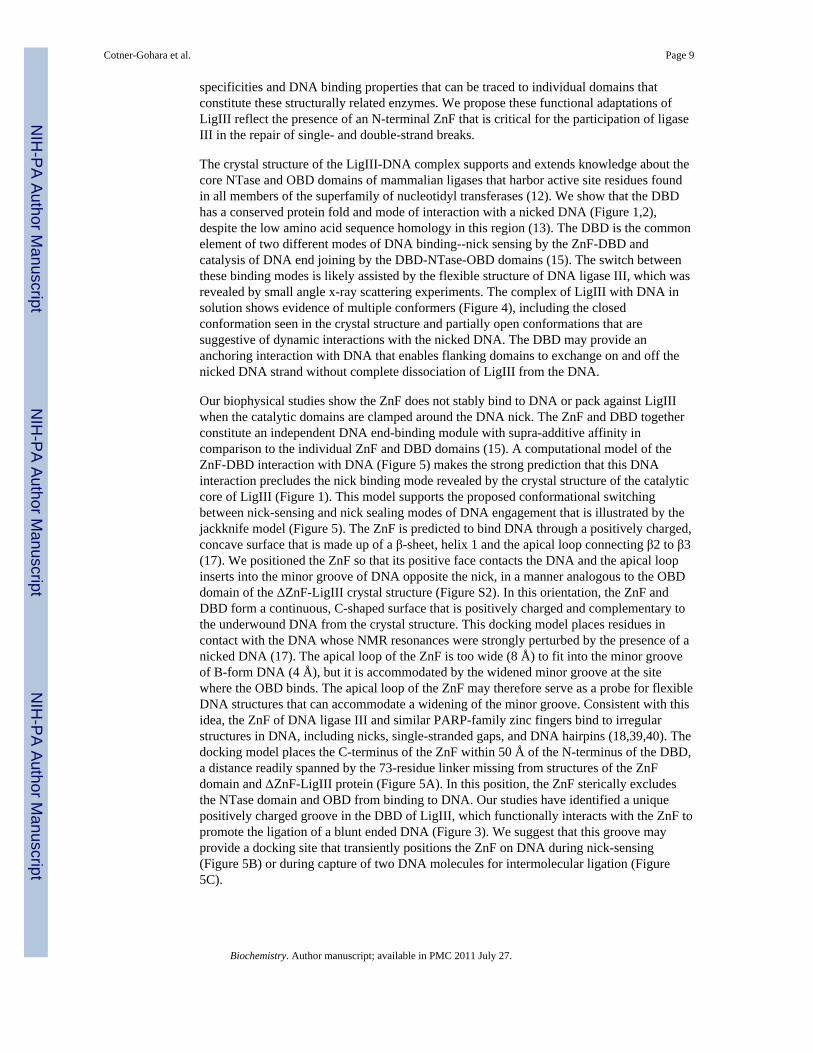
specificities and DNA binding properties that can be traced to individual domains thatconstitute these structurally related enzymes. We propose these functional adaptations ofLigIII reflect the presence of an N-terminal ZnF that is critical for the participation of ligaseIII in the repair of single- and double-strand breaks.
The crystal structure of the LigIII-DNA complex supports and extends knowledge about thecore NTase and OBD domains of mammalian ligases that harbor active site residues foundin all members of the superfamily of nucleotidyl transferases (12). We show that the DBDhas a conserved protein fold and mode of interaction with a nicked DNA (Figure 1,2),despite the low amino acid sequence homology in this region (13). The DBD is the commonelement of two different modes of DNA binding--nick sensing by the ZnF-DBD andcatalysis of DNA end joining by the DBD-NTase-OBD domains (15). The switch betweenthese binding modes is likely assisted by the flexible structure of DNA ligase III, which wasrevealed by small angle x-ray scattering experiments. The complex of LigIII with DNA insolution shows evidence of multiple conformers (Figure 4), including the closedconformation seen in the crystal structure and partially open conformations that aresuggestive of dynamic interactions with the nicked DNA. The DBD may provide ananchoring interaction with DNA that enables flanking domains to exchange on and off thenicked DNA strand without complete dissociation of LigIII from the DNA.
Our biophysical studies show the ZnF does not stably bind to DNA or pack against LigIIIwhen the catalytic domains are clamped around the DNA nick. The ZnF and DBD togetherconstitute an independent DNA end-binding module with supra-additive affinity incomparison to the individual ZnF and DBD domains (15). A computational model of theZnF-DBD interaction with DNA (Figure 5) makes the strong prediction that this DNAinteraction precludes the nick binding mode revealed by the crystal structure of the catalyticcore of LigIII (Figure 1). This model supports the proposed conformational switchingbetween nick-sensing and nick sealing modes of DNA engagement that is illustrated by thejackknife model (Figure 5). The ZnF is predicted to bind DNA through a positively charged,concave surface that is made up of a β-sheet, helix 1 and the apical loop connecting β2 to β3(17). We positioned the ZnF so that its positive face contacts the DNA and the apical loopinserts into the minor groove of DNA opposite the nick, in a manner analogous to the OBDdomain of the ΔZnF-LigIII crystal structure (Figure S2). In this orientation, the ZnF andDBD form a continuous, C-shaped surface that is positively charged and complementary tothe underwound DNA from the crystal structure. This docking model places residues incontact with the DNA whose NMR resonances were strongly perturbed by the presence of anicked DNA (17). The apical loop of the ZnF is too wide (8 Å) to fit into the minor grooveof B-form DNA (4 Å), but it is accommodated by the widened minor groove at the sitewhere the OBD binds. The apical loop of the ZnF may therefore serve as a probe for flexibleDNA structures that can accommodate a widening of the minor groove. Consistent with thisidea, the ZnF of DNA ligase III and similar PARP-family zinc fingers bind to irregularstructures in DNA, including nicks, single-stranded gaps, and DNA hairpins (18,39,40). Thedocking model places the C-terminus of the ZnF within 50 Å of the N-terminus of the DBD,a distance readily spanned by the 73-residue linker missing from structures of the ZnFdomain and ΔZnF-LigIII protein (Figure 5A). In this position, the ZnF sterically excludesthe NTase domain and OBD from binding to DNA. Our studies have identified a uniquepositively charged groove in the DBD of LigIII, which functionally interacts with the ZnF topromote the ligation of a blunt ended DNA (Figure 3). We suggest that this groove mayprovide a docking site that transiently positions the ZnF on DNA during nick-sensing(Figure 5B) or during capture of two DNA molecules for intermolecular ligation (Figure5C).
Cotner-Gohara et al. Page 9
Biochemistry. Author manuscript; available in PMC 2011 July 27.
NIH
-PA Author Manuscript
NIH
-PA Author Manuscript
NIH
-PA Author Manuscript

Although the addition of the purified ZnF in trans inhibits DNA joining by ΔZnF-LigIIIβ,the ZnF domain of LigIIIβ enhances nick-joining activity, in particular at higher saltconcentrations (15). To explain this apparent paradox, we proposed a sequential handoff ofthe DNA from the initial nick sensing by the ZnF-DBD to the engagement of the DNA nickby the catalytic core (15). In this model, the flat DNA-binding surface of the DBD acts as aplatform that presents the DNA to different domains of ligase III during each step of DNAend joining. This proposed mechanism of ZnF-dependent loading of ligase III onto DNA isnamed the “jackknife model,” which likens LigIII to a pocketknife with multiple domainsthat have specialized functions during the ligation reaction. The crystal structure and SAXSdata clearly demonstrate that the closed conformation of LigIII, with the conserved domains(DBD-NTase-OBD) encircling the nicked DNA, precludes binding by the ZnF. Thejackknife model attempts to reconcile the functional importance of the ZnF for substratediscrimination and catalytic efficiency of LigIII with the apparent transient nature ofinteractions between the ZnF and the DNA substrate. We suggest that the conformationalflexibility of LigIII revealed by SAXS experiments (Figure 4) enables LigIII to efficientlyundergo the large scale, dynamic changes necessary to transition between these twostructurally distinct DNA binding modes, nick-sensing and nick ligation. This two-stepbreak recognition mechanism is important primarily for DNA breaks that require furtherprocessing prior to ligation. Our docking model of the ZnF-DBD bound to DNA suggeststhat the ZnF might sense DNA flexibility by inserting into the minor groove, enablingselective binding to gaps, flaps, bulges, or double strand breaks (Figure 5).
Though the requirement for the ZnF in ligation of nicks remains unclear, the ZnF isabsolutely required for the intermolecular joining of blunt ended DNA molecules (15). Herewe envision that the two DNA binding modules of ligase III, spanning the ZnF-DBD and theNTase-OBD regions (15), could bridge between two DNA molecules and align them forligation by the DBD-NTase-OBD domains, in a manner analogous to the nick joiningreaction depicted by the crystal structure (Figure 5B). This intermolecular joining activity ofLigIII is compatible with its involvement in an alternative non-homologous end joiningpathway that repairs DNA double strand breaks (2). Interestingly, this alternative to thedominant, Ku-dependent pathway generates large deletions and chromosomal translocations,genomic rearrangements that are frequently detected in a cancer cells (41). In addition,cancer cells appear to be more dependent upon this pathway for the repair of DNA doublestrand breaks than normal cells (3), suggesting that cancer cells will be hypersensitive toinhibitors of backup NHEJ. Our biochemical and biophysical studies delineating the roles ofthe ZnF and DBD in intermolecular ligation by LigIII provide a rationale for theidentification of small molecules that target the ZnF and/or the positively-charged groove inthe DBD as selective inhibitors of alternative non-homologous end-joining.
Although the core structure of DNA ligase III is based on the same framework as othermammalian ligases, this enzyme has unique adaptations that contribute to the catalyticproperties of LigIII, improving the catalytic efficiency of DNA single strand break repairand enabling efficient ligation of two blunt-ended DNAs (15). These distinctive DNAbinding and enzymatic properties of ligase III contribute to its biological specialization, inaddition to protein-protein interactions that target the enzyme to sites of DNA damage.Alternative modes of DNA interaction may also contribute to the fidelity of DNA endjoining and provide opportunities for regulating ligation activity to prevent the formation ofdead end intermediates that inhibit repair of the break (42). The flexible structure of ligaseIII may facilitate recruitment to single-strand breaks while leaving the DNA accessible toother modifying enzymes for a sequential hand-off of repair intermediates that promotesefficient repair of DNA damage while avoiding toxic or mutagenic outcomes.Understanding these biologically important handoffs, which may also be critical for baseexcision repair (43,44), will require the integration of structural models of conformational
Cotner-Gohara et al. Page 10
Biochemistry. Author manuscript; available in PMC 2011 July 27.
NIH
-PA Author Manuscript
NIH
-PA Author Manuscript
NIH
-PA Author Manuscript

change with the rates and mechanisms of enzymatic functions, as developed here for DNAligase III using crystallography, SAXS, and mutational analyses.
Supplementary MaterialRefer to Web version on PubMed Central for supplementary material.
AcknowledgmentsJohn Pascal kindly provided us with RNA substrates.
Abbreviations
LigIII DNA ligase III
NTase nucleotidyl transferase domain
OBD oligonucleotide binding domain
DBD DNA binding domain
ZnF zinc finger domain
NHEJ nonhomologous end joining
BRCT BRCA1-related C-terminal domain
LigI DNA ligase I
LigIV DNA ligase IV
SAXS small angle x-ray scattering
PARP1 poly (ADP-ribose) polymerase I
References1. Tomkinson AE, Vijayakumar S, Pascal JM, Ellenberger T. DNA ligases: structure, reaction
mechanism, and function. Chemical Reviews. 2006; 106:687–699. [PubMed: 16464020]2. Wang H, Rosidi B, Perrault R, Wang M, Zhang L, Windhofer F, Iliakis G. DNA ligase III as a
candidate component of backup pathways of nonhomologous end joining. Cancer Research. 2005;65:4020–4030. [PubMed: 15899791]
3. Sallmyr A, Tomkinson AE, Rassool FV. Up-regulation of WRN and DNA ligase IIIalpha in chronicmyeloid leukemia: consequences for the repair of DNA double-strand breaks. Blood. 2008;112:1413–1423. [PubMed: 18524993]
4. Puebla-Osorio N, Lacey DB, Alt FW, Zhu C. Early embryonic lethality due to targeted inactivationof DNA ligase III. Molecular and Cellular Biology. 2006; 26:3935–3941. [PubMed: 16648486]
5. Dulic A, Bates PA, Zhang X, Martin SR, Freemont PS, Lindahl T, Barnes DE. BRCT domaininteractions in the heterodimeric DNA repair protein XRCC1-DNA ligase III. Biochemistry. 2001;40:5906–5913. [PubMed: 11352725]
6. Nash RA, Caldecott KW, Barnes DE, Lindahl T. XRCC1 protein interacts with one of two distinctforms of DNA ligase III. Biochemistry. 1997; 36:5207–5211. [PubMed: 9136882]
7. Mackey ZB, Ramos W, Levin DS, Walter CA, McCarrey JR, Tomkinson AE. An alternativesplicing event which occurs in mouse pachytene spermatocytes generates a form of DNA ligase IIIwith distinct biochemical properties that may function in meiotic recombination. Molecular andCellular Biology. 1997; 17:989–998. [PubMed: 9001252]
8. Husain I, Tomkinson AE, Burkhart WA, Moyer MB, Ramos W, Mackey ZB, Besterman JM, ChenJ. Purification and characterization of DNA ligase III from bovine testes. Homology with DNA
Cotner-Gohara et al. Page 11
Biochemistry. Author manuscript; available in PMC 2011 July 27.
NIH
-PA Author Manuscript
NIH
-PA Author Manuscript
NIH
-PA Author Manuscript
ligase II and vaccinia DNA ligase. The Journal of Biological Chemistry. 1995; 270:9683–9690.[PubMed: 7721901]
9. Lakshmipathy U, Campbell C. Mitochondrial DNA ligase III function is independent of Xrcc1.Nucleic Acids Research. 2000; 28:3880–3886. [PubMed: 11024166]
10. Lakshmipathy U, Campbell C. The human DNA ligase III gene encodes nuclear and mitochondrialproteins. Molecular and Cellular Biology. 1999; 19:3869–3876. [PubMed: 10207110]
11. McKinnon PJ, Caldecott KW. DNA strand break repair and human genetic disease. Annu RevGenomics Hum Genet. 2007; 8:37–55. [PubMed: 17887919]
12. Shuman S, Schwer B. RNA capping enzyme and DNA ligase: a superfamily of covalentnucleotidyl transferases. Molecular Microbiology. 1995; 17:405–410. [PubMed: 8559059]
13. Martin IV, MacNeill SA. ATP-dependent DNA ligases. Genome Biology. 2002;3:REVIEWS3005. [PubMed: 11983065]
14. Pascal JM, O'Brien PJ, Tomkinson AE, Ellenberger T. Human DNA ligase I completely encirclesand partially unwinds nicked DNA. Nature. 2004; 432:473–478. [PubMed: 15565146]
15. Cotner-Gohara E, Kim IK, Tomkinson AE, Ellenberger T. Two DNA-binding and nick recognitionmodules in human DNA ligase III. The Journal of Biological Chemistry. 2008; 283:10764–10772.[PubMed: 18238776]
16. Mackey ZB, Niedergang C, Murcia JM, Leppard J, Au K, Chen J, de Murcia G, Tomkinson AE.DNA ligase III is recruited to DNA strand breaks by a zinc finger motif homologous to that ofpoly(ADP-ribose) polymerase. Identification of two functionally distinct DNA binding regionswithin DNA ligase III. The Journal of Biological Chemistry. 1999; 274:21679–21687. [PubMed:10419478]
17. Kulczyk AW, Yang JC, Neuhaus D. Solution structure and DNA binding of the zinc-finger domainfrom DNA ligase IIIalpha. Journal of Molecular Biology. 2004; 341:723–738. [PubMed:15288782]
18. Taylor RM, Whitehouse CJ, Caldecott KW. The DNA ligase III zinc finger stimulates binding toDNA secondary structure and promotes end joining. Nucleic Acids Research. 2000; 28:3558–3563. [PubMed: 10982876]
19. Van Duyne GD, Standaert RF, Karplus PA, Schreiber SL, Clardy J. Atomic structures of thehuman immunophilin FKBP-12 complexes with FK506 and rapamycin. Journal of MolecularBiology. 1993; 229:105–124. [PubMed: 7678431]
20. Otwinowski Z, Minor W. Processing of X-ray Diffraction Data Collected in Oscillation Mode.Methods in Enzymology. 1997; 276:307–326.
21. Pflugrath JW. The finer things in X-ray diffraction data collection. Acta Crystallographica. 1999;55:1718–1725.
22. Terwilliger TC, Berendzen J. Automated MAD and MIR structure solution. ActaCrystallographica. 1999; 55:849–861.
23. de La Fortelle, E.; Bricogne, G.; Carter, Charles W, Jr. Methods in Enzymology. Academic Press;1997. [27] Maximum-likelihood heavy-atom parameter refinement for multiple isomorphousreplacement and multiwavelength anomalous diffraction methods; p. 472-494.
24. Emsley P, Cowtan K. Coot: model-building tools for molecular graphics. Acta Crystallographica.2004; 60:2126–2132.
25. Murshudov GN, Vagin AA, Dodson EJ. Refinement of macromolecular structures by themaximum-likelihood method. Acta Crystallographica. 1997; 53:240–255.
26. Baker NA, Sept D, Joseph S, Holst MJ, McCammon JA. Electrostatics of nanosystems: applicationto microtubules and the ribosome. Proceedings of the National Academy of Sciences of the UnitedStates of America. 2001; 98:10037–10041. [PubMed: 11517324]
27. Hura GL, Menon AL, Hammel M, Rambo RP, Poole FL 2nd, Tsutakawa SE, Jenney FE Jr,Classen S, Frankel KA, Hopkins RC, Yang SJ, Scott JW, Dillard BD, Adams MW, Tainer JA.Robust, high-throughput solution structural analyses by small angle X-ray scattering (SAXS).Nature Methods. 2009; 6:606–612. [PubMed: 19620974]
28. Guinier, A.; Fournet, F. Small Angle Scattering of X-rays. Wiley Interscience; New York: 1955.29. Svergun D. Determination of the regularization Parameter in Indirect-Transform Methods using
perceptual criteria. J Appl Cryst. 1992; 25:495–503.
Cotner-Gohara et al. Page 12
Biochemistry. Author manuscript; available in PMC 2011 July 27.
NIH
-PA Author Manuscript
NIH
-PA Author Manuscript
NIH
-PA Author Manuscript

30. Pelikan M, Hura GL, Hammel M. Structure and Flexibility within proteins as identified throughsmall angle X-ray scattering. General Physiology and Biophysics. 2009; 28:174–189. [PubMed:19592714]
31. Nandakumar J, Nair PA, Shuman S. Last stop on the road to repair: structure of E. coli DNA ligasebound to nicked DNA-adenylate. Molecular Cell. 2007; 26:257–271. [PubMed: 17466627]
32. Nair PA, Nandakumar J, Smith P, Odell M, Lima CD, Shuman S. Structural basis for nickrecognition by a minimal pluripotent DNA ligase. Nature Structural & Molecular Biology. 2007
33. Tomkinson AE, Roberts E, Daly G, Totty NF, Lindahl T. Three distinct DNA ligases inmammalian cells. The Journal of Biological Chemistry. 1991; 266:21728–21735. [PubMed:1939197]
34. Bhagwat AS, Sanderson RJ, Lindahl T. Delayed DNA joining at 3' mismatches by human DNAligases. Nucleic Acids Research. 1999; 27:4028–4033. [PubMed: 10497267]
35. Koch MH, Vachette P, Svergun DI. Small-angle scattering: a view on the properties, structures andstructural changes of biological macromolecules in solution. Q Rev Biophys. 2003; 36:147–227.[PubMed: 14686102]
36. Putnam CD, Hammel M, Hura GL, Tainer JA. X-ray solution scattering (SAXS) combined withcrystallography and computation: defining accurate macromolecular structures, conformations andassemblies in solution. Quarterly Reviews of Biophysics. 2007; 40:191–285. [PubMed: 18078545]
37. Pelikan M, Hura GL, Hammel M. Structure and flexibility within proteins as identified throughsmall angle X-ray scattering. Gen Physiol Biophys. 2009; 28:174–189. [PubMed: 19592714]
38. Ellenberger T, Tomkinson AE. Eukaryotic DNA ligases: structural and functional insights. AnnualReview of Biochemistry. 2008; 77:313–338.
39. Petrucco S. Sensing DNA damage by PARP-like fingers. Nucleic Acids Research. 2003; 31:6689–6699. [PubMed: 14627802]
40. Petrucco S, Volpi G, Bolchi A, Rivetti C, Ottonello S. A nick-sensing DNA 3'-repair enzyme fromArabidopsis. The Journal of Biological Chemistry. 2002; 277:23675–23683. [PubMed: 11948185]
41. Wang H, Perrault AR, Takeda Y, Qin W, Wang H, Iliakis G. Biochemical evidence for Ku-independent backup pathways of NHEJ. Nucleic Acids Research. 2003; 31:5377–5388. [PubMed:12954774]
42. Ahel I, Rass U, El-Khamisy SF, Katyal S, Clements PM, McKinnon PJ, Caldecott KW, West SC.The neurodegenerative disease protein aprataxin resolves abortive DNA ligation intermediates.Nature. 2006; 443:713–716. [PubMed: 16964241]
43. Garcin ED, Hosfield DJ, Desai SA, Haas BJ, Bjoras M, Cunningham RP, Tainer JA. DNAapurinic-apyrimidinic site binding and excision by endonuclease IV. Nature Structural &Molecular Biology. 2008; 15:515–522.
44. Parikh SS, Putnam CD, Tainer JA. Lessons learned from structural results on uracil-DNAglycosylase. Mutation Research. 2000; 460:183–199. [PubMed: 10946228]
45. Ciarrocchi G, Lestingi M, Wright G, Montecucco A. Bacteriophage T4 and human type I DNAligases relax DNA under joining conditions. Nucleic Acids Research. 1993; 21:5934–5939.[PubMed: 8290355]
Cotner-Gohara et al. Page 13
Biochemistry. Author manuscript; available in PMC 2011 July 27.
NIH
-PA Author Manuscript
NIH
-PA Author Manuscript
NIH
-PA Author Manuscript

Figure 1. The structure of ΔZnF-LigIIIβ bound to DNA(A) Ligase III bound to a nicked DNA substrate. The adenylated ΔZnF-Ligase IIIβ proteinwas captured in a pre-Step 2 complex with DNA prior to transfer of AMP to the DNA. The5′ PO4 and 3′ ddC termini of the nicked DNA strand are highlighted in green and yellow,respectively. The crystallographic model contains 537 residues of the 697-residue proteinfragment that was crystallized and 43 of the 44 nucleotides. The C-terminal 100 amino acids(residues 747–862) are disordered, indicating that this region is flexible. However,comparison to ligase homologues indicates that the entire OBD domain is observed. Inaddition, there are three disordered loops in the protein: two in the DNA-binding domain(DBD; residues 207–213 and 376–383) and one in the OB-fold domain (OBD; residues666–691). All three of these loops are located on the exterior of the protein, away from theDNA and domain interfaces. (B) The surface view of ΔZnF-Ligase IIIβ bound to DNAshows that Ligase III forms a continuous ring about the DNA during Step 2 with the nickedDNA strand sequestered in the active site pocket of the NTase domain. (C) The DBD ofLigase III forms a platform for the highly distorted DNA, which is partially unwound andhas an offset in the helical axis about the nick (left). The DBD interacts with the DNAthrough two helix-hairpin-helix motifs that insert into the minor groove on either side of thenick (45).
Cotner-Gohara et al. Page 14
Biochemistry. Author manuscript; available in PMC 2011 July 27.
NIH
-PA Author Manuscript
NIH
-PA Author Manuscript
NIH
-PA Author Manuscript

Figure 2. Human LigIII structural alignment to human LigI(A) Structural alignment of LigI and LigIII shows the contrast between the high sequenceconservation of the NTase and OBD domains and the low sequence conservation of theDBD domain. The secondary structure of LigIII, colored-coded by domain, is indicatedabove the amino acid sequence. Residues contacting DNA (green) or other domains (purple)are highlighted. The two helices that form a positively charged groove in unique to ligase IIIare highlighted in yellow, with key residues highlighted in bright yellow. (B) The domainstructure of ligase III.
Cotner-Gohara et al. Page 15
Biochemistry. Author manuscript; available in PMC 2011 July 27.
NIH
-PA Author Manuscript
NIH
-PA Author Manuscript
NIH
-PA Author Manuscript

Figure 3. A unique positively charged groove in the DBD is important for intermolecular DNAligation(A) Structural comparison of DBDs between ligase I (left) and ligase III reveals a uniquepositively charged groove of ligase III. (B) Close view of the positively charged groove,which is boxed in (A). (C) LigIIIβ, ΔZnF, and two mutants of LigIIIβ (K323E and R327E)with residue substitutions in the positively charged groove were assayed for blunt-endjoining activity in a single turnover assay. 100 nM of proteins were reacted with 4 nM DNAsubstrates at 22 °C with different time points (10 sec, 20 sec, 40 sec, 1 min, 1.5 min, 2 minand 3 min). (D) The ligated fraction for each protein each protein in (C) [WT (●), ΔZnF (○),K323E (▼) and R327E (△)] is plotted. Error bars are standard deviations from threeseparate experiments. (E) The reciprocal charge-reversal mutants (K323E/E265K andR327E/D262R) and their parental single mutants were assayed for blunt-end and nickjoining activity. 100 nM protein were reacted with DNA substrates at 22 °C for 1.5 min.
Cotner-Gohara et al. Page 16
Biochemistry. Author manuscript; available in PMC 2011 July 27.
NIH
-PA Author Manuscript
NIH
-PA Author Manuscript
NIH
-PA Author Manuscript

Figure 4. Small angle X-ray scattering of LigIII domains and DNA substrate complexes(A) Normalized pair distribution (P(r)) functions for the ΔZnF755 protein in the presence andabsence of DNA reveal a large conformational transition of the three conserved domains ofLigIII (DBD-NTase-OBD) from an extended to a compact structure. (B) Comparisons ofP(r) functions for LigIII755 in the presence and absence of DNA show that ZnF-containingLigIII proteins are elongated even when bound to DNA, suggesting that the ZnF adoptsflexible conformations during end joining. (C–F) Rigid body modeling of ΔZnF755 (C),LigIII755 (D), ΔZnF755-DNA complex (E), and LigIII755-DNA complex (F) by MDsimulation and MES. For each protein complex, the top left panel shows a comparison of theexperimental P(r) curve with those calculated from single best fit model (red) or the MESensemble (green). In the ΔZnF755-DNA complex (E), the crystal structure of LigIIIβ ΔZnF-DNA complex is used as the single model. In the top right panel, a comparison of Dmaxvalues for 10,000 models with their Rg values is shown. The best single model and the bestfit group of MES conformers are indicated by red and green circles, respectively, with theirrelative volume fractions. The bottom panels show the single best fit model and three MESconformers with their relative volume fractions. The LigIII domains are colored as in Figure2.
Cotner-Gohara et al. Page 17
Biochemistry. Author manuscript; available in PMC 2011 July 27.
NIH
-PA Author Manuscript
NIH
-PA Author Manuscript
NIH
-PA Author Manuscript

Figure 5. Jackknife Mechanism of DNA Substrate Recognition(A) Model of nick sensing by LigIII. The ZnF solution structure (PDB ID: 1uw0) wasdocked onto the DBD and DNA from the ΔZnF crystal structure to demonstrate how theZnF-DBD module could bind to DNA in a nick-sensing mode. 73 residues of ligase III aremissing from the two structures used to construct the model. This missing linker between theZnF and NTase domains may interact with the positively charged groove in the DBD, asindicated by the black dots. (B) Unliganded ligase III adopts an extended conformation, asdemonstrated by SAXS experiments. In the substrate recognition step, the ZnF is proposedto insert into the minor groove at the nick, recognizing flexibility in the DNA substrate. Inthe catalytic step, we hypothesize that the ZnF disengages to allow the DBD, NTase andOBD to fully encircle the nick, resembling in the ΔZnF crystal structure. (C) The two DNAbinding regions of ligase III (the ZnF-DBD and NTase-OBD) could allow simultaneousbinding of two DNAs to stimulate intermolecular ligations.
Cotner-Gohara et al. Page 18
Biochemistry. Author manuscript; available in PMC 2011 July 27.
NIH
-PA Author Manuscript
NIH
-PA Author Manuscript
NIH
-PA Author Manuscript

NIH
-PA Author Manuscript
NIH
-PA Author Manuscript
NIH
-PA Author Manuscript
Cotner-Gohara et al. Page 19
Tabl
e 1
Cry
stal
logr
aphi
c D
ata
Stat
istic
s
Nat
ive
SeM
et-1
SeM
et-2
MA
D1 λ 1
MA
D λ
2M
AD
λ3
Bea
mlin
eN
SLS
24-I
DN
SLS
24-I
DA
LS 4
.2.2
ALS
4.2
.2A
LS 4
.2.2
Wav
elen
gth
(λ)
0.97
910.
9791
0.97
911
0.97
935
0.96
412
Res
olut
ion
(Å)
3.0
(3.1
1-3.
0)3.
15 (3
.26-
3.15
)3.
1 (3
.21-
3.1)
3.11
(3.2
2-3.
11)
3.2
(3.3
1-3.
2)
Com
plet
enes
s (%
)95
.5 (7
3.5)
96.7
(79.
2)10
0 (1
00)
99.9
(100
.0)
99.9
(100
.0)
Red
unda
ncy
12.9
(5.5
)5.
3 (2
.4)
7.11
(7.2
4)7.
12 (7
.26)
7.16
(7.3
6)
Rsy
m2
.057
(.60
2).0
94 (.
579)
.145
(.67
9).1
44 (.
663)
.2 (.
7)
I/sig
ma(
I)25
.0 (2
.25)
11.2
(1.9
)6.
9 (1
.9)
6.9
(2.0
)5.
6 (1
.8)
Phas
ing
Res
olut
ion
(Å)
3.00
3.15
3.10
3.11
3.2
Rcu
llis3
Iso
(cen
/ace
n)/a
no.8
25/.8
39/-
-/-/.8
44.7
21/.6
93/.8
85.8
10/.7
92/.9
62
Phas
ing
Pow
er3
Iso
(cen
/ace
n)/a
no.0
721/
.679
/--/-
/1.0
09.5
64/.5
37/.5
98.6
34/.5
51/.2
71
Ref
inem
ent
Res
olut
ion
(Å)
3.00
Rcr
yst/R
free
40.
257/
0.28
8
r.m.s.
bon
d le
ngth
s (Å
)0.
003
r.m.s.
bon
d an
gles
(°)
0.81
6
1 Mul
tiwav
elen
gth
anom
alou
s dis
pers
ion
(MA
D) x
-ray
dat
a st
atis
tics f
rom
one
wav
elen
gth
(λ) o
f a th
ree-λ
MA
D e
xper
imen
t.
2 Rsy
m =
Σ|I-
<I>|
/Σ<I
>, w
here
I is
the
refle
ctio
n in
tens
ity a
nd <
I> is
the
aver
age
inte
nsity
of m
ultip
le sy
mm
etry
-rel
ated
refle
ctio
ns.
3 As r
epor
ted
by S
HA
RP.
4 Rcr
yst =
Σ|F
o-F c
|/Σ||F
o|, w
here
Fo
and
F c a
re th
e ob
serv
ed a
nd c
alcu
late
d st
ruct
ure
fact
or a
mpl
itude
s. R
free
was
cal
cula
ted
with
a te
st se
t of r
efle
ctio
ns (5
% o
f the
dat
a).
5 Stat
istic
s rep
orte
d in
par
enth
eses
repr
esen
t hig
hest
reso
lutio
n sh
ell.
Biochemistry. Author manuscript; available in PMC 2011 July 27.

NIH
-PA Author Manuscript
NIH
-PA Author Manuscript
NIH
-PA Author Manuscript
Cotner-Gohara et al. Page 20
Table 2
Structural parameters from SAXS data
Experimental parameters Rigid body modeling
SAXS sample Dmax (Å) Rg (Å) χ2 single model/MES
LigIIIβ ~194 48.5 ± 0.2
LigIIIβ + DNA ~195 48.4 ± 0.2
LigIII755 ~177 45.5 ± 0.2 6.1/3.9
LigIII755 + DNA ~180 45.9 ± 0.2 6.3/4.5
ΔZnF ~167 42.9 ± 0.3
ΔZnF + DNA ~162 40.3 ± 0.2
ΔZnF755 ~132 36.6 ± 0.2 7.3/3.6
ΔZnF755 + DNA ~125 31.8 ± 0.1 18.1/3.9
Crystal structure of ΔZnF-DNA complex ~103 28.5 ± 0.2
Rg radius of gyration given by the Guinier approximation (28)
Dmax maximum protein distance estimated from P(r) function as shown in Figure 3.
χ2 single model /MES, goodness of fit χ2 for best-fit atomic model and multiconformational fit χ2 for MES – models. In ΔZnF755 + DNA
complex, χ2 for single model was calculated from the crystal structure of ΔZnF-DNA complex.
Biochemistry. Author manuscript; available in PMC 2011 July 27.