Cellular responses against Schistosoma mansoni in immunized Balb/c mice with soluble proteins from...
13
Tanzania Journal of Health Research Doi: http://dx.doi.org/10.4314/thrb.v14i4.7 Volume 14, Number 4, October 2012 Cellular responses against Schistosoma mansoni in immunized Balb/c mice with soluble proteins from intermediate host, Biomphalaria pfeifferi KENNEDY M. KURIA 1* , REBECCA WAIHENYA 2 , HELLEN KUTIMA 2 , PENINNAH NJOKI 1 and DORCAS S. YOLE 3 1 Mount Kenya University, P.O Box 342, Thika, Kenya 2 Jomo Kenyatta University of Agriculture and Technology P.O. Box 62000, Nairobi, Kenya 3 Institute of Primate Research, P.O Box 24481 Karen, Nairobi, Kenya ___________________________________________________________________________ Abstract: Scores of millions of people around the world are infected by Schistosoma mansoni causing considerable morbidity, mortality and loss of productivity. Safe chemotherapeutic agents have been used though there are challenges of re-infection due to resistance. Both epidemiological and experimental data suggest that acquired cell mediated immunity play significant roles in regulating the intensity of S. mansoni infection as well as its patho-physiologic sequelae. Improved control of this trematode parasite may be obtained with immunization to enhance the resistance of individuals to risk of infection. This study investigated the cellular responses of mice immunized with soluble proteins from foot and digestive gland of the vector snail and challenged with S. mansoni. The proteins were used to immunize the experimental groups then challenged with the S. mansoni. The experimental groups were FT (immunized with foot protein) and DG (immunized with digestive gland). The parameters, which were analyzed to demonstrate protection, included; the worm counts and cellular (IFN-γ, IL-5 cytokines) responses. It was observed that, the experimental groups showed significant protection in terms of worm reduction and immune responses. The group vaccinated with foot protein showed higher protection (87.5%) as compared to the group vaccinated with the digestive gland (50%) in terms of worm reduction. Cytokines (IFN-γ and IL-5) production was present in different levels during the assay time points which showed an aspect of protection. The Foot protein of the vector showed more immunizing power than the digestive gland. Research towards utilizing the two proteins as feasible vaccine candidates is encouraged. ________________________________________________________________________________________ Keywords: Schistosoma mansoni, Biomphalaria pfeifferi, IFN-γ, IL-5 cytokines, mice Introduction Schistosomiasis remains one of the neglected diseases whose search for a vaccine remains at large. No effective vaccine for the disease exists yet. Schistosomiasis affects over 200 million people worldwide, mainly in rural agricultural and peri-urban areas of developing countries, and approximately 10% suffer severe health complications from the infection (McManus et al., 2008). While chemotherapeutic drugs, such as praziquantel, oxamniquine and metrifonate, are currently considered safe and effective for the treatment of schistosomiasis, reinfection occurs frequently following drug treatment, thus a vaccine is sought to provide long-term solution (Oliveira et al., 2008). Several studies have determined that an age-dependent resistance to reinfection after cure of schistosomiasis through chemotherapeutic treatment occurs in some patients, suggesting that immunity to Schistosoma infection can be acquired and vaccination may be an effective option (McManus et al., 2008). * Correspondence: Kennedy M. Kuria; E-mail: [email protected]
Transcript of Cellular responses against Schistosoma mansoni in immunized Balb/c mice with soluble proteins from...
Tanzania Journal of Health Research Doi: http://dx.doi.org/10.4314/thrb.v14i4.7Volume 14, Number 4, October 2012
Cellular responses against Schistosoma mansoni in immunized Balb/c mice withsoluble proteins from intermediate host, Biomphalaria pfeifferi
KENNEDY M. KURIA1*, REBECCA WAIHENYA2, HELLEN KUTIMA2, PENINNAH NJOKI1 andDORCAS S. YOLE3
1Mount Kenya University, P.O Box 342, Thika, Kenya2Jomo Kenyatta University of Agriculture and Technology P.O. Box 62000, Nairobi, Kenya3Institute of Primate Research, P.O Box 24481 Karen, Nairobi, Kenya___________________________________________________________________________Abstract: Scores of millions of people around the world are infected by Schistosoma mansoni causingconsiderable morbidity, mortality and loss of productivity. Safe chemotherapeutic agents have beenused though there are challenges of re-infection due to resistance. Both epidemiological andexperimental data suggest that acquired cell mediated immunity play significant roles in regulatingthe intensity of S. mansoni infection as well as its patho-physiologic sequelae. Improved control of thistrematode parasite may be obtained with immunization to enhance the resistance of individuals torisk of infection. This study investigated the cellular responses of mice immunized with solubleproteins from foot and digestive gland of the vector snail and challenged with S. mansoni. Theproteins were used to immunize the experimental groups then challenged with the S. mansoni. Theexperimental groups were FT (immunized with foot protein) and DG (immunized with digestivegland). The parameters, which were analyzed to demonstrate protection, included; the worm countsand cellular (IFN-γ, IL-5 cytokines) responses. It was observed that, the experimental groups showedsignificant protection in terms of worm reduction and immune responses. The group vaccinated withfoot protein showed higher protection (87.5%) as compared to the group vaccinated with thedigestive gland (50%) in terms of worm reduction. Cytokines (IFN-γ and IL-5) production waspresent in different levels during the assay time points which showed an aspect of protection. TheFoot protein of the vector showed more immunizing power than the digestive gland. Researchtowards utilizing the two proteins as feasible vaccine candidates is encouraged.________________________________________________________________________________________Keywords: Schistosoma mansoni, Biomphalaria pfeifferi, IFN-γ, IL-5 cytokines, mice
Introduction
Schistosomiasis remains one of the neglected diseases whose search for a vaccine remains atlarge. No effective vaccine for the disease exists yet. Schistosomiasis affects over 200 millionpeople worldwide, mainly in rural agricultural and peri-urban areas of developingcountries, and approximately 10% suffer severe health complications from the infection(McManus et al., 2008). While chemotherapeutic drugs, such as praziquantel, oxamniquineand metrifonate, are currently considered safe and effective for the treatment ofschistosomiasis, reinfection occurs frequently following drug treatment, thus a vaccine issought to provide long-term solution (Oliveira et al., 2008). Several studies have determinedthat an age-dependent resistance to reinfection after cure of schistosomiasis throughchemotherapeutic treatment occurs in some patients, suggesting that immunity toSchistosoma infection can be acquired and vaccination may be an effective option (McManuset al., 2008).
* Correspondence: Kennedy M. Kuria; E-mail: [email protected]

2
Consequently, the development of a vaccine against schistosomes remains a priorityand immunological responses to infection remain a key component of this effort. Thedevelopment of an anti-schistosome vaccine must take into account the association betweenanti-parasite immune responses and the development of severe disease. This is becauseprotection and pathology in schistosomiasis are so self associated (Wilson, 1990).Additionally, experimental vaccination efforts have been successful in animal models ofSchistosomiasis (McManus et al., 2008).
The essential function of antibodies in protective immunity is likely to be theinduction of cytotoxic destruction of schistosomulum stage. Antibody-dependent cell-mediated cytotoxicity (ADCC) appears to be the main mechanism of killing parasites inrodent, primate and human schistosomiasis (Capron & Dessaint, 1985). Antibody-dependentcell-mediated cytotoxicity mechanisms involving pro-inflammatory cell populations(macrophages, eosinophils and platelets) as cellular partners and unusual antibody isotypeas IgE or subclasses of IgG with anaphylactic properties have been implicated (Capron et al.,1987). These observations of IgE dependent cell-mediated killing, initially observed inrodents and confirmed in human schistosomiasis, raised the protective role of IgE antibodiesagainst schistosomes (Capron & Capron, 1994).
IgG4 antibody class is believed to counteract this IgE-mediated immunity since theodds of re-infection increase with decreasing IgE levels and increasing IgG4 levels.Therefore, IgG4 may inhibit the ADCC response by competing with IgE antibodies andblocking them from binding to the epitopes on the schistosome. Thus, immunity to re-infection is more related to the IgE–IgG4 balance than to the absolute levels of these twoisotypes (Demeure et al., 1993).
The aim of this research was to investigate for a potential vaccine candidate againstS. mansoni. The zoonotic nature of this species means that mice can be used as experimentalhosts, which might reflect the host/parasite situation in humans better. The replication ofschistosomes does not occur in the human host; hence a partially effective vaccine wouldprolong and reduce the development of morbidity, the signs of which do not appear until asufficient number of egg-associated granulomas have been produced. According toReynolds et al. (2004), the snail vector of S. mansoni, B. pfeifferi shares same antigens withthe parasites explaining why the parasite is able to develop in this host. Therefore solubleproteins obtained from parts of the snail could offer protection against S. mansoni when usedto immunize the host, hence this study sought to address this phenomenon. The solubleproteins from the foot and digestive glands were used to induce the cellular response ofmice after which protection was assessed. The procedures done to the mice were accordingto the institute’s guidelines on animal handling.
Materials and Methods
Study site and study populationThe study was carried out at the Institute of Primate Research (IPR) which is a biomedicalresearch institute using both non-human primate models and rodents.Seventy two mice of eight weeks old BALC /c mice were used in the study. They werepurchased from KEMRI and maintained at in IPR Animal Resources Department at rodents’holding facilities. The mice were fed with nutrients pellets (Laboratory chow from UngaFeeds® CO) and supplemented with carrots and kale leaves. Water was supplied to them adlibitum. Biomphalaria pfeifferi snails were collected from irrigation canals in Mwea (Kirinyaga

3
District). The snails were scooped out of the water using a sieve attached to a long pole.The snails collected from Mwea were carried in plastic containers lined with damp cottonwool and transported to the snail room at IPR laboratory. The snails were screened for thepresence of schistosomes.
Experimental DesignBALB/c mice were placed into four groups of 18 mice each, of foot protein (FT), digestivegland (DG), infected control (IC) and normal control (N). In group FT, mice wereimmunized three times with foot protein extract from snail, and challenged with normalcercaria of S. mansoni; Group DG, were vaccinated three times with digestive gland proteinextract, then challenged with S. mansoni cercariae; Group IC, mice were used as infectedcontrol and were infected with normal S. mansoni cercariae. Group N, mice were used asnaive control. The groups were differentiated by markings with picric acid on their body. Sixmice were sampled from every group during every sampling time point except for naivecontrol. Sampling involved; obtaining blood for serum, lymph nodes and spleen to preparecells for culture, at week two and four post infection (p.i). At week six post challenge (p.c),blood for serum was obtained from all mice that were perfused to recover adult worms.
Preparation of immunizing agentOff springs of B. pfeifferi snails maintained at IPR for over five years, were used forpreparation of soluble proteins. Foot and digestive gland were obtained from the snail andplaced in labeled separate eppendorf tubes containing phosphate buffered saline (PBS x1)(Yole et al. 1996). The samples were homogenized using a glass mortar and pestle. Thehomogenate was then centrifuged for 1 hr at 14,000 g at 4 degrees Celsius. The supernatantwas obtained and assayed for protein concentration using the microtitre plate technique.Protein assay estimation was done in serial doubling dilution where the BIO-RAD dye wasadded to the aliquots, then incubated for 1 hr. The protein concentrations were read usingELISA reader at wavelength of 595nm (Yole et al., 1996).
Immunization of BALB/c mice with foot protein and digestive glandThe protein extracts were used as immunizing agents. Fifty micrograms of each protein wasmixed with 100 µl of complete Freud’s adjuvant and used for immunizing the mice duringthe first vaccination. In the second and third booster 50 µg of each protein was mixed with100 µl of incomplete Freud’s adjuvant. The immunization was done intraperitoneally using1 ml syringe and needle. The vaccination was done to two groups, one receiving foot protein(FT) and the other digestive gland (DG). The mice in the two groups were immunized threetimes, at intervals of two weeks. One week after the final immunization, the mice werechallenged with the normal S. mansoni cercariae (Yole et al., 1996).
Parasite and intermediate hostTwenty four hour faecal samples from olive baboons, housed at IPR primate colony, withchronic S. mansoni infection, were screened for S. mansoni eggs. The eggs were hatched and5-8 miracidia were picked up from the Petri dish using a drawn out Pasteur pipette with arubber bulb, under the dissecting microscope. The miracidia were dispensed into each wellof a 24 well culture plate (Nunclon, Denmark). Snails from the malacology laboratory weretransferred individually into the wells and the plates covered to prevent the snails fromcrawling out. The set-up was left for 30 minutes to allow parasite penetration after which the

4
snails were transferred to snail tanks. The patent period for S. mansoni is five weeks. At fourweeks p.i, the tanks were covered with dark cloth to prevent trickle shedding of cercariae.The procedure was carried out similar to that of Yole et al. (1996).
Shedding of cercaria from infected snailsInfected snails were shed and cercariae concentration in the suspension was estimated bycounting three 50 µl aliquots under the dissecting microscope. An average of cercarialnumber was taken and used to calculate the concentration of cercariae per milliliter. Thiswas used to determine the volume of the suspension that was required to infect each mousewith 250 cercariae ( Smithter & Terry 1965).
Challenge of mice with the S. mansoniMice in groups FT (immunized with snail’s foot protein), DG (immunized with snail’sdigestive gland protein) and IC (not immunized), were anaesthetized withketamine/xylazine mixture (20:1 made by adding 0.5 ml of xylazine to 10 ml of ketamine).Anaesthesia was delivered as 0.02 ml per 30 g mouse body weight intraperitoneally. Onceunconscious, the mice were arranged on a wooden infecting rack. Cotton wool, dipped inwater, was used to clean and wet the shaven area to allow easy penetration of the cercariae.Metal rings, 1 cm diameter, were placed on the shaven area and cercarial suspensiondispensed into the metal ring using a 1 ml micropipette. Cercariae were allowed to penetratefor 30 minutes (Smithter & Terry, 1965).
Sampling and preparation of lymph nodesAt each sampling point (weeks 2, 4 and 6) post challenge (p.c), six mice from each groupwere anaesthetized. A small incision was made at the centre of the abdominal skin and theskin torn around the waist of the mouse. The skin was then pulled up to expose the thoracicregion and the abdominal wall was exposed by pulling the mouse skin to the groin region.Inguinal lymph nodes and the auxillary lymph nodes were removed carefully andtransferred into incomplete medium (RPMI 1640, 0.1% Gentamycin, 5x10⁻⁵ Bmecaptoethanol) in a Petri-dish (Yole et al., 1996) in a sterile biosafety hood. They wereteased using a sharp sterile forceps to release lymphocytes. The suspension was dispensedin 15 ml centrifuge tube and sterile incomplete medium (RPMI 1640, 0.1% Gentamycin, 5x10-
5 B mecaptoethanol) added up to 10 ml mark. The cells were centrifuged at 450 g for 10minutes for effective sedimentation. The supernatants were discarded leaving a pellet. Thecells in the pellet were resuspended and washing was done two times. After the final wash,the cells were resuspended in 1 ml of complete medium (incomplete medium fortified with10 % fetal calf serum) and counted in the haemocytometer chamber using Trypan blue. Thecounting was done under a microscope using X40 magnification. The cells were thenadjusted to 3 x 10⁶ cells/ml and cells were used for culture supernatant (Yole et al., 1996).
Sampling and preparation of spleenThe spleen, from each mouse was removed and placed into incomplete medium in a Petridish in a sterile culture hood. A 10 ml syringe piston was used to squash the spleen. Thedispersed cells were then dispensed in 15 ml tube and topped up with incomplete media.The cells were washed and counted. However, the cells were resuspended in 4 ml ofcomplete medium in readiness for counting. The cells were used to culture supernatant forthe cytokine assays (Yole et al., 1996).

5
Immunological assays
Culture Supernatant: Flat-bottomed 48-well microtitre plate was used for culture and 6 x 10⁵
cells from lymph nodes and spleen. Duplicate wells were set for each regime. Negativecontrol had only medium and cells. Positive control had 1μg/ml of Concanavalin A and testwells, 10 μg/ml well of Soluble worm antigen preparation (SWAP) and 0-3 hrs releaseprotein. The total volume of culture medium per well was 400 μl. The plates were incubatedat 37ºC, 5% CO2 for 48hrs for Con A and 72hrs for the other set-ups. At the end of the
specified incubation period, supernatants were collected and used for ELISA (Yole et al.,1996).
Antibody assay (IL-5 and IFN-γ): Nunc-ImmunoTM (MaxiSorpTM Surface) ELISA plateswere coated with 50 μl of 5 μg/ml solution of monoclonal anti IL-5-antibody and IFN-γantibody. They were incubated overnight at 4ºC. The plates were then washed six timeswith 0.05 % Tween 20 in PBS and 100 μl/well blocking buffer, 0.1 % Bovine Serum Albumin(BSA) was added. The plates were incubated for 1 hr at 37ºC and thereafter washed six timesand 50 μl/well of samples and standards, in duplicates, were added. Plates were incubatedfor 2 hrs at 37ºC. They were then washed six times and 50 μl/well of 3 μg/ml rabbit anti-mouse IL-5 and IFN-γ (mAb TRFK4 - biotin) added. The plates were then incubated for 1 hrat 37ºC. The plates were washed six times and 50 μl/well of Streptavidin – HRP (conjugate)added. They were incubated for 1 hr at 37ºC and washed six times and 50 μl/well of thesubstrate (Sure Blue TM TMB) added. The plates were incubated at 37oC in the dark for 30minutes. Optical density was read at 630 nm in an ELISA microplate reader (DynatechMRX).
Data analysisThe student’s t-test was used to analyze significance levels. P values < 0.05 were consideredsignificant.
Results
IFN – γ production to 0 – 3 hr antigen by lymph node cells following infection with S.mansoniThe IFN – γ production to 0-3hr antigens are shown in Figure 1. For group DG, at week 2 p.ithe production was 12032pg/ml which then dropped to low levels of 109pg/ml at week 4.There was no measurable level of IFN – γ for FT, at week 2 p.i. However, at week 4 thegroup recorded a production of 84pg/ml. The production for IC, at week 2 was 1141pg/mlwhich then reduced to 335.75pg/ml at week 4. There was a significance difference (p<0.05)between week 2 and week 4 in DG, unlike groups FT and IC which showed no significancedifference (p>0.05) between week 2 and week 4. The production of IFN – γ in DG wassignificantly higher than IC at week 2 (p<0.05). Group FT at week 2, showed no productionfor IFN – γ as compared to group IC which had a higher production than FT, but thedifference was not significant (p>0.05). At week 4, the levels of IFN – γ in DG had reducedwith no significant difference (p>0.05) compared to IC. At week 4, the levels of IFN – γ in FThad increased slightly with no significance difference (p>0.05) compared to IC. At week 2,the response of IFN – γ in DG was significantly higher (p<0.05) than in group FT. At week 4,Groups DG and FT showed no significant difference in the IFN – γ production (Figure 1).
6
Figure 1: IFN – γ production to 0–3 hr antigen in lymph node cells at weeks 2 and 4 followinginfection with S. mansoni
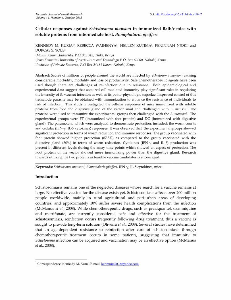
IFN – γ production to SWAP antigen by lymph node cells following infection with S.mansoniThe IFN – γ production to SWAP antigen are shown in Figure 2. The response of IFN – γ inDG, at week 2 p.i was 109pg/ml, but increased to higher levels of 11279.5pg/ml at week 4.Group FT, showed low production (84pg/ml) at week 2 p.i, but at week 4 there was someslight increase in the production (304.5pg/ml). At week 2, the IC group showed a productionof 335.75pg/ml which increased to 2779.5pg/ml at week 4. There was a significant difference(p<0.05) in DG between week 2 and week 4, unlike groups FT and IC which showed nosignificant difference (p>0.05) between these two time points. The production of IFN – γ inDG was lower than group IC at week 2, but there was no significant difference (p>0.05).Group FT at week 2, showed a lower production of IFN – γ than group IC though notsignificantly different (p>0.05). At week 2, the production of IFN – γ in groups DG and FTwas not significantly different (p<0.05). At week 4, the levels of IFN – γ in DG had increasedsignificantly (p<0.05) when compared to IC. At week 4, the levels of IFN – γ in FT hadincreased slightly with no significant difference (p>0.05) as compared to group IC. At week4, Groups DG and FT showed significant difference (p<0.05) in the production of IFN – γ;DG had a higher production than group FT.
Time post-infection
7
Figure 2: IFN – γ production to SWAP antigen by lymph node cells at weeks 2 and 4 followinginfection with S. mansoni
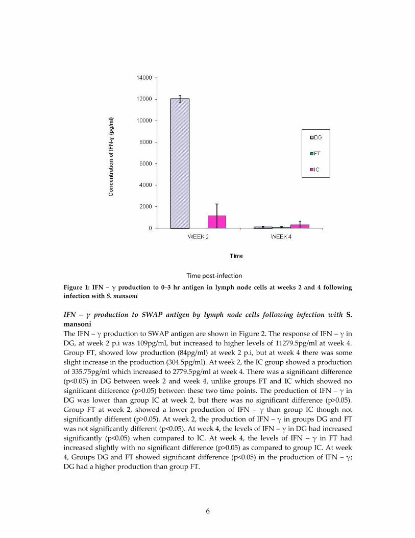
IFN – γ production to 0 – 3 hr antigen by spleen cells following infection with S. mansoniThe IFN – γ production to 0-3hr cercarie antigens are shown in Figure 3. The response ofIFN – γ in spleen cells in DG, at week 2 p.i was high (13624.5pg/ml) but decreased at week 4to 445pg/ml. The response of the FT group, was low at week 2 p.i (18.5pg/ml), however atweek 4 there were no measurable levels. In IC, at week 2, the response was minimal but atweek 4 there was a response of 560pg/ml. Groups DG and IC at week 2 and week 4 showeda significant difference (P<0.05); FT response between week 2 and week 4 was not significant(P>0.05). The response of IFN – γ in spleen cells in the DG was higher than IC at week 2; thisshowed significant difference (P<0.05) between the two groups. At week 2, FT had a slightlyhigher response as compared to IC but the response was not significantly different (P>0.05).At week 2, the response of IFN – γ in groups DG was significantly higher (P<0.05) thangroup FT. At week 4, the levels of IFN – γ in spleen between DG and IC was notsignificantly different (P>0.05). At week 4, the levels of IFN – γ in group FT was lower thanIC, and the difference was significant (p<0.05). At week 4, Groups DG and FT showedsignificant difference (P<0.05) in the response to IFN – γ, where group DG had a higherproduction than group FT.
Time post-infection

8
Figure 3: IFN – γ production to 0 – 3 hr antigen by spleen cells at weeks 2 and 4 following S.mansoni infection
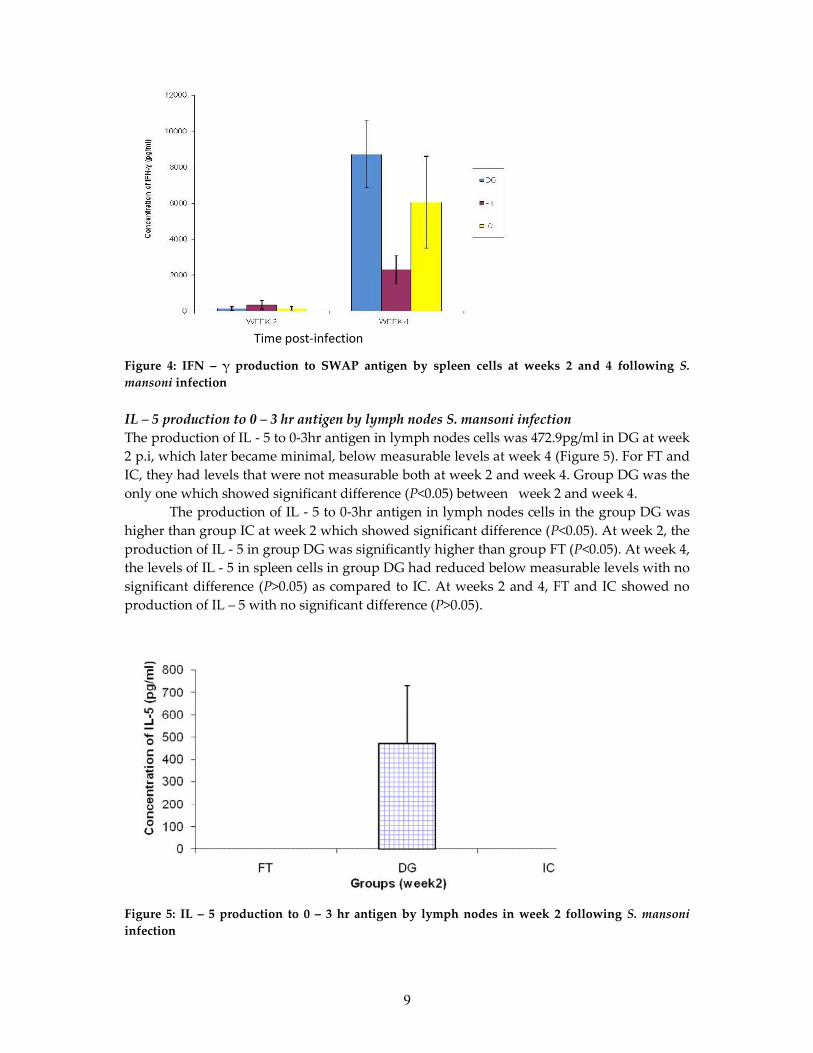
IFN – γ production to SWAP antigen by spleen cells following infection with S. mansoniThe production of IFN – γ to SWAP antigen in spleen cells is as shown in Figure 4. In DG, atweek 2 p.i the production was low (144pg/ml) but it increased to a high level at week 4 to8717.5pg/ml. In FT, the production was low of 350.25pg/ml at week 2 p.i, but at week 4 theproduction had increased to 2312.5pg/ml. In group IC, at week 2, there was low productionof 157.25pg/ml which increased at week 4 to 6052.5pg/ml. There was significant difference(P<0.05) of all groups between week 2 and week 4. The production of IFN – γ in spleen cellsin the DG was lower than group IC at week 2 with no significant difference (P>0.05). Atweek 2, FT showed higher production of IFN – γ than group IC but the difference was notsignificant (P>0.05). At week 2, the production of IFN – γ in groups FT was higher than DGwith no significant difference (P>0.05). At week 4, the production in DG had increased butwith no significant difference (P>0.05) as compared to IC. At week 4, the levels of IFN – γ inIC was higher than group FT with significant difference (P<0.05). At week 4, DG and FTshowed significant difference (P<0.05) in the production of IFN – γ, where group DG had ahigher concentration than group FT.
Time post-infection
9
Figure 4: IFN – γ production to SWAP antigen by spleen cells at weeks 2 and 4 following S.mansoni infection
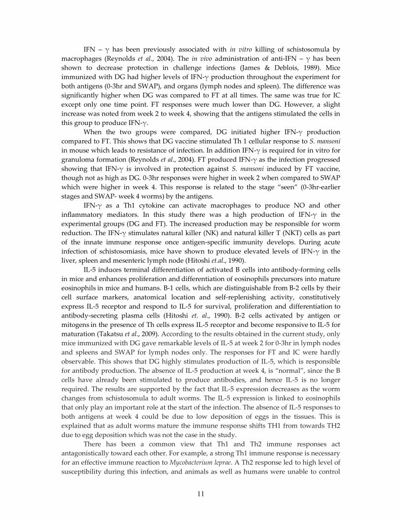
IL – 5 production to 0 – 3 hr antigen by lymph nodes S. mansoni infectionThe production of IL - 5 to 0-3hr antigen in lymph nodes cells was 472.9pg/ml in DG at week2 p.i, which later became minimal, below measurable levels at week 4 (Figure 5). For FT andIC, they had levels that were not measurable both at week 2 and week 4. Group DG was theonly one which showed significant difference (P<0.05) between week 2 and week 4.
The production of IL - 5 to 0-3hr antigen in lymph nodes cells in the group DG washigher than group IC at week 2 which showed significant difference (P<0.05). At week 2, theproduction of IL - 5 in group DG was significantly higher than group FT (P<0.05). At week 4,the levels of IL - 5 in spleen cells in group DG had reduced below measurable levels with nosignificant difference (P>0.05) as compared to IC. At weeks 2 and 4, FT and IC showed noproduction of IL – 5 with no significant difference (P>0.05).
Figure 5: IL – 5 production to 0 – 3 hr antigen by lymph nodes in week 2 following S. mansoniinfection
Time post-infection

10
IL – 5 production to SWAP antigen by lymph nodes following S. mansoni infectionThe production of IL - 5 to SWAP in lymph nodes cells was 746.8pg/ml for DG at week 2 p.i,which later became minimal at week 4. In FT and IC, levels of IL-5 which were minimal atweek 2. The production for the groups FT, DG and IC was hardly noticeable at week 4.Group DG showed significant difference (P<0.05) between week 2 and week 4. Theproduction of IL - 5 to SWAP antigen in lymph nodes cells in the DG was higher than groupIC at week 2 which showed significant difference (P<0.05). At week 2, the production of IL -5 in group DG was significantly higher than group FT (P<0.05). At week 4, the levels of IL - 5in spleen cells in group DG had reduced below measurable levels with no significantdifference (P>0.05) as compared to IC. At weeks 2 and 4, FT and IC showed no production ofIL – 5 with no significant difference (P>0.05).
IL – 5 production to 0 – 3 hr antigen by spleen cells following S. mansoni infectionThe production of IL - 5 to 0-3hr antigen in lymph nodes cells was 291.25pg/ml in DG atweek 2 p.i, which later became minimal at week 4. Groups FT and IC had minimum levelswhich were not measurable both at week 2 and week 4. The production of the groups wasminimal at week 4. Group DG was the only one that showed significant difference (P<0.05)between week 2 and week 4. The production of IL - 5 in spleen cells in the group DG wassignificantly higher than IC at week 2 (P<0.05). At week 4, the levels of IL - 5 in spleen cellsin group DG had reduced below measurable levels with no significant difference (p>0.05)between the groups (DG and IC). At weeks 2 and 4, groups FT and IC showed noproduction of IL – 5 with no significant difference (P>0.05). At week 2, the production of IL -5 in group DG was significantly higher than group FT (P<0.05).
IL – 5 production to SWAP antigen by spleen cells following S. mansoni infectionThe production of IL - 5 in spleen cells was below measurable levels in the groups DG, FTand IC both at weeks 2 and 4 p.i.
Discussion
It is speculated that S. mansoni infections induce an immune response dominated by Th2cells, whereas a Th1 predominant response strongly correlates with resistance to infection. Akey distinction between the Th1 versus Th2 pathways lies in the source of differentcytokines involved. Th1 responses are typically characterized by the secretion of IFN-γ andIL-2. However, Th2 responses are characterized by the secretion of IL-4, IL-5, IL-6 and IL-10.The vaccination-induced Th1-type response plays an important role in anti-schistosomeinfection by producing cytokines, such as IFN-γ and IL-2. It has been shown that, at an earlystage of infection, the host’s response against the parasite is a Th1-type one. Epidemiologicalsurveys of schistosomiasis showed the individual with a high level of IFN-γ wassignificantly correlated with resistance to schistosome infection. In animal models withschistosome infection, it has been observed that IFN-γ can suppress granuloma formation invivo, and decrease the size of pulmonary granulomas and the extent of hepatic fibrosis(Reynolds et al., 2004).
With regard to cytokines assessed in this study, it sought to asses those that arereported to be differentially produced by two different T-helper subsets. Mosmann et al.,1986, have shown that those cells designated TH-1 produce IFN – γ, whereas thosedesignated TH-2 produce IL – 5.
11
IFN – γ has been previously associated with in vitro killing of schistosomula bymacrophages (Reynolds et al., 2004). The in vivo administration of anti-IFN – γ has beenshown to decrease protection in challenge infections (James & Deblois, 1989). Miceimmunized with DG had higher levels of IFN-γ production throughout the experiment forboth antigens (0-3hr and SWAP), and organs (lymph nodes and spleen). The difference wassignificantly higher when DG was compared to FT at all times. The same was true for ICexcept only one time point. FT responses were much lower than DG. However, a slightincrease was noted from week 2 to week 4, showing that the antigens stimulated the cells inthis group to produce IFN-γ.
When the two groups were compared, DG initiated higher IFN-γ productioncompared to FT. This shows that DG vaccine stimulated Th 1 cellular response to S. mansoniin mouse which leads to resistance of infection. In addition IFN-γ is required for in vitro forgranuloma formation (Reynolds et al., 2004). FT produced IFN-γ as the infection progressedshowing that IFN-γ is involved in protection against S. mansoni induced by FT vaccine,though not as high as DG. 0-3hr responses were higher in week 2 when compared to SWAPwhich were higher in week 4. This response is related to the stage “seen” (0-3hr-earlierstages and SWAP- week 4 worms) by the antigens.
IFN-γ as a Th1 cytokine can activate macrophages to produce NO and otherinflammatory mediators. In this study there was a high production of IFN-γ in theexperimental groups (DG and FT). The increased production may be responsible for wormreduction. The IFN-γ stimulates natural killer (NK) and natural killer T (NKT) cells as partof the innate immune response once antigen-specific immunity develops. During acuteinfection of schistosomiasis, mice have shown to produce elevated levels of IFN-γ in theliver, spleen and mesenteric lymph node (Hitoshi et.al., 1990).
IL-5 induces terminal differentiation of activated B cells into antibody-forming cellsin mice and enhances proliferation and differentiation of eosinophils precursors into matureeosinophils in mice and humans. B-1 cells, which are distinguishable from B-2 cells by theircell surface markers, anatomical location and self-replenishing activity, constitutivelyexpress IL-5 receptor and respond to IL-5 for survival, proliferation and differentiation toantibody-secreting plasma cells (Hitoshi et. al., 1990). B-2 cells activated by antigen ormitogens in the presence of Th cells express IL-5 receptor and become responsive to IL-5 formaturation (Takatsu et al., 2009). According to the results obtained in the current study, onlymice immunized with DG gave remarkable levels of IL-5 at week 2 for 0-3hr in lymph nodesand spleens and SWAP for lymph nodes only. The responses for FT and IC were hardlyobservable. This shows that DG highly stimulates production of IL-5, which is responsiblefor antibody production. The absence of IL-5 production at week 4, is “normal”, since the Bcells have already been stimulated to produce antibodies, and hence IL-5 is no longerrequired. The results are supported by the fact that IL-5 expression decreases as the wormchanges from schistosomula to adult worms. The IL-5 expression is linked to eosinophilsthat only play an important role at the start of the infection. The absence of IL-5 responses toboth antigens at week 4 could be due to low deposition of eggs in the tissues. This isexplained that as adult worms mature the immune response shifts TH1 from towards TH2due to egg deposition which was not the case in the study.
There has been a common view that Th1 and Th2 immune responses actantagonistically toward each other. For example, a strong Th1 immune response is necessaryfor an effective immune reaction to Mycobacterium leprae. A Th2 response led to high level ofsusceptibility during this infection, and animals as well as humans were unable to control

12
the infection (Verhagen et. al., 1998). In C57BL/6 mice Leishmania major induces an effectiveTh1 immune response, while in BALB/c mice a nonprotective Th2 immune response isinduced (Etges & Muller, 1998). According to Scott (1991) gamma interferon (IFN-γ) as atypical Th1 cytokine and interleukin-5 (IL-5) as a typical Th2 cytokine have antagonisticeffects. This explains why group DG had more worms recovered than group FT. Group DGhad both IFN-γ and IL-5 responses which were antagonistic to each other. On the otherhand, FT had high responses of IFN-γ and low responses of IL-5 which was minimal, hencelower worm counts. In conclusion, immunization with Foot protein (FT) and Digestivegland (DG) reduced worm burden. DG had higher cellular responses as shown by higherlevels of IFN-γ initially and thereafter.
Acknowledgements
We thank Mr. Kiio Kithome, Sammy Kisara, Simon Kiarie and Collins Kisara for theirexcellent technical assistance. The Institute of Primate Research Centre, Kenya is thanked forproviding the laboratory facility to conduct this research.
References
Capron, A. & Dessaint, J.P. (1985) Effector and Regulatory Mechanisms in Immunity toSchistosomes. Annual Review in Immunology 3, 455-476.
Capron, A., Dessaint, J.P., Capron, M., Ouma, J.H. & Butterworth, A. (1987) Immunity toschistosomes: Progress towards Vaccine. Science 238, 1065-1072.
Capron, M. & Capron, A. (1994) Immunology and Effector Cells in schistosomiasis. Science264, 187.
Demeure, C.E., Rihet, P., Abel, L., Ouattara, M., Bourgois, A. & Dessein, J. (1993) Resistanceto schistosomiasis mansoni in humans: influence of IgE\IgG4 balance and IgG2 inimmunity to reinfection after chemotherapy. Journal of Infectious Diseases 168, 1000-1008.
Etges, R. & Muller, I. (1998) Progressive disease or protective immunity to Leishmania majorinfection: the result of a network of stimulatory and inhibitory interactions. Journal ofMolecular Medicine 76, 372-390.
Hitoshi, Y., Yamaguchi, N., Mita, S., Takaki, S., Tominaga, A. & Takatsu, K. (1990)Distribution of IL-5 receptor-positive B cells. Expression of IL-5 receptor on Ly-1(CD5)+ B cells. Journal of Immunology 144, 42-18.
James, S.L. & Deblois, L.A. (1986) Induction of protective immunity against Schistosomamansoni by a non living vaccine. II. Response of mouse strains with selectiveimmune defects. Journal of Immunology 136, 3864-3871.
McManus, D.P. & Loukas, A. (2008) Current status of vaccines for schistosomiasis. ClinicalMicrobiology Reviews 21, 225–242.
Mosmann, T.R., Cherwinski, H., Bond, M.W., Giedlin, M.A. & Coffman, R.L. (1986) Twotypes of murine helper T-cell clones. I. Definition according to profiles of lymphokineactivities and secreted proteins. Journal of Immunology 136, 2348-2357.
Oliveira, S.C., Fonseca, C.T., Cardoso, F.C., Farias, L.P. & Leite, L.C.C. (2008) Recentadvances in vaccine research against schistosomiasis in Brazil. Acta Tropica 108, 256–262.
13
Reynolds, S.R., Dahl, C.E. & Harn, D.A. (2004) T and B epitope determination and analysisof multiple antigenic peptides for the Schistosoma mansoni experimental vaccinetriose-phosphate isomerase. Journal of Immunology 152, 193-200.
Scott, P. (1991) IFN-gamma modulates the early development of Th1 and Th2 responses in amurine model of cutaneous leishmaniasis. Journal of Immunology 147, 3149-3155.
Smithers, S.R. & Terry, R.J. (1965) The infection of laboratory hosts with the cercariae ofmansoni and the recovery of adults’ worms. Parasitology 5, 695-700.
Takatsu, K., Kouro, T. & Nagai, Y. (2009) Interleukin 5 in the link between innate andacquired immune response. Advanced Immunology 101, 191-192.
Verhagen, C.E., van T.C., Pouw, K.A., Buffing, A., Chand, M.A., Faber, W.R., Aarden, L.A. &Das, P.K. (1998) Type 1- and type 2-like lesional skin-derived Mycobacterium leprae-responsive T cell clones are characterized by coexpression of IFN-gamma/TNF-alphaand IL-4/IL-5/IL-13, respectively. Journal of Immunology 160, 2380-2387.
Wilson, R.A. (1990) ’Leakey Livers’: portal shunting and immunity to schistosomes.Parasitology Today 6, 354-358.
Yole, D.S., Pemberton, R., Reid, G.D. & Wilson, R.A. (1996) Protective immunity toSchistosoma mansoni induced in the Olive baboon, Papio anubis, by the irradiatedcercariae vaccine. Parasitology 12, 37-46.