Diesel exhaust particulates affect cell signaling, mucin profiles, and apoptosis in trachea explants...
12
Diesel Exhaust Particulates Affect Cell Signaling, Mucin Profiles, and Apoptosis in Trachea Explants of Balb/C Mice Robson Seriani, 1 Mara de Souza Junqueira, 2 Alessandra Choqueta de Toledo, 3 Milton Arruda Martins, 3 Marcelo Seckler, 4 Adriano Mesquita Alencar, 5 Elnara Marcia Negri, 1 Luiz Fernando Ferraz Silva, 1 Tha ıs Mauad, 1 Paulo Hil ario Nascimento Saldiva, 1 Mariangela Macchione 1 1 Laboratory of Experimental Air Pollution, Department of Pathology, School of Medicine, University of S~ ao Paulo, S~ ao Paulo, SP, Brazil 2 Central Biotery Laboratory, School of Medicine, University of S~ ao Paulo, S~ ao Paulo, SP, Brazil 3 Experimental Therapeutics Laboratory, Department of Medicine, School of Medicine, Univer- sity of S~ ao Paulo, S~ ao Paulo, SP, Brazil 4 Department of Chemistry Engineering, Polytechnic School, University of S~ ao Paulo, S~ ao Paulo, SP, Brazil 5 Department of General Physics - Institute of Physics, University of S~ ao Paulo, S~ ao Paulo, SP, Brazil Received 12 July 2013; revised 10 January 2014; accepted 15 April 2014 ABSTRACT: Particulate matter from diesel exhaust (DEP) has toxic properties and can activate intracellu- lar signaling pathways and induce metabolic changes. This study was conducted to evaluate the activa- tion of extracellular signal-regulated kinase (ERK) and c-Jun N-terminal kinase (JNK) and to analyze the mucin profile (acid (AB 1 ), neutral (PAS 1 ), or mixed (AB/PAS 1 ) mucus) and vacuolization (V) of tracheal explants after treatment with 50 or 100 lg/mL DEP for 30 or 60 min. Western blot analyses showed small increases in ERK1/2 and JNK phosphorylation after 30 min of 100 lg/mL DEP treatment compared with the control. An increase in JNK phosphorylation was observed after 60 min of treatment with 50 lg/mL DEP compared with the control. We did not observe any change in the level of ERK1/2 phosphorylation after treatment with 50 lg/mL DEP. Other groups of tracheas were subjected to histological sectioning and stained with periodic acid-Schiff (PAS) reagent and Alcian Blue (AB). The stained tissue sections were then subjected to morphometric analysis. The results obtained were compared using ANOVA. Treat- ment with 50 lg/mL DEP for 30 min or 60 min showed a significant increase (p < 0.001) in the amount of acid mucus, a reduction in neutral mucus, a significant reduction in mixed mucus, and greater vacuoliza- tion. Our results suggest that compounds found in DEPs are able to activate acid mucus production and enhance vacuolization and cell signaling pathways, which can lead to airway diseases. V C 2014 Wiley Periodi- cals, Inc. Environ Toxicol 00: 000–000, 2014. Keywords: histopathology; DEP; biomarkers; signal transduction; explants; MAPK Correspondence to: M. Macchione; e-mail: [email protected] Supported by: Conselho Nacional de Desenvolvimento Cient ıfico e Tecnol ogico CNPq Processo: 484189/2007-7, Laborat orio de Investigac¸~ ao M edica LIM05 do Hospital da Cl ınicas – Faculdade de Medicina da Universidade de S~ ao Paulo- HC- FMUSP. Published online 00 Month 2014 in Wiley Online Library (wileyonlinelibrary.com). DOI: 10.1002/tox.22000 V C 2014 Wiley Periodicals, Inc. 1
-
Upload
independent -
Category
Documents
-
view
2 -
download
0
Transcript of Diesel exhaust particulates affect cell signaling, mucin profiles, and apoptosis in trachea explants...
Diesel Exhaust Particulates Affect Cell Signaling,Mucin Profiles, and Apoptosis in TracheaExplants of Balb/C Mice
Robson Seriani,1 Mara de Souza Junqueira,2 Alessandra Choqueta de Toledo,3
Milton Arruda Martins,3 Marcelo Seckler,4 Adriano Mesquita Alencar,5
Elnara Marcia Negri,1 Luiz Fernando Ferraz Silva,1 Tha�ıs Mauad,1
Paulo Hil�ario Nascimento Saldiva,1 Mariangela Macchione1
1Laboratory of Experimental Air Pollution, Department of Pathology, School of Medicine,University of S~ao Paulo, S~ao Paulo, SP, Brazil
2Central Biotery Laboratory, School of Medicine, University of S~ao Paulo, S~ao Paulo, SP, Brazil
3Experimental Therapeutics Laboratory, Department of Medicine, School of Medicine, Univer-sity of S~ao Paulo, S~ao Paulo, SP, Brazil
4Department of Chemistry Engineering, Polytechnic School, University of S~ao Paulo, S~aoPaulo, SP, Brazil
5Department of General Physics - Institute of Physics, University of S~ao Paulo, S~ao Paulo, SP, Brazil
Received 12 July 2013; revised 10 January 2014; accepted 15 April 2014
ABSTRACT: Particulate matter from diesel exhaust (DEP) has toxic properties and can activate intracellu-lar signaling pathways and induce metabolic changes. This study was conducted to evaluate the activa-tion of extracellular signal-regulated kinase (ERK) and c-Jun N-terminal kinase (JNK) and to analyze themucin profile (acid (AB1), neutral (PAS1), or mixed (AB/PAS1) mucus) and vacuolization (V) of trachealexplants after treatment with 50 or 100 lg/mL DEP for 30 or 60 min. Western blot analyses showed smallincreases in ERK1/2 and JNK phosphorylation after 30 min of 100 lg/mL DEP treatment compared withthe control. An increase in JNK phosphorylation was observed after 60 min of treatment with 50 lg/mLDEP compared with the control. We did not observe any change in the level of ERK1/2 phosphorylationafter treatment with 50 lg/mL DEP. Other groups of tracheas were subjected to histological sectioningand stained with periodic acid-Schiff (PAS) reagent and Alcian Blue (AB). The stained tissue sectionswere then subjected to morphometric analysis. The results obtained were compared using ANOVA. Treat-ment with 50 lg/mL DEP for 30 min or 60 min showed a significant increase (p< 0.001) in the amount ofacid mucus, a reduction in neutral mucus, a significant reduction in mixed mucus, and greater vacuoliza-tion. Our results suggest that compounds found in DEPs are able to activate acid mucus production andenhance vacuolization and cell signaling pathways, which can lead to airway diseases. VC 2014 Wiley Periodi-
cals, Inc. Environ Toxicol 00: 000–000, 2014.
Keywords: histopathology; DEP; biomarkers; signal transduction; explants; MAPK
Correspondence to: M. Macchione;
e-mail: [email protected]
Supported by: Conselho Nacional de Desenvolvimento Cient�ıfico e Tecnol�ogicoCNPq Processo: 484189/2007-7, Laborat�orio de Investigac~ao M�edica LIM05 do
Hospital da Cl�ınicas – Faculdade de Medicina da Universidade de S~ao Paulo- HC-FMUSP.
Published online 00 Month 2014 in Wiley Online Library
(wileyonlinelibrary.com). DOI: 10.1002/tox.22000
VC 2014 Wiley Periodicals, Inc.
1

INTRODUCTION
Diesel exhaust particles (DEP) are a major contributor to
air particulate mass (PM) in urban areas (Cao et al.,
2007). DEP nanoparticles (diameter <100 nm) penetrate
deep into the respiratory tract. These particles consist of
toxic compounds such as hydrocarbons, sulfur and metals
that are adsorbed on the surface of the DEPs (Vermylen
et al., 2005; Brook et al., 2010). Air pollution is known to
induce a series of cellular responses in the airway epithe-
lium, including mucus hypersecretion and apoptosis
(Franco et al., 2009).
Polycyclic aromatic hydrocarbons (PAHs) and nitroar-
enes are distributed between the particle and gas phase of
diesel exhaust. Nitroarenes result from the incomplete com-
bustion of fuel from gasoline burners and diesel engines and
have mutagenic and carcinogenic properties (Rosenkranz,
1996; Zwirner-Baier and Neumann, 1999; National Toxicol-
ogy Program, U.S, 2011). Organic compounds such as PAHs
and nitroarenes present in DEPs can lead to the activation of
cellular signaling pathways. For example, they can induce
the phosphorylation of mitogen-activated protein kinase
(MAPK), which initiates an inflammatory response even at
nontoxic concentrations (Thomas et al., 1997; Bayram et al.,
1998; Ohtoshi et al., 1998; Steerenberg et al., 1998; Boland
et al., 1999; Bonvallot et al., 2000). Similarly, in vitro and invivo studies have shown that transition metals such as Fe,
Cu, Zn, Ni, or V present in DEPs can produce reactive oxy-
gen species (ROS) or catalyze the formation of H2O2 by OH.
radicals via the Fenton reaction and the Haber-Weiss reac-
tion. These compounds trigger a cellular response mediated
by the activation of intracellular signaling pathways (Dreher
et al., 1996; Ghio et al., 2002), which culminate in prolifera-
tion, cell transformation or cell death (Fantl et al., 1993; Hill
and Treisman, 1995).
The extracellular signal-regulated kinase (ERK/MAPK)
pathway typically transduces growth factor signals that lead
to cell differentiation or proliferation (Marais and Marshall,
1996) and plays an important role in acute lung injury
(Schuh and Pahl, 2009), airway remodeling (Guan et al.,
2007; Raidl et al. 2007), mucus hyperproduction in chronic
airway diseases (Imamura et al., 2004), and allergic airway
inflammation (Duan et al., 2004). Moreover, when activated
via the classical Ras-Raf-MEK1/2-ERK1/2 pathway, ERK
has been implicated in the disruption of the actin cytoskele-
ton (Barros and Marshall, 2005). Alternatively, the activa-
tion of the c-Jun N-terminal kinase (JNK/MAPK) and p38/
MAPK pathways results in stress responses, arrested growth,
and apoptosis (Xia et al., 1995).
Airways are the first mechanical barrier against air pollu-
tion. They are formed by ciliated and mucus-producing epi-
thelial cells (Toledo et al., 2011), and the mucociliary
apparatus is the primary defense of the pulmonary system
against noxious inhaled agents. Explants of the trachea and
lung contain several different cell types, including ciliated,
pseudostratified, and columnar epithelial cells; microvilli-
covered cells; goblet cells; and undifferentiated and differen-
tiating epithelial cells (Pittet et al., 2010). Therefore, these
explants are good models to study the effects of air pollution
in respiratory cells.
We hypothesized that exposure to DEPs leads to
increased mucus production and apoptosis, which is medi-
ated via the activation of MAPKs. To this end, we evaluated
the effects of acute DEP exposure on the phosphorylation of
ERK and JNK proteins, mucus production and apoptosis
using tracheal explants.
MATERIALS AND METHODS
Composition of Diesel Exhaust Particles(DEP)
In this study, diesel particles were collected in 2005 during
one day of routine operation of a bus from the S~ao Paulo
metropolitan fleet, which was equipped with a Mercedes
Benz MB1620 210-hp engine with a Euro III emission pro-
file. However, the bus did not have electronic fuel injection
control, and no post-treatment of the emissions occurred at
the exhaust pipe. The diesel particles were stored for toxico-
logical studies at 4�C. The diesel fuel used in Sao Paulo
vehicles during this collection period contained 500 ppm of
sulfur.
The diesel particle composition was previously character-
ized by Laks et al. (2008) and Zin et al. (2012). The fre-
quency distribution of particle diameters showed that the
diameter of 90% of the DEPs used in this study was <22.60
lm, 50% of the particles were smaller than 6.80 lm, and
10% were smaller than 1.54 lm. The average volume and
surface sizes of the particles were 10.02 and 3.60 lm,
respectively. For elemental analysis, RX fluorescence spec-
trometry was used. The concentration of PAHs was eval-
uated using high-performance liquid chromatography (Laks
et al., 2008). The concentrations of the following PAHs
were determined: benzo[b]fluoranthene, benzo[k]fluoran-
thene, benzo[a]pyrene, dibenz[ab]anthracene, and indeno
[123-cd]anthracene. Table I shows the metal content (ppb)
and mean 6 standard deviation of polycyclic aromatic
hydrocarbons (ng/g) of the intact DEPs (Table I).
Scanning Electron Microscopy
We performed qualitative analyses of the DEP samples
diluted in culture medium (50 and 100 lg/mL) using a Field
Emission Gun Scanning Electron Microscope, model JEOL
JSM-7401F (Tokyo, Japan) at the Institute of Chemistry,
University of S~ao Paulo (IQ-USP). The samples were ana-
lyzed under magnifications of 10,000 and 40,0003. The
samples were filtered using MilliporeVR filters (0.49 lm
pore) and were subsequently dried for 4 h at 37�C. The
material was then removed from the filter using double-
2 SERIANI ET AL.
Environmental Toxicology DOI 10.1002/tox

sided tape and coated with gold (3 nm coat) to prevent sam-
ple degradation.
Determination of the DEP ConcentrationUsed in Tracheal Explant Treatments
We used the MTT assay [3-(4,5 dimethylthiazol-2yl)-2,5
diphenyltetrazolium bromide] (Sigma Chemical, St. Louis,
MO) to determine DEP toxicity (Fig. 2). The assay was con-
ducted using BEAS-2B cells (kindly provided by Dr. M. Si-
Tahar).
Briefly, the cells were seeded in 96-well plates contain-
ing 4 3 104 BEAS-2B cells/well in 180 lL of medium
and were cultured for 12 h to allow attachment. DEP sus-
pensions were added at concentrations of 5 lg/mL to 250
lg/mL, and the cells were exposed to the DEPs for 60
min. Next, 5 mg/mL MTT and 90 lL of LHC-9 medium
was added, and the cells were incubated for 4 h in a CO2
incubator. After 4 h, the plates were washed with 100 lL
of dimethyl sulfoxide (DMSO) per well and homogenized
for 30 min. The ability of the cells to reduce MTT was
shown by the production of the formazan, which is an
indicator of mitochondrial integrity and cell viability.
Then, the plates were read using an ELISA (Enzyme-
Linked Immunosorbent Assay) reader (Spectra Max 250,
Molecular Devices CA, USA) at 540 nm (Carvalho-Souza
et al., 2011). The cell viability values are expressed as the
percentage of the absorbance observed for the control
cells. This protocol was performed to determine the best
concentration of DEPs to use in the tracheal explants.
The exposure concentration was determined based on the
surface area of the culture dish. The dish had a diameter of
3.5 cm, corresponding to an area of 9.61 cm2. We used 150
and 300 lg of particles in 3 mL of media to achieve 50 and
100 lg/mL, respectively. Thus, the exposure concentrations
per cm2 were 15.7 lg/cm2 and 31.2 lg/cm2, respectively.
Animals
All animals received humane care in compliance with the
“Principles of Laboratory Animal Care” formulated by the
National Society for Medical Research and the “Guiding
Principles in the Care and Use of Animals” approved by the
Council of the American Physiological Society. Our Institu-
tional Animal Care and Use Committee approved all of the
protocols in this study.
Organ Culture
A total of twenty tracheas from 12-week-old adult male
BALB/c mice were each divided into four 1-mm pieces. The
explants were maintained on plates in Dulbecco’s Modified
Eagles Medium (DMEM) and Ham’s F12 medium (Sigma
Chemical) plus supplements and antibiotics (Lankford et al.,
2005) for 24 h at 37�C and 5% CO2. Subsequently, they
were divided into five groups containing sixteen samples per
group and were treated with 50 and 100 lg/mL of particulate
matter from diesel exhaust diluted in DMEM-Ham’s F12 for
30 min or 60 min (Sigma Chemical). The control group was
maintained in DMEM-Ham’s F12 medium (Sigma Chemi-
cal) only.
Western Blotting
After treatment, the protein from forty tracheal explants
(n 5 8 in each group) was extracted using lysis buffer (1%
Triton X-100, 150 mM NaCl, 50 mM Tris pH 7.5, 1 mM
Na3VO4, 1 mM PMSF, and 2 lg/mL aprotinin) (Sigma
Chemical). The tracheal explants were homogenized (Poly-
tron), and the protein concentration was determined using
the bicinchoninic acid (BCA) protein assay (Pierce, Rock-
ford, IL). The lysate (40 lg of total protein per lane) was
subjected to electrophoretic separation using 10% SDS
PAGE and then transferred to polyvinylidene fluoride
(PVDF) membranes (Millipore).
The blots were blocked with 5% BSA in TBST (25
mM Tris pH 7.8, 125 mM NaCl, and 0.1% Tween-20)
(Sigma Chemical) for 1 h. The blots were then incubated
overnight with specific primary polyclonal antibodies
against phospho-JNK1/2 (Calbiochem, San Diego, CA)
and phospho-ERK1/2 (Calbiochem). The membranes
were washed with TBST and were then incubated with
horseradish peroxidase-conjugated goat anti-rabbit IgG
(Sigma Chemical). The proteins were subsequently visual-
ized in an ImageQuant LAS 4000 (GE Healthcare, Fair-
field, CT) using ECL detection reagents (GE Healthcare).
To ensure equal protein loading, the same blot was subse-
quently incubated with anti-MAP Kinase ERK1/2 (Cal-
biochem) and b-actin (Sigma Chemical). The blots were
developed using horseradish peroxidase-conjugated goat
anti-rabbit IgG and horseradish peroxidase-conjugated
anti-mouse IgG, respectively. After development, the
bands were quantified by densitometry.
The amount of protein extracted in each experiment was
not enough to perform duplicates or triplicates. Therefore,
we were not able to perform statistics. The data are densito-
metric values normalized to b-actin from Figure 3.
TABLE I. Mean 6 standard deviation of the polycyclicaromatic hydrocarbons (ng/g) and metal content (ppb)of intact DEP (Laks et al., 2008)
Metals (Mean 6 SD) PAHs (ng/g)
Nickel (Ni) 181 6 37 Naphthalene 49.23
Sulfur (S) 626 6 416 Acenaphthylene 179.48
Iron (Fe) 74,556 6 2,2 Fluorene 683.94
Vanadium (V) 37 6 13 Anthracene 94.73
Lead (Pb) 50 6 47 Pyrene 12,838.27
Cadmium (Cd) 29 6 8 Benz[a]anthracene 1,162.73
Chromium (Cr) 161 6 116 Benzo[b]fluoranthene 789.93
Copper (Cu) 17 6 1 Benzo[k]fluoranthene 562.28
Benzo[a]pyrene 1,642.28
DEP AFFECT CELL SIGNALING, MUCIN PROFILES, AND APOPTOSIS 3
Environmental Toxicology DOI 10.1002/tox

Morphometry
The tissues from forty tracheal explants were embedded
in paraffin and processed according to routine histological
procedures. The samples were analyzed by two investiga-
tors who were unaware of the origin of the material. The
sections were stained with a combination of periodic
acid–Schiff’s reagent and Alcian blue (PAS/AB) at a pH
of 2.5. With this technique, the neutral and acidic glyco-
proteins are stained red and blue, respectively (Jones and
Reid, 1978). The mucus content (acid, neutral, and mixed)
and vacuolization of the respiratory epithelium of the tra-
chea were quantified by conventional morphometry.
Using a microscope coupled to a video camera and an
image analysis system, we digitized the microscopic
image and displayed it on a monitor using a high-
resolution video coupled to an eyepiece with a known
area containing 884 squares and 3536 points. The volume
proportion of the neutral and acid mucus contained in the
trachea was determined by point counting (Weibel, 1990;
Pires-Neto et al., 2006). Briefly, the number of points hit-
ting on each type of mucosal and nonsecretory area of the
epithelium was counted in each field. We then calculated
the number of points corresponding to the total area of the
epithelium tissue in each field. The same procedure was
used to quantify TUNEL-positive cells. Using a Leica
DMR microscope attached to both a JVC TK-C 1380
color video camera and an image analysis software system
(Image pro-plus), we digitized the microscopic image in a
high-resolution video coupled to an eyepiece with a
known area. The average thickness of the epithelium was
determined by measuring the space between the basal
membrane limit and the apical membrane limit (magnifi-
cation of 13803).
The quantification of mucus acid (AB1), neutral mucus
(PAS1), mixed mucus (AB/PAS1), and the level of vacuoli-
zation (V) was determined using the value of the total num-
ber of points, N, and the thickness of the epithelium, d, in
lm2 using the following formula:
AB1 or PAS1 or AB=PAS1 or V=3:81 N=d
where 3.81 is the area of each point.
TUNEL Technique
The TUNEL technique (Roche, Indianapolis, IN) (Gavriele
et al., 1992) was used to determine epithelial apoptosis.
After hydration, deparaffinized slides were incubated with
proteinase K (20 lg/mL) for 15 min at room temperature.
The endogenous peroxidase activity was blocked using 3%
hydrogen peroxide in PBS. We conducted primary antibody
incubation in a moist chamber at 4�C overnight. After stabi-
lization at room temperature and washing with PBS, the
slides were incubated with the secondary antibodies for 1 h
at room temperature. After washing with PBS, the slides
were incubated with a streptavidin–peroxidase complex for
30 min. The slides were then treated with DAB (diamino-
benzidine) substrate. The number of apoptotic cells was cal-
culated from the value of the total number of points, N, and
the thickness of the epithelium, in lm2, using the following
formula:
apoptotic cells 5 3:81 N=d
where 3.81 is the area of each point.
Statistical Analysis
The data are expressed as the mean 6 standard deviation. To
compare differences between the control (DMEM/F12) and
DEP groups (30 min and 60 min) regarding acid, neutral,
and mixed mucus and vacuolization, pairwise multiple com-
parison procedures (Holm–Sidak method) were used. The
Sigmastat v9.0 program was used for the analyses, and the
significance level was set at 5%.
RESULTS
Characteristics of DEPs
Two different populations of particles were observed. One
population was composed of spherical particles, measuring
between 1 and 2 lm in diameter. The second population
consisted of very small particles, which formed large
agglomerates (Fig. 1).
Determination of the DEP ConcentrationUsed in Tracheal Explants Based on theMTT Assay
The results of the MTT assay using BEAS-2B cells were
used to determine the most effective concentration and
length of DEP treatment to be used in the tracheal explant
experiments. We determined that treatment with 50 lg/mL
and 100 lg/mL DEPs for 60 min enhanced metabolic activ-
ity (Fig. 2).
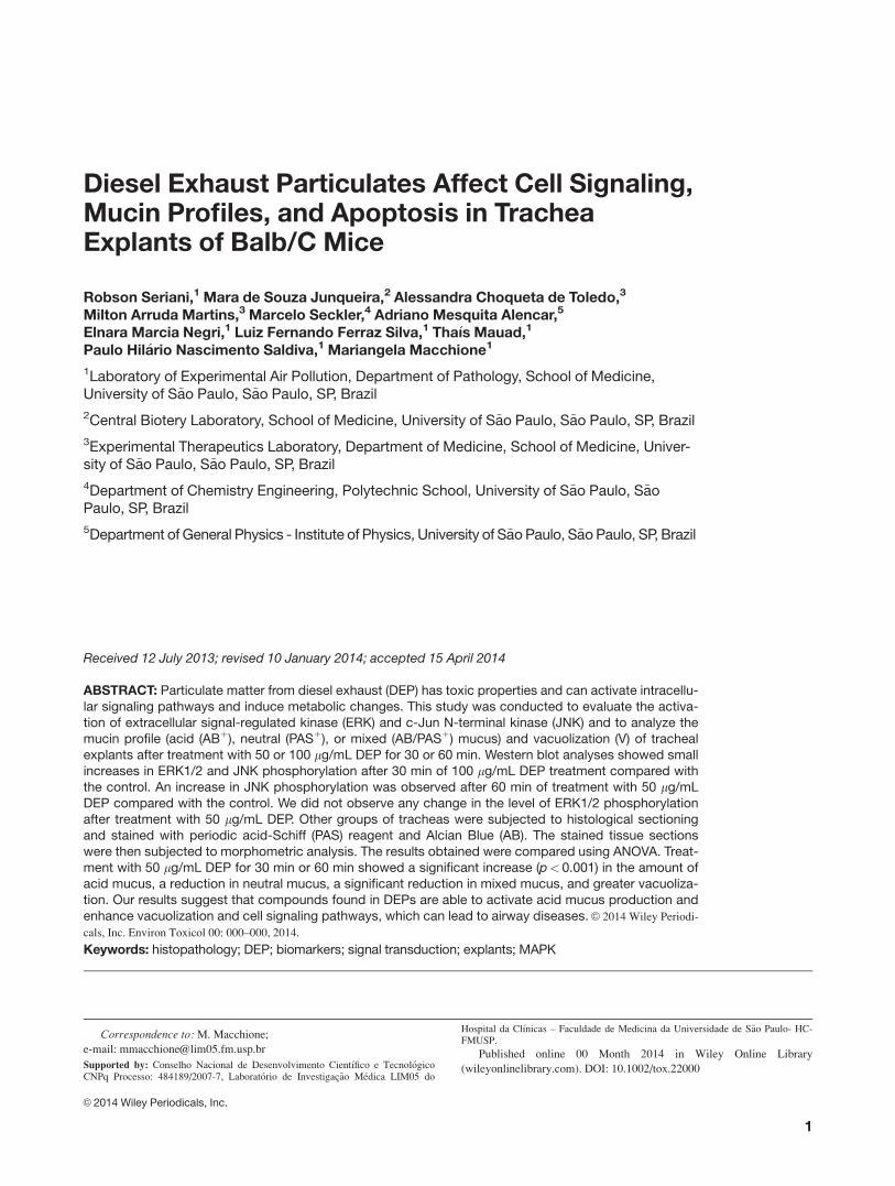
Activation of ERK and JNK after Exposure toDEP
Thirty minutes of exposure to 50 lg/mL DEPs led to an
increase in the phosphorylation levels of ERK1/2. Further-
more, the phosphorylation levels of JNK1/2 decreased. After
sixty minutes of exposure to 50 lg/mL DEPs, the phospho-
rylation levels of ERK1/2 decreased; however, JNK1/2
phosphorylation increased.
Thirty minutes of exposure to 100 lg/mL DEPs caused
the phosphorylation levels of both ERK1/2 and JNK1/2 to
increase. After 60 min of exposure to 100 lg/mL DEPs, the
4 SERIANI ET AL.
Environmental Toxicology DOI 10.1002/tox

phosphorylation levels of ERK1/2 increased; however, the
phosphorylation of JNK1/2 decreased (Fig. 3).
Mucin Profile after DEP Exposure
Figure 4 shows representative photomicrographs of tracheal
sections from the five experimental groups stained with AB/
PAS. Treatment with 50 lg/mL DEPs for 30 min prompted a
significant increase (p< 0.001) in the amount of acid mucus.
This increase in acid mucus was associated with a decrease in
neutral mucus, as shown in Figure 4 (p< 0.001). Treatment
with 50 lg/mL DEPs caused a significant reduction
(p< 0.001) in the mixed mucus after 30 min or 60 min. The
tracheas treated with 50 lg/mL DEPs for 30 min displayed
greater vacuolization (p< 0.001), which may be linked to the
release of higher amounts of neutral mucus. No statistically
significant differences were observed in the explants treated
with 100 lg/mL DEPs for 30 min or 60 min compared with
the control group.
Apoptosis
Overall, the groups treated with 50 lg/mL DEPs for 30 min
or 60 min showed a statistically significant increase in the
number of TUNEL-positive cells when compared with the
controls (p< 0.001). The group treated with 100 lg/mL
DEPs for 30 min had a higher number of TUNEL-positive
cells (p< 0.001) than all of the other groups. The group
treated with 100 lg/mL DEPs for 60 min had a lower num-
ber of TUNEL-positive cells compared to the other DEP
groups; however, considerable epithelial loss was observed
in this group (Fig. 5). Eight tracheal explants embedded in
Fig. 2. MTT assay (tetrazolium salt, 3-4,5 dimethylthiazol-2,5diphenyl tetrazolium bromide) of BEAS-2B cells exposed todifferent concentrations (5 lg/mL to 250 lg/mL) of DEPs for60 min.
Fig. 1. Scanning Electron Microscopy (SEM) of (A) Dulbecco’s Modified Eagles Medium (DMEM) and Hams F12 medium(Sigma Chemical, St. Louis, MO) plus supplements and antibiotics (control) at 10,0003 magnification; (B) 50 lg/mL DEPsplus medium at 10,0003 magnification; (C) 100 lg/mL DEPs plus medium; and (D) 100 lg/mL DEPs at 40,0003 magnifica-tion to show smaller sized particles. In this SEM micrograph, it is possible to observe two size populations: one composed ofspherical particles measuring between 1 and 2 lm in diameter and a second population of smaller particles that formedaggregates with large agglomerates.
DEP AFFECT CELL SIGNALING, MUCIN PROFILES, AND APOPTOSIS 5
Environmental Toxicology DOI 10.1002/tox
each slide were used for each group. The integrity of the epi-
thelium for most of the explants was not adequate to permit
the measurement of the TUNEL-positive cells with the
desired accuracy. Therefore, we analyzed 3–4 tracheal
explants on each slide for all of the groups (Fig. 4).
DISCUSSION
In this study, we observed an increase in the concentration
of phosphorylated ERK and JNK in tracheal explants
shortly after exposure to DEPs. These findings were asso-
ciated with alterations in the mucus profile of the tracheal
cells, including cellular vacuolization, which suggests
mucus extrusion The DEP-exposed cells demonstrated
increased apoptosis.
Studies performed using lung and tracheal explants
enable the analysis of the response of different cell types
to a given stimulus, which differs from the study of cell
lines (Pittet et al., 2010). Tracheal explants display many
key features of in vivo airways, such as mucus coverage,
mucociliary clearance and cell structure (Kitson et al.,
1999). Moreover, tracheal tissues add a further dimension
by providing the opportunity to examine the interactions
between the epithelium and the underlying airway muco-
sal structural cells ex vivo (Abeynaike et al., 2010). In
addition, tracheal explants have been previously used to
investigate the effects of mineral dust and air pollution
particles on tracheal function (Dai et al., 1998; Dai et al.,
2003). Therefore, tracheal explants may be useful for the
study of complex mixtures in numerous substances, such
as diesel exhaust. These substances contain various toxic
compounds and have physical characteristics that can
result in the materials being retained in upper airways (by
inflammatory cells) or reaching the lungs.
Previous studies using animals have demonstrated that air
pollution modifies the epithelial cell profile by increasing the
number of mucus-producing cells and modifying the physi-
cal and chemical characteristics of mucus (Saldiva et al.,
1992). Ishihara and Kagawa (2003) observed qualitative
changes in the types of glycoproteins present in the mucus
granules of goblet cells, and these changes depended on the
concentration and the length of exposure to particulate air
pollution involving diesel exhaust.
In our study, we verified the activation of the MAPK
pathway (ERK and JNK) in tracheal explants exposed to
particulate matter from diesel exhaust. The MAPKs are
involved in metabolic disorders, proliferation and apoptosis.
ERK was the first MAPK cascade protein demonstrated to
be important in different cellular signaling processes such as
growth, proliferation, and survival. There are two predomi-
nant ERK isoforms (ERK1 and ERK2), which are referred
to as p42/p44 MAP kinases (Boulton et al., 1990, 1991).
ERK1 and ERK2 phosphorylate active substrates in all cellu-
lar compartments, including membrane proteins (CD120a,
Syk, and calnexin), nuclear substrates (SRC-1, Pax6, NF-
AT, Elk-1, MEF2, c-Fos, c-Myc, and STAT3), cytoskeletal
proteins, and kinases (Chen et al., 2001). The abnormal regu-
lation of MAPK pathways has been reported in a wide range
of diseases including many cancers and pulmonary diseases,
such as asthma (Duan et al. 2004; Pelaia et al. 2005), emphy-
sema (Mercer et al., 2004), and COPD (Renda et al., 2008).
The ERK1/2 pathway controls the activity or abun-
dance of the BCL-2 protein family, cytochrome c from
mitochondria, the activation of caspases and cell death
(Balmanno and Cook, 2009). According to Hewson et al.
(2003), in airways, ERK is a critical tyrosine kinase
involved in the induction of MUC5AC expression and
goblet cell metaplasia in response to a variety of stimuli.
Amara et al. (2007) showed that ERK1/2 mediates the
activation of NOX4 and MMP-1 in human lung epithelial
cells exposed to DEPs, and Pourazar et al. (2008) showed
that ERK activation transduces proliferative and differen-
tiation responses in the bronchial epithelium. Li et al.
(1998) indicated that ERK/MAPK activation via the c-src-
MAPK-pp90rsk pathway can also be a sufficient signal
for NF-jB activation and MUC2 in epithelial cells. Fur-
thermore, Li et al. (2012) showed that a significant
increase in phosphorylated ERK induces MUC5AC
expression in bronchial epithelial cells.
The c-Jun NH2-terminal kinase (JNK) pathway
includes mitogen-activated protein kinases that are acti-
vated primarily by cytokines and exposure to many
Fig. 3. Effect of DEPs on the phosphorylation of ERK1/2 (A) and JNK (B) in tracheal explants. The tracheal explants wereexposed to one of two concentrations of DEPs (50 or 100 lg/mL) for 30 min or 60 min.
6 SERIANI ET AL.
Environmental Toxicology DOI 10.1002/tox

stresses, including changes in the physical and chemical
properties of the environment. Additionally, this pathway
contributes to apoptotic signal transduction (Davis, 2000;
Weston and Davis 2003). Tournier et al. (2000) implicated
JNK in apoptosis because jnk12/2jnk22/2 mice were
resistant to apoptosis induced by UV irradiation,
Fig. 4. Mucin-occupying area in the tracheal explants of BALB/c mice exposed to 50 lg/mL or 100 lg/mL DEP. (A) Anincrease in the content of acid mucus after treatment with 50 lg/ml DEP for 30 min or 60 min (*p< 0.001) and (B) a reductionin neutral (*p< 0.001), and (C) mixed mucus (*p< 0.001) were observed. (D) A significant increase in vacuolization (*p<0.001)was observed only at a concentration of 50 lg/mL after 30 min. [Color figure can be viewed in the online issue, which is avail-able at wileyonlinelibrary.com.]
DEP AFFECT CELL SIGNALING, MUCIN PROFILES, AND APOPTOSIS 7
Environmental Toxicology DOI 10.1002/tox

anisomycin and MMS. According to Davis (2000) and
Weston and Davis (2007), JNK is important in other forms
of cell death, including necrosis and autophagy because
the mitochondria are a primary target of JNK-mediated
pro-apoptotic signaling.
JNK-signaling studies in mammals suggest that JNK
induces apoptosis by modulating the pro-apoptotic Bcl-2
family protein BIM (Lei et al., 2003; Nateri et al., 2005).
Sakurai et al. (2006) also support a role for JNK in tumor
development. Thus, this pro-oncogenic role of JNK may be
related to its known ability to promote proliferation: the
deregulation of JNK has been implicated in cancer (Davis,
2000; Chang and Karin, 2001; Shaulin and Karin, 2002;
Weston and Davis, 2007).
Other properties of JNK activation have been described
by Jaeschke et al. (2005). These authors showed that JNK2
controls the Th1/Th2 balance of the immune response,
thereby protecting against autoimmune disease. JNK1 is
implicated in autoimmune disease, where it plays an impor-
tant role in regulating the expression of the anti-
inflammatory cytokine IL-10. In addition, Choi et al. (2010)
observed mucus overproduction and MUC5AC protein
expression associated with JNK expression in the epithelia
of airways exposed to smoke.
DEPs have been shown to induce apoptosis in airway
cells via MAPK activation and oxidative stress (Franco et al.
2009). The activation of JNK has been proposed to have a
role in promoting apoptosis. The chemical composition of
DEPs is important in this process due to the presence of met-
als and PAHs. These compounds activate several signaling
proteins during biotransformation and produce ROS, which
induce the activation of AP-1, MEK-1, ERK, JNK, NF-jB
and p53 and cause DNA damage. The activation of several
signaling pathways has been shown to be mediated by the
inhibition of protein tyrosine phosphatases, and the stimula-
tion of tyrosine residue phosphorylation triggers or potenti-
ates cell apoptosis (Ye et al., 1999; Huang et al., 2000; Au
et al., 2006).
According to Reardon et al. (1999) and Shen et al.
(2003), the pro-survival ERK and pro-apoptotic JNK path-
ways act in a dynamic equilibrium, with the ERK pathway
acting to inhibit the JNK pathway or vice versa. This interac-
tion could possibly explain the increases and decreases in
ERK and JNK in our measurements at 30 min to 50 or 100
lg/mL DEPs.
However, we cannot exclude that other pathways leading
to apoptosis could have influenced our results. According to
Xia et al. (2004), the aromatic fraction of DEPs can directly
Fig. 5. Images and graphic representation of the proportion of cells undergoing apoptosis after treatment with 50 or 100 lg/mL DEPs for 30 min or 60 min. Statistically significant differences in apoptosis were observed after treatment with 50 lg/mLDEPs for 30 min (*p 5 0.004), 50 lg/mL DEPs for 60 min (#p 5 0.006), and 100 lg/mL DEPs for 30 min (†p< 0.001 comparedwith the control group. (B) Light microscopy of cells stained using the TUNEL assay depicts dark brown cells undergoingapoptosis (403). [Color figure can be viewed in the online issue, which is available at wileyonlinelibrary.com.]
8 SERIANI ET AL.
Environmental Toxicology DOI 10.1002/tox

induce mitochondria swelling and depolarization leading to
calcium overload in the matrix and causing cellular death.
Moreover, the high levels of non-carcinogenic metals (i.e.,
cobalt, lead, iron, and zinc) present in DEPs have often been
shown to provoke the production of ROS (e.g., H2O2), which
leads to apoptosis through the mitochondrial pathway.
Lemos et al. (1994) and Pires-Neto et al. (2006) showed
an increase in the concentration of acid mucus in the nasal
epithelium of rats exposed to urban particulate matter. Yosh-
izaki et al. (2010) showed that exposure of mice to diesel
particulate matter for 60 days resulted in an increase in
Muc5ac protein expression in the lungs and an increase in
acid mucus in the nasal epithelium. Mucin acidification
induces alterations in the physico-chemical properties of
mucus, with an increase in viscosity that damages mucocili-
ary clearance and the defenses of the respiratory epithelium
(Lemos et al., 1994; Pires-Neto et al., 2006; Yoshizaki et al.,
2010). In this study, we also showed that an increase in acid
mucus occurs primarily after treatment with a low concen-
tration of DEPs (50 lg/mL), which may be due to a ceiling
effect. In addition, other factors, such as apoptosis, may
have influenced the results (Fig. 2). The in vivo secretion of
mucins, which is usually initiated by a secretagogue, occurs
rapidly, within seconds to minutes; however, the genetic reg-
ulation and biosynthesis of mucins requires hours (Neutra
et al., 1987; Verdugo, 1991; Davis, 2002). In our study, the
exposure was conducted for a maximum of one hour, which
may explain the hypersecretion of produced mucin.
In a mouse model of smoke inhalation injury, Choi et al.
(2010) showed that mucus overproduction is associated with
an increase in epithelial Muc5ac protein expression, which is
dependent on the activation of the JNK pathway. These
results suggest that the JNK pathway may be a potential tar-
get for regulating mucus overproduction. ERK is a critical
tyrosine kinase involved in the induction of MUC5ACex-
pression and goblet cell metaplasia in response to a variety
of stimuli (Hewson et al., 2003; Songs et al., 2005). Our
results corroborate these previous studies, particularly those
using a DEP concentration of 50 lg/mL.
It is possible that the vacuoles observed in our study are
the result of endocytosis of the DEPs by tracheal cells, as
observed by Geiser (2002) and Bao et al. (2007) in CHO-K1
cells. However, we cannot exclude the possibility that the
vacuoles may represent mucus extrusion. Mucin secretion invivo is typically initiated by a secretagogue, and its extrusion
can occur within minutes (Rose and Voynow, 2006). In the
present study, the explants were maintained for 24 h in cul-
ture medium after 30 or 60 min of DEP exposure, and DEPs
may have acted as a secretagogue. We showed increased
vacuolization at the 30 min timepoint and a slight increase at
the 60 min timepoint after treatment with 50 lg/mL DEPs,
which can be explained by the observed extrusion of neutral
and mixed mucus. ERK activation was present at 30 min
and may be responsible for the depolymerization of actin
and the release of vesicles by the secretory cells. JNK activa-
tion, which inhibits the polymerization of microtubules and
induces apoptosis, was observed after treatment for 60 min
with 50 and 100 lg/mL DEPs. According to Barros and
Marshall (2005) and Tricker et al. (2011), ERK1/2 signaling
has been shown to lead to the disruption of actin stress fibers
and the loss of focal adhesion by several mechanisms.
MEKK1 is a mitogen-activated protein kinase kinase kinase
(MAP3K) that activates JNK and is required for microtubule
inhibitor-induced apoptosis in B cells. Active cytoskeletal
rearrangements have also been shown to accompany many
events associated with vesicle transport and fusion in a vari-
ety of cell systems (Koffer et al., 1990; Vitale et al., 1995;
Lang et al. 2000).
We have used BEAS-2B as a surrogate of tracheal epithe-
lial cells because these cells respond to diesel toxicity. We
believe both cell types have similar responses to cytotoxicity.
However, to our knowledge, there are no studies comparing
the effects of DEPs in BEAS-2B and tracheal explants. Future
studies should be performed to clarify these points.
In Sao Paulo, doses of 200 lg/m3 PM10 are not uncom-
mon in traffic corridors (Yoshizaki et al., 2010). If we
assume that humans breathe 6 L/min, the volume inspired in
one hour will be 360 L, equivalent to 0.36 m3, corresponding
to 70 lg of inhaled PM. Considering that particle retention
in trachea is approximately 20%, 14 lg is retained in the tra-
chea/hour. Healthy subjects produce a mean of 50 mL of
mucus/day 10–100 mL (Rubin, 2002). Thus, the amount of
mucus produced in the trachea will be approximately 2.0
mL in 1 h. Thus, the expected concentration of PM in the
mucus lining the trachea in one hour is 7 lg/mL/h. In our
study, we used DEP concentrations of 50 and 100 lg/mL for
30 min or 1 h. The doses used here would then correspond
to 12–14 h of exposure to urban air pollution. Other similar
studies have used up to 500 lg/cm2 (Dai and Churg, 2000;
Dai, et al., 2002; Dai, et al., 2003).
The concentrations used in our study were low doses and
were used to show the toxic effects of DEPs in airways and
the risks of exposure to these concentrations. We observed
that higher concentrations of DEPs in the culture medium
tended to form large agglomerates. It is possible that at a
concentration of 50 lg/mL, the DEPs would be more dis-
persed in the medium and penetrate epithelial cells more eas-
ily than at the 100 lg/mL concentration. Accordingly, the
stress response to injury in the form of acid mucus was more
pronounced in the cells exposed to the lower concentration
of DEPs.
In conclusion, these results suggest that compounds found
in particulate matter from diesel exhaust activate cell signal-
ing pathways, thereby inducing the production of acid
mucus, vacuolization and apoptosis in tracheal explants.
We thank Prof. Dr. Roger Chammas and researcher Dr Moni-
que Matsuda and Dr. Carla Lima for their valuable scientific opin-
ions and the use of laboratory supplies. Additionally, we thank
Sandra de Morais Fernezlian, Esmeralda Miristene Eher and
DEP AFFECT CELL SIGNALING, MUCIN PROFILES, AND APOPTOSIS 9
Environmental Toxicology DOI 10.1002/tox

Rodrigo Soares Vaz de Camargo for the preparation of histological
slides and Bernardo Saraiva Ferracini for the preparation of figures.
REFERENCES
Abeynaike L, Meeusen ENT, Bischof RJ. 2010. An ovine tracheal
explant culture model for allergic airway inflammation.
J Inflamm 7:46.
Balmanno K, Cook SJ. 2009. Tumour cell survival signalling by
the ERK1/2 pathway. Cell Death Diff 16:368–377.
Bao L, Chen S, Wu L, Hei TK, Wu Y, Yu Z, Xu A. 2007. Mutage-
nicity of diesel exhaust particles mediated by cell-particle inter-
action in mammalian cells. Toxicology 229:91–100.
Barros JC, Marshall CJ. 2005. Activation of either ERK1/2 or
ERK5 MAP kinase pathways can lead to disruption of the actin
cytoskeleton. J Cell Sci 118:1663–1671.
Bayram H, Devalia JL, Sapsford RJ, Ohtoshi T, Miyabara Y,
Saga€ı M, Davies RJ. 1998. The effect of diesel exhaust particles
on cell function and release of inflammatory mediators from
human bronchial epithelial cells in vitro. Am J Respir Cell Mol
Biol 18:441–448.
Benbrahim-Tallaa L, Baan RA, Grosse Y, Lauby-Secretan B, El
Ghissassi F, Bouvard V, Guha N, Loomis D, Straif K. 2012.
International Agency for Research on Cancer Monograph Work-
ing Group. Carcinogenicity of diesel-engine and gasoline-engine
exhausts and some nitroarenes. Lancet Oncol 13:663–664.
Boeckenholt C, Begrow F, Verspohl EJ. 2012. Effect of Silymarin
and Harpagoside on inflammation reaction of BEAS-2B cells,
on ciliary beat frequency (CBF) of trachea explants and on
mucociliary clearance (MCC). Planta Med 78:761–766.
Boland S, Baeza A, Fournier T, Houcine O, Gendron MC,
Chevrier M, Jouvenot P, Coste A, Aubier M, Marano F. 1999.
Diesel exhaust particles are taken up by human airway epithe-
lial cells in vitro and alter cytokine production. Am J Physiol
276:604–613.
Bonvallot V, Baeza-Squiban A, Boland S, Marano F. 2000. Activa-
tion of transcription factors by diesel exhaust particles in human
bronchial epithelial cells in vitro. Inhal Toxicol 12:359–364.
Boulton TG, Yancopoulos GD, Gregory JS, Slaughter C,
Moomaw C, Hsu J, Cobb MH. 1990. An insulin-stimulated pro-
tein kinase similar to yeast kinases involved in cell cycle con-
trol. Science 249:64–67.
Boulton TG, Yancopoulos GD, Slaughter C, Moomaw C, Hsu J,
Cobb MH. 1991. ERKs: A family of protein-serine/threonine
kinases that are activated and tyrosine phosphorylated in
response to insulin and NGF. Cell 65:663–675.
Brook RD, Rajagopalan S, Pope CA, Brook JR, Bhatnagar A,
Diez-oux AV, Holguin F, Hong Y, Luepker RV, Mittleman
MA, Peters A, Siscovick D, Smith SC Jr, Whitsel L, Kaufman
JD. 2010. Particulate matter air pollution and cardiovascular
disease: An update to the scientific statement from the Ameri-
can Heart Association. Circulation 12:2331–2378.
Carvalho-Sousa CE, Cruz-Machado SdS, Tamura EK, Fernandes
PACM, Pinato L, Muxel SM, Cecon E, Markus RP. 2011.
Molecular basis for defining the pineal gland and pinealocytes
as targets for tumor necrosis factor. Front Endocrin 2:10.
Cao D, Tal TL, Graves LM, Gilmour I, Linak W, Reed W,
Bromberg PA, Samet JM. 2007. Diesel exhaust particulate-
induced activation of Stat-3 requires activities of EGFR and Src
in airway epithelial cells Am J Physiol Lung Cell Mol Physiol
292:422–429.
Chang L, Karin M. 2001. Mammalian MAP kinase signalling cas-
cades. Nature 410:37–40.
Chen ZTB, Robinson GF, Silvestro L, Pearson G, Xu B, Wright
A, Vanderbilt C, Cobb MH. 2001. MAP kinases. Chem Rev
101:2449–2476.
Choi W, Syrkina O, Kwon KY, Quinn DA, Hales CA. 2010. JNK
activation is responsible for mucus overproduction in smoke
inhalation injury. Respir Res 11:172.
Dai J, Gilks B, Price K, Churg A. 1998. Mineral dusts directly
induce epithelial and interstitial fibrogenic mediators and
matrix components in the airway wall. Am J Respir Crit Care
Med. 158:1907–1913.
Dai J, Churg A. 2001. Relationship of fiber surface iron and active
oxygen species to expression of procollagen, PDGF-A, and
TGF-beta(1) in tracheal explants exposed to amosite asbestos.
Am J Respir Cell Mol Biol 24:427–435.
Dai J, Xie C, Churg A. 2002. Iron loading makes a nonfibrogenic
model air pollutant particle fibrogenic in rat tracheal explants.
Am J Respir Cell Mol Biol 26:685–693.
Dai J, Xie C, Vincent R, Churg A. 2003. Air pollution particles
produce airway wall remodeling in rat tracheal explants. Am J
Respir Cell Mol Biol 29:352–358.
Davis RJ. 2000. Signal transduction by the JNK group of MAP
kinases. Cell 103:239–252.
Davis CW. 2002. Regulation of secretion from in vitro cellular
models. Novartis Founding Symp 248:113–125.
Dreher K, Jurkat R, Kodanvanti U, Lehmann J, Winsett D, Costa
D. 1996. Soluble transition metals mediate acute pulmonary
injury and airway hyperresponsiveness induced by residual oil
fly ash particles. Chest 109:33S–34S.
Duan W, Chan JH, Wong CH, Leung BP, Wong WS. 2004. Anti-
inflammatory effects of mitogen-activated protein kinase inhibitor
U0126 in an asthma mouse model. J Immunol 172:7053–7059.
Fantl WJ, Johnson DE, WIlliams LT. 1993. Signalling by receptor
tyrosine kinases. Annu Ver Biochem 62:453–481.
Franco R, S�anchez-Olea R, Reyes-Reyes EM, Panayiotidis MI.
2009. Environmental toxicity, oxidative stress and apoptosis:
m�enage �a trois. Mutat Res 674:3–22.
Geiser M. 2002. Morphological aspects of particle uptake by lung
phagocytes. Microsc Res Tech 57:512–522.
Ghio AJ, Silbajoris R, Carson JL, Samet JM. 2002. Biologic
effects of oil fly ash. Environ Health Perspect 110:89–94.
Gong JH, Shin D, Han SY, Kim JL, Kang YH. 2012. Kaempferol
suppresses eosionphil infiltration and airway inflammation in
airway epithelial cells and in mice with allergic asthma. J Nutr
142:47–56.
Guan XJ, Zhang WX, Li CC, Zheng YM, Lin L, Ye LP, Chen XF,
Luo YC, Cai, XH, Dong L, Zhang HL, Zhou XC. 2007. The
role of external signal regulated kinase and transforming
growth factor beta(1) in asthma airway remodeling and
10 SERIANI ET AL.
Environmental Toxicology DOI 10.1002/tox

regulation of glucocorticoids. Zhonghua Yi Xue Za Zhi 87:
1767–1772.
Hill CS, Treisman R. 1995. Transcriptional regulation by extracel-
lular signals: Mechanisms and specificity. Cell 80:199–211.
Hewson CA, Edbrooke MR, Johnston SL. 2004. PMA induces the
MUC5AC respiratory mucin in human bronchial epithelial
cells, via PKC, EGF/TGF-a, Ras/Raf, MEK, ERK and Sp1-
dependent mechanisms. J Mol Biol 344:683–695.
Imamura Y, Yanagihara K, Mizuta Y, Seki M, Ohno H,
Higashiyama Y, Miyazaki Y, Tsukamoto K, Hirakata Y,
Tomono K, Kadota J, Kohno S. 2004. Azithromycin inhibits
MUC5AC production induced by the Pseudomonas aeruginosaautoinducer N-(3-oxododecanoyl) homoserine lactone in NCI-
H292 cells. Antimicrob Agents Chemother 48:3457–3461.
Ishihara Y, Kagawa J. 2003. Chronic diesel exhaust exposures of
rats demonstrate concentration and time-dependent effects on
pulmonary inflammation. Inhal Toxicol 25:473–492.
Jaeschke A, Rincon M, Doran B, Reilly J, Neuberg D, Greiner
DL, Shultz LD, Rossini AA, Flavell RA, Davis RJ. 2005. Dis-
ruption of the Jnk2 (Mapk9) gene reduces destructive insulitis
and diabetes in a mouse model of type I diabetes. Proc Natl
Acad Sci USA 102:6931–6935.
Kitson C, Angel B, Judd D, Rothery S, Severs NJ, Dewar A,
Huang L, Wadsworth SC, Cheng SH, Geddes DM, Alton EW.
1999. The extra- and intracellular barriers to lipid and
adenovirus-mediated pulmonary gene transfer in native sheep
airway epithelium. Gene Ther 6:534–546.
Krishna M, Narang H. 2008. The complexity of mitogen-activated
protein kinases (MAPKs) made simple. Cell Mol Life Sci 65:
3525–3544.
Koffer A, Tatham PER, Gomperts BD. 1990. Changes in the state
of actin during the exocytotic reaction of permeabilized rat
mast cells. J Cell Biol 111:919–927.
Laks D, Oliveira RC, de Andr�e PA, Macchione M, Lemos M,
Faffe D, Saldiva PH, Zin WA. 2008. Composition of diesel par-
ticles influences acute pulmonary toxicity: An experimental
study in mice. Inhal Toxicol 20:1037–42.
Lang T, Wacker I, Wunderlich I, Rohrbach A, Giese G, Soldati T,
Almers W. 2000. Role of actin cortex in the subplamalemal
transport of secretory granules in PC-12 cells. Biophys J 78:
2863–2877.
Lemos M, Lichtenfelds AJ, Amaro J�unior E, Macchione M,
Martins MA, King M, B€ohm GM, Saldiva PH. 1994. Quantita-
tive pathology of nasal passages in rats exposed to urban levels
of air pollution. Environ Res 66:87–95.
Li JD, Feng W, Gallup M, Kim JH, Gum J, Kim Y, Basbaum C.
1998. Activation of NF-kappaB via a Src-dependent Ras-
MAPK-pp90rsk pathway is required for Pseudomonas
aeruginosa-induced mucin overproduction in epithelial cells.
Proc Natl Acad Sci USA 95:5718–5723.
Li N, Hao M, Phalen RF, Hinds WC, Nel AE. 2003. Particulate
air pollutants and asthma. A paradigm for the role of oxidative
stress in PM-induced adverse health effects. Clin Immunol 109:
250–265.
Li N, Li Q, Zhou XD, Juliy VP, Perelman M. 2012. Chronic
mechanical stress induces mucin5AC expression in human
bronchial epithelial cells through ERK dependent pathways.
Mol Biol Rep 39:1019–1028.
Lei K, Davis RJ. 2003. JNK phosphorylation of Bim-related mem-
bers of the Bcl2 family induces Bax-dependent apoptosis. Proc
Natl Acad Sci USA 100:2432–2437.
Mercer BA, Kolesnikova N, Sonett J, D’Armiento J. 2004. Extrac-
ellular regulated kinase/mitogen activated protein kinase is up-
regulated in pulmonary emphysema and mediates matrix
metalloproteinase-1 induction by cigarette smoke. J Biol Chem
279:17690–17696.
Mehlen P, Rabizadeh S, VanArsdale T, Zhang H, Shin H, Wang
JJ, Leo E, Zapata J, Hauser CA, Reed JC, Bredesen DE. 1999.
TRAF family proteins interact with the common neurotrophin
receptor and modulate apoptosis induction. J Biol Chem 274:
30202–30208.
Marais R, Marshall CJ. 1996. Control of the ERK MAP kinase
cascade by Ras and Raf. Cancer Surv 27:101–125.
Nateri AS, Spencer-Dene B, Behrens A. 2005. Interaction of
phosphorylated c-Jun with TCF4 regulates intestinal cancer
development. Nature 437:281–285.
Neutra MR, Forstner JF. 1987. Gastrointestinal mucus: synthesis,
secretion and function. In: Johnson LR, editor. Procedings of
Gastrointestinal Tract. New York: Raven. pp 975–1009.
Ohtoshi T, Takizawa H, Okazaki H, Kawasaki S, Takeuchi N,
Ohta K, Ito K. 1998. Diesel exhaust particles stimulate
human airway epithelial cells to produce cytokines relevant to
airway inflammation in vitro. J Allergy Clin Immunol 101:
778–785.
Pelaia G, Cuda G, Vatrella A, Gallelli L, Caraglia M, Marra M,
Abbruzzese A, Caputi M, Maselli R, Costanzo FS, Marsico AS.
2005. Mitogen-activated protein kinases and asthma. J Cell
Physiol 202:642–653.
Pires-Neto RC, Lichtenfels AJ, Soares SR, Macchione M, Saldiva
PH, Dolhnikoff M. 2006. Effects of S~ao Paulo air pollution on
the upper airways of mice. Environ Res 101:356–361.
Pittet LA, Hall-Stoodley L, Harmsen AG. 2010. Influenza virus
infection decreases tracheal mucociliary velocity and clearance
of Streptococcus pneumoniae. Am J Respir Cell Mol Biol 42:
450–460.
Pourazar J, Blomberg A, Kelly FJ, Davies DE, Wilson SJ,
Holgate ST, Sandstr€om T. 2008. Diesel exhaust increases
EGFR and phosphorylated C-terminal Tyr 1173 in the bron-
chial epithelium. Particle Fibre Toxicol 5:8.
Raidl M, Sibbing B, Strauch J, Muller K, Nemat A, Schneider
PM, Hag H, Erdmann E, Koch A. 2007. Impaired TNFalpha-
induced VEGF expression in human airway smooth muscle
cells from smokers with COPD: Role of MAPkinases and his-
tone acetylation—effect of dexamethasone. Cell Biochem Bio-
phys 49:98–110.
Reardon DB, Contessa JN, Mikkelson RB, Valerie K, Amir C,
Dent P, Scmidt-Ulrich RK. 1999. Dominant negative EGFR-
CD533 and inhibition of MAPK modify JNK1 activation and
enhance radiation toxicity of human mammary carcinoma cells.
Oncogene 18:4756–4766.
Renda T, Baraldo S, Pelaia G, Bazzan E, Turato G, Papi A,
Maestrelli P, Maselli R, Vatrella A, Fabbri LM, Zuin R,
DEP AFFECT CELL SIGNALING, MUCIN PROFILES, AND APOPTOSIS 11
Environmental Toxicology DOI 10.1002/tox

Marsico SA, Saetta M. 2008. Increased activation of p38
MAPK in COPD. Eur Respir J 31:62–69.
Report on Carcinogens, U.S. Department of Health and Human
Services, Public Health Service: Twelfth Edition (2011). http://
ntp.niehs.nih.gov/go/roc12.
Rose MC, Voynow JA. 2006. Respiratory tract mucin genes and
mucin glycoproteins in health and disease. Physiol Rev 86:
245–278.
Rosenkranz HS. 1996. Mutagenic nitroarenes, diesel emissions,
particulate-induced mutations and cancer: An essay on cancer-
causation by a moving target. Mutat Res 367:65–72.
Saldiva PH, King M, Delmonte VL, Macchione M, Parada MA,
Daliberto ML, Sakae RS, Criado PM, Silveira PL, Zin WA,
G.M. B€ohm. 1992. Respiratory alterations due to urban air pol-
lution: An experimental study in rats. Environ Res 57:19–33.
Sakurai T, Maeda S, Chang L, Karin M. 2006. Loss of hepatic
NF-k B activity enhances chemical hepatocarcinogenesis
through sustained c-Jun N-terminal kinase 1 activation. Proc
Natl Acad Sci USA 103:10544–10551.
Schuh K, Pahl A. 2009. Inhibition of the MAP kinase ERK pro-
tects from lipopolysaccharide-induced lung injury. Biochem
Pharmacol 77:1827–1834.
Shaulian E, Karin M. 2002. AP-1 as a regulator of cell life and
death. Nat Cell Biol 4:E131–E136.
Shen YH, Godlewski, J, Zhu, J, Sathyanarayana P, Leaner V,
Birrer MJ, Rana A, Tzivion G. 2003. Cross-talk between JNK/
SAPK and ERK/MAPK pathways: Sustained activation of JNK
blocks ERK activation by mitogenic factors. J Biol Chem 278:
26715–26721.
Song JS, Cho KS, Yoon HK, Moon HS, Park SH. 2005. Neutro-
phil elastase causes MUC5AC mucin synthesis via EGF recep-
tor, ERK and NF-kB pathways in A549 cells. Korean J Intern
Med 20:275–283.
Steerenberg PA, Zonnenberg JAJ, Dormans JAMA, Joon PNT,
Wouters IM. 1998. Diesel exhaust particles induced release of
interleukin 6 and 8 by (primed) human bronchial epithelial cells
(BEAS 2B) in vitro. Exp Lung Res 24:85–100.
Tanaka A, Jin Y, Lee SJ, Zhang M, Kim HP, Stolz DB, Ryter
SW, Choi AM. 2012. Hyperoxia-induced LC3B interacts with
the Fas apoptotic pathway in epithelial cell death. Am J Respir
Cell Mol Biol 46:507–514.
Toledo AC, Arantes-Costa FM, Macchione M, Saldiva PH, Negri
EM, Lorenzi-Filho G, Martins MA. 2011. Salbutamol improves
markers of epithelial function in mice with chronic allergic pul-
monary inflammation. Respir Physiol Neurobiol 177:155–161.
Totlandsdal AI, Cassee FR, Schwarze P, Refsnes M, Lag M.
2010. Diesel exhaust particles induce CYP1A1 and pro-
inflammatory responses via differential pathways in human
bronchial epithelial cells. Part Fibre Toxicol 7:41.
Thomas RS, Tymms MJ, McKinlay LH, Shannon MF, Seth A,
Kola I. 1997. ETS1, NF-_B and AP-1 synergistically transacti-
vate the human GM-CSF promoter. Oncogene 14:2845–2855.
Tournier C, Hess P, Yang DDJ, Turner TK, Nimnual A, Bar-Sagi
D, Jones SN, Flavell RA, Davis RJ. 2000. Requirement of JNK
for stress-induced activation of the cytochrome c-mediated
death pathway. Science 288:870–874.
Tricker E, Arvand A, Kwan R, Chen GY, Gallagher E, Cheng G.
2011. Apoptosis Induced by Cytoskeletal Disruption Requires
Distinct Domains of MEKK1. PLoS One 6:e17310.
Verdugo P. 1991. Mucin exocytosis. Am Rev Respir Dis 144:
33–37.
Vermylen J, Nemmar A, Nemery B, Hoylaerts MF. 2005. Ambi-
ent air pollution and acute myocardial infarction. J Thromb
Haemost 3:1955–1961.
Vitale ML, Seward EP, Trifaro JM. 1995. Chromaffin cell cortical
actin network dynamics control the size of the release-ready
vesicle pool and the initial rate of exocytosis. Neuron 14:353–
363.
Weibel ER. 1990. Morphometry: stereological theory and practi-
cal methods. In: Gil, editor. Models of Lung Disease, 1st ed.
Dekker: New York. pp 199–252.
Weston CR, Davis RJ. 2002. The JNK signal transduction path-
way. Curr Opin Genet Dev 12:14–21.
Xia T, Korge P, Weiss JN, Li N, Venkatesen MI, Sioutas C, Nel
A. 2004. Quinones and aromatic chemical compounds in partic-
ulate matter induce mitochondrial dysfunction: Implications for
ultrafine particle toxicity. Environ Health Perspect 112:1347–
1358.
Xia Z, Dickens M, Raingeaud J, Davis RJ, Greenberg ME. 1995.
Opposing effects of ERK and JNK-p38 MAP kinases on apo-
ptosis. Science 270:1326–1331.
Yoshizaki K, Brito JM, Toledo AC, Nakagawa NK, Piccin VS,
Junqueira MS, Negri EM, Carvalho AL, Oliveira AP, Lima
WT, Saldiva PH, Mauad T, Macchione M. 2010. Subchronic
effects of nasally instilled diesel exhaust particulates on the
nasal and airway epithelia in mice. Inhal Toxicol 22:610–
617.
Zin WA, Silva AG, Magalh~aes CB, Carvalho GM, Riva DR, Lima
CC, Leal-Cardoso JH, Takiya CM, Valenca SS, Saldiva PH,
Faffe DS. 2012. Eugenol attenuates pulmonary damage induced
by diesel exhaust particles. J Appl Physiol 112:911–917.
Zwirner-Baier I, Neumann HG. 1999. Polycyclic nitroarenes
(nitro-PAHs) as biomarkers of exposure to diesel exhaust.
Mutat Res 441:135–144.
12 SERIANI ET AL.
Environmental Toxicology DOI 10.1002/tox