Bacillus subtilis spores as vaccine adjuvants: further insights into the mechanisms of action
Upload
inelearnfishCategory
view
1download
0
�ISSN 0372-5480Printed in Croatia
Veterinarski arhiV 77 (1), �-17, 2007
Influence of adjuvants Saponin and Montanide ISA 50 on immune response of calves immunized with purified midgut antigen of
Hyalomma marginatum isaaci
Gurusamy Ponnudurai1*, Lalitha John2, and Tirunelveli Jayagopal Harikrishnan3
1Department of Veterinary Parasitology, Veterinary College and research institute, namakkal, tamil nadu, india
2Department of Veterinary Parasitology, Madras Veterinary College, Chennai, india3Department of Veterinary Parasitology, Veterinary College and research institute, namakkal, tamil nadu,
india
PONNUDURAI, G., L. JOHN, T. J. HARIKRISHNAN: Influence of adjuvants Saponin and Montanide ISA 50 on immune response of calves immunized with purified midgut antigen of Hyalomma marginatum isaaci. Vet. arhiv 77, 9-17, 2007.
AbSTRAcTImmune status of calves, immunized with purified midgut antigen of hyalomma marginatum isaaci,
was assessed by standard immunological tests. Leukocyte migration inhibition test showed inhibition from day 21 post-infection in calves that received midgut antigen with Saponin as adjuvant (Saponin group) while significant inhibition was observed from 28 DPI in the Montanide group. Intradermal test revealed immediate hypersensitivity reaction within 15 m. and the highest skin fold thickness was observed after 24 h. of inoculation of antigen (14.50 ± 1.50 mm in the Saponin group and 12.00 ± 0.70 mm in the Montanide group). Similarly, a significant humoral response as judged by ELISA was observed from 7 DPI (P<0.01) in the Saponin group, with a peak value on 42 DPI, whereas the significant antibody was observed only on 21 DPI, with a peak value on 30 DPI in the Montanide group.
Key words: immune response, hyalomma marginatum isaaci, midgut antigen, calves
*Part of Ph.D. Thesis submitted to Tamil Nadu Veterinary and Animal Sciences University, Chennai, by the first author
________*Contact address:Dr. G. Ponnudurai, M.V.Sc., Ph.D., Assist. Professor, Department of Veterinary Parasitology, Veterinary College and Research Institute, Namakkal-637 001, Tamil Nadu, India, Phone: +91 4286 266491; Fax: +91 4286 266484; E-mail: [email protected]
10 Vet. arhiv 77 (1), �-17, 2007
IntroductionCattle ticks are one of the major constraints in the livestock industry, particularly in
tropical and subtropical areas, mainly because of the diseases they transmit and the costs involved in their control (CASTRO DE and NEWSON, 1993). The ixodid tick hyalomma is the predominant one of tick genera prevalent in much of Asia and an important vector for theileria annulata in India (CHHABRA and KHURANA, 2005). Conventional methods of tick control using acaricides have certain limitations. As an alternative, immunological control of ticks has emerged as the area of research in recent years. Host immune response to any antigen is largely determined by adjuvants used for immunization. Hence, the antigen should have to be tagged on to potential adjuvants to produce maximum immune response for elimination target organisms.
The present study was undertaken in order to discover suitable adjuvants for immunizing calves against tick species hyalomma marginatum isaaci.
Materials and methodstick antigen. Adult female h. m. isaaci ticks, fed for 4-5 days (partially engorged)
on cross-bred calves were used for antigen preparation. Midgut diverticula were removed and placed in 0.15 M phosphate buffered saline (pH 7.2). Harvested gut tissues were homogenized at 1.500 cycles for 15 m. in Potter’s homogenizer (Eitek, Mumbai). The homogenates were sonicated in a sonicator (B- Braun Bio-tech International, Germany) at a standard probe of 6 constant points for a period of 10 m. with an interval of 1 m. for each cycle.
The sonicated homogenates were centrifuged at 10.000 ×g for 30 m. (Remi C-24, Mumbai, India) to obtain supernatant and pellet fractions. The supernatant was pipetted out and filtered through a 0.45 µm disposable membrane filter unit. Phenyl methyl sulfonyl fluoride (PMSF) was added to supernatant antigen at the rate of 1 µL/mL of antigen (GHOSH and KHAN, 1997).
Gel filtration chromatography. Supernatant antigens (8 mL) were applied to 100 × 2 cm columns of Sephadex-G-200 (Sigma, U.S.A). The fractions were eluted with 18 mL/hr in PBS (pH 7.2) with 0.2 per cent sodium azide. A total of 70 fractions of midgut antigen in 3 mL volume were collected. The protein concentration of each fraction was determined (LOWRY et al., 1�51). Indirect ELISA was carried out for identification of antigenic fractions. Based on the ELISA titer values, the antigenic fractions were pooled and diluted with PBS (pH 7.2) so that each aliquot contained 1mg /mL of antigen, which were stored at -20 oC until further use.
adjuvants. Two adjuvants were used in the immunization trials and their immunopotentiating properties were assessed.
G. Ponnudurai et al.: Influence of adjuvants Saponin and Montanide ISA 50 on immune response of calves
11Vet. arhiv 77 (1), �-17, 2007
saponin. Saponin powder (Sigma, USA) from the South American tree Quillaja saponaria was used as 0.1 per cent suspension.
Montanide isa 50. Montanide oleate derivative in mineral oil suspension (Seppic SA- Paris) was used at a level of 50 per cent.
immunization. A total of 12 calves were divided into three groups, four calves in each group. All calves in group I (Saponin group) and group II (Montanide group) were immunized with column-purified midgut antigen, while group III was kept as an unimmunized control. Immunization schedule, dose, adjuvant and route of inoculation are presented in Table 1. In this study, tick naïve status of calves was confirmed by immunodiffusion test (AGPT).
Table1. Schedule of immunization of calves with midgut antigen of H. m. isaaci
ImmunizationDay
Experiment
Dose RouteGroup I Group II Group IIIPrimary 0 S + Ag M + Ag S + Buf 1 mg s/cFirst booster 14 S + Ag M + Ag M+ Buf 1 mg s/cSecond booster 28 Ag Ag Buf 1 mg s/c
S = Saponin, M = Montanide, Ag = Antigen and Buf = Buffer
Heparinised blood and serum samples were collected at weekly intervals from immunized and control calves from 0 - 91 days. Cell-mediated immunity was assessed by leukocyte migration inhibition test and intradermal test, while humoral immune response was assessed by performing ELISA.
assessment of cell mediated immune response. Leukocyte migration inhibition test (LMit). Heparinised blood samples were
subjected to LMIT and the test was carried out as per the method of CLAUSEN (1976). Percentage migration was arrived at by:
Average migration of cells in the presence of antigenAverage migration of cells in the absence of antigen
Per cent inhibition = 100 - per cent migration.
intradermal test. Skin test was performed 2 weeks after the last injection of antigen. All calves were slowly injected intradermally with 0.5 mL of antigen in the right flank region
× 100
G. Ponnudurai et al.: Influence of adjuvants Saponin and Montanide ISA 50 on immune response of calves
12
with a control injection of 0.5 mL of PBS in the adjoining site. The skin fold thickness of calves was measured (in mm) using Vernier Callipers at 0, 15, 30 m., 1, 2, 4, 24, 48 and 72 h. post-inoculation of antigen. The cell mediated immune response was assessed in terms of increased skin fold thickness at the antigen inoculated site in immunized calves and compared to that of the control calves. To study the histopathological changes skin biopsies were taken from immunized and control animals 48 h. after intradermal injection.
assessment of humoral immune response. enzyme linked immunosorbent assay (eLisa). Microtiter plates were coated with
100 µL of 1: 100 purified midgut antigen diluted in carbonate bicarbonate buffer (pH 9.6) and incubated at 4 0C overnight. After incubation the plates were washed three times with phosphate buffer saline containing 0.05 % Tween 20 (PBS-T), followed by blocking with 100 µL of PBS containing 1% Bovine serum albumin per well and incubated for 1h. at room temperature. After washing the plates with 3 changes of PBS-T, 100 µL of 1: 100 dilution serum from both immunized and control calves was placed in a duplicate well and kept at room temperature for 1 h. The plates were again washed with PBS-T 3 times and 100 µL of 1:1000 of anti-bovine IgG of horse radish peroxidase conjugate (Genei, Bangalore) was added to each well. After 1 h. of incubation the plates were washed and 100 µL of substrate solution containing 2’-2 azino diethyl benzthiozoline-6 sulfonic acid (ABTS) was added to each well. Separate conjugate, substrate, positive and negative control wells, were also maintained. The plates were kept in the dark for 30 m. and optical density (OD) was measured in a spectrometer (UV-180 spectrometer, U.K.) at 405 nm.
Data obtained in this study were analyzed by Student’s t-test.
ResultsThe mean protein concentration of the purified midgut antigen obtained in the present
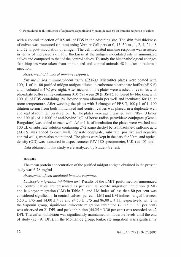
study was 6.78-mg/mL.assessment of cell mediated immune response. Leukocyte migration inhibition test. Results of the LMIT performed on immunized
and control calves are presented as per cent leukocyte migration inhibition (LMI) and leukocyte migration (LM) in Table 2., and LM index of less than 80 per cent was considered significant. In control calves, per cent LMI and LM indices ranged between 5.50 ± 1.75 and 14.00 ± 4.33 and 94.50 ± 1.75 and 86.00 ± 4.33, respectively, while in the Saponin group, significant leukocyte migration inhibition (20.25 ± 3.83 per cent) was observed on 21 DPI, and peak inhibition (44.25 ± 3.30 per cent) was recorded on 42 DPI. Thereafter, inhibition was significantly maintained at moderate levels until the end of study (i.e., 91 DPI). In the Montanide group, leukocyte migration was significantly
Vet. arhiv 77 (1), �-17, 2007
G. Ponnudurai et al.: Influence of adjuvants Saponin and Montanide ISA 50 on immune response of calves
13Vet. arhiv 77 (1), �-17, 2007
elevated after 28 DPI (39.00 ± 5.95 per cent) with a peak inhibition on 35 DPI (43.00 ± 2.41 per cent) that declined from 56 DPI onwards.
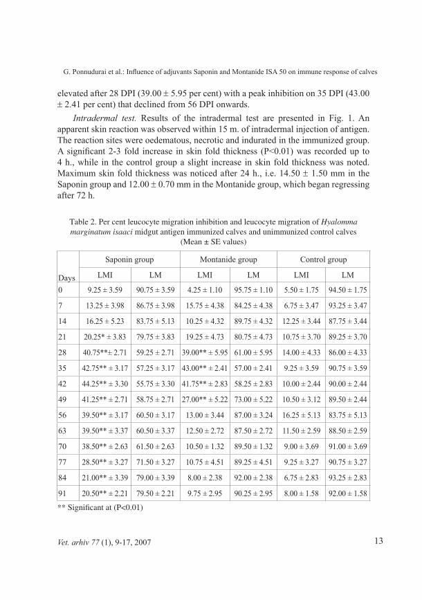
intradermal test. Results of the intradermal test are presented in Fig. 1. An apparent skin reaction was observed within 15 m. of intradermal injection of antigen. The reaction sites were oedematous, necrotic and indurated in the immunized group. A significant 2-3 fold increase in skin fold thickness (P<0.01) was recorded up to 4 h., while in the control group a slight increase in skin fold thickness was noted. Maximum skin fold thickness was noticed after 24 h., i.e. 14.50 ± 1.50 mm in the Saponin group and 12.00 ± 0.70 mm in the Montanide group, which began regressing after 72 h.
Table 2. Per cent leucocyte migration inhibition and leucocyte migration of Hyalomma marginatum isaaci midgut antigen immunized calves and unimmunized control calves
(Mean ± SE values)
Days
Saponin group Montanide group Control group
LMI LM LMI LM LMI LM
0 9.25 ± 3.59 90.75 ± 3.59 4.25 ± 1.10 95.75 ± 1.10 5.50 ± 1.75 94.50 ± 1.75
7 13.25 ± 3.98 86.75 ± 3.98 15.75 ± 4.38 84.25 ± 4.38 6.75 ± 3.47 93.25 ± 3.47
14 16.25 ± 5.23 83.75 ± 5.13 10.25 ± 4.32 89.75 ± 4.32 12.25 ± 3.44 87.75 ± 3.44
21 20.25* ± 3.83 79.75 ± 3.83 19.25 ± 4.73 80.75 ± 4.73 10.75 ± 3.70 89.25 ± 3.70
28 40.75**± 2.71 59.25 ± 2.71 39.00** ± 5.95 61.00 ± 5.95 14.00 ± 4.33 86.00 ± 4.33
35 42.75** ± 3.17 57.25 ± 3.17 43.00** ± 2.41 57.00 ± 2.41 9.25 ± 3.59 90.75 ± 3.59
42 44.25** ± 3.30 55.75 ± 3.30 41.75** ± 2.83 58.25 ± 2.83 10.00 ± 2.44 90.00 ± 2.44
4� 41.25** ± 2.71 58.75 ± 2.71 27.00** ± 5.22 73.00 ± 5.22 10.50 ± 3.12 89.50 ± 2.44
56 39.50** ± 3.17 60.50 ± 3.17 13.00 ± 3.44 87.00 ± 3.24 16.25 ± 5.13 83.75 ± 5.13
63 39.50** ± 3.37 60.50 ± 3.37 12.50 ± 2.72 87.50 ± 2.72 11.50 ± 2.59 88.50 ± 2.59
70 38.50** ± 2.63 61.50 ± 2.63 10.50 ± 1.32 89.50 ± 1.32 9.00 ± 3.69 91.00 ± 3.69
77 28.50** ± 3.27 71.50 ± 3.27 10.75 ± 4.51 89.25 ± 4.51 9.25 ± 3.27 90.75 ± 3.27
84 21.00** ± 3.39 79.00 ± 3.39 8.00 ± 2.38 92.00 ± 2.38 6.75 ± 2.83 93.25 ± 2.83
�1 20.50** ± 2.21 79.50 ± 2.21 9.75 ± 2.95 90.25 ± 2.95 8.00 ± 1.58 92.00 ± 1.58
** Significant at (P<0.01)
G. Ponnudurai et al.: Influence of adjuvants Saponin and Montanide ISA 50 on immune response of calves
14
Skin biopsies taken from immunized and control calves after 48 h. at inoculation revealed leukocytic infiltration, predominantly with eosinophils. Perivascular infiltration of eosinophils and a few polymorphs in the dermis were also noticed (Fig. 2.)
Vet. arhiv 77 (1), �-17, 2007
Fig. 2. Skin-delayed type of hypersensitivity reaction with infiltration of polymorphs and eosinophils after 48 h. of intradermal inoculation of midgut antigen. H&E; ×320.
Fig. 1. Intradermal test (skin fold thickness) response to midgut antigen of H. m. isaaci
G. Ponnudurai et al.: Influence of adjuvants Saponin and Montanide ISA 50 on immune response of calves
15
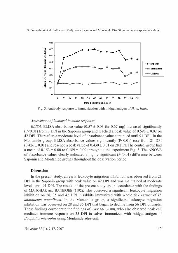
assessment of humoral immune response. eLisa. ELISA absorbance value (0.57 ± 0.03 for 0.67 mg) increased significantly
(P<0.01) from 7 DPI in the Saponin group and reached a peak value of 0.698 ± 0.02 on 42 DPI. Thereafter, a moderate level of absorbance value continued until 91 DPI. In the Montanide group, ELISA absorbance values significantly (P<0.01) rose from 21 DPI (0.426 ± 0.01) and reached a peak value of 0.430 ± 0.01 on 28 DPI. The control group had a mean of 0.153 ± 0.00 to 0.189 ± 0.00 throughout the experiment Fig. 3. The ANOVA of absorbance values clearly indicated a highly significant (P<0.01) difference between Saponin and Montanide groups throughout the observation period.
DiscussionIn the present study, an early leukocyte migration inhibition was observed from 21
DPI in the Saponin group with peak value on 42 DPI and was maintained at moderate levels until 91 DPI. The results of the present study are in accordance with the findings of MANOHAR and BANERJEE (1992), who observed a significant leukocyte migration inhibition on 28, 35 and 42 DPI in rabbits immunized with whole tick extract of h. anatolicum anatolicum. In the Montanide group, a significant leukocyte migration inhibition was observed on 28 and 35 DPI that began to decline from 56 DPI onwards. These findings corroborate the findings of RAMAN (2000), who also observed peak cell mediated immune response on 35 DPI in calves immunized with midgut antigen of Boophilus microplus using Montanide adjuvant.
Vet. arhiv 77 (1), �-17, 2007
Fig. 3. Antibody response to immunization with midgut antigen of H. m. isaaci
G. Ponnudurai et al.: Influence of adjuvants Saponin and Montanide ISA 50 on immune response of calves
16
In the present study, the intradermal injection of midgut antigen of h. m. isaaci revealed immediate hypersensitivity reaction within 15 m. of inoculation; peak skin fold thickness was recorded 24 h. after inoculation. The immediate hypersensitivity reaction could be due to either the vasoactive property of certain components from tick antigens, or mediators released from the sensitized cells during antigen antibody interaction. The results of the present study are in agreement with the findings of GILL et al. (1986) who observed skin reaction within 10 m. of inoculation of salivary gland antigen of h. a. anatolicum in calves. These studies further support the findings of WILLADSEN et al. (1�78), who stated that the release of histamine resulted in immediate hypersensitivity reaction, which has been found to have a direct effect on tick attachment.
The changes observed in skin biopsies taken after 48 h. are similar to the findings of GILL et al. (1986) who also observed cellular infiltration of eosinophils and neutrophils in skin biopsies taken after 48 h.
A significant serum antibody response was noticed from 7 DPI, with a peak value on 42 DPI in the Saponin group, while in the Montanide group, a significant level of ELISA absorbance value was observed from 21 DPI, with a peak value on 28 DPI. These results are in total conformity with the findings of RODRIGUEZ et al. (1995) who reported that the ELISA antibody titer against Bm 86 antigens were highest two weeks after the third vaccination. Similarly, SHARMA et al. (2001) observed notable antibody response from 7 DPI, and peak value on 42 DPI with a moderate level of response until the end of the study.
conclusionBased on the results of the present study, we concluded that the midgut antigen at
a dose rate of 3 mg in three divided doses with Saponin as adjuvant could be an ideal vaccination schedule for immunization of cattle against hyalomma marginatum isaaci ticks. As antibody response was moderate until the end of the study in the Saponin group, this immunization schedule endorses the need for a booster dose after 4-5 months for sustainable control of ticks.
_______AcknowledgementsThe first author is grateful to the University authorities for permitting him to pursue the doctoral programme on a part-time basis, and to the Dean, Veterinary College and Research Institute, Namakkal, for the facilities provided for the research programme.
ReferencesCASTRO DE, J. J. R. M. NEWSON (1993): Host resistance in cattle tick control. Parasitology
Today 9, 13-17. CHHABRA, M. B., K. L. KHURANA (2005): Immunity and immunization against hyalomma tick
vectors of bovine tropical theileriosis - A review. J. Vet. Parasitol. 19, 9-17
Vet. arhiv 77 (1), �-17, 2007
G. Ponnudurai et al.: Influence of adjuvants Saponin and Montanide ISA 50 on immune response of calves
17
CLAUSEN, J. E. (1976): Leucocyte migration inhibition factor. Manual of Clinical Immunology. (Rose, N. R., H. Friedman, Eds.) American Society of Microbiology, Washington, pp. 101.
GHOSH, S., M. H. KHAN (1997): Immunological control of ticks III. Shared antigens in larval and adult stages of Boophilus microplus. J. Vet. Parasitol. 11, 149-154.
GILL, H. S., R. BOID, C. A. ROSS (1986): Isolation and characterization of salivary antigens from hyalomma anatolicum anatolicum. Parasit. Immunol. 8, 11-25.
LOWRY, O. H., N. J. ROSE BROUGH, A. L. FARR, R. J. RANDALL (1951): Protein measurement with the folin phenol reagent. J. Biol. Chem. 193, 265-275.
MANOHAR, G. S., D. P. BANERJEE (1992): Immune responses in rabbits immunized against the tick hyalomma anatolicum anatolicum. Indian J. Anim. Sci. 62, 505-512.
RAMAN, M. (2000): Immunoprophylaxis against Boophilid ticks. Ph. D. Thesis submitted to the Tamil Nadu Veterinary and Animal Sciences University, Chennai-51.
RODRIGUEZ, M., C. L. MASSARD, DA A. H. FONSECA, N. F. RAMOS, H. MACHADO, V. LABARTA, J. DE LA FUENTE, A. H. DA FONSECA, J. DE LA FUENTE (1995): Effects ofEffects of vaccination with a recombinant Bm86 antigen preparation on natural infestations of Boophilus microplus in grazing dairy and beef pure and cross bred cattle in Brazil. Vaccine 13, 1804-1808.
SHARMA, J. K., S. GHOSH, M. H. KHAN G. DAS, (2001): Immunoprotective efficacy of a purified 39 KDa nymphal antigen of hyalomma anatolicum anatolicum. Trop. Anim. Hlth. Prod. 33, 103-116.
WILLADSEN, P., P. G. WILLIAMS, J. S. ROBERTS, J. D. KERR (1978): Responses of cattle to allergens from Boophilus microplus. Int. J. Parasitol. 8, 89-95.Int. J. Parasitol. 8, 89-95.
Received: 16 May 2005Accepted: 21 December 2006
PONNUDURAI, G., L. JOHN, T. J. HARIKRISHNAN: Učinak adjuvansa saponina i montanida ISA 50 na imunološki odgovor teladi imunizirane pročišćenim antigenom srednjega crijeva krpelja Hyalomma marginatum isaaci. Vet. arhiv 77, 9-17, 2007.
SAŽETAKImunosni status teladi imunizirane pročišćenim antigenom srednjega crijeva krpelja Hyalomma marginatum
isaaci procijenjen je upotrebom standardnih imunoloških testova. Testom inhibicije migracije leukocita dokazana je inhibicija 21. dana nakon invazije u teladi imunizirane antigenom srednjega crijeva krpelja u kombinaciji sa saponinom kao adjuvansom, dok je značajna inhibicija u skupini u kojoj je primijenjen montanid bila dokazana 28. dana nakon invazije. Intradermalnim testom dokazana je reakcija rane preosjetljivosti već za 15 minuta. Najveće zadebljanje kože dokazano je 24 sata nakon inokulacije antigena (14,50 ± 1,50 mm u skupini u kojoj je primijenjen saponin i 12,00 ± 0,7 mm u skupini u kojoj je primijenjen montanid). Znatan humoralni odgovor određen imunoenzimnim testom zabilježen je 7. dana nakon invazije (P<0,01) u skupini u kojoj je bio primijenjen saponin s najvišom vrijednošću 42. dana poslije inokulacije, dok je porast titra protutijela zabilježen 21. dana poslije inokulacije u skupini u kojoj je primijenjen montanid s vršnom vrijednošću zabilježenom 30. dana nakon primjene.
Ključne riječi: imunosni odgovor, hyalomma marginatum isaaci, antigen srednjega crijeva, telad
Vet. arhiv 77 (1), �-17, 2007
G. Ponnudurai et al.: Influence of adjuvants Saponin and Montanide ISA 50 on immune response of calves

.
Copyright © 2022 FDOKUMEN