New Frontiers on Adjuvants Drug Strategies and Treatments ...
16
Scientia Pharmaceutica Review New Frontiers on Adjuvants Drug Strategies and Treatments in Periodontitis Gaetano Isola 1, * , Alessandro Polizzi 1 , Simona Santonocito 1 , Domenico Dalessandri 2 , Marco Migliorati 3 and Francesco Indelicato 1 Citation: Isola, G.; Polizzi, A.; Santonocito, S.; Dalessandri, D.; Migliorati, M.; Indelicato, F. New Frontiers on Adjuvants Drug Strategies and Treatments in Periodontitis. Sci. Pharm. 2021, 89, 46. https://doi.org/10.3390/scipharm 89040046 Academic Editor: Helen D. Skaltsa Received: 12 August 2021 Accepted: 19 October 2021 Published: 22 October 2021 Publisher’s Note: MDPI stays neutral with regard to jurisdictional claims in published maps and institutional affil- iations. Copyright: © 2021 by the authors. Licensee MDPI, Basel, Switzerland. This article is an open access article distributed under the terms and conditions of the Creative Commons Attribution (CC BY) license (https:// creativecommons.org/licenses/by/ 4.0/). 1 Department of General Surgery and Surgical-Medical Specialties, School of Dentistry, University of Catania, Via S. Sofia 78, 95123 Catania, Italy; [email protected] (A.P.); [email protected] (S.S.); [email protected] (F.I.) 2 Department of Medical and Surgical Specialties, Radiological Sciences and Public Health, Dental School, University of Brescia, Piazzale Spedali Civili 1, 25123 Brescia, Italy; [email protected] 3 Department of Orthodontics, School of Dentistry, University of Genova, Largo Rossana Benzi 10, 16132 Genova, Italy; [email protected] * Correspondence: [email protected]; Tel.: +39-095-7435359 Abstract: Causes of the progression of periodontitis such as an imbalance between the immune response by the host by the release of inflammatory mediators in the response of the oral pathogenic dysbiotic biofilm have been identified. New insights on specific cell signaling pathways that appear during periodontitis have attracted the attention of researchers in the study of new personalised ap- proaches for the treatment of periodontitis. The gold standard of non-surgical therapy of periodontitis involves the removal of supra and subgingival biofilm through professional scaling and root planing (SRP) and oral hygiene instructions. In order to improve periodontal clinical outcomes and overcome the limitations of traditional SRP, additional adjuvants have been developed in recent decades, includ- ing local or systemic antibiotics, antiseptics, probiotics, anti-inflammatory and anti-resorptive drugs and host modulation therapies. This review is aimed to update the current and recent evolution of therapies of management of periodontitis based on the adjunctive and target therapies. Moreover, we discuss the advances in host modulation of periodontitis and the impact of targeting epigenetic mechanisms approaches for a personalised therapeutic success in the management of periodontitis. In conclusion, the future goal in periodontology will be to combine and personalise the periodontal treatments to the colonising microbial profile and to the specific response of the individual patient. Keywords: periodontitis; adjuvants; drugs; antimicrobials; chemotherapeutic agents; dysbiosis; bacteria 1. Introduction Periodontitis is a disease with an infectious aetiology characterised by inflammation of the supporting tissues of a tooth that can lead, if not properly treated, to the destruction of both periodontal tissues and alveolar bone, and, in the long term, cause tooth loss [1]. However, periodontitis’s onset and subsequent progression also occur as a host’s unbalanced immune reaction to a dysbiotic organised biofilm. Among dysbiotic biofilms, the periodontopathogenic bacteria produce metabolites and enzymes that determine the alteration and destruction of parts of the extracellular matrix, including the collagen; and the increase of the permeability of the cellular membranes of the host in order to determine a subsequent tissue invasion which increases periodontal damage over time [1,2]. All this induces the host immune response leading to site-specific local inflammation of the tissue and increased immune cell response [2]. Among the main known periodontal pathogens, there are bacteria such as Actinobacillus actinomycetemcomitans and Porphyromonas gingivalis (P.gingivalis), which produce multiple factors underlying the tissue damage found during periodontitis such as peptidoglycans, various integrins and outer membrane proteins, lipopolysaccharide and cellular superficial fimbriae degradation connective tissue [3–7]. Sci. Pharm. 2021, 89, 46. https://doi.org/10.3390/scipharm89040046 https://www.mdpi.com/journal/scipharm
-
Upload
khangminh22 -
Category
Documents
-
view
0 -
download
0
Transcript of New Frontiers on Adjuvants Drug Strategies and Treatments ...

Scientia
Pharmaceutica
Review
New Frontiers on Adjuvants Drug Strategies and Treatmentsin Periodontitis
Gaetano Isola 1,* , Alessandro Polizzi 1 , Simona Santonocito 1, Domenico Dalessandri 2 , Marco Migliorati 3
and Francesco Indelicato 1
�����������������
Citation: Isola, G.; Polizzi, A.;
Santonocito, S.; Dalessandri, D.;
Migliorati, M.; Indelicato, F. New
Frontiers on Adjuvants Drug
Strategies and Treatments in
Periodontitis. Sci. Pharm. 2021, 89, 46.
https://doi.org/10.3390/scipharm
89040046
Academic Editor: Helen D. Skaltsa
Received: 12 August 2021
Accepted: 19 October 2021
Published: 22 October 2021
Publisher’s Note: MDPI stays neutral
with regard to jurisdictional claims in
published maps and institutional affil-
iations.
Copyright: © 2021 by the authors.
Licensee MDPI, Basel, Switzerland.
This article is an open access article
distributed under the terms and
conditions of the Creative Commons
Attribution (CC BY) license (https://
creativecommons.org/licenses/by/
4.0/).
1 Department of General Surgery and Surgical-Medical Specialties, School of Dentistry, University of Catania,Via S. Sofia 78, 95123 Catania, Italy; [email protected] (A.P.); [email protected] (S.S.);[email protected] (F.I.)
2 Department of Medical and Surgical Specialties, Radiological Sciences and Public Health, Dental School,University of Brescia, Piazzale Spedali Civili 1, 25123 Brescia, Italy; [email protected]
3 Department of Orthodontics, School of Dentistry, University of Genova, Largo Rossana Benzi 10,16132 Genova, Italy; [email protected]
* Correspondence: [email protected]; Tel.: +39-095-7435359
Abstract: Causes of the progression of periodontitis such as an imbalance between the immuneresponse by the host by the release of inflammatory mediators in the response of the oral pathogenicdysbiotic biofilm have been identified. New insights on specific cell signaling pathways that appearduring periodontitis have attracted the attention of researchers in the study of new personalised ap-proaches for the treatment of periodontitis. The gold standard of non-surgical therapy of periodontitisinvolves the removal of supra and subgingival biofilm through professional scaling and root planing(SRP) and oral hygiene instructions. In order to improve periodontal clinical outcomes and overcomethe limitations of traditional SRP, additional adjuvants have been developed in recent decades, includ-ing local or systemic antibiotics, antiseptics, probiotics, anti-inflammatory and anti-resorptive drugsand host modulation therapies. This review is aimed to update the current and recent evolution oftherapies of management of periodontitis based on the adjunctive and target therapies. Moreover,we discuss the advances in host modulation of periodontitis and the impact of targeting epigeneticmechanisms approaches for a personalised therapeutic success in the management of periodontitis.In conclusion, the future goal in periodontology will be to combine and personalise the periodontaltreatments to the colonising microbial profile and to the specific response of the individual patient.
Keywords: periodontitis; adjuvants; drugs; antimicrobials; chemotherapeutic agents; dysbiosis; bacteria
1. Introduction
Periodontitis is a disease with an infectious aetiology characterised by inflammationof the supporting tissues of a tooth that can lead, if not properly treated, to the destructionof both periodontal tissues and alveolar bone, and, in the long term, cause tooth loss [1].
However, periodontitis’s onset and subsequent progression also occur as a host’sunbalanced immune reaction to a dysbiotic organised biofilm. Among dysbiotic biofilms,the periodontopathogenic bacteria produce metabolites and enzymes that determine thealteration and destruction of parts of the extracellular matrix, including the collagen; andthe increase of the permeability of the cellular membranes of the host in order to determinea subsequent tissue invasion which increases periodontal damage over time [1,2]. All thisinduces the host immune response leading to site-specific local inflammation of the tissueand increased immune cell response [2]. Among the main known periodontal pathogens,there are bacteria such as Actinobacillus actinomycetemcomitans and Porphyromonas gingivalis(P.gingivalis), which produce multiple factors underlying the tissue damage found duringperiodontitis such as peptidoglycans, various integrins and outer membrane proteins,lipopolysaccharide and cellular superficial fimbriae degradation connective tissue [3–7].
Sci. Pharm. 2021, 89, 46. https://doi.org/10.3390/scipharm89040046 https://www.mdpi.com/journal/scipharm
Sci. Pharm. 2021, 89, 46 2 of 16
Once these pathogenic bacteria trigger immune and inflammatory processes, the bodyinduces leukocytes, fibroblasts or other inflammatory cells to release various substances inorder to protect tissues from infection, including metalloproteinases, cytokines, transglu-taminases, prostaglandins and proteolytic enzymes [8]. Proteases cause the degradation ofcollagen in periodontal tissues and, therefore, can create incursions for further infiltrationof leukocytes [9]. Despite the production and release of several inflammatory mediators,tissue destruction occurs mainly due to an imbalance between the matrix metalloproteinaselevel and their endogenous inhibitors [10]. Subsequently, throughout the stimulation ofsome pro-inflammatory cytokines, such as including interleukin (IL)-1b, IL-6, tumor necro-sis factor, and (TNF)-a, the inflammatory infiltrate from the periodontal tissues starts thetissue and alveolar bone destruction [7,10–14]. The inflammatory-related mediators cancontrol the release of ligand-receptor factor Kappa B (NF-KB), RANKL and osteoprotegerin,which stimulate osteoclastic activity by inducing alveolar bone destruction [15].
Host modulation therapy is among the most widely used approaches to halting theprogression of periodontitis and alveolar bone loss. This uses pharmacological therapies andconventional periodontal treatment to ameliorate the destructive aspects of the host inflam-matory response by inhibiting microbial growth or modulating the host response [16–18],through reduction of the production of metalloproteinases or related inflammatory cy-tokines [19]. Several classes of anti-inflammatory drugs have been tested, including metal-loproteinase inhibitors (doxycycline) [20] and NSAIDs (flurbiprofen) [21], but these havenumerous limitations in clinical use [22].
In recent years, a better understanding of the pathways of inflammatory cells thatresult in the release of tissue-destroying proteins has led to an analysis of the possibilityof developing new therapies for the long-term success of periodontitis [23], but thereis still a long way to go before they are routinely used in clinical practice. Numerousstudies have attempted to describe the main bacterial species that determine the tissuedamage of periodontitis, which species are influenced by specific therapies, and what arethe innovative therapeutic effects of reducing the bacterial and inflammatory mediatorsreleased during periodontitis, but there are still several aspects to be explored further.
The objectives of this review were to analyse and update the present knowledge ofthe conventional and adjunctive therapies for periodontitis. Furthermore, we discussedthe impact of innovative and target therapies of periodontitis for long-term therapeuticsuccess based on the latest scientific evidence. The data included in this review have beencollected through a free literature search by three independent authors.
2. Conventional Therapies of Periodontitis: Endpoint, Strategies and Limitations2.1. Endpoints
The success of periodontitis therapy, both in the short- and long term, depends onthe effect of physical disintegration of the supra and subgingival periodontal pathogenspresented in the biofilm [24]. The initial causal therapy mainly consisted of oral hygieneinstruction and professional scaling and root planing (SRP). Both the supra- and subgingivalpathogens and calculus are mechanically disrupted, with combined and synergic effectsinduced by periodontal debridement and domiciliary oral hygiene instructions [25]. Inthis regard, the gold standard of non-surgical therapy of periodontitis is represented bythe SRP performed by both hand and ultrasonic instruments alone, with a demonstratedmicrobial and clinical effectiveness in the short-term period [26,27] (Figure 1).
It has been previously shown that the mechanical disruption of supra- and subgingivalbiofilm by the non-surgical periodontal therapy performed by SRP alone can reduce theplaque index (PI) and the bleeding on probing (BOP) in around 45% of periodontal sites [28].On this regard, following non-surgical periodontal therapy, a reduction in the probingdepth (PD) has been demonstrated in a range of 1.29 mm for periodontal pockets with aninitial PD of 3–4 mm and of 2.2 mm for the pockets ranged 5–6 mm and an improvementin the clinical attachment level (CAL) in a range of 0.5–2 mm [29,30].
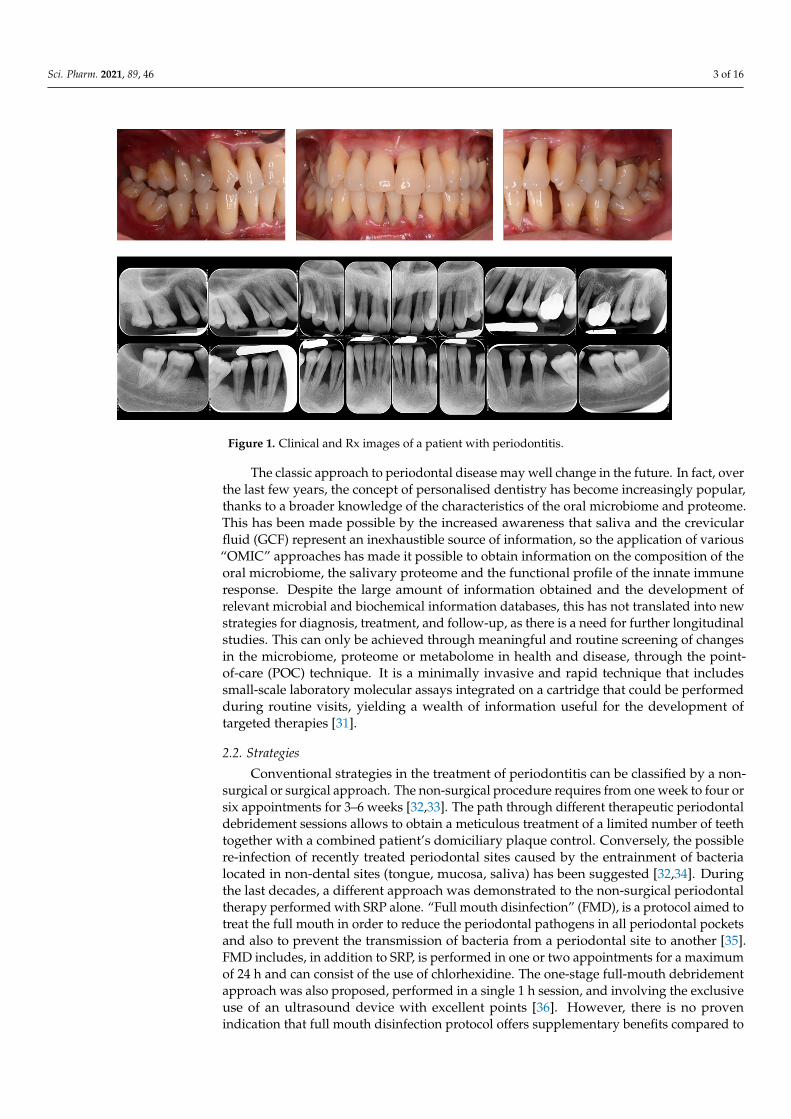
Sci. Pharm. 2021, 89, 46 3 of 16Sci. Pharm. 2021, 89, x FOR PEER REVIEW 3 of 16
Figure 1. Clinical and Rx images of a patient with periodontitis.
It has been previously shown that the mechanical disruption of supra‐ and subgingi‐
val biofilm by the non‐surgical periodontal therapy performed by SRP alone can reduce
the plaque index (PI) and the bleeding on probing (BOP) in around 45% of periodontal
sites [28]. On this regard, following non‐surgical periodontal therapy, a reduction in the
probing depth (PD) has been demonstrated in a range of 1.29 mm for periodontal pockets
with an initial PD of 3–4 mm and of 2.2 mm for the pockets ranged 5–6 mm and an im‐
provement in the clinical attachment level (CAL) in a range of 0.5–2 mm [29,30].
The classic approach to periodontal disease may well change in the future. In fact,
over the last few years, the concept of personalised dentistry has become increasingly
popular, thanks to a broader knowledge of the characteristics of the oral microbiome and
proteome. This has been made possible by the increased awareness that saliva and the
crevicular fluid (GCF) represent an inexhaustible source of information, so the application
of various “OMIC” approaches has made it possible to obtain information on the compo‐
sition of the oral microbiome, the salivary proteome and the functional profile of the in‐
nate immune response. Despite the large amount of information obtained and the devel‐
opment of relevant microbial and biochemical information databases, this has not trans‐
lated into new strategies for diagnosis, treatment, and follow‐up, as there is a need for
further longitudinal studies. This can only be achieved through meaningful and routine
screening of changes in the microbiome, proteome or metabolome in health and disease,
through the point‐of‐care (POC) technique. It is a minimally invasive and rapid technique
that includes small‐scale laboratory molecular assays integrated on a cartridge that could
be performed during routine visits, yielding a wealth of information useful for the devel‐
opment of targeted therapies [31].
2.2. Strategies
Conventional strategies in the treatment of periodontitis can be classified by a non‐
surgical or surgical approach. The non‐surgical procedure requires from one week to four
or six appointments for 3–6 weeks [32,33]. The path through different therapeutic perio‐
dontal debridement sessions allows to obtain a meticulous treatment of a limited number
of teeth together with a combined patient’s domiciliary plaque control. Conversely, the
possible re‐infection of recently treated periodontal sites caused by the entrainment of
bacteria located in non‐dental sites (tongue, mucosa, saliva) has been suggested [32,34].
Figure 1. Clinical and Rx images of a patient with periodontitis.
The classic approach to periodontal disease may well change in the future. In fact, overthe last few years, the concept of personalised dentistry has become increasingly popular,thanks to a broader knowledge of the characteristics of the oral microbiome and proteome.This has been made possible by the increased awareness that saliva and the crevicularfluid (GCF) represent an inexhaustible source of information, so the application of various“OMIC” approaches has made it possible to obtain information on the composition of theoral microbiome, the salivary proteome and the functional profile of the innate immuneresponse. Despite the large amount of information obtained and the development ofrelevant microbial and biochemical information databases, this has not translated into newstrategies for diagnosis, treatment, and follow-up, as there is a need for further longitudinalstudies. This can only be achieved through meaningful and routine screening of changesin the microbiome, proteome or metabolome in health and disease, through the point-of-care (POC) technique. It is a minimally invasive and rapid technique that includessmall-scale laboratory molecular assays integrated on a cartridge that could be performedduring routine visits, yielding a wealth of information useful for the development oftargeted therapies [31].
2.2. Strategies
Conventional strategies in the treatment of periodontitis can be classified by a non-surgical or surgical approach. The non-surgical procedure requires from one week to four orsix appointments for 3–6 weeks [32,33]. The path through different therapeutic periodontaldebridement sessions allows to obtain a meticulous treatment of a limited number of teethtogether with a combined patient’s domiciliary plaque control. Conversely, the possiblere-infection of recently treated periodontal sites caused by the entrainment of bacterialocated in non-dental sites (tongue, mucosa, saliva) has been suggested [32,34]. Duringthe last decades, a different approach was demonstrated to the non-surgical periodontaltherapy performed with SRP alone. “Full mouth disinfection” (FMD), is a protocol aimed totreat the full mouth in order to reduce the periodontal pathogens in all periodontal pocketsand also to prevent the transmission of bacteria from a periodontal site to another [35].FMD includes, in addition to SRP, is performed in one or two appointments for a maximumof 24 h and can consist of the use of chlorhexidine. The one-stage full-mouth debridementapproach was also proposed, performed in a single 1 h session, and involving the exclusiveuse of an ultrasound device with excellent points [36]. However, there is no provenindication that full mouth disinfection protocol offers supplementary benefits compared to

Sci. Pharm. 2021, 89, 46 4 of 16
the multi-staged non-surgical periodontal approach. Both therapeutic approaches are valid,and there were demonstrated only small differences between the two therapies; therefore,the choice of the strategy should be taken on the specific cases at the patient’s level [37].
2.3. Limitations
Only 10–15 bacterial species, among the over 700 present in the oral biofilm, havebeen related to the beginning and evolution of periodontitis dysbiosis [38]. Despite thehigh composition of the oral biofilm, the traditional periodontal therapy is non-specific,as it consists mainly of the mechanical disruption of the supra- and subgingival biofilms.However, SRP alone is sufficient to determine positive results in the long-term in some pa-tients with a mild form of periodontitis [39]; nevertheless, a significant percentage of pocketsites may not have a good response in periodontal patients. This can be partly explaineddue to the inherent limitations of mechanical debridement, including the problematicaccess to deep and winding pockets, furcations, and vertical defects. Furthermore, therapyperformed by SRP alone can represent limited effects on some periodontal pathogens, suchas bacteria of the red complex of Socransky [40] and does not allow the elimination ofperiodontopathogens in non-native oral microflora (e.g., oral mucosa, tongue) [41]. SRPalone can also determine some minor side effects, such as gingival recession and dentinalhypersensitivity [42]. These limitations in therapy can be partially overcome through theuse of a series of alternative technologies [43]. For these reasons, additional adjuvants,which includes local or systemic antibiotics, antiseptics, probiotics, and host modulationtherapies, have been developed in the last few decades.
3. Microbial Modulation of Periodontitis
Almost all forms of periodontitis are determined by the presence of a pathogen biofilmlocated into the periodontal pocket, which can require therapy to modulate the microbialbiofilm. In general, oral infections caused by periodontal pathogens are tricky to modulatewith antibiotics, particularly if the biofilm is not mechanically removed [44]. Therefore,according to the current consensus, correct antimicrobial therapy should be led by anaccurate periodontal debridement that destroys the supra and subgingival biofilm [45].Adjuvant antimicrobial therapies are both administered at a local and systemic level. Theadvantages and limits of local and systemic antimicrobials are presented in Table 1.
Table 1. Potential of antimicrobials in periodontology.
Characteristics Local Administration Systemic Administration
Distribution Narrow range of effectiveness Wide
Concentration Good topic concentration, bad effectiveness far from local Variables in different body districts
Therapeutic potential Better locally More effective in reaching all microbes
Issues Local reinfections Systemic side effects
Limitations Limited to treated sites Requires good patient compliance
The potential advantage of the systemic dispensation of antimicrobials is the possibilityof reducing all periodontopathogens present in the mouth and those located in other sitessuch as the tonsils and dorsum of the tongue. Nevertheless, this approach necessitatesa strong collaboration by the patient and could determine undesirable side effects at asystemic level that could cause the onset of bacterial resistance [46]. On the contrary, localadministration of antimicrobials does not request particular compliance of the patient andallows the application of the active principle directly at the local infection site at a highintensity that not be achieved systemically [46].
3.1. Chlorhexidine
Antiseptic solutions are often used for the control of biofilms, especially pathogenicones, during the active phases of periodontal treatment. A recent review of the literature
Sci. Pharm. 2021, 89, 46 5 of 16
found a similar result in the comparison between chlorhexidine irrigation associated withSRP and SRP alone [47]. Chlorhexidine (CHX) gluconate is an antiseptic, antifungal andbactericidal chemical agent that acts successfully on both Gram-positive and -negativeperiodontopathogens. It also has a bacteriostatic effect that determines the inhibition ofbacterial proliferation [48]. This bisbiguanide antiseptic is also used as a slow local releasesystem available in the form of gel, paint or chip. In order to increase the effectivenessof non-surgical periodontal therapy, the concept of chemomechanical treatment based onsequential SRP and the additional subgingival administration of 35% of CHX paint [49]and subgingival positioning of a biodegradable CHX chip was introduced [50].
In the last few decades, several clinical studies have demonstrated that a CHX chipused after SRP is an effective protocol that allows the active reduction of periodontitis forabout 6–9 months [51]. The use of CHX in the form of mouthwash has produced betterclinical and microbial results compared with SRP alone or SRP associated with professionalplaque control [52] or SRP associated with placebo [53]. However, the adverse effects ofCHX, such as brown tooth and mucosal staining, altered taste perception, and augmentedcalculus deposition should be taken into account [54].
3.2. Antibiotics
Some clinical studies suggest the hypothesis that in specific clinical situations, includ-ing particular forms of active and rapidly evolving periodontitis, in the presence of deeppockets or with particular pathological profiles, the additional use of systemic antimicro-bials associated with SRP may determine clinically relevant advantages in the medium andlong term.
The combined use of amoxicillin and metronidazole for periodontitis was initiallyintroduced to counteract Aggregatibacter actinomycetemcomitans (A. actinomicetemcomitans)on periodontal tissues [55], although subsequent studies have also shown a role for severalother pathogens during the active phases of periodontitis [56]. Furthermore, not only a lowprevalence of A. actinomicetemcomitans during periodontitis has been demonstrated [57],but moreover, it has been shown that the use of amoxicillin and metronidazole in additionto SRP is associated with adverse effects, including systemic resistance to antibiotics, andalso could not be valid against other periodontopathogenic bacteria, especially P. gingivalisand other periodontopathogens, after 3–6 months of therapy [35].
Regarding the use of topical antibiotics in periodontics, some studies have shownlimited usefulness to sites refractory to traditional therapy with SRP alone or in patientswith localised lesions [58]. Antibiotics used topically (e.g., chips, gels, topical solutions),compared with antibiotics systemically, lead to fewer adverse events, including bacterialresistance, and are better tolerated by the patient [59]. The use of local antibiotics has beensuggested, in some studies, as an alternative for surgical therapy [60], but at the sametime, a real long-term efficacy of local applications has not been demonstrated, and somecommercial products have been withdrawn from the market [61].
The most effective antimicrobial agents, such as tetracycline fibers, slow-release doxy-cycline, and minocycline, resulted in an average PD reduction of between 0.5 and 0.7 mm;while less effective agents such as CHX and metronidazole determined an average PDgain of 0.1–0.4 mm [62]. Some randomised controlled trials with a follow-up ≤ 6 monthsdemonstrated a moderate increase in the CAL level of 0.40 mm using CHX chips, a meanpercentage of 0.64 mm for doxycycline gel, and 0.24 mm microspheres of minocycline [63].However, a precise evaluation of the usefulness of topical antimicrobials for routine clinicaluse is prevented by significant limitations and high acquisition costs [47,59]. More specifi-cally, a 3-rear RCT study showed that the adjunctive use of locally delivered doxycyclinegel determined positive short-term effects on BOP, PD and CAL, but repeated annual ap-plications were not useful compared to SRP alone in maintenance patients [64]. The resultsof a recent study suggested that nanostructured doxycycline gel (nDOX) compared to con-ventional doxycycline gel and placebo used as an adjunct to SRP determined a significantPD and CAL reduction; however, these observations are limited to the short term [65].
Sci. Pharm. 2021, 89, 46 6 of 16
Research is currently focusing on different therapeutic strategies to limit the useof antibiotics, especially systemic antibiotics, to extremely severe cases of periodontitis.Among the strategies being explored are antiviral strategies against leucotoxin (LtxA),which is produced by A. actinomycetemcomitans strains [66]. This toxin specifically killshuman white blood cells, reducing the host’s ability to respond to infection [67]. Underlyingthis antivirulence strategy is the idea that by inhibiting the activity of this particular toxin,the virulence of the bacteria would be reduced, resulting in a decrease in the severityof the infection. Obvious advantages include slower development of resistance due toreduced selective pressure (compared to antibiotics) and targeted treatment that wouldnot impact on the healthy microbiota. However, further studies and research are neededbefore this therapy can be used in clinical practice [66]. Several studies have pointedthe role of Aggregatibacter actinomycetemcomitans in the development of periodontitis withrapid progression of attachment loss (AL) in adolescents, as they observed that subjectswith a highly virulent clone of A. actinomycetemcomitans, called genotype JP2, have apronounced risk for disease progression [67–70]. Therefore, therapies targeting specificvirulence factors, such as the one against LtxA could have an important preventive effectin these individuals [68].
3.3. Antimicrobial Proteins
Peptides and antimicrobial proteins are classes of host defense particles that act inorder to reduce the proportions and augmentation of oral bacteria and similar pathogens.These compounds have aroused huge attention in periodontal innate immunity medicineand as an alternative resource of typical antimicrobials (e.g., antibiotics) during thelast decade [71].
Several antimicrobial peptides such as tissue-specific human beta-defensins (hBD)-3and cathelicidin antimicrobial peptides (LL)-37 have been analysed against different typesand different strains of oral periodontal pathogens [72]. Regarding main peptides involvedagainst periodontitis, Streptococcus gordonii subtype M5 and 10,558 presents a full rangesusceptibility against P. gingivalis. On the other hand, LL-37 has been shown to be poorlysensitive against P. gingivalis, while hBD3 has been proven to possess a high susceptibilityin some in vitro periodontitis models in which gingival epithelial cells were exposed toperiodontal bacteria [72].
More than twenty systemic genetic diseases have been identified as associated withperiodontitis related to alterations in the expression of the antimicrobial peptide similarlyto periodontitis, which may have the same characteristics of augmented susceptibility toperiodontal pathogen infections [73].
Several reports have analysed other antimicrobial peptides associated such as lacto-ferrin and mucin-7, both associated with periodontitis. Among these, mucin-7 (MG2)and lactoferrin have been shown to decrease significantly three times in subjects withperiodontitis compared to healthy controls, thus suggesting antimicrobial properties ofthese peptides [74].
3.4. Probiotics
The term “probiotic” was defined in 2005 as a set of live microorganisms that, given inadequate quantities, confer benefits to the host [75]. There are likely to be slight changes inthe definition of this term in the literature, which may be due to the new criteria of evalua-tion and new discoveries. For example, some reports have shown that certain inactivatedmicroorganisms or the derivatives of their components have potentially beneficial effectson health, expanding the concept of probiotics as a therapeutic use [76].
In periodontology, probiotic strains have been evaluated in addition to SRP and havebeen demonstrated to interfere with bacterial recolonisation [77]. Most of the studieson probiotics have analysed Lactobacillus-derived products such as ones that contain Lac-tobacillus reuteri. A recent meta-analysis has demonstrated that the use of probiotics inperiodontal therapy determined a CAL gain of 0.42 mm and PD reduction of 0.18 mm in
Sci. Pharm. 2021, 89, 46 7 of 16
moderate pockets and 0.67 mm in deep pockets after periodontal therapy [78]. Moreover,continuous intake of probiotics has been provided with the best favourable results whenused, in periodontal treatment, as an adjuvant to SRP, even if the heterogeneity in thedesign of the analysed studies does not allow one to draw any important conclusions,especially as concerns clinical utility in the long term [79].
3.5. Lasers
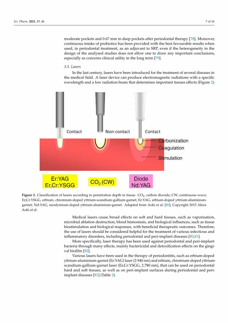
In the last century, lasers have been introduced for the treatment of several diseases inthe medical field. A laser device can produce electromagnetic radiations with a specificwavelength and a low radiation beam that determines important tissues effects (Figure 2).
Sci. Pharm. 2021, 89, x FOR PEER REVIEW 7 of 16
periodontitis compared to healthy controls, thus suggesting antimicrobial properties of
these peptides [74].
3.4. Probiotics
The term “probiotic” was defined in 2005 as a set of live microorganisms that, given
in adequate quantities, confer benefits to the host [75]. There are likely to be slight changes
in the definition of this term in the literature, which may be due to the new criteria of
evaluation and new discoveries. For example, some reports have shown that certain inac‐
tivated microorganisms or the derivatives of their components have potentially beneficial
effects on health, expanding the concept of probiotics as a therapeutic use [76].
In periodontology, probiotic strains have been evaluated in addition to SRP and have
been demonstrated to interfere with bacterial recolonisation [77]. Most of the studies on
probiotics have analysed Lactobacillus‐derived products such as ones that contain Lactoba‐
cillus reuteri. A recent meta‐analysis has demonstrated that the use of probiotics in perio‐
dontal therapy determined a CAL gain of 0.42 mm and PD reduction of 0.18 mm in mod‐
erate pockets and 0.67 mm in deep pockets after periodontal therapy [78]. Moreover, con‐
tinuous intake of probiotics has been provided with the best favourable results when
used, in periodontal treatment, as an adjuvant to SRP, even if the heterogeneity in the
design of the analysed studies does not allow one to draw any important conclusions,
especially as concerns clinical utility in the long term [79].
3.5. Lasers
In the last century, lasers have been introduced for the treatment of several diseases
in the medical field. A laser device can produce electromagnetic radiations with a specific
wavelength and a low radiation beam that determines important tissues effects (Figure 2).
Figure 2. Classification of lasers according to penetration depth in tissue. CO2, carbon dioxide; CW, continuous wave;
Er,Cr:YSGG, erbium, chromium‐doped yttrium‐scandium‐gallium‐garnet; Er:YAG, erbium‐doped yttrium‐aluminium‐
garnet; Nd:YAG, neodymium‐doped yttrium‐aluminium‐garnet. Adapted from Aoki et al. [80], Copyright 2015 Akira
Aoki et al.
Medical lasers cause broad effects on soft and hard tissues, such as vaporisation, mi‐
crobial ablation destruction, blood hemostasis, and biological influences, such as tissue
biostimulation and biological responses, with beneficial therapeutic outcomes. Therefore,
Figure 2. Classification of lasers according to penetration depth in tissue. CO2, carbon dioxide; CW, continuous wave;Er,Cr:YSGG, erbium, chromium-doped yttrium-scandium-gallium-garnet; Er:YAG, erbium-doped yttrium-aluminium-garnet; Nd:YAG, neodymium-doped yttrium-aluminium-garnet. Adapted from Aoki et al. [80], Copyright 2015 AkiraAoki et al.
Medical lasers cause broad effects on soft and hard tissues, such as vaporisation,microbial ablation destruction, blood hemostasis, and biological influences, such as tissuebiostimulation and biological responses, with beneficial therapeutic outcomes. Therefore,the use of lasers should be considered helpful for the treatment of various infectious andinflammatory disorders, including periodontal and peri-implant diseases [80,81].
More specifically, laser therapy has been used against periodontal and peri-implantbacteria through many effects, mainly bactericidal and detoxification effects on the gingi-val biofilm [82].
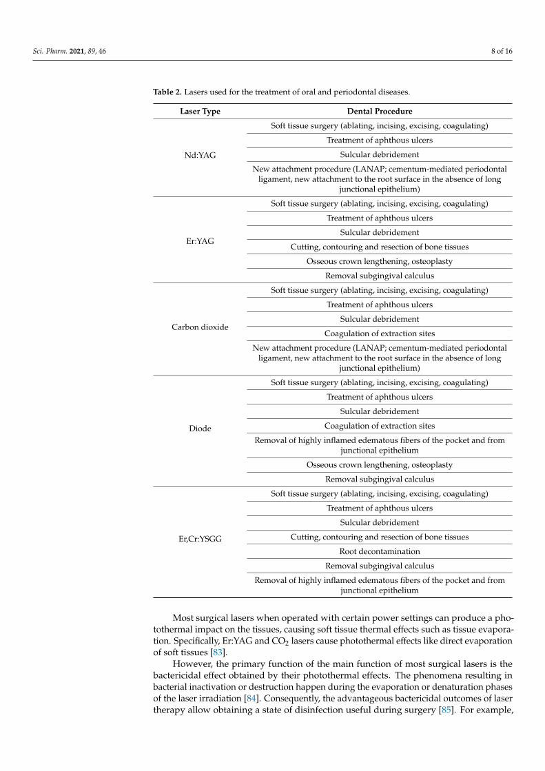
Various lasers have been used in the therapy of periodontitis, such as erbium-dopedyttrium-aluminium-garnet (Er:YAG) laser (2.940 nm) and erbium, chromium-doped yttrium-scandium-gallium-garnet laser (Er,Cr:YSGG, 2.780 nm), that can be used on periodontalhard and soft tissues, as well as on peri-implant surfaces during periodontal and peri-implant diseases [81] (Table 2).
Sci. Pharm. 2021, 89, 46 8 of 16
Table 2. Lasers used for the treatment of oral and periodontal diseases.
Laser Type Dental Procedure
Nd:YAG
Soft tissue surgery (ablating, incising, excising, coagulating)
Treatment of aphthous ulcers
Sulcular debridement
New attachment procedure (LANAP; cementum-mediated periodontalligament, new attachment to the root surface in the absence of long
junctional epithelium)
Er:YAG
Soft tissue surgery (ablating, incising, excising, coagulating)
Treatment of aphthous ulcers
Sulcular debridement
Cutting, contouring and resection of bone tissues
Osseous crown lengthening, osteoplasty
Removal subgingival calculus
Carbon dioxide
Soft tissue surgery (ablating, incising, excising, coagulating)
Treatment of aphthous ulcers
Sulcular debridement
Coagulation of extraction sites
New attachment procedure (LANAP; cementum-mediated periodontalligament, new attachment to the root surface in the absence of long
junctional epithelium)
Diode
Soft tissue surgery (ablating, incising, excising, coagulating)
Treatment of aphthous ulcers
Sulcular debridement
Coagulation of extraction sites
Removal of highly inflamed edematous fibers of the pocket and fromjunctional epithelium
Osseous crown lengthening, osteoplasty
Removal subgingival calculus
Er,Cr:YSGG
Soft tissue surgery (ablating, incising, excising, coagulating)
Treatment of aphthous ulcers
Sulcular debridement
Cutting, contouring and resection of bone tissues
Root decontamination
Removal subgingival calculus
Removal of highly inflamed edematous fibers of the pocket and fromjunctional epithelium
Most surgical lasers when operated with certain power settings can produce a pho-tothermal impact on the tissues, causing soft tissue thermal effects such as tissue evapora-tion. Specifically, Er:YAG and CO2 lasers cause photothermal effects like direct evaporationof soft tissues [83].
However, the primary function of the main function of most surgical lasers is thebactericidal effect obtained by their photothermal effects. The phenomena resulting inbacterial inactivation or destruction happen during the evaporation or denaturation phasesof the laser irradiation [84]. Consequently, the advantageous bactericidal outcomes of lasertherapy allow obtaining a state of disinfection useful during surgery [85]. For example,
Sci. Pharm. 2021, 89, 46 9 of 16
Nd:YAG lasers have been shown to possess selective absorption in pigments, effectivein killing some of biofilms such as those of P. gingivalis in periodontitis models [84]. Fur-thermore, lasers can reduce the release of toxic substances, such as lipopolysaccharideendotoxins [86]. Thanks to these additional effects of decontamination and detoxification,lasers can promote better wound healing when compared to conventional therapies usedin periodontal pockets.
Moreover, it can be argued that irradiation of the root surface by a laser may determinea microbe-inhibiting effect on the adhesion and colonisation of oral bacteria, an importantstage in the stability of healing of the periodontal pocket [82].
It is also hypothesised that, following laser therapy, the effects of photobiomodulationare simultaneously determined, including the stimulation of proliferation and differenti-ation of cell tissues and the anti-inflammatory influences, and that this should promotetissue healing. However, the photobiomodulation effect induced by laser therapy onwound healing is not still well understood and has been demonstrated to vary betweendifferent types of lasers.
Yamasaki et al. [87] stated that the low-level laser therapy induced by CO2 laserled to the heat shock protein expression with different intensities and patterns to thoseexpressed after surgery performed with the scalpel. In fact, on the day following laserapplication therapy, growth in the percentage of connective tissue cells marked withbromodeoxyuridine in the laser wound was demonstrated compared to the wound healingof traditional surgery, with a more quickly wound repair process induced by the lasertherapy than through conventional surgery.
It seems that another significant effect induced by lasers through a low pulsed emission(especially with the CO2 laser) is the coagulation process that produces an acceleration inthe repair process and also that promotes the progression remodelling of gingival tissueswith a good influence on wound healing [88].
However, at present, clinical trials aimed at analysing the effectiveness of lasers onperiodontal and tissue regeneration are still limited. Given these promising pivotal results,further research is needed to clarify better the possible useful effects of lasers on soft tissuewound healing.
3.6. Desiccant Agents
Given the key role of biofilms in the development of periodontitis, some additionaltreatments were developed to improve the effectiveness of SRP in subjects with periodonti-tis by selectively reducing periodontal pathogens.
Microorganisms in biofilms live in an extracellular polymeric substance (EPS) matrixcomposed of proteins, polysaccharides, and DNA that can adhere on both root surfacesor material surfaces (e.g., implants) [89]. The presence of EPS allows oral bacteria tosurvive and protects them, at the same time, from the degrading action of any antimicrobialagents that are unable to reach the bacteria present at the subgingival level [90]. For thesereasons, some strategies have been developed in the last few decades, such as the localdelivery agents.
In dentistry, a type of local delivery agent, a desiccant agent, has been employedfor the treatment of oral aphthous stomatitis [91]. Successively, a further generation ofdesiccant had been developed in order to be applied in periodontal pockets. A desiccantagent is a liquid or gel blend containing a mixture of sulphonic and sulfuric acids [92]. Thedesiccant agents include aqueous mixture solutions of concentrated hydroxybenzenesul-fonic and hydroxymethoxybenzenesulphonic and sulfuric acid that present a hygroscopicsurface and a denaturing action. The sulfate group present in the desiccant mixture hasa particular internal polar structure and oxygen atoms on the external surface, present-ing a solid negative surface superficial charge. This feature allows this solution, thanksto their high effect, to constrain the water of the matrix of the oral bacterial biofilm, toadhere and detach, quickly destroy the biofilm, and eradicate it from the gingival surfaceand the root surface [93]. Encouraging results have been reported in a pivotal study by
Sci. Pharm. 2021, 89, 46 10 of 16
Bracke et al. [92], which demonstrated that the additional use of desiccant in the SRPwas effective for reducing the mean levels of some periodontal clinical mediators afterperiodontal therapy. Moreover, in a further RCT study with a split-mouth protocol, thedesiccant agent associated with SRP performed with ultrasonic and hand instruments wasdemonstrated to be efficacious in disrupting the biofilm and in reducing the microbial andinflammatory mediators when used in combination to SRP [94].
On this regard, a molecular report aimed at detecting the activity of the main peri-odontal bacteria such as P. gingivalis, T. forsythia, and T. denticola after 2 weeks of activeperiodontal therapy found a significant decrease in about 99% for bacteria of the redcomplex and 96% in the percentage of the total bacterial count after treatment with theadjunctive use of a desiccant agent plus SRP [95]. In accordance, another research reportedfavourable results of the desiccant solutions useful in the elimination of pathogenic biofilmon the peri-implant surfaces [96].
3.7. Anti-Inflammatory Drugs and Bisphosphonates
Different cell types in the periodontium (neutrophils, macrophages, fibroblasts andepithelial cells) in response to bacterial lipopolysaccharide release prostaglandins, po-tent pro-inflammatory mediators that are relevant for the progression of periodontaldisease; for example, prostaglandin E2 is known to induce osteoclastic bone resorption [97].Various studies showed that both topical and systemic non-steroidal anti-inflammatorydrugs (NSAIDs) short-term application induced a reduction of gingival bleeding, whereaslong-term intake promoted an improvement in terms of bone loss [98–101]. However asystematic review has pointed out some important limitations in their use for periodontitis:lacking of long-term observational studies, and systemic side effects (mainly gastrointesti-nal and cardiotoxic) [102].
Bisphosphonates are drugs capable of inhibiting bone resorption and are mainly usedin patients with osteoporosis or bone metastases. The main side effect is osteonecrosis ofthe jaws, which is more common in patients taking intravenous bisphosphonates [103,104].The anti-resorptive properties of bisphosphonates have been of interest among researcherson periodontal disease as an adjunctive therapy for the possible benefits that have beenobserved in animal and human studies [105–107]. However, although there may be thepossibility that bisphosphonates can alter the severity of periodontal disease, their use isstill debated both for the risk of osteonecrosis and for the potential equivocal data relatedto the fact that osteoporosis could influence the progression of periodontitis [106,108].
3.8. Other Adjunctive Therapies for Periodontitis
Statins are drugs used to lower cholesterol levels, but they also have anti-inflammatoryproperties that may be of interest in the management of periodontal disease. Large cohortstudies adjusted for confounding factors have however shown little or no evidence on thepotential beneficial effects of statins [109,110]. In contrast, improvements in periodontalparameters were reported in a smaller study, not adjusted for all possible confounders [111].However, more recent studies have reported beneficial effects in the additional topicaluse of statins + SRP [112–115]. However, further studies are needed to confirm or not thepossible use of statins as adjuvant therapy in periodontitis.
Metformin is a hypoglycaemic drug that inhibits hepatic gluconeogenesis and de-creases peripheral insulin resistance. It has been shown that it is able to stimulate osteoblas-tic differentiation and bone formation [116,117]. Doses of 50 mg/kg induced a reductionof inflammation, oxidative stress and bone loss in induced periodontitis rats [117]. Inhumans local application of 1% metformin gel into the periodontal pockets induced animprovement in PD, CAL and intrabody defects reduction compared to placebo in adjunctto SRP [118]. Two recent meta-analysis concluded that the use of adjunctive metformin toSRP may induce additional benefits in the treatment of periodontal disease [119,120].
Omega-3 polyunsaturated fatty acids (PUFAs) have anti-inflammatory propertiesthat have been studied in several diseases, including periodontal disease [121]. An RCT
Sci. Pharm. 2021, 89, 46 11 of 16
study demonstrated that SRP associated with dietary supplementation of omega-3 PUFAs(300 mg/die for twelve weeks) significantly reduced gingival inflammation, PD and CALcompared to SRP and placebo in patients with moderate-severe periodontitis [122]. Incontrast, another group showed that omega-3 PUFAs dietary supplementation (+ SRP)induced a reduction of salivary TNF-α but without significant impact on periodontalclinical parameters in patients with periodontitis [123]. Further studies are needed toclarify their possible role in the treatment of periodontitis.
4. Conclusions
The treatment of periodontal disease in its various forms has evolved over the pastcentury, initially on an empirical basis and then on an increasing number of studies basedon scientific evidence. In periodontology, it has been recognised that the removal of supraand subgingival plaque deposits and tartar had a favourable influence on periodontaltissues. The biofilm removal, both through periodontal therapy using SRP and thoroughhome oral hygiene, has become the milestone of modern periodontal treatment.
However, it has been shown that changing the proportions of periodontopathogenicbacteria (especially the red and orange Socransky complex bacteria) and the speed ofrecolonisation of the biofilm is regulated and improved by the use of additional agents totraditional mechanical procedures. The addition of chemotherapeutic agents administeredsystemically or locally, measured by clinical, microbial, and inflammatory outcomes hasbeen demonstrated to possess good results; however, in the short and mid-term.
In light of the scientific evidence analysed in this review, it is also clear that the subjectsaffected by periodontal diseases differ from each other in the composition and proportion ofsub-gingival microorganisms (especially those that are most periodontal pathogens). Thisis because periodontal pathogens present in biofilms are colonised by different pathogenicspecies, which are influenced by various environmental factors. It can be assumed that thevariances in the clinical, microbiological, and inflammatory response are important bothfor the host’s ability to react to the infection of periodontal bacteria and for the “cluster”typology of the supra and subgingival biofilm, species that are deposited on the periodontaltissues with particular specific pathways. Furthermore, such evidence suggests that eachsubject responds differently to equal periodontal treatment, and these studies are stillunclear on the reason. Certainly, the goal of future research in periodontology will be tocombine and prepare a periodontal treatment, personalising it to the colonising microbialprofile and to the specific response of the individual patient.
The future of health care will surely be based on better prevention of disease andpersonalisation of the single therapy. Only through regular preventive care and periodicperiodontal visits with the relative risk factors reduction (e.g., smoking, diabetes, car-diovascular disease prevention, etc.) at a patient-level can prevent the development ofperiodontitis and, at the same time, will reduce both from a perspective of economic andbiological saving for the patient.
A better understanding of the epigenetic modification of periodontal tissues thatinteract with the dysbiotic biofilm, perhaps through the analysis of transgenerationalgenomic modifications, will be one of the fundamental steps better to understand theaetiology, development and progression of periodontitis.
The next challenge will be to carry out integrated studies that will combine basic andclinical research knowledge to explore in a combined way therapeutic paths that will surelyhave a synergistic therapeutic impact in the long-term therapy of periodontitis.
Author Contributions: Conceptualisation, G.I.; methodology, A.P., S.S.; validation, D.D., F.I.; datacuration, M.M.; writing—review and editing, G.I., A.P., S.S. All authors have read and agreed to thepublished version of the manuscript.
Funding: This work was performed through funds from the Department of General Surgery andSurgical-MedicalF Specialties, School of Dentistry, University of Catania, Catania, Italy.
Conflicts of Interest: The authors declare no conflict of interest.
Sci. Pharm. 2021, 89, 46 12 of 16
References1. Tonetti, M.S.; Greenwell, H.; Kornman, K.S. Staging and grading of periodontitis: Framework and proposal of a new classification
and case definition. J. Periodontol. 2018, 89 (Suppl. S1), S159–S172. [CrossRef]2. Bainbridge, B.W.; Darveau, R.P. Porphyromonas gingivalis lipopolysaccharide: An unusual pattern recognition receptor ligand
for the innate host defense system. Acta Odontol. Scand. 2001, 59, 131–138. [CrossRef]3. Sugita, N.; Kimura, A.; Matsuki, Y.; Yamamoto, T.; Yoshie, H.; Hara, K. Activation of transcription factors and IL-8 expression
in neutrophils stimulated with lipopolysaccharide from Porphyromonas gingivalis. Inflammation 1998, 22, 253–267. [CrossRef][PubMed]
4. Olsen, I.; Chen, T.; Tribble, G.D. Genetic exchange and reassignment in Porphyromonas gingivalis. J. Oral. Microbiol. 2018,10, 1457373. [CrossRef]
5. Olsen, I.; Singhrao, S.K. Is there a link between genetic defects in the complement cascade and Porphyromonas gingivalis inAlzheimer’s disease? J. Oral. Microbiol. 2020, 12, 1676486. [CrossRef]
6. Olsen, I.; Singhrao, S.K. Importance of heterogeneity in Porhyromonas gingivalis lipopolysaccharide lipid A in tissue specificinflammatory signalling. J. Oral. Microbiol. 2018, 10, 1440128. [CrossRef]
7. Olsen, I.; Yilmaz, O. Possible role of Porphyromonas gingivalis in orodigestive cancers. J. Oral. Microbiol. 2019, 11, 1563410.[CrossRef] [PubMed]
8. Graves, D.T.; Jiang, Y.; Valente, A.J. The expression of monocyte chemoattractant protein-1 and other chemokines by osteoblasts.Front Biosci 1999, 4, D571–D580. [CrossRef]
9. Graves, D.T.; Oskoui, M.; Volejnikova, S.; Naguib, G.; Cai, S.; Desta, T.; Kakouras, A.; Jiang, Y. Tumor necrosis factor modulatesfibroblast apoptosis, PMN recruitment, and osteoclast formation in response to P. gingivalis infection. J. Dent. Res. 2001,80, 1875–1879. [CrossRef] [PubMed]
10. Uchida, M.; Shima, M.; Shimoaka, T.; Fujieda, A.; Obara, K.; Suzuki, H.; Nagai, Y.; Ikeda, T.; Yamato, H.; Kawaguchi, H.Regulation of matrix metalloproteinases (MMPs) and tissue inhibitors of metalloproteinases (TIMPs) by bone resorptive factorsin osteoblastic cells. J. Cell Physiol. 2000, 185, 207–214. [CrossRef]
11. Olsen, I. Relationship between serine dipeptide lipids of commensal Bacteroidetes and atherosclerosis. J. Oral. Microbiol. 2018,10, 1453224. [CrossRef]
12. Olsen, I. Organization of supragingival plaque at the micron scale. J. Oral. Microbiol. 2018, 10, 1438722. [CrossRef] [PubMed]13. Olsen, I.; Singhrao, S.K.; Potempa, J. Citrullination as a plausible link to periodontitis, rheumatoid arthritis, atherosclerosis and
Alzheimer’s disease. J. Oral. Microbiol. 2018, 10, 1487742. [CrossRef]14. Olsen, I.; Yamazaki, K. Can oral bacteria affect the microbiome of the gut? J. Oral. Microbiol. 2019, 11, 1586422. [CrossRef]
[PubMed]15. Aubin, J.E.; Bonnelye, E. Osteoprotegerin and its ligand: A new paradigm for regulation of osteoclastogenesis and bone resorption.
Medscape Womens Health 2000, 5, 5. [CrossRef]16. Olsen, I.; Hajishengallis, G. Major neutrophil functions subverted by Porphyromonas gingivalis. J. Oral. Microbiol. 2016, 8, 30936.
[CrossRef]17. Olsen, I.; Lambris, J.D.; Hajishengallis, G. Porphyromonas gingivalis disturbs host-commensal homeostasis by changing
complement function. J. Oral. Microbiol. 2017, 9, 1340085. [CrossRef] [PubMed]18. Olsen, I.; Hicks, S.D. Oral microbiota and autism spectrum disorder (ASD). J. Oral. Microbiol. 2020, 12, 1702806. [CrossRef]19. Chen, T.; Olsen, I. Porphyromonas gingivalis and its CRISPR-Cas system. J. Oral. Microbiol. 2019, 11, 1638196. [CrossRef]20. Preshaw, P.M.; Hefti, A.F.; Jepsen, S.; Etienne, D.; Walker, C.; Bradshaw, M.H. Subantimicrobial dose doxycycline as adjunctive
treatment for periodontitis: A review. J. Clin. Periodontol. 2004, 31, 697–707. [CrossRef]21. Williams, R.C.; Jeffcoat, M.K.; Howard Howell, T.; Rolla, A.; Stubbs, D.; Teoh, K.W.; Reddy, M.S.; Goldhaber, P. Altering the
progression of human alveolar bone loss with the non-steroidal anti-inflammatory drug flurbiprofen. J. Periodontol. 1989,60, 485–490. [CrossRef]
22. Preshaw, P.M. Host modulation therapy with anti-inflammatory agents. Periodontol 2000 2018, 76, 131–149. [CrossRef]23. Rakmanee, T.; Calciolari, E.; Olsen, I.; Darbar, U.; Griffiths, G.S.; Petrie, A.; Donos, N. Expression of growth mediators in the
gingival crevicular fluid of patients with aggressive periodontitis undergoing periodontal surgery. Clin. Oral Investig. 2019,23, 3307–3318. [CrossRef] [PubMed]
24. Lindhe, J.; Socransky, S.S.; Nyman, S.; Haffajee, A.; Westfelt, E. “Critical probing depths” in periodontal therapy. J. Clin. Periodontol.1982, 9, 323–336. [CrossRef]
25. Laleman, I.; Cortellini, S.; De Winter, S.; Rodriguez Herrero, E.; Dekeyser, C.; Quirynen, M.; Teughels, W. Subgingival debridement:End point, methods and how often? Periodontol 2000 2017, 75, 189–204. [CrossRef] [PubMed]
26. Hallmon, W.W.; Rees, T.D. Local anti-infective therapy: Mechanical and physical approaches. A systematic review. Ann Periodontol2003, 8, 99–114. [CrossRef] [PubMed]
27. Smiley, C.J.; Tracy, S.L.; Abt, E.; Michalowicz, B.S.; John, M.T.; Gunsolley, J.; Cobb, C.M.; Rossmann, J.; Harrel, S.K.; Forrest, J.L.; et al.Evidence-based clinical practice guideline on the nonsurgical treatment of chronic periodontitis by means of scaling and root planingwith or without adjuncts. J. Am. Dent. Assoc. 2015, 146, 525–535. [CrossRef] [PubMed]
28. Cobb, C.M. Clinical significance of non-surgical periodontal therapy: An evidence-based perspective of scaling and root planing.J. Clin. Periodontol. 2002, 29 (Suppl. S2), 6–16. [CrossRef]
Sci. Pharm. 2021, 89, 46 13 of 16
29. Cobb, C.M. Non-surgical pocket therapy: Mechanical. Ann. Periodontol. 1996, 1, 443–490. [CrossRef]30. Van der Weijden, G.A.; Timmerman, M.F. A systematic review on the clinical efficacy of subgingival debridement in the treatment
of chronic periodontitis. J. Clin. Periodontol. 2002, 29 (Suppl. S3), 55–71, discussion 90–51. [CrossRef]31. Belibasakis, G.N.; Bostanci, N.; Marsh, P.D.; Zaura, E. Applications of the oral microbiome in personalized dentistry. Arch. Oral
Biol. 2019, 104, 7–12. [CrossRef] [PubMed]32. Badersten, A.; Nilveus, R.; Egelberg, J. Effect of nonsurgical periodontal therapy. I. Moderately advanced periodontitis. J. Clin.
Periodontol. 1981, 8, 57–72. [CrossRef] [PubMed]33. Badersten, A.; Nilveus, R.; Egelberg, J. Effect of nonsurgical periodontal therapy. II. Severely advanced periodontitis. J. Clin.
Periodontol. 1984, 11, 63–76. [CrossRef] [PubMed]34. van Winkelhoff, A.J.; van der Velden, U.; de Graaff, J. Microbial succession in recolonizing deep periodontal pockets after a single
course of supra- and subgingival debridement. J. Clin. Periodontol. 1988, 15, 116–122. [CrossRef]35. Keestra, J.A.; Grosjean, I.; Coucke, W.; Quirynen, M.; Teughels, W. Non-surgical periodontal therapy with systemic antibiotics in
patients with untreated aggressive periodontitis: A systematic review and meta-analysis. J. Periodontal. Res. 2015, 50, 689–706.[CrossRef]
36. Tomasi, C.; Bertelle, A.; Dellasega, E.; Wennstrom, J.L. Full-mouth ultrasonic debridement and risk of disease recurrence: A 1-yearfollow-up. J. Clin. Periodontol. 2006, 33, 626–631. [CrossRef] [PubMed]
37. Lang, N.P. Commentary: Bacteria play a critical role in the etiology of periodontal disease. J. Periodontol. 2014, 85, 211–213.[CrossRef]
38. Socransky, S.S.; Haffajee, A.D. The bacterial etiology of destructive periodontal disease: Current concepts. J. Periodontol. 1992,63, 322–331. [CrossRef]
39. Drisko, C.H. Nonsurgical periodontal therapy. Periodontol 2000 2001, 25, 77–88. [CrossRef]40. Renvert, S.; Wikstrom, M.; Dahlen, G.; Slots, J.; Egelberg, J. Effect of root debridement on the elimination of Actinobacillus
actinomycetemcomitans and Bacteroides gingivalis from periodontal pockets. J. Clin. Periodontol. 1990, 17, 345–350. [CrossRef]41. Danser, M.M.; Timmerman, M.F.; van Winkelhoff, A.J.; van der Velden, U. The effect of periodontal treatment on periodontal
bacteria on the oral mucous membranes. J. Periodontol. 1996, 67, 478–485. [CrossRef] [PubMed]42. Haffajee, A.D.; Cugini, M.A.; Dibart, S.; Smith, C.; Kent, R.L., Jr.; Socransky, S.S. The effect of SRP on the clinical and microbiological
parameters of periodontal diseases. J. Clin. Periodontol. 1997, 24, 324–334. [CrossRef]43. Sanz, I.; Alonso, B.; Carasol, M.; Herrera, D.; Sanz, M. Nonsurgical treatment of periodontitis. J. Evid. Based Dent. Pract. 2012,
12, 76–86. [CrossRef]44. Marsh, P.D. Controlling the oral biofilm with antimicrobials. J. Dent. 2010, 38 (Suppl. S1), S11–S15. [CrossRef]45. Garcia Canas, P.; Khouly, I.; Sanz, J.; Loomer, P.M. Effectiveness of systemic antimicrobial therapy in combination with scaling
and root planing in the treatment of periodontitis: A systematic review. J. Am. Dent. Assoc. 2015, 146, 150–163. [CrossRef]46. Quirynen, M.; Teughels, W.; van Steenberghe, D. Microbial shifts after subgingival debridement and formation of bacterial
resistance when combined with local or systemic antimicrobials. Oral Dis. 2003, 9 (Suppl. S1), 30–37. [CrossRef]47. Hanes, P.J.; Purvis, J.P. Local anti-infective therapy: Pharmacological agents. A systematic review. Ann. Periodontol. 2003, 8, 79–98.
[CrossRef] [PubMed]48. Heasman, P.A.; Heasman, L.; Stacey, F.; McCracken, G.I. Local delivery of chlorhexidine gluconate (PerioChip) in periodontal
maintenance patients. J. Clin. Periodontol. 2001, 28, 90–95. [CrossRef] [PubMed]49. Cosyn, J.; Wyn, I.; De Rouck, T.; Sabzevar, M.M. Subgingival chlorhexidine varnish administration as an adjunct to same-day
full-mouth root planing. I. Clinical observations. J. Periodontol. 2007, 78, 430–437. [CrossRef] [PubMed]50. Jeffcoat, M.K.; Palcanis, K.G.; Weatherford, T.W.; Reese, M.; Geurs, N.C.; Flashner, M. Use of a biodegradable chlorhexidine chip
in the treatment of adult periodontitis: Clinical and radiographic findings. J. Periodontol. 2000, 71, 256–262. [CrossRef]51. Soskolne, W.A.; Chajek, T.; Flashner, M.; Landau, I.; Stabholtz, A.; Kolatch, B.; Lerner, E.I. An in vivo study of the chlorhexidine
release profile of the PerioChip in the gingival crevicular fluid, plasma and urine. J. Clin. Periodontol. 1998, 25, 1017–1021.[CrossRef] [PubMed]
52. Feres, M.; Gursky, L.C.; Faveri, M.; Tsuzuki, C.O.; Figueiredo, L.C. Clinical and microbiological benefits of strict supragingivalplaque control as part of the active phase of periodontal therapy. J. Clin. Periodontol. 2009, 36, 857–867. [CrossRef] [PubMed]
53. Faveri, M.; Gursky, L.C.; Feres, M.; Shibli, J.A.; Salvador, S.L.; de Figueiredo, L.C. Scaling and root planing and chlorhexidinemouthrinses in the treatment of chronic periodontitis: A randomized, placebo-controlled clinical trial. J. Clin. Periodontol. 2006,33, 819–828. [CrossRef]
54. Loe, H.; Schiott, C.R.; Karring, G.; Karring, T. Two years oral use of chlorhexidine in man. I. General design and clinical effects. J.Periodontal. Res. 1976, 11, 135–144. [CrossRef]
55. Pavicic, M.J.; van Winkelhoff, A.J.; Douque, N.H.; Steures, R.W.; de Graaff, J. Microbiological and clinical effects of metronidazoleand amoxicillin in Actinobacillus actinomycetemcomitans-associated periodontitis. A 2-year evaluation. J. Clin. Periodontol. 1994,21, 107–112. [CrossRef]
56. Guerrero, A.; Nibali, L.; Lambertenghi, R.; Ready, D.; Suvan, J.; Griffiths, G.S.; Wilson, M.; Tonetti, M.S. Impact of baselinemicrobiological status on clinical outcomes in generalized aggressive periodontitis patients treated with or without adjunctiveamoxicillin and metronidazole: An exploratory analysis from a randomized controlled clinical trial. J. Clin. Periodontol. 2014,41, 1080–1089. [CrossRef] [PubMed]

Sci. Pharm. 2021, 89, 46 14 of 16
57. Ng, V.W.; Bissada, N.F. Clinical evaluation of systemic doxycycline and ibuprofen administration as an adjunctive treatment foradult periodontitis. J. Periodontol. 1998, 69, 772–776. [CrossRef]
58. Bonito, A.J.; Lux, L.; Lohr, K.N. Impact of local adjuncts to scaling and root planing in periodontal disease therapy: A systematicreview. J. Periodontol. 2005, 76, 1227–1236. [CrossRef]
59. Etienne, D. Locally delivered antimicrobials for the treatment of chronic periodontitis. Oral Dis. 2003, 9 (Suppl. S1), 45–50.[CrossRef]
60. Killoy, W.J. The clinical significance of local chemotherapies. J. Clin. Periodontol. 2002, 29 (Suppl. S2), 22–29. [CrossRef]61. Mombelli, A.; Cionca, N.; Almaghlouth, A. Does adjunctive antimicrobial therapy reduce the perceived need for periodontal
surgery? Periodontol 2000 2011, 55, 205–216. [CrossRef]62. Matesanz-Perez, P.; Garcia-Gargallo, M.; Figuero, E.; Bascones-Martinez, A.; Sanz, M.; Herrera, D. A systematic review on the
effects of local antimicrobials as adjuncts to subgingival debridement, compared with subgingival debridement alone, in thetreatment of chronic periodontitis. J. Clin. Periodontol. 2013, 40, 227–241. [CrossRef]
63. Smiley, C.J.; Tracy, S.L.; Abt, E.; Michalowicz, B.S.; John, M.T.; Gunsolley, J.; Cobb, C.M.; Rossmann, J.; Harrel, S.K.; Forrest, J.L.; et al.Systematic review and meta-analysis on the nonsurgical treatment of chronic periodontitis by means of scaling and root planing withor without adjuncts. J. Am. Dent. Assoc. 2015, 146, 508–524.e5. [CrossRef] [PubMed]
64. Bogren, A.; Teles, R.P.; Torresyap, G.; Haffajee, A.D.; Socransky, S.S.; Wennström, J.L. Locally delivered doxycycline duringsupportive periodontal therapy: A 3-year study. J. Periodontol. 2008, 79, 827–835. [CrossRef] [PubMed]
65. Madi, M.; Pavlic, V.; Samy, W.; Alagl, A. The anti-inflammatory effect of locally delivered nano-doxycycline gel in therapy ofchronic periodontitis. Acta Odontol. Scand. 2018, 76, 71–76. [CrossRef] [PubMed]
66. Krueger, E.; Brown, A.C. Aggregatibacter actinomycetemcomitans leukotoxin: From mechanism to targeted anti-toxin therapeu-tics. Mol. Oral Microbiol. 2020, 35, 85–105. [CrossRef]
67. Johansson, A. Aggregatibacter actinomycetemcomitans leukotoxin: A powerful tool with capacity to cause imbalance in the hostinflammatory response. Toxins 2011, 3, 242–259. [CrossRef] [PubMed]
68. Ennibi, O.K.; Claesson, R.; Akkaoui, S.; Reddahi, S.; Kwamin, F.; Haubek, D.; Johansson, A. High salivary levels of JP2 genotypeof Aggregatibacter actinomycetemcomitans is associated with clinical attachment loss in Moroccan adolescents. Clin. Exp. Dent.Res. 2019, 5, 44–51. [CrossRef]
69. Claesson, R.; Höglund-Åberg, C.; Haubek, D.; Johansson, A. Age-related prevalence and characteristics of Aggregatibacteractinomycetemcomitans in periodontitis patients living in Sweden. J. Oral. Microbiol. 2017, 9, 1334504. [CrossRef]
70. Ennibi, O.K.; Benrachadi, L.; Bouziane, A.; Haubek, D.; Poulsen, K. The highly leukotoxic JP2 clone of Aggregatibacteractinomycetemcomitans in localized and generalized forms of aggressive periodontitis. Acta Odontol. Scand. 2012, 70, 318–322.[CrossRef]
71. Gordon, Y.J.; Romanowski, E.G.; McDermott, A.M. A review of antimicrobial peptides and their therapeutic potential asanti-infective drugs. Curr. Eye Res. 2005, 30, 505–515. [CrossRef]
72. Ji, S.; Hyun, J.; Park, E.; Lee, B.L.; Kim, K.K.; Choi, Y. Susceptibility of various oral bacteria to antimicrobial peptides and tophagocytosis by neutrophils. J. Periodontal. Res. 2007, 42, 410–419. [CrossRef] [PubMed]
73. Hart, T.C.; Atkinson, J.C. Mendelian forms of periodontitis. Periodontol 2000 2007, 45, 95–112. [CrossRef] [PubMed]74. Groenink, J.; Ligtenberg, A.J.; Veerman, E.C.; Bolscher, J.G.; Nieuw Amerongen, A.V. Interaction of the salivary low-molecular-
weight mucin (MG2) with Actinobacillus actinomycetemcomitans. Antonie Van Leeuwenhoek 1996, 70, 79–87. [CrossRef] [PubMed]75. Guarner, F.; Perdigon, G.; Corthier, G.; Salminen, S.; Koletzko, B.; Morelli, L. Should yoghurt cultures be considered probiotic?
Br. J. Nutr. 2005, 93, 783–786. [CrossRef] [PubMed]76. Suez, J.; Zmora, N.; Segal, E.; Elinav, E. The pros, cons, and many unknowns of probiotics. Nat. Med. 2019, 25, 716–729. [CrossRef]
[PubMed]77. Iniesta, M.; Herrera, D.; Montero, E.; Zurbriggen, M.; Matos, A.R.; Marin, M.J.; Sanchez-Beltran, M.C.; Llama-Palacio, A.; Sanz, M.
Probiotic effects of orally administered Lactobacillus reuteri-containing tablets on the subgingival and salivary microbiota inpatients with gingivitis. A randomized clinical trial. J. Clin. Periodontol. 2012, 39, 736–744. [CrossRef]
78. Martin-Cabezas, R.; Davideau, J.L.; Tenenbaum, H.; Huck, O. Clinical efficacy of probiotics as an adjunctive therapy to non-surgical periodontal treatment of chronic periodontitis: A systematic review and meta-analysis. J. Clin. Periodontol. 2016,43, 520–530. [CrossRef]
79. Matsubara, V.H.; Bandara, H.M.; Ishikawa, K.H.; Mayer, M.P.; Samaranayake, L.P. The role of probiotic bacteria in managingperiodontal disease: A systematic review. Expert Rev. Anti. Infect. 2016, 14, 643–655. [CrossRef]
80. Aoki, A.; Mizutani, K.; Schwarz, F.; Sculean, A.; Yukna, R.A.; Takasaki, A.A.; Romanos, G.E.; Taniguchi, Y.; Sasaki, K.M.; Zeredo,J.L.; et al. Periodontal and peri-implant wound healing following laser therapy. Periodontol 2000 2015, 68, 217–269. [CrossRef]
81. Schwarz, F.; Aoki, A.; Becker, J.; Sculean, A. Laser application in non-surgical periodontal therapy: A systematic review. J. Clin.Periodontol. 2008, 35, 29–44. [CrossRef] [PubMed]
82. Ishikawa, I.; Okamoto, T.; Morita, S.; Shiramizu, F.; Fuma, Y.; Ichinose, S.; Okano, T.; Ando, T. Blue-violet light emitting diode(LED) irradiation immediately controls socket bleeding following tooth extraction: Clinical and electron microscopic observations.Photomed Laser Surg 2011, 29, 333–338. [CrossRef]
83. Walsh, J.T., Jr.; Cummings, J.P. Effect of the dynamic optical properties of water on midinfrared laser ablation. Lasers Surg. Med.1994, 15, 295–305. [CrossRef] [PubMed]
Sci. Pharm. 2021, 89, 46 15 of 16
84. Akiyama, F.; Aoki, A.; Miura-Uchiyama, M.; Sasaki, K.M.; Ichinose, S.; Umeda, M.; Ishikawa, I.; Izumi, Y. In vitro studies ofthe ablation mechanism of periodontopathic bacteria and decontamination effect on periodontally diseased root surfaces byerbium:yttrium-aluminum-garnet laser. Lasers Med. Sci. 2011, 26, 193–204. [CrossRef] [PubMed]
85. Kojima, T.; Shimada, K.; Iwasaki, H.; Ito, K. Inhibitory effects of a super pulsed carbon dioxide laser at low energy density onperiodontopathic bacteria and lipopolysaccharide in vitro. J. Periodontal. Res. 2005, 40, 469–473. [CrossRef]
86. Folwaczny, M.; Aggstaller, H.; Mehl, A.; Hickel, R. Removal of bacterial endotoxin from root surface with Er:YAG laser. Am. J.Dent. 2003, 16, 3–5.
87. Yamasaki, A.; Ito, H.; Yusa, J.; Sakurai, Y.; Okuyama, N.; Ozawa, R. Expression of heat shock proteins, Hsp70 and Hsp25, in therat gingiva after irradiation with a CO2 laser in coagulation mode. J. Periodontal. Res. 2010, 45, 323–330. [CrossRef]
88. Yamasaki, A.; Tamamura, K.; Sakurai, Y.; Okuyama, N.; Yusa, J.; Ito, H. Remodeling of the rat gingiva induced by CO2 lasercoagulation mode. Lasers Surg. Med. 2008, 40, 695–703. [CrossRef]
89. Hall-Stoodley, L.; Costerton, J.W.; Stoodley, P. Bacterial biofilms: From the natural environment to infectious diseases. Nat. Rev.Microbiol. 2004, 2, 95–108. [CrossRef]
90. Marsh, P.D. Dental plaque: Biological significance of a biofilm and community life-style. J. Clin. Periodontol. 2005, 32 (Suppl. S6), 7–15.[CrossRef] [PubMed]
91. Rhodus, N.L.; Bereuter, J. An evaluation of a chemical cautery agent and an anti-inflammatory ointment for the treatment ofrecurrent aphthous stomatitis: A pilot study. Quintessence Int. 1998, 29, 769–773.
92. Bracke, J.; Basara, M.; Savord, E.; Dunaway, A.; Watkins, M. Pilot Evaluation of a Simple Adjunctive Method for ImprovedRemoval of Oral Biofilm during Conventional Scaling and Root Planing Therapy. J. Biol. Regul. Homeost Agents 2015, 29, 6–9.
93. Porter, S.R.; Al-Johani, K.; Fedele, S.; Moles, D.R. Randomised controlled trial of the efficacy of HybenX in the symptomatictreatment of recurrent aphthous stomatitis. Oral Dis. 2009, 15, 155–161. [CrossRef]
94. Isola, G.; Matarese, G.; Williams, R.C.; Siciliano, V.I.; Alibrandi, A.; Cordasco, G.; Ramaglia, L. The effects of a desiccant agent inthe treatment of chronic periodontitis: A randomized, controlled clinical trial. Clin. Oral Investig. 2018, 22, 791–800. [CrossRef]
95. Antonelli, A.; Giovannini, L.; Baccani, I.; Giuliani, V.; Pace, R.; Rossolini, G.M. In Vitro Antimicrobial Activity of the DecontaminantHybenX((R)) Compared to Chlorhexidine and Sodium Hypochlorite against Common Bacterial and Yeast Pathogens. Antibiotic2019, 8, 188. [CrossRef] [PubMed]
96. Pini-Prato, G.; Magnani, C.; Rotundo, R. Treatment of Acute Periodontal Abscesses Using the Biofilm Decontamination Approach:A Case Report Study. Int. J. Periodontics Restor. Dent. 2016, 36, 55–63. [CrossRef]
97. Howell, T.H.; Williams, R.C. Nonsteroidal antiinflammatory drugs as inhibitors of periodontal disease progression. Crit. Rev.Oral Biol. Med. 1993, 4, 177–196. [CrossRef]
98. Heasman, P.; Seymour, R. An association between long-term non-steroidal anti-inflammatory drug therapy and the severity ofperiodontal disease. J. Clin. Periodontol. 1990, 17, 654–658. [CrossRef]
99. Ali, T.T.; Waite, I. The effect of systemic ibuprofen on gingival inflammation in humans. J. Clin. Periodontol. 1993, 20, 723–728.[CrossRef] [PubMed]
100. Waite, I.; Saxton, C.; Young, A.; Wagg, B.; Corbett, M. The periodontal status of subjects receiving non-steroidal anti-inflammatorydrugs. J. Periodontal Res. 1981, 16, 100–108. [CrossRef] [PubMed]
101. Heasman, P.; Seymour, R.; Boston, R. The effect of a topical non-steroidal anti-inflammatory drug on the development ofexperimental gingivitis in man. J. Clin. Periodontol. 1989, 16, 353–358. [CrossRef]
102. Salvi, G.; Lang, N. The effects of non-steroidal anti-inflammatory drugs (selective and non-selective) on the treatment ofperiodontal diseases. Curr. Pharm. Des. 2005, 11, 1757–1769. [CrossRef]
103. Mücke, T.; Krestan, C.R.; Mitchell, D.A.; Kirschke, J.S.; Wutzl, A. Bisphosphonate and medication-related osteonecrosis of the jaw:A review. In Seminars in Musculoskeletal Radiology; Thieme Medical Publishers: Lipsia, Germany, 2016; pp. 305–314.
104. Sigua-Rodriguez, E.A.; da Costa Ribeiro, R.; de Brito, A.C.R.; Alvarez-Pinzon, N.; de Albergaria-Barbosa, J.R. Bisphosphonate-related osteonecrosis of the jaw: A review of the literature. Int. J. Dent. 2014, 192320. [CrossRef]
105. Bhavsar, N.; Trivedi, S.; Dulani, K.; Brahmbhatt, N.; Shah, S.; Chaudhri, D. Clinical and radiographic evaluation of effect ofrisedronate 5 mg as an adjunct to treatment of chronic periodontitis in postmenopausal women (12-month study). Osteoporos. Int.2016, 27, 2611–2619. [CrossRef]
106. Akram, Z.; Abduljabbar, T.; Kellesarian, S.V.; Abu Hassan, M.I.; Javed, F.; Vohra, F. Efficacy of bisphosphonate as an adjunct tononsurgical periodontal therapy in the management of periodontal disease: A systematic review. Br. J. Clin. Pharmacol. 2017,83, 444–454. [CrossRef] [PubMed]
107. Badran, Z.; Kraehenmann, M.A.; Guicheux, J.; Soueidan, A. Bisphosphonates in periodontal treatment: A review. Oral Health PrevDent 2009, 7, 3–12. [PubMed]
108. Chambrone, L. Current status of the influence of osteoporosis on periodontology and implant dentistry. Curr. Opin. Endocrinol.Diabetes Obes. 2016, 23, 435–439. [CrossRef] [PubMed]
109. Saver, B.G.; Hujoel, P.P.; Cunha-Cruz, J.; Maupomé, G. Are statins associated with decreased tooth loss in chronic periodontitis? J.Clin. Periodontol. 2007, 34, 214–219. [CrossRef] [PubMed]
110. Cunha-Cruz, J.; Saver, B.; Maupome, G.; Hujoel, P.P. Statin use and tooth loss in chronic periodontitis patients. J. Periodontol. 2006,77, 1061–1066. [CrossRef] [PubMed]
Sci. Pharm. 2021, 89, 46 16 of 16
111. Meisel, P.; Kroemer, H.K.; Nauck, M.; Holtfreter, B.; Kocher, T. Tooth loss, periodontitis, and statins in a population-basedfollow-up study. J. Periodontol. 2014, 85, e160–e168. [CrossRef] [PubMed]
112. Sinjab, K.; Zimmo, N.; Lin, G.H.; Chung, M.P.; Shaikh, L.; Wang, H.L. The effect of locally delivered statins on treating periodontalintrabony defects: A systematic review and meta-analysis. J. Periodontol. 2017, 88, 357–367. [CrossRef]
113. Pradeep, A.; Garg, V.; Kanoriya, D.; Singhal, S. 1.2% rosuvastatin versus 1.2% atorvastatin gel local drug delivery and redeliveryin treatment of intrabony defects in chronic periodontitis: A randomized placebo-controlled clinical trial. J. Periodontol. 2016,87, 756–762. [CrossRef] [PubMed]
114. Rosenberg, D.R.; Andrade, C.X.; Chaparro, A.P.; Inostroza, C.M.; Ramirez, V.; Violant, D.; Nart, J. Short-Term Effects of 2%Atorvastatin Dentifrice as an Adjunct to Periodontal Therapy: A Randomized Double-Masked Clinical Trial. J. Periodontol. 2015,86, 623–630. [CrossRef]
115. Estanislau, I.M.G.; Terceiro, I.R.C.; Lisboa, M.R.P.; Teles, P.d.B.; Carvalho, R.d.S.; Martins, R.S.; Moreira, M.M.S.M. Pleiotropiceffects of statins on the treatment of chronic periodontitis–a systematic review. Br. J. Clin. Pharmacol. 2015, 79, 877–885. [CrossRef]
116. Jang, W.G.; Kim, E.J.; Bae, I.-H.; Lee, K.-N.; Kim, Y.D.; Kim, D.-K.; Kim, S.-H.; Lee, C.-H.; Franceschi, R.T.; Choi, H.-S. Metformininduces osteoblast differentiation via orphan nuclear receptor SHP-mediated transactivation of Runx2. Bone 2011, 48, 885–893.[CrossRef]
117. Araújo, A.A.; Pereira, A.; Medeiros, C.; Brito, G.A.C.; Leitão, R.F.C.; Araújo, L.S.; Guedes, P.M.M.; Hiyari, S.; Pirih, F.Q.;Araújo Júnior, R.F. Effects of metformin on inflammation, oxidative stress, and bone loss in a rat model of periodontitis. PLoSONE 2017, 12, e0183506. [CrossRef]
118. Pradeep, A.; Rao, N.S.; Naik, S.B.; Kumari, M. Efficacy of varying concentrations of subgingivally delivered metformin in thetreatment of chronic periodontitis: A randomized controlled clinical trial. J. Periodontol. 2013, 84, 212–220. [CrossRef]
119. Akram, Z.; Vohra, F.; Javed, F. Locally delivered metformin as adjunct to scaling and root planing in the treatment of periodontaldefects: A systematic review and meta-analysis. J. Periodontal Res. 2018, 53, 941–949. [CrossRef] [PubMed]
120. Nicolini, A.C.; Grisa, T.A.; Muniz, F.W.M.G.; Rösing, C.K.; Cavagni, J. Effect of adjuvant use of metformin on periodontaltreatment: A systematic review and meta-analysis. Clin. Oral Investig. 2019, 23, 2659–2666. [CrossRef]
121. Serhan, C.N.; Chiang, N.; Van Dyke, T.E. Resolving inflammation: Dual anti-inflammatory and pro-resolution lipid mediators.Nat. Rev. Immunol. 2008, 8, 349–361. [CrossRef]
122. Deore, G.D.; Gurav, A.N.; Patil, R.; Shete, A.R.; NaikTari, R.S.; Inamdar, S.P. Omega 3 fatty acids as a host modulator in chronicperiodontitis patients: A randomised, double-blind, palcebo-controlled, clinical trial. J. Periodontal Implant. Sci. 2014, 44, 25–32.[CrossRef] [PubMed]
123. Keskiner, I.; Saygun, I.; Bal, V.; Serdar, M.; Kantarci, A. Dietary supplementation with low-dose omega-3 fatty acids reducessalivary tumor necrosis factor-α levels in patients with chronic periodontitis: A randomized controlled clinical study. J. PeriodontalRes. 2017, 52, 695–703. [CrossRef] [PubMed]