Effect of different adjuvants on the longevity and strength of ...
17
Publisher’s version / Version de l'éditeur: Vous avez des questions? Nous pouvons vous aider. Pour communiquer directement avec un auteur, consultez la première page de la revue dans laquelle son article a été publié afin de trouver ses coordonnées. Si vous n’arrivez pas à les repérer, communiquez avec nous à [email protected]. Questions? Contact the NRC Publications Archive team at [email protected]. If you wish to email the authors directly, please see the first page of the publication for their contact information. https://publications-cnrc.canada.ca/fra/droits L’accès à ce site Web et l’utilisation de son contenu sont assujettis aux conditions présentées dans le site LISEZ CES CONDITIONS ATTENTIVEMENT AVANT D’UTILISER CE SITE WEB. Vaccines, 7, 4, pp. 1-16, 2019-12-03 READ THESE TERMS AND CONDITIONS CAREFULLY BEFORE USING THIS WEBSITE. https://nrc-publications.canada.ca/eng/copyright NRC Publications Archive Record / Notice des Archives des publications du CNRC : https://nrc-publications.canada.ca/eng/view/object/?id=e1da88c3-5699-4bd7-96aa-688cd52fad23 https://publications-cnrc.canada.ca/fra/voir/objet/?id=e1da88c3-5699-4bd7-96aa-688cd52fad23 NRC Publications Archive Archives des publications du CNRC This publication could be one of several versions: author’s original, accepted manuscript or the publisher’s version. / La version de cette publication peut être l’une des suivantes : la version prépublication de l’auteur, la version acceptée du manuscrit ou la version de l’éditeur. For the publisher’s version, please access the DOI link below./ Pour consulter la version de l’éditeur, utilisez le lien DOI ci-dessous. https://doi.org/10.3390/vaccines7040204 Access and use of this website and the material on it are subject to the Terms and Conditions set forth at Effect of different adjuvants on the longevity and strength of humoral and cellular immune responses to the HCV envelope glycoproteins Akache, Bassel; Deschatelets, Lise; Harrison, Blair A.; Dudani, Renu; Stark, Felicity C.; Jia, Yimei; Landi, Amir; Law, John L. M.; Logan, Michael; Hockman, Darren; Kundu, Juthika; Tyrrell, D. Lorne; Krishnan, Lakshmi; Houghton, Michael; Mccluskie, Michael J.
-
Upload
khangminh22 -
Category
Documents
-
view
5 -
download
0
Transcript of Effect of different adjuvants on the longevity and strength of ...

Publisher’s version / Version de l'éditeur:
Vous avez des questions? Nous pouvons vous aider. Pour communiquer directement avec un auteur, consultez la
première page de la revue dans laquelle son article a été publié afin de trouver ses coordonnées. Si vous n’arrivez pas à les repérer, communiquez avec nous à [email protected].
Questions? Contact the NRC Publications Archive team at
[email protected]. If you wish to email the authors directly, please see the first page of the publication for their contact information.
https://publications-cnrc.canada.ca/fra/droits
L’accès à ce site Web et l’utilisation de son contenu sont assujettis aux conditions présentées dans le site
LISEZ CES CONDITIONS ATTENTIVEMENT AVANT D’UTILISER CE SITE WEB.
Vaccines, 7, 4, pp. 1-16, 2019-12-03
READ THESE TERMS AND CONDITIONS CAREFULLY BEFORE USING THIS WEBSITE.
https://nrc-publications.canada.ca/eng/copyright
NRC Publications Archive Record / Notice des Archives des publications du CNRC :https://nrc-publications.canada.ca/eng/view/object/?id=e1da88c3-5699-4bd7-96aa-688cd52fad23
https://publications-cnrc.canada.ca/fra/voir/objet/?id=e1da88c3-5699-4bd7-96aa-688cd52fad23
NRC Publications ArchiveArchives des publications du CNRC
This publication could be one of several versions: author’s original, accepted manuscript or the publisher’s version. / La version de cette publication peut être l’une des suivantes : la version prépublication de l’auteur, la version acceptée du manuscrit ou la version de l’éditeur.
For the publisher’s version, please access the DOI link below./ Pour consulter la version de l’éditeur, utilisez le lien DOI ci-dessous.
https://doi.org/10.3390/vaccines7040204
Access and use of this website and the material on it are subject to the Terms and Conditions set forth at
Effect of different adjuvants on the longevity and strength of humoral
and cellular immune responses to the HCV envelope glycoproteinsAkache, Bassel; Deschatelets, Lise; Harrison, Blair A.; Dudani, Renu; Stark, Felicity C.; Jia, Yimei; Landi, Amir; Law, John L. M.; Logan, Michael; Hockman, Darren; Kundu, Juthika; Tyrrell, D. Lorne; Krishnan, Lakshmi; Houghton, Michael; Mccluskie, Michael J.

Article
Effect of Different Adjuvants on the Longevity andStrength of Humoral and Cellular Immune Responsesto the HCV Envelope Glycoproteins
Bassel Akache 1, Lise Deschatelets 1, Blair A. Harrison 1, Renu Dudani 1, Felicity C. Stark 1 ,Yimei Jia 1, Amir Landi 2, John L. M. Law 2, Michael Logan 2, Darren Hockman 2, Juthika Kundu 2,D. Lorne Tyrrell 2, Lakshmi Krishnan 1, Michael Houghton 2 and Michael J. McCluskie 1,*
1 National Research Council Canada, Human Health Therapeutics, 1200 Montreal Rd, Ottawa,ON K1T 0H1, Canada; [email protected] (B.A.); [email protected] (L.D.);[email protected] (B.A.H.); [email protected] (R.D.);[email protected] (F.C.S.); [email protected] (Y.J.); [email protected] (L.K.)
2 Li Ka Shing Institute of Virology, Department of Medical Microbiology & Immunology, University ofAlberta, 6-010 Katz Group-Rexall Centre for Health Research, Edmonton, AB T6G 2E1, Canada;[email protected] (A.L.); [email protected] (J.L.M.L.); [email protected] (M.L.);[email protected] (D.H.); [email protected] (J.K.); [email protected] (D.L.T.);[email protected] (M.H.)
* Correspondence: [email protected]
Received: 7 November 2019; Accepted: 30 November 2019; Published: 3 December 2019 �����������������
Abstract: Infection by Hepatitis C virus (HCV) can lead to liver cirrhosis/hepatocellular carcinomaand remains a major cause of serious disease morbidity and mortality worldwide. However, currenttreatment regimens remain inaccessible to most patients, particularly in developing countries, and,therefore, the development of a novel vaccine capable of protecting subjects from chronic infectionby HCV could greatly reduce the rates of HCV infection, subsequent liver pathogenesis, and insome cases death. Herein, we evaluated two different semi-synthetic archaeosome formulations asan adjuvant to the E1/E2 HCV envelope protein in a murine model and compared antigen-specifichumoral (levels of anti-E1/E2 IgG and HCV pseudoparticle neutralization) and cellular responses(numbers of antigen-specific cytokine-producing T cells) to those generated with adjuvant formulationscomposed of mimetics of commercial adjuvants including a squalene oil-in-water emulsion, aluminumhydroxide/monophosphoryl lipid A (MPLA) and liposome/MPLA/QS-21. In addition, we measuredthe longevity of these responses, tracking humoral, and cellular responses up to 6 months followingvaccination. Overall, we show that the strength and longevity of anti-HCV responses can beinfluenced by adjuvant selection. In particular, a simple admixed sulfated S-lactosylarchaeol(SLA) archaeosome formulation generated strong levels of HCV neutralizing antibodies andpolyfunctional antigen-specific CD4 T cells producing multiple cytokines such as IFN-γ, TNF-α, andIL-2. While liposome/MPLA/QS-21 as adjuvant generated superior cellular responses, the SLA E1/E2admixed formulation was superior or equivalent to the other tested formulations in all immuneparameters tested.
Keywords: hepatitis C; glycoprotein E1/E2; archaeosome; SLA; vaccine; adjuvant; glycolipid
1. Introduction
Hepatitis C virus (HCV) is a highly pathogenic virus infecting over 70 million people globally [1].Chronic infection can lead to liver cirrhosis and hepatocellular carcinoma, causing approximately~400,000 deaths per year worldwide. As viral transmission occurs through exposure to contaminated
Vaccines 2019, 7, 204; doi:10.3390/vaccines7040204 www.mdpi.com/journal/vaccines

Vaccines 2019, 7, 204 2 of 16
blood, the recent surge in intravenous illicit drug use in the US has also led to a concomitant increasein the rates of HCV infection [2]. Antiviral drugs such as Vesovi® (sofosbuvir, velpatasvir, andvoxilaprevir combination regimen) have been recently approved by the FDA for the treatment of HCVinfection and have demonstrated >95% cure rate in clinical trials [3]. However, low rates of diseasediagnosis accompanied by the high cost of the treatment regimen could restrict access to the drug for alarge segment of the affected population, particularly in developing countries.
Vaccines remain amongst the most efficacious and cost-effective approaches to reduce rates ofinfectious disease transmission. Multiple HCV vaccine candidates have been evaluated in non-humanprimates and/or human clinical trials [4]. MF59®-adjuvanted vaccines targeting glycoproteins E1 andE2 on the viral envelope surface have been shown to suppress viral infection in chimpanzees andgenerate neutralizing antibodies in human subjects [5,6]. Antigen-specific CD4 helper T cells werealso detected in the vaccinated subjects. Importantly, the antibodies generated in the clinical trialwere cross-neutralizing. Although the vaccine antigens were derived from a single HCV genotype(1a), antibodies in certain patients were capable of neutralizing infection by multiple clades of HCVin vitro [7]. Interestingly, the only HCV vaccine candidate shown to significantly reduce the incidence ofchronic persistent infection in the chimpanzee model after prophylactic immunization and experimentalchallenge using either homologous or heterologous HCV 1a is a formulation consisting of E1/E2 antigenderived from a single 1a genotype combined with oil-in-water-based adjuvants [8,9].
While selecting the proper antigen is crucial for the development of an efficacious HCV vaccine, itis also important to include an appropriate adjuvant ideally capable of inducing robust, long-lasting,humoral, and cell-based immune responses. Archaeal lipid-based adjuvants have been previouslyshown to induce both antibody and cellular immune responses against multiple antigens, includingovalbumin and Hepatitis B surface antigen (HBsAg) in preclinical mouse models [10–13]. They havenot been tested clinically yet. Traditional archaeosomal formulations consisted of liposomes formedfrom total polar lipids (TPL) derived from archaea such as the methanogen Methanobrevibactersmithii (MS). These archaeosomes were shown to effectively activate professional antigen-presentingcells [14–16] and generate robust cellular and humoral immune response to encapsulated antigen inboth cancer and infectious disease models [11,12,17]. More recently, a novel simpler semi-syntheticarchaeosome formulation composed of a sulfated disaccharide group covalently linked to the free sn-1hydroxyl backbone of an archaeal core lipid (sulfated S-lactosylarchaeol, SLA), has been developed [18].Archaeosomes formed with SLA alone or in combination with lactosylarchaeol (LA) retain a similarlevel of adjuvanticity to MS TPL archaeosomes, but consist of a simpler formulation with manyadvantages including consistency of production, reduced costs, and ease of synthesis [10,14,19].A recent comparison of SLA to a wide panel of commercial adjuvants (e.g., aluminum salts, TLRagonists, and water/oil emulsions) when paired with the model antigens ovalbumin and HBsAgdemonstrated that SLA archaeosome formulations had robust adjuvant activity that was superior tomany of the other tested adjuvants [10]. Interestingly, a novel SLA archaeosome formulation, wherebythe antigen is simply admixed with preformed SLA archaeosomes, has also been shown to stimulateequal or superior antigen-specific responses as formulations where the antigen is entrapped withinthe archaeosome [20]. As the efficiency of antigen entrapment within archaeosomes formulationsis typically variable and relatively low (5–40%) [21,22], this new admixed formulation provides aconvenient easy to mix format with no loss of antigen during the formulation process, thereby reducingcosts and standardizing the amount of archaeal lipid in the final vaccine formulations.
Previous studies evaluated the immunogenicity of the HCV E1/E2-based vaccine with a panel ofcommercial and experimental adjuvants [13], including a squalene based oil-in-water emulsion usedas a mimetic of MF59® (the oil-in-water emulsion found in the marketed influenza vaccine Fluad™)and Alum/MPL, an aluminum salt combined with the TLR4 agonist Monophosphoryl lipid A (MPL)as a mimetic of AS04™ (the adjuvant combination contained in the commercial Hepatitis B vaccineFendrix™) [23,24]. In addition, SLA/LA-based archaeosomes and cyclic di-AMP STING agonists werealso evaluated in the same study [13]. These latter two adjuvant formulations induced superior E1/E2

Vaccines 2019, 7, 204 3 of 16
immune responses, generating more robust antigen-specific T cell responses. Herein, we wanted toextend these studies to evaluate further our novel admixed SLA archaeosome formulation and alsocompare with a mimetic of AS01™ (an adjuvant system found in in the newly approved Shinglesvaccine, Shingrix™ that combines two immunostimulatory molecules, MPL, and the saponin QS-21,with liposomes) [25]. In addition, the longevity of the E1/E2 humoral and cellular responses wasevaluated to ensure that the tested adjuvants were capable of inducing long-lasting HCV-specificimmune responses. Overall, it is shown that the admixed SLA formulation, along with the AS01™ andMF59® mimetics, induce robust and long-lived immune responses detectable in mice up to 6 monthsfollowing the last vaccination.
2. Materials and Methods:
2.1. Mice
6–8 week old female C57BL/6 x BALB/c F1 mice were obtained from Charles River Laboratories(Saint-Constant, Canada). Mice were maintained at the small animal facility of the National ResearchCouncil Canada (NRC) in accordance with the guidelines of the Canadian Council on Animal Care.All procedures performed on animals in this study were in accordance with regulations and guidelinesreviewed and approved in animal use protocol 2016.08 by the NRC Human Health TherapeuticsAnimal Care Committee.
2.2. Vaccine Preparation
Vaccine antigen consisting of HCV H77 recombinant E1/E2 heterodimer was prepared as previouslydescribed [26]. 1 µg of E1/E2 was diluted in phosphate-buffered saline (PBS; Thermo Fisher Scientific,Waltham, MA, USA) and administered alone or in combination with various adjuvants in a finalvolume of 50 µL. E1/E2 encapsulated within SLA archaeosomes, SLA (Enc), or simply admixed withpre-formed empty archaeosomes, SLA (Adm) was prepared as previously described [20] and contained0.115 and 1 mg of SLA per dose, respectively. AddaVax™ (Invivogen, San Diego, CA, USA), a mimeticof the squalene-oil-in-water emulsion adjuvant MF-59, was admixed 1:1 v/v with E1/E2. Aluminumhydroxide/monophosphoryl lipid A (alum/MPLA), a mimetic of the AS04™ adjuvant formulation wasprepared as described previously [13] using alum (Alhydrogel® “85”, aluminum hydroxide, 100 µgAl3+, Brenntag Biosector, Frederikssund, Denmark), and MPL (TLR4 agonist—monophosphoryl LipidA from S. minnesota R595 VacciGrade, 10 µg, Invivogen), prepared as per manufacturer’s instructionsand combined prior to the addition of E1/E2. Finally, a liposome/MPLA/QS-21 formulation wasprepared as a mimetic for AS01B based on published methods [27]. In brief, E1/E2 was incorporatedinto liposomes composed of L-α-phosphatidylcholine derived from egg (Millipore Sigma, Oakville,ON, Canada) and cholesterol (Millipore Sigma). Non-entrapped E1/E2 was removed by centrifugationand liposomes washed in water. The E1/E2 concentration was determined by gel electrophoresis usingdensitometry, and the solution diluted to 40 µg/mL E1/E2. Finally, QS-21 (Desert King International,San Diego, CA, USA) and MPLA (Invivogen) were added to the E1/E2-containing liposomes at a finalconcentration of 100 µg/mL each, diluting the E1/E2 down to a final concentration of 20 µg/mL. As such,each vaccine dose contained 1 µg of E1/E2and 5 µg of each adjuvant (i.e., MPLA and QS-21). Adjuvantdose levels were based on data from previous studies.
2.3. Immunization of Mice and Sample Collection
Mice (n = 10/group) were immunized by intramuscular (i.m.) injection (50 µL) into the lefttibialis anterior (T.A.) muscle on days 0, 21, and 35 with a total dose per injection of 1 µg HCV E1/E2alone or formulated with the various adjuvant formulations. Negative control groups consisted ofunimmunized naïve mice. Groups contained 2 cohorts of 5 animals with Cohort 1 euthanized onday 42 to evaluate cellular responses 7 days following final vaccination, and Cohort 2 euthanized onday 224 to evaluate the longevity of cellular responses approximately 6 months later. To recall the

Vaccines 2019, 7, 204 4 of 16
antigen-specific T cells, all animals in Cohort 2, regardless of group, were injected i.m. with 0.2 µg ofantigen alone on day 220. Spleens were collected from euthanized animals for measurement of cellularimmune responses by IFN-γ ELISpot and/or intracellular cytokine staining. Animals were bled via thesubmandibular vein on Days 20, 42, 121, 219 and 224, and recovered serum was used for quantificationof antigen-specific IgG antibody levels.
2.4. Anti-E1/E2 ELISA
Anti-E1/E2 total IgG titers in mouse serum were quantified by ELISA. Briefly, 96–well high-bindingELISA plates (Thermo Fisher Scientific) were coated overnight at room temperature (RT) with 100 µL of0.15 µg/mL E1/E2 protein (same as used for immunization) diluted in PBS. Plates were washed 5 timeswith PBS/0.05% Tween20 (PBS-T; Sigma-Aldrich, St. Louis, Missouri, USA), and then blocked for 1 h at37 ◦C with 200 µL 10% fetal bovine serum (FBS; Thermo Fisher Scientific) in PBS. After the plates werewashed 5 times with PBS-T, 3.162-fold serially diluted samples in PBS-T with 10% FBS was added in100 µL volumes and incubated for 1 h at 37 ◦C. After 5 washes with PBS-T (Sigma-Aldrich), 100 µLof goat anti-mouse IgG -HRP (1:4000, Southern Biotech, Birmingham, AL USA) was added for 1 h at37 ◦C. After 5 washes with PBS-T, 100 µL/well of the substrate o-phenylenediamine dihydrochloride(OPD, Sigma-Aldrich) diluted in 0.05 M citrate buffer (pH 5.0) was added. Plates were developedfor 30 min at RT in the dark. The reaction was stopped with 50 µL/well of 4N H2SO4. Bound IgGAbs were detected spectrophotometrically at 450 nm. Titers for IgG in serum were defined as thedilution that resulted in an absorbance value (OD 450) of 0.2 and was calculated using XLfit software(ID Business Solutions, Guildford, UK). No detectable titers were measured in serum samples fromnaïve control animals.
2.5. HCV Pseudoparticle Neutralization Assay
The neutralization assay was performed as described previously [13]. Briefly, the ability ofHCV pseudoparticles (HCVpp; retroviral vectors encoding luciferase and expressing HCV E1/E2proteins on the capsid surface) to transduce Huh7.5 liver cell lines following incubation with sera fromimmunized mice were measured. Serum was heat-inactivated at 56 ◦C for 30 min and diluted 1:250 inDulbecco’s modified eagle medium (DMEM; Thermo Fisher Scientific) containing 3% FBS (ThermoFisher Scientific), 1% penicillin/streptomycin (P/S; Thermo Fisher Scientific), 20 mM HEPES (ThermoFisher Scientific), and 4 µg/mL polybrene (Merck Millipore, Burlington, MA, USA). An equal volumeof HCVpp was added and samples incubated for 1 h at 37 ◦C with 5% CO2. Huh7.5 cells cultured on96-well plates were spinoculated with the HCVpp:serum mixtures by centrifugation for 1 h at RT at1200 rpm. Plates were incubated at 37 ◦C with 5% CO2. After 5 h, the media was removed, and 100 µLof DMEM containing 10% FBS, 1% P/S, 1% glutamine, and 1% non-essential amino acids (ThermoFisher Scientific) was added. After ~44 h, the cells were washed with PBS and lysed with 50 µL of lysisbuffer (Promega Corporation, Madison, WI, USA) at room temperature for 5 min. 50 µL of luciferin(Promega Corporation) was then added, and luminescence of the solution was measured 15 min laterusing a luminescence plate reader (Tecan Group Limited, Männedorf, Switzerland) in 96-well whiteFalcon® plates (Corning, Tewksbury, MA, USA). Percent neutralization was calculated as follows: %neutralization = 100 − (100 × (luminescence in the presence of mouse serum)/(luminescence in theabsence of mouse serum)). For analysis purposes, samples with calculated values ≤1 were assigned avalue of 1.
2.6. ELISpot
The levels of E1/E2 specific T cells were quantified by ELISpot using a mouse IFN-γ kit (MabtechInc., Cincinnati, OH, USA). Spleens were mechanically minced with the frosted ends of two glassslides and splenocytes were isolated in Roswell Park Memorial Institute (RPMI) media (Thermo FisherScientific) containing 10% FBS (Thermo Fisher Scientific), 1% penicillin/streptomycin (Thermo FisherScientific), 1% glutamine (Thermo Fisher Scientific), and 55 µM 2-Mercaptoethanol (Thermo Fisher

Vaccines 2019, 7, 204 5 of 16
Scientific). Cells were passed through a 70 µm cell strainer and cell yields determined on a Cellometer(Nexcelom, Lawrence, MA). 4 × 10⁵ cells were stimulated in duplicate with an E1/E2 peptide library(GL Biochem Ltd., Shanghai, China) consisting of 55 20mer peptides overlapping by 10 amino acids at aconcentration of 5 µg/mL. The final volume per well was 0.2 mL. Cells were also incubated without anystimulants to measure background responses. Plates were incubated for ~20 h at 37 ◦C with 5% CO2,at which point the plates were washed and developed according to the manufacturer’s instructions.AEC substrate (Becton Dickenson, Franklin Lakes, NJ, USA) was used to visualize the spots. Spotswere counted using an automated ELISpot plate reader (Zellnet consulting, Fort Lee, NJ, USA).
2.7. Intracellular Cytokine Staining
The phenotype (CD4 vs. CD8) and polyfunctionality (expression of IFN-γ, TNF-α, and/or IL-2)of E1/E2 specific T cells were determined by intracellular cytokine staining of splenocytes. Cells(2 × 10⁶ per sample) were stimulated with the E1/E2 peptide pool as described above in the presence ofGolgiplug™ (Becton Dickenson) for ~20 h at 37 ◦C with 5% CO2. Cells were also incubated without anypeptides to measure background responses. Following incubation, splenocytes were washed with PBS(Thermo Fisher Scientific) and stained with the fixable blue dead cell stain (Thermo Fisher Scientific).Cells were then stained with an antibody cocktail to identify immune cell types through binding ofcell surface markers: Anti-CD14-BV510 (Becton Dickenson), anti-CD16-BV510 (Becton Dickenson),α-CD19-BV510 (Becton Dickenson), anti-CD4-APC-Cy7 (Becton Dickenson), and anti-CD8-PerCp-Cy5.5(Becton Dickenson) diluted in staining buffer (PBS+ 2% FBS; Thermo Fisher Scientific). Cells were thenwashed in staining buffer and permeabilized for intracellular staining using the BD Cytofix/Cytoperm™kit (Becton Dickenson) according to the manufacturer’s instructions. Samples were then stained withan antibody cocktail to anti-CD3-AF700 (eBioscience, San Diego, CA, USA), anti-CD69-PE-CF594(Becton Dickenson), anti-IFN-γ-AF488 (Becton Dickenson), anti-TNF-α-BV421 (Becton Dickenson),anti-IL-2-APC (Becton Dickenson), and Granzyme B-PE-Cy7 (eBioscience) diluted in permeabilizationwash buffer (Becton Dickenson). All samples were washed and resuspended in staining buffer foracquisition with a BD Fortessa flow cytometer (Becton Dickenson). Cell populations were characterizedas follows: Non-T cells and dead cells were excluded based on staining for BV510 and the fixable dye,respectively. Activated CD3+CD4+ or CD3+CD8+ T cells were identified through positive stainingof the CD69 activation marker prior to classifying them as IFN- γ, TNF-α, and/or IL-2 positive cells.Expression of the cytolytic marker Granzyme B was also evaluated on cytokine-producing CD8 T cells.The general gating strategy used to identify cytokine positive T cells is depicted in Figure S1.
2.8. Statistical Analysis
Data were analyzed using GraphPad Prism® version 8 (GraphPad Software, San Diego, CA).Statistical significance of the difference between groups was calculated by one-way analysis of variance(ANOVA) followed by post-hoc analysis using Dunnett’s (comparison with control unadjuvantedgroup) multiple comparison test. Antibody titers and cytokine levels were log-transformed prior tostatistical analysis. For all analyses, differences were considered to be not significant with p > 0.05.Significance was indicated in the graphs as follows: * p < 0.05, ** p < 0.01, *** p < 0.001 and **** p < 0.0001.Correlation between data sets was also determined using GraphPad Prism by calculating the Pearsoncorrelation coefficient. Unless otherwise indicated in the figure legends, grouped data were presentedas the mean + standard error of the mean (SEM).
3. Results
3.1. Humoral Response to E1/E2-Adjuvanted Vaccine Formulations in Mice
Mice were immunized on days 0, 21, and 35 with E1/E2 antigen alone or in combinationwith different adjuvants including Addavax (an MF59® mimetic), Alum/MPL (AS04™ mimetic),Lipo/QS-21/MPL (AS01™ mimetic), and E1E2 either encapsulated within SLA archaeosomes, SLA

Vaccines 2019, 7, 204 6 of 16
(Enc), or simply admixed with pre-formed empty archaeosomes, SLA (Adm). Anti-E1/E2 titers wereassessed on day 20 (20 days post single vaccination) and on day 42 (7 days post third vaccination).Following a single vaccine dose, no measurable antibody titers were detected with the unadjuvantedformulation, while all the adjuvanted formulations had geometric mean titers (GMT) >10 (the lowerlimit of detection of the assay; Figure 1A). The titers in mice immunized with E1/E2 adjuvanted withSLA (Adm), Addavax, and Lipo/QS-21/MPL were higher than those obtained in mice immunized withantigen alone (p < 0.0001) with GMT (lower and upper 95% confidence interval (CI)) of 85.5 (43.5 and168.1), 90.9 (38 and 217.1), and 106.4 (49.6 and 228.5), respectively.
Vaccines 2019, 7, x FOR PEER REVIEW 6 of 16
formulation, while all the adjuvanted formulations had geometric mean titers (GMT) >10 (the lower limit of detection of the assay; Figure 1A). The titers in mice immunized with E1/E2 adjuvanted with SLA (Adm), Addavax, and Lipo/QS-21/MPL were higher than those obtained in mice immunized with antigen alone (p <0.0001) with GMT (lower and upper 95% confidence interval (CI)) of 85.5 (43.5 and 168.1), 90.9 (38 and 217.1), and 106.4 (49.6 and 228.5), respectively.
Figure 1. Anti-E1/E2 IgG titers and Hepatitis C virus (HCV) neutralization activity in the serum of immunized mice. C57Bl/6 x Balb/c F1 mice (n = 10/group) were immunized intramuscular (i.m.) with HCV E1/E2 protein (1 µg) with or without adjuvant on days 0, 21, and 35. Serum was obtained from all mice on days 20 (Panel A) and 42 (Panel B) with serum analyzed for anti-E1/E2 IgG antibody by ELISA. Grouped data is presented as GMT + 95% CI. Serum from Day 42 was also tested for its ability to neutralize HCVpp entry into Huh7.5 liver cells in vitro (Panel C).
On day 42, following an additional two vaccine doses, titers in all groups increased (Figure 1B). The largest fold-increase was seen with Alum/MPL, with a ~580-fold increase in GMT from day 20 to 42. Meanwhile, repeated vaccination with the other adjuvanted formulations yielded an ~140 to 230-fold increase in GMT over the same timeframe. All adjuvanted formulations induced E1/E2-specific IgG titers greater than those observed with antigen alone on day 42 (p < 0.0001). Again, the highest titers were observed in mice immunized with E1/E2 adjuvanted with SLA (Adm), Addavax, and Lipo/QS-21/MPL with GMT (lower and upper 95% CI) of 19,836 (11,794 and 33,360), 17,390 (9,118 and 33,165), and 15,054 (11,194 and 20,244), respectively.
Importantly, these antibodies were capable of neutralizing HCV pseudoparticles and preventing their entry into a human-derived liver cell line in vitro. In a HCV neutralization assay, expression of a reporter gene was inhibited significantly by sera from mice immunized using SLA (Adm) (p < 0.01), Addavax (p < 0.01) and Lipo/QS-21/MPL (p < 0.05) as adjuvants (Figure 1C) with a mean % reduction (SEM) of 85.8% (4.1), 86.6% (3.6) and 60.1% (5.1), respectively. While sera from E1/E2-Alum/MPL
Figure 1. Anti-E1/E2 IgG titers and Hepatitis C virus (HCV) neutralization activity in the serum ofimmunized mice. C57Bl/6 x Balb/c F1 mice (n = 10/group) were immunized intramuscular (i.m.) withHCV E1/E2 protein (1 µg) with or without adjuvant on days 0, 21, and 35. Serum was obtained from allmice on days 20 (A) and 42 (B) with serum analyzed for anti-E1/E2 IgG antibody by ELISA. Groupeddata is presented as GMT + 95% CI. Serum from Day 42 was also tested for its ability to neutralizeHCVpp entry into Huh7.5 liver cells in vitro (C). Meanings of the asterisks show in Section 2.8.
On day 42, following an additional two vaccine doses, titers in all groups increased (Figure 1B).The largest fold-increase was seen with Alum/MPL, with a ~580-fold increase in GMT from day 20to 42. Meanwhile, repeated vaccination with the other adjuvanted formulations yielded an ~140 to230-fold increase in GMT over the same timeframe. All adjuvanted formulations induced E1/E2-specificIgG titers greater than those observed with antigen alone on day 42 (p < 0.0001). Again, the highesttiters were observed in mice immunized with E1/E2 adjuvanted with SLA (Adm), Addavax, andLipo/QS-21/MPL with GMT (lower and upper 95% CI) of 19,836 (11,794 and 33,360), 17,390 (9,118 and33,165), and 15,054 (11,194 and 20,244), respectively.
Importantly, these antibodies were capable of neutralizing HCV pseudoparticles and preventingtheir entry into a human-derived liver cell line in vitro. In a HCV neutralization assay, expressionof a reporter gene was inhibited significantly by sera from mice immunized using SLA (Adm)
Vaccines 2019, 7, 204 7 of 16
(p < 0.01), Addavax (p < 0.01) and Lipo/QS-21/MPL (p < 0.05) as adjuvants (Figure 1C) with amean % reduction (SEM) of 85.8% (4.1), 86.6% (3.6) and 60.1% (5.1), respectively. While sera fromE1/E2-Alum/MPL immunized mice did result in 68.6% (8.8) neutralization, this did not reach a level ofstatistical significance. The level of neutralization with sera from unadjuvanted and SLA (Enc) -E1/E2formulations were similarly low at 34% (11.1) and 18.2% (7.6), respectively.
3.2. Cellular Response to E1/E2-Adjuvanted Vaccine Formulations in Mice
E1/E2-specific T cell responses were assessed through IFN-γ ELISpot and intracellular cytokinestaining (ICCS). Seven days following the third vaccination, splenocytes from mice immunized withLipo/QS-21/MPL had significantly higher levels of IFN-γ+ T cells induced by a E1/E2 peptide librarythan those obtained in the unadjuvanted group with a group mean (SEM) of 1616 (269) vs. 17.5(3.9) IFN-γ positive spot-forming cells (SFC)/106 splenocytes, (p < 0.0001; Figure 2). SLA (Enc) andSLA (Adm)-adjuvanted formulations also induced significantly higher numbers of IFN-γ positivecells/106 splenocytes than antigen alone with a mean (SEM; p-value) of 294 (191; p < 0.01) and 91 (21;p < 0.05), respectively.
Vaccines 2019, 7, x FOR PEER REVIEW 7 of 16
immunized mice did result in 68.6% (8.8) neutralization, this did not reach a level of statistical significance. The level of neutralization with sera from unadjuvanted and SLA (Enc) -E1/E2 formulations were similarly low at 34% (11.1) and 18.2% (7.6), respectively.
3.2 Cellular Response to E1/E2-Adjuvanted Vaccine Formulations in Mice
E1/E2-specific T cell responses were assessed through IFN-γ ELISpot and intracellular cytokine staining (ICCS). Seven days following the third vaccination, splenocytes from mice immunized with Lipo/QS-21/MPL had significantly higher levels of IFN-γ+ T cells induced by a E1/E2 peptide library than those obtained in the unadjuvanted group with a group mean (SEM) of 1616 (269) vs. 17.5 (3.9) IFN-γ positive spot-forming cells (SFC)/106 splenocytes, (p < 0.0001; Figure 2). SLA (Enc) and SLA (Adm)-adjuvanted formulations also induced significantly higher numbers of IFN-γ positive cells/106
splenocytes than antigen alone with a mean (SEM; p-value) of 294 (191; p < 0.01) and 91 (21; p < 0.05), respectively.
Figure 2. E1/E2-specific T cells as determined by IFN- γ ELISpot with splenocytes of immunized mice. C57Bl/6 x Balb/c F1 mice were immunized i.m. with HCV E1/E2 protein (1 µg) with or without adjuvant on days 0, 21, and 35. Splenocytes were harvested on day 42 (n = 5/group) and analyzed by IFN-γ ELISpot when stimulated by media alone or an E1/E2 peptide pool. Values obtained with media alone were subtracted from those measured in the presence of the peptide pool.
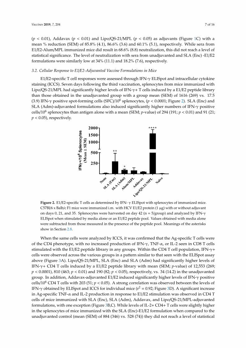
When the same cells were analyzed by ICCS, it was confirmed that the Ag-specific T cells were of the CD4 phenotype, with no increased production of IFN-γ, TNF-α, or IL-2 seen in CD8 T cells stimulated with the E1/E2 peptide library in any groups. Within the CD4 T cell population, IFN-γ+ cells were observed across the various groups in a pattern similar to that seen with the ELISpot assay above (Figure 3A). Lipo/QS-21/MPL, SLA (Enc) and SLA (Adm) had significantly higher levels of IFN-γ+ CD4 T cells induced by a E1/E2 peptide library with mean (SEM; p-value) of 12,553 (269; p < 0.0001), 810 (463; p < 0.01) and 190 (82; p < 0.05), respectively, vs. 34 (14.2) in the unadjuvanted group. In addition, Addavax-adjuvanted E1/E2 induced significantly higher levels of IFN-γ positive cells/106 CD4 T cells with 203 (51; p < 0.05). A strong correlation was observed between the levels of IFN-γ obtained by ELISpot and ICCS for individual mice (r2 = 0.92; Figure 3D). A significant increase in Ag-specific TNF-α and IL-2 production in response to E1/E2 stimulation was observed in CD4 T cells of mice immunized with SLA (Enc), SLA (Adm), Addavax, and Lipo/QS-21/MPL-adjuvanted formulations, with one exception (Figure 3B,C). While levels of IL-2+ CD4+ T cells were slightly higher in the splenocytes of mice immunized with the SLA (Enc)-E1/E2 formulation when compared to the unadjuvanted control (mean (SEM) of 884 (346) vs. 528 (74)) they did not reach a level of statistical significance. A large proportion of the antigen-specific CD4 cells were positive for multiple cytokines (i.e., 22%–72%; Figure 3E). The percentage of cytokine-positive CD4 T cells expressing more
Figure 2. E1/E2-specific T cells as determined by IFN- γ ELISpot with splenocytes of immunized mice.C57Bl/6 x Balb/c F1 mice were immunized i.m. with HCV E1/E2 protein (1 µg) with or without adjuvanton days 0, 21, and 35. Splenocytes were harvested on day 42 (n = 5/group) and analyzed by IFN-γELISpot when stimulated by media alone or an E1/E2 peptide pool. Values obtained with media alonewere subtracted from those measured in the presence of the peptide pool. Meanings of the asterisksshow in Section 2.8.
When the same cells were analyzed by ICCS, it was confirmed that the Ag-specific T cells wereof the CD4 phenotype, with no increased production of IFN-γ, TNF-α, or IL-2 seen in CD8 T cellsstimulated with the E1/E2 peptide library in any groups. Within the CD4 T cell population, IFN-γ+
cells were observed across the various groups in a pattern similar to that seen with the ELISpot assayabove (Figure 3A). Lipo/QS-21/MPL, SLA (Enc) and SLA (Adm) had significantly higher levels ofIFN-γ+ CD4 T cells induced by a E1/E2 peptide library with mean (SEM; p-value) of 12,553 (269;p < 0.0001), 810 (463; p < 0.01) and 190 (82; p < 0.05), respectively, vs. 34 (14.2) in the unadjuvantedgroup. In addition, Addavax-adjuvanted E1/E2 induced significantly higher levels of IFN-γ positivecells/106 CD4 T cells with 203 (51; p < 0.05). A strong correlation was observed between the levels ofIFN-γ obtained by ELISpot and ICCS for individual mice (r2 = 0.92; Figure 3D). A significant increasein Ag-specific TNF-α and IL-2 production in response to E1/E2 stimulation was observed in CD4 Tcells of mice immunized with SLA (Enc), SLA (Adm), Addavax, and Lipo/QS-21/MPL-adjuvantedformulations, with one exception (Figure 3B,C). While levels of IL-2+ CD4+ T cells were slightly higherin the splenocytes of mice immunized with the SLA (Enc)-E1/E2 formulation when compared to theunadjuvanted control (mean (SEM) of 884 (346) vs. 528 (74)) they did not reach a level of statistical
Vaccines 2019, 7, 204 8 of 16
significance. A large proportion of the antigen-specific CD4 cells were positive for multiple cytokines(i.e., 22–72%; Figure 3E). The percentage of cytokine-positive CD4 T cells expressing more than onecytokine in mice immunized with SLA (Enc), SLA (Adm), Addavax, and Lipo/QS-21/MPL -adjuvantedformulations was 42%, 34%, 37%, and 72%, respectively.
Vaccines 2019, 7, x FOR PEER REVIEW 8 of 16
than one cytokine in mice immunized with SLA (Enc), SLA (Adm), Addavax, and Lipo/QS-21/MPL -adjuvanted formulations was 42%, 34%, 37%, and 72%, respectively.
Figure 3. Polyfunctionality and phenotype of E1/E2-specific T cells as determined by intracellular cytokine staining of splenocytes of immunized mice. C57Bl/6 x Balb/c F1 mice were immunized i.m. with HCV E1/E2 protein (1 µg) with or without adjuvant on days 0, 21, and 35. Splenocytes were harvested on day 42 (n = 5/group) and analyzed by intracellular cytokine staining (ICCS) when stimulated by an E1/E2 peptide pool or media alone. The number of E1/E2-specific IFN-γ+ (Panel A), TNF-α+ (Panel B), and IL-2+ (Panel C) per million CD4 T cells were determined. Values obtained with media alone were subtracted from those measured in the presence of the peptide pool. Correlation of IFN-γ responses as determined by ELISpot and ICCS was also performed (Panel D). The frequency of cells expressing IFN-γ, TNF-α, and IL-2 alone or in combination is displayed (geomean per group) with the total number of cytokine-positive cells per million CD4+ T cells indicated below the pie chart (Panel E).
Figure 3. Polyfunctionality and phenotype of E1/E2-specific T cells as determined by intracellularcytokine staining of splenocytes of immunized mice. C57Bl/6 x Balb/c F1 mice were immunizedi.m. with HCV E1/E2 protein (1 µg) with or without adjuvant on days 0, 21, and 35. Splenocyteswere harvested on day 42 (n = 5/group) and analyzed by intracellular cytokine staining (ICCS) whenstimulated by an E1/E2 peptide pool or media alone. The number of E1/E2-specific IFN-γ+ (A), TNF-α+
(B), and IL-2+ (C) per million CD4 T cells were determined. Values obtained with media alone weresubtracted from those measured in the presence of the peptide pool. Correlation of IFN-γ responses asdetermined by ELISpot and ICCS was also performed (D). The frequency of cells expressing IFN-γ,TNF-α, and IL-2 alone or in combination is displayed (geomean per group) with the total number ofcytokine-positive cells per million CD4+ T cells indicated below the pie chart (E). Meanings of theasterisks show in Section 2.8.

Vaccines 2019, 7, 204 9 of 16
3.3. Longevity of E1/E2-Specific Humoral and Cellular Immune Responses in Mice
The longevity of immune responses generated by E1/E2-based vaccines was also assessed in miceto confirm the ability of the different adjuvants to generate long-lasting antigen-specific humoral andcellular immune responses. Mice were maintained for ~6 months post-last vaccination, and sera werecollected at multiple timepoints (i.e., days 121, 219, and 224) to track anti-E1/E2 antibody titers over time.On day 220, mice received a low 0.2 µg dose of E1/E2 to assist in the evaluation of the antigen-specificrecall response in the mice. The E1/E2-specific IgG titers did decrease over time with a 2 to 15-folddecrease in GMT from days 42 to 219 seen across the various groups (Figure 4A). The largest folddecrease was seen in animals immunized with the Alum/MPL-adjuvanted formulation (i.e., 15-fold),although it did not reach a level of statistical significance (p = 0.1316). Over the same timepoints,statistically significant decreases were seen with animals immunized with E1/E2 adjuvanted with eitherSLA (Adm) (4-fold, p < 0.05) and Lipo/QS-21/MPL (3-fold, p < 0.05). Interestingly, the administration ofthe low dose E1/E2 antigen on day 220 led to a significant rise in antibody titers in animals previouslyvaccinated with E1/E2 adjuvanted with SLA (Adm), Addavax or Lipo/QS-21/MPL with 2-fold (p < 0.01),3-fold (p < 0.05) and 2-fold (p < 0.0001) increases in antibody GMT seen between days 219 and 224,respectively. Animals in the other study groups did not have statistically significant different antibodytiters between these timepoints.

Vaccines 2019, 7, 204 10 of 16Vaccines 2019, 7, x FOR PEER REVIEW 10 of 16
Figure 4. Longevity of anti-E1/E2 IgG titers and HCV neutralization activity in serum of immunized mice. C57Bl/6 x Balb/c F1 mice were immunized i.m. with HCV E1/E2 protein (1 µg) with or without adjuvant on days 0, 21, and 35. Sera were obtained from mice (n = 5/group) at multiple timepoints (i.e., days 20, 42, 121, 219, and 224) to track anti-E1/E2 antibody titers over time. On day 220, mice were injected i.m. with 0.2 µg of E1/E2 without adjuvant to look at the antigen-specific recall response. Anti-E1/E2 IgG titers over the course of the study (Panel A) and specifically on Day 219 (Panel B) were determined by ELISA. Grouped data is presented as GMT + 95% CI. Serum from Day 219 was also tested for its ability to neutralize HCVpp entry into Huh7.5 liver cells in vitro (Panel C).
Figure 4. Longevity of anti-E1/E2 IgG titers and HCV neutralization activity in serum of immunizedmice. C57Bl/6 x Balb/c F1 mice were immunized i.m. with HCV E1/E2 protein (1 µg) with or withoutadjuvant on days 0, 21, and 35. Sera were obtained from mice (n = 5/group) at multiple timepoints(i.e., days 20, 42, 121, 219, and 224) to track anti-E1/E2 antibody titers over time. On day 220, micewere injected i.m. with 0.2 µg of E1/E2 without adjuvant to look at the antigen-specific recall response.Anti-E1/E2 IgG titers over the course of the study (A) and specifically on Day 219 (B) were determinedby ELISA. Grouped data is presented as GMT + 95% CI. Serum from Day 219 was also tested for itsability to neutralize HCVpp entry into Huh7.5 liver cells in vitro (C). Meanings of the asterisks show inSection 2.8.

Vaccines 2019, 7, 204 11 of 16
Importantly, antibodies in the serum of mice 6 months following last vaccination (i.e., from day219) were still significantly higher in mice receiving E1/E2 adjuvanted with SLA (Adm), Addavax, orLipo/QS-21/MPL vs. E1/E2 alone (Figure 4B; p < 0.001). In addition, serum from mice immunized withthe SLA (Adm) and Addavax -adjuvanted formulations were capable of significantly neutralizing HCVpseudoparticles in vitro, with a mean % reduction (SEM) of 94.6% (4.1) and 88.4% (4.7), respectively,compared to 21% (8.4) with the unadjuvanted formulation (Figure 4C; p < 0.05).
To confirm the longevity of the E1/E2-specific T cell responses, splenocytes collected on day224 were stimulated with E1/E2-derived peptide pool and IFN-γ production measured by ELISpot.As on day 42, mice immunized with Lipo/QS-21/MPL had the highest levels of IFN-γ+ SFC/106
splenocytes with group mean (SEM) of 242.8 (38.8) vs. 9.8 (3.1) obtained in the unadjuvanted group,(p < 0.0001; Figure 5). SLA (Enc), SLA (Adm), and Addavax-adjuvanted formulations also inducedsignificantly higher numbers of IFN-γ positive cells/106 splenocytes than immunization with antigenalone with group mean (SEM; p-value) of 49.5 (7.7; p < 0.0001), 30.3 (5.1; p < 0.01) and 22.3 (3.7;p < 0.05), respectively.
Vaccines 2019, 7, x FOR PEER REVIEW 11 of 16
Importantly, antibodies in the serum of mice 6 months following last vaccination (i.e., from day 219) were still significantly higher in mice receiving E1/E2 adjuvanted with SLA (Adm), Addavax, or Lipo/QS-21/MPL vs. E1/E2 alone (Figure 4B; p < 0.001). In addition, serum from mice immunized with the SLA (Adm) and Addavax -adjuvanted formulations were capable of significantly neutralizing HCV pseudoparticles in vitro, with a mean % reduction (SEM) of 94.6% (4.1) and 88.4% (4.7), respectively, compared to 21% (8.4) with the unadjuvanted formulation (Figure 4C; p < 0.05).
To confirm the longevity of the E1/E2-specific T cell responses, splenocytes collected on day 224 were stimulated with E1/E2-derived peptide pool and IFN-γ production measured by ELISpot. As on day 42, mice immunized with Lipo/QS-21/MPL had the highest levels of IFN-γ+ SFC/106 splenocytes with group mean (SEM) of 242.8 (38.8) vs. 9.8 (3.1) obtained in the unadjuvanted group, (p < 0.0001; Figure 5). SLA (Enc), SLA (Adm), and Addavax-adjuvanted formulations also induced significantly higher numbers of IFN-γ positive cells/106 splenocytes than immunization with antigen alone with group mean (SEM; p-value) of 49.5 (7.7; p < 0.0001), 30.3 (5.1; p < 0.01) and 22.3 (3.7; p < 0.05), respectively.
Figure 5. Longevity of E1/E2-specific T cell responses in splenocytes of immunized mice. C57Bl/6 x Balb/c F1 mice were immunized i.m. with HCV E1/E2 protein (1 µg) with or without adjuvant on days 0, 21, and 35. On day 220, mice were injected i.m. with 0.2 µg of E1/E2 without adjuvant. Splenocytes were harvested on Day 224 (n = 5/group) and analyzed by IFN-γ ELISpot when stimulated by media alone or an E1/E2 peptide pool. Values obtained with media alone were subtracted from those measured in the presence of the peptide pool.
4. Discussion
The HCV E1/E2 heterodimer plays an essential role in virus entry, with E2 binding directly to the receptor CD81 on the surface of the host cell. While HCV-neutralizing antibodies have been shown to bind epitopes on E1 or E2, the majority of HCV-neutralizing antibodies in humans target E2, interrupting its interaction with its cell surface receptor [28]. E1/E2-based vaccines have been shown in a preclinical setting to induce robust immune responses with generated antibodies capable of neutralizing HCV in vitro and preventing infection in non-human primate models in vivo [5]. E1/E2 antigen derived from a single 1a genotype combined with oil-in-water-based adjuvants remains the only HCV vaccine candidate shown to significantly reduce the incidence of chronic persistent infection in the chimpanzee model after prophylactic immunization and experimental challenge using either homologous or heterologous 1a virus [8,9]. As with other subunit vaccines, a clinically efficacious E1/E2 vaccine formulation would likely require inclusion of an adjuvant capable of inducing strong and long-lived antigen-specific immune responses. In Phase I studies, a MF59®-adjuvanted E1/E2 vaccine formulation was well tolerated and induced antigen-specific lymphoproliferative and cross-neutralizing antibody responses in healthy human subjects [6,7,29].
Figure 5. Longevity of E1/E2-specific T cell responses in splenocytes of immunized mice. C57Bl/6x Balb/c F1 mice were immunized i.m. with HCV E1/E2 protein (1 µg) with or without adjuvanton days 0, 21, and 35. On day 220, mice were injected i.m. with 0.2 µg of E1/E2 without adjuvant.Splenocytes were harvested on Day 224 (n = 5/group) and analyzed by IFN-γ ELISpot when stimulatedby media alone or an E1/E2 peptide pool. Values obtained with media alone were subtracted fromthose measured in the presence of the peptide pool. Meanings of the asterisks show in Section 2.8.
4. Discussion
The HCV E1/E2 heterodimer plays an essential role in virus entry, with E2 binding directly tothe receptor CD81 on the surface of the host cell. While HCV-neutralizing antibodies have beenshown to bind epitopes on E1 or E2, the majority of HCV-neutralizing antibodies in humans targetE2, interrupting its interaction with its cell surface receptor [28]. E1/E2-based vaccines have beenshown in a preclinical setting to induce robust immune responses with generated antibodies capableof neutralizing HCV in vitro and preventing infection in non-human primate models in vivo [5].E1/E2 antigen derived from a single 1a genotype combined with oil-in-water-based adjuvants remainsthe only HCV vaccine candidate shown to significantly reduce the incidence of chronic persistentinfection in the chimpanzee model after prophylactic immunization and experimental challengeusing either homologous or heterologous 1a virus [8,9]. As with other subunit vaccines, a clinicallyefficacious E1/E2 vaccine formulation would likely require inclusion of an adjuvant capable of inducingstrong and long-lived antigen-specific immune responses. In Phase I studies, a MF59®-adjuvantedE1/E2 vaccine formulation was well tolerated and induced antigen-specific lymphoproliferative andcross-neutralizing antibody responses in healthy human subjects [6,7,29]. The ability of this vaccineformulation to protect against chronic HCV infection has yet to be determined. Likewise, it is unknown

Vaccines 2019, 7, 204 12 of 16
whether superior immune responses would be obtained with other commercially approved adjuvantssuch as AS01™ and AS04™ if incorporated in an E1/E2-based vaccine. SLA-based adjuvants enhancethe generation of antigen-specific immune responses through increased local cytokine production,immune cell trafficking, and antigen uptake at the injection site, leading to increased protection inmurine models of infectious disease (e.g., influenza virus) and cancer [10,14,19,20,30]. In previousstudies, an encapsulated SLA-based formulation was able to enhance E1/E2-specific cellular responsesbut did not significantly enhance antibody-mediated HCVpp neutralization [13]. Therefore, in thisstudy we compared the ability of both the SLA (Enc) and SLA (Adm) formulations to enhanceimmune responses to E1/E2 with mimetics of three adjuvants currently used in approved vaccines,namely the squalene-based oil-in-water emulsion Addavax (a mimetic of MF59®), the TLR4 agonistMPL combined with aluminum salts (a mimetic of AS04™), and a liposome formulation containingMPL/QS-21 (a mimetic of AS01™). These adjuvants were selected as they are mimetics of adjuvantsapproved for human use and cover a range of different mechanisms of action. In addition, we sought tocompare the longevity of the immune responses generated by these formulations, which has not beenpreviously assessed for experimental vaccines containing SLA-based archaeosomes. Previously, thelevels of antigen-specific antibodies and cellular responses were assessed within a few weeks of finalimmunization [10]. Long-lived immune responses are critical in the context of prophylactic vaccineswhere exposure to an infectious agent may occur years post-vaccination.
Multiple novel adjuvants have been approved for clinical use in the past 20 years. MF59® has beenlicensed for use in Europe since 1997 and in the United States since 2015 as part of the influenza vaccine,Fluad® [23]. The AS04™ adjuvant system, containing an endotoxin-derived TLR4 agonist, MPL, alongwith aluminum salt, is currently formulated in marketed vaccines such as Fendrix® and Cervarix®
approved for the prevention of infections by pneumococcal bacteria and human papillomavirus,respectively [24]. Most recently, AS01™, containing MPL along with the strongly immunostimulatorysaponin QS-21 isolated from the bark of the Quillaja saponaria tree, was approved as part of thevaricella-zoster vaccine, Shingrix [31]. While these vaccine adjuvants are approved, they may not besuitable for all indications, as different types/magnitudes of immune response may be necessary fora specific vaccine. In addition, due to their proprietary nature, access to these adjuvants is largelyrestricted to the pharmaceutical companies that developed them. Our results demonstrate that SLAarchaeosome formulations, in particular, the SLA (Adm), along with the mimetics of MF59® andAS01™were capable of inducing long-lived humoral and cellular E1/E2-specific immune responses.The humoral response (E1/E2-specific IgG titers and HCVpp neutralization) induced by formulationscontaining these three adjuvants was largely similar in most of the assessed readouts. As previouslyreported by Landi et al. [13], when compared to the antigen alone, an archaeosome formulation with anencapsulated antigen induced significantly higher titers of antigen-specific IgG antibodies, but levelsof HCVpp neutralization were not significantly different between the two groups. Administrationof a low antigen dose 6 months following last vaccination induced a significant and rapid increasein antibody titers in the groups immunized with vaccine formulations adjuvanted with SLA (Adm),Addavax or Lipo/QS-21/MPL, indicative of a functional memory response. As the levels of memorycells were not directly measured in this study, future studies could address this by looking at theimpact of various adjuvants on memory cell formation. Although we did not evaluate the ability of theantigen-specific antibodies generated in this study to cross-neutralize HCVpp coated with the E1/E2proteins from other HCV genotypes, human subjects immunized with the same E1/E2 antigens didgenerate antibodies capable of neutralizing multiple strains of HCV [7].
While antibodies would be the main mediators of HCV viral particle neutralization, a robustantigen-specific CD4 T cell response could contribute to the quality and longevity of the humoralresponse. In addition, cytotoxic CD4 and CD8 T cells targeting E1/E2 would facilitate the clearanceof virally-infected cells in infected individuals. Both SLA formulations, along with Lipo/QS-21/MPLwere able to significantly enhance the levels of antigen-specific T cells when compared to antigenalone as determined by IFN-γ ELISpot. When evaluated by ICCS, it appears that none of the vaccine

Vaccines 2019, 7, 204 13 of 16
formulations were able to induce antigen-specific CD8 T cells as the cytokine secretion in response tothe E1/E2 peptide pool was restricted to T cells of the CD4 phenotype. This was not unexpected, asprevious reports have shown that T cell responses to E1/E2 recombinant antigen, when combined withvarious adjuvants, were mainly CD4-specific, but that expression of the E1/E2 from a viral-based DNAvector could induce antigen-specific CD8 T cells [13,32]. SLA archaeosomes have been shown to bestrong and consistent inducers of CD8 T cell responses to model antigens such as OVA and HBsAg.The lack of CD8 responses in our model may be due to the nature of the antigen platform and/or thelack of processed E1/E2 epitopes with high binding affinity to the MHC molecules expressed in themouse model used. In contrast, strong CD4 T cell responses were seen with many of the adjuvantedvaccine formulations. These responses were polyfunctional with the antigen-specific expression ofIFN-γ, TNF-α, and IL-2 seen in the CD4 T cells of vaccinated mice. Alum/MPL was the weakestinducer of T cell responses with no significant differences seen in any of the cellular immune readoutswhen compared to antigen alone. The AS01™mimetic, Lipo/QS-21/MPL, was the strongest inducerof E1/E2-specific CD4 T cells in our system. This agrees with the known profile of AS01™, whichhas been shown to robustly induce CD4 T cells to malaria and varicella-zoster antigens in preclinicaland clinical studies [31]. The SLA (Enc)-adjuvanted formulation did induce strong CD4-mediatedexpression of IFN-γ and TNF-α, as previously reported [13]. Meanwhile, the SLA (Adm) formulationinduced a significant increase in antigen-specific IL-2 production in addition to IFN-γ and TNF-α.Increased polyfunctionality of T cells is associated with more efficacious immune responses to certainpathogens [33]. As such, the use of SLA (Adm) instead of SLA (Enc) may offer a further advantage inaddition to the superior humoral responses discussed above. The observed differences in the immuneresponse between the two formulations may be due to the SLA lipid dose and/or antigen context (i.e.,free protein vs. incorporated within the liposome). It would be of interest in future studies to evaluatethese factors more closely to determine if they are contributing to the differences in activity of thesetwo archaeosome formulations. In addition, mice immunized with the Lipo/QS-21/MPL-adjuvantedformulation did induce a large number of antigen-specific CD4 T cells shortly after immunization,but these were largely short-lived (1616 vs. 243 SFC/106 splenocytes at 1 week and 6 months postlast vaccination, respectively). A decrease over the same two timepoints was also seen with micereceiving the SLA (Adm)-adjuvanted formulation, but the decrease was ~3-fold (from 91 to 30 SFC/106
splenocytes). We are currently evaluating combinations of other immunostimulatory molecules withdiffering mechanisms of action (such as TLR agonists and/or saponins) with the SLA (Adm) formulationas this approach has been shown to play a role in the immunogenicity of AS01™ [31]. As cytotoxicCD4 T cells have been shown to target multiple viral pathogens [34], and the tested adjuvanted vaccineformulations (especially Lipo/QS-21/MPL) induced E1/E2-specific CD4 T cells, it is possible that thesevaccines could induce both cytotoxic and humoral immune responses targeting HCV despite thelack of a detectable E1/E2-specific CD8 responses in our system. It would be of interest in futurestudies to determine if the SLA formulation could be adapted to adjuvant responses generated withnucleic acid-based vaccine vectors to increase the number of antigen-specific CD8 T cells to HCV E1/E2.In addition, future studies could compare responses using an SLA-adjuvanted protein vaccine withthose induced by an E1/E2 expressing viral-based DNA vaccine.
In summary, we have previously demonstrated that SLA archaeosomes are capable of inducingstrong humoral and cellular immune responses against multiple antigens [10,19,20]. We have alsoshown that they are well-tolerated with a favorable safety profile when administered intramuscularlyin vivo and can stimulate strong local cytokine secretion, immune cell recruitment, and antigen uptakeat the vaccination site [14]. Herein, we have shown that while the E1/E2 antigen was immunogenic whenadministered alone, the magnitude and longevity of the antigen-specific responses were significantlyenhanced by the inclusion of certain adjuvants. SLA glycolipids, particularly the simple admixedSLA formulation, can induce potent and long-lived anti-HCV E1/E2 responses in mice. In addition,these responses were equivalent or better to those obtained with various adjuvants (except for cellularresponses vs. Lipo/QS-21/MPL). In addition, the overall cost of preparing the SLA admixed formulation

Vaccines 2019, 7, 204 14 of 16
is much reduced when compared to the encapsulated formulation as there is no antigen loss vs.60–95% antigen loss when preparing the antigen-encapsulated SLA archaeosomes. Due to the limitedavailability of commercial adjuvants and its strong immunostimulatory profile, further developmentand characterization of the robust SLA adjuvant system with HCV E1/E2 is warranted.
Supplementary Materials: The following are available online at http://www.mdpi.com/2076-393X/7/4/204/s1,Figure S1: Gating strategy for intracellular cytokine staining data.
Author Contributions: Conceptualization, B.A., L.K., M.H. and M.J.M.; methodology, B.A., L.D., B.A.H., R.D.,F.C.S., Y.J., A.L., J.L.M.L., M.L., D.H., J.K.; supervision, B.A., M.J.M., D.L.T.; formal analysis, B.A.; writing—originaldraft preparation, B.A. and M.J.M.; writing—review and editing, B.A., L.D., B.A.H., R.D., F.C.S., Y.J., A.L., J.L.M.L.,M.L., D.H., J.K., D.L.T., L.K., M.H., and M.J.M.
Funding: This research was funded with a grant from Alberta Innovates Health Solutions (Reference number201201140).
Acknowledgments: The authors would like to acknowledge John Shelvey and Perry Fleming for producing thearchaeal biomass and Dean Williams, Janelle Sauvageau and Mohammad P. Jamshidi for the synthesis of SLA.
Conflicts of Interest: Lakshmi Krishnan is an inventor on various archaeosome patents and patent applications.
References
1. World Health Organization. Available online: https://www.who.int/news-room/fact-sheets/detail/hepatitis-c(accessed on 9 July 2019).
2. Zibbell, J.E.; Asher, A.K.; Patel, R.C.; Kupronis, B.; Iqbal, K.; Ward, J.W.; Holtzman, D. Increases in AcuteHepatitis C Virus Infection Related to a Growing Opioid Epidemic and Associated Injection Drug Use,United States, 2004 to 2014. Am. J. Public Health 2018, 108, 175–181. [CrossRef] [PubMed]
3. Bourliere, M.; Gordon, S.C.; Flamm, S.L.; Cooper, C.L.; Ramji, A.; Tong, M.; Ravendhran, N.; Vierling, J.M.;Tran, T.T.; Pianko, S.; et al. Sofosbuvir, Velpatasvir, and Voxilaprevir for Previously Treated HCV Infection.N. Engl. J. Med. 2017, 376, 2134–2146. [CrossRef] [PubMed]
4. Liang, T.J. Current progress in development of hepatitis C virus vaccines. Nat. Med. 2013, 19, 869–878.[CrossRef] [PubMed]
5. Choo, Q.L.; Kuo, G.; Ralston, R.; Weiner, A.; Chien, D.; Van, N.G.; Han, J.; Berger, K.; Thudium, K.; Kuo, C.Vaccination of chimpanzees against infection by the hepatitis C virus. Proc. Natl. Acad. Sci. USA 1994,91, 1294–1298. [CrossRef] [PubMed]
6. Frey, S.E.; Houghton, M.; Coates, S.; Abrignani, S.; Chien, D.; Rosa, D.; Pileri, P.; Ray, R.; Di Bisceglie, A.M.;Rinella, P.; et al. Safety and immunogenicity of HCV E1E2 vaccine adjuvanted with MF59 administered tohealthy adults. Vaccine 2010, 28, 6367–6373. [CrossRef]
7. Law, J.L.; Chen, C.; Wong, J.; Hockman, D.; Santer, D.M.; Frey, S.E.; Belshe, R.B.; Wakita, T.; Bukh, J.; Jones, C.T.;et al. A hepatitis C virus (HCV) vaccine comprising envelope glycoproteins gpE1/gpE2 derived from a singleisolate elicits broad cross-genotype neutralizing antibodies in humans. PLoS ONE 2013, 8, e59776. [CrossRef]
8. Houghton, M.; Abrignani, S. Prospects for a vaccine against the hepatitis C virus. Nature 2005, 436, 961–966.[CrossRef]
9. Houghton, M. Prospects for prophylactic and therapeutic vaccines against the hepatitis C viruses.Immunol. Rev. 2011, 239, 99–108. [CrossRef]
10. Akache, B.; Stark, F.C.; Jia, Y.; Deschatelets, L.; Dudani, R.; Harrison, B.A.; Agbayani, G.; Williams, D.;Jamshidi, M.P.; Krishnan, L.; et al. Sulfated archaeol glycolipids: Comparison with other immunologicaladjuvants in mice. PLoS ONE 2018, 13, e0208067. [CrossRef]
11. Conlan, J.W.; Krishnan, L.; Willick, G.E.; Patel, G.B.; Sprott, G.D. Immunization of mice with lipopeptideantigens encapsulated in novel liposomes prepared from the polar lipids of various Archaeobacteria elicitsrapid and prolonged specific protective immunity against infection with the facultative intracellular pathogen,Listeria monocytogenes. Vaccine 2001, 19, 3509–3517.
12. Krishnan, L.; Deschatelets, L.; Stark, F.C.; Gurnani, K.; Sprott, G.D. Archaeosome adjuvant overcomes toleranceto tumor-associated melanoma antigens inducing protective CD8 T cell responses. Clin. Dev. Immunol. 2010,2010, 578432. [CrossRef]

Vaccines 2019, 7, 204 15 of 16
13. Landi, A.; Law, J.; Hockman, D.; Logan, M.; Crawford, K.; Chen, C.; Kundu, J.; Ebensen, T.; Guzman, C.A.;Deschatelets, L.; et al. Superior immunogenicity of HCV envelope glycoproteins when adjuvanted withcyclic-di-AMP, a STING activator or archaeosomes. Vaccine 2017, 35, 6949–6956. [CrossRef]
14. Akache, B.; Stark, F.C.; Iqbal, U.; Chen, W.; Jia, Y.; Krishnan, L.; McCluskie, M.J. Safety and biodistribution ofsulfated archaeal glycolipid archaeosomes as vaccine adjuvants. Hum. Vaccin Immunother. 2018, 14, 1746–1759.[CrossRef] [PubMed]
15. Gurnani, K.; Kennedy, J.; Sad, S.; Sprott, G.D.; Krishnan, L. Phosphatidylserine receptor-mediated recognitionof archaeosome adjuvant promotes endocytosis and MHC class I cross-presentation of the entrapped antigenby phagosome-to-cytosol transport and classical processing. J. Immunol. 2004, 173, 566–578. [CrossRef]
16. Krishnan, L.; Gurnani, K.; Dicaire, C.J.; van Faassen, H.; Zafer, A.; Kirschning, C.J.; Sad, S.; Sprott, G.D. Rapidclonal expansion and prolonged maintenance of memory CD8+ T cells of the effector (CD44highCD62Llow)and central (CD44highCD62Lhigh) phenotype by an archaeosome adjuvant independent of TLR2. J. Immunol.2007, 178, 2396–2406. [CrossRef]
17. Stark, F.C.; McCluskie, M.J.; Krishnan, L. Homologous Prime-Boost Vaccination with OVA Entrapped inSelf-Adjuvanting Archaeosomes Induces High Numbers of OVA-Specific CD8(+) T Cells that Protect AgainstSubcutaneous B16-OVA Melanoma. Vaccines 2016, 4, 44. [CrossRef]
18. Whitfield, D.M.; Sprott, G.D.; Krishnan, L. Sulfated-Glycolipids as Adjuvants for Vaccines. U.S. PatentApplication WO 2016/004512 A1, 14 January 2016.
19. McCluskie, M.J.; Deschatelets, L.; Krishnan, L. Sulfated archaeal glycolipid archaeosomes as a safe andeffective vaccine adjuvant for induction of cell-mediated immunity. Hum. Vaccin Immunother. 2017,13, 2772–2779. [CrossRef]
20. Jia, Y.; Akache, B.; Deschatelets, L.; Qian, H.; Dudani, R.; Harrison, B.A.; Stark, F.C.; Chandan, V.; Jamshidi, M.P.;Krishnan, L.; et al. A comparison of the immune responses induced by antigens in three differentarchaeosome-based vaccine formulations. Int. J. Pharm. 2019, 561, 187–196. [CrossRef]
21. Krishnan, L.; Dicaire, C.J.; Patel, G.B.; Sprott, G.D. Archaeosome vaccine adjuvants induce strong humoral,cell-mediated, and memory responses: Comparison to conventional liposomes and alum. Infect. Immun.2000, 68, 54–63. [CrossRef]
22. Sprott, G.D.; Yeung, A.; Dicaire, C.J.; Yu, S.H.; Whitfield, D.M. Synthetic archaeosome vaccines containingtriglycosylarchaeols can provide additive and long-lasting immune responses that are enhanced byarchaetidylserine. Archaea 2012, 2012, 513231. [CrossRef]
23. Di Pasquale, A.; Preiss, S.; Tavares Da, S.F.; Garcon, N. Vaccine Adjuvants: From 1920 to 2015 and Beyond.Vaccines 2015, 3, 320–343. [CrossRef] [PubMed]
24. Garcon, N.; Di, P.A. From discovery to licensure, the Adjuvant System story. Hum. Vaccin Immunother. 2017,13, 19–33. [CrossRef] [PubMed]
25. James, S.F.; Chahine, E.B.; Sucher, A.J.; Hanna, C. Shingrix: The New Adjuvanted Recombinant HerpesZoster Vaccine. Ann. Pharmacother. 2018, 52, 673–680. [CrossRef] [PubMed]
26. Logan, M.; Law, J.; Wong, J.A.J.; Hockman, D.; Landi, A.; Chen, C.; Crawford, K.; Kundu, J.; Baldwin, L.;Johnson, J.; et al. Native Folding of a Recombinant gpE1/gpE2 Heterodimer Vaccine Antigen from a PrecursorProtein Fused with Fc IgG. J. Virol. 2017, 91, e01552-16. [CrossRef]
27. Vandepapeliere, P. Vaccine Compositions Comprising a Saponin Adjuvant. U.S. Patent Application US2011/0206758 A1, 25 August 2011.
28. Freedman, H.; Logan, M.R.; Law, J.L.; Houghton, M. Structure and Function of the Hepatitis C Virus EnvelopeGlycoproteins E1 and E2: Antiviral and Vaccine Targets. ACS Infect. Dis. 2016, 2, 749–762. [CrossRef]
29. Stamataki, Z.; Coates, S.; Abrignani, S.; Houghton, M.; McKeating, J.A. Immunization of human volunteerswith hepatitis C virus envelope glycoproteins elicits antibodies that cross-neutralize heterologous virusstrains. J. Infect. Dis. 2011, 204, 811–813. [CrossRef]
30. Stark, F.C.; Akache, B.; Ponce, A.; Dudani, R.; Deschatelets, L.; Jia, Y.; Sauvageau, J.; Williams, D.;Jamshidi, M.P.; Agbayani, G.; et al. Archaeal glycolipid adjuvanted vaccines induce strong influenza-specificimmune responses through direct immunization in young and aged mice or through passive maternalimmunization. Vaccine 2019, 37, 7108–7116. [CrossRef]
31. Didierlaurent, A.M.; Laupeze, B.; Di, P.A.; Hergli, N.; Collignon, C.; Garcon, N. Adjuvant system AS01:Helping to overcome the challenges of modern vaccines. Expert Rev. Vaccines 2017, 16, 55–63. [CrossRef]

Vaccines 2019, 7, 204 16 of 16
32. Lin, Y.; Kwon, T.; Polo, J.; Zhu, Y.F.; Coates, S.; Crawford, K.; Dong, C.; Wininger, M.; Hall, J.; Selby, M.; et al.Induction of broad CD4+ and CD8+ T-cell responses and cross-neutralizing antibodies against hepatitis Cvirus by vaccination with Th1-adjuvanted polypeptides followed by defective alphaviral particles expressingenvelope glycoproteins gpE1 and gpE2 and nonstructural proteins 3, 4, and 5. J. Virol. 2008, 82, 7492–7503.
33. Boyd, A.; Almeida, J.R.; Darrah, P.A.; Sauce, D.; Seder, R.A.; Appay, V.; Gorochov, G.; Larsen, M.Pathogen-Specific T Cell Polyfunctionality Is a Correlate of T Cell Efficacy and Immune Protection. PLoS ONE2015, 10, e0128714.
34. Muraro, E.; Merlo, A.; Martorelli, D.; Cangemi, M.; Dalla, S.S.; Dolcetti, R.; Rosato, A. Fighting Viral Infectionsand Virus-Driven Tumors with Cytotoxic CD4(+) T Cells. Front. Immunol. 2017, 8, 197. [CrossRef] [PubMed]
© 2019 by the authors. Licensee MDPI, Basel, Switzerland. This article is an open accessarticle distributed under the terms and conditions of the Creative Commons Attribution(CC BY) license (http://creativecommons.org/licenses/by/4.0/).