Current Drug Repurposing Strategies for Rare ... - Frontiers
Upload
khangminh22Category
view
3download
0
Resveratrol-LoadedTPGS-Resveratrol-Solid LipidNanoparticles for Multidrug-ResistantTherapy of Breast Cancer: In Vivo andIn Vitro StudyWenrui Wang1*, Mengyang Zhou1,2, Yang Xu1, Wei Peng1, Shiwen Zhang1, Rongjie Li 1,Han Zhang1, Hui Zhang1, Shumin Cheng1, Youjing Wang1, Xinyu Wei3, Chengxu Yue1,Qingling Yang3* and Changjie Chen3*
1Anhui Province Key Laboratory of Translational Cancer Research, Department of Biotechnology, Bengbu Medical College,Anhui, China, 2Department of Life Sciences, Anhui Medical University, Anhui, China, 3Department of Biochemistry, School ofLaboratory Medicine Bengbu Medical College, Anhui, China
Multidrug resistance (MDR) is a serious problem during cancer therapy. The purpose of thepresent study was to formulate D-α-Tocopheryl polyethylene glycol 1000 succinate-resveratrol-solid lipid nanoparticles (TPGS-Res-SLNs) to improve its therapeuticefficacy against breast cancer. In this study, the solvent injection method was used toprepare the TPGS-Res-SLNs. It was found that the TPGS-Res-SLNs exhibited zetapotential and drug-loading of −25.6 ± 1.3 mV and 32.4 ± 2.6%, respectively.Therefore, it was evident that the TPGS-Res-SLNs can increase cellular uptake ofchemotherapeutic drugs, induce mitochondrial dysfunction, and augment tumortreatment efficiency by inducing apoptosis. Moreover, it was found that SKBR3/PRcells treated with TPGS-Res-SLNs exhibited significant inhibition of cell migration andinvasion, as compared with free resveratrol. In addition, results from in vivo SKBR3/PRxenograft tumor models revealed that TPGS-Res-SLNs has better efficacy in promotingapoptosis of tumor cells owing to high therapeutic outcomes on tumors when comparedwith the efficacy of free resveratrol. In conclusion, the findings of the present study indicatesignificant potential for use of TPGS-Res-SLNs as an efficient drug delivery vehicle toovercome drug resistance in breast cancer therapy.
Keywords: multidrug resistance, drug delivery, resveratrol, solid lipid nanoparticle, breast cancer
INTRODUCTION
Globally, breast cancer (BC) is the second leading cause of cancer-related deaths in women (Malvezziet al., 2019). The American Cancer Society predicts that, an estimated 281,550 new cases of invasivebreast cancer will be diagnosed, resulting in 43,600 deaths in 2021 (Siegel et al., 2021). Reportsindicate that genetic mutations, endocrine disorders, and a decline in immune function increases therisk of developing breast cancer (Sun et al., 2017; Sui et al., 2015; Rojas and Stuckey, 2016).
Current methods of treatment for this condition include surgery, chemotherapy, radiotherapy, orcombined strategy (Untch et al., 2014; Willers et al., 1997). For instance, use of paclitaxel (PTX) hasbeen recognized as the first-line chemotherapy in treatment of breast cancer. However, the efficacy of
Edited by:Jinbing Xie,
Southeast University, China
Reviewed by:Jiao Wang,
Purdue University, United StatesLing Zhi Wu,
Nanjing University of Posts andTelecommunications, China
*Correspondence:Wenrui Wang
[email protected] Chen
[email protected] Yang
Specialty section:This article was submitted to
Biomaterials,a section of the journal
Frontiers in Bioengineering andBiotechnology
Received: 22 August 2021Accepted: 04 November 2021Published: 07 December 2021
Citation:Wang W, Zhou M, Xu Y, Peng W,Zhang S, Li R, Zhang H, Zhang H,Cheng S, Wang Y, Wei X, Yue C,
Yang Q and Chen C (2021)Resveratrol-Loaded TPGS-
Resveratrol-Solid Lipid Nanoparticlesfor Multidrug-Resistant Therapy of
Breast Cancer: In Vivo and InVitro Study.
Front. Bioeng. Biotechnol. 9:762489.doi: 10.3389/fbioe.2021.762489
Frontiers in Bioengineering and Biotechnology | www.frontiersin.org December 2021 | Volume 9 | Article 7624891
ORIGINAL RESEARCHpublished: 07 December 2021
doi: 10.3389/fbioe.2021.762489
PTX is often limited by multidrug resistance (MDR) (Xu et al.,2016). Therefore, there is need to develop a novel strategy forovercoming this limitation and provide a safe, economically-friendly, and effective therapeutic agent against BC.
The development of MDR poses a major challenge to theexisting conventional chemotherapies against BC. Functionally,an ATP-binding cassette (ABC) transporter family, comprisingP-glycoprotein (P-gp), multidrug resistance-associated protein(MRP) and BC resistance protein (BCRP), utilizes ATP-derivedenergy to pump chemotherapy drugs out of tumor cells andprotect tumor tissues from chemical toxicity (Yuan et al., 2016;Tang et al., 2016). This leads to insufficient intracellular levels ofchemotherapeutic drugs and hence a poor therapeutic efficiencyof the drugs. Effective reversal of P-gp-mediated MDR andmaintaining accumulation of chemotherapeutic drugs in tumortissues is imperative to management of MDR and for successfultreatment of BC.
Over the past decades, considerable attempts have been madeto develop P-gp inhibitors to overcome MDR. Consequently,several inhibitors, such as verapamil and disulfiram, have beenidentified with their co-encapsulation into nanocarriers found toreduce MDR of cancer cells in vitro. However, results from in vivostudies have been unsatisfactory, mainly because of poor aqueoussolubility and low intracellular concentration (Chen et al., 2015;Alamolhodaei et al., 2017; Zhang et al., 2018). Furthermore, MDRinhibitors may have inherent toxicity on the normal cells.
D-α-Tocopheryl polyethylene glycol 1000 succinate (TPGS),as a derivative of natural vitamin E (α-tocopherol). It has beenwidely applied in the food and drug industry as a solubilizer,absorption enhancer, and a vehicle for nanodrug delivery system.The biological and physicochemical properties of the compoundprovide multiple advantages for its applications in drug delivery,including enhancement of drug solubility, improvement of drugpermeation, and selective antitumor activity (Yang et al., 2018;Zhang et al., 2017; Gorain et al., 2018). Notably, TPGS can inhibitactivity of ATP-dependent P-gp and act as a potent excipient forovercoming MDR in tumor tissues.
Nanotechnology has rapidly developed and plays animportant role in anti-tumor, immunology as well asinflammation research (Cho, et al., 2008; Zhu et al., 2018;Zheng et al., 2019). Recently, solid lipid nanoparticles (SLNs)have attracted the most attention among all known nanocarriersin the field of drug delivery. This, owing to several uniquematerial properties of SLNs such as small particle size,biocompatibility, chemical, and mechanical stability as well aseasy functionalization potential (Banerjee et al., 2019; Shen et al.,2019; Wang et al., 2015). In addition, SLNs can modulate releaseof kinetics, improve blood circulation time, and increase overalltherapeutic efficacy of anticancer drugs (Jiang et al., 2016). In thepresent study, resveratrol was successfully loaded into SLNs fortreatment of breast cancer (Wang et al., 2017a). Moreover, someresearch studies have shown that SLNs is a promising platformfor the delivery of therapeutic agents for MDR cancerchemotherapy (Majidinia et al., 2020). Based on theseconsiderations, the current study designed TPGS-functionalized SLNs for loading drugs to overcome MDR inbreast cancer.
In this study, a novel drug delivery system was developed toovercome MDR in BC, based on the findings of our previousresearch. First, TPGS-Res-SLNs was successfully synthesizedusing emulsification and low-temperature solidificationmethods and then characterized them using transmissionelectron microscopy (TEM) and zeta potential detection. Therelease behaviors of TPGS-Res-SLNs in vitro in PBS at differentpH values was then measured. Second, anti-tumor efficacy of thesynthesized compounds in the SKBR3/paclitaxel resistant(SKBR3/PR) cells was explored in vitro. The effect of thesecompounds on cell uptake and cytotoxicity against SKBR3/PRcells as well as anti-tumor efficacy in mice bearing an SKBR3/PRtumor were then evaluated. Further, animal studies were used tovalidate the treatment with TPGS-Res-SLNs, and compare theresulting effects to those of free drug. Finally, the present studyexamined the probable underlying mechanism of MDRinhibition in breast cancer.
MATERIALS AND METHODS
MaterialsResveratrol, D-α-Tocopheryl polyethylene glycol 1000 succinate(TPGS) was obtained from Aladdin Chemicals (Shanghai,China). Stearic acid, lecithin chloroform, and Tween80 wereacquired from Sinopharm Chemical Reagent Co, Ltd.(Shanghai, China). Dulbecco’s Modified Eagle Medium(DMEM), fetal bovine serum (FBS), penicillin G, and trypsin-EDTA were purchased from Thermo Fisher Scientific (MA,United States). Other chemicals used in this study were ofanalytical grade. The primary antibodies used in theexperiment including P-gp, BCRP, N-cadherin, Vimentin,MMP-2, and MMP-9 were purchased from Proteintech(Beverly, MA, United States). Horseradish peroxidase (HRP)which are conjugated secondary antibodies used against rabbitor mouse immunoglobulin were purchased from Santa CruzBiotechnology (Santa Cruz).
Preparation of Res-SLNs andTPGS-Res-SLNsD-α-Tocopheryl polyethylene glycol 1000 succinate-resveratrol-solid lipid nanoparticles (TPGS-Res-SLNs) were prepared usingthe emulsification and low-temperature solidification method. Atotal of 10 ml solution containing resveratrol (0.15 g), stearic acid(0.2 g), lecithin (0.1 g), and TPGS (0.1 g) were prepared inchloroform and then added into 30 ml of H2O containingMyrj52 (0.25 g) under fast stirring. The mixture was stirred at1,000 rpm at 75°C for about 1 h till the total volume reduced to5 ml, 10 ml of cold water was then added and the solution wasstirred at 1,000 rpm for another 2 h at ice bath. To remove bothimpurities and large particles, the formulated TPGS-Res-SLNswere centrifuged at 20,000 rpm (Avanti J25 centrifuge, JA25.50rotor, Beckman) for 1 h to collect the synthesized materials. Theproductive TPGS-Res-SLNs was dried for 24 h at −56°C invacuum. Blank SLNs were developed following the sameprocedure without the addition of resveratrol whereas the
Frontiers in Bioengineering and Biotechnology | www.frontiersin.org December 2021 | Volume 9 | Article 7624892
Wang et al. TPGS-SLNs for Breast Cancer

preparation of Res-SLNs was done using the same protocolwithout the use of TPGS.
Morphological, ζ-potential Characterizationof Res-SLNs and TPGS-Res-SLNsThe Res-SLNs and TPGS-Res-SLNs formulation was initiallyimaged under TEM to investigate its morphology. A drop of adiluted suspension of Res-SLNs and TPGS-Res-SLNsformulation was placed on the carbon-coated copper grid ofTEM to form a thin liquid film. This was followed by stainingthem with phosphotungstic acid solution (2%, w/v) for 5 min.The prepared samples were examined under a transmissionelectron microscope (JEOL, Tokyo, Japan). Theζ-potential ofRes-SLNs and TPGS-Res-SLNs were determined using aMalvern Zeta-size Nano ZS (Malvern Instruments, Malvern,United Kingdom).
Quantifying the Loading Efficiency ofResveratrol by UV-Vis SpectroscopyThe current study also determined the drug loading of Res-SLNsand TPGS-Res-SLNs. Briefly, 500 µl diluted Res-SLNs and TPGS-Res-SLNs formulation was placed into a 0.5 ml centrifugalAmicon® filter tube (Millipore, Carrigtwohill, Ireland) andcentrifuged at 14,000 rpm for 30 min at 4°C using aneppendorf centrifuge (Eppendorf AG, Germany). The loadedSLNs remained in the filter tube and the free resveratrolpassed through the filter. The amount of free resveratrol in thesupernatant was then quantified by UV-vis at 304 nm. The drugloading of SLNs were determined using the following equations:
DL% � Wtotal −Wfree
Wlipids× 100%
Where: Wfree is the amount of resveratrol measured in thesupernatant; Wtotal is the theoretical amount of resveratrol thatwas added;Wlipids is the total mass of lipids in the SLNs or in theTPGS-SLNs.
In Vitro Drug Release KineticsTo increase the solubility of resveratrol, the in vitro release ofresveratrol from TPGS-Res-SLNs was tested using pH 7.4 and pH5.5 buffer solutions [1:1 mixture of water and ethanol (v/v)].Briefly, 5 ml freshly prepared TPGS-Res-SLNs solution(equivalent resveratrol dose of 30 μM) was put into dialysistube and dialyzed against 50 ml of the medium with constantshaking at 37°C. At predetermined time points, 1 ml of thedialysis solution was removed and an equal volume of freshmedium was added. Resveratrol release was determined bymeasuring the absorption intensity at 304 nm using UV-visiblespectrometer.
In Vitro CytotoxicityThe relative cytotoxicity of free resveratrol, Res-SLNs, and TPGS-Res-SLNs were separately evaluated in vitro through CCK-8assay. The SKBR3/PR cells were seeded in 96-well plates at a
density of 5 × 10³ cells per well in 100 μl DMEM and grew for24 h. Subsequently, cells were incubated with free Resveratrol,Res-SLNs, and TPGS-Res-SLNs at different concentrations (0, 20,30, and 40 μM) for 24 or 48 h. Cell viability was measured using aCCK-8 kit (Dojindo Laboratories, Japan) according to themanufacturer’s instructions and absorbance was measured at450 nm wavelength using a spectrophotometer.
Cell UptakeConsidering the no fluorescent property of Resveratrol, C6was chosen as the probe for investigating the cellular uptakebehavior of SKBR3/PR cells (Al-Kassas et al., 2017). The cellswere seeded overnight in a 24-well plate and incubated withfree C6 or C6-loaded-TPGS-SLNs at an equivalentconcentration of C6 (100 ng/ml) in a medium supplementwith 10% FBS. After 0.5- and 4-h incubation, the cells werewashed twice with precooled PBS, fixed using 4%paraformaldehyde, and stained with DAPI dye solution for5 min. Fluorescent images of the cells were analyzed under aTCS SP2 confocal microscope (Leica, Germany) to investigatethe cellular uptake of free C6 and C6-TPGS-SLNs.
P-gp Activity AssayTo investigate the transport activity of P-gp, Rho-123 comprisingfluorescent substrates of P-gp were used (Kumar et al., 2020).Briefly, SKBR3/PR cells were pretreated with Resveratrol, Res-SLNs, and TPGS-Res-SLNs in culture medium in the dark andthen co-treated with Rho-123 (0.5 μg/ml) for 1 h at 37°C. Afteraccumulation of Rho-123, the cells were washed with ice-coldPBS and collected using trypsinization. Intracellular fluorescenceof Rho-123 was measured using a FACS can flow cytometer (BDBiosciences).
Scratch Wound Healing AssayThe wound healing assay was carried out to confirm themigration ability. The SKBR3/PR cells were first seeded insix-well plates and cultured to confluency. A single wound wascreated by scratching the confluent monolayer of the cell usinga 10-μl sterile pipette tip. The cells were then washed twotimes with phosphate buffered saline (PBS) and incubatedwith DMEM supplemented with 1% FBS. Images were thentaken using an inverted microscope at 0 and 24 h.
Transwell Invasion AssayThe invasion assays of transfected SKBR3/PR cells wereperformed using Transwell cell-culture chambers (Corning,United States). The chamber was coated with the admixture ofMatrigel (BD, United States). A total of 3,000 cells were theseeded into upper chamber with 100 μl serum-free medium.On the other hand, the lower chamber was supplemented with750 μl medium containing 10% FBS. A total of 3,000 treatedcells were seeded into the upper chamber with 100 μl serum-free medium and the bottom chamber was supplemented with750 μl medium containing 10% FBS. After 24 h of incubation,these cells were washed, fixed with 4% polyoxymethylene andthen stained with 0.1% crystal violet. The invaded cells were
Frontiers in Bioengineering and Biotechnology | www.frontiersin.org December 2021 | Volume 9 | Article 7624893
Wang et al. TPGS-SLNs for Breast Cancer

then counted and photographed under a microscope (HanrongCompany, Shanghai). Five visual fields were selected and theaverage number was taken.
Cell ApoptosisTo quantitatively analyze apoptosis in SKBR3/PR cells, AnnexinV-FITC/PI double staining was performed in the current study.The SKBR3/PR cells were seeded into a six-wells plate at thedensity of 2×10⁵ per well and cultured for 24 h. The SKBR3/PRcells were later washed with PBS (the culture medium) and wasthen replaced with TPGS-Res-SLNs, Res-SLNs or free resveratrolat a concentration equivalent to 30 μM Res for 24 h at 37°C and5% CO2. The cells with no treatment were used as the control.Thereafter, the cells were washed with cold PBS and harvestedusing 0.25% trypsin (no EDTA). The collected cells were washedtwice with cold PBS and resuspended in 500 μl 1×binding buffer.Five of Annexin V-Fluorescein isothiocyanate (FITC) and 5 μlPropidium Iodide (PI) were then into the resuspended cellsaccording to the manufacture’s protocol. The plate wasincubated in the dark for 30 min at 37°C before it was placedin an ice bath in preparation for flow cytometry analysis (BDBiosciences). Results were analyzed using Flowjo7.6 software.
Cell CycleThe SKBR3/paclitaxel resistant cells were seeded in six-wellsplates at the destiny of 2 × 10⁵ per well for 24 h and thentreated with TPGS-Res-SLNs, Res-SLNs or free Res for 24 h(equivalent dose of resveratrol 30 μM). The cells were thenwashed with cold PBS and harvested after trypsinization with0.25% trypsin (no EDTA). Then, the cells were washed with PBSand fixed in cold 70% ethanol for 24 h. After fixation, the cellswere spun again and washed with cold PBS and sequentiallytreated with RNase (100 μg/ml) and propidium iodide (50 μg/ml)for 20 min at 37°C in the dark. Cell cycle stages were determinedand analyzed using flow cytometry.
Immunofluorescent StainingThe SKBR3/PR cells were grown on surface-treated coverslips in 24well plates (approximately 50,000 cells per well) for 24 h untilbetween 50 and 60% confluence. The cells were later exposed todifferent formulations of Resveratrol, Res-SLNs, andTPGS-Res-SLNs(equivalent dose of free resveratrol 30 μM). After a 12-h period ofdrug treatment, the cells were fixed with 4% paraformaldehyde(Solarbio, Beijing, China) for 18min, permeabilized for 8minwith 0.2% Triton X-100, and blocked for 55min with 1% bullserum albumin (Amresco) at room temperature. The cells wereprobed overnight with a primary antibody against E-Cadherin(Sigma; 1:100 dilution) at 4°C and Alexa Fluor 488-conjugatedgoat anti rabbit IgG (Molecular Probes, Eugene, OR, 1:100) in thedark for 1 h at room temperature. After three further washes, the cellswere counterstained using 4, 6-diamidine-2-phenylindole (Beyotime,1:1000) in the dark for 5min at room temperature. All reagents werediluted in phosphate buffered saline (PBS) with all the steps followedby three-times-5min PBS washing. Images were captured using ZENversion 2012 software (Zeiss, Gottingen, Germany) under a laserscanning confocal microscope LSM 780 (Zeiss).
Western Blot AnalysisThe SKBR3/PR cells (approximately 5 × 10⁵) were seeded insix-well plates (cells per well) and exposed to resveratrol, Res-SLNs, or TPGS-Res-SLNs (equivalent dose of free resveratrol30 μM) for 24 h. The medium was removed and the cellswashed several times using PBS (pH 7.4) in a lysis bufferfor 15 min. After centrifugation at 12,000 rpm, a BCA proteinassay kit (KeyGen Biotech, Nanjing, China) was used tomeasure the total protein concentration of the supernatantwith cell lysates. Then, equal amounts of total protein (30 μg)were analyzed on 10% SDS-polyacrylamide gelelectrophoresis and transferred onto a polyvinylidenefluoride (PVDF) membranes. The samples were blockedwith 5% BSA and incubated overnight with primaryantibodies at 4°C. The blots were incubated withhorseradish peroxidase (HRP)-coupled secondary antibody,and the bands were visualized using ImageQuant LAS4000mini (GE Healthcare Life Science, United States). The primaryantibodies used in the experiment included P-gp, BCRP,N-cadherin, Vimentin, MMP-2, MMP-9, and β-actin(included as a control). HRP-conjugatedsecondary antibodies were purchased from KeyGEN(Nanjing, China).
Xenograft Models in Nude MiceAll animal assay were performed in accordance with theGuidelines for Care and Use of Laboratory Animals ofBengbu medical University and approved by the AnimalEthics Committee of Animal Experiments at Bengbumedical University (Permit No. SYXK Wan 2019081).Female BALB/c nude mice, aged between 5 and 6 weekswere use in this study. They were obtained by theExperimental Animal Center of the Bengbu medicalcollege. The SKBR3/PR cells we suspended in PBS, at aconcentration of 2 × 10⁷/ml. A 10 μl of the cell suspensionwas subcutaneously injected into the dorsal right flank of eachmouse. When tumor volume reached ∼100 mm3, the micewere randomized into four groups as follows (n � 4 pergroup): 1) the control group: where mice wereadministered with a daily dose of 100 μl PBS; 2) Resveratrolgroup: in which mice were treated with 20 mg/kg resveratrol;3) Resveratrol-SLNs group: where mice were treated withResveratrol-SLNs at doses equivalent to 20 mg/kgresveratrol; 4) TPGS-Res-SLNs group: in which mice weretreated with TPGS-Res-SLNs at doses equivalent to 20 mg/kgresveratrol. The tail intravenous injection is a difficulttechnique and it is hard to ensure the accuracy of the dosegiven to mice every time. Therefore, all treatments were givenintraperitoneally (IP) daily for five times, to guarantee theequivalent concentration of resveratrol and improve theaccuracy of this study. The shortest (S) and longest (L)diameters of the mouse tumors were measured andrecorded every after 2 days using a Vernier caliper and thetumor volume (V) was calculated using the following formula:
Tumor volume(V) � L × S2/2
Frontiers in Bioengineering and Biotechnology | www.frontiersin.org December 2021 | Volume 9 | Article 7624894
Wang et al. TPGS-SLNs for Breast Cancer

After 21 days, the mice were anesthetized, the tissues wereexcised, and the major organs (heart, liver, spleen, lung, andkidney) were collected for subsequent analyses.
Immunohistochemical StainingExpression of E-cadherin and N-cadherin was examined byimmunohistochemical (IHC) staining. Briefly, paraffin-embedded tumor tissues were sliced into 3 μm sections,deparaffinized with xylene, rehydrated with alcohol gradientand then rinsed using deionized water. The tissue antigen wasthe retrieved by heating it for about 10 min in Tris-EDTAsolution (pH 8.0), and the sections were blocked in 3% H2O2
solution. The blocked sections were incubated overnight withantibodies against E-cadherin (diluted 1:200) or Vimentin(diluted 1:100) at 4°C, washed three times with PBS, and thenincubated with goat anti-rabbit IgG for 30 min. The sections werestained with freshly-prepared 3, 3′-diaminobenzidine (DAB),observed under the microscope, and pictures taken for analysisusing the image processing software.
In Vivo TUNEL AssayIn the current study, TUNEL assays were performed to detectDNA fragmentation, which is characteristic of apoptotic cells(Tang, et al., 2010). Tumor sections (4 μm) weredeparaffinized in xylene and rehydrated in a series ofgraded ethanol to water. Thereafter, 50 ml of proteinase K(50 mg/ml) in TBS (pH 8.0) was added onto the slides andincubated for 30 min at room temperature. The slides wererinsed three times with PBS and then incubated for 1 h with50 ml tunel cocktail (a mixture of 2 ml enzyme solution and48 ml label solution) at 37°C in a humidified atmosphere in thedark. Finally, 1 μl of 1,000× DAPI stock (0.2 mg/ml) wasallowed to stand in the dark for a few minutes, coveredusing slips and the slides analyzed under CLSM using488 nm excitation and 530 nm emission. The cells showing
green fluorescence were categorized as apoptotic. Cell nucleistaining using DAPI was also performed and themean fluorescence intensity was measured using ImageJsoftware.
Histological ExaminationIn the current study histopathological analysis was performed onorgans and tumor tissues, using hematoxylin and eosin (H&E)staining. The main organs (heart, lung, liver, spleen, and kidney)were harvested and fixed overnight in 4% paraformaldehyde andthen embedded in paraffin. The tissues were cut into 5 µmsections for H&E staining according to the standardprocedures. This was followed by examination of thetissues under a light microscope (Olympus Corporation,Tokyo, Japan).
Statistical AnalysisAll results of the present study were presented as means ±standard deviation (SD). The statistical difference between twogroups was tested using one-way analysis of variance (ANOVA)followed by t-test. The data were considered as statisticallysignificant at p < 0.05 or 0.01.
RESULTS
Characterization of Res-SLNs andTPGS-Res-SLNsGenerally, the Res-SLNs and TPGS-Res-SLNs were successfullyprepared in the present study. The morphology and size of thesecompounds were first characterized through transmission electronmicroscopy (TEM). Both Res- and TPGS-Res-SLNs were sphericalin shape with mean diameters of 169 and 203 nm, respectively(Figures 1A,B). The zeta potential of Res-SLNs and TPGS-Res-SLNswas −27.8 ± 1.6 and −25.6 ± 1.3 mV, respectively (Figures 1C,D).
FIGURE 1 | Characterization and in vitro drug release of TPGS-Res-SLNs. TEM image (scale bar: 500 nm) of (A) Res-SLNs and (B) TPGS-Res-SLNs in thesuspension. The Zeta potentials of (C) Res-SLNs and (D) TPGS-Res-SLNs. (E) In vitro release profiles of Resveratrol from TPGS-Res-SLNs at different pH (pH 7.4 andpH 5.5). Data are expressed as the mean ± S.D. (n � 3).
Frontiers in Bioengineering and Biotechnology | www.frontiersin.org December 2021 | Volume 9 | Article 7624895
Wang et al. TPGS-SLNs for Breast Cancer

The surface charge and size of nanoparticles are importantparameters that affect cellular damage of cancer cells. Theresultant uniform sizes of the Res-SLNs and TPGS-Res-SLNsenables better solubility and bioavailability (Bai et al., 2013).
Zeta potential is a significant factor in maintaining stability ofnanoparticles in suspension through electrostatic repulsionbetween particles (Gao et al., 2011). In this study, zetapotential of the studied particles was negative enough tosustain the stability of TPGS-Res-SLNs dispersed system. The
drug-loading of Res by TPGS-SLNs was 32.4 ± 2.6%. In addition,the release curves of resveratrol from TPGS-Res-SLNs wereinvestigated under different pH conditions ranging from 5.5 to7.4. Results of the current study showed that 30.27% of resveratrolwas released from the TPGS-Res-SLNs at 48 h in PBS buffers (pH7.4), whereas 45.8% of resveratrol in the PBS with pH 5.5(Figure 1E). These results suggested that the TPGS-SLNscould be utilized for controlling the release of resveratrol in apH-sensitive manner.
FIGURE 2 | In vitro cell viability assay using CCK8 reagents. The cell viability of SKBR3/PR cell treated with SLNs, free resveratrol, Res-SLNs and TPGS-Res-SLNsafter 24 h (A) and 48 h (B). Data are presented as the mean ± S.D. (n � 3 wells). The significant difference of the results was analyzed. **P < 0.01, *P < 0.05.
FIGURE 3 | Cellular uptake and P-gp inhibition ability. (A) Confocal laser scanning microscopy (CLSM) images of SKBR3/PR cells after 0.5 and 4 h incubation withthe coumarin-6-loaded-TPGS-SLNs (C6-TPGS-SLNs), respectively. The image was obtained from FITC channel (green), DAPI channel (blue) and the overlay of the twochannels. The C6-TPGS-SLNs were green and the nucleus was stained by DAPI (blue). (B) P-gp inhibition ability of Resveratrol, Res-SLNs and TPGS-Res-SLNsincreased uptake of Rho-123 in SKBR3/PR cells.
Frontiers in Bioengineering and Biotechnology | www.frontiersin.org December 2021 | Volume 9 | Article 7624896
Wang et al. TPGS-SLNs for Breast Cancer
Cytotoxicity of Res-SLNs andTPGS-Res-SLNs on SKBR3/PRTo assess the potential of different drug formulations to inhibitgrowth of SKBR3/PR cells, a CCK8 assay was performed at 24 and48 h. It was found that there was no toxicity caused by thenanocarrier against the SKBR3/PR cells (Figures 2A,B). After24 h of incubation, the TPGS-Res-SLNs showed a slightly highertoxicity than free resveratrol and Res-SLNs to SKBR3/PR cells.After the incubation period, a higher cytotoxicity was recorded inTPGS-Res-SLNs relative to Res-SLNs and free Res. These resultscan be attributed to the fact that free resveratrol have lowsolubility and instability in aqueous solution. The solubilityand stability of resveratrol were enhanced by loading it inSLNs or in TPGS-SLNs and this resulted in prolongedinhibition of proliferation to drug resistant cancer cells.Additionally, the rate of cell survival for TPGS-Res-SLNs was
always lower than that of Res-SLNs and free resveratrol at eachdose level (Figure 2).
Efficient Cellular Uptake of TPGS-Res-SLNsThe uptake of different drug formulations in drug-resistantcell-line SKBR3/PR was also studied under a fluorescencemicroscope using the fluorescence dye, coumarin-6 (C6),and the cellular uptake was compared between C6 loadedTPGS-SLNs and coumarin-6 solution. The C6@TPGS-SLNsexhibited stronger fluorescence intensities compared to C6 after0.5 h of incubation (Figure 3A). Thia suggested that there was arapid internalization of C6@TPGS-SLNs into the cells within theperiod. Prolonging incubation time to 4 h, resulted into theentry of C6 into the SKBR/PR and into a marked increaseintensity of fluorescence. When compared with C6, C6@TPGS-SLNs showed significantly stronger green
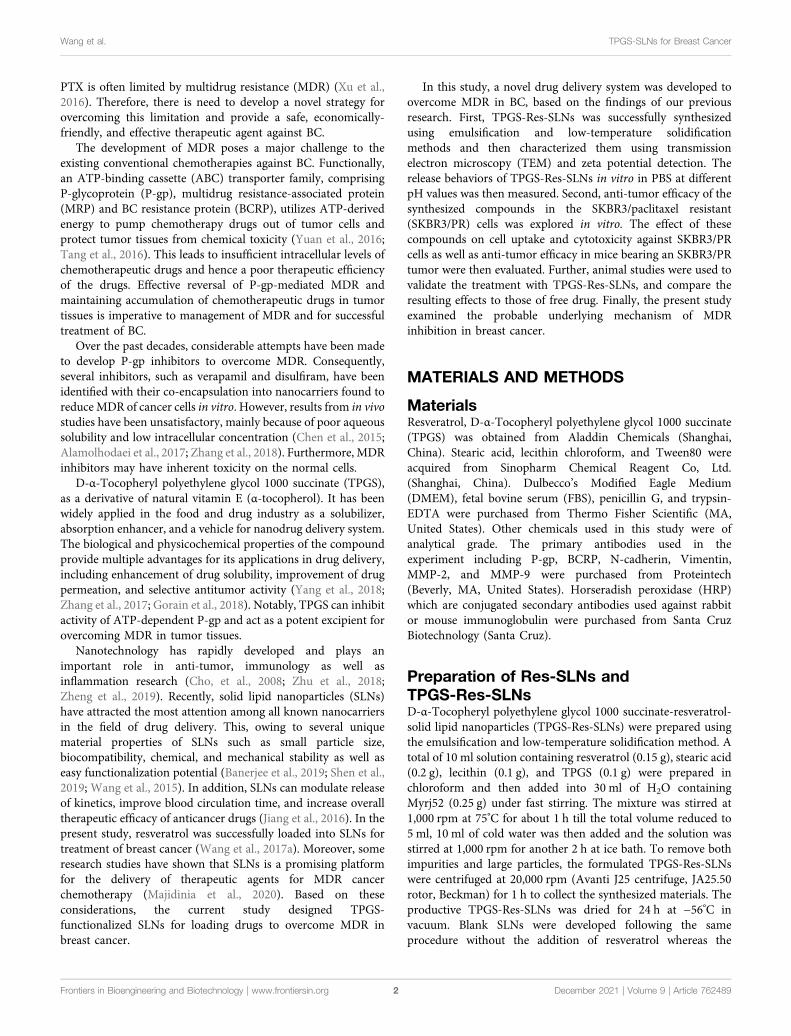
FIGURE 4 | TPGS-Res-SLNs induced apoptosis of SKBR3/PR cells. (A) Assessment of cellular death (apoptosis and necrosis) using Annexin V-FITC/PIstaining after incubation for 24 h. SKBR3/PR cells were incubated with Resveratrol, Res-SLNs and TPGS-Res-SLNs for 24 hours. (B) Flow cytometry analysisof cell cycle phase distribution in SKBR3/PR cells. (C) Histogram showing the cell cycle distribution after treatment with Resveratrol, Res-SLNs and TPGS-Res-SLNs.
Frontiers in Bioengineering and Biotechnology | www.frontiersin.org December 2021 | Volume 9 | Article 7624897
Wang et al. TPGS-SLNs for Breast Cancer

fluorescence after 4 h of incubation. The results of this studyindicated that uptake of C6 loaded TPGS-SLNs and C6solution was time dependent.
P-gp Inhibition Improves IntracellularAccumulationThe P-gp has been recognized as an active efflux transporter andplays an important role in paclitaxel resistance in variouscancers including breast cancer (Che et al., 2019). Toinvestigate the functions of free resveratrol, Res-SLNs, andTPGS-Res-SLNs on P-gp mediated drug efflux, theirinfluence on intracellular cell uptake of Rhodamine-123(Rho123) in SKBR3/PR cells was also assessed in thepresent study.
It was evident that the cells treated with free resveratrolshowed significantly enhanced fluorescence relative to thecontrol group (Figure 3B). On the other hand, the cellstreated with TPGS-Res-SLNs exhibited strongerfluorescence as compared with those treated with Res-SLNsand Resveratrol. These results showed that TPGS significantlyenhanced Rho123 accumulation in SKRR3/PR cells and thisindicates that the inhibition of p-gp by TPGS-Res-SLNs canprevent drug efflux and increase chemosensitivity of SKBR3/PR cells.
Effect of TPGS-Res-SLNs on Cell Cycle andApoptosisTo determine the anticancer effect of free resveratrol, Res-SLNs,and TPGS-Res-SLNs, themethod of AnnexinV-FITC&PIDoubleStaining was used in the present study. A 21.27% of apoptotic cells(early apoptosis 4.27% plus late apoptosis17%) was recorded in thesample incubated with free Resveratrol as compared with theuntreated control group (Figure 4A). It was found that whenthe cells were treated with Res-SLNs, the total apoptotic SKBR3/PRcells increased to 39.80% (7.70 and 32.10% for early and lateapoptosis, respectively). In addition, the percentage of totalapoptotic cells treated with TPGS-Res-SLNs was 57.40%, whichrepresented 45.60 and 11.80% for early and late apoptosis,respectively. These findings suggest that TPGS-Res-SLNs displaythe strongest anti-apoptotic effect in SKBR3/PR cells.
To determine the mechanism of cytotoxicity, the present studyanalyzed progression of the cell cycle andDNA content in SKBR3/PRcells after treatment with free resveratrol, Res-SLNs, and TPGS-Res-SLNs through flow cytometry. Incubating the cells with the threecompounds revealed an increase at theG2/Mphase from9.5 to 13.92,21.66, and 28.43%, respectively (Figures 4B,C). This indicated that allthe three formulations could induce different levels of cell cycle arrestat the G2/M phase. In particular, TPGS-Res-SLNs resulted in themost significant cell cycle arrest in SKBR3/PR cells. Furthermore,these findings were consistent with the results of in vitro cytotoxicity.
FIGURE 5 | TPGS-Res-SLNs inhibited invasion and migration ability of SKBR3/PR cells in vitro (A) Transwell migration assay was used to detect the number oftrans-membrane cells. Representative microphotographs of the Boyden chamber assay of SKBR3/PR cells. (B) The quantitative data for the Boyden chamberassay. The bar graph represents the number of invasive cells present per unit area in different treated groups. (C)Wound-healing assays were performed to assesscell migration. Cells were treated with Resveratrol, Res-SLNs and TPGS-Res-SLNs for 0 and 24 h or untreated. Representative images of treated anduntreated cells are shown. (D) Distance migrated by cells at the 24 h are shown. The values represent the mean ± SD of three independent experiments. (**p < 0.01,*p < 0.05).
Frontiers in Bioengineering and Biotechnology | www.frontiersin.org December 2021 | Volume 9 | Article 7624898
Wang et al. TPGS-SLNs for Breast Cancer

Effect of TPGS-Res-SLNs on Cell Migrationand InvasionTo further investigate whether the TPGS-Res-SLNs treatmenthad a negative effect on cell invasion, a transwell invasion assaywas conducted using matrigel. It was found that there was asignificant reduction in the number of invasive cells when treatedwith TPGS-Res-SLNs for 24 h as compared with when treatedwith Res-SLNs and the free resveratrol (Figures 5A,B).
The effect of TPGS-Res-SLNs on SKBR3/PR migration was alsoinvestigated in the current study using a wound-healing assay(Figures 5C,D). After TPGS-Res-SLNs treatment, the cellmigration distance was notably shorter. This indicate thatresveratrol has a negative effect on cell migration when carriedby the nanoparticles. In addition, it was noted that cellular motilitywas also inhibited after different treatment times (0 and 24 h). Thecell images and quantitative analyses revealed that there were threetimes fewermigratory cells in the TPGS-Res-SLNs-treated group ascompared with those treated with resveratrol group.
Mechanism by TPGS-Res-SLNsPrevents MDRPrevious studies have reported that SKBR3/PR cells showsenhanced migratory ability, which is associated with EMTphenotype (Smith and Bhowmick, 2016; Wang et al., 2017b;Laiolo et al., 2018). Therefore, the present study explored the
effect of nano formulations on SKBR3/PR cells by transformingthe EMT phenotype. Considering that EMT is characterized by aloss of epithelial markers including E-cadherin and upregulationof mesenchymal markers including N-cadherin and vimentin, thepresent study investigated the expressions of EMT-relatedproteins using western blot analysis and immunofluorescent(IF) assay. Immunofluorescence analysis revealed elevatedE-cadherin protein levels were in SKBR3/PR cells (Figure 6A).Conversely, results of the western blot analysis showed thattreatment with Resveratrol, Res-SLNs, and TPGS-Res-SLNsresulted in decreased levels of vimentin and N-cadherin(Figure 6B).
Multi-drug resistance (MDR) is common in patients and hasbeen linked to overexpression of drug efflux transporters P-gpand BCRP (Chiu et al., 2016). Overexpression of P-gp isassociated with resistance to a wide range of anticancer drugs,including several natural product substances such as paclitaxel(Wang et al., 2017c). Previous studies eports have implicatedsome nanodrugs in inhibition of MDR in tumor cells and down-regulation of P-gp expression, thus reducing drug efflux (Wanget al., 2019). Based on this, the present study used western blotanalysis to investigate the effects of different nano-formulationson P-gp and BCRP expression in SKBR3/PR cells. It was evidentthat treatment with free resveratrol did not result in a significantdecrease in P-gp and BCRP protein levels, but a significantreduction in expression of P-gp and BCRP in the two (Res-
FIGURE 6 | The effect of TPGS-res-SLNs on the EMT-related protein level of SKBR3/PR cells by Immunofluorescence assay and Western blot. (A)Immunofluorescence assay was used to detect EMT re lated proteins. SKBR3/PR cell treated with Resveratrol, Res-SLNs and TPGS-Res-SLNs for 48 h. Untreated cellsserved as the control. E-cadherin was stained in green, and nuclei stained with DAPI were in blue. (B) Expression level of P-gp, BCRP, E-cadherin, N-cadherin, Vimentin,MMP-2, and MMP-9 in protein extracted from SKBR3/PR cells analyzed by Western blot.
Frontiers in Bioengineering and Biotechnology | www.frontiersin.org December 2021 | Volume 9 | Article 7624899
Wang et al. TPGS-SLNs for Breast Cancer

SLNs, and TPGS-Res-SLNs) drug formulations was observed(Figure 6B).
Many herbal medicines against drug resistance have beenreported in multiple cancer types. Resveratrol was assessed onvarious types of cancers as a chemotherapy sensitizer includingpancreatic cancer, breast cancer, and colon cancer. Themechanisms by which resveratrol chemosensitizes cancer cellsinclude by inhibition of tumor cell proliferation, metastasis, andangiogenesis. Resveratrol was assessed as a chemotherapysensitizer on various types of cancers, including pancreaticcancer, breast cancer, and colon cancer. The mechanisms bywhich resveratrol chemosensitizes cancer cells include byinhibition of tumor cell proliferation, metastasis, andangiogenesis.
Docetaxel–resveratrol combined treatment provides apromising future for gastric cancer patients to postpone drugresistance and prolong survival. D-α-Tocopheryl polyethyleneglycol 1000 succinate can inhibit P-gp, thus sensitizing MDRcells. In the present study, the effect of SLNs with TPGS onsensitizing MDR cells was investigated in SKBR3/PR cells. Solidlipid nanoparticles serve as potential anticancer drug deliverynanocarriers, because they exhibit great superiority to modulatedrug release, improve anticancer activity, and overcome MDR.Res-SLNs exhibited high cytotoxicity and allowed efficientintracellular drug delivery. This dual inhibitory strategy can
have a significant potential in the clinical management ofMDR in cancer.
Matrix metalloproteinases, especially MMP-2/-9, playimportant roles in breaking down the extracellular matrix(ECM) (Dia and Pangloli, 2017). Loss of the ECM of blood orlymph vessels allows cancer cells to invade into the blood orlymphatic system and spread to other tissues and organs. Thepresent study evaluated whether different nano-formulationssuppresses cancer cell invasion and motility by affecting theexpression of matrix metalloproteinases. This was performedthrough a western blot analysis assay to examine theexpression of MMP-2 and MMP-9 in cancer cells aftertreatment with different nano-formulations. Particularly, it wasfound that there was a significant decrease in MMP-2/MMP-9expression in SKBR3/PR cells following treatment withresveratrol, Res-SLNs, TPGS-Res-SLNs as compared with thecontrols (Figure 6B). These results corroborated with thefindings obtained from the transwell-based migration assay.
In Vivo Anti-Tumor Efficacy in SKBR3/PRSubcutaneous Bearing Nude MiceHaving confirmed the good performance of TPGS-Res-SLNsthrough in vitro experiments, we further investigated theantitumor effect of the nanoparticles in vivo in SKBR3/PR
FIGURE 7 | The in vivo efficacies of Resveratrol, Res-SLNs and TPGS-Res-SLNs onmice bearing SKBR3/PR xenografts. (A) Representative images of mice at the16th day in the different treatment groups. (B) Digital images of tumors excised from representative mice after the indicated treatments. (C) Body weight vs. time curvesfor mice treated with the indicated formulations. (D) Tumor weight of mice in the different treatment groups. (E) Tumor volume vs. time curves for mice treated with theindicated formulations. he data represented mean ± SD (n � 4). *p < 0.05, **p < 0.01.
Frontiers in Bioengineering and Biotechnology | www.frontiersin.org December 2021 | Volume 9 | Article 76248910
Wang et al. TPGS-SLNs for Breast Cancer

FIGURE 8 | The antitumor effect of TPGS-Res-SLNs on SKBR3/PR tumor bearing BALB/c nudemice. (A) Apoptosis detection by TUNEL assay in slices of tumorscollected from different groups of mice two days after the end of the indicated treatments. Original magnification ×400. (B) IHC staining of E-cadherin (E-Ca), N-cadherin(N-Ca), and positive cells of differently treated subcutaneous tumor. (C) H8E staining of vital organs of SKBR3-bearing nude mice after intravenous injection of PBS,Resveratrol, Res-SLNs and TPGS-Res-SLNs.
FIGURE 9 | Schematic illustration of the mechanism of TPGS-Res-SLNs regulates paclitaxel resistance in breast cancer.
Frontiers in Bioengineering and Biotechnology | www.frontiersin.org December 2021 | Volume 9 | Article 76248911
Wang et al. TPGS-SLNs for Breast Cancer

tumor-bearing mice. Different formulations of drugs wereintraperitoneally injected into the mice. Of note, the bodyweights of mice were not significantly altered in all treatmentgroups during the entire experimental period (Figure 7C). Micetreated with PBS showed faster tumor growth, whereas thosetreated with free resveratrol only showed slightly slower tumorgrowth. Comparatively, Res-SLNs and TPGS-Res-SLNs groupsshowed higher anti-tumor effects than groups treated with PBSand free resveratrol (Figure 7A). Images of excised tumorsrevealed that TPGS-Res-SLNs had a remarkably higherinhibition effect compared to other treatments (Figure 7B). Inaddition, mice treated with Res-SLNs and TPGS-Res-SLNs hadlower tumor weight compared to control groups (Figure 7D).Analysis of tumor growth curves showed that TPGS-Res-SLNshad significantly (p < 0.05) higher antitumor activity than freeResveratrol and Res-SLNs (Figure 7E). These results suggestedthat TPGS-Res-SLNs had stronger tumor growth suppressioneffect due to two reasons; it had a higher cellular uptake, andprevented the inhibitory effect of TPGS on paclitaxel efflux in aP-gp-dependent manner.
To investigate anti-tumor effects at the cellular level, theTUNEL assay was performed to analyze cell apoptosis. Similarto the tumor growth inhibition assay, we found that PBS-treatedgroups had no tumor cell apoptosis (Figure 8A). Groups treatedwith TPGS-Res-SLNs had significantly higher apoptosiscompared to other groups. These findings reinforced theconclusion that TPGS-Res-SLNs had the highest therapeuticefficacy of all treatments. Taken together, these resultsdemonstrate that TPGS-Res-SLNs can effectively loadresveratrol and enhance its efficacy against breast cancer in vivo.
Previous studies have shown that EMT is associated withpaclitaxel resistance in breast cancer cells (Tian et al., 2017).During EMT, several epithelial surface markers, primarilyE-cadherin, are downregulated, whereas mesenchymal markerssuch as N-cadherin are upregulated. This phenomenonpredominantly occurs at the invasive front (IF) of the tumor.We, therefore, examined expression of E-cadherin andN-cadherin in the tumor through immunohistochemistry.Results showed that E-cadherin expression was higher inTPGS-Res-SLNs than in free resveratrol and Res-SLNs-treatedgroups (Figure 8B). By contrast, N-cadherin expression wassignificantly lower in TPGS-Res-SLNs compared to freeresveratrol and Res-SLNs treated groups.
Generally, cancer chemotherapeutics have been shown tocause severe toxicity to normal tissues. Therefore, we furtherexplored the effect of each treatment on histopathology of majororgans, heart, liver, spleen, lung, and kidney through H&Estaining. It was evident that free Resveratrol, Res-SLNs, andTPGS-Res-SLNs treatments induced no obvious histologicalchanges (Figure 8C). These results demonstrated that TPGS-Res-SLNs treatment did not induce organ damage, and cantherefore be considered as a safe and effective therapeuticagent for breast cancer treatment.
CONCLUSION
Although several strategies have been developed to combat multi-drug resistance in cancer cells, it remains an obstacle that limitefficacy of chemotherapy. In this study, we developed a TPGS-SLNs-based delivery strategy as illustrated in Figure 9. The TPGS-Res-SLNs and TPGS-SLNs were successfully fabricated, thenloaded with Resveratrol. The morphology and size distributionof these nanoparticles were examined and their inhibitory effectson growth, apoptosis, and invasion of SKBR3/PR cells wereinvestigated. The therapeutic effects of different formulations ofthese compounds were explored in vivo using nude mice models ofSKBR3/PR, and expression of P-gp, BCRP, E-cadherin,N-cadherin, MMP-2 and MMP-9 was quantified using westernblots to reveal the mechanism by which the TPGS-Res-SLNs exerttheir therapeutic effects. Collectively, the results demonstrated thatTPGS-Res-SLNs showed great potential to treat breast cancer.
DATA AVAILABILITY STATEMENT
The raw data supporting the conclusions of this article will bemade available by the authors, without undue reservation.
ETHICS STATEMENT
The animal study was reviewed and approved by the AnimalEthics Committee of Bengbu Medical University (Permit No.SYXK Wan 2019081).
AUTHOR CONTRIBUTIONS
WW, CC, and QY conceived the idea of study. WW wrote themanuscript. MZ, YX, WP, SZ, RL, HaZ, and HuZ conducted theexperiments and analyzed the data. YW, SC, YW, and CYperformed the data analyses. All authors contributed to thearticle and approved the submitted version.
FUNDING
This work was supported by the 512 Talent CultivationProgram of Bengbu Medical College (by51201206), the KeyProjects of Anhui Province University Outstanding YouthTalent Support Program (gxyqZD2019037), the NatureScience Research Project of Anhui province(2108085MH294), the Science and Technology Program ofBengbu City (BYLK201811), the Major Program of AnhuiEducational Committee (KJ2019ZD28), and the program forgraduates research of Bengbu Medical College (China)(Byycxz20013).
Frontiers in Bioengineering and Biotechnology | www.frontiersin.org December 2021 | Volume 9 | Article 76248912
Wang et al. TPGS-SLNs for Breast Cancer
REFERENCES
Al-Kassas, R., Bansal, M., and Shaw, J. (2017). Nanosizing Techniques forImproving Bioavailability of Drugs. J. Controlled Release 260, 202–212.doi:10.1016/j.jconrel.2017.06.003
Alamolhodaei, N. S., Tsatsakis, A. M., Ramezani, M., Hayes, A. W., and Karimi, G.(2017). Resveratrol as MDR Reversion Molecule in Breast Cancer: AnOverview. Food Chem. Toxicol. 103, 223–232. doi:10.1016/j.fct.2017.03.024
Bai, F., Wang, C., Lu, Q., Zhao, M., Ban, F.-Q., Yu, D.-H., et al. (2013).Nanoparticle-mediated Drug Delivery to Tumor Neovasculature to CombatP-Gp Expressing Multidrug Resistant Cancer. Biomaterials 34, 6163–6174.doi:10.1016/j.biomaterials.2013.04.062
Banerjee, I., De, M., Dey, G., Bharti, R., Chattopadhyay, S., Ali, N., et al. (2019). APeptide-Modified Solid Lipid Nanoparticle Formulation of PaclitaxelModulates Immunity and Outperforms Dacarbazine in a Murine MelanomaModel. Biomater. Sci. 7, 1161–1178. doi:10.1039/c8bm01403e
Che, L., Liu, Z., Wang, D., Xu, C., Zhang, C., Meng, J., et al. (2019). Computer-assisted Engineering of Programmed Drug Releasing MultilayerNanomedicine via Indomethacin-Mediated Ternary Complex forTherapy against a Multidrug Resistant Tumor. Acta Biomater. 97,461–473. doi:10.1016/j.actbio.2019.07.033
Chen, H. H., Huang, W. C., Chiang, W. H., Liu, T. I., Shen, M. Y., Hsu, Y. H., et al.(2015). pH-Responsive Therapeutic Solid Lipid Nanoparticles for ReducingP-Glycoprotein-Mediated Drug Efflux of Multidrug Resistant Cancer Cells. Int.J. Nanomedicine 10, 5035–5048. doi:10.2147/IJN.S86053
Chiu, K.-Y.,Wu, C.-C., Chia, C.-H., Hsu, S.-L., and Tzeng, Y.-M. (2016). Inhibition ofGrowth, Migration and Invasion of Human Bladder Cancer Cells by Antrocin, aSesquiterpene Lactone Isolated from Antrodia Cinnamomea, and its MolecularMechanisms. Cancer Lett. 373, 174–184. doi:10.1016/j.canlet.2015.11.046
Cho, K., Wang, X., Nie, S., Chen, Z., and Shin, D. M. (2008). TherapeuticNanoparticles for Drug Delivery in Cancer. Clin. Cancer Res. 14,1310–1316. doi:10.1158/1078-0432.ccr-07-1441
Dia, V. P., and Pangloli, P. (2017). Epithelial-to-mesenchymal Transition inPaclitaxel-Resistant Ovarian Cancer Cells Is Downregulated by Luteolin.J. Cel. Physiol. 232, 391–401. doi:10.1002/jcp.25436
Gao, Y., Chen, Y., Ji, X., He, X., Yin, Q., Zhang, Z., et al. (2011). ControlledIntracellular Release of Doxorubicin in Multidrug-Resistant Cancer Cells byTuning the Shell-Pore Sizes of Mesoporous Silica Nanoparticles. ACS Nano 5,9788–9798. doi:10.1021/nn2033105
Gorain, B., Choudhury, H., Pandey, M., and Kesharwani, P. (2018). PaclitaxelLoaded Vitamin E-TPGS Nanoparticles for Cancer Therapy.Mater. Sci. Eng. C91, 868–880. doi:10.1016/j.msec.2018.05.054
Jiang, S., Zhu, R., He, X., Wang, J., Wang, M., Qian, Y., et al. (2016). EnhancedPhotocytotoxicity of Curcumin Delivered by Solid Lipid Nanoparticles. Ijn 12,167–178. doi:10.2147/ijn.s123107
Laiolo, J., Tomašič, T., Vera, D. M. A., González, M. L., Lanza, P. A., Gancedo, S. N.,et al. (2018). Analogues of the Lignan Pinoresinol as Novel lead Compounds forP-Glycoprotein (P-gp) Inhibitors. ACS Med. Chem. Lett. 9 (12), 1186–1192.doi:10.1021/acsmedchemlett.8b00324
Majidinia, M., Mirza-Aghazadeh-Attari, M., Rahimi, M., Mihanfar, A., Karimian,A., Safa, A., et al. (2020). Overcoming Multidrug Resistance in Cancer: RecentProgress in Nanotechnology and New Horizons. IUBMB Life 72, 855–871.doi:10.1002/iub.2215
Malvezzi, M., Carioli, G., Bertuccio, P., Boffetta, P., Levi, F., La Vecchia, C., et al.(2019). European Cancer Mortality Predictions for the Year 2019 with Focus onBreast Cancer. Ann. Oncol. 30, 781–787. doi:10.1093/annonc/mdz051
Rojas, K., and Stuckey, A. (2016). Breast Cancer Epidemiology and Risk Factors.Clin. Obstet. Gynecol. 59, 651–672. doi:10.1097/grf.0000000000000239
Senthil Kumar, C., Thangam, R.,Mary, S. A., Kannan, P. R., Arun, G., andMadhan, B.(2020). Targeted Delivery and Apoptosis Induction of Trans-resveratrol-ferulicAcid Loaded Chitosan Coated Folic Acid Conjugate Solid Lipid Nanoparticles incolon Cancer Cells. Carbohydr. PolymersPolym 231, 115682. doi:10.1016/j.carbpol.2019.115682
Shen, M.-Y., Liu, T.-I., Yu, T.-W., Kv, R., Chiang, W.-H., Tsai, Y.-C., et al. (2019).Hierarchically Targetable Polysaccharide-Coated Solid Lipid Nanoparticles asan Oral Chemo/thermotherapy Delivery System for Local Treatment of colonCancer. Biomaterials 197, 86–100. doi:10.1016/j.biomaterials.2019.01.019
Siegel, R. L., Miller, K. D., and Dvm, A. J. (2021). Cancer Statisics. 2021ca-cancerJ. Clin. 71, 33. doi:10.3322/caac.21654
Smith, B., and Bhowmick, N. (2016). Role of EMT in Metastasis and TherapyResistance. Jcm 5, 17. doi:10.3390/jcm5020017
Sui, X., Wang, X., Han, W., Li, D., Xu, Y., Lou, F., et al. (2015). MicroRNAs-mediated Cell Fate in Triple Negative Breast Cancers. Cancer Lett. 361, 8–12.doi:10.1016/j.canlet.2015.02.048
Sun, Y.-S., Zhao, Z., Yang, Z.-N., Xu, F., Lu, H.-J., Zhu, Z.-Y., et al. (2017). RiskFactors and Preventions of Breast Cancer. Int. J. Biol. Sci. 13, 1387–1397.doi:10.7150/ijbs.21635
Tang, D.-W., Yu, S.-H., Ho, Y.-C., Mi, F.-L., Kuo, P.-L., and Sung, H.-W. (2010).Heparinized Chitosan/poly(γ-Glutamic Acid) Nanoparticles for Multi-Functional Delivery of Fibroblast Growth Factor and Heparin. Biomaterials31, 9320–9332. doi:10.1016/j.biomaterials.2010.08.058
Tang, Y., Wang, Y., Kiani, M. F., and Wang, B. (2016). Classification, TreatmentStrategy, and Associated Drug Resistance in Breast Cancer. Clin. Breast Cancer16, 335–343. doi:10.1016/j.clbc.2016.05.012
Tian, J., Xu, S., Deng, H., Song, X., Li, X., Chen, J., et al. (2017). Fabrication of Self-Assembled Chitosan-Dispersed LDL Nanoparticles for Drug Delivery with aOne-step green Method. Int. J. Pharmaceutics 517, 25–34. doi:10.1016/j.ijpharm.2016.11.030
Untch, M., Konecny, G. E., Paepke, S., and von Minckwitz, G. (2014). Current andFuture Role of Neoadjuvant Therapy for Breast Cancer. The Breast 23, 526–537.doi:10.1016/j.breast.2014.06.004
Wang, J., Wang, H., Zhu, R., Liu, Q., Fei, J., and Wang, S. (2015). Anti-inflammatory Activity of Curcumin-Loaded Solid Lipid Nanoparticles in IL-1β Transgenic Mice Subjected to the Lipopolysaccharide-Induced Sepsis.Biomaterials 53, 475–483. doi:10.1016/j.biomaterials.2015.02.116
Wang, W., Zhang, L., Chen, T., Guo, W., Bao, X., Wang, D., et al. (2017a).Anticancer Effects of Resveratrol-Loaded Solid Lipid Nanoparticles on HumanBreast Cancer Cells. Molecules 22, 1814. doi:10.3390/molecules22111814
Wang, W., Zhang, L., Wang, Y., Ding, Y., Chen, T., Wang, Y., et al. (2017b).Involvement of miR-451 in Resistance to Paclitaxel by Regulating YWHAZ inBreast Cancer. Cell. Death Dis 8, e3071. doi:10.1038/cddis.2017.460
Wang, Y., Gong, T., Zhang, Z., and Fu, Y. (2017c). Matrix StiffnessDifferentially Regulates Cellular Uptake Behavior of Nanoparticles inTwo Breast Cancer Cell Lines. ACS. Appl. Mater. Inter. 9, 31.doi:10.1021/acsami.7b08751
Wang, Y. Y., Yan, L., Yang, S., Xu, H. N., Chen, T. T., Dong, Z. L., et al. (2019). LongNoncoding RNA AC073284.4 Suppresses Epithelial-Mesenchymal Transitionby Sponging miR-18b-5p in Paclitaxel-Resistant Breast Cancer Cells. J. Cel.Physiol 234, 23202–23215. doi:10.1002/jcp.28887
Willers, H., Würschmidt, F., Janik, I., Bünemann, H., and Heilmann, H. P. (1997).Combined Breast-Preserving Surgery, Chemotherapy and Radiotherapy in theTreatment of Breast Carcinoma. Strahlenther. Onkol 173, 148–154. doi:10.1007/BF03039273
Xu, J. H., Hu, S. L., Shen, G. D., and Shen, G. (2016). Tumor Suppressor Genes andTheir Underlying Interactions in Paclitaxel Resistance in Cancer Therapy.Cancer Cel. Int 16, 13. doi:10.1186/s12935-016-0290-9
Yang, C., Wu, T., Qi, Y., and Zhang, Z. (2018). Recent Advances in the Applicationof Vitamin E TPGS for Drug Delivery. Theranostics 8, 464–485. doi:10.7150/thno.22711
Yuan, Y., Cai, T., Xia, X., Zhang, R., Chiba, P., and Cai, Y. (2016).Nanoparticle Delivery of Anticancer Drugs Overcomes MultidrugResistance in Breast Cancer. Drug Deliv. 23, 3350–3357. doi:10.1080/10717544.2016.1178825
Zhang, D., Kong, Y., Sun, J., Huo, S., Zhou, M., Gui, Y., et al. (2017). Co-deliveryNanoparticles with Characteristics of Intracellular Precision Release Drugs forOvercoming Multidrug Resistance. Int. J. Nanomed 12, 2081–2108.doi:10.2147/ijn.s128790
Zhang, Y., Wu, J., Ye, M., Wang, B., Sheng, J., Shi, B., et al. (2018). ETS1 IsAssociated with Cisplatin Resistance through IKKα/NF-Κb Pathway in CellLine MDA-MB-231. Cancer.Cell. Int. 18, 86. doi:10.1186/s12935-018-0581-4
Zheng, N., Liu, W., Li, B., Nie, H., Liu, J., Cheng, Y., et al. (2019). Co-delivery ofSorafenib and Metapristone Encapsulated by CXCR4-Targeted PLGA-PEGNanoparticles Overcomes Hepatocellular Carcinoma Resistance to Sorafenib.J. Exp. Clin. Cancer Res. 38, 232. doi:10.1186/s13046-019-1216-x
Frontiers in Bioengineering and Biotechnology | www.frontiersin.org December 2021 | Volume 9 | Article 76248913
Wang et al. TPGS-SLNs for Breast Cancer
Zhu, J., Huo, Q., Xu, M., Yang, F., Li, Y., Shi, H., et al. (2018). Bortezomib-catecholConjugated Prodrug Micelles: Combining Bone Targeting and Aryl Boronate-Based pH-Responsive Drug Release for Cancer Bone-Metastasis Therapy.Nanoscale 10, 1039. doi:10.1039/c8nr03899f
Conflict of Interest: The authors declare that the research was conducted in theabsence of any commercial or financial relationships that could be construed as apotential conflict of interest.
Publisher’s Note: All claims expressed in this article are solely those of the authorsand do not necessarily represent those of their affiliated organizations, or those of
the publisher, the editors and the reviewers. Any product that may be evaluated inthis article, or claim that may be made by its manufacturer, is not guaranteed orendorsed by the publisher.
Copyright © 2021 Wang, Zhou, Xu, Peng, Zhang, Li, Zhang, Zhang, Cheng, Wang,Wei, Yue, Yang and Chen. This is an open-access article distributed under the termsof the Creative Commons Attribution License (CC BY). The use, distribution orreproduction in other forums is permitted, provided the original author(s) and thecopyright owner(s) are credited and that the original publication in this journal iscited, in accordance with accepted academic practice. No use, distribution orreproduction is permitted which does not comply with these terms.
Frontiers in Bioengineering and Biotechnology | www.frontiersin.org December 2021 | Volume 9 | Article 76248914
Wang et al. TPGS-SLNs for Breast Cancer
Copyright © 2022 FDOKUMEN