Co-Administration of Molecular Adjuvants Expressing NF-Kappa B Subunit p65/RelA or Type-1...
20
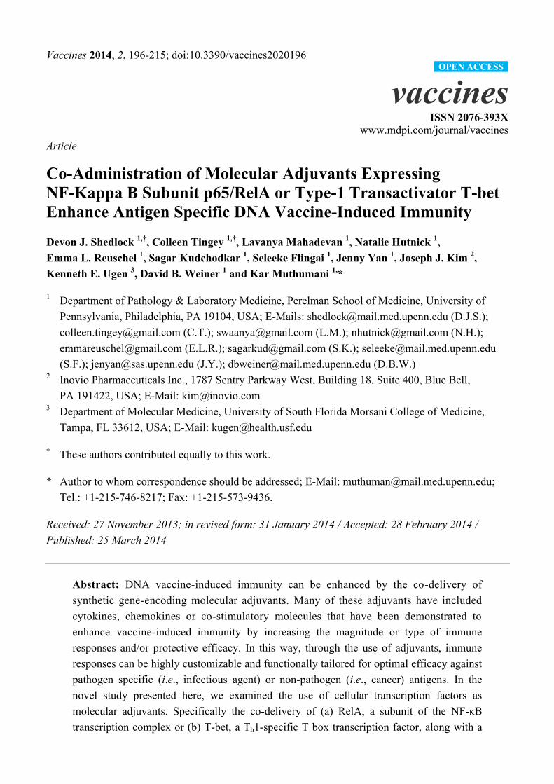
Vaccines 2014, 2, 196-215; doi:10.3390/vaccines2020196 vaccines ISSN 2076-393X www.mdpi.com/journal/vaccines Article Co-Administration of Molecular Adjuvants Expressing NF-Kappa B Subunit p65/RelA or Type-1 Transactivator T-bet Enhance Antigen Specific DNA Vaccine-Induced Immunity Devon J. Shedlock 1,† , Colleen Tingey 1,† , Lavanya Mahadevan 1 , Natalie Hutnick 1 , Emma L. Reuschel 1 , Sagar Kudchodkar 1 , Seleeke Flingai 1 , Jenny Yan 1 , Joseph J. Kim 2 , Kenneth E. Ugen 3 , David B. Weiner 1 and Kar Muthumani 1, * 1 Department of Pathology & Laboratory Medicine, Perelman School of Medicine, University of Pennsylvania, Philadelphia, PA 19104, USA; E-Mails: [email protected] (D.J.S.); [email protected] (C.T.); [email protected] (L.M.); [email protected] (N.H.); [email protected] (E.L.R.); [email protected] (S.K.); [email protected] (S.F.); [email protected] (J.Y.); [email protected] (D.B.W.) 2 Inovio Pharmaceuticals Inc., 1787 Sentry Parkway West, Building 18, Suite 400, Blue Bell, PA 191422, USA; E-Mail: [email protected] 3 Department of Molecular Medicine, University of South Florida Morsani College of Medicine, Tampa, FL 33612, USA; E-Mail: [email protected] † These authors contributed equally to this work. * Author to whom correspondence should be addressed; E-Mail: [email protected]; Tel.: +1-215-746-8217; Fax: +1-215-573-9436. Received: 27 November 2013; in revised form: 31 January 2014 / Accepted: 28 February 2014 / Published: 25 March 2014 Abstract: DNA vaccine-induced immunity can be enhanced by the co-delivery of synthetic gene-encoding molecular adjuvants. Many of these adjuvants have included cytokines, chemokines or co-stimulatory molecules that have been demonstrated to enhance vaccine-induced immunity by increasing the magnitude or type of immune responses and/or protective efficacy. In this way, through the use of adjuvants, immune responses can be highly customizable and functionally tailored for optimal efficacy against pathogen specific (i.e., infectious agent) or non-pathogen (i.e., cancer) antigens. In the novel study presented here, we examined the use of cellular transcription factors as molecular adjuvants. Specifically the co-delivery of (a) RelA, a subunit of the NF-κB transcription complex or (b) T-bet, a T h 1-specific T box transcription factor, along with a OPEN ACCESS
Transcript of Co-Administration of Molecular Adjuvants Expressing NF-Kappa B Subunit p65/RelA or Type-1...
Vaccines 2014, 2, 196-215; doi:10.3390/vaccines2020196
vaccines ISSN 2076-393X
www.mdpi.com/journal/vaccines
Article
Co-Administration of Molecular Adjuvants Expressing
NF-Kappa B Subunit p65/RelA or Type-1 Transactivator T-bet
Enhance Antigen Specific DNA Vaccine-Induced Immunity
Devon J. Shedlock 1,†
, Colleen Tingey 1,†
, Lavanya Mahadevan 1, Natalie Hutnick
1,
Emma L. Reuschel 1, Sagar Kudchodkar
1, Seleeke Flingai
1, Jenny Yan
1, Joseph J. Kim
2,
Kenneth E. Ugen 3, David B. Weiner
1 and Kar Muthumani
1,*
1 Department of Pathology & Laboratory Medicine, Perelman School of Medicine, University of
Pennsylvania, Philadelphia, PA 19104, USA; E-Mails: [email protected] (D.J.S.);
[email protected] (C.T.); [email protected] (L.M.); [email protected] (N.H.);
[email protected] (E.L.R.); [email protected] (S.K.); [email protected]
(S.F.); [email protected] (J.Y.); [email protected] (D.B.W.) 2
Inovio Pharmaceuticals Inc., 1787 Sentry Parkway West, Building 18, Suite 400, Blue Bell,
PA 191422, USA; E-Mail: [email protected] 3
Department of Molecular Medicine, University of South Florida Morsani College of Medicine,
Tampa, FL 33612, USA; E-Mail: [email protected]
† These authors contributed equally to this work.
* Author to whom correspondence should be addressed; E-Mail: [email protected];
Tel.: +1-215-746-8217; Fax: +1-215-573-9436.
Received: 27 November 2013; in revised form: 31 January 2014 / Accepted: 28 February 2014 /
Published: 25 March 2014
Abstract: DNA vaccine-induced immunity can be enhanced by the co-delivery of
synthetic gene-encoding molecular adjuvants. Many of these adjuvants have included
cytokines, chemokines or co-stimulatory molecules that have been demonstrated to
enhance vaccine-induced immunity by increasing the magnitude or type of immune
responses and/or protective efficacy. In this way, through the use of adjuvants, immune
responses can be highly customizable and functionally tailored for optimal efficacy against
pathogen specific (i.e., infectious agent) or non-pathogen (i.e., cancer) antigens. In the
novel study presented here, we examined the use of cellular transcription factors as
molecular adjuvants. Specifically the co-delivery of (a) RelA, a subunit of the NF-κB
transcription complex or (b) T-bet, a Th1-specific T box transcription factor, along with a
OPEN ACCESS
Vaccines 2014, 2 197
prototypical DNA vaccine expressing HIV-1 proteins was evaluated. As well, all of the
vaccines and adjuvants were administered to mice using in vivo electroporation (EP), a
technology demonstrated to dramatically increase plasmid DNA transfection and subsequent
transgene expression with concomitant enhancement of vaccine induced immune responses.
As such, this study demonstrated that co-delivery of either adjuvant resulted in enhanced T
and B cell responses, specifically characterized by increased T cell numbers, IFN-γ
production, as well as enhanced antibody responses. This study demonstrates the use of
cellular transcription factors as adjuvants for enhancing DNA vaccine-induced immunity.
Keywords: DNA vaccine; transcription factors; adjuvant-enhanced immunity; T cell
immunity; antibody responses
1. Introduction
DNA vaccination has once again became elevated to the forefront of efforts aimed at developing
vaccines against challenging infectious diseases including HIV/AIDS, emerging strains of influenza as
well as SARS. As well, it has a reemerging role as a delivery method for tumor immunotherapy [1–8].
While “first-generation” DNA vaccines were poorly immunogenic, recent technological advances have
dramatically improved their ability to drive immunity, including cellular based responses, in
preclinical immunogenicity and efficacy studies [9–14] as well as in clinical trials [15–18]. The
transfection rate of plasmid DNA and subsequent expression of their encoded antigens (Ages) are
significantly enhanced when highly-concentrated plasmid vaccine formulations are delivered through
in vivo electroporation (EP), a technology using brief square-wave electric pulses at the vaccination
site to facilitate entry and expression of DNA plasmids into transiently permeabilized cells resulting in
improved immunogenicity and efficacy of the vaccines [19]. In theory, a cocktail of plasmids could be
assembled for directing a highly-specialized immune response against any number of variable antigens
(Ag), which, in turn, could induce a more robust and efficacious immune response. In addition,
“consensus-engineering” of the Ag amino acid sequences has been effectively used to help bias
vaccine-induced immunity towards particular divergent, circulating, or virulent strains such as
enhancing protection among divergent strains of HIV and influenza virus [20,21]. Due, in part, to these
technological developments, immunization regimens including these “enhanced” DNA (E-DNA)
vaccines are extremely customizable and highly versatile.
Immunity can be further directed by co-delivery of the vaccine with plasmid-based molecular
adjuvants encoding species-specific immunomodulatory proteins. These have typically included cytokines,
chemokines, and surface expressed co-stimulatory molecules [18,22–34]. Such a gene adjuvant
approach substantially enhanced immune potency in numerous vaccine studies [16,18,29,35,36]. As a
candidate for molecular adjuvant development, transcription factors regulate the gene expression of
numerous inflammatory factors and promote activation and maturation of the adaptive immune
response [37–39]. An established pro-inflammatory mediator is the NF-kappa B protein complex
which regulates the expression of cytokines (TNF-α, IL-1β, IL-6, IL-2, etc.), induces DC maturation
(characterized by upregulation of CD80, CD86 and CD40), and promotes cell survival by the
Vaccines 2014, 2 198
regulation of Bcl-XL and IAPs, and protecting against TNF-α induced cell death [40–42]. This
transcription factor consists of five members including p65 (RelA), c-Rel, RelB, p50 (NF-κB1) and
p52 (NF-κB2) [42]. Importantly, RelA is a vital component in inflammation and cell survival,
possesses transcriptional activating capabilities, and can potently activate kappa B-dependent
transcription. Upregulation of this subunit may regulate the gene expression of multiple inflammatory
and survival factors that may lead to improved adaptive immunity [43–46].
An additional candidate for development as a molecular adjuvant is T-bet, a T helper 1 (Th1)-specific
transcription factor, which would ideally help to promote the induction of Th1-type immunity. This
T-box family member regulates lineage commitment in CD4 Th cells by directly activating
transcription of the IFN-γ gene. It also exhibits the property of redirecting committed Th 2 populations
to a Th1 phenotype [47,48]. The importance of T-bet in Th1 immunity has been most clearly illustrated
and reported in cases where CD4+ T cells lacking T-bet are severely impaired in their ability to
produce IFN-γ, yet secrete elevated levels of the opposing Th2 subset cytokines, IL-4 and IL-5.
Co-expression of this protein along with a vaccine against tuberculosis has been demonstrated to
increase IgG2a antibody (Ab) responses as well as the production of IFN-γ and IL-2 [47]. Thus, a
T-bet-expressing molecular adjuvant delivered by E-DNA vaccination would ideally enhance the
magnitude and type of immunity induced by immunization. Together, RelA and T-bet present attractive
candidates, as molecular adjuvants, for the enhancement of immunity following E-DNA vaccination.
In this study, we investigated the ability of molecular adjuvants expressing transcription factors
RelA or T-bet, as developed herein, to enhance in mice, immunity of an E-DNA vaccine encoding
either HIV-1 Env or Gag. Their potential ability to modify immunity was then assessed. Co-administration
of either pRelA or pTbet in conjunction with the pEnv or pGag vaccine significantly increased T cell
immunity, as measured by INF-γ production by ELISpot and proliferation. As well, B-cell/antibody
levels were enhanced as indicated by an increase in B-cell numbers as well as antigen specific
antibody titers. Consistent with these findings, the total amount of antigen specific IgG in serum was
increased following the co-administration of plasmids expressing the transcription factors. This study
builds on recent successes in demonstrating the potency of E-DNA vaccination and suggests that
transcription factors may serve as an effective adjuvant to increase vaccine-induced immunity.
2. Experimental
2.1. Plasmid Vaccine Constructs
The pRelA plasmid DNA constructs encode the full-length mouse NF-κB subunit p65/RelA
(GenBank #TF65_MOUSE) and Type-1 transactivator T-bet (GenBank #TBX21_MOUSE), respectively.
In addition, the Ig heavy chain epsilon-1 signal peptide (GenBank#AAB59424) was fused to the
N-terminus of each sequence, replacing the N-terminal methionine, which facilitates expression [11,49].
Each gene was genetically optimized for expression in mice, including codon- and RNA-optimization,
among other proprietary modifications for enhancing protein expression (GenScript, Piscataway, NJ,
USA). The optimized genes were then sub-cloned into modified pVax1 mammalian expression vectors
(Invitrogen, Carlsbad, CA, USA) under the control of the cytomegalovirus immediate-early (CMV)
promoter. These reagents were then used as the molecular adjuvants in this study. The pGag [50]

Vaccines 2014, 2 199
and pEnv [51] plasmids, expressing the HIV-1 proteins Gag and Env respectively, have been
previously described.
2.2. Transfections and Western Blot Analysis
Human Embryonic Kidney (HEK) 293T cells were maintained in Dulbecco‟s modified Eagle
medium (Life Technologies, Grand Island, NY, USA), supplemented with 10% heat-inactivated fetal
calf serum (FCS), 100 IU of penicillin per mL, 100 μg of streptomycin per mL and 2 mM L-glutamine [9].
Briefly, cells were transfected using TurboFection 8.0 (OriGene, Rockville, MD, USA) per the
manufacturer‟s protocol and subsequently incubated for 24–48 h. Cells were harvested with ice cold
PBS, centrifuged and washed, and then pelleted for Western immunoblot analysis [52]. Nuclear
extracts (107 cells) were made according to the method of Muthumani et al. [52]. The nuclear proteins
from the transfected cells were then dissolved in 20 mM Hepes (pH 7.9) containing 0.4 M NaCl, 1 mM
EDTA, 1 mM EGTA, 1 mM DTT, 1 mM PMSF and a cocktail of protease inhibitors (Promega Corp,
Madison, WI, USA). The protein concentration of each extract was measured by the Bio-Rad protein
assay kit (Bio-Rad, Hercules, CA, USA), and extracts were stored in aliquots at −70 °C until used.
Standard western blotting analysis was performed. Cells were treated with protein lysis buffer (0.01 M
Tris-HCl buffer pH 7.4, containing 1% Triton X-100, 1% sodium deoxycholate, 0.1% SDS)
supplemented with protease inhibitors (Protease Inhibitor Cocktail tablets; Roche, Indianapolis, IN,
USA). Proteins in lysates were then separated using 12% SDS-PAGE [53]. Protein-specific detection
antibodies for RelA and T-bet (Cell Signaling Technology, Danvers, MA, USA) were incubated with
the blots and expression visualized using the enhanced chemiluminescence (ECL) Western blot
detection system (GE Healthcare, Piscataway, NJ, USA).
2.3. Confirmation of Transcription Activity of RelA/p65 and T-Bet by Luciferase Reporter Assay and
IFN-Gamma Production
A RelA/p65 expressing vector, which co-expresses luciferase (pNF-κB-Luc) was used to confirm
the functionality of RelA/p65, which is necessary before it being used the “adjuvanted” vaccine study.
The luciferase reporter assay was performed as described previously [52,54,55]. Briefly, 293T cells
(105 cells/well) were seeded in a 96-well plate for 24 h. The cells were then transfected with the
RelA/p65 Luc expressing plasmid followed by incubation for 6 h. After incubation, the cell culture
medium was removed and replaced with fresh medium. Two days post transfection cells were treated
with 20 ng/mL of recombinant TNF-α for 6 h followed by measurement of luciferase activity by using
Microlumat plus luminometer (LUMAT LB9501, Berthold Technologies, Oak Ridge, TN, USA).
For confirmation of pT-bet function, the production of IFN-γ from pT-bet transfected CD4+ T cells
was measured. The impetus for measurement of IFN-γ is based on previously published studies that
demonstrated a direct correlation between T-bet and IFN-γ production [56]. Briefly in this analysis
naïve CD4+ T cells, isolated from the spleens of Balb/C mice, were purified using a CD4
+ T cell
isolation kit (Miltenyibiotec, San Diego, CA, USA). These cells were maintained in RPMI media
supplemented with 10% FBS, 100 U/mL penicillin and 200 µg/mL streptomycin and subsequently
transfected with pT-bet or pVax1 as a negative control. Two days post-transfection, cells were
Vaccines 2014, 2 200
stimulated overnight with anti-CD3 plus anti-CD28 Abs (1 µg/mL). IFN-γ levels in the supernatants
collected from the cultured CD4+ T cells were subsequently measured by a standard ELISA [36].
2.4. Animals and Vaccination Regimen
Adult female BALB/cJ (H-2d) mice were purchased from The Jackson Laboratory (Bar Harbor,
ME, USA). All animal experimentation was conducted according to University of Pennsylvania
(UPENN) IACUC approved protocols and performed in accordance with recommendations in the
Guide for the Care and Use of Laboratory Animals of NIH. UPENN complies with NIH policy as
stated in the Animal Welfare Act, and all other applicable federal, state and local laws. Mice were
immunized intramuscularly (i.m.) by needle injection into the left-thigh quadriceps muscle with 25 µg
of plasmid resuspended in 25 µL of PBS. Vaccinations were immediately followed by EP, at the same
site, and repeated at a two-week interval. For EP mediate delivery, a three-pronged CELLECTRA®
adaptive constant current Minimally Invasive Device (MID) was used, supplied by Inovio
Pharmaceuticals, Inc. (Blue Bell, PA, USA). Specifically, square-wave pulses were delivered through
a triangular 3-electrode array (inserted 2 mm intradermally) consisting of 26-gauge solid stainless steel
electrodes and two constant-current pulses of 0.1 Amps were delivered for 52 msec/pulse separated by
a 1 s delay. During the vaccination/molecular adjuvant administration regimen, and through the termination
for the study, all mice were monitored every 3 days for the development of potential adverse effects.
2.5. Splenocyte, T Cell Isolation and Cytokine Quantitation
Spleens were harvested 7–8 days following the third immunization as previously described [12].
Briefly, spleens were placed in RPMI 1640 medium (Mediatech, Manassas, VA, USA) supplemented
with 10% FBS, 1X Antibiotic-Antimycotic (Life Technologies, Grand Island, NY, USA), and
1× β-ME (Life Technologies, Grand Island, NY, USA). Splenocytes were isolated by mechanical
disruption of the spleen using a Stomacher machine (Seward Laboratory Systems, Bohemia, NY,
USA), and the resulting product was filtered using a 40 μm cell strainer (BD Biosciences, San Jose,
CA, USA). The cells were then treated for 5 min with ACK lysis buffer (Lonza, Walkersville, MD,
USA) for lysis of RBCs, washed in PBS, and then resuspended in RPMI medium for use in the
ELISPOT assay. CD4 naïve T cells were purified from the spleens using a naïve CD4+ T cell isolation
kit (Miltenyi Biotec, Auburn, CA, USA). These cells were maintained in RPMI medium supplemented
with 10% FBS, 100 U/mL penicillin, 200 μg/mL streptomycin, and stimulated with anti-CD3 plus
anti-CD28 (1 μg/mL each). Upon stimulation with anti-CD3 plus anti-CD28 antibodies, cytokine
production levels in the culture supernatants of cultured cells were examined by enzyme-linked
immunosorbent assay (ELISA) as described previously [3,13].
2.6. ELISPOT Analysis
The standard IFN-γ ELISPOT assay used in this study has been previously described [9,11,12].
Briefly, 96-well plates (Millipore, Billerica, MA, USA) were coated with anti-mouse IFN-γ capture
antibody and incubated for 24 h at 4 °C (R&D Systems, Minneapolis, MN, USA). The following day,
plates were washed with PBS and then incubated for 2 h with blocking buffer (1% BSA and 5%
Vaccines 2014, 2 201
sucrose in PBS). CD4+ or CD8
+ T cells (5 × 10
5 cells/well plated in triplicate) were MACS-purified
(Miltenyibiotec, San Diego, CA, USA) from splenocytes and subsequently stimulated with HIV-1 Gag
(consensus subtype B) or Env (subtype B (MN)) peptides (15-mers overlapping by 11 amino acids,
spanning the lengths of their respective protein (NIH AIDS Reagent Program, Bethesda, MD, USA).
After 18–24 h of stimulation overnight at 37 °C in 5% CO2, the plates were washed in PBS and
subsequently incubated for an additional 24 h at 4 °C with biotinylated anti-mouse IFN-γ monoclonal
antibody (mAb) purchased from R&D Systems (Minneapolis, MN, USA). The plates were then
washed again in PBS, and streptavidin-alkaline phosphatase (MabTech, Nacka Strand, Sweden) was
added to each well and incubated for 2 h at RT. Lastly, the plates were washed again in PBS followed
by incubation with BCIP/NBT Plus substrate (MabTech, Cincinnati, OH, USA) for 5–30 min. Upon
completion of spot development based on visual inspection, the plate was rinsed with distilled water
and then dried overnight at RT. Spots were enumerated using an automated ELISPOT reader (Cellular
Technology, Shaker Heights, OH, USA).
2.7. T Cell Proliferation Assay
Proliferative responses were measured in vitro by incubating 105 splenocytes in culture medium per
well in 96-well U-bottom plates in the presence of serial dilutions (5, 1, and 0.1 μg/mL) of recombinant
HIV-1 IIIB pr55 (Gag) (NIH AIDS Reagent Program, Bethesda, MD) or HIV-1 MN IIIB gp160 (Env)
(Protein Sciences, Meriden, CT, USA) and incubated at 37 °C with 5% CO2. Incorporation of tritiated
(3H)-thymidine was measured by pulsing with 1 μCi/well of (
3H)-thymidine during a 0–24 h time
period as described previously [57]. The plate was then harvested and incorporated 3H-thymidine was
measured in a Beta plate reader (Wallac, Waltham, MA, USA). The proliferative response is expressed
as a stimulation index (SI), calculated by dividing the mean cpm (counts per minute) of Ag-stimulated
wells by the mean cpm of non-stimulated wells.
2.8. ELISA
Sera from vaccinated mice harvested 7 days following the third vaccination were tested for
antibody responses against recombinant HIV-1 Env (NIH AIDS Reagent Program) by ELISA. Briefly,
96-well ELISA plates were coated with recombinant HIV-1 Env protein (Protein Sciences) and
incubated at 4 °C and washed subsequently with PBS and 0.1% Tween-20. Plates were then blocked
for 2 h with PBS and 0.2% Tween-20. After removal of the blocking solution, 100 μL of the pre-diluted
(1:50, 1:100, 1:500, 1:1000) mouse serum was added and incubated for 1 h. Plates were then washed
four times and incubated with a peroxidase-coupled anti-mouse IgG mAb (Sigma-Aldrich, St. Louis,
MO, USA). Lastly, plates were washed again followed by addition of 200 μL of substrate solution
(R&D Systems, Minneapolis, MN, USA) per well. The optical density at (OD405 nm) was subsequently
measured after a 15 min incubation. All assays were performed in triplicate.
2.9. Flow Cytometry
Muscle tissues (i.e., from the site of injection/vaccination) were removed aseptically, rinsed in
Hanks‟ balanced salt solution (Life Technologies, Grand Island, NY, USA), minced into approximately
Vaccines 2014, 2 202
1 × 2-mm squares, and digested in 20 mL of collagenase A (1 mg/mL, Life Technologies, Grand
Island, NY, USA) at 37 °C for 45 min, with occasional agitation. The cellular digest was filtered
through a sterile 31 μm nylon mesh, centrifuged at 400 g for 10 min, and washed twice in 10%
FCS-DMEM. The cell pellet was then resuspended in 4 mL of 10% FCS-DMEM.
For flow cytometric analysis, 106 cells from the immunized mice cells were washed in suspension
with ice-cold buffer A (PBS/0.1% BSA/0.01% NaN3) and incubated for 20 min at 4 °C with 50 μL of
a 1:100 diluted fluorescent-labeled specific antibodies using methods described previously [58]. The
fluorescently conjugated Abs utilized were FITC-CD11c, PE-CD4, PE-Cy7-CD45R (B220) (eBioscience,
San Diego, CA, USA), Alexa Fluor-750-CD8α, and PerCP-Cy5.5-CD11b (BD Biosciences, San Jose,
CA, USA). Cells were washed twice and immediately analyzed on a flow cytometer (Becton
Dickinson FACS, San Jose, CA, USA). All incubations and washes were performed at 4 °C with
ice-cold buffer A. Cells were gated on singlets and live cells. The flow cytometric data were analyzed
using FlowJo software (Tree Star, Ashland, OR, USA).
2.10. Statistical Analysis
Group analyses were completed by a matched, two-tailed, unpaired t-test with all values are
presented as mean ± SEM. Mann-Whitney analysis was used to determine statistical differences. All
data were analyzed using GraphPad Prism5 Software [59]. Statistically significant differences between
groups were defined as * p < 0.1, ** p < 0.01, *** p < 0.001, and **** p < 0.0001.
3. Results
3.1. Adjuvant Construction and Expression
The pRelA and pTbet plasmids encode the full-length mouse NF-kappa B subunit p65/RelA and
Type-1 transactivator T-bet, respectively. Each was genetically optimized, synthesized, and subcloned
into modified pVax1 mammalian expression vectors (Figure 1A). To test for expression of these
plasmids, HEK 293T cells were transfected with each and protein production was assessed by standard
Western immunoblotting. An approximately 65 kDa protein corresponding to RelA was detected,
using a specific Ab, in cell lysates harvested both 24 h and 48 h post-transfection (Figure 1B).
Likewise, T-bet was detected as an approximately 56 kDa protein using an anti-T-bet Ab. Binding was
specific for their respective proteins since neither bound to lysates from cells transfected with empty
vector control plasmid pVax1. These data demonstrate that each of the molecular adjuvants expresses
their respective encoded proteins upon in vitro transfection of HEK 293T cells. Further, IκB-dependent
transcription was accessed in the HeLa cells luciferase expressing cell system (Figure 1C) to confirm
the activation of RelA (p65). An increase in RelA expression as measured by relative luciferase
activity was observed in a dose dependent manner. That is, increasing the plasmid from 3 μg to 5 μg
or 10 μg resulted in an increase in the relative luciferase activity approximately 1.5 or 2.5 fold. T-bet
expression correlates with IFN-γ expression in T cell and NK cells [60] and therefore in this assay
IFN-γ serves as surrogate for the functional expression of T-bet (Figure 1D).

Vaccines 2014, 2 203
Figure 1. Molecular adjuvant construction and expression. (A) Mouse RelA or T-bet
primary sequences were genetically optimized, synthesized, and then subcloned into modified
pVax1 expression vectors. Optimization entailed inclusion of a IgE leader peptide (IgE),
preceded by a Kozak sequence, fused at the N-terminus. The figure indicates the
restrictions enzymes used for subcloning, the translation initiation site (forward arrow), IgE
leader peptide (IgE; hatched bar), protein length (aa), and transgenes (black with white
lettering); (B) Protein expression from the nuclear extract was analyzed by Western
immunoblotting following transfection of HEK 293T cells with pRelA, pTbet, or empty
vector control (pVax1). The relative size (kDa) of the proteins are determined by detection
analysis using protein-specific Abs as indicated; (C) Over expression of RelA potently induces
κB dependent transcription. HeLa cells were transiently transfected with a NF-κB-dependent
luciferase reporter gene together with expression vectors encoding RelA/p65. The cotransfected
cells were subsequently grown for 48 h, and the luciferase activity was determined as
described in the Materials and Methods; (D). Overexpression of T-bet stimulates
production of IFN-γ: Naive CD4 T cells were transfected with either pT-bet or pVax1 and
stimulated with anti-CD3 plus anti-CD28 followed the measurement of IFN-γ production
by enzyme-linked immunosorbent assay (ELISA) as described Materials and Methods.
IFN-γ levels are expressed as μg/mL

Vaccines 2014, 2 204
3.2. Enhanced Cellular Immunity
The contribution of pRelA and pTbet, in terms of enhancing vaccine-induced immunity, was then
assessed. Balb/C mice (n = 4/group) were vaccinated three times with 25 µg of pEnv or pGag either
with or without 25 µg of pRelA or pTbet, 25 µg of pRelA or pTbet alone, or with 25 µg of a control
plasmid (pVax1; Figure 2). The vaccines and adjuvants were delivered in 25 µL of PBS by in vivo EP.
Animals were sacrificed on day 35, (i.e., seven days after the third vaccination) followed by isolation
of splenocytes for immune analysis by IFN-γ ELISpot. In this assay, HIV-1 Env or Gag peptide pools
were used for stimulation of MACS-purified CD4+ or CD8
+ T cells and the IFN-γ ELISpot results are
displayed in Figure 2. Both CD4+ and CD8
+ T-cell responses were observed to be significantly
increased in mice vaccinated with pEnv and co-administrated pRelA compared with pEnv alone.
Likewise, immunization with pEnv with co-administrated pTbet compared to pEnv alone demonstrated
significant increases in CD4+ and CD8
+ T cell responses (Figure 2B).
Figure 2. Transcription factor adjuvants enhance antigen specific DNA vaccine induced T
cell immunity. (A) Balb/C mice (n= 4/group) were vaccinated three times at two week
intervals with HIV-1 pGag or pEnv alone, pGag or pEnv with co delivery of either pRelA
or pTbet. Other control groups were pRelA or pTbet alone, or a pVax1 control. T cell
responses (CD8+ and CD4
+) were analyzed by IFN-γ ELISPOT one week following the
third immunization and results for IFN-γ+ spot forming cells (SFC) per 106 MACS-purified
T cells are indicated following re-stimulation with subtype B HIV-1 Env (B) or Gag (C)
peptide pools. Samples were performed in triplicate, error bars represent SEM, and
statistically significant values are shown; ** p < 0.01, *** p < 0.001 and **** p < 0.0001,
referring to comparison between the indicated vaccination groups provided in the graph.
Experiments were performed twice independently with similar results.
Vaccines 2014, 2 205
Figure 2. Cont.
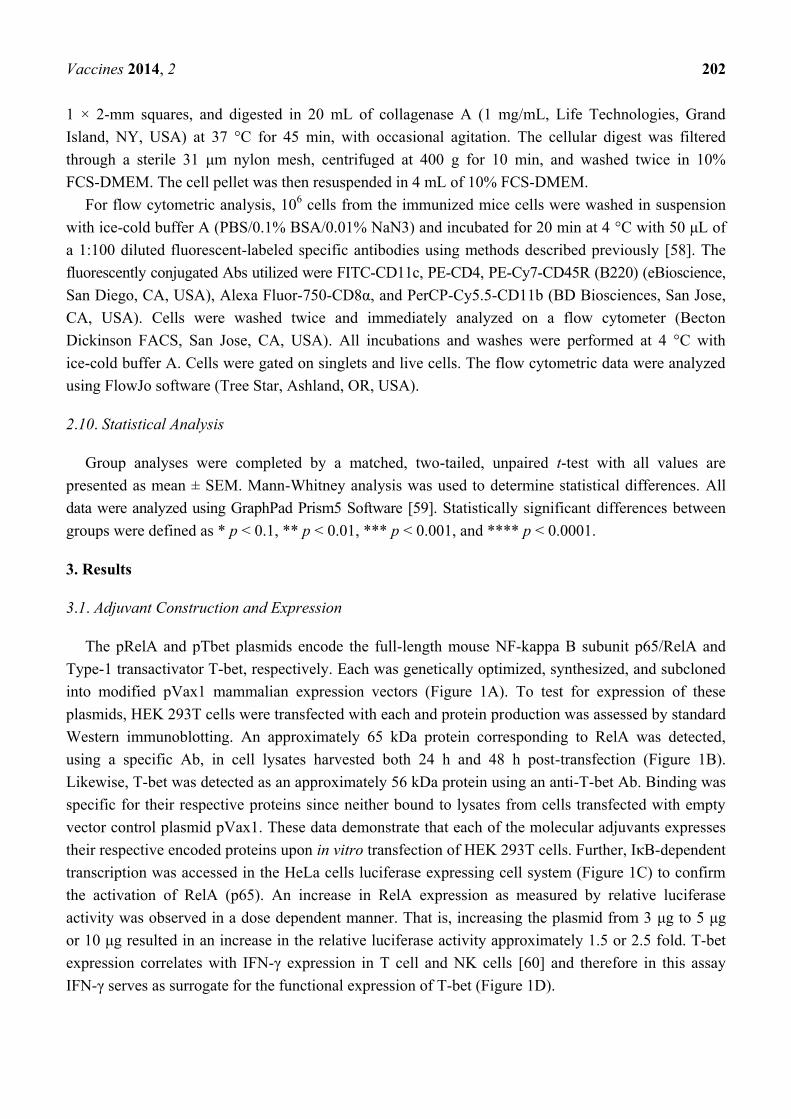
To confirm the enhancing effects of these two adjuvants on T cell IFN-γ production for a different
Ag, we also vaccinated animals with the HIV-1Gag either with or without pRelA or pTbet, similarly as
performed above. Analogous to the pEnv group, CD4+ T cell responses were increased in mice
immunized with pGag plus co-administrated pRelA, when compared with mice immunized with pGag
alone (Figure 2C). There was an even greater enhancement of the CD8+ T cell response in mice vaccinated
with pGag and co-administrated pRelA compared to immunization with pGag alone (Figure 2C).
Further, immunization with HIV-1 Gag along with concomitant administration of pTbet demonstrated
increased CD8+ T-cell responses when compared to immunization with pGag alone (Figure 2C).
However, CD4+ T cell responses were not as significantly increased as observed with co-delivery of
pRelA. Also, administration of either pRelA or pTbet alone did not markedly activate either CD4+ or
CD8+ T cells against Gag or Env as measured by IFN-γ production. Therefore, these data demonstrate
that co-administration of the transcription factor adjuvants promoted enhanced T cell responses against
two separate antigens with the data suggesting that expanding the breadth of vaccine-elicited cellular
immune responses was stimulated by administration of an immune adjuvant.
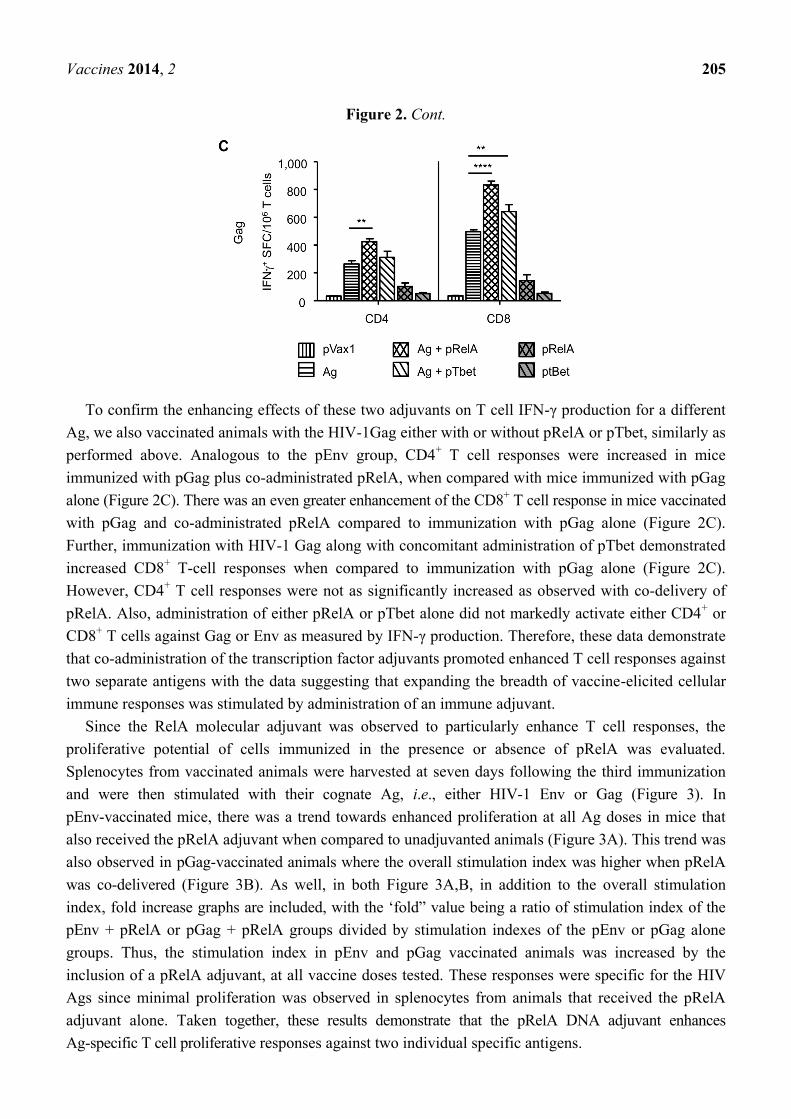
Since the RelA molecular adjuvant was observed to particularly enhance T cell responses, the
proliferative potential of cells immunized in the presence or absence of pRelA was evaluated.
Splenocytes from vaccinated animals were harvested at seven days following the third immunization
and were then stimulated with their cognate Ag, i.e., either HIV-1 Env or Gag (Figure 3). In
pEnv-vaccinated mice, there was a trend towards enhanced proliferation at all Ag doses in mice that
also received the pRelA adjuvant when compared to unadjuvanted animals (Figure 3A). This trend was
also observed in pGag-vaccinated animals where the overall stimulation index was higher when pRelA
was co-delivered (Figure 3B). As well, in both Figure 3A,B, in addition to the overall stimulation
index, fold increase graphs are included, with the „fold” value being a ratio of stimulation index of the
pEnv + pRelA or pGag + pRelA groups divided by stimulation indexes of the pEnv or pGag alone
groups. Thus, the stimulation index in pEnv and pGag vaccinated animals was increased by the
inclusion of a pRelA adjuvant, at all vaccine doses tested. These responses were specific for the HIV
Ags since minimal proliferation was observed in splenocytes from animals that received the pRelA
adjuvant alone. Taken together, these results demonstrate that the pRelA DNA adjuvant enhances
Ag-specific T cell proliferative responses against two individual specific antigens.
Vaccines 2014, 2 206
Figure 3. Increased T-cell proliferative potential following DNA vaccination plus
co-administration of pRelA. Proliferative responses were measured seven days following
the third vaccination with either pEnv or pGag alone, pEnv or pGag with pRelA molecular
adjuvant, or empty vector control pVax1 alone. Splenocytes were incubated with recombinant
HIV-1 Env (A) or Gag (B) at various concentrations: 0.5 (white bars), 1.0 (light gray bars),
and 5.0 (dark gray bars) and subsequently pulsed with tritiated (3H)-thymidine for 24 h.
Incorporated thymidine was expressed as a stimulation index (SI) calculated by dividing
the mean cpm (counts per minute) of Ag-stimulated wells by the mean cpm of non-stimulated
wells. Fold increase in SI for pRelA-adjuvanted mice are displayed for each concentration
of Env (A, right panel) or Gag (B, right panel). Samples were tested in triplicate. Error bars
represent the SEM, and statistically significant values are provided for the indicated group
comparison shown in the graphs. **** p < 0.0001.
3.3. Enhanced Antibody Responses with Adjuvanted Vaccination
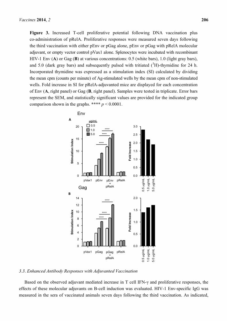
Based on the observed adjuvant mediated increase in T cell IFN-γ and proliferative responses, the
effects of these molecular adjuvants on B-cell induction was evaluated. HIV-1 Env-specific IgG was
measured in the sera of vaccinated animals seven days following the third vaccination. As indicated,
Vaccines 2014, 2 207
mice received pEnv either with or without co-administered pRelA or pTbet, pRelA or pTbet alone, or a
pVax1 control plasmid (Figure 4). Measurable IgG responses were induced by pEnv alone at dilutions
ranging from 1:50 to 1:500, but were non longer measurable at a dilution of 1:1000. Importantly, these
responses were augmented at all dilutions by the inclusion of the pRelA or pTbet adjuvant when
compared to the pEnv group alone. Specifically, differences were observed at the 1:50 sera dilution,
where administration of pRelA and pTbet significantly enhanced the induction of HIV-1 Env-specific
IgG responses (p = 0.0388 and p = 0.0062, respectively). Enhanced IgG responses were specific for
Env since minimal antibody responses were observed in the sera from mice that were administered the
pRelA or pTbet adjuvant alone. These data suggest that both transcription factor adjuvants elicited an
enhanced humoral immune response that was analogous and consistent with the elevated IFN-γ levels
and T cell proliferative responses observed following vaccination with pRelA or pTbet.
One potential mechanism for the ability of the transcription factors to enhance antibody responses
may be thorough increase in the number of activated B-cells. To access whether this was occurring, the
pRelA administered muscle at the site of vaccination was biopsied three days after pEnv immunization
with co-administrated pRelA followed by quantification of number of B220+ B-cells at the site of
injection. The results indicated that pRelA and pEnv alone caused only a slight increase in B-cell
trafficking to the site of injection compared to pVax1 administration alone (Figure 5). This is indicated
by the MFI (mean fluorescent intensity) values shown in the individual FACS scans, which are directly
proportional to the level of B220+ B cells. However, the addition of a pRelA adjuvant in combination
with the pEnv vaccine further enhanced the number of B-cells at the site of injection.
Figure 4. Improved B cell responses with pEnv vaccination and co-administered transcriptional
molecular adjuvant. B cell/antibody responses were assessed in the sera of vaccinated mice
(n = 4/group) seven days following the third immunization with pEnv alone, pEnv in
combination with either pRelA or pTbet, each of the molecular adjuvants alone, or with
empty vector control plasmid (pVax1). Anti-Env p120 antibody-binding titers were
determined by ELISA. Data are presented as the mean endpoint titers. Statistically
significant values are indicated; *** p < 0.001 (comparison between pEnv alone and pEnv
+ pRelA or pEnv + pT-bet) and **** p < 0.0001 (comparison between pRelA alone and
pEnv + pRelA or pT-bet alone and pEnv + pT-bet).

Vaccines 2014, 2 208
Figure 5. Molecular adjuvants enhance populations of B-cells at the site of immunization.
Cells cultures from the muscle were analyzed by flow cytometry for expression of B220. The
isolated cells were incubated in culture media for three days and these cells and then
stained with DC subsets (CD11c+/CD11b
+), B cells (B220
+), T cells (CD4
+ and CD8
+
subsets), to distinguish monocytes/dendritic, B cells, T cells, respectively. Such differential
staining allowed the exclusion of dendritic and T cells from subsequent analysis of B220
expression. Histograms show the B220+ expression on B cells exclusively using a specific
mAb as well as an isotype-matched, irrelevant mAb as a control. The profile of an
isotype-matched irrelevant Ab, used as a control (shaded area) is also indicated in the
panels. MFI = mean fluorescent intensity which is proportional to the level of B220
expressing B cells.
4. Discussion
The study reported here is the first to report the ability of plasmid-expressed RelA and T-bet to
function as molecular adjuvants for antigen specific DNA vaccine induced immunity. While the potential
utility of RelA and T-bet are evident, the exact mechanism by which they enhance immunogenicity is
currently unknown. For the RelA molecular adjuvant, it is likely that the over expression of this
molecule directly drives NF-κB activation, much like many current adjuvants do indirectly. For
example, many bacterial-derived carbohydrates such as those present in the GSK AS04 adjuvant and
many veterinary adjuvants exert their pro-inflammatory effects through activating this pathway.
Likewise, the recently approved MPL adjuvantTLR-4, which activates NF-κB and leads to enhanced
Th1 and antibody responses. In the present study, we hypothesized that directly expressing RelA/p65 at
the site of injection would potentially impact multiple cell types and multiple signaling pathways
within a given cell resulting in NF-κB activation. This could lead to the stimulation of numerous
pro-inflammatory signals for the induction of vaccine-specific Th1-type responses. NF-κB is expressed
in all hematological cells and the pairing of the p65 subunit leads to multiple functional outcomes
including activation. In this study, we also reported an approach to enhance the immunogenicity of
Vaccines 2014, 2 209
DNA-based vaccines using T-bet as an adjuvant. Enhancement of antigen specific immune responses
was confirmed by the fact that the T cell immune response was elicited in the T-bet co-administered
mice compared to the pVax1 control. The enhanced immunity observed with molecular immune
adjuvants may also have been the result of direct transfection of local DCs at the site of injection. It is
known that the nuclear translocation of p65 is associated with the activation and maturation of DCs [61].
In addition, NF-kappa B activation controls the expression of critical co-stimulatory molecules
such as CD80, CD86 and MHC class II along with pro-inflammatory cytokines such as IL-12, IL-6
and TNF-α [62,63]. Over expression of RelA in local DCs could likely increase cytokine production
and co-stimulatory molecule expression, both of which could lead to improved T cell induction.
Therefore, it is likely that the inclusion of a RelA-expressing molecular adjuvant could result in
NF-kappa B activation in multiple cells at the site of injection, including resident DC and potentially
could explain the improved T cell phenotype observed in this study.
In addition to stimulation of the T cell responses, enhanced antigen specific IgG production was
also observed when a transcription adjuvant was used in combination with the vaccine. For the RelA
adjuvant, NF-kappa B activation enhances the expression of adhesion molecules required for immune
cell accumulation at the site of inflammation [64]. Further, studies in knockout mice suggest p65 is
required for B-cell proliferation in response to BCR signaling [41]. Therefore, the inclusion of pRelA
may facilitate the accumulation of Ag-specific cells at the site of vaccination. For the T-bet molecular
adjuvant, in vitro over expression in B cell lines has been shown to result in Ab class switching,
thereby increasing the level of IgG2a [65]. It is possible that IgG2a responses are enhanced herein
since T-bet expression regulates the transcription of mature B cell receptors, which are necessary for
the survival of memory B cells. In addition, the differential transcriptional stimulation determined by
T-bet may regulate B cell potential for cytokine secretion, trafficking, and survival that ultimately
permits flexibility in long-term Ag-specific immunoprotection [65]. Thus, data herein indicate that
vaccination with co-expressed transcription factors associated with pro-inflammation and Th1 type
development may contribute to the enhancement of vaccine-induced B cell responses.
Several viral and bacterial infection models have been tested extensively to define the transcription
factors that may have a role in the development, differentiation and maturation of immune cells on
particularly the CD8+ T-cell effector and memory populations [46]. Our results are in agreement with a
model supporting a role for several transcriptional factors in the enhancement of primary antigen
specific cellular and humoral responses. Overall, the work presented here suggests that pRelA and
T-bet plays a significant role in creating the immune environment that influences the development and
function of the strong vaccine induced CD8+ T-cell response and antibody production.
While the molecular adjuvanted E-DNA vaccine approaches presented herein have demonstrated
the potential for increased Ag-specific immunity, it is important to consider possible safety issues that
may be caused by transcription factor expression. Importantly, while increased NF-kappa B expression
can lead to multiple immunological disorders and cancers [66,67] we observed no significant adverse
safety events in mice vaccinated with the pRelA or pTbet molecular adjuvants. This may be explained
by transient expression of these proteins by plasmid DNA that is known to persist for approximately
14–30 days. In addition, local expression of the plasmid DNA expressed transcription factors may help
to drive immunity at the vaccination site while minimizing the possibility of systemic or off-target
effects. Thus, it would be unlikely that a transcription factor-encoding genetic adjuvant would induce

Vaccines 2014, 2 210
any serious long-term negative side effects. Furthermore, our group and others have studied extensively
the use of various immune modulating plasmid-expressed adjuvants without any evidence for the
development of adverse effects. This is an important safety consideration and demonstrates that
administration of plasmid based transcriptional factors does not lead to global immune deregulation.
However, future studies should further investigate the safety of the proposed adjuvants in preclinical
studies utilizing nonhuman primates, before advancing to the clinic. In conclusion, this is the first
report, to our knowledge, to demonstrate the ability of several transcription factors, when delivered as
DNA expression plasmid, to enhance the immunogenicity of antigen specific DNA vaccines.
5. Conclusions
The use of cellular transcription factors RelA and T-bet as molecular adjuvants for enhancing
DNA vaccine-induced immunity was investigated in this report. When co-delivered along with a
prototypical DNA vaccine by in vivo electroporation (EP), either of these putative adjuvants stimulated
enhanced antigen-specific T and B cell responses as indicated by increased T cell numbers and IFN-γ
production, as well as by an increase in antibody levels. This study builds on recent achievements
demonstrating the potency of the enhanced DNA (E-DNA) vaccination method and establishes that
transcription factors may serve as effective molecular adjuvants to boost vaccine-induced immunity.
Acknowledgments
This work was supported by grants funded to DBW through the National Institutes of Health
including NIH-CFAR, NIAIDS-HVDDT and NIH-NIAID-HIVRAD. KM was supported by a National
Institute of Health-Center For AIDS Research Training Supplemental Grant-5-P30-AI-045008-13.
This work was supported in part by the core facilities of the Penn Center for AIDS Research and the
Abramson Cancer Center Core facilities.
Author Contributions
Conceived and designed the experiments: Kar Muthumani, David B. Weiner. Performed the
experiments: Devon J. Shedlock, Colleen Tingey, Lavanya Mahadevan, Natalie Hutnick, Emma L.
Reuschel, Sagar Kudchodkar, Seleeke Flingai, Jenny Yan and Kar Muthumani. Contributed
reagents/materials/analysis tools: Joseph J. Kim, Kenneth E. Ugen and David B. Weiner. Wrote the
paper: Kar Muthumani and David B. Weiner. Obtained funding: Kar Muthumani and David B.
Weiner. Study supervision: Kar Muthumani and David B. Weiner. Critical revisions of manuscript:
Kar Muthumani, Kenneth E. Ugen and David B. Weiner.
Conflicts of Interest
The laboratory of DBW has grant funding and collaborations as well as service in scientific review,
consulting and advising capacities for commercial entities and therefore there exists possible conflicts
associated with this work with Pfizer, Inovio, BMS, Virxsys, Ichor, Merck, Althea, VGXI, J&J,
Aldevron, and possibly others. The authors have no other relevant affiliations or financial involvement
with any organization or entity with a financial interest in or financial conflict with the subject matter
Vaccines 2014, 2 211
or materials discussed in the manuscript apart from those disclosed. No writing assistance was utilized
in the production of this manuscript.
Reference
1. Zeng, Q.; Gomez, B.P.; Viscidi, R.P.; Peng, S.; He, L.; Ma, B.; Wu, T.C.; Hung, C.F.
Development of a DNA vaccine targeting merkel cell polyomavirus. Vaccine 2012, 30, 1322–1329.
2. Mascola, J.R.; Montefiori, D.C. The role of antibodies in HIV vaccines. Annu. Rev. Immunol.
2010, 28, 413–444.
3. Laddy, D.J.; Yan, J.; Khan, A.S.; Andersen, H.; Cohn, A.; Greenhouse, J.; Lewis, M.;
Manischewitz, J.; King, L.R.; Golding, H.; et al. Electroporation of synthetic DNA antigens offers
protection in nonhuman primates challenged with highly pathogenic avian influenza virus. J. Virol.
2009, 83, 4624–4630.
4. Korber, B.; Gnanakaran, S. AIDS/HIV Converging on an HIV vaccine. Science 2011, 333,
1589–1590.
5. Zhou, T.; Georgiev, I.; Wu, X.; Yang, Z.Y.; Dai, K.; Finzi, A.; Kwon, Y.D.; Scheid, J.F.; Shi, W.;
Xu, L.; et al. Structural basis for broad and potent neutralization of HIV-1 by antibody VRC01.
Science 2010, 329, 811–817.
6. Moir, S.; Malaspina, A.; Fauci, A.S. Prospects for an hiv vaccine: Leading B cells down the right
path. Nat. Struct. Mol. Biol. 2011, 18, 1317–1321.
7. Leitner, W.W.; Baker, M.C.; Berenberg, T.L.; Lu, M.C.; Yannie, P.J.; Udey, M.C. Enhancement
of DNA tumor vaccine efficacy by gene gun-mediated codelivery of threshold amounts of
plasmid-encoded helper antigen. Blood 2009, 113, 37–45.
8. Martin, J.E.; Louder, M.K.; Holman, L.A.; Gordon, I.J.; Enama, M.E.; Larkin, B.D.;
Andrews, C.A.; Vogel, L.; Koup, R.A.; Roederer, M.; et al. A sars DNA vaccine induces neutralizing
antibody and cellular immune responses in healthy adults in a phase I clinical trial. Vaccine 2008,
26, 6338–6343.
9. Shedlock, D.J.; Aviles, J.; Talbott, K.T.; Wong, G.; Wu, S.J.; Villarreal, D.O.; Myles, D.J.;
Croyle, M.A.; Yan, J.; Kobinger, G.P.; et al. Induction of broad cytotoxic T cells by protective
DNA vaccination against marburg and ebola. Mol. Ther. 2013, 21, 1432–1444.
10. Shedlock, D.J.; Silvestri, G.; Weiner, D.B. Monkeying around with HIV vaccines: Using rhesus
macaques to define “gatekeepers” for clinical trials. Nat. Rev. Immunol. 2009, 9, 717–728.
11. Shedlock, D.J.; Talbott, K.T.; Cress, C.; Ferraro, B.; Tuyishme, S.; Mallilankaraman, K.;
Cisper, N.J.; Morrow, M.P.; Wu, S.J.; Kawalekar, O.U.; et al. A highly optimized DNA vaccine
confers complete protective immunity against high-dose lethal lymphocytic choriomeningitis
virus challenge. Vaccine 2011, 29, 6755–6762.
12. Shedlock, D.J.; Talbott, K.T.; Wu, S.J.; Wilson, C.M.; Muthumani, K.; Boyer, J.D.; Sardesai, N.Y.;
Awasthi, S.; Weiner, D.B. Vaccination with synthetic constructs expressing cytomegalovirus
immunogens is highly T cell immunogenic in mice. Hum. Vaccine Immunother. 2012, 8, 1668–1681.
13. Mallilankaraman, K.; Shedlock, D.J.; Bao, H.; Kawalekar, O.U.; Fagone, P.; Ramanathan, A.A.;
Ferraro, B.; Stabenow, J.; Vijayachari, P.; Sundaram, S.G.; et al. A DNA vaccine against chikungunya
Vaccines 2014, 2 212
virus is protective in mice and induces neutralizing antibodies in mice and nonhuman primates.
PLoS Negl. Trop. Dis. 2011, 5, e928.
14. Muthumani, K.; Lankaraman, K.M.; Laddy, D.J.; Sundaram, S.G.; Chung, C.W.; Sako, E.; Wu, L.;
Khan, A.; Sardesai, N.; Kim, J.J.; et al. Immunogenicity of novel consensus-based DNA vaccines
against chikungunya virus. Vaccine 2008, 26, 5128–5134.
15. Bagarazzi, M.L.; Yan, J.; Morrow, M.P.; Shen, X.; Parker, R.L.; Lee, J.C.; Giffear, M.;
Pankhong, P.; Khan, A.S.; Broderick, K.E.; et al. Immunotherapy against HPV16/18 generates potent
th1 and cytotoxic cellular immune responses. Sci. Transl. Med. 2012, 4, doi:10.1126/scitranslmed.
3004414.
16. Kublin, J. Welcome to the HVTNEWS. Available online: http://www.hvtn.org/science/hvtnews/
HVTNews-SpecialEdition-Sept2011-web.pdf (accessed on 1 November 2013).
17. Kutzler, M.A.; Weiner, D.B. DNA vaccines: Ready for prime time? Nat. Rev. Genet. 2008, 9,
776–788.
18. Kalams, S.A.; Parker, S.D.; Elizaga, M.; Metch, B.; Edupuganti, S.; Hural, J.; de Rosa, S.;
Carter, D.K.; Rybczyk, K.; Frank, I.; et al. Safety and comparative immunogenicity of an HIV-1
DNA vaccine in combination with plasmid interleukin 12 and impact of intramuscular
electroporation for delivery. J. Infect. Dis. 2013, 208, 818–829.
19. Kee, S.T.; Gehl, J.; Lee, E.W. Clinical Aspects of Electroporation, 1st ed.; Springer: New York,
NY, USA, 2011; p. 268.
20. Laddy, D.J.; Yan, J.; Kutzler, M.; Kobasa, D.; Kobinger, G.P.; Khan, A.S.; Greenhouse, J.;
Sardesai, N.Y.; Draghia-Akli, R.; Weiner, D.B. Heterosubtypic protection against pathogenic
human and avian influenza viruses via in vivo electroporation of synthetic consensus DNA
antigens. PLoS One 2008, 3, e2517.
21. Yan, J.; Yoon, H.; Kumar, S.; Ramanathan, M.P.; Corbitt, N.; Kutzler, M.; Dai, A.; Boyer, J.D.;
Weiner, D.B. Enhanced cellular immune responses elicited by an engineered HIV-1 subtype B
consensus-based envelope DNA vaccine. Mol. Ther. 2007, 15, 411–421.
22. Henke, A.; Chiang, C.S.; Zell, R.; Stelzner, A. Co-expression of interleukin-2 to increase the
efficacy of DNA vaccine-mediated protection in coxsackievirus B3-infected mice. Antivir. Res.
2004, 64, 131–136.
23. Takahashi, S.; Yotnda, P.; Rousseau, R.F.; Mei, Z.; Smith, S.; Rill, D.; Younes, A.; Brenner, M.K.
Transgenic expression of CD40L and interleukin-2 induces an autologous antitumor immune
response in patients with non-hodgkin‟s lymphoma. Cancer Gene Ther. 2001, 8, 378–387.
24. Santra, S.; Barouch, D.H.; Sharpe, A.H.; Letvin, N.L. B7 co-stimulatory requirements differ for
induction of immune responses by DNA, protein and recombinant pox virus vaccination. Eur. J.
Immunol. 2000, 30, 2650–2659.
25. Beignon, A.S.; McKenna, K.; Skoberne, M.; Manches, O.; DaSilva, I.; Kavanagh, D.G.;
Larsson, M.; Gorelick, R.J.; Lifson, J.D.; Bhardwaj, N. Endocytosis of HIV-1 activates plasmacytoid
dendritic cells via toll-like receptor-viral rna interactions. J. Clin. Invest. 2005, 115, 3265–3275.
26. Palma, C.; Vendetti, S.; Cassone, A. Role of 4-1bb receptor in the control played by CD8+ T cells on
IFN-gamma production by mycobacterium tuberculosis antigen-specific CD4+ T cells. PLoS One
2010, 5, e11019.
Vaccines 2014, 2 213
27. Auten, M.W.; Huang, W.; Dai, G.; Ramsay, A.J. Cd40 ligand enhances immunogenicity of
vector-based vaccines in immunocompetent and CD4+ T cell deficient individuals. Vaccine 2012,
30, 2768–2777.
28. Sin, J.I. Myd88 signal is required for more efficient induction of Ag-specific adaptive immune
responses and antitumor resistance in a human papillomavirus E7 DNA vaccine model. Vaccine
2011, 29, 4125–4131.
29. Belisle, S.E.; Yin, J.; Shedlock, D.J.; Dai, A.; Yan, J.; Hirao, L.; Kutzler, M.A.; Lewis, M.G.;
Andersen, H.; Lank, S.M.; et al. Long-term programming of antigen-specific immunity from gene
expression signatures in the PBMC of rhesus macaques immunized with an SIV DNA vaccine.
PLoS One 2011, 6, e19681.
30. Hirao, L.A.; Wu, L.; Khan, A.S.; Satishchandran, A.; Draghia-Akli, R.; Weiner, D.B.
Intradermal/subcutaneous immunization by electroporation improves plasmid vaccine delivery
and potency in pigs and rhesus macaques. Vaccine 2008, 26, 440–448.
31. Fagone, P.; Shedlock, D.J.; Bao, H.; Kawalekar, O.U.; Yan, J.; Gupta, D.; Morrow, M.P.;
Patel, A.; Kobinger, G.P.; Muthumani, K.; et al. Molecular adjuvant HMGB1 enhances anti-influenza
immunity during DNA vaccination. Gene Ther. 2011, 18, 1070–1077.
32. Muthumani, G.; Laddy, D.J.; Sundaram, S.G.; Fagone, P.; Shedlock, D.J.; Kannan, S.; Wu, L.;
Chung, C.W.; Lankaraman, K.M.; Burns, J.; et al. Co-immunization with an optimized plasmid-
encoded immune stimulatory interleukin, high-mobility group box 1 protein, results in enhanced
interferon-gamma secretion by antigen-specific cd8 t cells. Immunology 2009, 128, e612–e620.
33. Kutzler, M.A.; Kraynyak, K.A.; Nagle, S.J.; Parkinson, R.M.; Zharikova, D.; Chattergoon, M.;
Maguire, H.; Muthumani, K.; Ugen, K.; Weiner, D.B. Plasmids encoding the mucosal chemokines
ccl27 and ccl28 are effective adjuvants in eliciting antigen-specific immunity in vivo. Gene Ther.
2010, 17, 72–82.
34. Boyer, J.D.; Robinson, T.M.; Kutzler, M.A.; Parkinson, R.; Calarota, S.A.; Sidhu, M.K.;
Muthumani, K.; Lewis, M.; Pavlakis, G.; Felber, B.; et al. SIV DNA vaccine co-administered with
IL-12 expression plasmid enhances CD8 SIV cellular immune responses in cynomolgus macaques.
J. Med. Primatol. 2005, 34, 262–270.
35. Hirao, L.A.; Wu, L.; Khan, A.S.; Hokey, D.A.; Yan, J.; Dai, A.; Betts, M.R.; Draghia-Akli, R.;
Weiner, D.B. Combined effects of IL-12 and electroporation enhances the potency of DNA
vaccination in macaques. Vaccine 2008, 26, 3112–3120.
36. Boyer, J.D.; Robinson, T.M.; Kutzler, M.A.; Vansant, G.; Hokey, D.A.; Kumar, S.; Parkinson, R.;
Wu, L.; Sidhu, M.K.; Pavlakis, G.N.; et al. Protection against simian/human immunodeficiency
virus (SHIV) 89.6 P in macaques after coimmunization with shiv antigen and IL-15 plasmid.
Proc. Natl. Acad. Sci. USA 2007, 104, 18648–18653.
37. Pietila, T.E.; Veckman, V.; Lehtonen, A.; Lin, R.; Hiscott, J.; Julkunen, I. Multiple NF-κb and
IFN regulatory factor family transcription factors regulate CCL19 gene expression in human
monocyte-derived dendritic cells. J. Immunol. 2007, 178, 253–261.
38. Gocke, A.R.; Hussain, R.Z.; Yang, Y.; Peng, H.; Weiner, J.; Ben, L.H.; Drew, P.D.; Stuve, O.;
Lovett-Racke, A.E.; Racke, M.K. Transcriptional modulation of the immune response by peroxisome
proliferator-activated receptor-{alpha} agonists in autoimmune disease. J. Immunol. 2009, 182,
4479–4487.
Vaccines 2014, 2 214
39. Gilmore, T.D.; Herscovitch, M. Inhibitors of NF-kappab signaling: 785 and counting. Oncogene
2006, 25, 6887–6899.
40. Kang, S.M.; Tran, A.C.; Grilli, M.; Lenardo, M.J. Nf-κ B subunit regulation in nontransformed
CD4+ T lymphocytes. Science 1992, 256, 1452–1456.
41. Doi, T.S.; Takahashi, T.; Taguchi, O.; Azuma, T.; Obata, Y. Nf-κb rela-deficient lymphocytes:
Normal development of T cells and B cells, impaired production of IGA and IGG1 and reduced
proliferative responses. J. Exp. Med. 1997, 185, 953–961.
42. Bouwmeester, T.; Bauch, A.; Ruffner, H.; Angrand, P.O.; Bergamini, G.; Croughton, K.;
Cruciat, C.; Eberhard, D.; Gagneur, J.; Ghidelli, S.; et al. A physical and functional map of the
human TNF-α/NF-κb signal transduction pathway. Nat. Cell Biol. 2004, 6, 97–105.
43. DiDonato, J.A. IKK alpha on center stage. Sci. STKE 2001, doi:10.1126/stke.2001.97.pe1.
44. DiDonato, J.A.; Hayakawa, M.; Rothwarf, D.M.; Zandi, E.; Karin, M. A cytokine-responsive
ikappab kinase that activates the transcription factor nf-kappab. Nature 1997, 388, 548–554.
45. DiDonato, J.A.; Mercurio, F.; Karin, M. Nf-κb and the link between inflammation and cancer.
Immunol. Rev. 2012, 246, 379–400.
46. Vallabhapurapu, S.; Karin, M. Regulation and function of NF-κb transcription factors in the
immune system. Annu. Rev. Immunol. 2009, 27, 693–733.
47. Hu, D.; Wu, J.; Zhang, R.; Chen, L. T-bet acts as a powerful adjuvant in AG85B DNA-based
vaccination against tuberculosis. Mol. Med. Rep. 2012, 6, 139–144.
48. Martins, G.A.; Hutchins, A.S.; Reiner, S.L. Transcriptional activators of helper T cell fate are
required for establishment but not maintenance of signature cytokine expression. J. Immunol.
2005, 175, 5981–5985.
49. Yang, J.S.; Kim, J.J.; Hwang, D.; Choo, A.Y.; Dang, K.; Maguire, H.; Kudchodkar, S.;
Ramanathan, M.P.; Weiner, D.B. Induction of potent Th1-type immune responses from a novel
DNA vaccine for west nile virus new york isolate (WNV-NY1999). J. Infect. Dis. 2001, 184,
809–816.
50. Egan, M.A.; Megati, S.; Roopchand, V.; Garcia-Hand, D.; Luckay, A.; Chong, S.Y.; Rosati, M.;
Sackitey, S.; Weiner, D.B.; Felber, B.K.; et al. Rational design of a plasmid DNA vaccine capable
of eliciting cell-mediated immune responses to multiple hiv antigens in mice. Vaccine 2006, 24,
4510–4523.
51. Muthumani, K.; Zhang, D.; Dayes, N.S.; Hwang, D.S.; Calarota, S.A.; Choo, A.Y.; Boyer, J.D.;
Weiner, D.B. Novel engineered HIV-1 east african clade-a gp160 plasmid construct induces
strong humoral and cell-mediated immune responses in vivo. Virology 2003, 314, 134–146.
52. Muthumani, K.; Choo, A.Y.; Zong, W.X.; Madesh, M.; Hwang, D.S.; Premkumar, A.;
Thieu, K.P.; Emmanuel, J.; Kumar, S.; Thompson, C.B.; et al. The HIV-1 vpr and glucocorticoid
receptor complex is a gain-of-function interaction that prevents the nuclear localization of PARP-1.
Nat. Cell Biol. 2006, 8, 170–179.
53. Muthumani, K.; Shedlock, D.J.; Choo, D.K.; Fagone, P.; Kawalekar, O.U.; Goodman, J.; Bian, C.B.;
Ramanathan, A.A.; Atman, P.; Tebas, P.; et al. HIV-mediated phosphatidylinositol 3-kinase/serine-
threonine kinase activation in APCS leads to programmed death-1 ligand upregulation and
suppression of HIV-specific CD8 T cells. J. Immunol. 2011, 187, 2932–2943.

Vaccines 2014, 2 215
54. Muthumani, K.; Choo, A.Y.; Shedlock, D.J.; Laddy, D.J.; Sundaram, S.G.; Hirao, L.; Wu, L.;
Thieu, K.P.; Chung, C.W.; Lankaraman, K.M.; et al. Human immunodeficiency virus type 1 Nef
induces programmed death 1 expression through a p38 mitogen-activated protein kinase-dependent
mechanism. J. Virol. 2008, 82, 11536–11544.
55. Muthumani, K. Transient transfection and luciferase assay. Protoc. Exchange 2006,
doi:10.1038/nprot.2006.362.
56. Szabo, S.J.; Kim, S.T.; Costa, G.L.; Zhang, X.; Fathman, C.G.; Glimcher, L.H. A novel
transcription factor, T-bet, directs Th1 lineage commitment. Cell 2000, 100, 655–669.
57. Shedlock, D.J.; Shen, H. Requirement for CD4 T cell help in generating functional CD8 T cell
memory. Science 2003, 300, 337–339.
58. Hirao, L.A.; Wu, L.; Satishchandran, A.; Khan, A.S.; Draghia-Akli, R.; Finnefrock, A.C.;
Bett, A.J.; Betts, M.R.; Casimiro, D.R.; Sardesai, N.Y.; et al. Comparative analysis of immune
responses induced by vaccination with SIV antigens by recombinant AD5 vector or plasmid DNA
in rhesus macaques. Mol. Ther. 2010, 18, 1568–1576.
59. GraphPad Prism5 Software. GraphPad Software, Inc.: La Jolla, CA, USA, 2013.
60. Zloza, A.; Kohlhapp, F.J.; Lyons, G.E.; Schenkel, J.M.; Moore, T.V.; Lacek, A.T.;
O‟Sullivan, J.A.; Varanasi, V.; Williams, J.W.; Jagoda, M.C.; et al. Nkg2d signaling on CD8+ T
cells represses T-bet and rescues CD4-unhelped CD8+ T cell memory recall but not effector
responses. Nat. Med. 2012, 18, 422–428.
61. Jung, M.Y.; Kim, H.S.; Hong, H.J.; Youn, B.S.; Kim, T.S. Adiponectin induces dendritic cell
activation via PLCGamma/JNK/NF-κb pathways, leading to Th1 and Th17 polarization. J. Immunol.
2012, 188, 2592–2601.
62. Tas, S.W.; de Jong, E.C.; Hajji, N.; May, M.J.; Ghosh, S.; Vervoordeldonk, M.J.; Tak, P.P.
Selective inhibition of NF-κb in dendritic cells by the nemo-binding domain peptide blocks maturation
and prevents T cell proliferation and polarization. Eur. J. Immunol. 2005, 35, 1164–1174.
63. Ouaaz, F.; Arron, J.; Zheng, Y.; Choi, Y.; Beg, A.A. Dendritic cell development and survival
require distinct NF-κb subunits. Immunity 2002, 16, 257–270.
64. Chen, C.C.; Rosenbloom, C.L.; Anderson, D.C.; Manning, A.M. Selective inhibition of E-selectin,
vascular cell adhesion molecule-1, and intercellular adhesion molecule-1 expression by inhibitors
of I kappa B-alpha phosphorylation. J. Immunol. 1995, 155, 3538–3545.
65. Wang, N.S.; McHeyzer-Williams, L.J.; Okitsu, S.L.; Burris, T.P.; Reiner, S.L.; McHeyzer-Williams,
M.G. Divergent transcriptional programming of class-specific B cell memory by T-bet and roralpha.
Nat. Immunol. 2012, 13, 604–611.
66. Perkins, N.D. The diverse and complex roles of NF-κb subunits in cancer. Nat. Rev. Cancer 2012,
12, 121–132.
67. Imanifooladi, A.A.; Yazdani, S.; Nourani, M.R. The role of nuclear factor-kappab in inflammatory
lung disease. Inflamm. Allergy Drug Targets 2000, 9, 197–205.
© 2014 by the authors; licensee MDPI, Basel, Switzerland. This article is an open access article
distributed under the terms and conditions of the Creative Commons Attribution license
(http://creativecommons.org/licenses/by/3.0/).