Identification of genes involved in interactions between Biomphalaria glabrata and Schistosoma...
17
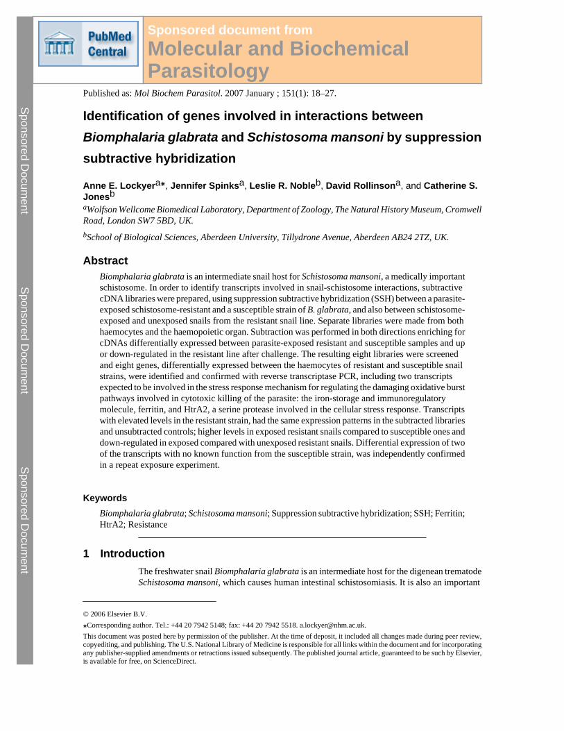
Identification of genes involved in interactions between Biomphalaria glabrata and Schistosoma mansoni by suppression subtractive hybridization Anne E. Lockyer a⁎ , Jennifer Spinks a , Leslie R. Noble b , David Rollinson a , and Catherine S. Jones b a Wolfson Wellcome Biomedical Laboratory, Department of Zoology, The Natural History Museum, Cromwell Road, London SW7 5BD, UK. b School of Biological Sciences, Aberdeen University, Tillydrone Avenue, Aberdeen AB24 2TZ, UK. Abstract Biomphalaria glabrata is an intermediate snail host for Schistosoma mansoni, a medically important schistosome. In order to identify transcripts involved in snail-schistosome interactions, subtractive cDNA libraries were prepared, using suppression subtractive hybridization (SSH) between a parasite- exposed schistosome-resistant and a susceptible strain of B. glabrata, and also between schistosome- exposed and unexposed snails from the resistant snail line. Separate libraries were made from both haemocytes and the haemopoietic organ. Subtraction was performed in both directions enriching for cDNAs differentially expressed between parasite-exposed resistant and susceptible samples and up or down-regulated in the resistant line after challenge. The resulting eight libraries were screened and eight genes, differentially expressed between the haemocytes of resistant and susceptible snail strains, were identified and confirmed with reverse transcriptase PCR, including two transcripts expected to be involved in the stress response mechanism for regulating the damaging oxidative burst pathways involved in cytotoxic killing of the parasite: the iron-storage and immunoregulatory molecule, ferritin, and HtrA2, a serine protease involved in the cellular stress response. Transcripts with elevated levels in the resistant strain, had the same expression patterns in the subtracted libraries and unsubtracted controls; higher levels in exposed resistant snails compared to susceptible ones and down-regulated in exposed compared with unexposed resistant snails. Differential expression of two of the transcripts with no known function from the susceptible strain, was independently confirmed in a repeat exposure experiment. Keywords Biomphalaria glabrata; Schistosoma mansoni; Suppression subtractive hybridization; SSH; Ferritin; HtrA2; Resistance 1 Introduction The freshwater snail Biomphalaria glabrata is an intermediate host for the digenean trematode Schistosoma mansoni, which causes human intestinal schistosomiasis. It is also an important © 2006 Elsevier B.V. ⁎Corresponding author. Tel.: +44 20 7942 5148; fax: +44 20 7942 5518. [email protected]. This document was posted here by permission of the publisher. At the time of deposit, it included all changes made during peer review, copyediting, and publishing. The U.S. National Library of Medicine is responsible for all links within the document and for incorporating any publisher-supplied amendments or retractions issued subsequently. The published journal article, guaranteed to be such by Elsevier, is available for free, on ScienceDirect. Sponsored document from Molecular and Biochemical Parasitology Published as: Mol Biochem Parasitol. 2007 January ; 151(1): 18–27. Sponsored Document Sponsored Document Sponsored Document
-
Upload
independent -
Category
Documents
-
view
1 -
download
0
Transcript of Identification of genes involved in interactions between Biomphalaria glabrata and Schistosoma...
Identification of genes involved in interactions betweenBiomphalaria glabrata and Schistosoma mansoni by suppressionsubtractive hybridization
Anne E. Lockyera⁎, Jennifer Spinksa, Leslie R. Nobleb, David Rollinsona, and Catherine S.JonesbaWolfson Wellcome Biomedical Laboratory, Department of Zoology, The Natural History Museum, CromwellRoad, London SW7 5BD, UK.
bSchool of Biological Sciences, Aberdeen University, Tillydrone Avenue, Aberdeen AB24 2TZ, UK.
AbstractBiomphalaria glabrata is an intermediate snail host for Schistosoma mansoni, a medically importantschistosome. In order to identify transcripts involved in snail-schistosome interactions, subtractivecDNA libraries were prepared, using suppression subtractive hybridization (SSH) between a parasite-exposed schistosome-resistant and a susceptible strain of B. glabrata, and also between schistosome-exposed and unexposed snails from the resistant snail line. Separate libraries were made from bothhaemocytes and the haemopoietic organ. Subtraction was performed in both directions enriching forcDNAs differentially expressed between parasite-exposed resistant and susceptible samples and upor down-regulated in the resistant line after challenge. The resulting eight libraries were screenedand eight genes, differentially expressed between the haemocytes of resistant and susceptible snailstrains, were identified and confirmed with reverse transcriptase PCR, including two transcriptsexpected to be involved in the stress response mechanism for regulating the damaging oxidative burstpathways involved in cytotoxic killing of the parasite: the iron-storage and immunoregulatorymolecule, ferritin, and HtrA2, a serine protease involved in the cellular stress response. Transcriptswith elevated levels in the resistant strain, had the same expression patterns in the subtracted librariesand unsubtracted controls; higher levels in exposed resistant snails compared to susceptible ones anddown-regulated in exposed compared with unexposed resistant snails. Differential expression of twoof the transcripts with no known function from the susceptible strain, was independently confirmedin a repeat exposure experiment.
KeywordsBiomphalaria glabrata; Schistosoma mansoni; Suppression subtractive hybridization; SSH; Ferritin;HtrA2; Resistance
1 IntroductionThe freshwater snail Biomphalaria glabrata is an intermediate host for the digenean trematodeSchistosoma mansoni, which causes human intestinal schistosomiasis. It is also an important
© 2006 Elsevier B.V.⁎Corresponding author. Tel.: +44 20 7942 5148; fax: +44 20 7942 5518. [email protected] document was posted here by permission of the publisher. At the time of deposit, it included all changes made during peer review,copyediting, and publishing. The U.S. National Library of Medicine is responsible for all links within the document and for incorporatingany publisher-supplied amendments or retractions issued subsequently. The published journal article, guaranteed to be such by Elsevier,is available for free, on ScienceDirect.
Sponsored document fromMolecular and BiochemicalParasitology
Published as: Mol Biochem Parasitol. 2007 January ; 151(1): 18–27.
Sponsored Docum
ent Sponsored D
ocument
Sponsored Docum
ent

model invertebrate and currently the subject of a large-scale genome project (seehttp://biology.unm.edu/biomphalaria-genome/). Interactions between snails and schistosomesare complex and there is a need to elucidate pathways involved in snail-parasite interactionsand identify those factors involved in the intricate balance between the snail internal defencesystem (IDS) and trematode infectivity mechanisms that determine the success or failure of aninfection (for a review see [1]). Compatible snail-trematode infections often reflect theparasite's capacity to avoid or interfere with the innate response of the snail. The term ‘resistant’can be applied to those individuals within a single snail species that are able to evade infectionby a species or strain of schistosome that is normally capable of parasitizing that species ofsnail.
Relatively little is known of the molluscan IDS compared to vertebrate immune systems.Molluscs appear to lack an adaptive immune system like that found in vertebrates and, instead,are considered to use various innate mechanisms involving cell-mediated and humoralreactions that interact to recognize and eliminate invading pathogens or parasites inincompatible or resistant snails (for reviews see [2–4]). However, a diverse family of fibrinogenrelated proteins (FREPs) containing immunoglobulin-like domains has been discovered in B.glabrata and may play a role in snail defence [5]. Circulating haemocytes (macrophage-likedefence cells) in the snail haemolymph are known to aggregate in response to trauma,phagocytose small particles (bacteria and fungi) and encapsulate larger ones, such astrematodes. Final killing is effected by haemocyte-mediated cytotoxicity mechanismsinvolving non-oxidative and oxidative pathways, including lysosomal enzymes and reactiveoxygen/nitrogen intermediates [6,7].
Despite many expression analyses aimed at identifying defence related genes involved in snail-trematode interactions, including subtractive hybridization [8], differential display [9–14] andexpressed sequence tag programmes [15,16], much remains to be discovered concerningspecific molecules mediating the defensive events in snail intermediate hosts, in particular theresponse shown by resistant or incompatible snails. In this study a sensitive, widely used PCR-based subtraction approach, suppression subtractive hybridization [17] and a PCR techniquefor amplifying the initial cDNA samples [18], were used to investigate how B. glabrataresponds to S. mansoni infection. Previous SSH studies in B. glabrata examined the wholehead-foot region [19], however, with the combined PCR approach it was possible to examineindividual tissues within the snail and focus on haemocytes, the primary effector cells involvedin the snail immune defence and the haemopoietic organ, responsible for producing thehaemocytes. SSH allows a comparison of two mRNA pools simultaneously and theexperiments were chosen to investigate gene expression in resistant snails. Parasite-exposedresistant and susceptible snail lines were compared as well as exposed and unexposed resistantsnails in order to see which genes are up or down-regulated in response to exposure to theparasite and to identify genes involved specifically in the defence response of resistant snails.
2 Materials and methods2.1 Snail material and parasite exposure
The first experiment used 60 adult B. glabrata snails from susceptible strain (NHM Accessionnumber 1742) and 60 from resistant strain (NHM Accession number 3017, derived from BS-90[20]). The snails were held overnight in autoclaved snail water with 100 μg/ml ampicillin tominimize bacterial contamination. Each snail was individually exposed to 10 schistosomemiracidia (Belo Horizonte strain). This experiment was also repeated to test the expression ofidentified transcripts. A second experiment used 120 snails from the resistant snail strain, butonly 60 of the snails were exposed to the parasite as described before, while 60 were keptunexposed in identical conditions as a control. The sampling regimes for both experimentswere the same. Twelve snails from each sample group were taken at 5 time periods, 2, 4, 6, 8
Lockyer et al. Page 2
Published as: Mol Biochem Parasitol. 2007 January ; 151(1): 18–27.
Sponsored Docum
ent Sponsored D
ocument
Sponsored Docum
ent

and 24 h after exposure to the parasite. The material from each time point was pooled to givesufficient RNA for the SSH and the extended sampling regime was necessary to allow timefor the snails to be dissected. This also meant that all the transcripts that are expressed over thefirst 24 h after infection would be obtained. Snails were swiftly killed by decapitation, and allthe exuded haemolymph collected. The haemolymph was pooled for each sampling time andsnail strain and the haemocytes pelleted by spinning at 10,000 × g at 4 °C for 20 min. The pelletwas frozen in liquid nitrogen and stored at −80 °C. Snails were preserved in 1 ml RNAlater(Ambion Inc., Texas, USA) and stored at 4 °C until dissection. The haemopoietic organ,responsible for producing haemocytes that have an active role in the snail internal defencesystem (reviewed in [6]) was dissected for each snail (preservation in RNAlater makes thisorgan easy to identify and dissect cleanly from surrounding tissues) and pooled for each strainfrom all time points.
2.2 RNA extraction and SMART cDNA synthesisTotal RNA was extracted from haemopoietic organ or haemocyte pools using the SV RNAextraction kit (Promega UK Ltd., Southampton, UK) according to the manufacturer's protocol.This kit includes DNAse treatment to eliminate genomic DNA contamination. cDNA synthesisand subsequent amplification was performed using 1 μg total RNA with the SMART™ PCRcDNA synthesis kit (CLONTECH) according to the manufacturer's instructions.
2.3 Suppression subtractive hybridization and library constructionEight independently subtracted libraries were made using the PCR select kit (BD Clontech).These were resistant minus susceptible and vice versa for both the haemopoietic organ andhaemocytes from the first experiment and exposed minus unexposed snails of the resistant lineand vice versa from the second experiment again both from the haemocytes and haemopoieticorgan. The subtraction was carried out as described in the kit manufacturer's instructions.However, six primary amplification PCRs, followed by a secondary PCR from each primaryPCR were carried out and then these PCRs were pooled for library construction. This multiplesampling of each subtracted pool was designed to minimize sampling effects, wherebytranscripts may be present or absent by chance. The libraries were made by ligating 3 μl ofeach pool of secondary PCRs into pGem-T easy vector (Promega) and transforming JM109cells according to the manufacturer's instructions. The entire library was plated out onto 10 LBagar plates. From each library 2× 96 white colonies were selected for overnight culture.
2.4 Differential screeningDuplicate filters were made for each library by spotting 2× 96 clones grown overnight in LB,onto Biodyne B membrane (PALL) using dot-blotting apparatus (ATTO Corp. Tokyo, Japan).The membrane was soaked in 2× SSC prior to loading in the press. Then 50 μl 2× SSC withdilute bromophenol blue was sucked through followed by 20 μl LB culture. The bacteria werethen denatured with 50 μl denaturing solution (1.5 M NaCl, 0.5 M NaOH), then neutralizedwith 50 μl of 1.5 M NaCl, 0.5 M Tris pH 7.5 solution for 2 min, and rinsed in 50 μl 2× SSC.The filters were air dried and stored at RT.
For each pair of duplicate filters, one was probed with the library from which it was made, andone with the reciprocal subtracted library, so for example the haemocyte resistant specificlibrary filters were probed with the haemocyte resistant specific library as a probe, as well asusing the susceptible haemocyte library as a probe. In each case the probe used was 10–15 μlof cleaned secondary PCR, previously digested with the restriction enzymes, SmaI and RsaI(Boerhinger Mannheim) to remove adapter sequences and labelled with digoxigenin using DIGHigh Prime DNA labeling detection starter kit II (Roche). The filters were visualized byautoradiography. Positive spots were identified as those that were clearly present on the filter
Lockyer et al. Page 3
Published as: Mol Biochem Parasitol. 2007 January ; 151(1): 18–27.
Sponsored Docum
ent Sponsored D
ocument
Sponsored Docum
ent

probed with the corresponding library while completely absent from the reverse subtractedlibrary.
2.5 Sequence analysisAll identified fragments were sequenced using BigDye and run on an ABI 377. The sequencetraces were examined using Sequencher and all vector and adaptor sequences removed.Sequences were compared to GenBank using BlastN BlastX and tBlastX using the programSeqtools (http://www.seqtools.dk/.) All sequences were submitted to GenBank (Accessionnumbers DY523246–DY523271).
2.6 Semi-quantitative RT-PCRPCR was carried out using the amplified fractions as a template; this confirms the presence orabsence of the fragment in the enriched libraries. PCR was also carried out on the un-subtractedcontrol fractions—these consist of material that was amplified from the same experiment, butwhich was not subtracted. It was necessary to use amplified material for these PCRs due toRNA being limited.
For each identified candidate specific primers were designed and synthesized. Semi-quantitative PCR used 0.4 μM Forward and Reverse primers (see supplementary material), 1×advantage polymerase buffer, 0.2 mM dNTPs and 0.4 μl 50× advantage polymerase (BDClontech). Cycling conditions were 94 °C for 30 s then cycles of 94 °C for 30 s, 58 °C for 30 sand 72 °C for 30 s. PCRs were cycled for a total of 33 cycles, but 5 μl of each reaction wasremoved after 18, 23, and 28 cycles to provide a semi-quantitative result—at lower cyclenumber the product in the reaction is proportional to the amount of starting material allowingus to ascertain if the levels differ, more effectively than after full cycling. These were runtogether on a 2% agarose gel to allow a semi-quantitative estimate of the presence of thetranscript in the template. Semi-quantitative RT-PCR of actin (using B. glabrata specific actinprimers F: TGGTGCCTTTTCTTCTCTAATTGC; R:GAAAGTGTGATGCCAGATCTTCTC) was used to assess the comparability of samples forthe unsubtracted control material to confirm that equivalent quantities of cDNA had been usedfor the specific primers. PCR and cycling conditions were the same as for specific PCRs. Sincethe subtracted libraries had non-differentially expressed genes (such as actin) subtracted fromthem, it is not possible to use this type of control for these samples.
For the repeat experiment, 1 μl cDNA (prior to SMART amplification) was used and RT-PCRperformed as above. For four of the transcripts, no amplification products were obtained fromthis cDNA so SMART-amplified cDNA was used as above. Actin PCRs were carried outsimultaneously on both unamplified and SMART-amplified cDNA as a control. To assessstatistically differential expression of these transcripts, integrated optical density (IOD)measurements were obtained for each amplified band, as a proxy for gene expression(Labworks™, Ultra Violet Products, Cambridge), and a matched paired t-test performed foreach transcript, using log IOD values normalized to actin.
3 Results3.1 Differential screening of cDNA libraries
SSH was used to make eight subtracted libraries; forward and reverse subtraction of resistantand susceptible material from haemocytes and the haemopoietic organ made two resistant-specific and two susceptible-specific libraries, one for each tissue type; and from the secondexperiment, forward and reverse subtraction of parasite-exposed and unexposed material fromthe resistant line, again from the same tissue types, made two exposed-specific and twounexposed-specific libraries. Differential screening of the 8 subtracted libraries identified 24
Lockyer et al. Page 4
Published as: Mol Biochem Parasitol. 2007 January ; 151(1): 18–27.
Sponsored Docum
ent Sponsored D
ocument
Sponsored Docum
ent
differentially expressed positive candidate transcripts, all of which were sequenced. Thesecandidates were selected by presence or absence on the filters (see supplementary data),quantitative differences were ignored since the filter contained spotted bacterial colonies ratherthan quantified DNA. Sequencing revealed 5 clusters (2 containing 6 clones each and 3 with2 clones each), and 6 unique sequences, resulting in 11 different candidate clusters orsequences.
Nine of the candidate sequences originated from haemocyte material, eight of which werederived from the first experiment comparing exposed resistant and susceptible snail strains.Five different sequences from the resistant minus the susceptible library were identified;Clusters 2–5 and one single sequence ZB9413. Three positives were identified from thesusceptible minus resistant library, two unique sequences (ZB9365 and ZBA105) and onecluster of 6 sequences (Cluster 1), which are transcripts specific to the haemocyte susceptiblelibrary. A single transcript (ZBA3283) was identified from the second experiment of resistant-exposed minus resistant-unexposed library.
Only 2 clear positive transcripts were obtained from the haemopoietic organ material, one(ZBA2946) from the resistant unexposed minus exposed library, and one (ZB9039) from theresistant exposed minus susceptible exposed library.
3.2 Blast resultsAll the sequences were compared to the non-redundant and expressed sequence tag (EST)sections of GenBank using BlastN, BlastX and tBlastX (for details see supplementarymaterial). Using the criteria of an E value of <1 × 10⁻⁴ for a significant match, only four hadsignificant BLAST hits in the non-redundant section of GenBank. Cluster 5 (six sequences)identified soma ferritin from Lymnaea stagnalis [21], both at the nucleotide and protein level,as well as a number of other ferritin heavy chain sequences. Cluster 4 (two sequences) identifieda fibrinogen related protein 3.2 from B. glabrata (AY028461) [22], but only at the nucleotidelevel—BlastX searches produced no matches. The section of sequence identified is withinintron 3 (bases 6282–6112) of this protein as it is described on GenBank. However a BlastNsearch of ESTs identified nine other B. glabrata ESTs, and since this transcript also containeda poly(A) tail (more than 26 bp long), a poly adenylation signal (24 bp upstream from the startof the polyA sequence) and a 147 amino acid long open reading frame, the sequence is definitelyderived from mRNA. The other three sequences from the resistant specific library identifiedno significant matches in the non-redundant section of GenBank, but all identified previouslysequenced B. glabrata transcripts from dbEST. Cluster 2 initially identified 34 B. glabrataESTs, but the resulting contig identified a further 11 ESTs producing a final 1493 bp contigcomposed of 45 ESTs, all Open Reading frame ESTs (ORESTES) [23] from both susceptibleand resistant snail lines, from whole body and ovotestis. This extended sequence still producedno other significant database matches, but contained a 369 aa long open reading frame (from1 to 1107 bp). Cluster 3 identified 46 ESTs, all from haemocyte libraries [16] apart from onefragment (CB350578) identified from a differential display experiment comparing the effectof S. mansoni and Echinostoma caproni excretory-secretory products on Bge cells, but notinvestigated further in that study [14]. The resulting 619 bp contig did not identify any furtherB. glabrata ESTs and the slightly extended sequence had no additional significant hits. Of thethree sequences from the susceptible library, only one (ZBA105) identified a sequence on thedatabase, but this was a putative hypothetical protein from C. elegans, which provides noinformation as to the function of this protein. The sequence from the exposed-specific libraryfrom the resistant snail line, ZBA3283, did not produce a significant hit, but examining boththe BlastN and BlastX results showed that all the sequences identified were mitogen activatedprotein kinase (MAPK), based on the first 120 bases of the transcript fragment. This translatedsection identified the 3′ section of the catalytic domain of serine/threonine kinase with a BlastP
Lockyer et al. Page 5
Published as: Mol Biochem Parasitol. 2007 January ; 151(1): 18–27.
Sponsored Docum
ent Sponsored D
ocument
Sponsored Docum
ent
search at NCBI entrez Blast interface (http://www.ncbi.nlm.nih.gov/entrez) (E value = 0.001)and suggests that the transcript fragment obtained from the SSH (which has a polyA tail)contains only the very end of the coding region of MAPK followed by the 3′ UTR, and thiscoding section was insufficient to produce a significant Blast result. It seems probable that thisis a MAPK-like sequence but more sequence would be required to confirm its homology toMAPK. One of the transcript fragments (ZB9039) from the haemopoietic organ showed nosignificant database match, while the other (ZBA2946) was significantly similar to a serineprotease HTRA2, from many organisms including mouse and human [24].
3.3 Confirmation of differential expressionSemi-quantitative RT-PCR of the enriched pool confirmed differential expression for 8 of the11 fragments (Figs. 1 and 2a), justifying the stringent selection criteria used to select candidatesfrom the filters. Specific PCRs for each identified transcript were carried out to determine theexpression of each not only in the subtracted fraction, which consists of the enriched pool ofproducts, but also in an unsubtracted control, which represents the original unsubtracted poolof transcripts. This amplified control fraction was used as only very small amounts of RNAcould be obtained from the tissues used in these experiments.
Seven of the nine transcripts obtained from haemocyte material were confirmed asdifferentially expressed, including four derived from resistant-specific library and all threefrom the susceptible-specific library (Fig. 1). Cluster 2 from the resistant minus susceptiblelibrary and Clone ZBA3283 (putative MAPK-like sequence) obtained from the secondexperiment of resistant exposed minus resistant unexposed library could not be confirmed asdifferentially expressed (results not shown). For both ferritin and ZB9413 the products arenoticeably present in both the subtracted fraction and the unsubtracted resistant-specificfraction and absent in both the susceptible-specific subtracted and unsubtracted fractions,demonstrating their presence in the original cDNA population from the resistant snails andabsence from the susceptible one (Fig. 1a and d). Clusters 3 and 4 each show products presentin the resistant subtracted fraction as well as in the original control pool, but absent or presentonly at higher cycle numbers in the susceptible subtracted fraction. Although there are productsseen in the original unsubtracted susceptible cDNA, they were only observed at higher cyclenumbers (Fig. 1b and c) indicating that although the sequences are not resistant specific, thereappears to be a difference in the level of expression between the two strains. The threesusceptible specific sequences, including one with a match to a putative hypothetical protein,also show confirmed differential expression (Fig. 1e–g).
Differential expression for only one out of the two transcripts obtained from the haemopoieticorgan libraries could be confirmed by semi-quantitative RT-PCR. Examining the unexposedversus exposed resistant snail libraries, the serine protease fragment is clearly present in theunexposed fraction while absent from the exposed (Fig. 2a). Semi-quantitative RT-PCR of thetranscript of unknown sequence, ZB9039, originating from resistant exposed minus susceptibleexposed haemopoietic organ libraries did not confirm a change in expression (result notshown).
3.4 Characterization of differentially expressed transcripts in other subtracted librariesHaving examined and confirmed the expression patterns of eight transcripts using the librariesfrom which they were isolated, we also investigated the expression of these transcripts in theother independently subtracted libraries (i.e. from the other experiment and tissue type), againusing semi-quantitative PCR on the subtracted and unsubtracted fractions obtained from theSSH.
Lockyer et al. Page 6
Published as: Mol Biochem Parasitol. 2007 January ; 151(1): 18–27.
Sponsored Docum
ent Sponsored D
ocument
Sponsored Docum
ent

3.4.1 Transcripts from the resistant libraries—The expression of the four transcriptswith elevated expression in haemocytes from resistant exposed snails (compared withsusceptible exposed strains) was independently examined in the exposed resistant comparedwith unexposed resistant subtractions (Fig. 3). This showed in all cases elevated expression inunexposed resistant snails relative to exposed snails. When expression of these transcripts fromhaemocyte libraries was examined in haemopoietic organ material from either resistantexposed versus susceptible exposed or exposed versus unexposed resistant libraries either nodifferences were detected or no PCR products were obtained for the majority of the fourtranscripts. Only with Cluster 3 did the material from the haemopoietic organ mirror that fromthe haemocytes with more product (at lower cycle number) in the material from the resistantsnails compared with that from the susceptible snails. Cluster 3 also showed, again only at lowcycle numbers, more product in the unexposed haemopoietic organ material compared withthe exposed, in the resistant snail line.
3.4.2 Transcripts from the susceptible libraries—Of the fragments with elevatedtranscript levels in the susceptible strain identified from haemocytes (Fig. 3) both Cluster 1and ZB9365 showed increased expression in the susceptible enriched libraries from thehaemopoietic organ, although in both cases, the transcript was not present at detectable levelsin the control fractions, only after enrichment. No product was detectable in the haemocytematerial from the exposed or unexposed material derived from the resistant line, but as theseare susceptible specific products this is not unexpected. Only ZBA105, hypothetical protein,was detected in the resistant material and was confirmed present at higher levels in haemocytematerial of unexposed resistant snails compared to exposed snails, but this fragment was notfound in haemopoietic organ derived material.
3.4.3 Transcripts from the unexposed libraries—The expression of ZBA2946(serine protease HtrA2), the fragment identified from the unexposed resistant library fromhaemopoietic organ was also examined in the independently subtracted haemocyte materialfrom the same experiment and this expression pattern seems to mirror that found in thehaemopoietic organ material (Fig. 2b). However, on examining the resistant versus susceptiblelibraries, in the haemopoietic organ the susceptible and resistant controls demonstrate nodifference in levels of the transcript although it is absent from the resistant specific fraction(Fig. 2c). Finally examining the material from the haemocytes, the fragment is present only inthe resistant fraction, both subtracted and unsubtracted control (Fig. 2d). This means thatalthough the serine protease HtrA2 was identified from different material, it shows the sameexpression patterns as ferritin and the other fragments identified from the resistant specificlibrary, present in higher levels in the resistant specific haemocyte libraries and demonstratingelevated levels in the unexposed compared to the exposed haemocyte libraries from theresistant material.
3.5 Confirmation of differential expression in exposed resistant and susceptible snailsSince all of the fragments identified demonstrated differential expression between resistantand susceptible snail haemocytes, this experiment was repeated and the expression of eachtranscript examined in the haemocytes of these snails (Fig. 4). Using integrated optical densitymeasurements for each band, significant differential expression was directly confirmed for twosusceptible specific sequences, Cluster 1 and ZB9365, both transcripts with unknown function(Fig. 4a). Significantly different expression was not confirmed for two other sequencesidentified from the resistant library, Clusters 3 and 4 (FREP-intron sequence) (Fig. 4a). Theother fragments could not be detected directly in cDNA from the repeated experiment (resultsnot shown) suggesting the expression of these genes was very low. All were detected inamplified cDNA but did not demonstrate significantly different expression in this repeatexperiment (Fig. 4b). Since these transcripts were all found to be down-regulated in resistant
Lockyer et al. Page 7
Published as: Mol Biochem Parasitol. 2007 January ; 151(1): 18–27.
Sponsored Docum
ent Sponsored D
ocument
Sponsored Docum
ent
snails after exposure it seems likely that in the repeated experiment these genes were affectedby parasite exposure in both the resistant and susceptible snails, making detection difficult.
4 DiscussionIn this study, haemocyte suppressive subtractive libraries of S. mansoni-exposed resistant andsusceptible B. glabrata lines were created to identify defence-relevant genes that are involvedspecifically in the resistant response. We also examined parasite-exposed and unexposedresistant snails to see which genes were differentially regulated in response to S. mansoniinfection. These aims were also addressed using haemopoietic organ (tissue responsible forproducing the haemocytes) SSH libraries using the same snail lines and exposures. Differentialscreening of 8 subtracted libraries produced 11 sequence fragments that were strong candidatesfor differential expression. RT-PCR using the original subtracted and control materialconfirmed differential expression of eight of these transcripts, seven of which were derivedfrom haemocyte libraries. Only four had significant Blast hits in GenBank, three fromhaemocyte libraries (ferritin, FREP intron sequence, hypothetical protein) and one from ahaemopoietic organ library (serine protease HtrA2), while three show no homology tocharacterized genes on GenBank, although several do identify previously sequenced ESTs withno function ascribed to them. All the fragments demonstrating elevated levels in the resistantstrain, had the same expression patterns in the subtracted libraries and unsubtracted controls;higher levels in exposed resistant snails compared to susceptible ones and down-regulated inexposed compared with unexposed resistant snails. Clear differential expression of twosusceptible-specific fragments (Cluster 1 and ZB9365) with no known function was shown tobe repeatable.
4.1 Fibrinogen related proteins (FREPs)Cluster 4 identified a section of sequence within intron 3 of one FREP molecule, a fibrinogen-related protein 3-2 precursor (GenBank accession number AY028461) [22]. FREPs arefibrinogen related proteins, which demonstrate increased expression following parasiteinfection, and bind to parasite antigens [22]. The sequences in Cluster 4 clearly contained apoly (A) tail, a poly adenylation signal and an 147 amino acid long open reading frame whichstrongly support the supposition that they are expressed genes and this suggests the interestingpossibility that there may be coding regions contained in the published FREP intron sequenceswhich should be investigated further, particularly since alternative splicing to producetruncated forms is a feature of the FREP gene family [25]. Blast searches with Cluster 4sequence identified nine other B. glabrata ESTs, four of which contained this section ofsequence that identified the FREP gene. No ESTs from other organisms were identified withthis sequence. In some cases, however, these other ESTs did not align outside this region.Therefore another explanation for finding this sequence in the intron could be that it is acommon B. glabrata genomic sequence motif. However, without recovering more sequencefrom this transcript and resolving or refuting its homology with FREP genes it is not possibleto speculate further upon its function.
4.2 Cellular stress response proteins4.2.1 Ferritin—Cluster 5 had significant sequence homology to soma ferritin from thefreshwater snail L. stagnalis, which has similarities to the H-subunit type of vertebrate ferritinsand contains the iron-responsive element (IRE) of vertebrate ferritin mRNAs [21]. Ferritin isthe major iron repository in eukaryotic cells and plays a role in regulating iron homeostasis,exhibiting a high degree of structural conservation in bacteria, plants and animals [26]. As wellas iron regulation, ferritin H subunit demonstrates ferroxidase activity and may have a role asa protectant against oxygen free radical-mediated damage by limiting the amount of Fe(II)available for the generation of reactive oxygen species (ROS) [27–31].
Lockyer et al. Page 8
Published as: Mol Biochem Parasitol. 2007 January ; 151(1): 18–27.
Sponsored Docum
ent Sponsored D
ocument
Sponsored Docum
ent
Two B. glabrata ESTs with homology to ferritin have been previously reported. Nowak et al.[19] identified a 303 bp fragment (accession number CD760630) from a head/foot SSH libraryfrom resistant B. glabrata 12 h post infection with S. mansoni which demonstrated littledifferential expression, and Raghavan et al. [15] found a 492 bp EST (accession numberAW739853) from a resistant strain (BS-90) haemocyte library 5 h post-exposure to S.mansoni. These two sequences are identical for the 303 bp, which they both contain. However,Blast analysis with Cluster 5 revealed that it was different to these ferritin-like transcripts fromearlier studies; although all identified the soma ferritin from L. stagnalis as the closest proteinmatch, Cluster 5 sequence had only 80% identity with CD760630 (127/158 bp) and 79% withAW739853 (127/224 bp) from the Blast results. Cluster 5 did identify two other B. glabrataESTs AW740322 and AW740403 from the haemocytes of unexposed BS-90 (resistant) snails[15] with 100% and 97% identities, respectively. Ferritin H subunit genes form a multigenefamily [32,33], and therefore it seems likely that we have identified a second B. glabrata ferritingene. Two ferritin genes have also been identified in the oyster Crassostrea gigas [34,35] andin L. stagnalis,[21,36] where the sequences of both are more similar to ferritin H subunits thanL type and contain H-specific amino acid residues [21].
The function of ferritin in invertebrates is less well characterized than in mammals, but it hasbeen shown to be upregulated by LPS in echinoderms [37], in white spot syndrome virus(WSSV)-resistant shrimp compared to susceptible individuals [38], in response to anoxicconditions in Litterina littorea [39] and by nitric oxide (NO) in the snail Helix pomatia [40].The significance of elevated levels of ferritin transcripts in resistance snails could be linked tothe oxidative burst demonstrated by snails in response to parasite exposure (reviewed in [6])since H2O2 induces ferritin in HeLa cells [31] and in B. glabrata the production of H2O2demonstrated when the respiratory burst is artificially stimulated, appears greater in a resistantsnail line [41]. Cytosolic copper/zinc superoxide dismutase (Cu/Zn SOD), which catalyses theconversion of superoxide to H2O2, has been shown to have a constitutive difference in mRNAlevels between one mostly resistant snail line and susceptible snails [42] and a significantassociation of one allele of the Cu/Zn SOD gene to the resistance phenotype has been identified[43]. Therefore, given the role of ferritin in protecting against oxidative damage in vertebrates,the increased levels of ferritin observed in resistant snails when compared to susceptible, maybe part of their response to parasite infection.
4.2.2 Serine protease HtrA2—Clone ZBA2946 sequence showed considerablehomology to a mouse serine protease HtrA2 [24]. HtrA2 (also known as Omi) removes proteinsdamaged or denatured after elevated temperatures or oxidative stress (reviewed in [44]). HtrA2interacts with Mxi2 (an alternatively spliced form of p38 stress activated kinase) [45], and isthought to be involved in the cellular stress response in mammals [24].
The role of HtrA2 in B. glabrata is unknown, but, given the function of homologous proteinsin other organisms, it may be involved in the cellular stress response. Previous findings ofserine protease activity in B. glabrata include Bahgat et al. [46], who described trypsin-likeserine protease activity from B. glabrata haemocytes, a finding consistent with thesephagocytic cells mounting a cytotoxic attack against schistosome sporocysts. However,although the serine protease's activity varied among individual snails, there was no detectabledifference between snails susceptible or resistant to schistosome infection. One EST cluster(GenBank Accession no. CK989867) identified in a haemocyte cDNA library from snailsmaintained in non-axenic conditions [16], exhibits a portion of the Tryp-SPc (trypsin-likeserine protease), but this shows no homology to our sequence at the nucleotide level.
Lockyer et al. Page 9
Published as: Mol Biochem Parasitol. 2007 January ; 151(1): 18–27.
Sponsored Docum
ent Sponsored D
ocument
Sponsored Docum
ent
4.3 ConclusionsBoth ferritin and HtrA2, as stress response genes, would be expected to be stimulated inresponse to schistosome challenge, and elevated levels in resistant snails are consistent withthis. How then to account for the reduction in mRNA for both these genes that appears inparasite-exposed resistant snails compared to unexposed snails? One possible explanation maylie with the interaction between snail and parasite. The influence of the parasite must beconsidered since it manipulates the normal snail defence response, and this effect may begreater in susceptible snails than in resistant ones. It is possible that some effect of the parasitethat reduces the snails’ normal response to oxidative stress occurs, but is less significant inresistant snails, allowing them to overcome the challenge. This would result in the expressionpatterns seen for these genes and would also account for the low levels detected in the repeatedexperiment.
Two identified susceptible-specific transcripts (Cluster1 and ZB9365) were independentlyconfirmed as being present only in susceptible snails in the repeat experiment. Furthercharacterization of these transcripts with unknown function is required to determine if thesetranscripts are involved in the response of susceptible snails to parasite exposure and how thatresponse differs to that of resistant snails. Surprisingly six of the identified candidates did notshow differential expression in the repeat experiment. This could be due to a number of factors,including variability in the conditions of parasite exposure, loss of signal due to pooling thesamples over the 24 h time period (meaning that small or transient changes might not bedetected) and the fact that for four of the samples a product could only be detected faintly afteramplifying the cDNA, which again might prevent the detection of small differences inexpression.
In summary, we have identified several transcripts by differential screening of SSH librariesfrom B. glabrata that are expected to be involved in the stress response mechanism forregulating the damaging oxidative burst pathways involved in cytotoxic killing of the parasite.While no single transcript is likely to be solely responsible for conferring differingsusceptibility to S. mansoni infection, differences in gene expression demonstrate a differencein the snails’ response to infection and may indicate which mechanisms are employed by thesnails in their defence response. Whilst interesting candidate genes have been found using SSH,it is not a particularly efficient method for the analysis of gene expression, especially giventhat many of the candidates identified in this way did not show differential expression in arepeat experiment. A different approach to transcriptome analysis, such as using microarraytechnology, will enable a more thorough examination of changes in gene expression in B.glabrata, as a much larger number of genes can be analyzed simultaneously, using smalleramounts of RNA. The SSH libraries produced in this study, as well as larger libraries from amodification of the EST strategy, termed ORESTES (Open Reading Frame ESTs; [23]), havebeen used to make the first version of a custom B. glabrata cDNA microarray. This will enablea more detailed investigation of the transcriptome in response to trematode infection in thissnail intermediate host. This approach has been used successfully for expression studies ofgender-associated gene transcripts in S. mansoni [47,48]. Differentially expressed snailtranscripts reported here are also included on the array, enabling them to undergo furtherscreening [49] for a more thorough understanding of their role within the snail internal defencesystem.
Appendix A Supplementary dataRefer to Web version on PubMed Central for supplementary material.
Lockyer et al. Page 10
Published as: Mol Biochem Parasitol. 2007 January ; 151(1): 18–27.
Sponsored Docum
ent Sponsored D
ocument
Sponsored Docum
ent

Acknowledgements
This work was carried out with funding from the Wellcome Trust. We would like to thank Jayne King and MikeAnderson, NHM, for snail and parasite culture and Julia Llewellyn-Hughes and Claire Griffin, NHM, for sequencingassistance. Cathy Jones was in receipt of a Royal Society of Edinburgh Sabbatical Fellowship whilst co-authoring thisarticle.
References[1]. Lockyer A.E. Jones C.S. Noble L.R. Rollinson D. Trematodes and snails: an intimate association.
Can J Zool 2004;82:251–269.[2]. van der Knaap W.P.W. Loker E.S. Immune mechanisms in trematode-snail interactions. Parasitol
Today 1990;6:175–182. [PubMed: 15463334][3]. Adema, C.M.; Loker, E.S. Specificity and immunobiology of larval digenean snail associations. In:
Fried, B.; Graczyk, T.K., editors. Advances in Trematode Biology. CRC Press; Florida: 1997. p.230-253.
[4]. Loker E.S. Adema C.M. Zhang S.M. Kepler T.B. Invertebrate immune systems—not homogeneous,not simple, not well understood. Immunol Rev 2004;198:10–24. [PubMed: 15199951]
[5]. Zhang S.M. Adema C.M. Kepler T.B. Loker E.S. Diversification of Ig superfamily genes in aninvertebrate. Science 2004;305:251–254. [PubMed: 15247481]
[6]. Bayne C.J. Hahn U.K. Bender R.C. Mechanisms of molluscan host resistance and of parasitestrategies for survival. Parasitology 2001;123:S159–S167. [PubMed: 11769280]
[7]. Bayne C.J. Origins and evolutionary relationships between the innate and adaptive arms of immunesystems. Integr Comp Biol 2003;43:293–299.
[8]. Miller A.N. Ofori K. Lewis F. Knight M. Schistosoma mansoni: use of a subtractive cloning strategyto search for RFLPs in parasite-resistant Biomphalaria glabrata. Exp Parasitol 1996;84:420–428.[PubMed: 8948331]
[9]. Schneider O. Zelck U.E. Differential display analysis of hemocytes from schistosome-resistant andschistosome-susceptible intermediate hosts. Parasitol Res 2001;87:489–491. [PubMed: 11411951]
[10]. Miller A.N. Raghavan N. FitzGerald P.C. Lewis F.A. Knight M. Differential gene expression inhaemocytes of the snail Biomphalaria glabrata: effects of Schistosoma mansoni infection. Int JParasitol 2001;31:687–696. [PubMed: 11336750]
[11]. Lockyer A.E. Jones C.S. Noble L.R. Rollinson D. Use of differential display to detect changes ingene expression in the intermediate snail host Biomphalaria glabrata upon infection withSchistosoma mansoni. Parasitology 2000;120:399–407. [PubMed: 10811281]
[12]. Lockyer A.E. Noble L.R. Rollinson D. Jones C.S. Schistosoma mansoni: resistant specific infection-induced gene expression in Biomphalaria glabrata identified by fluorescent-based differentialdisplay. Exp Parasitol 2004;107:97–104. [PubMed: 15208043]
[13]. Jones C.S. Lockyer A.E. Rollinson D. Noble L.R. Molecular approaches in the study ofBiomphalaria glabrata—Schistosoma mansoni interactions: linkage analysis and gene expressionprofiling. Parasitology 2001;123:S181–S196. [PubMed: 11769282]
[14]. Coustau C. Mitta G. Dissous C. Schistosoma mansoni and Echinostoma caproni excretory-secretoryproducts differentially affect gene expression in Biomphalaria glabrata embryonic cells.Parasitology 2003;127:533–542. [PubMed: 14700189]
[15]. Raghavan N. Miller A.N. Gardner M. Comparative gene analysis of Biomphalaria glabratahemocytes pre- and post-exposure to miracidia of Schistosoma mansoni. Mol Biochem Parasitol2003;126:181–191. [PubMed: 12615317]
[16]. Mitta G. Galinier R. Tisseyre P. Gene discovery and expression analysis of immune-relevant genesfrom Biomphalaria glabrata hemocytes. Dev Comp Immunol 2005;29:393–407. [PubMed:15707661]
[17]. Diatchenko L. Lau Y.F.C. Campbell A.P. Suppression subtractive hybridization: a method forgenerating differentially regulated or tissue-specific cDNA probes and libraries. Proc Natl AcadSci USA 1996;93:6025–6030. [PubMed: 8650213]
Lockyer et al. Page 11
Published as: Mol Biochem Parasitol. 2007 January ; 151(1): 18–27.
Sponsored Docum
ent Sponsored D
ocument
Sponsored Docum
ent

[18]. Petalidis L. Bhattacharyya S. Morris G.A. Collins V.P. Freeman T.C. Lyons P.A. Globalamplification of mRNA by template-switching PCR: linearity and application to microarrayanalysis. Nucl Acids Res 2003:31.
[19]. Nowak T.S. Woodards A.C. Jung Y.H. Adema C.M. Loker E.S. Identification of transcriptsgenerated during the response of resistant Biomphalaria glabrata to Schistosoma mansoni infectionusing suppression subtractive hybridization. J Parasitol 2004;90:1034–1040. [PubMed: 15562603]
[20]. Paraense W.L. Correa L.R. Variation in susceptibility of populations of Austrolorbis glabratus to astrain of Schistosoma mansoni. Rev Inst Med Trop Sao Paulo 1963;5:15–22. [PubMed: 13941355]
[21]. von Darl M. Harrison P.M. Bottke W. cDNA cloning and deduced amino acid sequence of 2 ferritins—soma ferritin and yolk ferritin, from the snail Lymnaea stagnalis L. Eur J Biochem 1994;222:353–366. [PubMed: 7517354]
[22]. Adema C.M. Hertel L.A. Miller R.D. Loker E.S. A family of fibrinogen-related proteins thatprecipitates parasite-derived molecules is produced by an invertebrate after infection. Proc NatlAcad Sci USA 1997;94:8691–8696. [PubMed: 9238039]
[23]. Dias-Neto E. Correa R.G. Verjovski-Almeida S. Shotgun sequencing of the human transcriptomewith ORF expressed sequence tags. Proc Natl Acad Sci USA 2000;97:3491–3496. [PubMed:10737800]
[24]. Gray C.W. Ward R.V. Karran E. Characterization of human HtrA2, a novel serine protease involvedin the mammalian cellular stress response. Eur J Biochem 2000;267:5699–5710. [PubMed:10971580]
[25]. Zhang S.M. Loker E.S. The FREP gene family in the snail Biomphalaria glabrata: additionalmembers, and evidence consistent with alternative splicing and FREP retrosequences. Dev CompImmunol 2003;27:175–187. [PubMed: 12590969]
[26]. Theil E.C. Ferritin—structure, gene regulation, and cellular function in animals, plants, andmicroorganisms. Annu Rev Biochem 1987;56:289–315. [PubMed: 3304136]
[27]. Balla G. Jacob H. Balla J. Ferritin: a cytoprotective antioxidant strategem of endothelium. J BiolChem 1992;267:18148–18153. [PubMed: 1517245]
[28]. Cairo G. Tacchini L. Pogliaghi G. Anzon E. Tomasi A. Bernelli-Zazzera A. Induction of ferritinsynthesis by oxidative stress. J Biol Chem 1995;270:700–703. [PubMed: 7822298]
[29]. Rosen C.F. Poon R. Drucker D.J. UVB radiation-activated genes induced by transcriptional andposttranscriptional mechanisms in rat keratinocytes. Am J Physiol-Cell Physiol 1995;37:C846–C855.
[30]. Cozzi A. Corsi B. Levi S. Santambrogio P. Albertini A. Arosio P. Overexpression of wild type andmutated human ferritin H-chain in HeLa cells. In vivo role of ferritin ferroxidase activity. J BiolChem 2000;275:25122–25129. [PubMed: 10833524]
[31]. Orino K. Lehman L. Tsuji Y. Ayaki H. Torti S.V. Torti F.M. Ferritin and the response to oxidativestress. Biochem J 2001;357:241–247. [PubMed: 11415455]
[32]. Boyd D. Jain S.K. Crampton J. Barrett K.J. Drysdale J. Isolation and characterization of a cDNAclone for human ferritin heavy chain. Proc Natl Acad Sci USA 1984;81:4751–4755. [PubMed:6589621]
[33]. Costanzo F. Santoro C. Colantuoni V. Cloning and sequencing of a full length cDNA coding for ahuman apoferritin H-chain—evidence for a multigene family. EMBO J 1984;3:23–27. [PubMed:6323167]
[34]. Gueguen Y. Cadoret J.P. Flament D. Immune gene discovery by expressed sequence tags generatedfrom hemocytes of the bacteria-challenged oyster, Crassostrea gigas. Gene 2003;303:139–145.[PubMed: 12559575]
[35]. Durand J.P. Goudard F. Pieri J. Escoubas J.M. Schreiber N. Cadoret J.P. Crassostrea gigas ferritin:cDNA sequence analysis for two heavy chain type subunits and protein purification. Gene2004;338:187–195. [PubMed: 15315822]
[36]. Bottke W. Burschyk M. Volmer J. On the origin of the yolk protein ferritin in snails. Rouxs ArchDev Biol 1988;197:377–382.
[37]. Beck G. Ellis T.W. Habicht G.S. Schluter S.F. Marchalonis J.J. Evolution of the acute phaseresponse: iron release by echinoderm (Asterias forbesi) coelomocytes, and cloning of an echinodermferritin molecule. Dev Comp Immunol 2002;26:11–26. [PubMed: 11687259]
Lockyer et al. Page 12
Published as: Mol Biochem Parasitol. 2007 January ; 151(1): 18–27.
Sponsored Docum
ent Sponsored D
ocument
Sponsored Docum
ent
[38]. Pan D. He N.H. Yang Z.Y. Liu H.P. Xu X. Differential gene expression profile in hepatopancreasof WSSV-resistant shrimp (Penaeus japonicus) by suppression subtractive hybridization. DevComp Immunol 2005;29:103–112. [PubMed: 15450750]
[39]. Larade K. Storey K.B. Accumulation and translation of ferritin heavy chain transcripts followinganoxia exposure in a marine invertebrate. J Exp Biol 2004;207:1353–1360. [PubMed: 15010486]
[40]. Xie M. Hermann A. Richter K. Engel E. Kerschbaum H.H. Nitric oxide up-regulates ferritin mRNAlevel in snail neurons. Eur J Neurosci 2001;13:1479–1486. [PubMed: 11328343]
[41]. Bender R.C. Broderick E.J. Goodall C.P. Bayne C.J. Respiratory burst of Biomphalaria glabratahemocytes: Schistosoma mansoni-resistant snails produce more extracellular H2O2 thansusceptible snails. J Parasitol 2005;91:275–279. [PubMed: 15986600]
[42]. Goodall C.P. Bender R.C. Broderick E.J. Bayne C.J. Constitutive differences in Cu/Zn superoxidedismutase mRNA levels and activity in hemocytes of Biomphalaria glabrata (Mollusca) that areeither susceptible or resistant to Schistosoma mansoni (Trematoda). Mol Biochem Parasitol2004;137:321–328. [PubMed: 15383302]
[43]. Goodall C.P. Bender R.C. Brooks J.K. Bayne C.J. Biomphalaria glabrata cytosolic copper/zincsuperoxide dismutase (SOD1) gene: association of SOD1 alleles with resistance/susceptibility toSchistosoma mansoni. Mol Biochem Parasitol 2006;147:207–210. [PubMed: 16564582]
[44]. Pallen M.J. Wren B.W. The HtrA family of serine proteases. Mol Microbiol 1997;26:209–221.[PubMed: 9383148]
[45]. Zervos A.S. Faccio L. Gatto J.P. Kyriakis J.M. Brent R. Mxi2, a mitogen-activated protein kinasethat recognizes and phosphorylates Max protein. Proc Natl Acad Sci USA 1995;92:10531–10534.[PubMed: 7479834]
[46]. Bahgat M. Doenhoff M. Kirschfink M. Ruppel A. Serine protease and phenoloxidase activities inhemocytes of Biomphalaria glabrata snails with varying susceptibility to infection with the parasiteSchistosoma mansoni. Parasitol Res 2002;88:489–494. [PubMed: 12107469]
[47]. Hoffmann K.F. Johnston D.A. Dunne D.W. Identification of Schistosoma mansoni gender-associated gene transcripts by cDNA microarray profiling. Genome Biol 2002;3:1–12.
[48]. Fitzpatrick J.M. Johnston D.A. Williams G.W. An oligonucleotide microarray for transcriptomeanalysis of Schistosoma mansoni and its application/use to investigate gender-associated geneexpression. Mol Biochem Parasitol 2005;141:1–13. [PubMed: 15811522]
[49]. Yang G.P. Ross D.T. Kuang W.W. Brown P.O. Weigel R.J. Combining SSH and cDNA microarraysfor rapid identification of differentially expressed genes. Nucl Acids Res 1999;27:1517–1523.[PubMed: 10037815]
Lockyer et al. Page 13
Published as: Mol Biochem Parasitol. 2007 January ; 151(1): 18–27.
Sponsored Docum
ent Sponsored D
ocument
Sponsored Docum
ent
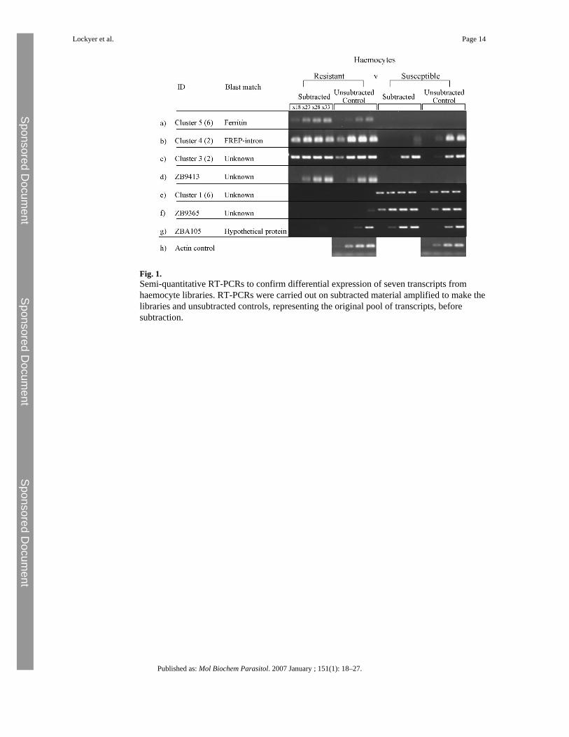
Fig. 1.Semi-quantitative RT-PCRs to confirm differential expression of seven transcripts fromhaemocyte libraries. RT-PCRs were carried out on subtracted material amplified to make thelibraries and unsubtracted controls, representing the original pool of transcripts, beforesubtraction.
Lockyer et al. Page 14
Published as: Mol Biochem Parasitol. 2007 January ; 151(1): 18–27.
Sponsored Docum
ent Sponsored D
ocument
Sponsored Docum
ent
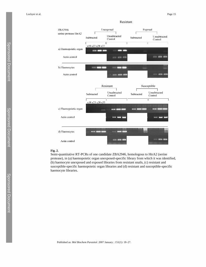
Fig. 2.Semi-quantitative RT-PCRs of one candidate ZBA2946, homologous to HtrA2 (serineprotease), in (a) haemopoietic organ unexposed-specific library from which it was identified,(b) haemocyte unexposed and exposed libraries from resistant snails, (c) resistant andsusceptible-specific haemopoietic organ libraries and (d) resistant and susceptible-specifichaemocyte libraries.
Lockyer et al. Page 15
Published as: Mol Biochem Parasitol. 2007 January ; 151(1): 18–27.
Sponsored Docum
ent Sponsored D
ocument
Sponsored Docum
ent

Fig. 3.Semi-quantitative RT-PCRs showing levels of gene expression of the confirmed differentiallyexpressed genes in the other subtracted libraries. SSH library—the library from which eachfragment was originally identified. Haem: Haemocyte; HO: Haemopoietic organ; R: resistant;S: susceptible; Ex: Parasite-exposed resistant; Un: unexposed resistant. M: MarkerHyperladder IV (Bioline); sub: subtracted fraction; unsub control: unsubtracted controlfraction.
Lockyer et al. Page 16
Published as: Mol Biochem Parasitol. 2007 January ; 151(1): 18–27.
Sponsored Docum
ent Sponsored D
ocument
Sponsored Docum
ent

Fig. 4.Semi-quantitative RT-PCRs with specific primers for the confirmed differentially expressedfragments on haemocytes from the repeated experiment, exposed resistant and susceptiblesnails. (a) PCRs using cDNA and (b) PCRs using amplified cDNA (for those fragments notdetected directly from cDNA) 0—no DNA control. For each candidate integrated opticaldensities were calculated from the bands and compared using a matched pairs t-test. *Significantly different expression (p < 0.05).
Lockyer et al. Page 17
Published as: Mol Biochem Parasitol. 2007 January ; 151(1): 18–27.
Sponsored Docum
ent Sponsored D
ocument
Sponsored Docum
ent