Nosocomial acquisition of Candida parapsilosis: An epidemiologic study

Contribution of CgPDR1-Regulated Genes in EnhancedVirulence of Azole-Resistant Candida glabrataSelene Ferrari1, Maurizio Sanguinetti2, Riccardo Torelli2, Brunella Posteraro2, Dominique Sanglard1*
1 Institute of Microbiology, University of Lausanne and University Hospital Center, Lausanne, Switzerland, 2 Institute of Microbiology, Universita Cattolica del Sacro Cuore,
Rome, Italy
Abstract
In Candida glabrata, the transcription factor CgPdr1 is involved in resistance to azole antifungals via upregulation of ATPbinding cassette (ABC)-transporter genes including at least CgCDR1, CgCDR2 and CgSNQ2. A high diversity of GOF (gain-of-function) mutations in CgPDR1 exists for the upregulation of ABC-transporters. These mutations enhance C. glabratavirulence in animal models, thus indicating that CgPDR1 might regulate the expression of yet unidentified virulence factors.We hypothesized that CgPdr1-dependent virulence factor(s) should be commonly regulated by all GOF mutations inCgPDR1. As deduced from transcript profiling with microarrays, a high number of genes (up to 385) were differentiallyregulated by a selected number (7) of GOF mutations expressed in the same genetic background. Surprisingly, thetranscriptional profiles resulting from expression of GOF mutations showed minimal overlap in co-regulated genes. Onlytwo genes, CgCDR1 and PUP1 (for PDR1 upregulated and encoding a mitochondrial protein), were commonly upregulatedby all tested GOFs. While both genes mediated azole resistance, although to different extents, their deletions in an azole-resistant isolate led to a reduction of virulence and decreased tissue burden as compared to clinical parents. As expectedfrom their role in C. glabrata virulence, the two genes were expressed as well in vitro and in vivo. The individualoverexpression of these two genes in a CgPDR1-independent manner could partially restore phenotypes obtained in clinicalisolates. These data therefore demonstrate that at least these two CgPDR1-dependent and -upregulated genes contribute tothe enhanced virulence of C. glabrata that acquired azole resistance.
Citation: Ferrari S, Sanguinetti M, Torelli R, Posteraro B, Sanglard D (2011) Contribution of CgPDR1-Regulated Genes in Enhanced Virulence of Azole-ResistantCandida glabrata. PLoS ONE 6(3): e17589. doi:10.1371/journal.pone.0017589
Editor: Robert Cramer, Montana State University, United States of America
Received November 30, 2010; Accepted January 27, 2011; Published March 9, 2011
Copyright: � 2011 Ferrari et al. This is an open-access article distributed under the terms of the Creative Commons Attribution License, which permitsunrestricted use, distribution, and reproduction in any medium, provided the original author and source are credited.
Funding: This work was supported by a grant of the Swiss Research National Foundation 31003A_127378 to DS. M.S. was supported by a grant from the Istitutodi Ricovero e Cura a Carattere Scientifico (IRCCS) ‘‘Lazzaro Spallanzani’’ (Strategic Research Program 2006, Italy) and from Universita Cattolica del S. Cuore (LineaD1, 2010). The funders had no role in study design, data collection and analysis, decision to publish, or preparation of the manuscript.
Competing Interests: The authors have declared that no competing interests exist.
* E-mail: [email protected]
Introduction
Candida glabrata is a haploid member of Ascomycetes normally
not found in the environment but which has rather adapted to
conditions found in mammals [1]. Among human fungal
pathogens, C. glabrata is often reported as the second most
prevalent species after Candida albicans [2,3]. C. glabrata can cause
mucosal and bloodstream infection (BSI) mainly in immuno-
compromised hosts. Worldwide, C. glabrata accounts for an average
11% of infections caused by Candida species, however this
proportion varies from 7 to 20% depending on geographical
locations [4].
C. glabrata infections can be treated with several antifungal
agents including amphotericin B, azoles and echinocandins [5,6].
However, C. glabrata can develop antifungal resistance and
especially to the class of azole antifungals. Azole resistance
surveillance studies have revealed a proportion varying from 10
to 20% of isolates with MIC values reaching clinical breakpoints
(e.g. 64 mg/ml for fluconazole, based on CLSI standards). Several
countries reported an increase in the proportion of azole-resistant
isolates from 2001 to 2007 [4]. C. glabrata is also known for
exhibiting intrinsically higher azole MIC values than C. albicans.
For example, the average of fluconazole MIC values of a C. glabrata
wild type population is near a value of 4 mg/ml, while it is
approximately 32-fold lower for C. albicans [7,8]. We and others
showed that azole resistance in C. glabrata was mediated almost
exclusively by enhanced drug efflux and overexpression of
multidrug transporters of the ATP Binding Cassette (ABC)
transporters. Several genes encoding these transporters were
identified including CgCDR1, CgCDR2 (PDH1) and CgSNQ2
[8,9,10,11,12]. Azole resistance in clinical isolates can be the
result of overexpression of single or several transporters [13]. The
understanding of regulatory circuits controlling the expression of
these genes has progressed in the recent years. A major regulator
of these genes, CgPDR1, was identified [14,15]. This gene belongs
to the family of zinc finger transcription factors and functionally
resembles PDR1 and PDR3 from the baker’s yeast Saccharomyces
cerevisiae. Deletion of CgPDR1 results in a loss of transcriptional
control of the major transporters involved in azole resistance and,
consequently, decreased resistance to these antifungals [14,15].
CgPDR1 exhibits mutations, so called gain-of-function (GOF)
mutations, which are responsible for intrinsic high expression of
ABC transporters and therefore constitute the molecular basis of
azole resistance in C. glabrata [13,14,15]. One striking feature of
GOF mutations is their high diversity among CgPDR1 alleles from
azole-resistant isolates. As many as 67 mutations conferring azole
resistance are described up to now [13,14,15,16,17]. GOF
mutations are found within several domains of the transcription
PLoS ONE | www.plosone.org 1 March 2011 | Volume 6 | Issue 3 | e17589
factor corresponding to putative functional elements inferred from
comparison to the S. cerevisiae PDR1 and PDR3 and including the
transcriptional activation domain, a regulatory domain and a so-
called middle homology region (MHR) which is found in several
zinc finger proteins [13,16].
Not only are GOF mutations in CgPDR1 important for azole
resistance in C. glabrata but also for fungal-host interactions. We
showed that GOF mutations were associated with enhanced
virulence and fitness in animal models of systemic infection [13].
This was unexpected since it is generally accepted that the
development of drug resistance in other microbes is usually
associated with costs in virulence or fitness. Secondary compen-
satory mechanisms can however restore the costs of resistance
development [18,19].
In this study we addressed in C. glabrata the identification of
genes behind the GOF-dependent virulence of CgPDR1. Because
we rationalized that some genes commonly expressed by GOF
mutations could be responsible for this effect, we analysed with
transcript profiling analysis C. glabrata isolates containing individ-
ual GOF mutations but in identical genetic backgrounds. Only
two genes (CgCDR1 and PUP1) were identified. We describe here
their relevance in the enhanced virulence mediated by CgPDR1
GOF mutations.
Results
Transcriptional analysis of GOF mutationsIn a previous study, we reported a high variety of gain-of-
function (GOF) mutations in the transcriptional activator CgPDR1
[13]. These mutations conferred azole resistance through the
differentiated upregulation of several ABC transporters including
CgCDR1, CgCDR2 and CgSNQ2. It is known that CgPDR1 controls
the expression of many other genes, some of which contain a
regulatory domain in their promoter matching the PDRE
(Pleiotropic Drug Responsive Element) described in S. cerevisiae
(TCCRYGSR) [14,16].
We were therefore interested to test whether the differentiated
expression pattern observed for a few genes as described earlier
[13] could be generalized to the entire transcriptome of C. glabrata.
In order to achieve this goal, labeled cRNA from mRNA isolated
in triplicates from strains containing seven different CgPDR1 GOF
was applied to oligonucleotides custom arrays. The selection of
GOFs was based on their occurrence in putative CgPdr1
functional domains including the regulatory domain (L280F,
R376W), the MHR (Y584C, T588A) and the activation domain
(D1082G, E1083Q). The GOF P822L was also selected since it
was previously associated with a strong upregulation of CgSNQ2 as
compared to other ABC-transporters [9]. The format of one-color
hybridization was chosen since it allows direct comparisons
between any strains. The strains containing the different GOF
were obtained by re-introduction of CgPDR1 alleles at the genomic
locus and were described in our previous study [13].
As summarized in Table 1, the number of genes differentially
regulated ($2-fold) by individual GOF as compared to the wild
type CgPDR1 ranges from 73 (for the R376W substitution) to 385
(for the T588A substitution) and no GOF regulated a similar
number of genes. A total of 626 genes were regulated by at least
one GOF (see File S1). The degree of similarity between
transcription profiles in the 626 genes regulated could also be
estimated with linear regression coefficients, which can establish
the extent of gene co-regulation by pairs of separate GOF. As
summarized in Table 2, approximately half of r2 values from
pairwise comparisons were above 0.5 (from 0.54 to 0.87) and thus
signified a moderate trend towards the co-regulation of the genes
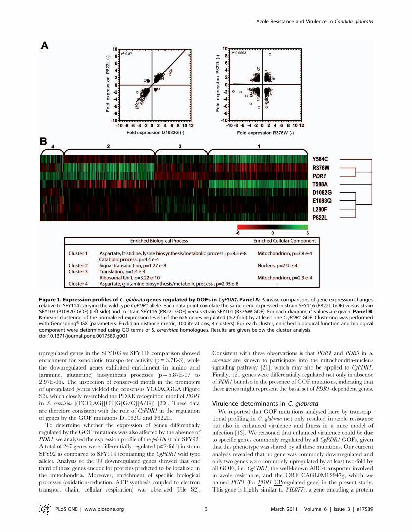
by these GOFs. The highest correlation (r2 = 0.87) was observed
between expression pattern of GOF D1082G (SFY103) with
P822L (SFY116) (Fig. 1A, left side). One GOF (R376W) in
SFY101 yielded systematically low r2 values with all other GOFs
(between 0.0003 and 0.058). Increasing the cut-off for differential
regulation to $3-fold did not significantly change r2 values (data
not shown). The expression of genes obtained from GOF P822L
(SFY116) and from R376W is shown to illustrate the low level of
gene co-regulation between both isolates (Fig. 1A, right side).
Taken together, these data support the concept that individual
GOF result each in distinct transcription profiles even though the
number of GOF analysed is probably only a portion of the entire
mutation spectrum.
Given the diversity of transcriptional profiles provided by each
GOF, the generated transcriptional data were clustered in a
separate analysis in order to group sets of genes co-regulated by
the different GOFs. Four separated groups were thus identified
which were enriched in specific biological processes (Fig. 1B). It is
noteworthy that genes from cluster 1 and 4 are enriched in
processes related to amino acid metabolism, while others are
enriched in signal transduction and protein metabolic processes.
We closely inspected the transcription profiles of two isolates,
one carrying the GOF mutation D1082G (SFY103) and the other
the mutation P822L (SFY116). This choice was based on the fact
that these profiles show the highest correlation (r2 = 0.87) and
similar numbers of up-and downregulated genes, thus facilitating
comparisons (Table 1 and 2). Between the two GOFs, 86 genes
were co-regulated (32 upregulated and 54 downregulated) from
the total of 626 genes regulated by at least one GOF. The
Table 1. Number of C. glabrata genes regulated by $twofoldin PDR1 GOF mutants as compared to the wild type.
StrainCgPDR1 GOFmutation
Genesupregulated
Genesdownregulated Total
SFY101 R376W 27 46 73
SFY103 D1082G 53 77 130
SFY105 T588A 235 150 385
SFY109 E1083Q 58 103 161
SFY111 Y584C 197 132 329
SFY115 L280F 67 132 199
SFY116 P822L 71 89 160
doi:10.1371/journal.pone.0017589.t001
Table 2. Correlation coefficients of transcriptional profiles.
GOF inCgPDR1allele L280F R376W Y584C T588A P822L D1082G E1083Q
L280F 1 0.016 0.6111 0.3107 0.7761 0.6979 0.8391
R376W 0.016 1 0.0588 0.0316 0.0003 0.0055 0.0003
Y584C 0.6111 0.0588 1 0.5491 0.7798 0.7012 0.7321
T588A 0.3107 0.0316 0.5491 1 0.4591 0.5596 0.4023
P822L 0.7761 0.0003 0.7798 0.4591 1 0.8704 0.7741
D1082G 0.6979 0.0055 0.7012 0.5596 0.8704 1 0.6984
E1083Q 0.8391 0.0003 0.7321 0.4023 0.7741 0.6984 1
doi:10.1371/journal.pone.0017589.t002
Azole Resistance and Virulence in Candida glabrata
PLoS ONE | www.plosone.org 2 March 2011 | Volume 6 | Issue 3 | e17589
upregulated genes in the SFY103 vs SFY116 comparison showed
enrichment for xenobiotic transporter activity (p = 3.7E-3), while
the downregulated genes exhibited enrichment in amino acid
(arginine, glutamine) biosynthesis processes (p = 5.87E-07 to
2.97E-06). The inspection of conserved motifs in the promoters
of upregulated genes yielded the consensus YCCACGGA (Figure
S3), which closely resembled the PDRE recognition motif of PDR1
in S. cerevisiae ((TCC[AG][CT]G[G/C][A/G]) [20]. These data
are therefore consistent with the role of CgPDR1 in the regulation
of genes by the GOF mutations D1082G and P822L.
To determine whether the expression of genes differentially
regulated by the GOF mutations was also affected by the absence of
PDR1, we analysed the expression profile of the pdr1D strain SFY92.
A total of 247 genes were differentially regulated ($2-fold) in strain
SFY92 as compared to SFY114 (containing the CgPDR1 wild type
allele). Analysis of the 99 downregulated genes showed that one
third of these genes encode for proteins predicted to be localized in
the mitochondria. Moreover, enrichment of specific biological
processes (oxidation-reduction, ATP synthesis coupled to electron
transport chain, cellular respiration) was observed (File S2).
Consistent with these observations is that PDR1 and PDR3 in S.
cerevisiae are known to participate into the mitochondria-nucleus
signalling pathway [21], which may also be applied to CgPDR1.
Finally, 121 genes were differentially regulated not only in absence
of PDR1 but also in the presence of GOF mutations, indicating that
these genes might represent the basal set of PDR1-dependent genes.
Virulence determinants in C. glabrataWe reported that GOF mutations analysed here by transcrip-
tional profiling in C. glabrata not only resulted in azole resistance
but also in enhanced virulence and fitness in a mice model of
infection [13]. We reasoned that enhanced virulence could be due
to specific genes commonly regulated by all CgPDR1 GOFs, given
that this phenotype was shared by all these mutations. Our current
analysis revealed that no gene was commonly downregulated and
only two genes were commonly upregulated by at least two-fold by
all GOFs, i.e. CgCDR1, the well-known ABC-transporter involved
in azole resistance, and the ORF CAGL0M12947g, which we
named PUP1 (for PDR1 UPregulated gene) in the present study.
This gene is highly similar to YIL077c, a gene encoding a protein
Figure 1. Expression profiles of C. glabrata genes regulated by GOFs in CgPDR1. Panel A: Pairwise comparisons of gene expression changesrelative to SFY114 carrying the wild type CgPDR1 allele. Each data point correlate the same gene expressed in strain SFY116 (P822L GOF) versus strainSFY103 (P1082G GOF) (left side) and in strain SFY116 (P822L GOF) versus strain SFY101 (R376W GOF). For each diagram, r2 values are given. Panel B:K-means clustering of the normalized expression levels of the 626 genes regulated ($2-fold) by at least one CgPDR1 GOF. Clustering was performedwith GenespringH GX (parameters: Euclidian distance metric, 100 iterations, 4 clusters). For each cluster, enriched biological function and biologicalcomponent were determined using GO terms of S. cerevisiae homologues. Results are given below the cluster analysis.doi:10.1371/journal.pone.0017589.g001
Azole Resistance and Virulence in Candida glabrata
PLoS ONE | www.plosone.org 3 March 2011 | Volume 6 | Issue 3 | e17589
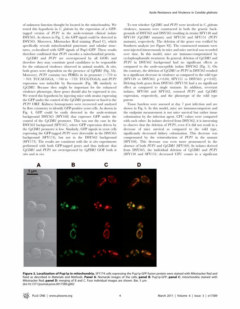
of unknown function thought be located in the mitochondria. We
tested this hypothesis in C. glabrata by the expression of a GFP-
tagged version of PUP1 in the azole-resistant clinical isolate
DSY565. As shown in Fig. 2, the GFP signal could be detected in
DSY565. Moreover, Mitotracker Red staining (Panel C), which
specifically reveals mitochondrial punctuate and tubular struc-
tures, co-localized with GFP signals of Pup1-GFP. These results
therefore confirmed that PUP1 encodes a mitochondrial protein.
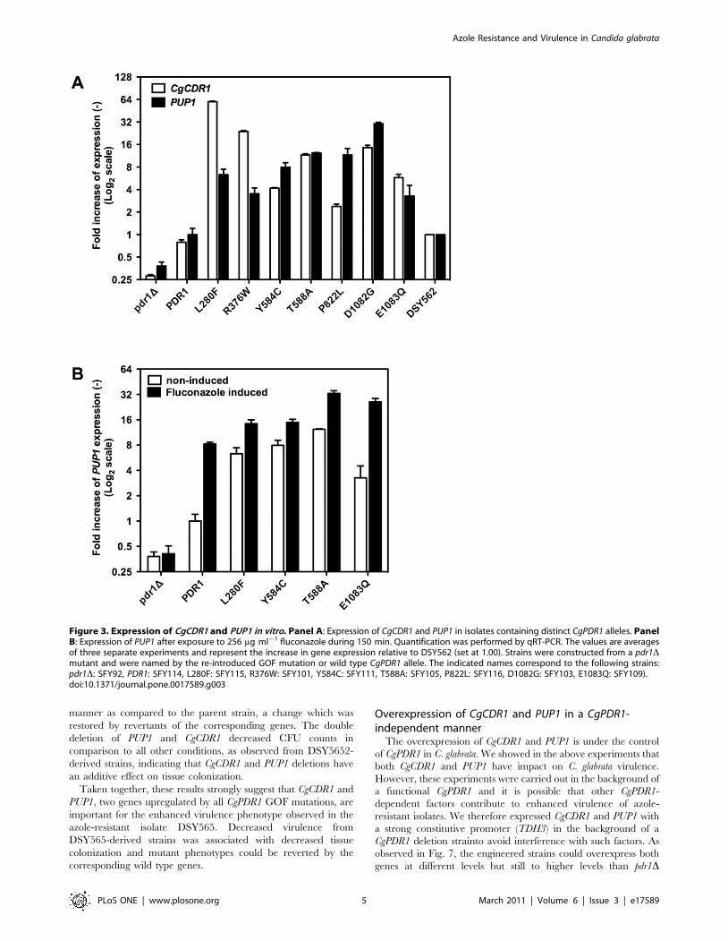
CgCDR1 and PUP1 are overexpressed by all GOFs and
therefore they may constitute good candidates to be responsible
for the enhanced virulence observed in animal models. In vitro,
both genes were dependent on the presence of CgPDR1 (Fig. 3A).
Moreover, PUP1 contains two PDREs in its promoter (2770 to
2763: TCCACGGA; 2740 to 2733: TCCGTGGA) and PUP1
expression was inducible by fluconazole (Fig. 3B) similarly to
CgCDR1. Because they might be important for the enhanced
virulence phenotype, these genes should also be expressed in vivo.
We tested this hypothesis by injecting mice with strains expressing
the GFP under the control of the CgCDR1 promoter or fused to the
PUP1 ORF. Kidneys homogenates were recovered and analysed
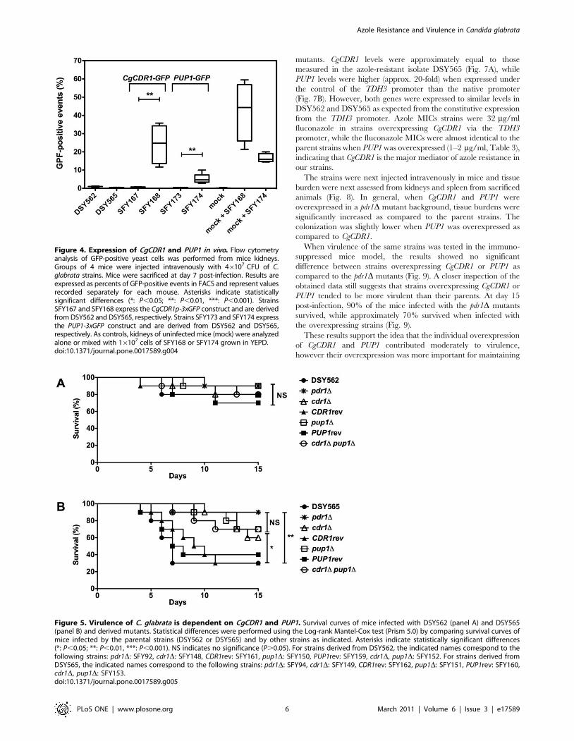
by flow cytometry to identify GFP-positive yeast cells. As shown in
Fig. 4, GFP could be easily detected in the azole-resistant
background DSY565 (SFY168) that expresses GFP under the
control of the CgCDR1 promoter. This was not the case in the
DSY562 background (SFY167), where GFP expression driven by
the CgCDR1 promoter is low. Similarly, GFP signals in yeast cells
expressing the GFP-tagged PUP1 were detectable in the DSY565
background (SFY174), but not in the DSY562 background
(SFY173). The results are consistent with the in vitro experiments
performed with both GFP-tagged genes and thus indicate that
CgCDR1 and PUP1 are overexpressed by CgPDR1 GOF both in
vitro and in vivo.
To test whether CgCDR1 and PUP1 were involved in C. glabrata
virulence, mutants were constructed in both the genetic back-
grounds of DSY562 and DSY565 resulting in strains SFY148 and
SFY149 (CgCDR1 mutants) and SFY150 and SFY151 (PUP1
mutants), respectively. The deletion of the genes was verified by
Southern analysis (see Figure S2). The constructed mutants were
next injected intravenously in mice and mice survival was recorded
over time. In this model, mice are immuno-compromised by
cyclophosphamide treatment. In general, deletion of CgCDR1 and
PUP1 in DSY562 background had no significant effects as
compared to the azole-susceptible isolate DSY562 (Fig. 5). On
the contrary, the deletion of CgCDR1 or PUP1 in DSY565 resulted
in a significant decrease in virulence as compared to the wild type
(SFY149 vs DSY565: p = 0.04; SFY151 vs DSY565: p = 0.02).
Deleting both genes from DSY565 (SFY170) had a no significant
effect as compared to single mutants. In addition, revertant
isolates, SFY160 and SFY162, restored PUP1 and CgCDR1
expression, respectively, and the phenotype of the wild type
parent.
Tissue burdens were assessed at day 7 post infection and are
shown in Fig. 6. In this model, mice are immunocompetent and
the endpoint measurement is not mice survival but rather tissue
colonization by the infection agent. CFU values were compared
with each other. In isolates derived from DSY562, it is interesting
to observe that the deletion of PUP1, even if it did not result in a
decrease of mice survival as compared to the wild type,
significantly decreased kidney colonization. This decrease was
compensated by the reintroduction of PUP1 in the mutant
(SFY160). This decrease was even more pronounced in the
absence of both PUP1 and CgCDR1 (SFY169). In isolates derived
from DSY565, the individual deletion of CgCDR1 and PUP1
(SFY150 and SFY151) decreased CFU counts in a significant
Figure 2. Localization of Pup1p in mitochondria. SFY174 cells expressing the Pup1p-GFP fusion protein were stained with Mitotracker Red andfixed as described in Materials and Methods. Panel A: Nomarski images of the cells; panel B: Pup1p-GFP; panel C: mitochondria stained withMitotracker Red; panel D: merging of B and C. Four individual images are shown. Bar, 5 mm.doi:10.1371/journal.pone.0017589.g002
Azole Resistance and Virulence in Candida glabrata
PLoS ONE | www.plosone.org 4 March 2011 | Volume 6 | Issue 3 | e17589
manner as compared to the parent strain, a change which was
restored by revertants of the corresponding genes. The double
deletion of PUP1 and CgCDR1 decreased CFU counts in
comparison to all other conditions, as observed from DSY5652-
derived strains, indicating that CgCDR1 and PUP1 deletions have
an additive effect on tissue colonization.
Taken together, these results strongly suggest that CgCDR1 and
PUP1, two genes upregulated by all CgPDR1 GOF mutations, are
important for the enhanced virulence phenotype observed in the
azole-resistant isolate DSY565. Decreased virulence from
DSY565-derived strains was associated with decreased tissue
colonization and mutant phenotypes could be reverted by the
corresponding wild type genes.
Overexpression of CgCDR1 and PUP1 in a CgPDR1-independent manner
The overexpression of CgCDR1 and PUP1 is under the control
of CgPDR1 in C. glabrata. We showed in the above experiments that
both CgCDR1 and PUP1 have impact on C. glabrata virulence.
However, these experiments were carried out in the background of
a functional CgPDR1 and it is possible that other CgPDR1-
dependent factors contribute to enhanced virulence of azole-
resistant isolates. We therefore expressed CgCDR1 and PUP1 with
a strong constitutive promoter (TDH3) in the background of a
CgPDR1 deletion strainto avoid interference with such factors. As
observed in Fig. 7, the engineered strains could overexpress both
genes at different levels but still to higher levels than pdr1D
Figure 3. Expression of CgCDR1 and PUP1 in vitro. Panel A: Expression of CgCDR1 and PUP1 in isolates containing distinct CgPDR1 alleles. PanelB: Expression of PUP1 after exposure to 256 mg ml21 fluconazole during 150 min. Quantification was performed by qRT-PCR. The values are averagesof three separate experiments and represent the increase in gene expression relative to DSY562 (set at 1.00). Strains were constructed from a pdr1Dmutant and were named by the re-introduced GOF mutation or wild type CgPDR1 allele. The indicated names correspond to the following strains:pdr1D: SFY92, PDR1: SFY114, L280F: SFY115, R376W: SFY101, Y584C: SFY111, T588A: SFY105, P822L: SFY116, D1082G: SFY103, E1083Q: SFY109).doi:10.1371/journal.pone.0017589.g003
Azole Resistance and Virulence in Candida glabrata
PLoS ONE | www.plosone.org 5 March 2011 | Volume 6 | Issue 3 | e17589
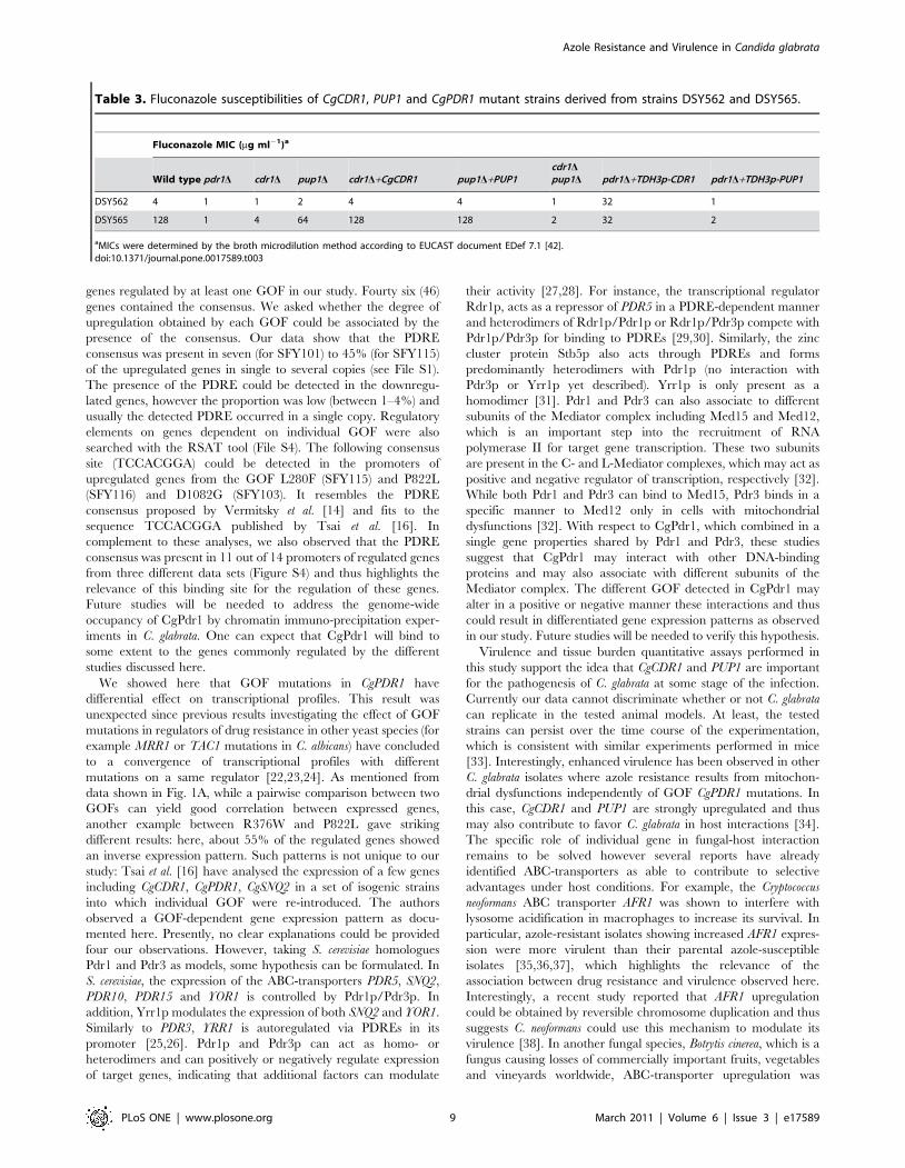
mutants. CgCDR1 levels were approximately equal to those
measured in the azole-resistant isolate DSY565 (Fig. 7A), while
PUP1 levels were higher (approx. 20-fold) when expressed under
the control of the TDH3 promoter than the native promoter
(Fig. 7B). However, both genes were expressed to similar levels in
DSY562 and DSY565 as expected from the constitutive expression
from the TDH3 promoter. Azole MICs strains were 32 mg/ml
fluconazole in strains overexpressing CgCDR1 via the TDH3
promoter, while the fluconazole MICs were almost identical to the
parent strains when PUP1 was overexpressed (1–2 mg/ml, Table 3),
indicating that CgCDR1 is the major mediator of azole resistance in
our strains.
The strains were next injected intravenously in mice and tissue
burden were next assessed from kidneys and spleen from sacrificed
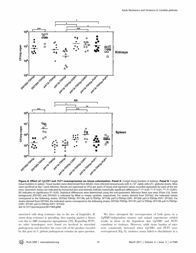
animals (Fig. 8). In general, when CgCDR1 and PUP1 were
overexpressed in a pdr1D mutant background, tissue burdens were
significantly increased as compared to the parent strains. The
colonization was slightly lower when PUP1 was overexpressed as
compared to CgCDR1.
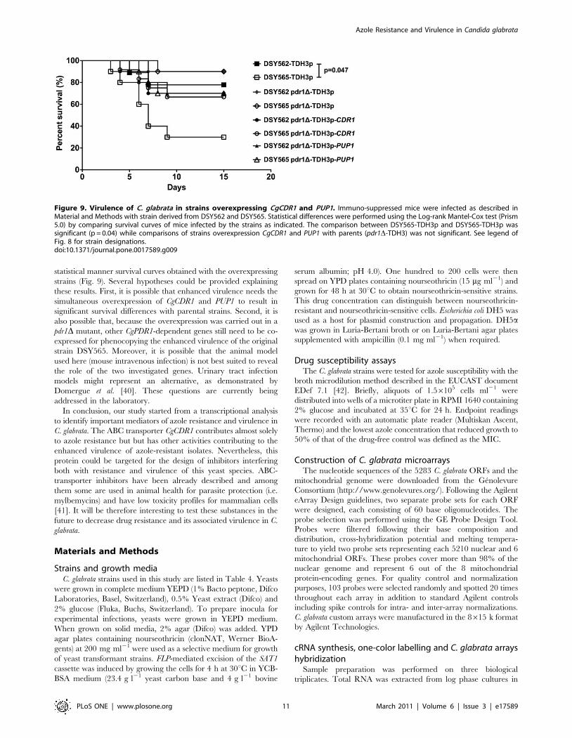
When virulence of the same strains was tested in the immuno-
suppressed mice model, the results showed no significant
difference between strains overexpressing CgCDR1 or PUP1 as
compared to the pdr1D mutants (Fig. 9). A closer inspection of the
obtained data still suggests that strains overexpressing CgCDR1 or
PUP1 tended to be more virulent than their parents. At day 15
post-infection, 90% of the mice infected with the pdr1D mutants
survived, while approximately 70% survived when infected with
the overexpressing strains (Fig. 9).
These results support the idea that the individual overexpression
of CgCDR1 and PUP1 contributed moderately to virulence,
however their overexpression was more important for maintaining
Figure 4. Expression of CgCDR1 and PUP1 in vivo. Flow cytometryanalysis of GFP-positive yeast cells was performed from mice kidneys.Groups of 4 mice were injected intravenously with 46107 CFU of C.glabrata strains. Mice were sacrificed at day 7 post-infection. Results areexpressed as percents of GFP-positive events in FACS and represent valuesrecorded separately for each mouse. Asterisks indicate statisticallysignificant differences (*: P,0.05; **: P,0.01, ***: P,0.001). StrainsSFY167 and SFY168 express the CgCDR1p-3xGFP construct and are derivedfrom DSY562 and DSY565, respectively. Strains SFY173 and SFY174 expressthe PUP1-3xGFP construct and are derived from DSY562 and DSY565,respectively. As controls, kidneys of uninfected mice (mock) were analyzedalone or mixed with 16107 cells of SFY168 or SFY174 grown in YEPD.doi:10.1371/journal.pone.0017589.g004
Figure 5. Virulence of C. glabrata is dependent on CgCDR1 and PUP1. Survival curves of mice infected with DSY562 (panel A) and DSY565(panel B) and derived mutants. Statistical differences were performed using the Log-rank Mantel-Cox test (Prism 5.0) by comparing survival curves ofmice infected by the parental strains (DSY562 or DSY565) and by other strains as indicated. Asterisks indicate statistically significant differences(*: P,0.05; **: P,0.01, ***: P,0.001). NS indicates no significance (P.0.05). For strains derived from DSY562, the indicated names correspond to thefollowing strains: pdr1D: SFY92, cdr1D: SFY148, CDR1rev: SFY161, pup1D: SFY150, PUP1rev: SFY159, cdr1D, pup1D: SFY152. For strains derived fromDSY565, the indicated names correspond to the following strains: pdr1D: SFY94, cdr1D: SFY149, CDR1rev: SFY162, pup1D: SFY151, PUP1rev: SFY160,cdr1D, pup1D: SFY153.doi:10.1371/journal.pone.0017589.g005
Azole Resistance and Virulence in Candida glabrata
PLoS ONE | www.plosone.org 6 March 2011 | Volume 6 | Issue 3 | e17589

Figure 6. C. glabrata tissue burdens in murine infection models. Fungal tissue burdens in kidneys (panel A) and spleen (panel B) from BALB/cmice infected intravenously with 46107 viable cells of C. glabrata strains. Mice were sacrificed at day 7 post-infection. Results are expressed as CFUsper gram of tissue and represent values recorded separately for each of the ten mice. Geometric means are indicated by horizontal bars. Statisticalcomparisons are summarized above each panel. Asterisks indicate statistically significant differences (*: P,0.05; **: P,0.01, ***: P,0.001). NSindicates no significance (P.0.05). The symbol ‘-’ indicates that the statistical comparison was not performed. Statistical differences were determinedusing the non-parametric Wilcoxon Rank sum tests (Prism 5.0). The origin of each strain is indicated; strain background (DSY562 and DSY565) isindicated by filled or empty symbols, respectively. See legend of Fig. 5 for strain designations.doi:10.1371/journal.pone.0017589.g006
Azole Resistance and Virulence in Candida glabrata
PLoS ONE | www.plosone.org 7 March 2011 | Volume 6 | Issue 3 | e17589

tissue colonization. Taken together, our results indicate that both
CgCDR1 and PUP1 are important mediators of C. glabrata
virulence, but that their individual overexpression per se is not
sufficient to mimic the increased virulence conferred by CgPDR1
GOF mutations.
Discussion
In this study we analysed the expression profiles of GOF
mutations obtained from azole-resistant isolates in a previous study
[13]. The analysis of transcription profiles gave only two genes
commonly upregulated by all GOFs, CgCDR1 and PUP1. Other
investigators have analysed transcription profiles of azole-resistant
isolates and thus enable comparisons with our study. Recently,
Tsai et al. [16] obtained the transcription profiles of seven clinical
pairs, each containing an azole-susceptible and an azole-resistant
isolate. The CgPDR1 GOF obtained from these strains were
different from those investigated here, except for the L280F GOF.
Their study highlighted 45 genes regulated (by $2-fold change as
compared to the susceptible parent) by at least one clinical pair.
Our study revealed a larger set of genes regulated by at least one
GOF (i.e. 626 genes). CgCDR1 and PUP1, the two genes selected in
our study were found commonly upregulated by all GOFs in the
Tsai et al. [16] study including by decreasing expression levels,
CgCDR1, CAGL0M12947g (PUP1), CAGL0F02717g (CgCDR2/
PDH1), CAGL0K00715g (RTA1), CAGL0C03289g (YBT1),
CAGL0G00242g (YOR1), CAGL0K09702g, CAGL0A00451g
(CgPDR1) and CAGL0G01122g. In a study published by
Vermitsky et al. [14], one azole-resistant isolate (F15) was
compared to an azole-susceptible parent. From the 109 genes
regulated by at least two-fold in the resistant isolate, 34 were found
regulated (out of 626 genes) in our study, among which CgCDR1
and PUP1, the latter being the most upregulated gene in their
study. The differences in transcriptional profiles could be
explained by several factors including experimental conditions,
type of array technology and intrinsic differenced between isolates
used in all three studies. One major difference between our study
and others is that we used an isogenic background in the
reintroduction of the seven individual CgPDR1 alleles, which
prevents intrinsic strain variations. This is perhaps a reason for the
difference between the number of genes regulated in at least one
condition in our study (626 genes regulated by at least one GOF)
and that of Tsai et al. [16] (45 genes regulated in at least one strain
pair). This view is supported by separate results obtained with the
transcriptional comparison of two related clinical strains, DSY717
and DSY2317, the latter containing the CgPDR1 GOF L1081F.
Between these two isolates, only 39 genes were regulated by at
least two-fold (File S3), including CgCDR1 and PUP1, thus
suggesting that intrinsic strain variations may mask the real effect
of GOF on the C. glabrata transcriptome.
The overlap between our study and others [14,16] falls into 14
regulated genes (Figure S4). Besides CgCDR1 and PUP1, which
were found consistently upregulated in all three studies, the other
genes may constitute a core set of genes regulated by CgPDR1. It is
interesting to observe that the 14 genes are almost all found
upregulated in the data provided by Vermitsky et al. [14] and Tsai
et al. [16], while in our case, the regulation of these genes is
dependent on the type of reintroduced GOF in the same genetic
background. Several hypotheses will be provided below.
Given that CgPDR1 is a major regulator of azole resistance in C.
glabrata and should act on regulated genes via PDRE binding
elements in the promoters of regulated genes, the consensus for
CgPDR1 binding (TCCRYGSR) was proposed and we searched
systematically for this motif in the promoter regions of the 626
Figure 7. Overexpression of CgCDR1 and PUP1 in a CgPDR1-independent manner. Panel A: TDH3-dependent expression of CgCDR1.Panel B: TDH3-dependent expression of PUP1. Quantification wasperformed by qRT-PCR. The values are averages of three separateexperiments and represent the increase in gene expression relative toSFY196 (set at 1.00). Strains derived from DSY562 are represented by blackbars and the indicated names correspond to the following strains: PDR1:SFY196, pdr1D: SFY198, pdr1D+TDH3p-CDR1: SFY200, pdr1D+TDH3p-PUP1:SFY202. Strains derived from DSY565 are represented by white bars and theindicated names correspond to the following strains: PDR1L280F: SFY197,pdr1D: SFY199, pdr1D+TDH3p-CDR1: SFY201, pdr1D+TDH3p-PUP1: SFY203.doi:10.1371/journal.pone.0017589.g007
Azole Resistance and Virulence in Candida glabrata
PLoS ONE | www.plosone.org 8 March 2011 | Volume 6 | Issue 3 | e17589
genes regulated by at least one GOF in our study. Fourty six (46)
genes contained the consensus. We asked whether the degree of
upregulation obtained by each GOF could be associated by the
presence of the consensus. Our data show that the PDRE
consensus was present in seven (for SFY101) to 45% (for SFY115)
of the upregulated genes in single to several copies (see File S1).
The presence of the PDRE could be detected in the downregu-
lated genes, however the proportion was low (between 1–4%) and
usually the detected PDRE occurred in a single copy. Regulatory
elements on genes dependent on individual GOF were also
searched with the RSAT tool (File S4). The following consensus
site (TCCACGGA) could be detected in the promoters of
upregulated genes from the GOF L280F (SFY115) and P822L
(SFY116) and D1082G (SFY103). It resembles the PDRE
consensus proposed by Vermitsky et al. [14] and fits to the
sequence TCCACGGA published by Tsai et al. [16]. In
complement to these analyses, we also observed that the PDRE
consensus was present in 11 out of 14 promoters of regulated genes
from three different data sets (Figure S4) and thus highlights the
relevance of this binding site for the regulation of these genes.
Future studies will be needed to address the genome-wide
occupancy of CgPdr1 by chromatin immuno-precipitation exper-
iments in C. glabrata. One can expect that CgPdr1 will bind to
some extent to the genes commonly regulated by the different
studies discussed here.
We showed here that GOF mutations in CgPDR1 have
differential effect on transcriptional profiles. This result was
unexpected since previous results investigating the effect of GOF
mutations in regulators of drug resistance in other yeast species (for
example MRR1 or TAC1 mutations in C. albicans) have concluded
to a convergence of transcriptional profiles with different
mutations on a same regulator [22,23,24]. As mentioned from
data shown in Fig. 1A, while a pairwise comparison between two
GOFs can yield good correlation between expressed genes,
another example between R376W and P822L gave striking
different results: here, about 55% of the regulated genes showed
an inverse expression pattern. Such patterns is not unique to our
study: Tsai et al. [16] have analysed the expression of a few genes
including CgCDR1, CgPDR1, CgSNQ2 in a set of isogenic strains
into which individual GOF were re-introduced. The authors
observed a GOF-dependent gene expression pattern as docu-
mented here. Presently, no clear explanations could be provided
four our observations. However, taking S. cerevisiae homologues
Pdr1 and Pdr3 as models, some hypothesis can be formulated. In
S. cerevisiae, the expression of the ABC-transporters PDR5, SNQ2,
PDR10, PDR15 and YOR1 is controlled by Pdr1p/Pdr3p. In
addition, Yrr1p modulates the expression of both SNQ2 and YOR1.
Similarly to PDR3, YRR1 is autoregulated via PDREs in its
promoter [25,26]. Pdr1p and Pdr3p can act as homo- or
heterodimers and can positively or negatively regulate expression
of target genes, indicating that additional factors can modulate
their activity [27,28]. For instance, the transcriptional regulator
Rdr1p, acts as a repressor of PDR5 in a PDRE-dependent manner
and heterodimers of Rdr1p/Pdr1p or Rdr1p/Pdr3p compete with
Pdr1p/Pdr3p for binding to PDREs [29,30]. Similarly, the zinc
cluster protein Stb5p also acts through PDREs and forms
predominantly heterodimers with Pdr1p (no interaction with
Pdr3p or Yrr1p yet described). Yrr1p is only present as a
homodimer [31]. Pdr1 and Pdr3 can also associate to different
subunits of the Mediator complex including Med15 and Med12,
which is an important step into the recruitment of RNA
polymerase II for target gene transcription. These two subunits
are present in the C- and L-Mediator complexes, which may act as
positive and negative regulator of transcription, respectively [32].
While both Pdr1 and Pdr3 can bind to Med15, Pdr3 binds in a
specific manner to Med12 only in cells with mitochondrial
dysfunctions [32]. With respect to CgPdr1, which combined in a
single gene properties shared by Pdr1 and Pdr3, these studies
suggest that CgPdr1 may interact with other DNA-binding
proteins and may also associate with different subunits of the
Mediator complex. The different GOF detected in CgPdr1 may
alter in a positive or negative manner these interactions and thus
could result in differentiated gene expression patterns as observed
in our study. Future studies will be needed to verify this hypothesis.
Virulence and tissue burden quantitative assays performed in
this study support the idea that CgCDR1 and PUP1 are important
for the pathogenesis of C. glabrata at some stage of the infection.
Currently our data cannot discriminate whether or not C. glabrata
can replicate in the tested animal models. At least, the tested
strains can persist over the time course of the experimentation,
which is consistent with similar experiments performed in mice
[33]. Interestingly, enhanced virulence has been observed in other
C. glabrata isolates where azole resistance results from mitochon-
drial dysfunctions independently of GOF CgPDR1 mutations. In
this case, CgCDR1 and PUP1 are strongly upregulated and thus
may also contribute to favor C. glabrata in host interactions [34].
The specific role of individual gene in fungal-host interaction
remains to be solved however several reports have already
identified ABC-transporters as able to contribute to selective
advantages under host conditions. For example, the Cryptococcus
neoformans ABC transporter AFR1 was shown to interfere with
lysosome acidification in macrophages to increase its survival. In
particular, azole-resistant isolates showing increased AFR1 expres-
sion were more virulent than their parental azole-susceptible
isolates [35,36,37], which highlights the relevance of the
association between drug resistance and virulence observed here.
Interestingly, a recent study reported that AFR1 upregulation
could be obtained by reversible chromosome duplication and thus
suggests C. neoformans could use this mechanism to modulate its
virulence [38]. In another fungal species, Botrytis cinerea, which is a
fungus causing losses of commercially important fruits, vegetables
and vineyards worldwide, ABC-transporter upregulation was
Table 3. Fluconazole susceptibilities of CgCDR1, PUP1 and CgPDR1 mutant strains derived from strains DSY562 and DSY565.
Fluconazole MIC (mg ml21)a
Wild type pdr1D cdr1D pup1D cdr1D+CgCDR1 pup1D+PUP1cdr1Dpup1D pdr1D+TDH3p-CDR1 pdr1D+TDH3p-PUP1
DSY562 4 1 1 2 4 4 1 32 1
DSY565 128 1 4 64 128 128 2 32 2
aMICs were determined by the broth microdilution method according to EUCAST document EDef 7.1 [42].doi:10.1371/journal.pone.0017589.t003
Azole Resistance and Virulence in Candida glabrata
PLoS ONE | www.plosone.org 9 March 2011 | Volume 6 | Issue 3 | e17589
associated with drug resistance due to the use of fungicides. B.
cinerea drug resistance is spreading, thus arguing against a fitness
cost due to ABC-transporter upregulation [39]. Regarding PUP1,
no other homologues were found yet involved in microbial
pathogenesis and therefore the exact role of the product encoded
by this gene in C. glabrata pathogenesis remains an open question.
We have attempted the overexpression of both genes in a
CgPDR1-independent manner and animal experiments yielded
results in favor of the hypothesis that CgCDR1 and PUP1
contribute to virulence. However, while tissue burden of mice
were consistently increased when CgCDR1 and PUP1 were
overexpressed (Fig. 8), virulence assays failed to discriminate in a
Figure 8. Effect of CgCDR1 and PUP1 overexpression on tissue colonization. Panel A: Fungal tissue burdens in kidneys. Panel B: Fungaltissue burdens in spleen. Tissue burden were determined from BALB/c mice infected intravenously with 46107 viable cells of C. glabrata strains. Micewere sacrificed at day 7 post-infection. Results are expressed as CFUs per gram of tissue and represent values recorded separately for each of the tenmice. Geometric means are indicated by horizontal bars and asterisks indicate statistically significant differences (*: P,0.05; **: P,0.01, ***: P,0.001).NS indicates no significance (P.0.05). Statistical differences were determined using the non-parametric Wilcoxon Rank sum tests (Prism 5.0). Strainbackground (DSY562 and DSY565) is indicated by filled or empty symbols, respectively. For strains derived from DSY562, the indicated namescorrespond to the following strains: DSY562-TDH3p: SFY196; pdr1D-TDH3p: SFY198; pdr1D-TDH3p-CDR1: SFY200 pdr1D-TDH3p-PUP1: SFY202. Forstrains derived from DSY565, the indicated names correspond to the following strains: DSY562-TDH3p: SFY197; pdr1D-TDH3p: SFY199; pdr1D-TDH3p-CDR1: SFY201 pdr1D-TDH3p-PUP1: SFY203.doi:10.1371/journal.pone.0017589.g008
Azole Resistance and Virulence in Candida glabrata
PLoS ONE | www.plosone.org 10 March 2011 | Volume 6 | Issue 3 | e17589
statistical manner survival curves obtained with the overexpressing
strains (Fig. 9). Several hypotheses could be provided explaining
these results. First, it is possible that enhanced virulence needs the
simultaneous overexpression of CgCDR1 and PUP1 to result in
significant survival differences with parental strains. Second, it is
also possible that, because the overexpression was carried out in a
pdr1D mutant, other CgPDR1-dependent genes still need to be co-
expressed for phenocopying the enhanced virulence of the original
strain DSY565. Moreover, it is possible that the animal model
used here (mouse intravenous infection) is not best suited to reveal
the role of the two investigated genes. Urinary tract infection
models might represent an alternative, as demonstrated by
Domergue et al. [40]. These questions are currently being
addressed in the laboratory.
In conclusion, our study started from a transcriptional analysis
to identify important mediators of azole resistance and virulence in
C. glabrata. The ABC transporter CgCDR1 contributes almost solely
to azole resistance but but has other activities contributing to the
enhanced virulence of azole-resistant isolates. Nevertheless, this
protein could be targeted for the design of inhibitors interfering
both with resistance and virulence of this yeast species. ABC-
transporter inhibitors have been already described and among
them some are used in animal health for parasite protection (i.e.
mylbemycins) and have low toxicity profiles for mammalian cells
[41]. It will be therefore interesting to test these substances in the
future to decrease drug resistance and its associated virulence in C.
glabrata.
Materials and Methods
Strains and growth mediaC. glabrata strains used in this study are listed in Table 4. Yeasts
were grown in complete medium YEPD (1% Bacto peptone, Difco
Laboratories, Basel, Switzerland), 0.5% Yeast extract (Difco) and
2% glucose (Fluka, Buchs, Switzerland). To prepare inocula for
experimental infections, yeasts were grown in YEPD medium.
When grown on solid media, 2% agar (Difco) was added. YPD
agar plates containing nourseothricin (clonNAT, Werner BioA-
gents) at 200 mg ml21 were used as a selective medium for growth
of yeast transformant strains. FLP-mediated excision of the SAT1
cassette was induced by growing the cells for 4 h at 30uC in YCB-
BSA medium (23.4 g l21 yeast carbon base and 4 g l21 bovine
serum albumin; pH 4.0). One hundred to 200 cells were then
spread on YPD plates containing nourseothricin (15 mg ml21) and
grown for 48 h at 30uC to obtain nourseothricin-sensitive strains.
This drug concentration can distinguish between nourseothricin-
resistant and nourseothricin-sensitive cells. Escherichia coli DH5 was
used as a host for plasmid construction and propagation. DH5awas grown in Luria-Bertani broth or on Luria-Bertani agar plates
supplemented with ampicillin (0.1 mg ml21) when required.
Drug susceptibility assaysThe C. glabrata strains were tested for azole susceptibility with the
broth microdilution method described in the EUCAST document
EDef 7.1 [42]. Briefly, aliquots of 1.56105 cells ml21 were
distributed into wells of a microtiter plate in RPMI 1640 containing
2% glucose and incubated at 35uC for 24 h. Endpoint readings
were recorded with an automatic plate reader (Multiskan Ascent,
Thermo) and the lowest azole concentration that reduced growth to
50% of that of the drug-free control was defined as the MIC.
Construction of C. glabrata microarraysThe nucleotide sequences of the 5283 C. glabrata ORFs and the
mitochondrial genome were downloaded from the Genolevure
Consortium (http://www.genolevures.org/). Following the Agilent
eArray Design guidelines, two separate probe sets for each ORF
were designed, each consisting of 60 base oligonucleotides. The
probe selection was performed using the GE Probe Design Tool.
Probes were filtered following their base composition and
distribution, cross-hybridization potential and melting tempera-
ture to yield two probe sets representing each 5210 nuclear and 6
mitochondrial ORFs. These probes cover more than 98% of the
nuclear genome and represent 6 out of the 8 mitochondrial
protein-encoding genes. For quality control and normalization
purposes, 103 probes were selected randomly and spotted 20 times
throughout each array in addition to standard Agilent controls
including spike controls for intra- and inter-array normalizations.
C. glabrata custom arrays were manufactured in the 8615 k format
by Agilent Technologies.
cRNA synthesis, one-color labelling and C. glabrata arrayshybridization
Sample preparation was performed on three biological
triplicates. Total RNA was extracted from log phase cultures in
Figure 9. Virulence of C. glabrata in strains overexpressing CgCDR1 and PUP1. Immuno-suppressed mice were infected as described inMaterial and Methods with strain derived from DSY562 and DSY565. Statistical differences were performed using the Log-rank Mantel-Cox test (Prism5.0) by comparing survival curves of mice infected by the strains as indicated. The comparison between DSY565-TDH3p and DSY565-TDH3p wassignificant (p = 0.04) while comparisons of strains overexpression CgCDR1 and PUP1 with parents (pdr1D-TDH3) was not significant. See legend ofFig. 8 for strain designations.doi:10.1371/journal.pone.0017589.g009
Azole Resistance and Virulence in Candida glabrata
PLoS ONE | www.plosone.org 11 March 2011 | Volume 6 | Issue 3 | e17589
liquid YEPD as previously described [8]. Briefly, after centrifuga-
tion of 5 ml culture (corresponding to 108 cells), the yeast cell pellet
was mixed with 0.3 g of glass beads, 300 ml of RNA extraction
buffer (0.1 M Tris-HCl at pH 7.5, 0.1 M LiCl, 10 mM EDTA,
0.5% SDS) and 300 ml of phenol-chloroform-isoamyl alcohol
(24:24:1). After 1 min of vortexing in a bead beater (Fastprep-24
Instrument, MP Biomedicals Switzerland, Zurich), the aqueous
phase was re-extracted with phenol-chloroform-isoamyl alcohol,
and RNA was precipitated with 600 ml of ethanol at 220uC for
1 h. The RNA pellet was resuspended in 50 ml of diethyl
pyrocarbonate-treated H2O. The integrity of the input template
RNA has been determined prior to labeling/amplification, using
Agilent RNA 6000 Nano LabChip kit and 2100 bioanalyzer
(Agilent Technologies). Agilent’s One-Color Quick Amp Labeling
Kit (Agilent Technologies) was used to generate fluorescent cRNA
according to the manufacturer’s instructions. Briefly, 1 mg of total
RNA from each sample was used to which a spike mix and T7
promoter primers were added, both of which are provided by the
manufacturer. cDNA synthesis was promoted by MMLV-RT
(Moloney Murine Leukemia Virus Reverse Transcriptase) in the
presence of dNTPs and RNaseOUT. Next, cRNA was produced
from this first reaction with T7 RNA polymerase, which
simultaneously amplifies target material and incorporates cyanine
3-labeled CTP. The labelled cRNAs were purified with RNeasy
Mini Kit (Qiagen) and quantified using NanoDrop ND-1000 UV-
VIS Spectrophotometer. 600 ng of Cy3-labelled cRNAs were
fragmented and hybridized for 17 h at 65uC to each array using
the Gene Expression Hybridization Kit (Agilent Technologies) and
a gasket slide with a 8 microarrays/slide format for sample
hybridization to separate each sample in specific sub-arrays of the
8615 K format.
Microarrays data analysisSlides were washed and processed according to the Agilent 60-
mer Oligo Microarray Processing protocol and scanned on a
Agilent microarray scanner G2565BA (Agilent Technologies).
Data were extracted from the images with Feature Extraction (FE)
software (Agilent Technologies). FE software flags outlier features,
and detects and removes spatial gradients and local backgrounds.
Data were normalized using a combined rank consistency filtering
with LOWESS intensity normalization.
The gene expression values obtained from FE software were
imported into GeneSpring 10.0.2 software (Agilent Technologies)
for preprocessing and data analysis. For inter-array comparisons, a
linear scaling of the data was performed using the 75th percentile
signal value of all of non-control probes on the microarray to
normalize one-color signal values. Probe sets with a signal intensity
value below the 20th percentile were considered as absent and
discarded from subsequent analysis. The expression of each gene
was normalized by its median expression across all samples. Genes
were included in the final data set if their expression changed by at
least 2-fold between each strain expressing a CgPDR1 GOF allele
and the strain SFY114 expressing the CgPDR1 wild type allele in at
least two independent experiments. Corrected p-value (,0.05) was
chosen as the cut-off for significance. Validation of genes found
regulated by microarray analysis was performed by qRT-PCR
analysis (see below for technical details) on a set of nine different
genes. In general, the correlation found between qRT-PCR and
microarray data was excellent (see Figure S1). Microarray data
have been uploaded to the NCBI GEO microarray repository.
The GEO accession number for the C. glabrata Agilent array is
GPL10713 and the accession numbers for the data are GSE23827,
GSE23828 and GSE23829.
Use of bioinformatic toolsThe analysis of consensus pattern on C. glabrata promoters
(2800 to 21) was performed using the Regulatory Sequence
Analysis Tools (RSAT: http://rsat.ulb.ac.be/rsat/index.html) and
implemented to the pattern discovery tool (oligo-analysis). The
settings were those supplied by default by the tool provider. The
position-specific scoring matrices (PSSM) consensus matrices were
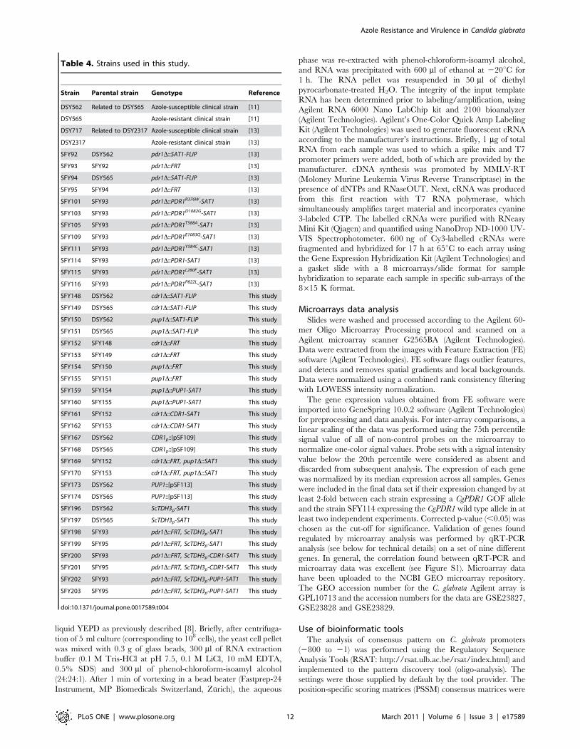
Table 4. Strains used in this study.
Strain Parental strain Genotype Reference
DSY562 Related to DSY565 Azole-susceptible clinical strain [11]
DSY565 Azole-resistant clinical strain [11]
DSY717 Related to DSY2317 Azole-susceptible clinical strain [13]
DSY2317 Azole-resistant clinical strain [13]
SFY92 DSY562 pdr1D::SAT1-FLIP [13]
SFY93 SFY92 pdr1D::FRT [13]
SFY94 DSY565 pdr1D::SAT1-FLIP [13]
SFY95 SFY94 pdr1D::FRT [13]
SFY101 SFY93 pdr1D::PDR1R376W-SAT1 [13]
SFY103 SFY93 pdr1D::PDR1D1082G-SAT1 [13]
SFY105 SFY93 pdr1D::PDR1T588A-SAT1 [13]
SFY109 SFY93 pdr1D::PDR1E1083Q-SAT1 [13]
SFY111 SFY93 pdr1D::PDR1Y584C-SAT1 [13]
SFY114 SFY93 pdr1D::PDR1-SAT1 [13]
SFY115 SFY93 pdr1D::PDR1L280F-SAT1 [13]
SFY116 SFY93 pdr1D::PDR1P822L-SAT1 [13]
SFY148 DSY562 cdr1D::SAT1-FLIP This study
SFY149 DSY565 cdr1D::SAT1-FLIP This study
SFY150 DSY562 pup1D::SAT1-FLIP This study
SFY151 DSY565 pup1D::SAT1-FLIP This study
SFY152 SFY148 cdr1D::FRT This study
SFY153 SFY149 cdr1D::FRT This study
SFY154 SFY150 pup1D::FRT This study
SFY155 SFY151 pup1D::FRT This study
SFY159 SFY154 pup1D::PUP1-SAT1 This study
SFY160 SFY155 pup1D::PUP1-SAT1 This study
SFY161 SFY152 cdr1D::CDR1-SAT1 This study
SFY162 SFY153 cdr1D::CDR1-SAT1 This study
SFY167 DSY562 CDR1p::[pSF109] This study
SFY168 DSY565 CDR1p::[pSF109] This study
SFY169 SFY152 cdr1D::FRT, pup1D::SAT1 This study
SFY170 SFY153 cdr1D::FRT, pup1D::SAT1 This study
SFY173 DSY562 PUP1::[pSF113] This study
SFY174 DSY565 PUP1::[pSF113] This study
SFY196 DSY562 ScTDH3p-SAT1 This study
SFY197 DSY565 ScTDH3p-SAT1 This study
SFY198 SFY93 pdr1D::FRT, ScTDH3p-SAT1 This study
SFY199 SFY95 pdr1D::FRT, ScTDH3p-SAT1 This study
SFY200 SFY93 pdr1D::FRT, ScTDH3p-CDR1-SAT1 This study
SFY201 SFY95 pdr1D::FRT, ScTDH3p-CDR1-SAT1 This study
SFY202 SFY93 pdr1D::FRT, ScTDH3p-PUP1-SAT1 This study
SFY203 SFY95 pdr1D::FRT, ScTDH3p-PUP1-SAT1 This study
doi:10.1371/journal.pone.0017589.t004
Azole Resistance and Virulence in Candida glabrata
PLoS ONE | www.plosone.org 12 March 2011 | Volume 6 | Issue 3 | e17589
converted using statistical parameters to consensus patterns and
viewed via Weblogo [43].
GO term enrichment analysis in the investigated genes
was carried out using the Generic Gene Ontology (GO) Term
Finder online tool available at http://quantbio.princeton.edu/
toolsResources.html.
Quantitative real-time RT-PCR (qRT-PCR)Total RNA was extracted from log phase cultures with an
RNeasy Protect Mini kit (Qiagen) by a process involving
mechanical disruption of the cells with glass beads and an
RNase-free DNase treatment step as previously described [44].
Expression of the CgCDR1, CgCDR2 and CgSNQ2 genes was
quantitatively assessed with real-time RT-PCR in an i-Cycler iQ
system (Bio-Rad). All primers and probes [44] were designed with
Beacon Designer 2 (version 2.06) software (Premier Biosoft
International) and synthesized by MWG Biotech (Ebersberg,
Germany). qRT-PCRwere carried out as previously described
[44]. Each reaction was run in triplicate on three separate
occasions. For relative quantification of the target genes, each set
of primer pairs and the Taqman probes were used in combination
with the primers and probe specific for the CgACT1 reference gene
in separate reactions [9].
CgPDR1 and PUP1 expression levels were determined by real-
time qRT-PCR in a StepOne Real-time PCR System (Applied
Biosystems) [13] using the Mesa Blue qPCR Mastermix Plus for
Sybr assay kit (Eurogentec). Each reaction was run in triplicate on
three separate occasions. CgPDR1 and PUP1 expression levels were
normalized by CgACT1 expression. Changes (n-fold) in gene
expression relative to that of control isolate SFY114 were
determined from CgACT1-normalized expression levels. The
primers used for PUP1 quantification are PUPa (59-cactggtgcgct-
gaaaggtg-39) and PUPb (59-tgtcccaggctatctttgcc-39). The primers
used for CgPDR1 and CgACT1 quantification were previously
described [13]. A two-fold increase in the expression level of each
gene was arbitrarily considered as significant [9].
Disruption and replacement of CgCDR1For the disruption of CgCDR1, the SAT1 flipping method was
employed (Reuss et al., 2004). The complete CgCDR1 ORF flanked
by 500 bp was amplified by PCR from genomic DNA of DSY562
using the primers CgCDR1-ApaI (59-gcgcaaaGGGCCCtacatgttgg-
caaacccagg-39) and CgCDR1-SacII (59-gcgcaaaCCGCGGttgga-
caattgaatcagccg-39) containing ApaI and SacII restriction sites,
respectively, and inserted into pBluescript II SK(+) to yield
pSF87. CgCDR1 deletion was created by PCR using the primers
CgCDR1-XhoI (59-gcgcaaaCTCGAGtgttacttttctttactttg-39) and
CgCDR1-NotI (59-gcgcaaaGCGGCCGCtaatttatttagcctgcgct-39)
and pSF87 as a template. The resulting PCR product was digested
with XhoI and NotI and ligated to a 4.7 kb XhoI-NotI fragment
containing the SAT1 flipper cassette from pSFS1 (referred as to
FLIP) [45] to yield pSF91. This plasmid was linearised by digestion
with ApaI and SacII and transformed into DSY562 and DSY565.
After selection of transformants on nourseothricin-containing
YEPD plates (200 mg/ml), the CgCDR1 deletion strains SFY148
and SFY149, respectively, were obtained.
For CgCDR1 replacement, the SAT1 cassette was excised in
SFY148 and SFY149 to obtain the nourseothricin-sensitive strains
SFY152 and 153 respectively. The 600-bp of the 39UTR of CgCDR1
ORF was amplified by PCR from DSY562 genomic DNA using the
primers CgCDR1-NotIb (59-gcgcaaaGCGGCCGCaaattttaga-
cagcgctcgg-39) and CgPDR1-SacIIb (59-gcgcaaaCCGCGGtttgcga-
caaattgggcagc-39) and inserted into pSFS1 to yield pSF97. The
complete CgCDR1 ORF flanked by 500-bp upstream and 250-bp
downstream was amplified using the primers CgCDR1-ApaI (see
above) and CgCDR1-XhoIb (59-gcgcaaaCTCGAGtatacctatgagca-
gatttc-39) and inserted into pSF97 to yield pSF103. This plasmid
was linearised by ApaI and SacII and transformed into SFY152 and
SFY153. After selection of transformants on, the CgCDR1 revertant
strains SFY161 and SFY162 were obtained.
Disruption and replacement of PUP1For the disruption of PUP1 (CAGL0M12947g), the complete
PUP1 ORF flanked by 500-bp was amplified using the primers
PUP-KpnI (59-gcgcaaaGGTACCcattcatacccattccgtgg-39) and
PUP-SacI (59-gcgcaaaGAGCTCtaggattcctgaaatgctgg-39) contain-
ing KpnI and SacI restriction sites, and inserted into pBluescript II
SK(+) to yield pSF90. PUP1 deletion was created by PCR using
the primers PUP-ApaI (59-gcgcaaaGGGCCCattgtaacttatgttgtctg-
39) and PUP-SacII (59-gcgcaaaCCGCGGagtgaccatactacacatta-39)
and pSF90 as a template. The resulting PCR product was digested
with ApaI and SacII and ligated to a 4.7 kb ApaI-SacII fragment
containing the SAT1 flipper cassette from pSFS1 [45] to yield
pSF94. This plasmid was linearised by digestion with KpnI and SacI
and transformed into DSY562 and DSY565 to obtain the PUP1
deletion strains SFY150 and SFY151, respectively.
Another PUP1 deletion cassette was constructed to obtain
strains with deletion in both CgCDR1 and PUP1. As described
above, pSF90 was amplified using the primers PUP-ApaI and
PUP-SacII. The SAT1 marker without the flipper system was
amplified using the primers SAT1-ApaI (59-gcaaaGGGCCCggac-
cacctttgattgtaaatagt-39) and SAT1-SacII 59-(ataagaatCCGCGGgt-
caaaactagagaataataaag-39) and pSFS1 as template. The resulting
PCR products were digested with ApaI and SacII and ligated to
yield pSF101. This plasmid was transformed into the CgCDR1
deletion strains SFY148 and SFY149 to obtain the CgCDR1 and
PUP1 double deletion strains SFY169 and SFY170, respectively.
For PUP1 replacement, the SAT1 cassette was excised in
SFY150 and SFY151 to obtain the nourseothricin-sensitive strains
SFY154 and SFY155 respectively. PUP1 replacement cassette was
created by PCR using the primers PUP-ApaIb (59-
gcgcaaaGGGCCCcgaatctattggtcgcaagg-39) and PUP-SacIIb (59-
gcgcaaaCCGCGGgtaagtcatggagcttatgc-39) and pSF90 as a tem-
plate. The resulting PCR product was digested with ApaI and SacII
and ligated to a 4.7 kb ApaI-SacII fragment containing the SAT1
flipper cassette from pSFS1 [45] to yield pSF98. This plasmid was
linearised by KpnI and SacI and transformed into SFY154 and
SFY155 to obtain the PUP1 revertant strains SFY159 and
SFY160. All above-constructed strains were verified by Southern
blot analysis (see Figure S2). Transformants were selected onto
nourseothricin-containing YEPD plates.
Overexpression of CgCDR1 and PUP1For CgCDR1 and PUP1 overexpression, the SAT1 marker was
amplified using the primers SAT1-NotI (59-ataagaatGCGGCC-
GCgtcaaaactagagaataataaag-39) and SAT1-BamHI (59-gcaaaG-
GATCCggaccacctttgattgtaaatagt-39) and inserted into the NotI-
BamHI restriction sites of pBluescript II SK(+) to yield pSF30. This
plasmid was then digested with XhoI and EcoRI and ligated to a
1.3 kb XhoI-EcoRI fragment containing the C. glabrata CEN-ARS from
pCgACU-5 (Kitada et al., 1996) to yield pSF126. The 0.7 kb EcoRI-
BamHI fragment from yEpGAP-Cherry-MCS [46] containing the
constitutive S. cerevisiae TDH3 promoter, was ligated into pSF126 to
yield pSF127. The complete CgCDR1 and PUP1 ORFs were
amplified by PCR from genomic DNA of DSY562 using the primers
CgCDR1-EcoRIfor (59-actGAATTCatgtctcttgcaagtgacaag-39) and
CgCDR1-EcoRIrev (59-ataGAATTCtatacctatgagcagatttc-39), and
PUP-EcoRIfor (59-actGAATTCatgtcagacagcagggaaat-39) and
Azole Resistance and Virulence in Candida glabrata
PLoS ONE | www.plosone.org 13 March 2011 | Volume 6 | Issue 3 | e17589

PUP-EcoRIrev (59-ataGAATTCcgaatctattggtcgcaagg-39), respec-
tively. The resulting PCR products were digested by EcoRI and
inserted downstream of the TDH3 promoter of pSF127 to yield the
CgCDR1 and PUP1 overexpressing vectors, pSF129 and pSF130,
respectively.
The plasmids pSF129 and pSF130 were transformed into the
PDR1 deletion strains SFY93 and SFY95 to obtain strains
overexpressing CgCDR1 (SFY200 and SFY201) or PUP1,
(SFY202 and SFY203). As controls, plasmid pSF127 was
introduced in strains DSY562, DSY565 and derivatives pdr1Dmutants SFY93 and SFY95 to yield strains SFY196, SFY197,
SFY198 and SFY 199, respectively. Transformants were selected
onto nourseothricin-containing YEPD plates.
Construction of the fusions CgCDR1p-3xGFP and PUP1-3xGFP
To express GFP under the control of the CgCDR1 promoter, the
SAT1 marker was amplified using the primers SAT1-StuI (59-
ataagaatAGGCCTgtcaaaactagagaataataaag-39) and SAT1-
BamHI (see above) and inserted into the StuI-BglII restriction sites
of pBS-3xGFP–TRP1 [47] containing three tandemly fused GFP
genes (3xGFP) to yield pSF104. Five hundred bp of the CgCDR1
promoter were amplified from genomic DNA of using the primers
CgCDR1p-BamHI (59-gcgcaaaGGATCCtacatgttggcaaacccagg-
39) and CgCDR1p-BclI (59-gcgcaaaTGATCAtgttacttttctttactttg-
3) containing BamHI and BclI restriction sites, respectively, and
inserted into the BamHI site of pSF104 to yield pSF109. This
plasmid was linearised by digestion with SphI and transformed into
DSY562 and DSY565 to obtain strains SFY167 and SFY168,
respectively.
To fuse the 3xGFP gene and the PUP1 ORF, the complete
PUP1 ORF was amplified from DSY562 genomic DNA using the
primers PUP-BglIIf (59-gcgcaaaAGATCTatgtcagacagcagggaaat-
39) and PUP-BglIIr (59-gcgcaaaAGATCTtgtatgatcattatcctt-39) and
inserted into the BamHI site of pSF104 to yield pSF113. This
plasmid was linearised by digestion with NcoI and transformed into
DSY562 and DSY565 to obtain strains SFY173 and SFY174,
respectively. Transformants were selected onto nourseothricin-
containing YEPD plates.
Confocal microscopyTo label mitochondria, log phase cultures of strain SFY174
were treated with 0.25 mM MitotrackerH Red CMXRos (Molec-
ular Probes) for 30 min and washed with PBS. C. glabrata cells were
fixed in 3.5% para-formaldehyde at 4uC for 5 min followed by
10 min at room temperature. Cells were then washed 3–5 min
with phosphate-buffered saline (10 mM Na2HPO4, 2 mM
KH2PO4, 140 mM NaCl, 3 mM KCl, pH 7.4). The remaining
fixative was quenched with 100 mM Tris-HCl, pH 8.0. Fluores-
cence was analyzed with a confocal fluorescence microscope (Zeiss
LSM 510 Meta, Jena, Germany).
Flow cytometryGroups of four female BALB/c mice (20 to 25 g; Charles-River)
were injected into their lateral vein with saline suspensions
containing 46107 colony-forming units (CFU) of the C. glabrata
strains (each in a volume of 250 ml). After seven days, mice were
sacrificed by CO2 inhalation, and kidneys were excised aseptically
and homogenized in 10 ml sterile water. Kidneys homogenates
were washed twice with FACS buffer (16PBS pH 7.4, 5% FCS,
2 mM EDTA pH 8.0) and resuspended in 2 ml FACS buffer.
Remaining tissue aggregates and cell clumps were eliminated by
filtration through 50-mm cell strainers. A FACSCaliburH system
(BD Bioscience) and the CellQuestTM software were used for
analysis.
Animal studiesFemale BALB/c mice (20 to 25 g) were purchased from Harlan
Italy S.r.l (San Pietro al Natisone, Udine, Italy) and inbred in-
house. The mice were housed in filter-top cages with free access to
food and water. To establish C. glabrata infection, mice were
injected into their lateral vein with saline suspensions of the C.
glabrata strains (each in a volume of 200 ml).
In virulence studies, a group of ten immuno-suppressed mice
was established for each yeast strain. Mice were rendered
neutropenic by intraperitoneal administration of cyclophospha-
mide (200 mg kg21 of body weight per day) three days before
challenge and on the day of infection. Mice were injected with
76107 colony-forming units (CFU) of each of the investigated
strains. For tissue burden experiments, immuno-competent mice
were inoculated with 46107 CFU. After seven days, mice were
sacrificed by use of CO2 inhalation, and target organs (spleen and
kidney) were excised aseptically, weighted individually, and
homogenized in sterile saline by using a Stomacher 80 device
(Pbi International, Milan, Italy) for 120 s at high speed. Organ
homogenates were diluted and plated onto YPD. Colonies were
counted after two days of incubation at 30uC, and the numbers of
CFU g21 of organ were calculated. For survival experiments, mice
were made neutropenic as previously described [48] and then
injected with 76107 CFUs of each of the strains studied. Mice
were monitored with twice-daily inspections and those that
appeared moribund or in pain were sacrificed by use of CO2
inhalation.
CFU counts were analyzed with non-parametric Wilcoxon
Rank sum tests, while mean survival times were compared among
groups by using the long-rank test. A P-value of less than 0.05 was
considered to be significant.
Ethics StatementThe animal experiments were performed under a protocol
approved by the Institutional Animal Use and Care Committee at
Universita Cattolica del S. Cuore, Rome, Italy (Permit number:
L21, 10/02/2008) and authorized by the Italian Ministry of
Health, according to Legislative Decree 116/92, which imple-
mented the European Directive 86/609/EEC on laboratory
animal protection in Italy. Animal welfare was routinely checked
by veterinarians of the Service for Animal Welfare.
Animal experiments carried out for in vivo detection of GFP-
tagged proteins (see above) were performed at the University of
Lausanne and University Hospital Center under the surveillance
of the local governmental veterinarian offices. These experiments
were approved by the local governmental veterinarian offices and
are registered under number 1734.2.
Supporting Information
Figure S1 Validation of microarrays results by qRT-PCR. Panel A: Gene expression relative to the strain SFY114
(containing the wild type CgPDR1 allele) obtained by microarray
analysis for each of the investigated GOF mutation in CgPDR1.
Color code for up- and downregulated genes is given. Panel B:
Gene expression relative to the strain SFY114 obtained by qRT-
PCR. The values are averages of three separate experiments and
represent increase in gene expression relative to SFY114 (set at
1.00). Primers used for CgPDR1, PUP1, CgCDR1 and the
normalization control CgACT1 are described in the Material and
Methods section. Other primers used for qRT-PCR are listed
Azole Resistance and Virulence in Candida glabrata
PLoS ONE | www.plosone.org 14 March 2011 | Volume 6 | Issue 3 | e17589
below. The comparison between qRT-PCR results and microarrays
was estimated by linear regression between relative expression
changes. R2 values ranged from 0.4 and 0.89 between comparisons.
Two comparisons including values obtained for CAGL0A00473g
and CAGL0A00451g (PDR1) gave low correlation coefficients. This
is explained by the fact that microarrays values of regulated genes
were 10–100 fold different than observed for qRT-PCR. However,
these discrepancies do not change the categorization of these genes
being up- and downregulated by a given GOF mutation and taking
a 2-fold change as a cut-off value. Forward and reverse primers are
the following for CAGL0K00715g: 59-TGCATCATCGAAGT-
CGTTGG-39 and 59-CCCACGAGTAACAGCACCACT-39; for
CAGL0E03894g: 59-AAGCCGCAGACAAAGAGCAG-39and 59-
CATCACCATTCTCGCCGTG-39; for CAGL0A00473g: 59-
CACTGGTGCGCTGAAAGGTG-39 and 59-TGTCCCAGGC-
TATCTTTGCC-39; for CAGL0F01111g: 59-GTTTGGCTA-
CTTGAGCACCGA-39 and 59-CGATCTCCCCTAGGCCATC-
-39; for CAGL0I09724g: 59-GCCTGAGAGCTTGGACCACT-39
and 59-TTGTTGGACGTGGTCTTCGA-39; for CAGL0D-
02662g: 59-CGCTGATGTTTCTGCGATGT-39 and 59-CACC-
GAATGCGATCATCAAA-39.
(TIF)
Figure S2 Southern blot analysis and diagram illustrat-ing strategies for disruption and replacement ofCgCDR1 and PUP1 in C. glabrata isolates. DNA was
purified from isolated colonies, digested with the restriction
enzyme PvuII, analyzed by gel electrophoresis and hybridized to
specific probes. Panel A: Analysis of CgCDR1 loci. The expected
sizes for CgCDR1 analysis are: 1.7 kb for DSY562 and DSY565
(wild type CgCDR1 locus); 6.1 kb for SFY148 and SFY149
(cdr1D::SAT1-FLIP); 1.3 kb for SFY152, SFY153, SFY169 and
SFY170 (cdr1D::FRT); 1.7 kb for SFY161 and SFY162
(cdr1D::CgCDR1-SAT1). Panel B: Analysis of PUP1 loci. The
expected sizes for PUP1 analysis are: 1.2 kb for DSY562 and
DSY565 (wild type PUP1 locus); 12.6 kb for SFY150 and SFY151
(pup1D::SAT1-FLIP); 7.8 kb for SFY154 and SFY155
(pup1D::FRT); 1.2 kb for SFY159 and SFY160 (pup1D::PUP1-
SAT1); 9.7 kb for SFY169 and SFY170 (pup1D::SAT1).
(TIF)
Figure S3 Promoter consensus analysis of genes upre-gulated in SFY103 (GOF mutation D1082G) and SFY116(GOF mutation P822L). The data was obtained using RSAT
(http://rsat.ulb.ac.be/rsat/index.html) and the oligo-analysis tool
with default settings.
(TIF)
Figure S4 Comparisons of transcript profiling experi-ments of azole resistance in C. glabrata. Panel A: Venn
diagram was obtained by comparisons of published studies [14,16]
with the present study and included all genes regulated by $2-fold.
Panel B: List of the 14 genes commonly regulated as reported by
published studies [14,16] and by the present study. Color codes
and abbreviations are detailed in File S1.
(TIF)
File S1 List of genes regulated by the GOF mutations inCgPDR1.(XLSX)
File S2 List of genes regulated by CgPDR1 in C.glabrata.(XLSX)
File S3 List of genes regulated in a pair of isolatecontaining an azole-susceptible (DSY717) and an azole-resistant isolate (DSY2317).(XLS)
File S4 Putative regulatory sequences in genes regulat-ed by GOF mutations in CgPDR1.(PDF)
Acknowledgments
The authors thank Francoise Ischer for excellent technical assistance,
Marilena La Sorda for performing animal experiments, Anthony Croxatto
for confocal microscopy and Albert Spicher for flow cytometry analysis.
The authors thank A. Coste and P. Hauser for critical reading of the
manuscript.
Author Contributions
Conceived and designed the experiments: SF DS MS. Performed the
experiments: SF RT MS. Analyzed the data: DS SF BP MS. Contributed
reagents/materials/analysis tools: DS SF BP MS. Wrote the paper: DS SF
MS.
References
1. Kaur R, Domergue R, Zupancic ML, Cormack BP (2005) A yeast by any other
name: Candida glabrata and its interaction with the host. Curr Opin Microbiol 8:
378–384.
2. Ruhnke M (2006) Epidemiology of Candida albicans infections and role of non-
Candida albicans yeasts. Curr Drug Targets 7: 495–504.
3. Pfaller MA, Diekema DJ (2007) Epidemiology of invasive candidiasis: a
persistent public health problem. Clin Microbiol Rev 20: 133–163.
4. Pfaller MA, Diekema DJ, Gibbs DL, Newell VA, Barton R, et al. (2010)
Geographic variation in the frequency of isolation and fluconazole and
voriconazole susceptibilities of Candida glabrata: an assessment from the
ARTEMIS DISK Global Antifungal Surveillance Program. Diagn Microbiol
Infect Dis 67: 162–171.
5. Sanglard D, Odds FC (2002) Resistance of Candida species to antifungal agents:
molecular mechanisms and clinical consequences. Lancet Infect Dis 2: 73–
85.
6. Lass-Florl C (2009) The changing face of epidemiology of invasive fungal disease
in Europe. Mycoses 52: 197–205.
7. Borst A, Raimer MT, Warnock DW, Morrison CJ, Arthington-Skaggs BA
(2005) Rapid acquisition of stable azole resistance by Candida glabrata isolates
obtained before the clinical introduction of fluconazole. Antimicrob Agents
Chemother 49: 783–787.
8. Sanglard D, Ischer F, Bille J (2001) Role of ATP-binding-cassette transporter
genes in high-frequency acquisition of resistance to azole antifungals in Candida
glabrata. Antimicrob Agents Chemother 45: 1174–1183.
9. Torelli R, Posteraro B, Ferrari S, De Carolis E, La Sorda M, et al. (2008) The ATP-
binding cassette transporter–encoding gene CgSNQ2 is contributor to the CgPdr1-
dependent azole resistance in Candida glabrata. Mol Microbiol 68: 186–201.
10. Bennett JE, Izumikawa K, Marr KA (2004) Mechanism of increased fluconazole
resistance in Candida glabrata during prophylaxis. Antimicrob Agents Chemother
48: 1773–1777.
11. Sanglard D, Ischer F, Calabrese D, Majcherczyk PA, Bille J (1999) The ATP
binding cassette transporter gene CgCDR1 from Candida glabrata is involved in the
resistance of clinical isolates to azole antifungal agents. Antimicrob Agents
Chemother 43: 2753–2765.
12. Izumikawa K, Kakeya H, Tsai HF, Grimberg B, Bennett JE (2003) Function of
Candida glabrata ABC transporter gene, PDH1. Yeast 20: 249–261.
13. Ferrari S, Ischer F, Calabrese D, Posteraro B, Sanguinetti M, et al. (2009) Gain
of function mutations in CgPDR1 of Candida glabrata not only mediate antifungal
resistance but also enhance virulence. PLoS Pathog 5: e1000268.
14. Vermitsky JP, Earhart KD, Smith WL, Homayouni R, Edlind TD, et al. (2006)
Pdr1 regulates multidrug resistance in Candida glabrata: gene disruption and
genome-wide expression studies. Mol Microbiol 61: 704–722.
Azole Resistance and Virulence in Candida glabrata
PLoS ONE | www.plosone.org 15 March 2011 | Volume 6 | Issue 3 | e17589
15. Tsai HF, Krol AA, Sarti KE, Bennett JE (2006) Candida glabrata PDR1, a
transcriptional regulator of a pleiotropic drug resistance network, mediates azole
resistance in clinical isolates and petite mutants. Antimicrob Agents Chemother
50: 1384–1392.
16. Tsai HF, Sammons LR, Zhang X, Suffis SD, Su Q, et al. (2010) Microarray and
molecular analyses of the azole resistance mechanism in Candida glabrata
oropharyngeal isolates. Antimicrob Agents Chemother 54: 3308–3317.
17. Berila N, Subik J (2010) Molecular analysis of Candida glabrata clinical isolates.
Mycopathologia 170: 99–105.
18. Andersson DI (2006) The biological cost of mutational antibiotic resistance: any
practical conclusions? Curr Opin Microbiol 9: 461–465.
19. Anderson JB (2005) Evolution of antifungal-drug resistance: mechanisms and
pathogen fitness. Nat Rev Microbiol 3: 547–556.
20. Devaux F, Marc P, Bouchoux C, Delaveau T, Hikkel I, et al. (2001) An artificial
transcription activator mimics the genome-wide properties of the yeast Pdr1
transcription factor. EMBO Rep 2: 493–498.
21. Gulshan K, Moye-Rowley WS (2007) Multidrug resistance in fungi. Eukaryot
Cell 6: 1933–1942.
22. Liu TT, Znaidi S, Barker KS, Xu L, Homayouni R, et al. (2007) Genome-wide
expression and location analyses of the Candida albicans Tac1p regulon. Eukaryot
Cell 6: 2122–2138.
23. Coste AT, Karababa M, Ischer F, Bille J, Sanglard D (2004) TAC1,
transcriptional activator of CDR genes, is a new transcription factor involved
in the regulation of Candida albicans ABC transporters CDR1 and CDR2.
Eukaryot Cell 3: 1639–1652.
24. Morschhauser J, Barker KS, Liu TT, Bla BWJ, Homayouni R, et al. (2007) The
transcription factor Mrr1p controls expression of the MDR1 efflux pump and
mediates multidrug resistance in Candida albicans. PLoS Pathog 3: e164.
25. Martens JA, Genereaux J, Saleh A, Brandl CJ (1996) Transcriptional activation
by yeast PDR1p is inhibited by its association with NGG1p/ADA3p. J Biol
Chem 271: 15884–15890.
26. Zhang X, Cui Z, Miyakawa T, Moye-Rowley WS (2001) Cross-talk between
transcriptional regulators of multidrug resistance in Saccharomyces cerevisiae. J Biol
Chem 276: 8812–8819.
27. Mamnun YM, Pandjaitan R, Mahe Y, Delahodde A, Kuchler K (2002) The
yeast zinc finger regulators Pdr1p and Pdr3p control pleiotropic drug resistance
(PDR) as homo- and heterodimers in vivo. Mol Microbiol 46: 1429–1440.
28. Saleh A, Lang V, Cook R, Brandl CJ (1997) Identification of native complexes
containing the yeast coactivator/repressor proteins NGG1/ADA3 and ADA2.
J Biol Chem 272: 5571–5578.
29. Akache B, Turcotte B (2002) New regulators of drug sensitivity in the family of
yeast zinc cluster proteins. J Biol Chem 277: 21254–21260.
30. Hellauer K, Akache B, MacPherson S, Sirard E, Turcotte B (2002) Zinc cluster
protein Rdr1p is a transcriptional repressor of the PDR5 gene encoding a
multidrug transporter. J Biol Chem 277: 17671–17676.
31. Akache B, MacPherson S, Sylvain MA, Turcotte B (2004) Complex interplay
among regulators of drug resistance genes in Saccharomyces cerevisiae. J Biol Chem
279: 27855–27860.
32. Shahi P, Gulshan K, Naar AM, Moye-Rowley WS (2010) Differential Roles of
Transcriptional Mediator Subunits in Regulation of Multidrug Resistance GeneExpression in Saccharomyces cerevisiae. Mol Biol Cell 21: 2469–2482.
33. Jacobsen ID, Brunke S, Seider K, Schwarzmuller T, Firon A, et al. (2010)
Candida glabrata persistence in mice does not depend on host immunosuppressionand is unaffected by fungal amino acid auxotrophy. Infection and Immunity 78:
1066–1077.34. Ferrari S, Sanguinetti M, De Bernardis F, Torelli R, Posteraro B, et al. (2011)
Loss of mitochondrial functions associated with azole resistance in Candida
glabrata also results in enhanced virulence in mice. Antimicrob AgentChemother; (in press).
35. Sanguinetti M, Posteraro B, La Sorda M, Torelli R, Fiori B, et al. (2006) Role ofAFR1, an ABC transporter-encoding gene, in the in vivo response to fluconazole
and virulence of Cryptococcus neoformans. Infect Immun 74: 1352–1359.36. Orsi CF, Colombari B, Ardizzoni A, Peppoloni S, Neglia R, et al. (2009) The
ABC transporter-encoding gene AFR1 affects the resistance of Cryptococcus
neoformans to microglia-mediated antifungal activity by delaying phagosomalmaturation. FEMS Yeast Research 9: 301–310.
37. Sionov E, Chang YC, Garraffo HM, Kwon-Chung KJ (2009) Heteroresistanceto fluconazole in Cryptococcus neoformans is intrinsic and associated with virulence.
Antimicrob Agents Chemother 53: 2804–2815.
38. Sionov E, Lee H, Chang YC, Kwon-Chung KJ (2010) Cryptococcus neoformans
overcomes stress of azole drugs by formation of disomy in specific multiple
chromosomes. PLoS Pathog 6: e1000848.39. Kretschmer M, Leroch M, Mosbach A, Walker AS, Fillinger S, et al. (2009)
Fungicide-driven evolution and molecular basis of multidrug resistance in fieldpopulations of the grey mould fungus Botrytis cinerea. PLoS Pathog 5: e1000696.
40. Domergue R, Castano I, De Las Penas A, Zupancic M, Lockatell V, et al. (2005)
Nicotinic acid limitation regulates silencing of Candida adhesins during UTI.Science 308: 866–870.
41. Dryden MW, Payne PA (2005) Preventing parasites in cats. Vet Ther 6:260–267.
42. EUCAST-AFST, (The, European, Committee, on, et al. (2008) EUCAST
Technical Note on fluconazole. Clin Microbiol Infect 14: 193–195.43. Thomas-Chollier M, Sand O, Turatsinze J-V, Janky Rs, Defrance M, et al.
(2008) RSAT: regulatory sequence analysis tools. Nucl Acids Res 36: W119–127.44. Sanguinetti M, Posteraro B, Fiori B, Ranno S, Torelli R, et al. (2005)
Mechanisms of azole resistance in clinical Isolates of Candida glabrata collectedduring a hospital survey of antifungal resistance. Antimicrob Agents Chemother
49: 668–679.
45. Reuss O, Vik A, Kolter R, Morschhauser J (2004) The SAT1 flipper, anoptimized tool for gene disruption in Candida albicans. Gene 341: 119–127.
46. Keppler-Ross S, Noffz C, Dean N (2008) A new purple fluorescent color markerfor genetic studies in Saccharomyces cerevisiae and Candida albicans. Genetics 179:
705–710.
47. Lee WL, Oberle JR, Cooper JA (2003) The role of the lissencephaly proteinPac1 during nuclear migration in budding yeast. J Cell Biol 160: 355–364.
48. Kaur R, Ma B, Cormack BP (2007) A family of glycosylphosphatidylinositol-linked aspartyl proteases is required for virulence of Candida glabrata. Proc Natl
Acad Sci U S A 104: 7628–7633.
Azole Resistance and Virulence in Candida glabrata
PLoS ONE | www.plosone.org 16 March 2011 | Volume 6 | Issue 3 | e17589
Copyright © 2022 FDOKUMEN