Isolation and partial sequencing of potentially odontoblast-specific/enriched rat cDNA clones...
12
Archives of Oral Biology 46 (2001) 249–260 Isolation and partial sequencing of potentially odontoblast-specific/enriched rat cDNA clones obtained by suppression subtractive hybridization Rama Dey a , Ho-Hyun Son b , Moon-Il Cho a, * a Department of Oral Biology, School of Dental Medicine, State Uniersity of New York at Buffalo, B-10 Foster Hall, Buffalo, NY 14214 -3092, USA b Department of Conseratie Dentistry, College of Dentistry, Seoul National Uniersity, Seoul 110 -744, South Korea Accepted 6 September 2000 Abstract Odontoblasts, which are responsible for dentine formation, are known to synthesize unique gene products such as dentine sialophosphoprotein. To further identify and clone novel odontoblast-specific genes, a suppression subtractive hybridization technique was used here. Differentially or predominantly expressed cDNAs in odontoblasts of rat incisors were obtained by subtracting the common cDNAs expressed in odontoblasts, osteoblasts and pulp cells. Clones were then partially sequenced and analysed for nucleotide sequence homology by the basic local alignment search tool program. From a total of 1290 clones analysed, 538 odontoblast-enriched clones were identified in the subtracted cDNA library. Out of 538 clones, 498 clones (92.6%) demonstrated high identity with genes in the GenBank database. In contrast, 31 clones (5.7%) showed low sequence identity with known genes, among which 18 clones (3.3%) were observed more than once, thereby possibly representing odontoblast-specific/enriched genes. The majority (390 clones; 72.5%) of the clones with high homology to known genes were found to be the rat/mouse dentine sialophosphate by dot-blot analysis (326 clones) and sequencing (64 clones). The second highest enrichment (39 clones) was for phosphate-regulating gene with homology to endopeptidase on the X-chromosome, which codes for a neutral endopeptidase. After suppression subtractive hybridization, several cDNAs that are commonly present in osteoblasts and odontoblasts appeared unsuppressed. Therefore, a rat odontoblast-specific/enriched subtraction cDNA library has been created from which a number of potentially novel genes for odontoblasts could be identified. © 2001 Elsevier Science Ltd. All rights reserved. Keywords: Subtraction cDNA library; Sequencing of cDNA clones; Suppression subtractive hybridization; Rat odontoblasts; Genes in odontoblasts www.elsevier.com/locate/archoralbio Abbreiations: BLAST, basic local alignment search tool; BSA, bovine serum albumin; DMEM, Dulbecco modified Eagle medium; FCS, fetal calf serum; G3PDH, glyceraldehyde-3-phosphate dehydrogenase; IPTG, isopropylthio--D-galactoside; PEX, phosphate-regulating gene with homology to endopeptidase on the X-chromosome; Poly(A) +, polyadenine residues; RT-PCR, reverse transcriptase-polymerase chain reaction; SDS, sodium dodecyl sulphate; SSPE, ; X-gal, 5-bromo-4-chloro-3-indolyl--D- galactoside. * Corresponding author. Tel.: +1-716-8292605; fax: +1-716-8293942. E-mail address: [email protected] (M.-I. Cho). 0003-9969/01/$ - see front matter © 2001 Elsevier Science Ltd. All rights reserved. PII:S0003-9969(00)00117-5
Transcript of Isolation and partial sequencing of potentially odontoblast-specific/enriched rat cDNA clones...

Archives of Oral Biology 46 (2001) 249–260
Isolation and partial sequencing of potentially
odontoblast-specific/enriched rat cDNA clones obtained by
suppression subtractive hybridization
Rama Dey a, Ho-Hyun Son b, Moon-Il Cho a,*a Department of Oral Biology, School of Dental Medicine, State Uni6ersity of New York at Buffalo, B-10 Foster Hall, Buffalo,
NY 14214-3092, USAb Department of Conser6ati6e Dentistry, College of Dentistry, Seoul National Uni6ersity, Seoul 110-744, South Korea
Accepted 6 September 2000
Abstract
Odontoblasts, which are responsible for dentine formation, are known to synthesize unique gene products such as
dentine sialophosphoprotein. To further identify and clone novel odontoblast-specific genes, a suppression subtractive
hybridization technique was used here. Differentially or predominantly expressed cDNAs in odontoblasts of rat
incisors were obtained by subtracting the common cDNAs expressed in odontoblasts, osteoblasts and pulp cells.
Clones were then partially sequenced and analysed for nucleotide sequence homology by the basic local alignment
search tool program. From a total of 1290 clones analysed, 538 odontoblast-enriched clones were identified in the
subtracted cDNA library. Out of 538 clones, 498 clones (92.6%) demonstrated high identity with genes in the
GenBank database. In contrast, 31 clones (5.7%) showed low sequence identity with known genes, among which 18
clones (3.3%) were observed more than once, thereby possibly representing odontoblast-specific/enriched genes. The
majority (390 clones; 72.5%) of the clones with high homology to known genes were found to be the rat/mouse
dentine sialophosphate by dot-blot analysis (326 clones) and sequencing (64 clones). The second highest enrichment
(39 clones) was for phosphate-regulating gene with homology to endopeptidase on the X-chromosome, which codes
for a neutral endopeptidase. After suppression subtractive hybridization, several cDNAs that are commonly present
in osteoblasts and odontoblasts appeared unsuppressed. Therefore, a rat odontoblast-specific/enriched subtraction
cDNA library has been created from which a number of potentially novel genes for odontoblasts could be identified.
© 2001 Elsevier Science Ltd. All rights reserved.
Keywords: Subtraction cDNA library; Sequencing of cDNA clones; Suppression subtractive hybridization; Rat odontoblasts; Genes
in odontoblasts
www.elsevier.com/locate/archoralbio
Abbre6iations: BLAST, basic local alignment search tool; BSA, bovine serum albumin; DMEM, Dulbecco modified Eagle
medium; FCS, fetal calf serum; G3PDH, glyceraldehyde-3-phosphate dehydrogenase; IPTG, isopropylthio-b-D-galactoside; PEX,
phosphate-regulating gene with homology to endopeptidase on the X-chromosome; Poly(A)+ , polyadenine residues; RT-PCR,
reverse transcriptase-polymerase chain reaction; SDS, sodium dodecyl sulphate; SSPE, ; X-gal, 5-bromo-4-chloro-3-indolyl-b-D-
galactoside.
* Corresponding author. Tel.: +1-716-8292605; fax: +1-716-8293942.
E-mail address: [email protected] (M.-I. Cho).
0003-9969/01/$ - see front matter © 2001 Elsevier Science Ltd. All rights reserved.
PII: S 0003 -9969 (00 )00117 -5

R. Dey et al. / Archi6es of Oral Biology 46 (2001) 249–260250
1. Introduction
Dentine and bone are mineralized tissues of the teeth
and the skeleton, respectively. Dentine is formed by
odontoblasts that are differentiated from ectomes-
enchymal cells (Ten Cate, 1998), while bone is synthe-
sized by osteoblasts derived from mesenchymal cells
(Friedenstein, 1976; Owen and Friedenstein, 1988). In
spite of distinct differences in the developmental origin
and functions of these tissues, both have a similar
matrix composition. Their organic matrices are com-
posed primarily of type I collagen and several non-col-
lagenous proteins, some of which are present in both;
these include osteonectin, osteopontin, osteocalcin and
bone sialoprotein (Butler, 1998). However, dentine also
contains unique proteins such as dentine phospho-
protein (Veis and Perry, 1967; Dickson et al., 1975;
George et al., 1998) and dentine sialoprotein (Butler,
1987; Ritchie et al., 1998). Recently, both were found to
be the product of a single gene for dentine sialophos-
phoprotein MacDougall et al., 1997). It remains un-
clear whether dentine has other unique matrix
components that are not present in bone.
While studies on the expression of osteoblast-specific
genes have provided important insights (Chen et al.,
1992; Lian and Stein, 1992; Lian et al., 1998), leading
to the identification of the osteogenic master gene,
Cbfa1 (Ducy and Karsenty, 1998), relatively little is
known about odontoblast differentiation and
odontogenesis.
As a first step to better understanding the regulatory
mechanisms responsible for odontoblast differentiation
and dentinogenesis, we have attempted to identify and
clone odontoblast-specific genes of the rat. There are
several forms of subtractive hybridization that can be
used to identify and isolate cDNAs of differentially
expressed genes (Duguid and Dinauer, 1990; Hara et
al., 1991; Lee et al., 1991). To selectively amplify differ-
entially expressed target cDNA fragments and simulta-
neously suppress non-target DNA amplification, a
suppression subtractive hybridization technique was in-
troduced (Diatchenko et al., 1996). This is a PCR-based
cDNA subtraction method that includes two important
procedures, normalization and subtraction. The nor-
malization step is designed to enrich differentially ex-
pressed cDNAs in the target sample, while the
subtraction step excludes commonly expressed cDNAs
between the target and the non-target samples. This
technique, therefore, can generate a target sample-spe-
cific cDNA library. We have applied this technique to
obtain differentially expressed cDNAs in odontoblasts
by subtracting those cDNAs that are common among
odontoblasts, osteoblasts, and pulp cells. The differen-
tially expressed cDNAs were then cloned to create a rat
odontoblast subtraction cDNA library. Here, we report
a compilation of partial sequences and dot-blot analysis
of 538 cDNA clones, some of which could be identified
as potentially novel odontoblast-specific genes.
2. Materials and methods
2.1. Extraction of mRNA
Total RNA was isolated from the odontoblasts/pulp
cells of rat mandibular incisors, and from cultured
dental papilla cells as well as osteoblastic cells to use as
tester and driver RNA samples, respectively. To isolate
total RNA from the odontoblasts/pulp cells, the
mandibular incisors were dissected out from 4-week-old
female Sprague–Dawley rats (Fig. 1). To eliminate
possible contamination with RNAs from enamel organ
and the periodontal ligament (Fig. 2), the unmineral-
ized proximal portion of the incisor was removed (Fig.
1), and all surrounding tissues were scraped off from
the root surface. The odontoblasts/pulp tissues were
carefully removed under a dissecting microscope after
splitting the incisors longitudinally into two halves. To
obtain total RNA from osteoblastic cells, calvariae of
2-day-old rat pups were collected, kept in liquid nitro-
gen and finely minced. To prepare total RNA from
dental papilla cells, cells were cultured as described
below. After homogenization of cultured cells or tis-
sues, total RNA was first isolated with Trizol reagent
Fig. 1. Diagrammatic representation of a longitudinal view of a rat mandible. To eliminate possible contamination with RNAs from
enamel organ and Hertwig’s epithelial root sheath, the unmineralized apical portion (dotted line) was removed.

R. Dey et al. / Archi6es of Oral Biology 46 (2001) 249–260 251
Fig. 2. Light micrographs of cross-sections (1 mm, toluidine
blue stain) of a rat mandible containing a mandibular incisor.
A low-magnification micrograph (a) shows the pulp chamber
(P), and surrounding tissues such as the alveolar bone (AB),
enamel organ (EO) and periodontal ligament (PDL). These
tissues were removed to prevent contamination with RNAs
from the tissues ×50. A high-magnification micrograph (b) of
the rectangular area in (a) demonstrates odontoblasts (O),
pulp cells (P), and their associated predentine (PD) and den-
tine (D). Odontoblasts and pulp cells were collected for the
preparation of a test RNA sample ×400.
were cultured in the absence of 10 mM b-glycerophos-
phate and 50 mg/ml ascorbic acid. At 5 days after
treatment, the expression of dentine sialophospho-
protein in total RNA was examined by RT-PCR as
described below.
2.3. RT-PCR of total RNA from odontoblasts/pulp
cells and cultured dental papilla cells
Total RNA from each cell type was digested with 6
units of RNase-free DNase (Promega, Madison, WI,
USA) in 100 mM Tris–HCl (pH 7.5), containing 50
mM MgCl2 at 37°C for 1 h. Using 1 ml of 0.5 mg/ml
oligo (dT) 12–18 primer, 2 mg of digested total RNA
was reverse-transcribed with 200 units of M-MLV
RNaseH− reverse transcriptase (SuperScript II RT;
Life Technologies. Gaithersburg, MD, USA) according
to the manufacturer’s instructions. In brief, the oligo
(dT) primer was annealed to 2 mg of total RNA from
each cell type in a total volume of 12 ml with water by
heating the mixture at 70°C for 10 min and cooling on
ice for 2 min. The annealed primer–RNA was then
mixed with 200 units of SuperScript II RT in 20 ml of
preheated (42°C) 20 mM Tris–HCl (pH 8.4), contain-
ing 50 mM KCl, 2.5 mM MgCl2, 10 mM dithiothreitol,
0.5 mM each of dATP, dCTP, dGTP and dTTP. The
mixture was incubated at 42°C for 50 min, terminated
at 70°C for 15 min and chilled on ice. The use of this
SuperScript preamplification system and subsequent
RNase H digestion (2 units) for 20 min at 37°C,
followed by PCR amplification using dentine sialophos-
phoprotein primers [dentine sialophosphoprotein sense
primer: 5%-CACATCCAGGAACCGCAGCACA-3%
(nucleotide 136–157) and antisense primer: 5%-CCT-
TACTCTCCTTTGCTTCCTC-3% (nucleotide 445–
466)], made it possible to detect dentine
sialophosphoprotein cDNA in these samples. Amplifi-
cation was performed under the following conditions:
initial denaturation at 94°C for 2 min; 25 cycles of
amplification at 94°C for 45 s, 50°C for 30 s, 72°C for
45 s; a final extension at 72°C for 7 min.
2.4. Tester double-stranded cDNA preparation
To generate an odontoblast-specific cDNA library,
tester double-stranded cDNAs were prepared using the
PCR-Select cDNA subtraction kit (Clontech Laborato-
ries Inc., Palo Alto, CA, USA) according to the manu-
facturer’s manuals. In order to synthesize first-strand
cDNA, 10 pmol of a cDNA synthesis primer was
annealed to 2 mg odontoblast/pulp cell poly(A)+
RNA.
Ligation efficiency of the adaptors to the tester dou-
ble stranded cDNAs was tested according to the manu-
facturer’s instructions, and products were analysed by
agarose gel electrophoresis.
(Life Technologies, Grand Island, NY, USA), and
poly(A)+ RNA was then extracted using the Oligotex
mRNA purification kit (Qiagen, Valencia, CA, USA)
with some modifications (Chien et al., 1999). Integrity
of the RNA was determined by electrophoresis on an
1% formaldehyde denaturing agarose gel.
2.2. Culture of dental papilla mesenchymal cells
To prepare mRNA from cultured dental papilla mes-
enchymal cells, dental papillae were isolated from the
developing first mandibular molars of 2-day-old
Sprague–Dawley rat pups. After dissecting out the
molars, approx. 1 mm thick mesiodistal pieces were cut
from the midplane of the molars. To eliminate a possi-
bility of contamination with odontoblasts, ameloblasts
and Hertwig’s root sheath epithelial cells, the newly
formed crown and its associated cells, and root sheath
epithelial cells in the apical portion of a developing
tooth were carefully removed under a dissecting micro-
scope. The remaining dental papilla was then finely
chopped and digested in 1 ml of DMEM containing 3
mg of collagenase/dispase (Sigma Chemical Co., St
Louis, MO) at 37°C for 30 min. Dissociated cells were
collected by centrifugation, plated at a density
of 5×10³ cells/cm2, and cultured in DMEM supple-
mented with 10% FCS and antibiotics. Confluent cells

R. Dey et al. / Archi6es of Oral Biology 46 (2001) 249–260252
2.5. Dri6er double-stranded cDNA preparation
Driver double stranded cDNAs were synthesized us-
ing 2 mg of poly(A)+ RNA each from calvarial os-
teoblasts and cultured dental papilla mesenchymal cells.
They were then blunt-ended by digesting with RsaI and
no adaptor was ligated to their ends. The RsaI-digested
driver cDNAs from the two cell types were mixed
together in equal proportions and used as driver
cDNA.
2.6. Subtracti6e hybridization
In order to obtain the differentially expressed cD-
NAs, two hybridizations were performed according to
the manufacturer’s instructions. The first hybridization
was to achieve equalization and enrichment of differen-
tially expressed sequences, while the second hybridiza-
tion was to form double-stranded tester molecules with
different adapters on each end, that correspond to
differentially expressed cDNAs.
2.7. Suppression PCR amplification
Two PCR amplifications were carried out according
to manufacturer’s protocols. The first was to amplify
exponentially double-stranded cDNAs with different
adaptor sequences on each end, and the second was to
enrich the differentially expressed cDNAs, and further
reduce background.
2.8. Efficiency of dentine sialophosphoprotein
amplification
In order to investigate the efficiency of enrichment of
odontoblast-specific/enriched cDNAs, the abundance of
dentine sialphosphoprotein in subtracted and unsub-
tracted secondary PCR products was assessed by PCR
amplification using the dentine sialphosphoprotein
primers. Samples were collected at 18th, 23rd, 28th and
33rd cycle intervals and analysed by gel electrophoresis.
We expected to observe a band representing the ap-
prox. 330-bp fragment of dentine sialophosphoprotein.
2.9. Cloning of the subtracted cDNA
The subtracted cDNAs obtained after secondary
PCR were cloned with a TA cloning kit (Invitrogen
Corporation, Carlsbad, CA, USA). The cDNAs were
inserted into the PCR 2.1 vector by incubating 3 ml of
the secondary PCR amplification and 2 ml of the vector
(25 ng/ml) with 4 Weiss units of T4 DNA ligase at 14°C
overnight. Escherichia coli (TOP 10F%) cells were trans-
formed with 2 ml of the plasmid, plated and incubated
at 37°C overnight on five 2× YT agar plates, contain-
ing 50 mg/ml ampicillin, IPTG and X-gal. Thus, a
library of putative odontoblast-specific/enriched cD-
NAs was constructed. The cDNA inserts were flanked
on both sides by EcoRI sites to facilitate their isolation.
2.10. Differential screening of subtracted cDNA library
In an attempt to screen the colonies with cDNAs
specific to odontoblasts, a differential screening proce-
dure was applied. For this purpose, dot-blotted mem-
branes with PCR reaction products from individual
colonies were prepared and screened by hybridization
with subtracted, unsubtracted and reverse cDNAs
probes.
2.10.1. Preparation of dot-blot membranes
Individual colonies on five selective 2×YT agar
plates were isolated and grown in 96-multiwell plates
containing 250 ml of 2×YT medium and 50 mg/ml
ampicillin. For PCR amplification, 2 ml from each well
was mixed with nested PCR primers 1 and 2 in 20 ml
reactions. PCR was performed with an initial denatura-
tion at 94°C for 30 s and then 25 cycles at 95°C for 30
s and 68°C for 3 min. A volume (15 ml) of each PCR
reaction product was mixed with 10 ml of 0.3 M NaOH/
15% Ficoll/0.5% bromophenol blue, and 0.2 ml of each
mixture was dot-blotted on to nylon membranes (Schle-
icher and Schuell, Inc., NH, USA) and cross-linked
with an ultraviolet linking device under 120 mJ.
2.10.2. Preparation of probes
2.10.2.1. Subtracted cDNA probes. Reaction products of
the secondary PCR of the subtracted cDNAs were
phenol-extracted, precipitated and digested with RsaI
and EaeI (New England Biolabs, Beverly, MA, USA)
to remove the adaptors from the ends of cDNAs. The
adaptors were then separated by agarose gel elec-
trophoresis, while cDNAs were purified using the Qia-
gen DNA purification kit (Qiagen, Valencia, CA,
USA). These served as the positive probes for screening
colonies.
2.10.2.2. Unsubtracted cDNA probes. In the same way,
the unsubtracted cDNAs from odontoblasts/pulp cells
were prepared and served as the unsubtracted probes
for screening colonies.
2.10.2.3. Re6erse cDNA probes. To prepare the reverse
cDNA probes, the entire procedure for subtraction was
repeated in reverse. In this case, the tester (odonto-
blasts/pulp cells) and the driver (calvarial osteoblasts
and cultured dental papilla mesenchymal cells) that
have been used in the previous subtraction procedures
served as the driver and the tester, respectively. Thus,
the recombinants with transcripts in the actual driver
cells could be identified and eliminated.

R. Dey et al. / Archi6es of Oral Biology 46 (2001) 249–260 253
2.10.3. Hybridization
Probes were labelled with [a-32P] dCTP according to
protocol (107 counts per min per 100 ng) using the
DECAprime II DNA labelling kit (Ambion, Austin,
TX, USA), and were hybridized to the DNA dot-blot
membranes in hybridization buffer containing 50% for-
mamide, 5× Denhardt’s solution, 0.5% SDS, 100 mg/
ml single-stranded DNA (Life Technologies, Grand
Island) and 5× SSPE at 42°C overnight. The blots
were washed twice with 2× SSPE/0.1% SDS at 37°C,
1× SSPE/0.1% SDS at 65°C, 0.1× SSPE/0.1% SDS at
65°C, and then autoradiographed.
2.11. Dentine sialophosphoprotein probe preparation
and hybridization
In order to avoid unnecessary sequencing of dentine
sialophosphoprotein clones, we attempted to identify
dentine sialphosphoprotein clones using dot-blot analy-
sis and eliminate them before their sequencing. Thus,
dentine sialophosphoprotein probes were prepared
from five clones that had been identified as dentine
sialophosphoprotein by sequence analysis. The inserts
were amplified using nested PCR primers 1 and 2 for 35
cycles at 94°C for 30 s, 55°C for 45 s, and 72°C for 45
s. The adaptors were removed by digestion with RsaI
and EaeI, and the amplified products were analysed by
1% agarose gel electrophoresis. The products were then
cut, purified using the Qiaex II gel extraction kit (Qia-
gen), labelled with [a-32P] dCTP using the DECAprime
II DNA labelling kit, and used to hybridize the same
dot-blot membranes that had previously been used for
differential screening under the conditions described
above.
2.12. DNA sequencing and homology search
For DNA sequencing, plasmid mini-preps of all po-
tentially positive clones were prepared using the Wizard
Plus Minipreps DNA Purification System (Promega,
Madison, WI, USA) and subjected to automated se-
quencing (Applied Biosystems, Foster City, CA, USA)
at the CAMBI facility, SUNY at Buffalo. Nucleic acid
homology search was performed against the GenBank
DNA database (non-redundant) using the advanced
BLAST program (Altschul et al., 1997) via a network
connection to the National Center for Biotechnology
Information (National Institute of Health, Bethesda).
Partial sequences ranging from 200 to 750 nucleotides
were used in the nucleic acid homology search. Quality
of match is given as % identity in base pairs for the
stretches of highest nucleotide match. Clones have been
divided into low homology (scores B100) for small
stretches, intermediate homology (scores 100–225) for
intermediate stretches, and high homology (scores \
225) for long stretches of highest nucleotide homology
to known genes in the GenBank DNA database.
2.13. Northern blot analysis for clones OD-314 and
OD-654 in rat tissues
It was interesting to know whether some of the
differentially expressed cDNAs that have been iden-
tified by suppressive subtractive hybridization were ac-
tually expressed specifically in odontoblasts/pulp cells.
To investigate this aspect, we selected clones OD-314
and OD-654 (that were detected more than once and
showed low homology) and studied their expression in
various rat tissues using Northern blot analysis as
described by Chien et al. (1999). Blot membranes were
prepared in our laboratory from agarose gels contain-
ing 10 mg total RNA in each lane from rat odontoblast/
pulp, calvaria, heart, kidney and liver. In order to
examine their expression in additional tissues, a multi-
ple-tissue blot membrane containing 1 mg mRNA each
from rat brain, heart, kidney, liver, lung, skeletal mus-
cle and spleen was purchased from Clontech. cDNA
probes used for blotting included the approx. 600-bp
insert from clone OD-314 and the approx. 650-bp insert
from clone OD-654, the rat G3PDH insert of 1.2-kb,
and the b-actin cDNA (Clontech). Both G3PDH and
b-actin cDNA probes were used to normalize the actual
amount of RNA loaded.
3. Results
3.1. Collection of odontoblasts/pulp tissues
The odontoblasts/pulp tissues within the fully miner-
alized dentine only were isolated (Fig. 1) and subjected
to total RNA isolation. Morphologically, these odonto-
blasts are fully differentiated and actively engaged in
dentinogenesis (Fig. 2).
3.2. Expression of dentine sialophosphoprotein in
odontoblasts/pulp cells and cultured dental papilla cells
To determine if dental papilla mesenchymal cells
differentiate into odontoblasts during a 5-day culture in
the absence of b-glycerophosphate and ascorbic acid,
we examined the amplification of dentine sialophospho-
protein in total RNA from these cells using RT-PCR,
and compared it with that of the odontoblast/pulp cells
as a positive control. The odontoblast/pulp tissue
clearly showed a distinct band representing an approx.
330-bp dentine sialophosphoprotein fragment, while the
cultured dental papilla cells did not reveal the approx.
330-bp dentine sialophosphoprotein fragment (Fig. 3).
These results indicate that dental papilla cells remain
undifferentiated under the culture condition and were
thus used to prepare one of the driver cDNAs for
creating a subtracted odontoblast-specific cDNA
library.

R. Dey et al. / Archi6es of Oral Biology 46 (2001) 249–260254
3.3. Ligation efficiency of adaptors to tester
double-stranded cDNA
Efficiency of ligation of the adaptors one or two to
the tester double stranded cDNAs was examined using
(1) G3PDH 3% and 5% primers as control, and (2)
G3PDH 3% primer and PCR primer 1 spanning the
adaptor-ligated junctions. As expected, an approx. 500-
bp band was observed using G3PDH 3% and 5% primers
(Fig. 4). When the G3PDH 3% primer and PCR primer
1 were used, an approx. 1.2 kb band in either adaptor
1- or adaptor 2-ligated tester cDNAs was amplified
similar (Fig. 4), indicating an efficient ligation of the
adaptors to tester cDNAs.
3.4. Efficiency of dentine sialophosphoprotein
amplification
The amplification of odontoblast-specific genes as
evidence of efficient enrichment was investigated by
assessing dentine sialophosphoprotein amplification in
subtracted as well as unsubtracted samples. In the
subtracted samples, a weak band appeared as early as
at the 18th cycle, which increased in intensity from the
23rd through the 33rd cycles. In contrast, no amplifica-
tion was observed in the unsubtracted samples (Fig. 5).
This is a clear indication that the subtracted sample was
highly enriched in odontoblast-specific cDNAs.
Fig. 4. PCR analysis of ligation efficiency of adaptor 1 (Ad 1)
or 2 (Ad 2) to tester double-stranded cDNAs (odontoblasts
and pulp cells). When G3PDH 5% and 3% primers were used as
control, an approx. 500-bp band with high intensity was
observed (lanes 1 and 3). When G3PDH 3% primer and PCR
primer 1 were used, an approx. 1.2-kb band (lanes 2 and 4)
was amplified with similar intensity to that of the control.
Lane M, DNA size markers.
3.5. Cloning and differential screening by blotting
A total of 1290 white bacterial colonies were iden-
tified on five selective 2× YT plates, suggesting highly
efficient cloning of the secondary PCR products into
PCR 2.1 vector. A few blue colonies were counted. To
identify odontoblast-specific/enriched clones, amplified
Fig. 5. Enriched dentine sialophosphoprotein (DSPP) amplifi-
cation in the subtracted samples by PCR. When PCR amplifi-
cation of DSPP was assessed in the subtracted sample using
antisense and sense DSPP primers, a weak band of An approx.
330-bp DSPP fragment appeared at the 18th, and its intensity
increased thereafter at the 23rd, 28th and 33rd cycle (lanes
1–4, respectively). The band was not observed at the same
cycles (lanes 5–8, respectively) in the unsubtracted sample.
Lane M, DNA size markers.
Fig. 3. PCR amplification of dentine sialophosphoprotein in
tester (odontoblasts and pulp cells) and driver (cultured dental
papilla mesenchymal cells) using DSSP sense and antisense
primers. PCR amplification of DSSP in cultured dental papilla
mesenchymal cells produced no amplification was found (lane
1). The odontoblasts/pulp tissue revealed amplification of an
approx. 330-bp DSSP fragment (lane 2). Lane M, DNA size
markers.
R. Dey et al. / Archi6es of Oral Biology 46 (2001) 249–260 255
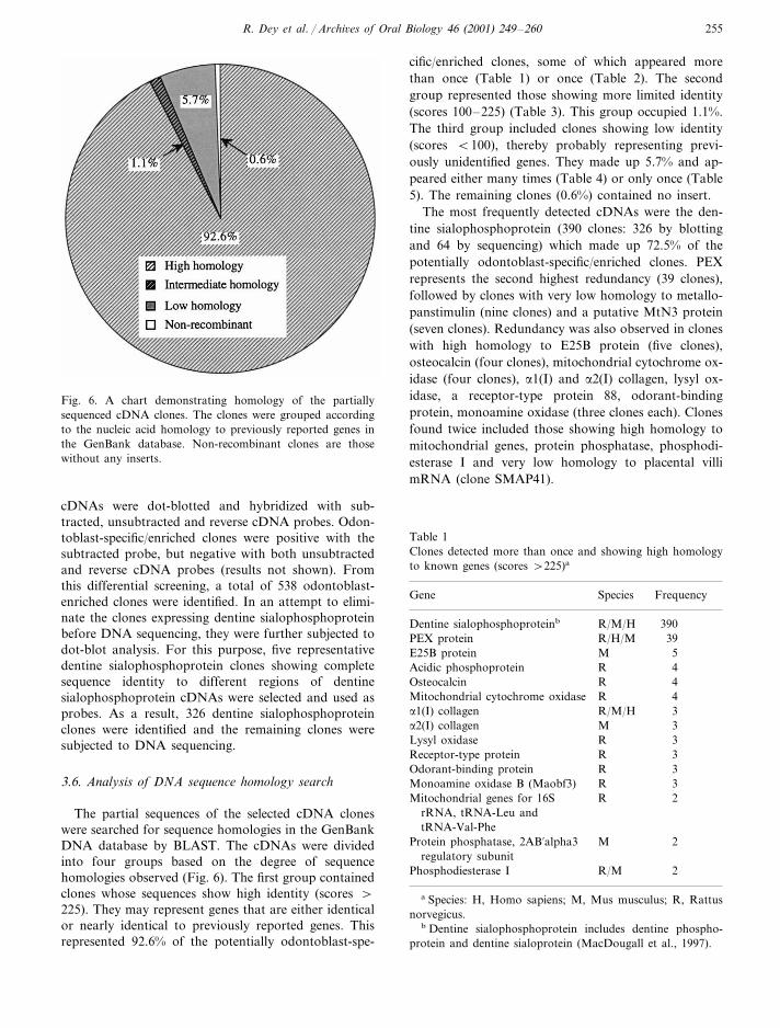
Fig. 6. A chart demonstrating homology of the partially
sequenced cDNA clones. The clones were grouped according
to the nucleic acid homology to previously reported genes in
the GenBank database. Non-recombinant clones are those
without any inserts.
cific/enriched clones, some of which appeared more
than once (Table 1) or once (Table 2). The second
group represented those showing more limited identity
(scores 100–225) (Table 3). This group occupied 1.1%.
The third group included clones showing low identity
(scores B100), thereby probably representing previ-
ously unidentified genes. They made up 5.7% and ap-
peared either many times (Table 4) or only once (Table
5). The remaining clones (0.6%) contained no insert.
The most frequently detected cDNAs were the den-
tine sialophosphoprotein (390 clones: 326 by blotting
and 64 by sequencing) which made up 72.5% of the
potentially odontoblast-specific/enriched clones. PEX
represents the second highest redundancy (39 clones),
followed by clones with very low homology to metallo-
panstimulin (nine clones) and a putative MtN3 protein
(seven clones). Redundancy was also observed in clones
with high homology to E25B protein (five clones),
osteocalcin (four clones), mitochondrial cytochrome ox-
idase (four clones), a1(I) and a2(I) collagen, lysyl ox-
idase, a receptor-type protein 88, odorant-binding
protein, monoamine oxidase (three clones each). Clones
found twice included those showing high homology to
mitochondrial genes, protein phosphatase, phosphodi-
esterase I and very low homology to placental villi
mRNA (clone SMAP41).
cDNAs were dot-blotted and hybridized with sub-
tracted, unsubtracted and reverse cDNA probes. Odon-
toblast-specific/enriched clones were positive with the
subtracted probe, but negative with both unsubtracted
and reverse cDNA probes (results not shown). From
this differential screening, a total of 538 odontoblast-
enriched clones were identified. In an attempt to elimi-
nate the clones expressing dentine sialophosphoprotein
before DNA sequencing, they were further subjected to
dot-blot analysis. For this purpose, five representative
dentine sialophosphoprotein clones showing complete
sequence identity to different regions of dentine
sialophosphoprotein cDNAs were selected and used as
probes. As a result, 326 dentine sialophosphoprotein
clones were identified and the remaining clones were
subjected to DNA sequencing.
3.6. Analysis of DNA sequence homology search
The partial sequences of the selected cDNA clones
were searched for sequence homologies in the GenBank
DNA database by BLAST. The cDNAs were divided
into four groups based on the degree of sequence
homologies observed (Fig. 6). The first group contained
clones whose sequences show high identity (scores \
225). They may represent genes that are either identical
or nearly identical to previously reported genes. This
represented 92.6% of the potentially odontoblast-spe-
Table 1
Clones detected more than once and showing high homology
to known genes (scores \225)a
SpeciesGene Frequency
R/M/H 390Dentine sialophosphoproteinb
R/H/MPEX protein 39
ME25B protein 5
RAcidic phosphoprotein 4
Osteocalcin R 4
Mitochondrial cytochrome oxidase R 4
R/M/Ha1(I) collagen 3
3Ma2(I) collagen
3RLysyl oxidase
3RReceptor-type protein
R 3Odorant-binding protein
RMonoamine oxidase B (Maobf3) 3
Mitochondrial genes for 16S R 2
rRNA, tRNA-Leu and
tRNA-Val-Phe
MProtein phosphatase, 2AB%alpha3 2
regulatory subunit
R/MPhosphodiesterase I 2
a Species: H, Homo sapiens; M, Mus musculus; R, Rattus
norvegicus.b Dentine sialophosphoprotein includes dentine phospho-
protein and dentine sialoprotein (MacDougall et al., 1997).

R. Dey et al. / Archi6es of Oral Biology 46 (2001) 249–260256
Table 2
Clones detected once and showing high homology to known genes (scores \225)a
Length sent Score SpeciesGeneClone
OD-13 34 kDa Phosphoprotein 415- 575 P
739 254CollagenOD-65 a1(XI) R
OD-82 Matrix metalloproteinase 663 357 S
491 686D6.1A proteinOD-124 R
255OD-126 32912q24.1 BAC RPCI11-412D9 H
350Dmx-like 1 464OD-127 H
675OD-167 Alpha subunit kidney-type Na+, K+-ATPase 718 R
656 844Sulfated glycoproteinOD-268 R
HelicaseOD-284 540 716 M
P190-BOD-310 487 652 M
656Ribosomal protein L5 pseudogene 250OD-330 H
Sulfonylurea receptor 2BOD-342 658 359 R
657NRD convertase 932OD-392 R
647OD-529 5733 b-hydroxysteroid dehydrogenase isomerase type II.2 R
642Cell-binding bone sialoprotein 492OD-588 R
640 654OD-596 L1ALB gene for LINE 1 repetitive seq R
225 375Mitochondrial cytochrome BOD-598 R
G-protein coupled thrombin receptorOD-618 631 944 R
Antizyme inhibitorOD-742 626 975 R
613Mitochondrial genome fragment 589OD-912 R
5%nucleotidaseOD-977 447 912 R
43034-kDa mov34 homologue 383OD-988 H
645 916OD-1008 Ribophorin II R
596Amelogenin 785OD-1033 R
603 700 ROD-1056 Ribosomal protein S4
653Heat shock protein 86 731OD-1161 M
Glycogen phosphorylase brainOD-1246 631 777 R
OD-1277 Osteonectin 644 337 R
a Species: H, Homo sapiens; M, Mus musculus; P, Plasmodium berghei; R, Rattus rattus; S, Sus scrofa.
3.7. Northern blot analysis
The expression of clones OD-314 and OD-654 was
found only in odontoblasts/pulp cells at high levels, but
was not observed in bone, brain, heart, kidney, liver,
lung, and skeletal muscle (Fig. 7). Interestingly, the
radioautograph of clone OD-314 demonstrated two
distinct bands (approx. 2.2 and 1.5 kb), whereas that of
OD-654 revealed several bands (ranging from approx.
7.2 to 2.4 kb).
4. Discussion
Interest in differentially expressed genes has provided
the impetus for the development of molecular tech-
niques to identify and isolate products of these genes.
Subtractive hybridization (Lee et al., 1991), differential
display of mRNA (Liang and Pardee, 1992), suppres-
sion subtractive hybridization (Diatchenko et al., 1996)
and more recently serial analysis of gene expression
(Velculescu et al., 1995) and DNA array (Nacht et al.,
1999) techniques have been introduced. We have used
the suppression subtractive hybridization method to
identify and clone the differentially expressed genes in
rat odontoblasts by subtracting the common genes
present in both osteoblasts and odontoblasts/pulp cells.
The method is based on the suppression PCR effect of
long inverted terminal repeats. When attached to DNA
fragments, these repeats can selectively suppress am-
plification of undesirable sequences in PCR procedures
(Siebert et al., 1995; Chenchik et al., 1996a,b). This
method can overcome the problem of differences in
mRNA abundance by incorporating a hybridization
step that equalizes sequence abundance during the
course of subtraction by standard hybridization kinetics
(Diatchenko et al., 1996). The major advantages of the
method include the requirement of small quantities of
poly(A)+ RNA from tissues and the ability to enrich
for rare sequences (Diatchenko et al., 1996). Indeed, we
found that this suppression subtractive hybridization is
a powerful method for generating an odontoblast
cDNA library that is highly enriched for differentially
expressed genes. However, the presence of a high pro-
portion of low abundant cDNAs with high nucleotide
sequence identity to known genes indicates that the

R. Dey et al. / Archi6es of Oral Biology 46 (2001) 249–260 257
subtraction may not be very efficient as previously
suggested (Diatchenko et al., 1996), and consequently
the screening was laborious.
Using suppression subtractive hybridization, we were
able to identify 538 odontoblast-enriched cDNA clones.
Knowing that the dentine sialophosphoprotein gene is
unique to odontoblasts, we expected that numerous
dentine sialophosphoprotein clones might be included
among the 538 clones. To avoid unnecessarily laborious
sequencing of dentine sialophosphoprotein clones, we
attempted to identify and eliminate them using dot-blot
analysis before DNA sequencing. For this purpose, five
representative dentine sialophosphoprotein clones that
had demonstrated identical sequences to different re-
gions of the dentine sialophosphoprotein cDNAs were
used as probes. As expected, this additional procedure
allowed us to identify 326 dentine sialophosphoprotein
clones and reduced the number of clones to be se-
quenced. The identification of a total of 390 dentine
sialophosphoprotein clones (326 by blotting and 64 by
sequencing) out of the 538 odontoblast-enriched cDNA
clones also provided strong evidence for the high degree
of selectivity of the suppression subtractive hybridiza-
tion technique in generating differentially expressed
cDNAs.
Interestingly, many clones that occurred multiple
times in this study represented those encoding extracel-
lular matrix proteins as observed in a rat incisor cDNA
library (Matsuki et al., 1995). These include cDNAs for
dentine sialophosphoprotein, osteocalcin, and type I
collagen. In addition, we found several cDNAs for lysyl
oxidase, bone sialoprotein and osteonectin that are
known to be highly enriched in both odontoblasts and
osteoblasts (Butler, 1998). The appearance of these
cDNAs indicates that the subtraction procedure may
not be effectively achieved, particularly for the highly
enriched genes. To eliminate common sequences, the
addition of excess driver cDNAs to the first hybridiza-
tion samples was recommended in the second hy-
bridization step during subtractive hybridization
(Diatchenko et al., 1996). In this study, we initially used
2 mg driver mRNA each from cultured dental papilla
mesenchymal cells and calvarial osteoblastic cells for
the subtraction procedure according to the manufac-
ture’s procedures. We speculate that the amount of
driver mRNA used may not be enough and the applica-
tion of a smaller amount of driver mRNA sample from
osteoblasts may have resulted in the incomplete sub-
traction. Fortunately, only those genes highly enriched
in both cell types were found to appear repeatedly.
Furthermore, we observed ribosomal protein L5
pseudogene and 28S ribosomal RNA that may be
present in various cell types. The reason for the appear-
ance of non-translated clones such as clone OD-330
(ribosomal protein L5 pseudogene) in the library is
unclear. However, our nucleotide sequence analysis of
this particular clone revealed the presence of long se-
quences of As. We speculate that this unique sequence
somehow led to initial recognition by the oligo (dT)
primer and subsequent amplification.
An intriguing finding was the appearance of an amel-
ogenin clone once. As the amelogenin gene is one of
ameloblast-specific genes, this result raises the possibil-
ity that the tester RNA sample (odontoblasts/pulp
cells) was contaminated partially with ameloblast RNA
in spite of our efforts to eliminate this problem. How-
ever, this contamination was not a serious problem, as
no other known ameloblast-specific gene was observed.
One interesting observation was the appearance of
the rat/human PEX gene as the second highest enriched
clone (39 clones) in a rat odontoblast subtraction
cDNA library. The PEX gene belongs to the neutral
endopeptidase family, which is composed of type II
integral membrane glycoproteins. These include neutral
endopeptidase (neprilysin), endothelin-converting en-
zymes and the erythrocyte surface protein and Kell
(Turner and Tanzawa, 1997). Specific defects in the
PEX gene are reportedly linked to human hypophos-
phataemic rickets that are characterized by the abnor-
mal development of bone and dentine (Nikiforuk and
Fraser, 1979; Abe et al., 1988; Shields et al., 1990). A
recent in situ hybridization study confirmed the expres-
sion of PEX mRNA in both osteoblasts and odonto-
blasts of the developing mouse (Ruchon et al., 1998),
although its role in both osteogenesis and odontogene-
sis remains unknown. It is of great interest to know
Table 3
Clones detected once and showing intermediate homology to known genes (scores 100–225)a
SpeciesClone Gene Length sent Score
OD-93 HGenomic DNA of 8p21.3 677 216
634OD-280 Translation Initiation factor 186 H
Major histocompatibility complex class I regionOD-313 316 M126
LOD-442 28S rRNA 508 182
204450 MSYTOD-1091
MOD-1247 F2 clone (C57BL/10XC3H) 147524
a Species: H, Homo sapiens; L, Latimeria chalumnae; M, Mus musculus.

R. Dey et al. / Archi6es of Oral Biology 46 (2001) 249–260258
Table 4
Clones detected once and showing limited regions of homolgy to known genes (scores B100)a
Species Accession LengthGene % IdentitybClone
H gb�U80456 27OD-106 100Transcription factor SIM2 short form
H dbj/AB019565Placental villi mRNA clone SMAP52 21OD-320 100
H gb�U29343OD-325 35Hyaluronan receptor (RHAMM) 94
H NM002132/HMMRHyaluronan-mediated motility receptor (HMMR) 25OD-348 100
Placental villi mRNA clone SMAP8526OD-361 H dbj/ab019570 27 100
H gb/AC004549BAC clone RG459N13 from 7p15 57OD-450 87
H gb/AC000403OD-551 53Genomic sequence Human 13 93
H dbj/AB019572Placental villi mRNA clone SMAP26 26OD-654 100
Chromosome II BAC F14B2OD-805 A gb/AC004450 27 96
H dbj/AB020860Genomic DNA of 8p21.3-p22 79OD-925 91
Clone an01-h01OD-1062 M gb�AF120321.1 20 100
20 of 103 of the complete genomeOD-1064 C gb�AF017104 20 100
H dbj�AB019568 27 100OD-1135 Placental villi mRNA clone SMAP8
a Species: A, Arabidopsis thaliana; C, Chlamydia pneumoniae; H, Homo sapiens; M, Mus musculus.b Quality of match is given as % identity in base pairs for the stretches of highest nucleotide match.
why the PEX clone appeared so many times, particu-
larly in a rat odontoblast subtraction cDNA library. To
understand the regulatory mechanism for the PEX ex-
pression, the sequencing of the gene in its entirety is
under investigation.
Despite the appearance of several clones in the rat
odontoblast cDNA library that are also expressed by
osteoblasts, our compilation of 390 dentine sialophos-
phoprotein clones out of 538 clones strongly suggests
that this technique was effective in identifying potential
odontoblast-specific/enriched cDNAs. Thus, there is a
good possibility that our approach, using the driver
samples from both cultured dental papilla cells and
calvarial osteoblasts, increases the chance of discover-
ing additional odontoblast-specific genes. The identifi-
cation of cDNA clones that appeared many times and
demonstrated low identity with previously reported
genes is indicative of potentially novel odontoblast-spe-
cific genes. This notion was further supported by the
results from our Northern blots. mRNAs for clones
OD-314 and OD-654 were expressed specifically in
odontoblasts/pulp cells, but not in other tissues includ-
ing bone, strongly suggesting that these clones represent
potentially new odontoblast-specific genes. In addition,
the presence of multiple bands in Northern blots sug-
gested that these clones may have multiple members of
each gene family or produce alternative spliced gene
products.
We speculate that novel genes may include a variety
of proteins, such as extracellular matrix proteins, en-
zymes, transmembrane receptors, growth factors,
proteins involved in signal-transduction pathways, and
transcription factors that may have important roles in
odontoblast differentiation and dentinogenesis. These
genes can be identified and characterized by full-length
sequencing of the cDNAs, recombinant protein produc-
tion, RT-PCR, in situ hybridization analysis of their
expression and location in tissues, and immunohisto-
chemical localization of their products using antibodies.
More importantly, functional analysis of the gene prod-
ucts could reveal the mechanisms responsible for odon-
toblast differentiation and dentinogenesis.
Acknowledgements
We thank Dr Jaro Sodek for his critical reading of
the manuscript. This study was supported in part by
USPHS grant DE 07034 and DE 4849.
Table 5
Clones detected more than once and showing low homology to
known genes (scores B100)a
Gene Species Frequency
S 9bMetallopanstimulin
7bDPutative MtN3-like protein
2bHPlacental villi SMAP41
a Species: S, Strongyloides ratti; H, Homo sapiens; D, Di-
anthus caryophyllus.b Some of the clones have been confirmed to be specific to
odontoblasts/pulp cells by Northern blot analyses (unpub-
lished data).

R. Dey et al. / Archi6es of Oral Biology 46 (2001) 249–260 259
Fig. 7. Northern blot analysis of mRNA expression in various
rat tissues using cDNAs derived from clones OD-314 and
OD-654. For agarose gel electrophoresis, 10 mg total RNA (A
and B) or 1 mg mRNA (C) was loaded in each lane. Radioau-
tographs for clones OD-314 (A and C) and OD-654 (B)
revealed their expression specifically in odontoblasts/pulp cells
(O/P), but not in other tissues such as brain (B), calvaria (Ca),
heart (H), kidney (K), liver (L), lung (Lu), and skeletal muscle
(SM). Their expression appeared in multiple bands (A and B).
Expression of G3PDH and b-actin, which are well-known
house-keeping genes, was observed in all the tissues examined.
Diatchenko, L., Lau, Y.F., Campbell, A.P., Chenchik, A.,
Moqadam, F., Huang, B., Lukyanov, S., Lukyanov, K.,
Gurskaya, N., Sverdlov, E.D., Siebert, P.D., 1996. Sup-
pression subtractive hybridization: a method for generating
differentially regulated or tissue-specific cDNA probes and
libraries. Proc. Natl. Acad. Sci. 93, 6025–6030.
Dickson, I.R., Dimuzio, M.T., Volpin, D., Anantha-
narayanan, S., Veis, A., 1975. The extraction of phospho-
proteins from bovine dentin. Calcif. Tissue Res. 19, 51–61.
Ducy, P., Karsenty, G., 1998. Genetic control of cell differen-
tiation in the skeleton. Curr. Opin. Cell. Biol. 10, 614–619.
Duguid, J.R., Dinauer, M.C., 1990. Library subtraction of in
vitro cDNA libraries to identify differentially expressed
genes in scrapie infection. Nucleic Acids Res. 18, 2789–
2792.
Friedenstein, A.J., 1976. Precursor cells of mechanocytes. Int.
Rev. Cytol. 47, 327–359.
George, A., Srinivasan, R., Thotakura, S., Veis, A., 1998. The
phosphophoryn gene family: identical domain structures at
the carboxyl end. Eur. J. Oral. Sci. 106 (Suppl. 1), 221–
226.
Hara, E., Kato, T., Nakada, S., Sekiya, S., Oda, K., 1991.
Subtractive cDNA cloning using oligo(dT)30-latex and
PCR: Isolation of cDNA clones specific to undifferentiated
human embryonal carcinoma cells. Nucleic Acids Res. 19,
7097–7104.
Lee, S.W., Tomasetto, C., Sager, R., 1991. Positive selection of
candidate tumor-suppressor genes by subtractive hy-
bridization. Proc. Natl. Acad. Sci. USA 88, 2825–2829.
Lian, J.B., Stein, G.S., 1992. Concepts of osteoblast growth
and differentiation: Basis for modulation of bone cell
development and tissue formation. Crit. Rev. Oral Biol.
Med. 3, 269–305.
Lian, J.B., Stein, G.S., Stein, J.L., van Wijnen, A.J., 1998.
Osteocalcin gene promoter: unlocking the secrets for regu-
lation of osteoblast growth and differentiation. J. Cell
Biochem. Suppl. 30–31, 62–72 Suppl.
Liang, P., Pardee, A., 1992. Differential display of eukaryotic
messenger RNA by means of the polymerase chain reac-
tion. Science 257, 967–970.
MacDougall, M., Simmons, D., Luan, X., Nydegger, J., Feng,
J., Gu, T.T., 1997. Dentin phosphoprotein and dentin
sialoprotein are cleavage products expressed from a single
transcript coded by a gene on human chromosome 4.
Dentin phosphoprotein DNA sequence determination. J.
Biol. Chem. 272, 835–842.
Matsuki, Y., Nakashima, M., Amizuka, N., Warshawsky, H.,
Goltzman, D., Yamada, K.M., Yamada, Y., 1995. A com-
pilation of partial sequences of randomly selected cDNA
clones from the rat incisor. J. Dent. Res. 74, 307–312.
Nacht, M., Ferguson, A.T., Zhang, W., Petroziello, J.M.,
Cook, B.P., Gao, Y.H., Maguire, S., Riley, D., Coppola,
G., Landes, G.M., Madden, S.L., Sukumar, S., 1999.
Combining serial analysis of gene expression and array
technologies to identify genes differentially expressed in
breast cancer. Cancer Res. 59, 5464–5470.
Nikiforuk, G., Fraser, D., 1979. Chemical determinants of
enamel hypoplasia in children with disorders of calcium
and phosphate homeostasis. J. Dent. Res. 58(Spec Issue B),
1014–1015.
Owen, M., Friedenstein, A.J., 1988. Stromal stem cells: mar-
References
Abe, K., Ooshima, T., Lily, T.S., Yasufuku, Y., Sobue, S.,
1988. Structural deformities of deciduous teeth in patients
with hypophosphatemic vitamin D-resistant rickets. Oral
Surg. Oral Med. Oral Pathol. 65, 191–198.
Altschul, S.F., Madden, T.L., Schaffer, A.A., Zhang, J.,
Zhang, Z., Miller, W., Lipman, D.J., 1997. Gapped
BLAST and PSI-BLAST: a new generation of protein
database search programs. Nucleic Acids Res. 25, 3389–
3402.
Butler, W.T., 1998. Dentin matrix proteins. Eur. J. Oral. Sci.
106 (suppl 1), 204–210.
Butler, W.T., 1987. Dentin-specific proteins. Methods Enzy-
mol. 145, 290–303.
Chen, J., Shapiro, H.S., Sodek, J., 1992. Development expres-
sion of bone sialoprotein mRNA in rat mineralized con-
nective tissues. J. Bone Miner. Res. 7, 987–997.
Chenchik, A., Diatchenko, L., Moqadam, F., Tarabykin, V.,
Lukyanov, S., Siebert, P.D., 1996a. Full-length cDNA
cloning and determination of mRNA 5% and 3% ends by
amplification of adaptor-ligated cDNA. BioTechniques 21,
526–534.
Chenchik, A., Moqadam, F., Siebert, P.D., 1996b. A new
method for full-length cDNA cloning by PCR. In: Kreig,
P.A. (Ed.), A Laboratory Guide to RNA Isolation, Analy-
sis and Synthesis. Wiley-Liss Inc., NY, pp. 273–321.
Chien, H.H., Lin, W.L., Cho, M.I., 1999. Interleukin-1b in-
duced release of matrix protein into culture media causes
inhibition of mineralization of nodules formed by peri-
odontal ligaments cells in vitro. Calcif. Tissue Int. 64,
402–413.

R. Dey et al. / Archi6es of Oral Biology 46 (2001) 249–260260
row-derived osteogenic precursors. Ciba Foundation Sym-
posium 136, 42–60.
Ritchie, H.H., Ritchie, D.G., Wang, L.H., 1998. Six decades
of dentinogenesis research. Historical and prospective
views on phosphophoryn and dentin sialoprotein. Eur. J.
Oral Sci. 106 (Suppl. 1), 211–220.
Ruchon, A.F., Marcinkiewicz, M., Siegfried, G., Tenenhouse,
H.S., DesGroseillers, L., Crine, P., Boileau, G., 1998. Pex
mRNA is localized in developing mouse osteoblasts and
odontoblasts. J. Histochem. Cytochem. 46, 459–468.
Shields, E.D., Scriver, C.R., Reade, T., Fujiwara, T.M., Mor-
gan, K., Ciampi, A., Schwartz, S., 1990. X-linked hy-
pophosphatemia: the mutant gene is expressed in teeth as
well as in kidney. Am. J. Hum. Genet. 46, 434–442.
Siebert, P.D., Chenchik, A., Kellogg, D.E., Lukyanov, K.A.,
Lukynov, S.A., 1995. An improved method for walking in
uncloned genomic DNA. Nucleic Acids Res. 23, 1087–
1088.
Ten Cate, A.R., 1998. Dentinogenesis. In: Ten Cate, A.R.
(Ed.), Oral Histology: Development, Structure and Func-
tion. Mosby-Year Book, Inc., St. Louis, Missouri, pp.
128–149.
Turner, A.J., Tanzawa, K., 1997. Mammalian membrane
metallopeptidases: NEP, ECE, KELL, and PEX. FASEB
J. 5, 355–364.
Veis, A., Perry, A., 1967. The phosphoprotein of the dentin
matrix. Biochemistry 6, 2409–2416.
Velculescu, V.E., Zhang, L., Vogelstein, B., Kinzler, K.W.,
1995. Serial analysis of gene expression. Science 270, 484–
487.
.