BIOL 213 Lab Handouts
38
BIOL 213 LAB HANDOUTS You are responsible for all of the information contained within these handouts. Lab practicals will be directly related to the terms and concepts outlined in this document. You are encouraged to bring these handouts with you to each lab to ensure that you fully understand the material. Remember, spelling counts on lab practicals, so be sure to practice spelling anatomical terms.
-
Upload
khangminh22 -
Category
Documents
-
view
1 -
download
0
Transcript of BIOL 213 Lab Handouts

BIOL213LABHANDOUTS
Youareresponsibleforallofthe informationcontainedwithinthesehandouts.Labpracticalswill be directly related to the terms and concepts outlined in this document. You areencouragedtobringthesehandoutswithyoutoeachlabtoensurethatyoufullyunderstandthe material. Remember, spelling counts on lab practicals, so be sure to practice spellinganatomicalterms.

INTRODUCTIONTOANATOMYANDPHYSIOLOGY
Identify the following terms on pictures and models. Be able to correctly use anatomicalterminology.BODYCAVITIES:PosteriorAspect
CranialCavityVertebralCanal
VentralCavityThoracicCavity
Mediastinum LeftPleuralCavity
RightPleuralCavity PericardialCavity
AbdominopelvicCavity AbdominalCavity PelvicCavityDiaphragmSEROUSMEMBRANES:ParietalPleuraVisceralPleuraPleuralCavityParietalPericardiumVisceralPericardiumPericardialCavityParietalPeritoneumVisceralPeritoneumPeritonealCavityANATOMICDIRECTIONS:Superior/InferiorAnterior/PosteriorMedial/LateralProximal/DistalSuperficial/DeepSECTIONSANDPLANES:Sagittal/Median(Midsagittal)Transverse(Horizontal)
Frontal(Coronal)ObliqueABDOMINOPELVICREGIONS:EpigastricRight/LeftHypochondriacUmbilicalRight/LeftLumbarHypogastricRight/LeftIliacABDOMINOPELVICQUADRANTS:RightUpperQuadrantRightLowerQuadrantLeftUpperQuadrantLeftLowerQuadrantREGIONALANATOMY:AbdominalAntebrachialAntecubitalAuricularAxillaryBrachialBuccalCalcanealCarpalCephalicCervicalCoxalCranialCruralDeltoidDigitalFemoralFrontalGlutealInguinal

ANATOMICALTERMINOLOGY(CONTINUED):LumbarMammaryManusMentalNasalOccipitalOlecranalOralOrbitalPalmarPatellarPectoral
PelvicPerinealPesPlantarPoplitealPubicSacralSternalSuralTarsalThoracicVertebral

MICROSCOPES
TERMSTOIDENTIFYONAMICROSCOPE:EyepieceHeadArmStageCoarseAdjustmentKnobFineAdjustmentKnobBaseCondenserIrisDiaphragmRotatingnosepieceObjectives(4x,10x,40x,100x)StageClipCONCEPTS:1.Calculatetotalmagnification.2.Correctlyfocusonanobjectthroughmultiplelenses.

MICROSCOPESWORKSHEET
PARTI:ORIENTATIONONAMICROSCOPECollectaslidewiththeletter“e”onit.Placetheslideonthestageandusethecliptosecure.The stage should be in the lowest position possible. Make sure the letter “e” is facing youcorrectly on the stage. Position the 4x objective (also called scanning) and use the coarseadjustment knob to focus on the letter. Continue focusing on the letter using the fineadjustmentknob.Describetheorientationoftheletter“e.”Isitthesame,upsidedown,etc.?______________________________________________________________________________Whenyoumovetheslidetotheright,whichwaydoestheimagemove?______________________________________________________________________________Whenyoumovetheslideup,whichwaydoestheimagemove?______________________________________________________________________________Nowmovetothe10xobjective(alsocalledlowpower).Focususingthefineadjustmentknobonly(doNOTusethecoarseadjustmentknobatthispoint).Describetheorientationoftheletter“e.”Didanythingchangefromthe4xobjective?______________________________________________________________________________PARTII:MAGNIFICATIONSofaryouhaveusedthe4xand10xobjectives. Inordertocalculatetotalmagnification,youmultiplythemagnificationoftheobjectivebythemagnificationoftheeyepiece(usuallythisis10x).Whatisthemagnificationoftheeyepieceonyourmicroscope?______________________________________________________________________________Whenusingthe4xobjective,thetotalmagnificationis:4(fromtheobjective)x10(fromtheeyepiece)=40xWhatisthetotalmagnificationwhenusingthe10xobjective?______________________________________________________________________________Nowmovetothe40xobjective(alsocalledhighpower).Focususingthefineadjustmentknobonly(doNOTusethecoarseadjustmentknobatthispoint).What is thetotalmagnificationwhenusingthe40xobjective?Whathappenedtolightintensitywhenmovingfrom10xto40xobjective?

CELLMEMBRANETRANSPORT
CompletethePhILS#1labforOsmosisandDiffusion:VaryingExtracellularConcentration.I. SOLUTIONS A. Asolutioniscomposedof:
1. Solvent(water)2. Solute(molecules)dissolvedinsolventBiologicalmembranesareselectivelypermeable,allowingonlycertainsolutesthrough.
II. DIFFUSIONA. Diffusion:movementofmoleculesfromareasofhigherconcentrationtoareasoflower
concentration.Agreatexampleofthisisthediffusionofoxygenintocellsandthediffusionofcarbondioxideoutofcells.
B. Facilitateddiffusion:usesachannel(example–potassiumchannel).
III. OSMOSIS
A. Osmolarity:concentrationofallmoleculesinasolution. B. Osmosis:diffusionofwaterdownitsconcentrationgradient. 1. Isotonic:nonetmovementofwater. 2. Hypotonic:waterenterscellanditswells(itcanburstorlyse). 3. Hypertonic:waterleavescellanditshrinks(crenate).
C. Mostcellmembraneshavefewopensodiumchannels,sowhencellsareplacedinsolutionsofNaCl,somesodiumwillmove,butmostlywaterwillmovebyosmosis.

IV. GOALSOFTHELAB1. Placeredbloodcellsinsolutionswithdifferentconcentrationsofsodiumchloride(0mMto240
mM).2. Measurethecolorofthesolutionsusingaspectrophotometer.
a. Normalsolutions:510nmwavelengthreflectedbycellmembranes.b. Hypotonicsolutions:watermovesintocells,theyburstandlesslightisreflected.
Therefore,amountoflighttransmittedincreases.c. Hypertonicsolutions:waterleavescellsandtheyshrink.Therefore,amountoflight
transmitteddecreases.
V. DATACOLLECTIONCompletethedatatablebelowwithyourresultsandplotthedataonthegraphprovided.
[NaCl] Transmittance0 50 100 120 140 160 180 200 220 240
Graphpaperimage:https://commons.wikimedia.org/wiki/File:Graph-paper.svg
VI. APPLICATIONQUESTIONS
1. Whathappenedtothecarrotplacedinthesaturatedsaltsolutionovernight?Howwouldyouclassifythissolution?Whathappenedtothecarrotplacedinthefilteredwatersolutionovernight?Howwouldyouclassifythissolution?
2. Basedonyourdata,duringapproximatelywhichrangeof[NaCl]areredbloodcellslosingwater
andhaveshrunkinsize?
3. TheprinciplesofosmosisareusedintheclinicwhenadministeringIVsolutions.Forexample,
whichsolutionwouldyouadministertoapatientsufferingfromcerebraledema(hypotonicorhypertonic)?Why?

HISTOLOGY
Identify the following tissues (and associated structures) under the microscope and usingpictures. For each, categorize as epithelial or connective tissue and givemain locations andfunctionsinthebody.EPITHELIALTISSUE:Simplesquamous(nucleus)Simplecuboidal(nucleus,lumen)Nonciliatedsimplecolumnar(nucleus)Ciliatedpseudostratifiedcolumnar(nucleus,cilia,gobletcells)Stratifiedsquamous(nucleus)Transitional(nucleus)
CONNECTIVETISSUEPROPER:LOOSECONNECTIVETISSUE:Areolar(fibroblast)Adipose(nucleus)Reticular(reticularfiber)DENSECONNECTIVETISSUE:Denseregular(fibroblastnucleus)Denseirregular(collagenfiber)Elastic(elasticfibers)
SUPPORTINGCONNECTIVETISSUE:HyalineCartilage(chondrocyteinlacuna)ElasticCartilage(chondrocyteinlacuna)Fibrocartilage(chondrocyteinlacuna)Bone(osteocyte,centralcanal)FLUIDCONNECTIVETISSUE:Blood(WBC,RBC,platelet)

INTEGUMENTARYSYSTEM
Identifythefollowingstructuresonmodelsandpictures.LAYERS:Epidermis
StratumcorneumStratumlucidumStratumgranulosum
Stratumspinosum StratumbasaleDermis
Dermalpapilla Meissner’scorpuscle Paciniancorpuscle Sweatgland(merocrineandapocrine)Subcutaneouslayer/HypodermisHAIR:HairfollicleHairshaftSebaceousglandArrectorpilimuscleHairpapilla

SKELETALSYSTEM
Classifybonesaccordingtotheirshape.BONESHAPES:LongShortSesamoidFlatIrregularIdentifythefollowingstructuresonalongbone.LONGBONE:ProximalepiphysisDistalepiphysisDiaphysisMetaphysisArticularcartilageEpiphysealplateEndosteumMedullarycavityIdentifythefollowingstructuresonamicroscopicbonemodel.MICROSCOPICBONE:SpongyboneTrabeculaeCompactboneOsteonCentralcanalPerforatingcanalOsteocyteLacunaCanaliculusPeriosteumLamella

SKELETALSYSTEM
Thereare206bonesintheadulthumanskeleton.Identifythesebonesinbotharticulatedanddisarticulatedforms.Includemarkingandbonenamewhenapplicable.SKULL:CranialBonesFrontalParietalOccipital Foramenmagnum OccipitalcondylesTemporal Mandibularfossa Mastoidprocess Styloidprocess ZygomaticprocessSphenoid Sellaturcica OpticcanalEthmoid Cribriformplate CristagalliHYOIDBONEVERTEBRALCOLUMN:Markingstoidentifyonanyvertebra:BodyVertebralforamenSpinousprocessTransverseprocessTypesofVertebraCervical(7) Transverseforamen
Atlas(C1) Axis(C2) Dens
FacialBonesMaxilla Infraorbitalforamen Palatineprocess IncisiveforamenPalatineZygomatic TemporalprocessLacrimalNasalVomerInferiornasalconchaeMandible MentalforamenSuturesCoronal,sagittal,squamous,lambdoidThoracic(12)Lumbar(5)SacrumCoccyxTHORACICCAGE:Trueribs(7pairs)Falseribs(5pairs)Floatingribs(2-3pairs)Sternum Manubrium Body Xiphoidprocess

PECTORALGIRDLE:Clavicle Sternalend AcromialendScapula Spine Acromion Coracoidprocess Glenoidcavity Supraspinousfossa Infraspinousfossa SubscapularfossaUPPERLIMB:Humerus Head Greatertubercle Lessertubercle Intertubercularsulcus Anatomicalneck Surgicalneck Olecranonfossa Coronoidfossa Trochlea CapitulumRadius Head Radialtuberosity StyloidprocessUlna Coronoidprcoess Olecranon Trochlearnotch StyloidprocessCarpals(8)Metacarpals(5)Phalanges(14)
PELVICGIRDLE:Coxa Ilium Iliaccrest Anteriorinferioriliacspine Ischium Ischialtuberosity Ischialspine PubisObturatorforamenAcetabulumLOWERLIMB:Femur Head Neck Foveacapitis Greatertrochanter Lessertrochanter Lineaaspera Lateralcondyle Medialcondyle Lateralepicondyle Medialepicondyle Tibia Lateralcondyle Medialcondyle Tibialtuberosity MedialmalleolusFibula Head LateralmalleolusPatellaTarsals(7) Talus CalcaneusMetatarsals(5)Phalanges(14)
CONCEPTS:! Compareandcontrasttheskullofaninfantwithanadult.! Compareandcontrastthemaleandfemaleskeleton.! Identifybonesintheaxialandappendicularskeletons.

ARTICULATIONS
Classify articulations (joints) based on structure and function and identify examples of each.Demonstratevariousjointmovementsandknowkeyligamentsofthefourmajorjointsinthebody.STRUCTURALCLASSIFICATION:Fibrous
SutureSyndesmosisGomphosis
Cartilaginous Synchondrosis SymphysisSynovial Ballandsocket Hinge Condylar Saddle Pivot Plane FUNCTIONALCLASSIFICATION:SynarthrosisAmphiarthrosisDiarthrosisJOINTMOVEMENTS:FlexionExtensionHyperextensionAbductionAdductionCircumductionSupinationPronationProtractionRetractionLateralrotationMedialrotationEversion
InversionPlantarflexionDorsiflexionElevationDepressionMAJORJOINTSANDTHEIRLIGAMENTS:Shoulder Coracohumeralligament Coracoacromialligament GlenohumeralligamentHip Iliofemoralligament Pubofemoralligament IschiofemoralligamentKnee Anteriorcruciateligament(ACL) Posteriorcruciateligament(PCL) Patellarligament Tibialcollateralligament FibularcollateralligamentElbow Ulnarcollateralligament Radialcollateralligament Anularligament

MUSCULARSYSTEM
Identifymuscle tissues (and associated structures) under themicroscope and using pictures.Foreach,givemainlocationsandfunctionsinthebody,typeofcontrol(voluntary/involuntary),andwhetherstriationsarepresentorabsent.MUSCLEHISTOLOGY:SkeletalMuscle(nucleus,striations)SmoothMuscle(nucleus)CardiacMuscle(nucleus,striations,intercalateddiscs)Identifythefollowingstructuresonthemicroscopicmusclemodel.MICROSCOPICMUSCLE:SarcolemmaNucleusTriad Twocisternaeofsarcoplasmicreticulum OnetransversetubuleMyofibrilSarcomere

MUSCULARSYSTEM
Identifythefollowingmusclesofthebodyusingmodelsandpictures.FACEANDNECK:FrontalisOccipitalisTemporalisMasseterBuccinatorOrbicularisorisOrbicularisoculiZygomaticusmajorZygomaticusminorSternocleidomastoidTORSO:PectoralismajorPectoralisminorSerratusanteriorTrapeziusLatissimusdorsiRectusabdominisExternalobliqueInternalobliqueTransversusabdominisUPPERLIMB:DeltoidRotatorcuffmuscles: Supraspinatus Infraspinatus Subscapularis TeresminorTeresmajorBicepsbrachiiTricepsbrachiiBrachioradialisPronatorteresFlexorcarpiradialisPalmarislongus
FlexorcarpiulnarisExtensordigitorumLOWERLIMB:GluteusmaximusGluteusmediusTensorfasciaelataeSartoriusGracilisAdductorlongusAdductormagnusQuadricepsfemorismuscles: Rectusfemoris Vastuslateralis Vastusmedialis VastusintermediusHamstringmuscles: Bicepsfemoris Semitendinosus SemimembranosusGastrocnemiusSoleusCalcanealtendonTibialisanteriorFibularislongus

MUSCULARSYSTEM
Definethetermsoriginandinsertion.Identifytheorigins,insertions,actions,andinnervationsforthefollowingmuscles.
Muscle Origin Insertion Action InnervationSupraspinatus
Supraspinousfossaofscapula
Greatertubercleofhumerus
Abductsarm Suprascapularnerve
Infraspinatus
Infraspinousfossaofscapula
Greatertubercleofhumerus
Adductsandlaterallyrotates
arm
Suprascapularnerve
Teresminor
Lateralborderofscapula
Greatertubercleofhumerus
Adductsandlaterallyrotates
arm
Axillarynerve
Subscapularis
Subscapularfossaofscapula
Lessertubercleofhumerus
Mediallyrotatesarm
Subscapularnerve
Bicepsfemoris
Ischialtuberosityandlineaaspera
offemur
Headoffibula Flexeslegandlaterallyrotates
leg
Tibialandfibulardivisionsofsciaticnerve
Semitendinosus
Ischialtuberosity Proximal,medialsurfaceoftibia
Flexeslegandmediallyrotates
leg
Tibialdivisionofsciaticnerve
Semimembranosus
Ischialtuberosity Medialcondyleoftibia
Flexeslegandmediallyrotates
leg
Tibialdivisionofsciaticnerve
Rectusfemoris
Anteriorinferioriliacspine
Patellaviapatellar
ligamenttotibialtuberosity
Extendsleg Femoralnerve
Vastusmedialis
Lineaasperaoffemur
Patellaviapatellar
ligamenttotibialtuberosity
Extendsleg Femoralnerve
Vastusintermedius
Anteriorandlateralsurfaces
offemur
Patellaviapatellar
ligamenttotibialtuberosity
Extendsleg Femoralnerve
Vastuslateralis
Greatertrochanterandlineaasperaof
femur
Patellaviapatellar
ligamenttotibialtuberosity
Extendsleg Femoralnerve

CONNECTINGCONCEPTS:Connect the skeletal, nervous, andmuscular systems to understand jointmovements in thebody.Forexample,thenervoussystemsendsasignalalongthefemoralnervetostimulatethequadricepsfemorisgroupofmuscles(rectusfemoris,vastus lateralis,vastus intermedius,andvastusmedialis). Contractionof thesemuscles pulls their insertions (patella/tibial tuberosity)toward their origins (coxa/femur) leading to extension at the knee joint. Be able to explainflexionatthekneejointandmovementsoftheshoulderjointbasedonthetableabove.

MUSCULARSYSTEM
CompletethePhILS#5labforSkeletalMuscleFunction:Stimulus-DependentForceGeneration.
I. MUSCLECONTRACTION
A. Musclescontractuponreceivingastimulusfromthenervoussytemattheneuromuscularjunction.Thisstimulusmustreachacriticallevelcalledthresholdinorderforthemusclefibertocontract.
B. Therearetwotypesofcontractions:1. Isometriccontraction:muscletenses,butlengthstaysthesame.
2. Isotoniccontraction:musclecontractsandchangeslength. a. Concentriccontraction:muscleshortens. b. Eccentriccontraction:musclelengthens.
II. MOTORUNITA. Motorunit:composedofonemotorneuronandallofthemusclefibersitinnervates.B. Motorunitrecruitment:withincreasedstimulation,moremotorunitsareactivatedtoincrease
musclecontractionforce.
III. GOALSOFTHELAB1. Dissectthegastrocnemius(calf)musclefromafrogandprepareforstimulationandrecording.2. Applyvaryingelectricalvoltagesandrecordmuscletensionasitcontracts.
IV. DATACOLLECTION Completethedatatablebelowwithyourresultsandplotthedataonthegraphprovided.
AppliedVoltage(V} MuscleTension(g)0 0.1 0.2 0.3 0.4 0.5 0.6 0.7 0.8 0.9 1.0 1.1 1.2 1.3 1.4 1.5 1.6
Graphpaperimage:https://commons.wikimedia.org/wiki/File:Graph-paper.svg

V. APPLICATIONQUESTIONS
1. Whywastherenomuscletensionresponsewhenlowvoltageshockswereapplied?2. Whatistheminimumvoltagerequiredtoproducemuscletension?
3. Whydoesapplyingmorevoltageproducegreatertension?
MUSCULARSYSTEM
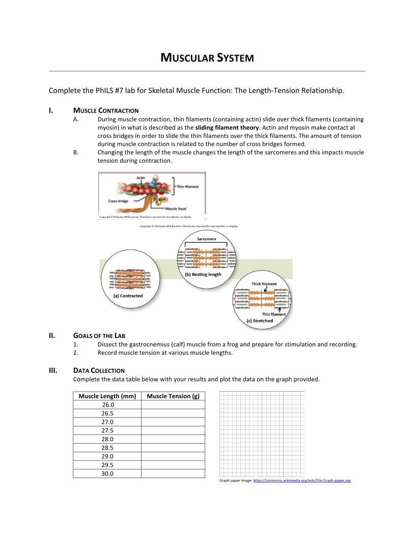
CompletethePhILS#7labforSkeletalMuscleFunction:TheLength-TensionRelationship.
I. MUSCLECONTRACTION
A. Duringmusclecontraction,thinfilaments(containingactin)slideoverthickfilaments(containingmyosin)inwhatisdescribedastheslidingfilamenttheory.Actinandmyosinmakecontactatcrossbridgesinordertoslidethethinfilamentsoverthethickfilaments.Theamountoftensionduringmusclecontractionisrelatedtothenumberofcrossbridgesformed.
B. Changingthelengthofthemusclechangesthelengthofthesarcomeresandthisimpactsmuscletensionduringcontraction.
II. GOALSOFTHELAB1. Dissectthegastrocnemius(calf)musclefromafrogandprepareforstimulationandrecording.2. Recordmuscletensionatvariousmusclelengths.
III. DATACOLLECTION Completethedatatablebelowwithyourresultsandplotthedataonthegraphprovided.
MuscleLength(mm) MuscleTension(g)26.0 26.5 27.0 27.5 28.0 28.5 29.0 29.5 30.0
Graphpaperimage:https://commons.wikimedia.org/wiki/File:Graph-paper.svg

IV. APPLICATIONQUESTIONS
1. Whatistheoptimummusclelengthformaximummuscletension?
2. Atwhatmusclelengthcanthemusclelifttheheaviestweight?
3. Whenmusclelengthistooshort,whydoestensiondecrease(explainatthemolecularlevel)?
4. Whenmusclelengthistoolong,whydoestensiondecrease(explainatthemolecularlevel)?

MUSCULARSYSTEM
CompletethePhILS#8labforSkeletalMuscleFunction:PrinciplesofSummationandTetanus.I. MUSCLECONTRACTION
A. Duringmusclecontraction,calciumisreleasedfromthesarcoplasmicreticulumtoinitiateactinandmyosinbinding.Calciumbindstothethinfilamentprotein,troponin,whichmovestropomyosinandrevealsbindingsitesonactin.Myosinbindstoactin,leadingtothepowerstrokeandslidingofthethinfilamentsoverthethickfilaments.
B. Duringcontraction,muscletensioncanbefurtherincreasedifcalciumreleaseisprolonged.Increasingthefrequencyofactionpotentialsincreasescalciumreleaseinaprocesscalledsummation.
II. TETANUSA. Whenamuscleisstimulatedrepeatedlybeforeitcanfullyreturntoarelaxedstate,muscle
tensionincreasesinaprocesscalledincompletetetanus.B. Ifamuscledoesnotrelaxatallbetweenstimulations,muscletensionismaximumincomplete
tetanus.
III. GOALSOFTHELAB1. Dissectthegastrocnemius(calf)musclefromafrogandprepareforstimulationandrecording.2. Recordmuscletensionatincreasingstimulationfrequencies.
IV. DATACOLLECTIONCompletethedatatablebelowwithyourresultsandmatchthegraphspresentedwiththeappropriateterm.
Timeinterval(milliseconds)
(A)Summation (B)IncompleteTetanus (C)CompleteTetanus

Figure1:MuscleTwitch(Baseline) Figure2
Figure3 Figure4
V. APPLICATIONQUESTIONS1. Figure1representsbaselinemuscletwitches.MatchtheotherthreeFigureswiththecorrect
termbelow:a. Summation–b. Incompletetetanus–
c. Completetetanus–
2. Asthefrequencyofstimulationincreases,muscletensionincreases.Whathappenstocalciuml evelsinthecytoplasmduringthistime?

NERVOUSSYSTEM
Identify nervous tissue (and associated structures) under themicroscope and using pictures.Givemainlocationsandfunctionsinthebody.NERVOUSHISTOLOGY:Nervoustissue(neuron,nucleusofneuron,neuronalprocesses,nucleiofglia)Identify the following structures on the neuron, brain, and spinal cord models. Identify thecomponentsofareflexarc.NEURONMODEL:DendritesCellbody Nucleus NisslbodiesAxon Axonhillock
NodesofRanvierNeurofibrilSchwanncell Myelinsheath NeurilemmaBRAINMODEL:Cerebrum Frontallobe Parietallobe Temporallobe OccipitallobeDiencephalon Thalamus Hypothalamus Pinealgland(inepithalamus)Brainstem Midbrain Pons MedullaoblongataPituitarygland
CorpuscallosumCerebellumGyrusSulcusSPINALCORDMODEL:Whitematter Lateralfuniculus Posteriorfuniculus AnteriorfuniculusGraymatter Dorsalhorn Ventralhorn LateralhornCentralcanalPosteriormediansulcusAnteriormedianfissureDorsalrootganglionDorsalrootVentralrootSpinalnerveREFLEXARC:ReceptorSensory(afferent)neuronInterneuronMotor(efferent)neuronEffector

NERVOUSSYSTEM
Identify the 12 pairs of cranial nerves including name, number, sensory/motor/mixed, andfunction.Completethecranialnervetestinglabtoapplyconceptstotheclinicalsetting.
Number Name Sensory,Motor,orMixed
Function
I Olfactory Sensory SmellII Optic Sensory VisionIII Oculomotor Motor Eyemovement,pupil
constrictionIV Trochlear Motor EyemovementV Trigeminal Mixed Sensory:scalp,face,
oralcavityfortouch,pain,and
temperature.Motor:musclesof
masticationVI Abducens Motor EyemovementVII Facial Mixed Sensory:taste
Motor:musclesoffacialexpression
VIII Vestibulocochlear Sensory Hearingandequilibrium
IX Glossopharyngeal Mixed Sensory:tasteMotor:one
pharyngealmuscleX Vagus Mixed Sensory:heart,lungs,
abdominalorgansMotor:mostpharynxmusclesandalllarynx
musclesXI Accessory Motor Trapeziusand
sternocleidomastoidXII Hypoglossal Motor Tonguemuscles

CRANIALNERVETESTING
Withapartner,testforcranialnervefunctionbyfollowingtheinstructionsbelow.Recordyourpersonalinformationcollectedinthesectionsprovided.CNI:OlfactoryHave your partner close his/her eyes and close one nostril. Present one of the three scentsprovidedandhaveyourpartneridentifythesmell.Afteraminutebreak,closethesamenostrilandpresentoneoftheotherscents.Repeatforthelastscent.Nowclosetheothernostrilandpresentthesamescentsinadifferentorder.Besuretoallowabreakinbetweenscents.LEFTNOSTRIL RIGHTNOSTRILScentused Recognized(YesorNo)? Scentused Recognized(YesorNo)?Peppermint Peppermint Orange Orange Coffee Coffee CNII:OpticHaveyourpartnercloseoneeyeandreadthesmallest linepossible from left torightonthevisualacuitychart.Nowclose theothereyeandreadthesmallest linepossible fromright toleft.Recordtheresponsebelowandstatewhetheritmatchesthechart. Response Matchesthechart(YesorNo)?Lefteye Righteye CNIII:OculomotorHaveyourpartnersitfacingyouandlookstraightahead.Useapenlighttoshinelightintooneeye(comeinfromtheside)anddetermine if thepupil inthateyeconstricts.Alsonote if thepupilintheothereyeconstricts.Afteraminutebreak,usethepenlighttoshinelightintotheothereyeanddetermineifthepupilconstricts. Constricts when light is
presentedtolefteye?Constricts when light ispresentedtorighteye?
Lefteye Righteye CNIV:TrochlearHave your partner sit facing you. Ask your partner to follow your finger while youmove itinferiorly. Track the eye movement and record if it is WNL (within normal limits) or O(abnormal).

EyemovementLefteye Righteye CNV:TrigeminalHaveyourpartnersitfacingyouandclosehis/hereyes.Useacottonswabandgentlymoveitacrossthefaceononeside.Haveyourpartneridentifywheretheyfeeltheobject.Repeatontheotherside.Next,haveyourpartneropenhis/hermouthagainstresistancetodeterminejawfunction.RecordasWNLorO. ResponsetotouchLeftside Rightside JawmovementResponse CNVI:AbducensHave your partner sit facing you. Ask your partner to follow your finger while youmove itlaterally(totheleftandtheright).TracktheeyemovementandrecordasWNLorO. EyemovementLefteye Righteye CNVII:FacialHaveyourpartnersmile,blink,andsquint.ObservefacialexpressionsonbothsidesandrecordasWNLorO. FacialexpressionLeftside Rightside CNVIII:VestibulocochlearHaveyourpartnersitfacingyou.Strikethetuningforkgentlyagainstyourpalmandplacethehandleonthetopofyourpartner’sheadinthemiddle.Askyourpartnerwheretheyhearthesound(abnormal isalongoneside,normal is inthemiddle).RecordWNLorOfortheWebertestbelow. WebertestResponse

Nowusethesametuningforkandgentlystrike itagainstyourpalm.Placethehandleonthemastoidprocessofyourpartnerononeside.Whenyourpartnerno longerhears thesound,place theendopposite to thehandlenear theopeningof thepinna. If yourpartner can stillhearthesound,thenhearingisnormalinthatear.RepeatwiththeotherearandrecordWNLorOfortheRinnetestbelow. RinneTestLeftear Rightear CNIX:GlossopharyngealHaveyourpartneropenhis/hermouthandsay“ahh.”Checkthatthesoftpalateelevatesandtheuvulastaysalongthemidline.RecordasWNLorO. SoftpalateanduvulaResponse CNX:VagusHaveyourpartnerswallowandaskifthereareanydifficultiescompletingthisaction.RecordasWNLorO.Note:thereisoverlapintestingforCNIXandX. SwallowingResponse CNXI:AccessoryHave your partner elevate shoulders (trapezius) and turn head side to side(sternocleidomastoid).RecordasWNLorO. ResponseTrapezius Sternocleidomastoid CNXII:HypoglossalHave your partner stick out his/her tongue. Determine if tongue deviates to one side(abnormal).RecordasWNLorO. TongueResponse

SPECIALSENSES
Identifythefollowingstructuresontheearandeyemodels.EARMODEL:Externalear:
Auricle/PinnaExternalacousticmeatus
TympanicmembraneMiddleear:
AuditorytubeAuditoryossicles:
Malleus Incus StapesOvalwindowInnerear:
CochleaSemicircularcanalsVestibule
Vestibulocochlearnerve
EYEMODEL:Externaltunic:
CorneaSclera
Middletunic:ChoroidIrisPupilCiliarybody
Internaltunic: Retina Foveacentralis OpticdiscOpticnerveAnteriorcavity
AqueoushumorPosteriorcavity
VitreoushumorLensSuspensoryligaments
Identifythefollowingmusclesassociatedwiththeeye including innervationandfunctionsforeach.EXTRINSICEYEMUSCLES:
Muscle Innervation ActionSuperiorrectus Oculomotornerve(III) Moveseyesuperiorlyand
mediallyInferiorrectus Oculomotornerve(III) Moveseyeinferiorlyand
mediallyMedialrectus Oculomotornerve(III) MoveseyemediallyLateralrectus Abducensnerve(VI) Moveseyelaterally
Superioroblique Trochlearnerve(IV) Moveseyeinferiorlyandlaterally
Inferioroblique Oculomotornerve(III) Moveseyesuperiorlyandlaterally

COWEYEDISSECTION
Identifythestructuresbelowbyfollowingthedissectioninstructions.Structurestoidentifyinthecoweye:
• Cornea• Sclera• Iris• Pupil• Anteriorcavity• Posteriorcavity• Vitreoushumor• Choroid• Retina• Opticnerve• Opticdisc• Ciliarybody• Lens• Suspensoryligaments
Dissectioninstructions:1)Thecoweyeyoureceivewillhaveseverallayersofconnectivetissue,muscle,andfatsurroundingit.Youwillneedtocutawaytheselayersbeforeyoucanbegindissectionontheeye.Becarefulnottocuttheopticnerveontheposteriorside.
Anterior Posterior
2)Onceyouremovethemuscleandfat,youwillbeabletoseethesclera,cornea,andopticnerveclearly.

Anterior Posterior
3)Carefullymakeacoronalcutthroughtheeyedividingitintotheanteriorandposteriorcavities.Usethescalpeltomakeasmallincisionandthenusethescissorstocutaroundtheeye.Youwillneedtogothroughall3layersoftheeye.Becarefulwhenseparatingthetwopartsasyouwanttokeeptheretinaintact.Youwillbeabletoseeandfeelthejelly-likevitreoushumoratthispoint.
4)Carefullyremovethevitreoushumorfromtheposteriorcavity.Youwanttoavoidtheretinadetachingfromthebackoftheeye.Theretinawillappearasalightbrownthinlayer.Youcangentlypullbackontheretinausingtheforceps.

5)Asyoupullawaytherestoftheretina,youwillnoticethatiteasilydetachesexceptatonespot-thisistheopticdiscorblindspot.
6)Next,youwillseeanirridescentlayer.Thisiscalledthetapetumlucidumandisusedtoaidinnightvision.Thisisoftenfoundinnocturnalanimals,likecatsandcows,butisnotseeninhumans.Theglowofcat’seyesseenwhenalightisreflectedintothemisduetothislayer.
7)Thedarkpimentedlayerunderneaththetapetumlucidumisthechoroid.

8)Movetotheanteriorportionoftheeyenow.Carefullyremovethevitreoushumorwhilekeepingthelensintact.Next,slowlypullthelensawayfromtheciliarybodyandnotethesuspensoryligaments.
9)Fromtheposteriorview,theciliarybodyisnowvisible.
10)Fromtheanteriorview,thecorneaisvisible.Cutawaythecornea(youwillneedtogothroughseveraltoughlayers)toseetheirisandthepupil.

11)Nowtakethelensandcarefullymakeamid-sagittalcut.Youwillseeseverallayersthatlooklikeanonion.Youcanpeelawaytheselayersofthelens.
LabClean-up:
! Alldissectedpartsshouldbeplacedintheplasticbagsprovidedbytheinstructor.! Dissectiontraysandtoolsshouldbecleanedwithsoapandwaterandthendried
thoroughly.! Labtablesshouldbewipedwithdisinfectantspray.! Glovesandpapertowelscanbedisposedofinthelabtrash.

SHEEPBRAINDISSECTION
Identifythestructuresbelowbyfollowingthedissectioninstructions.Structurestoidentifyinthesheepbrain:
• Frontallobe• Parietallobe• Temporallobe• Occipitallobe• Cerebellum• Gyrus• Sulcus• Longitudinalfissure• Pinealgland• Superiorcolliculus• Inferiorcolliculus• Olfactorybulb• Opticnerve• Opticchiasma• Optictract• Mammillarybody• Midbrain• Pons• Medullaoblongata• Corpuscallosum• Thalamus• Hypothalamus• Lateralventricle• Thirdventricle• Fourthventricle
Dissectioninstructions:1)Thesheepbrainyoureceivemayhavesomemeningesstillattached.Carefullycutawaytheselayersbeingsurenottodamagethebrainintheprocess.Placethebrainventralsidedowninthedissectiontrayandlocatethefollowingstructures:frontallobe,parietallobe,occipitallobe,longitudinalfissure,cerebellum.

2)Nowplacethebrainonthelateralsideandlocatethefollowingstructures:sulcus,gyrus,temporallobe.
3)Nowplacethebrainonthedorsalsideandlocatethefollowingstructures:olfactorybulb,opticnerve,opticchiasma,optictract,mamillarybody,midbrain,pons,medullaoblongata.

4)Withthedorsalsidefacingyou,carefullypullapartthecerebellumfromthecerebrum.Thiswillallowyoutolocatethefollowingstructures:pinealgland,superiorcolliculus,inferiorcolliculus,cerebellum.
5)Withthedorsalsidefacingyou,gentlyseparatethetwocerebralhemispheresalongthelongitudinalfissure.Youwillfindawhitebandoffibers,thecorpuscallosum,thatallowscommunicationbetweenthetwohemispheres.
6)Nowtakeascalpelandmakeamediancut.Yourcutsshouldbesmooth(don’tsawthetissue).Youwilllikelymakeseveralcutsasyouworkthroughthemediansection.Carefullyseparatethetwohalvesandlocatethefollowingstructures:corpuscallosum,opticchiasma,thalamus,hypothalamus,thirdventricle,pinealgland,mamillarybody,midbrain,pons,medullaoblongata,cerebellum,fourthventricle.

7)Notethelateralventriclenearthecorpuscallosum.
LabClean-up:
! Alldissectedpartsshouldbeplacedintheplasticbagsprovidedbytheinstructor.! Dissectiontraysandtoolsshouldbecleanedwithsoapandwaterandthendried
thoroughly.! Labtablesshouldbewipedwithdisinfectantspray.! Glovesandpapertowelscanbedisposedofinthelabtrash.